

Submitted:
15 October 2026
Posted:
16 October 2026
You are already at the latest version
Abstract
Hantavirus cardiopulmonary syndrome (HCPS) caused by Andes virus is a severe zoonotic disease characterized by a paradoxical cardiac phenotype: profound myocardial dysfunction that is frequently reversible in survivors and occurs without extensive structural damage. The mechanisms linking systemic inflammation to this reversible dysfunction remain unknown. This review examines the hypothesis that inflammatory signaling converges on the cardiac ryanodine receptor (RyR2), a major intracellular Ca²⁺-release channel, through post-translational modifications, generating reversible myocardial dysfunction in HCPS. In severe disease, elevated IL-6, reactive oxygen and nitrogen species, and nitrosative stress activate pathways that in experimental models converge on RyR2 through CaMKII-dependent phosphorylation at Ser2814, oxidative modification of cysteine residues, and S-nitrosylation, each destabilizing the channel and promoting diastolic Ca²⁺ leak. The resulting increase in spontaneous Ca²⁺ release reduces SR Ca²⁺ content, impairs contractility, and predisposes to arrhythmias. This framework identifies RyR2 as a plausible mechanistic hub for the reversible cardiac phenotype. Therapeutic implications are discussed, emphasizing the timing of interventions and the potential of rycals, drugs that stabilize the closed state of RyR2 and prevent diastolic Ca²⁺ leak, as an emerging strategy. This model provides a testable hypothesis for a disease that currently lacks specific treatment.
Keywords:
hantavirus pulmonary syndrome
; Andes virus
; ryanodine receptor calcium release channel
; calcium signaling
; inflammation
; calcium-calmodulin-dependent protein kinase type 2
; Interleukin-6
1. Introduction
Hantavirus cardiopulmonary syndrome (HCPS) is one of the most lethal infectious diseases encountered in critical care medicine. Caused predominantly by Andes virus (ANDV) in the Southern Cone of South America, HCPS can progress from a nonspecific febrile state to fulminant cardiopulmonary failure within hours, with reported lethality reaching up to 50% in some series [1,2,3,4]. Despite decades of clinical experience, no specific antiviral or immunomodulatory therapy has demonstrated benefit, and treatment remains largely supportive [3]. Although ANDV primarily targets the vascular endothelium [5,6], the clinical phenotype is dominated by cardiopulmonary failure that manifests as a profound myocardial dysfunction that is often refractory to conventional support [1,7,8,9]. Interestingly, this dysfunction is reversible in survivors, suggesting a functional process as the underlying cause rather than structural damage. However, the cellular mechanisms underlying this reversible cardiac dysfunction remain unknown.
A dysregulated host inflammatory response is a central feature of severe ANDV infection [6,10]. This response is characterized by a broad cytokine profile that includes Interleukin-6 (IL-6), Tumor Necrosis Factor α (TNFα), Interleukin 1 β (IL-1β), Interferon γ (IFN-γ), and several chemokines, alongside increased reactive oxygen species (ROS), reactive nitrogen species (RNS) and markers of nitrosative stress in the myocardium [6,11,12]. Among these, IL-6 has been specifically associated with disease severity and lethality in ANDV infection, with higher serum levels between fatal cases and survivors [13,14]. This milieu directly exposes the heart to a pro-inflammatory and oxidative context that has been shown to modify cardiomyocyte function. In experimental models of inflammatory cardiac disease, these same mediators converge on the cardiac ryanodine receptor (RyR2) through phosphorylation at Ser2814 via Ca²⁺/calmodulin-dependent protein kinase II (CaMKII), oxidative modification of cysteine residues, S-nitrosylation, and dissociation of the stabilizing subunit 12.6-kDa FK506 binding protein (FKBP12.6, also known as calstabin2) [15,16,17,18]. These post-translational modifications (PTMs) can transform RyR2 into a hyperactive channel that promotes diastolic SR Ca²⁺ leak, reduces SR Ca²⁺ content, impairs contractility, and generates proarrhythmic Ca²⁺ waves. Whether similar mechanisms operate in HCPS remains to be determined.
This review examines whether inflammatory signaling converges on RyR2 through PTMs to generate reversible myocardial dysfunction in HCPS. The framework integrates clinical, inflammatory, and molecular evidence to evaluate RyR2 as a mechanistic hub for the reversible cardiac phenotype observed in severe disease. The clinical evidence supporting this phenotype is first reviewed, followed by an examination of the inflammatory pathways activated in HCPS and the experimental evidence linking these pathways to RyR2 dysfunction. A mechanistic model integrating these findings is then proposed, and its therapeutic implications are discussed, with particular emphasis on the timing of interventions and the potential for RyR2 stabilization as an emerging strategy.
2. Clinical Evidence of Reversible Myocardial Dysfunction in HCPS
HCPS is an acute cardiopulmonary syndrome characterized by rapidly progressive respiratory failure, profound hemodynamic instability, and a high case-fatality rate despite advances in critical care management [1,2,3,10]. Although pulmonary capillary leak remains the pathological hallmark of the syndrome, the cardiovascular manifestations of severe HCPS have long been recognized as an integral component of the disease rather than a secondary consequence of respiratory failure. Clinical studies performed over the past three decades consistently describe a combination of circulatory collapse, depressed cardiac performance, and pulmonary edema, indicating that both pulmonary and cardiac dysfunction contribute to the clinical phenotype [1,2,3,7,10].
Despite the consistent recognition of cardiovascular involvement, the nature of myocardial dysfunction in HCPS remains incompletely understood. Whether the hemodynamic impairment reflects extensive structural myocardial injury or a predominantly reversible functional process has not been clearly established. In some cases, this cardiovascular compromise is accompanied by life-threatening ventricular arrhythmias [3,19], further underscoring the potential for severe electrophysiological disturbances. Addressing this distinction is essential because it determines the type of molecular mechanisms that should be considered when investigating cardiac dysfunction in HCPS.
Collectively, the available clinical evidence is more consistent with acute functional myocardial dysfunction than with extensive irreversible myocardial injury [1,8,20,21]. This distinction is critical because it provides a clear rationale for exploring mechanisms capable of producing severe but reversible cardiomyocyte dysfunction, including alterations in intracellular calcium handling.
The following summary is not intended as an exhaustive clinical review but rather as a focused presentation of the evidence supporting a reversible myocardial dysfunction phenotype in HCPS. Comprehensive clinical descriptions are available elsewhere [3,10,22].
2.1. The Clinical Syndrome
ANDV infection typically presents with a nonspecific febrile prodrome characterized by fever, chills, myalgias, headache, and gastrointestinal symptoms [1,3]. This initial phase can last from one to seven days and is clinically indistinguishable from other viral illnesses, rarely prompting specific diagnostic testing [3]. The transition to the cardiopulmonary phase (HCPS), when it occurs, is abrupt and marked by the rapid onset of bilateral interstitial pulmonary edema, severe hypoxemia, and hemodynamic collapse [1,3,10,22]. This biphasic evolution can progress from a nonspecific febrile illness to life-threatening cardiopulmonary failure within hours, necessitating intensive care admission in severe cases [3,10,22]. In its most critical form, HCPS culminates in refractory shock, acute respiratory distress syndrome, and multiorgan failure, with a case-fatality rate historically ranging from 25% to 40% [2,3,22]. The incubation period is unusually long for a viral hemorrhagic fever, extending up to six to eight weeks, which complicates early diagnosis and epidemiological tracing [3].
For patients who progress to the cardiopulmonary phase, the hallmark of this transition is the development of a shock syndrome. This arises from a distinctive combination of pathological processes that includes increased vascular permeability leading to noncardiogenic pulmonary edema and hypovolemia, together with intrinsic myocardial depression that reduces cardiac output despite elevated systemic vascular resistance (SVR) [1,3,7,10]. The coexistence of pulmonary capillary leak, reduced preload, and impaired contractility produces a hemodynamic profile that is neither purely cardiogenic nor purely distributive, distinguishing HCPS from other causes of acute respiratory failure and shock [1,7].
ANDV is endemic to the Southern Cone of South America, with the majority of reported cases concentrated in Chile and Argentina [3]. This regional concentration has generated substantial clinical experience and the largest published series of ANDV-HCPS patients, providing a robust foundation for characterizing severity and clinical course. First, in a contemporary national cohort from Chile, approximately one in four hospitalized patients required ECMO, with ~50% survival in this group [4]. Second, earlier series of HCPS caused by Sin Nombre virus, predominantly from North American centers, have reported survival rates of 60% to 80% among patients with refractory shock treated with ECMO [8,20,23]. Third, the disparity between these figures highlights the importance of early recognition and timely transfer to specialized centers for patients with refractory shock, as well as the potential influence of viral strain-specific factors on disease severity and outcomes. Fourth, and most importantly for the purposes of this review, the fact that a substantial proportion of patients with refractory shock can recover with adequate support indicates that this cardiac dysfunction is not necessarily progressive or functionally irreversible, a concept that will be developed in subsequent sections. This clinical trajectory, from an indistinct prodrome to fulminant cardiopulmonary failure with multiorgan involvement, establishes the context for understanding the systemic inflammatory and cardiovascular mechanisms underlying the syndrome.
2.2. Cardiovascular Phenotype of Severe HCPS
The pathophysiological hallmark of HCPS is a profound increase in vascular permeability that produces noncardiogenic pulmonary edema and a reduction in effective circulating volume [1,3]. Early hemodynamic characterization using pulmonary artery catheterization demonstrated that patients with severe HCPS have low pulmonary artery occlusion pressures (PAOP) despite bilateral interstitial edema, with an edema fluid-to-serum protein ratio over 80%, indicating that the edema fluid is rich in proteins and that the underlying mechanism is increased capillary permeability rather than elevated hydrostatic pressure [1]. Pleural effusions were present in 11 of 14 patients in this initial series, further supporting the systemic nature of the vascular leak [1]. More recent quantification using transpulmonary thermodilution confirmed this observation, documenting elevated extravascular lung water index (EVLWi) values of 13.1 mL/kg (reference range 3-7 mL/kg) and pulmonary vascular permeability index (PVPI) values of 3.2 (reference range 1-3) in patients with ANDV-HCPS [7]. Together, these findings strongly suggest that the pulmonary edema in HCPS is driven by increased vascular permeability rather than by fluid overload.
Additionally, the same cohort showed impaired cardiac performance. Because capillary leak reduces effective circulating volume, the low cardiac output observed in severe HCPS could in principle be attributed to hypovolemia alone [7]. However, several findings argue against this interpretation. Global ejection fraction (GEF), an index that relates stroke volume to volumetric preload, was reduced to 24% (reference range 25-35%), and stroke volume index (SI) was 34 mL/m² (reference range 40-60 mL/m²), indicating depressed systolic performance [7]. Global end-diastolic volume index (GEDVi) was 538 mL/m² (reference range 680-800 mL/m²), confirming that hypovolemia was present simultaneously [7]. Consistent with this, greater pulmonary vascular permeability correlated with lower GEF (r = -0.40, p < 0.01), one of several significant negative associations between pulmonary permeability and cardiac performance parameters reported in this cohort [7]. Importantly, the initial hemodynamic characterization of severe HCPS documented that myocardial depression persisted after volume expansion and was refractory to inotropic support [1], indicating that reduced preload alone does not account for the cardiac dysfunction observed in these patients. The resulting hemodynamic profile is not fully consistent with either cardiogenic or distributive shock, because it combines depressed cardiac performance with protein-rich edema fluid and filling pressures that remain low despite bilateral interstitial edema [1,7]. This mixed profile indicates that intrinsic myocardial dysfunction contributes independently to circulatory failure.
In the most severe cases, cardiac performance deteriorates to a point where mechanical circulatory support becomes necessary. Patients meet criteria associated with a historically predicted mortality approaching 100% without venoarterial extracorporeal membrane oxygenation (VA-ECMO) [8,23]. However, survival rates of 60% to 80% have been reported with VA-ECMO in historical clinical series [8,20,23]. In a contemporary national cohort of 31 hospitalized patients from Chile, 25.8% required VA-ECMO, with a survival rate of 50% in this group [4]. The fact that a substantial proportion of patients survive despite requiring such an extreme level of support indicates that the underlying myocardial dysfunction is not inevitably fatal when the heart is allowed to recover. This reversibility is a central feature of the cardiac phenotype in HCPS and distinguishes it from irreversible myocardial injury.
In addition to mechanical dysfunction and its reversibility, ventricular arrhythmias have also been documented as clinically relevant manifestations of severe HCPS. In a randomized controlled trial of high-dose methylprednisolone for ANDV-HCPS, ventricular tachycardia and ventricular fibrillation were included in the primary composite endpoint [19], underscoring their clinical significance in this context. The occurrence of these arrhythmias in the setting of severe myocardial dysfunction suggests that cardiac involvement in HCPS encompasses not only mechanical alterations but also electrical instability, a pattern that may point toward shared mechanisms affecting cardiomyocyte function.
2.3. A Severe but Predominantly Reversible Cardiac Phenotype
Despite the severity of myocardial dysfunction documented previously, substantial evidence indicates that cardiac function recovers in patients surviving HCPS. In patients who did not require ECMO support, clinical improvement can be rapid, with survivors extubated under six days after the onset of respiratory failure [1,7]. Even in patients with refractory shock and a historically predicted mortality approaching 100%, ECMO support was followed by recovery of cardiac and pulmonary function [8,20,23]. Thus, reversibility is observed across the full spectrum of disease severity, from patients who recover with conventional support to those who require mechanical circulatory support to survive.
Autopsy findings have been central in assessing whether this functional recovery is accompanied by structural myocardial damage. In fatal cases, hearts were initially described as structurally normal, with no evidence of myocardial necrosis, fibrosis, or infarction, and without elevation of cardiac enzymes such as creatine kinase MB and lactate dehydrogenase isoenzymes [1]. More recent histopathological analyses have documented myocarditis with mononuclear inflammatory infiltrates and scattered foci of myocyte necrosis, but not extensive infarction [24]. A separate autopsy series reported only mild myocardial inflammation in 4 of 10 cases, without documented necrosis [21]. Consistently, elevations in circulating markers of myocardial necrosis have not been a prominent feature of the disease, with only mild increases in high-sensitivity troponin T reported in a minority of patients [25]. This pattern is not what would be expected from ischemic injury. In a shock state with impaired tissue perfusion, an imbalance between myocardial oxygen supply and demand would typically produce type II infarction, with measurable myocyte death and persistent structural damage. Neither is observed in HCPS. The structural injury that does occur is focal and limited, and it does not account for the severity of the functional impairment or for the recovery observed in survivors.
Considering this scenario, ventricular arrhythmias in severe HCPS [19] are unlikely to result from ischemic scar or acute ischemic injury. They are more likely to reflect functional alterations in cardiomyocyte electrophysiology that coexist with the mechanical dysfunction. This coexistence suggests that both the contractile failure and the electrical instability may share a common functional origin, one that is intrinsic to the cardiomyocyte and independent of irreversible structural damage. The central mechanistic question that follows is what cellular mechanism can produce severe but reversible contractile dysfunction and arrhythmogenesis without myocardial infarction.
3. Inflammatory Mechanisms in HCPS and Their Cellular Consequences
Orthohantavirus infections trigger a complex inflammatory response involving multiple cytokines and chemokines, with the relative contribution of each mediator varying according to the infecting species and the host immune response [6]. Among these mediators, IL-6 has been consistently associated with disease severity in ANDV infection [13,14]. This dysregulated inflammatory response, characterized by endothelial activation and widespread cytokine release, is now recognized as a major determinant of cardiopulmonary dysfunction in severe HCPS [6,10].
Interestingly, these inflammatory mediators are not only markers of disease severity but also potent regulators of cardiomyocyte physiology. However, the mechanisms by which this inflammatory environment affects cardiomyocyte function in HCPS remain largely unexplored, and direct evidence from cardiomyocytes of HCPS patients is limited. Experimental models of inflammatory cardiac disease provide a framework for understanding these pathways. In these models, IL-6, TNFα, and related inflammatory mediators promote NO production, oxidative stress, mitochondrial dysfunction, and alterations in intracellular calcium homeostasis [26,27,28,29,30,31]. These downstream cellular pathways offer a plausible mechanistic context for understanding how the inflammatory milieu of severe HCPS may impair cardiomyocyte function.
3.1. Cytokine Response in HCPS
3.1.1. Endothelial Infection Initiates the Inflammatory Response
Current evidence indicates that endothelial cells are a primary target of pathogenic orthohantaviruses. Viral infection of the vascular endothelium does not produce extensive cytopathic injury. Instead, it initiates endothelial activation, increases vascular permeability, and recruits innate and adaptive immune cells [5,6,10].
The identification of protocadherin-1 (PCDH1) as an essential entry receptor for New World orthohantaviruses such as ANDV and Sin Nombre virus (SNV), the main agents of HCPS in South America and North America, respectively, provided a mechanistic explanation for the marked endothelial tropism observed in HCPS [5]. However, endothelial infection alone does not adequately explain the rapid cardiopulmonary deterioration observed in severe patients, indicating that the host response to infection, rather than direct viral cytopathology, is the primary determinant of tissue damage [6,10].
3.1.2. Cytokine Networks Associated with Disease Severity
Clinical studies consistently show that cytokine levels correlate with disease severity in HCPS. In a Chilean cohort of ANDV infection, serum IL-6 levels at hospital admission were 40-fold higher in severe cases than in mild cases and 28-fold higher in fatal cases than in survivors [13]. In multivariate analysis, IL-6 was the only cytokine that, together with respiratory distress, predicted disease outcome [13]. This association has been replicated in a larger Argentinian cohort of 93 patients with ANDV infection, in which IL-6 was the only serum marker independently associated with disease severity after adjusting for sex, age, and day of sampling, with an odds ratio of 2.25 [14]. This association has also been observed in infections caused by Choclo orthohantavirus (CHOV) in Panamá, which causes a mild to moderate form of HCPS, suggesting that the relationship between cytokine profiles and disease severity is a general feature of pathogenic orthohantaviruses rather than an ANDV-specific phenomenon [11].
Within this broad inflammatory environment, other mediators show a less consistent pattern of association with disease severity. In ANDV infection, TNFα, IFN-γ, and IL-10 are elevated compared with healthy controls [13,14]. However, their association with severity varies between studies. In the Chilean cohort, none of these cytokines discriminated between severe and mild cases [13]. However, in the larger Argentinian cohort, IL-10 and IFN-γ were elevated in fatal cases sampled during days 5-10, while TNFα did not discriminate fatal from non-fatal cases [14]. The association of IL-1β with disease severity is also inconsistent, as one cohort found no change compared with controls [13] while another reported elevated serum levels [14]. Thus, among the cytokines analyzed to date, IL-6 is distinguished by its specific and reproducible association with disease severity, rather than merely reflecting the presence of an inflammatory response.
Among the multiple inflammatory mediators associated with severe HCPS, this review focuses on IL-6 because it provides the most direct experimental link between systemic inflammation and impaired cardiomyocyte function. Although TNFα and IL-1β have been less consistently associated with disease severity in HCPS, both converge on cardiomyocyte calcium-handling pathways in experimental models [28,32]; thus, they will be considered in the mechanistic sections that follow. This focus does not imply that other mediators are irrelevant to HCPS pathogenesis.
3.2. Inflammation-Driven Cardiomyocyte Stress Pathways
Considering that IL-6 appears to be the cytokine most consistently associated with disease severity in HCPS, the following subsections examine the molecular pathways through which IL-6 and related inflammatory mediators can affect cardiomyocyte function. These pathways, characterized in experimental models, provide a framework for understanding how systemic inflammation may impair myocardial function in HCPS.
3.2.1. Nitro-Oxidative Signaling
Inflammatory cytokines do not merely reflect disease severity but can actively modify cardiomyocyte physiology through the induction of nitro-oxidative signaling pathways. Experimental evidence indicates that IL-6 and TNFα promote NO and ROS generation through partially distinct molecular mechanisms, ultimately altering intracellular calcium homeostasis and myocardial contractile function. These two convergent pathways are considered separately below, beginning with the IL-6/NO axis.
IL-6 was found to depress cardiomyocyte contractility through a JAK2/STAT3-dependent pathway. In isolated adult rat ventricular myocytes exposed to IL-6 (10 ng/mL), iNOS expression increased within 2 h through activation of JAK2/STAT3, resulting in enhanced NO production and sustained depression of contractility that persisted through 24 h of exposure [26,27]. Inhibition of JAK2 prevented STAT3 phosphorylation, abolished iNOS induction, suppressed NO production, and restored contractile performance, whereas ERK1/2 inhibition produced no comparable effects, indicating that the phenotype resulted from activation of a specific inflammatory pathway rather than from generalized kinase activation [26]. Downstream of NO production, activation of the NO-cGMP signaling cascade reduced sarcoplasmic reticulum (SR) Ca²⁺ content and diminished post-rest potentiation. Pharmacological inhibition of either iNOS or guanylate cyclase prevented these alterations, linking IL-6 signaling to impaired calcium homeostasis and myocardial depression [27].
Beyond the NO-cGMP cascade, IL-6 also impairs calcium handling through mitochondrial ROS generation. In adult mouse ventricular myocytes, exposure to IL-6 (1 ng/mL) increased mitochondrial ROS production, which rose slowly during the first 30 min and more rapidly thereafter, reaching a 2.3-fold increase at 2 h. This was accompanied by elevated diastolic Ca²⁺ levels, reduced Ca²⁺ transient amplitude, delayed Ca²⁺ transient decay, and increased frequency of spontaneous Ca²⁺ sparks and waves [29]. Scavenging of mitochondrial ROS largely prevented these events, indicating that mitochondrial oxidative stress links IL-6 signaling to calcium dysregulation [29]. A complementary pathway has been described for TNFα, which converges on the same mitochondrial ROS node. In isolated mouse atrial myocytes, acute exposure to TNFα (0.05 ng/mL, 2 h) increased mitochondrial ROS generation and the frequency of spontaneous Ca²⁺ sparks and Ca²⁺ waves [28]. Pharmacological scavenging of ROS attenuated this phenotype, supporting oxidative stress as a functional mediator rather than a secondary consequence of cellular injury [28]. These observations identify mitochondrial ROS as a shared node through which distinct inflammatory cytokines converge on a common calcium-handling phenotype.
Together, these findings indicate that inflammatory cytokines impair cardiomyocyte calcium handling through convergent nitro-oxidative effectors, including NO, oxidative stress, and mitochondrial ROS. Despite differences in the upstream triggers and experimental models, these effectors converge on a common functional phenotype characterized by increased spontaneous Ca²⁺ release, reduced SR Ca²⁺ content, and impaired contractility.
3.2.2. Mitochondrial and Organelle Stress
Beyond nitro-oxidative signaling, sustained inflammatory stress remodels mitochondrial function and intracellular organelle communication. Rather than representing isolated downstream consequences of cytokine exposure, these alterations constitute a set of interconnected cellular responses that regulate mitochondrial quality control, cellular energetics, and communication between the SR and mitochondria.
Experimental models of systemic inflammation indicate that inflammatory stress activates mitochondrial quality-control pathways. In murine lipopolysaccharide (LPS)-induced endotoxemia (5 mg/kg i.p., 18 h), activation of the PINK1/Parkin-dependent mitophagy pathway accompanied the development of septic cardiomyopathy [31]. Genetic deletion of Parkin impaired mitophagy and led to the accumulation of structurally abnormal mitochondria with disrupted cristae organization, reduced mitochondrial respiratory activity, impaired mitochondria-associated membrane (MAM) integrity, and worsened myocardial contractile dysfunction. Loss of Parkin was also associated with greater myocardial IL-6 production, suggesting that mitochondrial quality-control mechanisms normally function to limit inflammatory cardiac injury. Mitophagy therefore appears to constitute an adaptive response that preserves mitochondrial integrity during inflammatory stress [31].
At the molecular level, inflammatory remodeling of organelle communication appears to be mediated, at least in part, through activation of the IL-6/STAT3/FUNDC1 signaling axis. In murine models of LPS-induced endotoxemia (10 mg/kg i.p., 12 h) and in H9c2 and AC16 cardiomyocyte cell lines, activation of IL-6/STAT3 increased expression of the mitochondrial outer membrane protein FUNDC1 and promoted the formation of MAMs [30]. This structural remodeling was accompanied by enhanced intracellular calcium accumulation and mitochondrial fragmentation, followed by loss of mitochondrial membrane potential, reduced ATP production, and increased ROS generation, indicating coordinated dysfunction of both SR and mitochondria. Pharmacological inhibition of IL-6/STAT3 signaling with Bazedoxifene or Stattic, as well as genetic silencing of FUNDC1, markedly attenuated these alterations, supporting a mechanistic relationship between inflammatory signaling, organelle remodeling, and energetic dysfunction [30].
Taken together, these observations indicate that IL-6/STAT3 signaling coordinates several early adaptations of the inflamed cardiomyocyte. Beyond regulating nitro-oxidative signaling [26], activation of this pathway also promotes organelle remodeling through FUNDC1-dependent MAM formation [30]. These observations align with the broader cardiovascular literature on MAM biology [33], in which MAMs are characterized as dynamic signaling platforms that integrate Ca²⁺ exchange, mitochondrial metabolism, oxidative stress, and endoplasmic reticulum (ER) stress under both physiological and pathological conditions. These early adaptations may collectively condition the cardiomyocytes to subsequent calcium-handling abnormalities. However, the molecular target through which these diverse signals converge on a common calcium-handling phenotype remains to be identified. A pathway capable of integrating these inflammatory inputs is therefore essential for understanding how systemic inflammation generates the reversible myocardial dysfunction observed in HCPS.
4. RyR2 as an Integrative Hub of Inflammatory Signaling
Intracellular Ca2+ cycling provides the appropriate framework for identifying a molecular target that could integrate these inflammatory inputs. Figure 1 summarizes the main components of excitation-contraction coupling, including Ca²⁺ entry through the voltage-gated L-type Ca²⁺ channel (Cav1.2), Ca²⁺-induced Ca²⁺ release (CICR) through RyR2, reuptake into SR by SERCA2a, and regulation by accessory proteins such as FKBP12.6, and phospholamban (PLN).
Within this cycle, RyR2 is the principal SR Ca²⁺-release channel and determines the amount of Ca²⁺ available for contraction. It is also directly regulated by the PTMs induced by inflammatory signaling, including phosphorylation, oxidation, and S-nitrosylation [34,35]. Other regulators, such as SERCA2a, the sodium-calcium exchanger (NCX), and mitochondrial Ca²⁺ uptake, contribute to calcium homeostasis but are not regulated by the same range of inflammatory inputs. This positions RyR2 as a strong candidate for the molecular integrator of these inflammatory programs
Although direct evidence linking RyR2 dysfunction to HCPS is not currently available, the molecular foundation for such a connection is provided by experimental models of inflammatory cardiovascular disease. The following sections evaluate whether these programs converge on RyR2. If so, RyR2 would function as a signaling hub linking systemic inflammation to a reversible impairment of calcium handling, providing a mechanistic hypothesis for the reversible clinical phenotype described in Section 2.
4.1. Physiological Regulation of RyR2
RyR2 is a homotetramer of approximately 2.2 MDa, organized as a large cytosolic domain that constitutes about 80% of the protein mass and a smaller transmembrane domain that forms the ion-conducting pore [35]. This architecture places RyR2 at the interface between signaling events occurring in the cytosol and Ca²⁺ release from the SR lumen. The full CICR cycle and the main regulatory components of the RyR2 macromolecular complex are depicted in Figure 1.
RyR2 does not function as an isolated channel but as the central component of a macromolecular complex that integrates multiple regulatory inputs. Among its associated proteins, FKBP12.6 binds to the channel and stabilizes its closed state during diastole, thereby preventing spontaneous Ca²⁺ release [34,35]. The channel is also dynamically regulated by phosphorylation. Protein kinase A (PKA) phosphorylates RyR2 at Ser2030 and Ser2808 in response to β-adrenergic stimulation [34,35]. CaMKII targets Ser2808 and Ser2814, a site specifically associated with channel destabilization and diastolic Ca²⁺ leak [34,35]. Both kinases modulate channel activity according to the physiological demands of the heart, ensuring that Ca²⁺ release is precisely controlled in magnitude and timing [34]. These physiological regulatory inputs are summarized in Figure 2A.
4.2. Inflammatory Post-Translational Modifications of RyR2
Inflammatory signaling modifies RyR2 through three post-translational mechanisms that have been characterized in experimental models of inflammatory cardiac disease. These include CaMKII-dependent phosphorylation at Ser2814, oxidative and S-nitrosative modification of critical cysteine residues, and dissociation of the stabilizing subunit FKBP12.6 from the channel complex. The following subsections examine each of these mechanisms.
4.2.1. CaMKII-Dependent Phosphorylation of RyR2 and Calcium Handling
One of the most extensively characterized mechanisms through which inflammation destabilizes RyR2 is CaMKII-dependent phosphorylation at Ser2814. This site, located in the regulatory domain, has been validated as a critical effector through which diverse inflammatory triggers converge on RyR2 to promote diastolic Ca²⁺ leak [16,18,29,32].
In a murine model of polymicrobial sepsis (colon ascendens stent peritonitis, 24 h), elevated circulating cytokines were accompanied by increased myocardial levels of oxidized (active) CaMKII (ox-CaMKII) and enhanced phosphorylation of RyR2 at Ser2814 [18]. Isolated cardiomyocytes from these animals showed increased Ca²⁺ wave frequency, reduced SR Ca²⁺ content, and decreased Ca²⁺ transient amplitude, while echocardiography confirmed reduced ejection fraction and fractional shortening in vivo. Genetic ablation of the CaMKII phosphorylation site on RyR2 (RyR2-S2814A) prevented sepsis-induced contractile dysfunction and Ca²⁺ mishandling, despite persistent phosphorylation of other CaMKII targets such as PLN. This dissociation indicates that CaMKII-dependent phosphorylation of RyR2 at Ser2814 is specifically required for the development of contractile dysfunction in this model, rather than being a consequence of generalized CaMKII activation [18].
A distinct pathway linking IL-6 to CaMKII activation has been characterized during postoperative atrial fibrillation (POAF). In a murine model of POAF, atrial macrophage infiltration and local release of IL-6 activated STAT3 signaling in cardiomyocytes and transcriptionally upregulated expression of the cardiac-specific isoform of CaMKII [16]. This led to increased phosphorylation of RyR2 at Ser2814, enhanced SR Ca²⁺ leak, and triggered atrial arrhythmias. Genetic deletion of the IL-6 receptor in macrophages, cardiomyocyte-specific STAT3 knockout, or pharmacological STAT3 inhibition each prevented Ser2814 phosphorylation and reduced POAF, while RyR2-S2814A mice were protected despite persistent inflammation [16]. Furthermore, IL-6 can also activate CaMKII through a faster, PTM mechanism involving mitochondrial ROS, converging on the same phosphorylation site independently of STAT3-mediated transcription [29], as discussed in Section 3.
In an independent model of atrial fibrillation (AF) induced by sterile pericarditis, IL-6 increased atrial tissue levels of the cytokine, promoted RyR2 phosphorylation at both Ser2808 and Ser2814, and reduced total RyR2 expression [37]. These changes were accompanied by prolonged Ca²⁺ transient duration, increased Ca²⁺ transient heterogeneity, and delayed RyR2 refractoriness, leading to enhanced susceptibility to AF. Administration of a neutralizing anti-IL-6 antibody, given shortly after pericarditis induction, prevented RyR2 phosphorylation, preserved RyR2 expression, normalized Ca²⁺ handling parameters, and reduced AF inducibility, establishing a causal role for IL-6 under these conditions [37].
Evidence from human atrial tissue extends these findings to patients. In atrial myocytes from patients without a history of AF who were undergoing open-heart surgery and subsequently developed POAF, CaMKII expression and RyR2-Ser2814 phosphorylation were increased, together with enhanced SR Ca²⁺ leak and increased RyR2 Po [32]. These changes were accompanied by activation of the NLRP3 inflammasome, which generates IL-1β as a distinct upstream inflammatory trigger. Acute exposure of these myocytes to IL-1β exacerbated Ca²⁺ mishandling, indicating that the same Ser2814 site can be engaged by inflammatory signals distinct from the IL-6/STAT3 axis. RyR2 blockade with tetracaine normalized SR Ca²⁺ leak, and computational modeling confirmed that RyR2 inhibition abolished spontaneous Ca²⁺ release events and delayed afterdepolarizations (DADs) [32]. Additionally, human validation of the IL-6/STAT3/CaMKII axis in POAF was provided by analysis of pericardial fluid and atrial tissue from patients who developed this arrhythmia, confirming local IL-6 elevation and increased RyR2-Ser2814 phosphorylation [16].
In a distinct clinical context, atrial tissue obtained during cardiac surgery from patients with pre-existing AF showed increased CaMKII expression and activity, together with a 110% increase in RyR2-Ser2814 phosphorylation [38]. In isolated atrial myocytes from these patients, SR Ca²⁺ leak was increased nearly four-fold, leading to elevated diastolic Ca²⁺ levels. Inhibition of CaMKII with KN-93 normalized both SR Ca²⁺ leak and diastolic Ca²⁺ elevation, confirming that CaMKII-dependent RyR2 phosphorylation is the underlying mechanism [38]. Taken together, these findings identify CaMKII-dependent phosphorylation at Ser2814 as a convergent node through which distinct inflammatory triggers, including sepsis-related cytokine release, IL-6/STAT3 signaling, and IL-1β/NLRP3 activation, promote diastolic Ca²⁺ leak.
4.2.2. Redox Modifications: Oxidation and S-Nitrosylation
Phosphorylation is not the only PTM through which inflammatory signaling destabilizes RyR2. ROS/RNS can also modify the channel directly. Although these species can activate CaMKII, as discussed previously [29], their direct effects on RyR2 constitute a distinct mechanism. These redox modifications target critical cysteine residues in the channel, increasing its Po and promoting dissociation of the stabilizing subunit FKBP12.6 [34,35]. Specifically, two types of modifications have been characterized: oxidation of cysteine residues and S-nitrosylation.
The first of these mechanisms involves direct oxidative modification of RyR2 by mitochondrial ROS. In a murine model of polymicrobial sepsis (cecal ligation and puncture, 6 h), activation of Toll-like receptor 4 (TLR4) increased mitochondrial ROS production, which oxidized critical cysteine residues on RyR2 [17]. This oxidation promoted dissociation of FKBP12.6 from the channel complex, increased SR Ca²⁺ leak, reduced SR Ca²⁺ content, and depressed cardiomyocyte contractility. Pharmacological inhibition of TLR4 with TAK-242, ROS scavenging with Mito-TEMPO, or genetic deletion of TLR4 each prevented RyR2 oxidation and restored Ca²⁺ handling and cardiac function [17].
The direct oxidative modification of RyR2 by NO has also been reported. In a rat model of LPS-induced endotoxemia, elevated myocardial NO levels reduced RyR2 channel activity, measured as both calcium-induced calcium release from SR vesicles and [³H]ryanodine binding, an effect that was associated with oxidation of free thiol groups on the channel [36]. This oxidative modification occurred through a mechanism independent of cGMP signaling, as inhibition of guanylate cyclase did not prevent the reduction in RyR2 activity, whereas inhibition of iNOS partially restored it [36]. Notably, NO exerted a dual effect on RyR2 function depending on its concentration: at low levels, it stabilized the channel via cGMP-dependent mechanisms, whereas at high levels, it decreased channel activity through direct oxidative modification [36]. This concentration-dependent regulation highlights the complexity of redox modulation of RyR2 in inflammatory states, where NO production is markedly elevated.
A second redox mechanism has been characterized in the setting of ischemia-reperfusion (IR) injury. In a rat model of IR, elevated TNFα activated cardiac caspase-8, leading to increased S-nitrosylation of RyR2 [15]. In isolated cardiomyocytes, acute exposure to TNFα was sufficient to induce S-nitrosylation of the channel and increase its diastolic Po, confirming a direct effect of the cytokine on RyR2. Pharmacological inhibition of caspase-8 with Q-LETD-OPh prevented these changes in vivo, reduced infarct size, and improved long-term cardiac function [15].
Together, these findings indicate that oxidative and S-nitrosative modifications constitute a second route through which inflammatory signaling destabilizes RyR2. Both types of modification increase channel Po and promote SR Ca²⁺ leak, contributing to impaired contractility and cardiac dysfunction.
4.2.3. FKBP12.6 Dissociation as a Common Endpoint
FKBP12.6 is a small immunophilin that is associated with the RyR2 channel as part of its macromolecular complex [35,39]. Although RyR2 can gate autonomously in response to Ca²⁺, FKBP12.6 stabilizes the closed state of the channel during diastole without blocking the pore, limiting spontaneous Ca²⁺ release from the SR [35,39]. Its absence increases RyR2 Po and Ca²⁺ sensitivity, thereby favoring diastolic Ca²⁺ leak [39]. Thus, FKBP12.6 acts as a stabilizing component of the RyR2 complex, linking channel structure to the control of diastolic Ca²⁺ release.
Several inflammatory pathways converge on FKBP12.6 dissociation. In the setting of TLR4-mediated sepsis, LPS-induced ROS production oxidized RyR2 and reduced FKBP12.6 binding, an effect that was reversed by ROS scavenging or RyR2 stabilization [17]. Similarly, TNFα-driven caspase-8 activation led to S-nitrosylation of RyR2 and dissociation of FKBP12.6 from the channel complex, with consequent SR Ca²⁺ leak; preventing these changes with caspase-8 inhibition also reduced fibrosis and improved cardiac function [15]. These findings indicate that oxidation and S-nitrosylation, despite their distinct upstream triggers, converge on a common functional endpoint in the dissociation of FKBP12.6 from the RyR2 complex. In both cases, loss of FKBP12.6 binding is associated with increased SR Ca²⁺ leak and impaired contractile function.
The concept that FKBP12.6 dissociation generates a leaky RyR2 channel was originally established in the context of heart failure (HF), where PKA-mediated hyperphosphorylation of RyR2 at Ser2808 was proposed to reduce FKBP12.6 binding and increase channel Po [39]. Subsequent studies challenged this mechanism, reporting that Ser2808 phosphorylation does not affect FKBP12.6 association [40], and that even constitutive phosphorylation at this site fails to produce dysfunction in the absence of oxidative stress and FKBP12.6 depletion [41]. This unresolved controversy has been acknowledged in recent reviews [34,35] and questions the emphasis on PKA as a driver of FKBP12.6 dissociation. In contrast, the evidence linking oxidation and S-nitrosylation to FKBP12.6 loss is more direct and has been consistently replicated across independent models [15,17], making these redox pathways a more robust mechanistic foundation for inflammatory RyR2 destabilization.
Together, these observations identify FKBP12.6 dissociation as a common endpoint for oxidative and S-nitrosative modifications of RyR2. Loss of FKBP12.6 binding is associated with increased RyR2 Po and diastolic SR Ca²⁺ leak, providing a shared mechanism through which these redox pathways promote channel dysfunction.
5. A Mechanistic Model of Reversible Myocardial Dysfunction in HCPS
The evidence discussed so far indicates that inflammatory signaling converges on RyR2 through three types of PTMs. CaMKII-dependent phosphorylation at Ser2814, oxidative modification of cysteine residues, and S-nitrosylation each destabilize the channel, with the latter two converging on the dissociation of FKBP12.6 (Figure 2B). HCPS shares some of the inflammatory mediators described as regulators of RyR2. Elevated IL-6, oxidative stress, and increased NO production are present in severe disease [6,12,13], and markers of nitrosative stress have been documented in both ANDV and SNV infection [12]. These are the same mediators that, in experimental models, drive CaMKII-dependent RyR2 phosphorylation at Ser2814 [16,18,29] and modify the channel through oxidation and S-nitrosylation, promoting FKBP12.6 dissociation [15,17]. In fatal cases, this oxidative environment correlates with elevated cardiac nitrotyrosine adduct formation, alongside increased pulmonary iNOS expression [12].
The inflammatory milieu of HCPS also includes IL-1β and TNFα [13,14], which could further converge on RyR2 through additional mechanisms, such as NLRP3 inflammasome-mediated CaMKII activation and direct ROS-dependent channel modification [28,32]. Consistent with the relevance of these mediators in hantavirus disease, NO elevation is not restricted to ANDV. It has also been observed in Puumala orthohantavirus infection and in experimentally infected macaques [42,43,44], and cytokine profiles associated with disease severity have been documented in Choclo orthohantavirus infection [11]. Thus, the mediators present in HCPS are the same ones that, in experimental models, converge on RyR2 through phosphorylation, oxidation, and S-nitrosylation, leading to FKBP12.6 dissociation and diastolic SR Ca²⁺ leak. These observations support a model in which RyR2 dysfunction contributes to the myocardial impairment observed in HCPS, potentially accounting for the distinctive hemodynamic phenotype, including low-output shock with elevated vascular resistance and subsequent functional recovery with supportive care (Figure 3) [1,4,7,8].
If RyR2 is dysfunctional in HCPS, the predicted consequences follow a coherent sequence. In other cardiac inflammatory models, RyR2 destabilization produces diastolic SR Ca²⁺ leak, which reduces SR Ca²⁺ content, diminishes the amplitude of systolic Ca²⁺ release, and impairs myocardial contractility as illustrated in Figure 2 [15,16,17,18,29,38]. In HCPS, such contractile impairment could contribute to the reduced stroke volume and cardiac output observed during severe disease [1,7]. Altered Ca²⁺ handling may also impose an additional metabolic burden on mitochondria, potentially amplifying the energetic stress associated with inflammatory organelle dysfunction [18,30,31]. Together, impaired contractility and energetic stress could contribute to the limited hemodynamic reserve observed in severe HCPS, particularly when conventional interventions are insufficient to restore adequate cardiac output. These functional alterations, though severe, are consistent with the clinical recovery observed in survivors and with the absence of extensive necrosis reported in pathological studies of HCPS [21,24].
The same diastolic Ca²⁺ leak that impairs contractility can also generate arrhythmias through a distinct mechanism. Spontaneous Ca²⁺ waves activate the electrogenic sodium-calcium exchanger (NCX), producing DADs that can trigger ectopic activity [16,32,38]. In patients with AF, pharmacological RyR2 blockade with tetracaine abolished spontaneous Ca²⁺ release events and DADs in computational models, suggesting RyR2-mediated leak as the underlying trigger [32]. Although this evidence comes from atrial tissue, the same mechanism of RyR2-mediated triggered activity can operate in ventricular myocytes, where Ca²⁺ waves and DADs have been documented in experimental models of inflammatory cardiac disease [15,17]. Interestingly, ventricular arrhythmias are a recognized manifestation of severe HCPS [19]. This electrical instability could reflect a functional alteration of cardiomyocyte calcium handling rather than with ischemic scar or structural damage; thus, reinforcing the interpretation that both contractile and electrical dysfunction in HCPS may share a common origin at RyR2 level (Figure 2B).
This sequence remains a hypothesis rather than a demonstrated phenomenon in HCPS. Direct experimental evidence linking RyR2 dysfunction to ANDV infection is not available. Nevertheless, the convergence of molecular, functional, and clinical evidence supports its plausibility and provides a testable framework. Studies in relevant models of ANDV infection, or in human cardiomyocytes exposed to inflammatory mediators from HCPS patients, could directly assess the predicted changes in RyR2 phosphorylation, oxidation, S-nitrosylation, and FKBP12.6 dissociation, as well as the resulting SR Ca²⁺ leak. The therapeutic implications of this model are explored in the following section.
6. Therapeutic Implications and Emerging Opportunities
Clinical evidence summarized in Section 2 indicates that myocardial dysfunction in HCPS can be resolved in a substantial proportion of patients, even after severe physiological collapse requiring extracorporeal support [1,4,8,20]. Despite the potential for substantial functional recovery, disease-modifying therapeutic strategies have not demonstrated consistent benefit [3,19]. This discrepancy raises a fundamental question. If the heart can recover with time and supportive care, why have strategies designed to interrupt the underlying disease process been unsuccessful? The mechanistic model proposed in the preceding sections offers a possible explanation (Figure 2 and Figure 3). If the reversible dysfunction arises from PTMs of critical calcium-handling proteins rather than irreversible structural damage, then the failure of upstream interventions may reflect a fundamental timing mismatch. Such therapies are directed at the trigger, not at the established molecular phenotype. The present section examines this possibility by first reviewing the limitations of current therapeutic strategies and then exploring whether the mechanistic framework developed here points toward emerging intervention opportunities.
6.1. Current Therapeutic Strategies and Their Limitations
Several strategies have been evaluated in an attempt to modify the course of HCPS by targeting distinct points of the pathogenic cascade. A recent comprehensive clinical review [3] synthesizes the available evidence on these interventions. According to this, the antiviral ribavirin has not demonstrated benefit in high-quality clinical trials for HCPS. The only placebo-controlled trial, conducted in North America with SNV, showed no significant difference in survival or need for VA-ECMO and was terminated early for futility. In ANDV-HCPS, no high-quality clinical trial has demonstrated efficacy, and evidence remains limited to small case series with preliminary findings [3]. Similarly, high-dose intravenous methylprednisolone, tested in a randomized controlled trial in patients with ANDV-HCPS, did not improve mortality or the composite clinical endpoint, despite the biological rationale of modulating the inflammatory response [19]. Interestingly, the composite endpoint included death, severe hypoxemia, reduced cardiac output (measured as cardiac index), pulseless electrical activity, and ventricular tachycardia or fibrillation, underscoring the clinical relevance of arrhythmias in severe disease [19]. Alternatively, convalescent plasma, administered as neutralizing antibodies against ANDV, showed a reduction in mortality in an open-label nonrandomized study when compared with national historical data, with 4 deaths among 29 treated patients (14%) versus 63 deaths among 199 untreated cases (32%) during the same period (p = 0.049; OR 0.35; CI 0.12-0.99) [3]. However, this benefit was not significant after stratification by disease severity at entry, as measured by the Sequential Organ Failure Assessment (SOFA) Score. This finding is particularly instructive, as it suggests that patients in a more advanced disease stage do not benefit from passive immunization, whereas patients with less severe involvement might.
These interventions share a common limitation. They attempt to interrupt the pathogenic cascade at a point where the molecular alterations underlying myocardial dysfunction may already be established. The failure of these strategies does not imply that inflammation or viral replication are irrelevant to pathogenesis. Instead, it may be explained by the kinetics of the disease itself (Figure 4A). The transition from a nonspecific febrile prodrome to the cardiopulmonary phase is abrupt, and once established, the clinical course can be fulminant, with progression occurring within hours [3,10]. In most severe cases, death often occurs within 24 to 48 hours of hospital admission, leaving a very narrow window for intervention [45]. During this period, the inflammatory cascade and its downstream molecular consequences have already been engaged. By the time patients present and a specific diagnosis is confirmed, interventions targeting the initial trigger, whether viral replication or generalized inflammation, may be unable to reverse the functional phenotype. This relationship is illustrated by the contrast between the biological course of the disease (Figure 4A) and the clinical timeline of presentation and intervention (Figure 4B). This interpretation is consistent with the observation that supportive strategies, which do not depend on early diagnosis or specific timing, have been associated with improved survival in the most severe cases [10,45]. Thus, temporal constraints of the disease may define a fundamental limitation for therapies aimed at viral replication or the initial inflammatory response.
The most extensively studied supportive intervention is VA-ECMO. Although recovery rates vary among centers, recent national data from Chile show that 25.8% of hospitalized patients required VA-ECMO, with a mortality of 50% in this subgroup (Meza-Fuentes 2026). The clinical challenge is concentrated in this subgroup, for whom only symptomatic support is currently available. However, access to ECMO can be a limiting factor across different countries. Available information indicates that in Chile there are 15 ECMO centers with a total of 29 beds distributed across the country [46]. Thus, patients often require transfer over long distances, introducing delays and additional risks [46]. Although ECMO support is the best available treatment for refractory shock in HCPS [3,10], mortality remains substantial even when the treatment is accessible [4]. Since ECMO provides only temporary cardiopulmonary support without modifying the underlying pathophysiology, alternative support strategies have been explored.
Limited observational evidence has also explored high-volume hemofiltration (HVHF) as an alternative or adjunctive strategy. In a small case series of five patients with ANDV-HCPS and refractory shock, a targeted approach combining HVHF with aggressive metabolic correction avoided ECMO in three patients, all of whom survived [47]. Although HVHF is technically simpler than VA-ECMO, it is conceptually equivalent. Both strategies provide aggressive physiological support to sustain the patient while the inflammatory process resolves, without modifying the underlying pathophysiology. If the heart can recover with time and supportive care, could an intervention directed at the underlying molecular mechanism accelerate or improve that recovery?
6.2. RyR2 as an Emerging Therapeutic Target
The concept of modulating intracellular Ca2+ handling as a therapeutic strategy in cardiac disease has a precedent in the field of HF. More than a decade ago, clinical trials evaluated gene therapy targeting SERCA2a in patients with advanced HF, based on the rationale that enhancing SR Ca2+ reuptake would improve contractility and restore calcium homeostasis. The CUPID trial demonstrated that a single intracoronary infusion of an adeno-associated virus vector encoding SERCA2a (AAV1/SERCA2a) was safe and showed signals of biological activity [48]. However, the subsequent CUPID 2 trial failed to meet its primary efficacy endpoint, showing no reduction in recurrent HF events compared with placebo [49]. A later Phase 2 trial, AGENT-HF, similarly did not confirm a clear benefit on ventricular remodeling in patients with advanced systolic HF [50]. These findings, despite being derived from a different pathological context, may suggest that increasing SR Ca²⁺ uptake is insufficient when pathological Ca²⁺ release through RyR2 remains unresolved. This provides a complementary rationale for targeting the Ca²⁺-release channel itself, particularly in disease states in which RyR2 dysfunction is expected to contribute to the pathological phenotype
One approach to targeting the channel is pharmacological stabilization, pursued through a drug class known as rycals, which bind to the channel and stabilize the closed state by restoring the interaction with FKBP12.6 [51]. This approach builds on the mechanistic framework developed in Section 4. The first rycal to be characterized, the 1,4-benzothiazepine derivative JTV-519, was shown to prevent the dissociation of FKBP12.6 from RyR2 in a canine model of HF, reducing abnormal Ca²⁺ leak, preventing left ventricular remodeling, and preserving systolic and diastolic function [52]. A related compound, S107, was subsequently shown to inhibit diastolic SR Ca²⁺ leak and reduce HF progression in mouse models with constitutive PKA hyperphosphorylation of RyR2 [41]. Additionally, in models of IR injury and sepsis, RyR2 stabilization with S107 or JTV-519 reduced SR Ca²⁺ leak, improved contractile function, and decreased arrhythmogenesis, supporting the concept that pharmacological targeting of RyR2 can mitigate key features of dysfunction in settings of acute inflammatory stress [15,17]. More recently, structural studies have identified that RyR2 mutants associated with HF and arrhythmogenic disorders adopt a primed conformation that correlates with reduced affinity for FKBP12.6, and the rycal compound ARM210 reversed this state, restoring FKBP12.6 binding [53]. Moreover, dantrolene, a RyR1 stabilizer already approved for malignant hyperthermia and widely available in clinical settings, has been shown to attenuate cardiac hypertrophy and downregulate inflammatory signaling in a pressure-overload model, with its inhibitory action depending on the presence of FKBP12.6 in the RyR2 complex [54,55]. The clinical use of dantrolene for a RyR1-mediated condition demonstrates that pharmacological modulation of ryanodine receptors is not a theoretical strategy but an established therapeutic approach, providing additional support for the feasibility of targeting RyR2 in HCPS.
The mechanistic model proposed in the preceding sections provides a rationale for considering RyR2 stabilization in HCPS. The inflammatory environment characteristic of severe ANDV infection, characterized by elevated IL-6 and increased ROS and RNS, contains upstream signals that in experimental inflammatory cardiac models activate pathways converging on RyR2, including CaMKII-dependent phosphorylation at Ser2814, oxidative modification, and dissociation of FKBP12.6 [15,16,17,18]. If the reversible myocardial dysfunction described in Section 2 involves RyR2-mediated Ca²⁺ leak, then stabilizing the channel could restore calcium homeostasis and accelerate functional recovery (Figure 3).
Direct experimental validation of this hypothesis in HCPS is needed before any clinical translation can be considered. Studies in relevant models of ANDV infection, or in human cardiomyocytes exposed to inflammatory mediators from HCPS patients, could test whether RyR2 phosphorylation, oxidation, and SR Ca²⁺ leak are present in this context. If confirmed, preclinical evaluation of RyR2 stabilizers could follow. However, a practical challenge remains since patients typically present in advanced stages of the disease with a restricted therapeutic window for any intervention that requires early administration. A therapy directed at RyR2 would need to demonstrate efficacy even when the molecular alterations are already established, or alternatively, be deployable earlier in the clinical course, which would require improved diagnostic strategies. The hypothesis is testable, and the therapeutic tools are available; what is missing is direct evidence that RyR2 dysfunction is present in HCPS and that its correction translates into clinical benefit.
7. Conclusions
HCPS produces profound myocardial dysfunction that is reversible in survivors without extensive structural damage, and this paradox lacks a mechanistic explanation. The model proposed here addresses this gap by proposing that systemic inflammation converges on RyR2 through PTMs, generating diastolic Ca²⁺ leak, contractile dysfunction, and arrhythmogenesis, with recovery upon resolution of the inflammatory trigger.
This model is supported by three lines of evidence. First, the inflammatory mediators present in severe HCPS include elevated IL-6, increased ROS and RNS, and markers of nitrosative stress in the myocardium. Second, in experimental models of inflammatory cardiac disease, these mediators converge on RyR2 through CaMKII-dependent phosphorylation at Ser2814, oxidative modification, S-nitrosylation, and dissociation of FKBP12.6. Third, these PTMs transform RyR2 into a leaky channel that reduces SR Ca²⁺ content, impairs contractility, and generates proarrhythmic Ca²⁺ waves. Together, these findings identify RyR2 as a testable mechanistic candidate and point toward stabilization of the channel with rycals as a potential therapeutic strategy. Direct evidence in HCPS is still needed, and the narrow therapeutic window must be considered. The hypothesis is testable, and the tools are available.
The reversible cardiac phenotype of HCPS provides a unique clinical context in which to test this hypothesis. If confirmed, RyR2 stabilization could offer a mechanism-based therapeutic strategy for a disease that currently lacks specific treatment.
Author Contributions
Conceptualization, J.K. and C.V.; methodology, J.K.; validation, J.K. and C.V.; formal analysis, J.K. and C.V.; investigation, J.K.; resources, J.K.; data curation, J.K.; writing—original draft preparation, J.K.; writing—review and editing, J.K. and C.V.; visualization, J.K.; supervision, J.K.; project administration, J.K. All authors have read and agreed to the published version of the manuscript.
Funding
This research received no external funding.
Institutional Review Board Statement
Not applicable.
Informed Consent Statement
Not applicable.
Data Availability Statement
No new data were created or analyzed in this study.
Acknowledgments
During the preparation of this manuscript, the authors used GenAI tools for the purposes of text processing, language editing, and coherence review. The conceptual framework, literature selection, data extraction, interpretation of evidence, and figure design were developed entirely by the authors. The authors have reviewed and edited all AI-assisted output and take full responsibility for the content of this publication.
Conflicts of Interest
The authors declare no conflicts of interest.
Abbreviations
The following abbreviations are used in this manuscript:
| ANDV | Andes virus |
| HCPS | hantavirus cardiopulmonary syndrome |
| SNV | Sin Nombre virus |
| RyR2 | ryanodine receptor type 2 |
| CaMKII | Ca²⁺/calmodulin-dependent protein kinase II |
| PKA | protein kinase A |
| FKBP12.6 | 12.6-kDa FK506 binding protein |
| SR | sarcoplasmic reticulum |
| SERCA2a | sarco/endoplasmic reticulum Ca²⁺-ATPase 2a |
| PLN | phospholamban |
| NCX | sodium-calcium exchanger |
| PTM | post-translational modification |
| ROS | reactive oxygen species |
| RNS | reactive nitrogen species |
| IL-6 | interleukin 6 |
| TNFα | tumor necrosis factor α |
| TLR4 | Toll-like receptor 4 |
| MAM | mitochondria-associated membrane |
| DAD | delayed afterdepolarization |
| AF | atrial fibrillation |
| POAF | postoperative atrial fibrillation |
| HF | heart failure |
| IR | ischemia-reperfusion |
| VA-ECMO | venoarterial extracorporeal membrane oxygenation |
| HVHF | high-volume hemofiltration |
References
- Hallin, G.W.; Simpson, S.Q.; Crowell, R.E.; James, D.S.; Koster, F.T.; Mertz, G.J.; Levy, H. Cardiopulmonary Manifestations of Hantavirus Pulmonary Syndrome. Crit. Care Med. 1996, 24, 252–258. [Google Scholar] [CrossRef]
- MacNeil, A.; Ksiazek, T.G.; Rollin, P.E. Hantavirus Pulmonary Syndrome, United States, 1993–2009. Emerg. Infect. Dis. 2011, 17, 1195–1201. [Google Scholar] [CrossRef]
- Harris, G.H.; Sinchi, A.; Lawler, J.; Frank, M. Andes Virus — A Clinical Review. N. Engl. J. Med. 2026, 0. [Google Scholar] [CrossRef]
- Meza-Fuentes, G.; Delgado, I.; Vial, P.A.; López, R. Hantavirus Cardiopulmonary Syndrome: Organ Support Requirements and Outcomes from a National Chilean Cohort. Crit. Care 2026, 30, 330. [Google Scholar] [CrossRef]
- Jangra, R.K.; Herbert, A.S.; Li, R.; Jae, L.T.; Kleinfelter, L.M.; Slough, M.M.; Barker, S.L.; Guardado-Calvo, P.; Román-Sosa, G.; Dieterle, M.E.; et al. Protocadherin-1 Is Essential for Cell Entry by New World Hantaviruses. Nature 2018, 563, 559–563. [Google Scholar] [CrossRef]
- Saavedra, F.; Díaz, F.E.; Retamal-Díaz, A.; Covián, C.; González, P.A.; Kalergis, A.M. Immune Response during Hantavirus Diseases: Implications for Immunotherapies and Vaccine Design. Immunology 2021, 163, 262–277. [Google Scholar] [CrossRef]
- López, R.; Pérez-Araos, R.; Salazar, Á.; Ulloa, A.L.; Vial, C.; Vial, P.A.; Graf, J. Hemodynamic and Pulmonary Permeability Characterization of Hantavirus Cardiopulmonary Syndrome by Transpulmonary Thermodilution. Viruses 2019, 11, 900. [Google Scholar] [CrossRef]
- Dietl, C.A.; Wernly, J.A.; Pett, S.B.; Yassin, S.F.; Sterling, J.P.; Dragan, R.; Milligan, K.; Crowley, M.R. Extracorporeal Membrane Oxygenation Support Improves Survival of Patients with Severe Hantavirus Cardiopulmonary Syndrome. J. Thorac. Cardiovasc. Surg. 2008, 135, 579–584. [Google Scholar] [CrossRef]
- Valenzuela, G.; Barahona, K.; Rojas, C.; Barrera, A.; Henríquez, C.; Martínez-Valdebenito, C.; Potin, M.; Bedregal, P.; Ferrés, M. Beyond ECMO Survival: Long-Term Symptom Burden and Quality-of-Life Impairment in Hantavirus Cardiopulmonary Syndrome Survivors. Viruses 2025, 17, 1241. [Google Scholar] [CrossRef]
- Ulloa-Morrison, R.; Pavez, N.; Parra, E.; Lopez, R.; Mondaca, R.; Fernandez, P.; Kraunik, D.; Sanhueza, C.; Bravo, S.; Cornu, M.G.; et al. Critical Care Management of Hantavirus Cardiopulmonary Syndrome. A Narrative Review. J. Crit. Care 2024, 84, 154867. [Google Scholar] [CrossRef]
- Salinas, T.P.; Garrido, J.L.; Salazar, J.R.; Gonzalez, P.; Zambrano, N.; Fuentes-Villalobos, F.; Bravo, F.; Fica-Leon, V.; Salas-Burgos, A.; Calvo, M.; et al. Cytokine Profiles and Antibody Response Associated to Choclo Orthohantavirus Infection. Front. Immunol. 2021, 12. [Google Scholar] [CrossRef]
- Davis, I.C.; Zajac, A.J.; Nolte, K.B.; Botten, J.; Hjelle, B.; Matalon, S. Elevated Generation of Reactive Oxygen/Nitrogen Species in Hantavirus Cardiopulmonary Syndrome. J. Virol. 2002, 76, 8347–8359. [Google Scholar] [CrossRef]
- Angulo, J.; Martínez-Valdebenito, C.; Marco, C.; Galeno, H.; Villagra, E.; Vera, L.; Lagos, N.; Becerra, N.; Mora, J.; Bermúdez, A.; et al. Serum Levels of Interleukin-6 Are Linked to the Severity of the Disease Caused by Andes Virus. PLoS Negl. Trop. Dis. 2017, 11, e0005757. [Google Scholar] [CrossRef]
- Maleki, K.T.; García, M.; Iglesias, A.; Alonso, D.; Ciancaglini, M.; Hammar, U.; Ljunggren, H.-G.; Schierloh, P.; Martínez, V.P.; Klingström, J. Serum Markers Associated with Severity and Outcome of Hantavirus Pulmonary Syndrome. J. Infect. Dis. 2019, 219, 1832–1840. [Google Scholar] [CrossRef]
- Fauconnier, J.; Meli, A.C.; Thireau, J.; Roberge, S.; Shan, J.; Sassi, Y.; Reiken, S.R.; Rauzier, J.-M.; Marchand, A.; Chauvier, D.; et al. Ryanodine Receptor Leak Mediated by Caspase-8 Activation Leads to Left Ventricular Injury after Myocardial Ischemia-Reperfusion. Proc. Natl. Acad. Sci. 2011, 108, 13258–13263. [Google Scholar] [CrossRef]
- Keefe, J.A.; Aguilar-Sanchez, Y.; Navarro-Garcia, J.A.; Ong, I.; Li, L.; Paasche, A.; Abu-Taha, I.; Tekook, M.A.; Bruns, F.; Zhao, S.; et al. Macrophage-Mediated IL-6 Signaling Drives Ryanodine Receptor–2 Calcium Leak in Postoperative Atrial Fibrillation. J. Clin. Invest 2025, 135, e187711. [Google Scholar] [CrossRef]
- Yang, J.; Zhang, R.; Jiang, X.; Lv, J.; Li, Y.; Ye, H.; Liu, W.; Wang, G.; Zhang, C.; Zheng, N.; et al. Toll-like Receptor 4–Induced Ryanodine Receptor 2 Oxidation and Sarcoplasmic Reticulum Ca2+ Leakage Promote Cardiac Contractile Dysfunction in Sepsis. J. Biol. Chem. 2018, 293, 794–807. [Google Scholar] [CrossRef]
- Sepúlveda, M.; Gonano, L.A.; Viotti, M.; Morell, M.; Blanco, P.; López Alarcón, M.; Peroba Ramos, I.; Bastos Carvalho, A.; Medei, E.; Vila Petroff, M. Calcium/Calmodulin Protein Kinase II-Dependent Ryanodine Receptor Phosphorylation Mediates Cardiac Contractile Dysfunction Associated With Sepsis. Crit. Care Med. 2017, 45, e399. [Google Scholar] [CrossRef]
- Vial, P.A.; Valdivieso, F.; Ferres, M.; Riquelme, R.; Rioseco, M.L.; Calvo, M.; Castillo, C.; Díaz, R.; Scholz, L.; Cuiza, A.; et al. High-Dose Intravenous Methylprednisolone for Hantavirus Cardiopulmonary Syndrome in Chile: A Double-Blind, Randomized Controlled Clinical Trial. Clin. Infect. Dis. 2013, 57, 943–951. [Google Scholar] [CrossRef]
- Wernly, J.A.; Dietl, C.A.; Tabe, C.E.; Pett, S.B.; Crandall, C.; Milligan, K.; Crowley, M.R. Extracorporeal Membrane Oxygenation Support Improves Survival of Patients with Hantavirus Cardiopulmonary Syndrome Refractory to Medical Treatment. Eur. J. Cardiothorac. Surg. 2011, 40, 1334–1340. [Google Scholar] [CrossRef]
- dos Santos, J.P.V.; Adad, S.J.; Vergara, M.-L.S.; Micheletti, A.M.R. Clinical and Anatomopathological Aspects of Patients with Hantavirus Cardiopulmonary Syndrome in Uberaba, Minas Gerais, Brazil. Rev. Inst. Med. trop. S. Paulo 2019, 61, e55. [Google Scholar] [CrossRef]
- Fama, F.; Molà, F.; Rizzo, A.; Galli, L.; Fattore, R.; Bognetti, M.; de Capitani, G.; Genovese, C.; Mileto, D.; Meroni, L.; et al. Andes Virus–Associated Cardiopulmonary Syndrome: Diagnostic Approaches, Clinical Management, and Evidence Gaps—a Scoping Review. Clin. Microbiol. Infect. 2026. [Google Scholar] [CrossRef]
- Crowley, M.R.; Katz, R.W.; Kessler, R.; Simpson, S.Q.; Levy, H.; Hallin, G.W.; Cappon, J.; Krahling, J.B.; Wernly, J. Successful Treatment of Adults with Severe Hantavirus Pulmonary Syndrome with Extracorporeal Membrane Oxygenation. Crit. Care Med. 1998, 26, 409. [Google Scholar] [CrossRef]
- Saggioro, F.P.; Rossi, M.A.; Duarte, M.I.S.; Martin, C.C.S.; Alves, V.A.F.; Moreli, M.L.; Figueiredo, L.T.M.; Moreira, J.E.; Borges, A.A.; Neder, L. Hantavirus Infection Induces a Typical Myocarditis That May Be Responsible for Myocardial Depression and Shock in Hantavirus Pulmonary Syndrome. J. Infect. Dis. 2007, 195, 1541–1549. [Google Scholar] [CrossRef]
- Rasmuson, J.; Lindqvist, P.; Sörensen, K.; Hedström, M.; Blomberg, A.; Ahlm, C. Cardiopulmonary Involvement in Puumala Hantavirus Infection. BMC Infect. Dis. 2013, 13, 501. [Google Scholar] [CrossRef]
- Yu, X.; Kennedy, R.H.; Liu, S.J. JAK2/STAT3, Not ERK1/2, Mediates Interleukin-6-Induced Activation of Inducible Nitric-Oxide Synthase and Decrease in Contractility of Adult Ventricular Myocytes *. J. Biol. Chem. 2003, 278, 16304–16309. [Google Scholar] [CrossRef]
- Yu, X.-W.; Liu, M.-Y.G.; Kennedy, R.H.; Liu, S.J. Both cGMP and Peroxynitrite Mediate Chronic Interleukin-6-Induced Negative Inotropy in Adult Rat Ventricular Myocytes. J. Physiol. 2005, 566, 341–353. [Google Scholar] [CrossRef]
- Zuo, S.; Li, L.; Ruan, Y.; Jiang, L.; Li, X.; Li, S.; Wen, S.; Bai, R.; Liu, N.; Du, X.; et al. Acute Administration of Tumour Necrosis Factor-α Induces Spontaneous Calcium Release via the Reactive Oxygen Species Pathway in Atrial Myocytes. Europace 2018, 20, 1367–1374. [Google Scholar] [CrossRef]
- Zuo, S.; Li, L.; Jiang, L.; Jiang, C.; Li, X.; Li, S.; Wen, S.; Bai, R.; Du, X.; Dong, J.; et al. Pravastatin Alleviates Intracellular Calcium Dysregulation Induced by Interleukin-6 via the Mitochondrial ROS Pathway in Adult Ventricular Myocytes. J. Pharmacol. Sci. 2020, 143, 141–147. [Google Scholar] [CrossRef]
- Jiang, T.; Peng, D.; Shi, W.; Guo, J.; Huo, S.; Men, L.; Zhang, C.; Li, S.; Lv, J.; Lin, L. IL-6/STAT3 Signaling Promotes Cardiac Dysfunction by Upregulating FUNDC1-Dependent Mitochondria-Associated Endoplasmic Reticulum Membranes Formation in Sepsis Mice. Front. Cardiovasc. Med. 2022, 8. [Google Scholar] [CrossRef]
- Kim, M.; Nikouee, A.; Sun, Y.; Zhang, Q.-J.; Liu, Z.-P.; Zang, Q.S. Evaluation of Parkin in the Regulation of Myocardial Mitochondria-Associated Membranes and Cardiomyopathy During Endotoxemia. Front. Cell Dev. Biol. 2022, 10. [Google Scholar] [CrossRef]
- Heijman, J.; Muna, A.P.; Veleva, T.; Molina, C.E.; Sutanto, H.; Tekook, M.; Wang, Q.; Abu-Taha, I.H.; Gorka, M.; Künzel, S.; et al. Atrial Myocyte NLRP3/CaMKII Nexus Forms a Substrate for Post-Operative Atrial Fibrillation. Circ. Res. 2020, 127, 1036–1055. [Google Scholar] [CrossRef]
- Chen, X.; Yang, Y.; Zhou, Z.; Yu, H.; Zhang, S.; Huang, S.; Wei, Z.; Ren, K.; Jin, Y. Unraveling the Complex Interplay between Mitochondria-Associated Membranes (MAMs) and Cardiovascular Inflammation: Molecular Mechanisms and Therapeutic Implications. Int. Immunopharmacol. 2024, 141, 112930. [Google Scholar] [CrossRef]
- Benitah, J.-P.; Perrier, R.; Mercadier, J.-J.; Pereira, L.; Gómez, A.M. RyR2 and Calcium Release in Heart Failure. Front. Physiol. 2021, 12. [Google Scholar] [CrossRef]
- Gao, K.; Wang, W.; Ling, Y.; Li, B.; Xing, C.; Li, N.; Yin, X.; Tao, L.; Li, X.; Qiu, J.; et al. Structural and Functional Regulation of RyR2 in Cardiac Calcium Handling and Arrhythmogenesis. Biomedicines 2026, 14, 662. [Google Scholar] [CrossRef]
- Cohen, R.I.; Wilson, D.; Liu, S.F. Nitric Oxide Modifies the Sarcoplasmic Reticular Calcium Release Channel in Endotoxemia by Both Guanosine-3’,5’ (Cyclic) Phosphate-Dependent and Independent Pathways. Crit. Care Med. 2006, 34, 173–181. [Google Scholar] [CrossRef]
- Liao, J.; Zhang, S.; Yang, S.; Lu, Y.; Lu, K.; Wu, Y.; Wu, Q.; Zhao, N.; Dong, Q.; Chen, L.; et al. Interleukin-6-Mediated-Ca2+ Handling Abnormalities Contributes to Atrial Fibrillation in Sterile Pericarditis Rats. Front. Immunol. 2021, 12. [Google Scholar] [CrossRef]
- Neef, S.; Dybkova, N.; Sossalla, S.; Ort, K.R.; Fluschnik, N.; Neumann, K.; Seipelt, R.; Schöndube, F.A.; Hasenfuss, G.; Maier, L.S. CaMKII-Dependent Diastolic SR Ca2+ Leak and Elevated Diastolic Ca2+ Levels in Right Atrial Myocardium of Patients With Atrial Fibrillation. Circ. Res. 2010, 106, 1134–1144. [Google Scholar] [CrossRef]
- Marx, S.O.; Reiken, S.; Hisamatsu, Y.; Jayaraman, T.; Burkhoff, D.; Rosemblit, N.; Marks, A.R. PKA Phosphorylation Dissociates FKBP12.6 from the Calcium Release Channel (Ryanodine Receptor): Defective Regulation in Failing Hearts. Cell 2000, 101, 365–376. [Google Scholar] [CrossRef]
- Xiao, B.; Sutherland, C.; Walsh, M.P.; Chen, S.R.W. Protein Kinase A Phosphorylation at Serine-2808 of the Cardiac Ca2+-Release Channel (Ryanodine Receptor) Does Not Dissociate 12.6-kDa FK506-Binding Protein (FKBP12.6). Circ. Res. 2004, 94, 487–495. [Google Scholar] [CrossRef]
- Shan, J.; Betzenhauser, M.J.; Kushnir, A.; Reiken, S.; Meli, A.C.; Wronska, A.; Dura, M.; Chen, B.-X.; Marks, A.R. Role of Chronic Ryanodine Receptor Phosphorylation in Heart Failure and β-Adrenergic Receptor Blockade in Mice. J. Clin. Invest 2010, 120, 4375–4387. [Google Scholar] [CrossRef]
- Linderholm, M.; Tärnvik, A.; Groeneveld, P.H.P. Increased Production of Nitric Oxide in Patients with Hemorrhagic Fever with Renal Syndrome — Relation to Arterial Hypotension and Tumor Necrosis Factor. Infection 1996, 24, 337–340. [Google Scholar] [CrossRef]
- Groeneveld, P.H.P.; Colson, P.; Kwappenberg, K.M.C.; Clement, J. Increased Production of Nitric Oxide in Patients Infected with the European Variant of Hantavirus. Scand. J. Infect. Dis. 1995, 27, 453–456. [Google Scholar] [CrossRef]
- Klingström, J.; Plyusnin, A.; Vaheri, A.; Lundkvist, Å. Wild-Type Puumala Hantavirus Infection Induces Cytokines, C-Reactive Protein, Creatinine, and Nitric Oxide in Cynomolgus Macaques. J. Virol. 2002, 76, 444–449. [Google Scholar] [CrossRef]
- López, R.; Mertz, G.J.; Vial, P.A. What Intensivist Need to Know about Hantavirus Cardiopulmonary Syndrome. Intensive Care Med. 2026, 52, 1533–1536. [Google Scholar] [CrossRef]
- Martínez, D.; Salech, F.; Sint Jan, V.; Regueira, T.; Villalabeitia, E.; Rufs, J.; Fajardo, C.; Castillo, R.; Iñiguez, J.; Durán, L.; et al. Mobile ECMO in COVID-19 Patient: Case Report. J. Artif. Organs 2021, 24, 287–292. [Google Scholar] [CrossRef]
- López, R.; Pérez-Araos, R.; Salazar, Á.; Espinoza, M.; Vial, C.; Cuiza, A.; Vial, P.A.; Graf, J. Targeted High Volume Hemofiltration Could Avoid Extracorporeal Membrane Oxygenation in Some Patients with Severe Hantavirus Cardiopulmonary Syndrome. J. Med. Virol. 2021, 93, 4738–4747. [Google Scholar] [CrossRef]
- JASKI, B.E.; JESSUP, M.L.; MANCINI, D.M.; CAPPOLA, T.P.; PAULY, D.F.; GREENBERG, B.; BOROW, K.; DITTRICH, H.; ZSEBO, K.M.; HAJJAR, R.J. Calcium Upregulation by Percutaneous Administration of Gene Therapy in Cardiac Disease (CUPID Trial), a First-in-Human Phase 1/2 Clinical Trial. J. Card. Fail 2009, 15, 171–181. [Google Scholar] [CrossRef]
- Greenberg, B.; Butler, J.; Felker, G.M.; Ponikowski, P.; Voors, A.A.; Desai, A.S.; Barnard, D.; Bouchard, A.; Jaski, B.; Lyon, A.R.; et al. Calcium Upregulation by Percutaneous Administration of Gene Therapy in Patients with Cardiac Disease (CUPID 2): A Randomised, Multinational, Double-Blind, Placebo-Controlled, Phase 2b Trial. The Lancet 2016, 387, 1178–1186. [Google Scholar] [CrossRef]
- Hulot, J.-S.; Salem, J.-E.; Redheuil, A.; Collet, J.-P.; Varnous, S.; Jourdain, P.; Logeart, D.; Gandjbakhch, E.; Bernard, C.; Hatem, S.N.; et al. Effect of Intracoronary Administration of AAV1/SERCA2a on Ventricular Remodelling in Patients with Advanced Systolic Heart Failure: Results from the AGENT-HF Randomized Phase 2 Trial. Eur. J. Heart Fail. 2017, 19, 1534–1541. [Google Scholar] [CrossRef]
- Dridi, H.; Kushnir, A.; Zalk, R.; Yuan, Q.; Melville, Z.; Marks, A.R. Intracellular Calcium Leak in Heart Failure and Atrial Fibrillation: A Unifying Mechanism and Therapeutic Target. Nat. Rev. Cardiol. 2020, 17, 732–747. [Google Scholar] [CrossRef]
- Yano, M.; Kobayashi, S.; Kohno, M.; Doi, M.; Tokuhisa, T.; Okuda, S.; Suetsugu, M.; Hisaoka, T.; Obayashi, M.; Ohkusa, T.; et al. FKBP12.6-Mediated Stabilization of Calcium-Release Channel (Ryanodine Receptor) as a Novel Therapeutic Strategy against Heart Failure. Circulation 2003, 107, 477–484. [Google Scholar] [CrossRef]
- Miotto, M.C.; Reiken, S.; Wronska, A.; Yuan, Q.; Dridi, H.; Liu, Y.; Weninger, G.; Tchagou, C.; Marks, A.R. Structural Basis for Ryanodine Receptor Type 2 Leak in Heart Failure and Arrhythmogenic Disorders. Nat. Commun. 2024, 15, 8080. [Google Scholar] [CrossRef]
- Gao, Y.; Li, S.; Liu, X.; Si, D.; Chen, W.; Yang, F.; Sun, H.; Yang, P. RyR2 Stabilizer Attenuates Cardiac Hypertrophy by Downregulating TNF-α/NF-κB/NLRP3 Signaling Pathway through Inhibiting Calcineurin. J. Cardiovasc. Trans. Res. 2024, 17, 481–495. [Google Scholar] [CrossRef]
- Walweel, K.; Beard, N.; van Helden, D.F.; Laver, D.R. Dantrolene Inhibition of Ryanodine Channels (RyR2) in Artificial Lipid Bilayers Depends on FKBP12.6. J. Gen. Physiol. 2023, 155, e202213277. [Google Scholar] [CrossRef]
Figure 1.
Physiological calcium cycling in cardiomyocytes. Schematic representation of excitation-contraction coupling in ventricular cardiomyocytes. Sarcolemma and T-tubule are shown at the top, sarcoplasmic reticulum (SR) at the bottom, and contractile apparatus in the cytoplasm. Numbers indicate sequential steps: (1) Membrane excitation; depolarization of the sarcolemma activates voltage-gated L-type Ca²⁺ channels (Cav1.2) in the T-tubule, allowing extracellular Ca²⁺ to enter the cytoplasm. (2) Calcium-induced calcium release (CICR), Ca²⁺ influx through Cav1.2 activates ryanodine receptor type 2 (RyR2) channels on the SR membrane, triggering the release of Ca²⁺ from the SR lumen into the cytoplasm. FKBP12.6 is shown associated with RyR2, stabilizing the channel in its closed state during diastole. (3) Contraction cycle; Cytosolic Ca²⁺ binds to the contractile apparatus, activating actin-myosin cross-bridge cycling and sarcomere shortening. Relaxation occurs when sarco/endoplasmic reticulum Ca²⁺-ATPase (SERCA2a) pumps Ca²⁺ back into the SR, a process regulated by phospholamban (PLN). ATP consumption by SERCA2a is indicated. Regulatory kinases and accessory proteins are omitted for clarity.
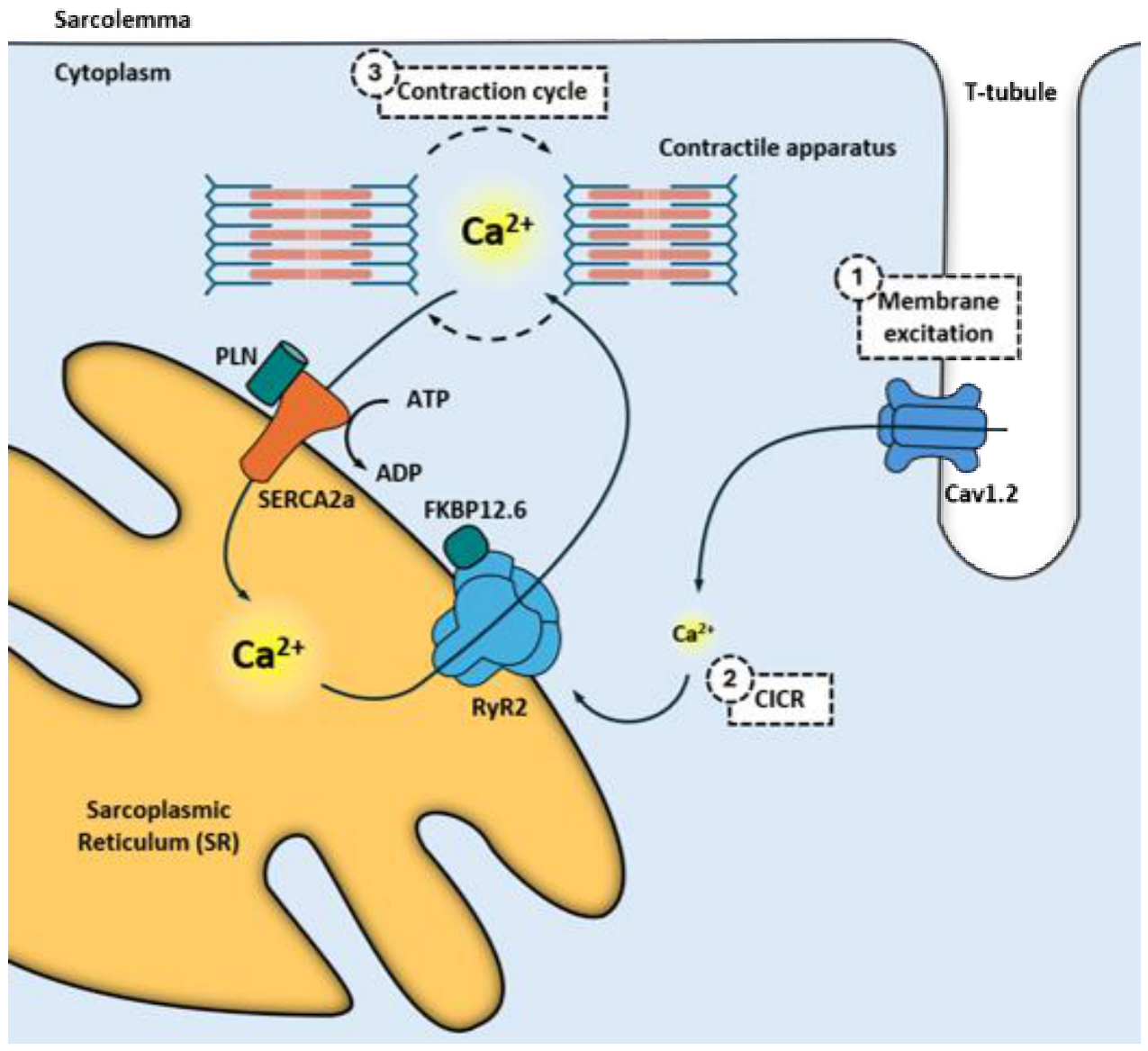
Figure 2.
Regulation of RyR2 and proposed mechanism linking RyR2 dysregulation to cardiac dysfunction in HCPS. (A) Physiological regulation of RyR2 by β-adrenergic/PKA signaling, Ca²⁺/calmodulin-dependent CaMKII activation, and FKBP12.6 association. (B) Proposed inflammatory pathways converging on RyR2. IL-6 signaling through JAK2/STAT3 is linked to CaMKII-dependent phosphorylation of RyR2 at Ser2814, while inflammatory redox signaling is represented by mitochondrial ROS, CaMKII oxidation, direct RyR2 oxidation, and NO/RNS-dependent modification. The dissociation of FKBP12.6 from RyR2 is shown as a convergent consequence of inflammatory redox signaling. Panel upper portion summarizes evidence-supported molecular mechanisms derived from different experimental models, including IL-6-mediated inflammation, postoperative atrial fibrillation, and sepsis/endotoxemia [16,17,18,26,29,36]. Dotted boundary indicates separation between evidence-supported molecular mechanisms and the proposed hypothesis (panel bottom portion). RyR2 dysregulation could be linked to spontaneous SR Ca²⁺ release, reduced SR Ca²⁺ content, impaired systolic Ca²⁺ release, contractile dysfunction, and delayed afterdepolarizations, thus representing a mechanistic inference for the cardiac phenotype of HCPS.
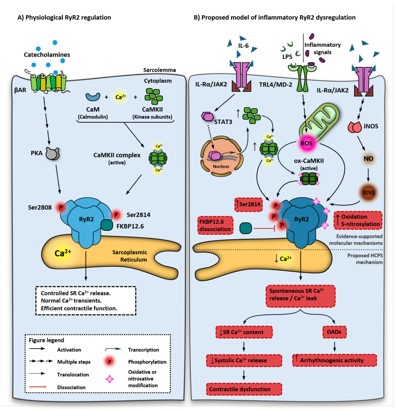
Figure 3.
Integrated mechanistic model. Proposed pathophysiological cascade linking ANDV infection to reversible myocardial dysfunction in HCPS. Three logical stages are shown: infection and inflammation (blue panels), establishment of pathology (red panels), and clinical resolution (green panel). Solid arrows indicate proposed causal relationships; dashed boxes and arrows denote hypothetical or unconfirmed elements; solid boxes mark terminal outcomes. Supportive care with VA-ECMO only buys time for spontaneous resolution of inflammation rather than modifying the pathophysiological mechanism, as indicated by the dashed arrow. Abbreviations: ANDV, Andes virus; HCPS, hantavirus cardiopulmonary syndrome; IL, interleukin; PTMs, post-translational modifications; ROS/RNS, reactive oxygen and nitrogen species; RyR2, ryanodine receptor type 2; TNFα, tumor necrosis factor α; VA-ECMO, venoarterial extracorporeal membrane oxygenation.
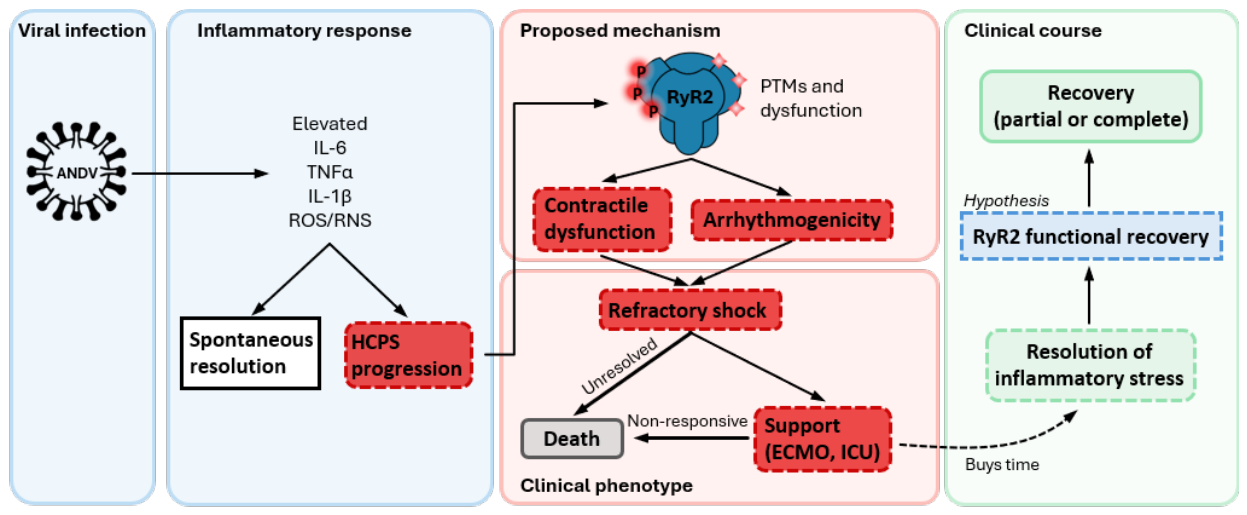
Figure 4.
Disease progression and timing of interventions in HCPS. (A) Clinical progression of HCPS from ANDV exposure to clinical recovery. The timeline depicts theoretical stages of disease progression and their approximate duration. Not all stages are clinically confirmed in every patient; in particular, incubation and prodrome are rarely diagnosed prospectively, as symptoms are nonspecific and hantavirus is typically not suspected until the cardiopulmonary phase. The therapeutic window (24-48 h) marks the period when most deaths occur. (B) Comparison of optimal intervention opportunities with real-world clinical practice using the same timeline. Optimal opportunities are inferred from the mechanism of action, not from demonstrated clinical efficacy, and are denoted with (?). In practice, patients typically present during the therapeutic window, when the cardiopulmonary phase is already established or in progress. Therefore, most interventions are initiated after their optimal opportunity. RyR2 stabilization with rycals is shown as a future therapeutic opportunity (dashed arrow), which would act on the established molecular mechanism rather than on upstream triggers. Time scale is not linear. Abbreviations: ANDV, Andes virus; ARDS, acute respiratory distress syndrome; GI, gastrointestinal tract; HCPS, hantavirus cardiopulmonary syndrome; ICU, intensive care unit; VA-ECMO, venoarterial extracorporeal membrane oxygenation. *, resolution with intensive care may extend beyond 48 h after ICU admission; however, this is not represented in the figure to preserve clarity.
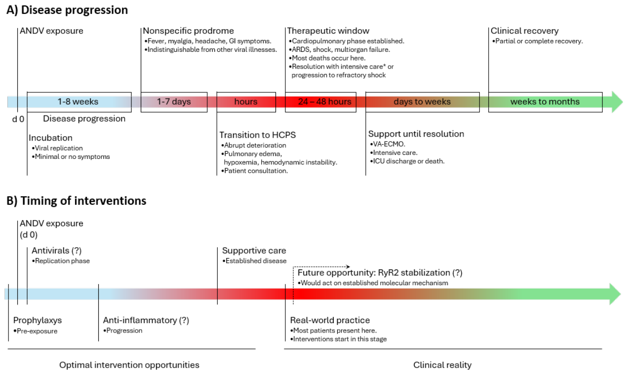
Disclaimer/Publisher’s Note: The statements, opinions and data contained in all publications are solely those of the individual author(s) and contributor(s) and not of MDPI and/or the editor(s). MDPI and/or the editor(s) disclaim responsibility for any injury to people or property resulting from any ideas, methods, instructions or products referred to in the content. |
© 2026 by the authors. Licensee MDPI, Basel, Switzerland. This article is an open access article distributed under the terms and conditions of the Creative Commons Attribution (CC BY) license (http://creativecommons.org/licenses/by/4.0/).
Copyright: This open access article is published under a Creative Commons CC BY 4.0 license, which permit the free download, distribution, and reuse, provided that the author and preprint are cited in any reuse.