

Submitted:
14 October 2026
Posted:
15 October 2026
You are already at the latest version
Abstract
Antimicrobial resistance (AMR) in Ecuador extends across human health, animal production, agriculture, and the environment, yet national evidence on bacteriophages as an alternative has remained scattered across indexed articles, theses, and institutional repositories. We mapped that evidence through a One Health lens, following the PRISMA extension for scoping reviews (PRISMA-ScR). Five bibliographic databases and publisher platforms (Web of Science, ScienceDirect, Springer, Scopus, and SciELO) and 21 Ecuadorian repositories were searched for studies published between 2021 and 2026. From 592 records, 38 reports met the eligibility criteria: 6 peer-reviewed articles and 32 gray-literature documents (7 postgraduate and 25 undergraduate). Inter-reviewer agreement was 89.1% (Fleiss' kappa = 0.773; 95% CI, 0.635-0.912). Research concentrated in animal health (44.7%) and human health (31.6%), with fewer reports in agriculture (10.5%), poultry (5.3%), aquaculture (5.3%), and the environment (2.6%). Institutional output was highest in Pichincha, Tungurahua, and Guayas; journal articles were affiliated with institutions in Pichincha, Guayas, and Azuay. No included reports were assigned to institutions in the Amazon or Galapagos regions. The most studied hosts were Salmonella enterica, Escherichia coli, Klebsiella pneumoniae, Vibrio spp., Listeria spp., and Ralstonia solanacearum. Methodologically, the corpus spans phenotypic characterization, PCR- and restriction-based differentiation, whole-genome sequencing, and functional validation, but these approaches coexist rather than form a maturation gradient. Genomic safety claims rested mainly on the absence of known lysogeny, resistance, and virulence genes, and functional transduction assays were largely absent. Ecuadorian bacteriophage research has begun to shift from demonstrating lysis toward molecularly defined products, but translational readiness is limited by fragmented publication, geographic concentration, and incomplete characterization. Decentralized laboratory capacity, national phage collections with deposited genomes, and standardized characterization pipelines would strengthen the field.
Keywords:
bacteriophages
; antimicrobial resistance
; one health
; phage-based biocontrol
; genomic characterization
; food safety
; sustainable agriculture
; Ecuador
1. Introduction
Antimicrobial resistance (AMR) occurs when bacteria, viruses, fungi, or parasites no longer respond to drugs used to prevent or treat infections. In bacteria, inappropriate and excessive use of antimicrobials is a major factor. AMR represents a threat, since the improper and excessive use of antibiotics favors the selection and spread of microorganisms resistant to multiple antimicrobials. This is a problem that transcends human health and relates to animal production, food, and the environment. In addition, antibiotics in soil and water favor the selection and spread of resistant microorganisms that can affect both animals and humans [1]. Its economic impact is worrisome because if resistance rates continue to rise, AMR-related healthcare costs will reach $159 billion annually by 2050, or 1.2% of global health spending. This poses a threat not only to health but also to global economic stability due to increased costs [2,3]. This growing health and economic burden, together with the reduction in therapeutic options against multidrug-resistant microorganisms, necessitates the development of alternatives for controlling bacterial infections [3].
One Health is a concept that focuses on animal, human, and environmental health. This indicates that these areas are closely related; therefore, it focuses on the work of multiple areas, disciplines, and communities [4]. Health, animals, and the environment are interrelated; therefore, One Health addresses disease control from prevention to detection, thus contributing to global health security. In this context, AMR is one of the most important problems within the One Health approach, since resistant bacteria and their genes circulate between humans, animals, and the environment. The use of antimicrobials in human health, animal production, or agriculture can lead to their release into the environment, creating a complex problem of potential resistant bacterial strains.
Bacteriophages (also called phages) are viruses that infect and replicate within bacteria. They are found anywhere where bacteria exist, such as in lakes, rivers, and even in the intestines of animals. Phages contain a nucleic acid genome within a protein capsid; their high host specificity can limit the impact on non-target bacteria. Lytic bacteriophages can destroy antibiotic-resistant bacteria, preventing their spread; therefore, they can be used as an alternative therapeutic option in animals and humans to combat multidrug-resistant microorganisms. Bacteriophages could be used in conjunction with antibiotics to treat multidrug-resistant bacteria and enhance treatment effectiveness [5]. Cocktails can also be made with different phages to expand the range of hosts, thus reducing the likelihood of resistance.
Bacteriophages can have applications in multiple areas of One Health. In human health, research has been conducted on phage therapy, a therapeutic approach that uses lytic bacteriophages to lyse pathogenic bacteria. Infection control can also be implemented to delay or prevent serious infections transmitted through contaminated water or animals, thereby preventing the spread of diseases between humans and animals [1]. Some bacteria form biofilms, which make several strains more resistant and harder to remove. However, certain phages can act against these biofilms by producing enzymes that degrade them and cause bacterial death [6]. In animal production, bacteriophages can be applied in pathogen control as an alternative to antibiotics in livestock and domestic animal infections [7]. Integrating bacteriophages into food or water can significantly improve gut health by enhancing digestion and reducing the load of enteric pathogens such as Salmonella spp. and Clostridium spp. In biotechnology, phages can be used as rapid diagnostic tools due to their high specificity, enabling monitoring of fermentation processes and diagnosis of infectious diseases [8]. In addition, phage engineering lets researchers modify phage genomes and direct them toward specific targets. Integrating systems such as CRISPR-Cas makes it easier to insert, delete, or modify genes, enhancing the potential of phages for therapeutic and diagnostic applications [9].
In Ecuador, in the clinical setting, during 2021, between 1400 and 2010 deaths attributable to antimicrobial resistance and between 5820 and 8580 deaths associated with this problem were estimated. Among the main pathogen-drug combinations associated with AMR load were methicillin-resistant Staphylococcus aureus, carbapenem-resistant Pseudomonas aeruginosa, and Acinetobacter baumannii. If current trends continue, it is projected that by 2030 deaths associated with AMR could reach between 7540 and 12700 [10]. In the field of animal production, a 2019 study evaluated antimicrobial resistance in Escherichia coli and Salmonella isolated from poultry in Ecuador. The highest proportions of resistance were observed against erythromycin (76% in E. coli and 85% in Salmonella) and tetracycline (71% and 90%, respectively). These findings show high levels of antimicrobial resistance among bacterial isolates obtained from poultry in Ecuador [11]. Likewise, in the environmental and agricultural fields, the presence of extended-spectrum beta-lactamase-producing Escherichia coli (ESBL) in irrigation water and agricultural products in 17 provinces of Ecuador was investigated. Of the samples, 11% tested positive for E. coli, and 165 isolates were recovered from the positive samples; 58% of the isolates exhibited an extended-spectrum beta-lactamase (ESBL) phenotype and high resistance to various antimicrobials. These findings show that resistant bacteria can also be found in agricultural and environmental systems [12]. Taken together, these antecedents show that AMR in Ecuador transcends the field of human health and involves animal production, agriculture and the environment, which reinforces the need to address it from the One Health perspective. This highlights the need to explore alternatives and strategies in the face of dependence on conventional antimicrobials.
Despite the national importance of AMR and the increased interest in bacteriophages as alternatives with great therapeutic, biocontrol, and biotechnological potential, the evidence generated in Ecuador remains fragmented between indexed scientific publications, theses, and institutional repositories. To date, there is no systematic synthesis to determine its distribution over time and territory, the institutions and areas of One Health that concentrate the research, the main bacterial hosts studied, and the applications with the greatest development. Similarly, there is a lack of evaluation at a methodological degree of maturity of the research, considering isolation and phenotypic characterization through genomic sequencing, safety evaluation, functional validation, and rational development of phage-based products. This absence of evidence limits the difference between the preliminary demonstration of lytic activity and the presence of candidates with sufficient characteristics to advance to subsequent stages of preclinical, productive, or environmental evaluation.
In this context, the purpose of this scoping review was to identify, organize and, critically analyze the scientific evidence and related gray literature on bacteriophages developed in Ecuador from a One Health approach. Specifically, it was proposed to characterize their temporal, geographical and, institutional distribution; to determine the bacterial hosts, the areas of application and, the experimental purposes; to map the distribution of phenotypic, molecular, genomic, and functional approaches and, to identify the main methodological and translational gaps that allow guiding future lines of research and application within the Ecuadorian context.
2. Materials and Methods
2.1. Review Design and Scope
This scoping review mapped experimental and computational research on bacteriophages in Ecuador through a One Health lens. Its objectives were to describe the temporal, institutional, and geographical distribution of the available reports; identify bacterial targets and application sectors; characterize the methodological approaches employed; and identify gaps in molecular characterization and evidence supporting potential applications. Reporting followed the Preferred Reporting Items for Systematic Reviews and Meta-Analyses extension for Scoping Reviews (PRISMA-ScR). No review protocol was registered.
2.2. Information Sources and Search Strategy
Five bibliographic databases and publisher platforms were searched: Web of Science, ScienceDirect, Springer, Scopus, and SciELO. The following search expression was used across these sources: (“bacteriophage” OR bacteriófago OR “phage therapy”) AND (Ecuador OR Ecuadorian) AND (bacteria OR antimicrobial OR biotechnology). Publication-year restrictions were applied to retrieve records published from 2021 to 2026. Searches were conducted on May 23, 2026; therefore, coverage of 2026 was limited to records available by that date.
Gray literature was sought through the institutional repositories of 21 Ecuadorian universities. These institutions were purposively selected through consensus among the authors, drawing on their academic experience and knowledge of the Ecuadorian university system. Collectively, the selected institutions have campuses or institutional locations across the Coast, the Andes, the Amazon, and the Galápagos Islands. Amazonian coverage included Universidad Regional Amazónica Ikiam, while the selected Universidad San Francisco de Quito repository also contains academic work from its Galápagos extension. The institutions and repository URLs are listed in the supplementary materials (Supplementary Table S2). This selection provided geographic reach across the four natural regions but did not encompass every Ecuadorian institutional repository.
Repository searches were conducted on July 1, 2026, through each repository’s own interface, including DSpace interfaces, using the terms “bacteriophage” and “bacteriófago”. This simplified approach accommodated differences in repository search functionality. Retrieved records were examined individually for publication year and relevance, and potentially eligible documents were assessed against the eligibility criteria.
2.3. Eligibility Criteria
Eligible reports comprised original journal articles and undergraduate or postgraduate theses published between 2021 and the corresponding search cutoff in 2026, written in Spanish or English, with accessible full text, and addressing bacteriophage-related research conducted in Ecuador with relevance to One Health. The scope included experimental phage isolation, characterization, and application, as well as computational analyses of phage-related sequences, prophages, and bacterial antiphage defense systems.
During study selection, the eligibility criteria were broadened to include computational analyses of phage-related sequences and bacterial antiphage defenses, as well as reports combining a literature review with original preliminary experimental findings. This post hoc amendment reflected their relevance to the broader mapping of Ecuadorian bacteriophage research. Their inclusion in descriptive counts did not imply equivalence to experimental evidence of phage efficacy or safety. Reports outside the publication period, of ineligible document types, or lacking the required thematic or One Health relevance were excluded.
2.4. Study Selection and Reviewer Agreement
Duplicate records were identified and removed using Mendeley before title and abstract screening. Potentially eligible reports were sought for full-text assessment. Nicolás Grijalva-Borja, Mariel Yela-Rubio, and Jaime Naranjo-Morán participated in study selection and eligibility assessment.
Inter-reviewer agreement was assessed across the 67 reports evaluated at full text, using the original individual eligibility decisions of Nicolás Grijalva-Borja, Mariel Yela-Rubio, and Jaime Naranjo-Morán before resolution of disagreements. Agreement was summarized using percentage agreement and Fleiss’ kappa, with its standard error and 95% confidence interval. Discrepancies were subsequently resolved through in-person meetings in which the reviewers examined the reports against the eligibility criteria until consensus was reached. Reasons for full-text exclusion were recorded, and the selection process was summarized in a PRISMA flow diagram.
2.5. Data Extraction and Units of Analysis
Data were extracted by Mariel Yela-Rubio and Nicolás Grijalva-Borja using a standardized extraction form and verified by Nicolás Grijalva-Borja. Extracted information included authorship, publication year, title, institution, One Health-related application sector, bacterial target, phage application, methodological approaches, and main findings.
The unit of synthesis was the report. Distinct publications or theses could therefore contribute separate entries to the descriptive mapping even when they shared phage material or datasets. Such reports were interpreted as related contributions rather than independent experimental replications.
Geographical summaries used the principal institutional assignment recorded for each report. They describe institutional affiliation rather than sampling locations, the geographical distribution of bacterial hosts, or the full territorial reach of collaborating institutions. In particular, an institutional assignment does not necessarily identify the campus or region in which all research activities occurred.
2.6. Classification of One Health-Related Application Sectors
Reports were organized into six operational application sectors relevant to One Health: human health, animal health, agriculture, poultry, aquaculture, and environment. These categories were used to describe the thematic distribution of the corpus; they were not intended as six independent or equivalent biological domains within a formal One Health taxonomy.
Human health encompassed reports primarily oriented toward human-associated pathogens or potential clinical applications. Animal health encompassed veterinary, animal-associated, and animal-microbiota research framed around animal health or host–microbe interactions. Agriculture encompassed plant-associated pathogens and crop protection. Poultry and aquaculture identified reports explicitly framed around applications in poultry production and aquatic production systems, respectively. Environment encompassed environmental surveillance and studies primarily addressing environmental reservoirs or circulation of phage-associated genetic material.
Each report contributed to one primary sector in the descriptive counts. Classification reflected the principal research objective and application context, rather than bacterial species or sample origin alone. Accordingly, research involving poultry-associated bacteria could be classified under animal health when its main focus was animal-associated microbial ecology or host–phage interactions, or under poultry when its main focus was an application in poultry production. The same distinction applied to aquatic animal research and aquaculture applications.
Poultry and aquaculture are substantively nested within broader animal-health and food-production contexts. Their separate presentation was intended to retain application-specific information and should not be interpreted as implying biological independence from animal health. Conversely, the animal-health category should not be interpreted as excluding all poultry- or aquatic-animal-associated research.
The categories were mutually exclusive for report-level counting but not conceptually exclusive. Cross-sector relevance was addressed in the narrative synthesis. Classification within a One Health-related sector did not, by itself, establish that a report investigated transmission, interactions, or integrated outcomes across human, animal, and environmental interfaces. Sector distributions were interpreted descriptively, without inferential comparisons or assumptions that the categories were equally likely.
2.7. Coding of Methodological Approaches and Evidence Synthesis
Methodological approaches were coded from the descriptions in Supplementary Table S1 using four non-exclusive binary categories: phenotypic assays; targeted molecular assays or profiling; genomic or sequence-based computational analysis; and applied or functional evaluation. A value of one indicated that an approach was reported, irrespective of its depth or the direction of its findings. A value of zero indicated that the approach was not described in the synthesis table and did not necessarily establish that it had not been performed.
Phenotypic approaches included observable laboratory measurements of phage activity or properties. Molecular approaches included targeted assays and profiling of phages or their bacterial hosts; host-directed molecular characterization was distinguished from direct characterization of phage genomes in the narrative synthesis. Genomic approaches included sequencing and computational analyses of phage-related sequences, prophages, or bacterial antiphage defenses. Applied approaches included functional evaluations directed toward an intended application.
Each report could contribute to more than one methodological category. The chord diagram counted report–approach occurrences, whereas heatmap percentages used the total number of reports assigned to each application sector as the denominator. Preliminary enrichment and lysis testing supported the phenotypic code for report 22, while computational analysis of bacterial antiphage defenses supported the genomic/computational code for report 2. Neither assignment implied complete phage characterization.
Descriptive counts and percentages were combined with a narrative synthesis organized around the review objectives. The methodological codes represented reported approaches rather than validated measures of study quality, biological safety, or translational readiness. Interpretation distinguished phenotypic activity, molecular characterization, genomic evidence, and functional or product-level evaluation without assuming a chronological progression among reports. References outside the eligible corpus were used to contextualize methods, biological mechanisms, and potential applications; they did not contribute to the descriptive counts or methodological coding of the 38 included reports.
2.8. Critical Appraisal
No formal methodological quality or risk-of-bias assessment was performed. This decision was consistent with the review’s objective of mapping the extent, characteristics, methodological approaches, and gaps of the available literature, rather than estimating pooled intervention effects or establishing the certainty of evidence for efficacy or safety. Eligible reports were retained without an additional quality-based exclusion threshold.
Methodological limitations were discussed narratively, but this discussion did not constitute a standardized critical appraisal. Consequently, the synthesis should be interpreted as a descriptive evidence map rather than as evidence establishing comparative effectiveness or certifying the safety of phage-based interventions.
3. Results
3.1. Study Selection and Reviewer Agreement
The study selection process is presented in Figure 1. The search identified 592 records, including 80 from scientific databases and 512 from gray literature sources. After removing nine duplicates, 583 records were screened by title and abstract, and 511 were excluded. Of the 72 reports sought for full-text retrieval, five could not be obtained. Consequently, 67 reports underwent full-text eligibility assessment. Twenty-nine reports were excluded: 16 because of an ineligible publication type, two because bacteriophages were not the primary focus, one because it did not address an application within the One Health framework, and 10 because they fell outside the predefined publication period. The final synthesis therefore comprised 38 eligible reports, comprising six journal articles and 32 theses (seven postgraduate and 25 undergraduate). Agreement among the three reviewers was 89.1%. Fleiss’ kappa was 0.773 (SE = 0.071; 95% CI: 0.635–0.912; z = 10.97; p < 0.001).
3.2. Publication Characteristics and Application Sectors
Using the principal institutional assignment recorded for each report, the six journal articles were distributed across Universidad San Francisco de Quito (USFQ; n = 2), Escuela Superior Politécnica del Litoral (ESPOL; n = 1), Universidad Espíritu Santo (UEES; n = 1), Universidad de Cuenca (UCuenca; n = 1), and Universidad Central del Ecuador (UCE; n = 1). These counts describe the extraction dataset and do not enumerate all collaborating affiliations. The rigor required for methodological development in bacteriophage research means that, in countries of the Global South, this research has an additional layer of inherent complexity.
According to the results of this scoping review, there are 32 theses, comprising seven postgraduate and 25 undergraduate reports. The phenomenon in which empirical research is not published in indexed journals is known as Unpublished Research Bias, where the Grey Literature Gap becomes evident [13]. As already mentioned, the possibility of research being published in a scientific journal depends on the nature of its results or the availability of economic resources, which often limits the publication of an empirical work in an indexed journal. In addition, there is another bias to take into account: pipeline bias [14], which explains that the time that elapses from the time a student defends their thesis work until —perhaps— an article is written, submitted, reviewed, and published is usually broken; therefore, work never comes out of the university “pipe”. Despite all this, it is important to report this grey literature, since without it, there is a tendency to exaggerate methodological biases and underestimate basic laboratory work carried out locally.
Based on our findings, the literature found about bacteriophages in Ecuador has been categorized into six research areas: human health, animal health, poultry, aquaculture, agriculture, and the environment—these sectors align with the One Health approach (Figure 2).
When analyzing the distribution of studies conducted in the 2021–2026 study period, it is observed that gray literature has been published across all analyzed areas, whereas the areas of interest of articles published in indexed journals were limited to animal and human health. The areas of greatest interest for Ecuadorian research on bacteriophages are animal health (44.7%, n = 17/38) and human health (31.6%, n = 12/38). Fewer reports addressed agriculture (10.5%, n = 4/38), poultry farming (5.3%, n = 2/38), aquaculture (5.3%, n = 2/38), and the environment (2.6%, n = 1/38) (Figure 3).
Historically, the main area of interest in bacteriophage research has been the area of human health [15,16,17], as this is driven by the global AMR crisis, focused on critical pathogens such as the ESKAPEE group [18], and secondly the area of animal health, as it has a direct role in the One Health framework as a bridge between the environment and humans [19,20]. In this area, the main aim is to reduce the use of antibiotics as growth promoters and to treat diseases that cause great economic losses, such as bovine mastitis [21]. In the Ecuadorian case, animal health studies accounted for 44.7% of the included reports, while human health studies accounted for 31.6%. Given the limited number of reports and the absence of a prespecified equivalence margin, this difference was interpreted descriptively rather than as evidence of equivalence between research areas.
Other areas related to more specific aspects within the One Health approach, such as agriculture, aquaculture, poultry, and the environment, are emerging in recent developments. The details of their approaches are discussed in the following sections of this scoping review.
If we analyze research on bacteriophages by the number of publications per year, there is no sustained growth between 2021 and 2026. In the case of grey literature, marked fluctuations are evident: in 2024, there was a peak of 12 published documents, which has since declined; at the time of writing this review (July 2026), only 1 has been published (Figure 4). These results should be interpreted with caution, as further articles may still be published later in the year. In contrast, articles published in indexed journals were limited throughout the period. Only in 2023 were there two articles; in 2025, one; and by the time of writing this review, three.
However, the small number of scientific articles and international variations make it impossible to establish a conclusive upward trend. Overall, the results show that Ecuadorian production on bacteriophages has been mainly concentrated in the grey literature, while its incorporation into the indexed scientific literature remains incipient. As already mentioned, this relates to Unpublished Research Bias, in which the Grey Literature Gap becomes evident. A bibliometric analysis of airborne microorganisms (a specific niche in which phages are studied) identified 1,087 articles published between 2011 and 2020, showing a clear upward trend that peaked in 2019 [22]. As of September 2024, 28,468 complete phage genomes have been sequenced and incorporated into INPHARED, the curated database of bacteriophage genomes [23]. Although the exact total number of articles on phage types and their study is not cited as a global figure, the sources consulted confirm that the field is experiencing exponential expansion, moving from anecdotal case reports to large-scale genomic and clinical studies.
3.3. Institutional and Geographical Distribution
To conclude the descriptive analysis of the state of bacteriophage research and considering the One Health approach in Ecuador, it is important to note the geographical distribution of this research across the country. The institutional production identified in this review was concentrated in the Andean corridor, with additional contributions from two coastal provinces. None of the included reports were affiliated with institutions located in the Ecuadorian Amazon or the Galápagos Islands. Because the map represents institutional affiliation rather than sampling location, these findings should not be interpreted as definitive evidence that no phage-related sampling or field activities have occurred in those regions.
Ecuadorian bacteriophage institutional affiliation showed a marked concentration in terms of territory and institutions. Of the 38 documents included, 31 (81.6%) came from institutions located in four provinces of the Andean region: Imbabura (n = 1), Tungurahua (n = 6), Azuay (n = 4), and Pichincha (n = 20). In the Coast region, only contributions from Guayas (n = 5) and El Oro (n = 2) were identified, accounting for 18.4% of the corpus. No documents associated with institutions in the Amazon or the island region were found. Due to the lack of records, the Galapagos Islands are not shown in Figure 5.
Geographically, the results describe a main production nucleus along the inter-Andean axis, complemented by two smaller coastal nuclei. The concentration was even more evident in Pichincha, where Quito—the capital city—is located and where six institutions contributed to the corpus analyzed. However, this production was not distributed evenly among the province’s universities either: the Universidad San Francisco de Quito contributed 10 of its 20 documents (see Figure 6). Likewise, the articles published in indexed journals were limited to Pichincha, Guayas, and Azuay, while the production from Tungurahua, El Oro, and Imbabura consisted exclusively of gray literature.
3.4. Methodological Approaches
Complementing the institutional distribution, Figure 7 graphically summarizes the methodological approaches reported in Supplementary Table S1 across the six One Health-related application sectors. The chord diagram displays report–approach occurrences, while the heatmap shows the proportion of reports within each sector documenting phenotypic, molecular, genomic/computational, and applied/functional approaches. These categories are non-exclusive, allowing individual reports to contribute to multiple approaches. Their implications for molecular characterization, genomic safety, and potential applications are examined in the Discussion.
4. Discussion
4.1. Research Distribution and Publication Patterns
The retrieved literature depicts an Ecuadorian bacteriophage research landscape documented predominantly through university theses, with comparatively few journal articles during the review period. Including institutional repositories was therefore essential to describe the range of research activities represented in the corpus. However, the predominance of theses does not, by itself, demonstrate selective publication according to study findings, identify financial barriers to publication, or establish that thesis research never reached a journal. Because the unit of analysis was the report, the observed distribution describes publication formats rather than the proportion of independent projects that remained unpublished. Annual fluctuations should also be interpreted cautiously because coverage of 2026 was incomplete and differed between bibliographic sources and repositories.
Between 2021 and the search cutoffs in 2026, Ecuadorian bacteriophage publications fluctuated without a sustained increase; the incomplete final year precludes a conclusion of long-term decline. According to the results presented in this scoping review, only six research articles published in indexed scientific journals explicitly discuss bacteriophages and their applications across various areas of development, including human and animal health. It is interesting to analyze this finding from an epidemiological perspective, where a paradox is impossible to ignore while advanced biomedical research is predominantly published in the Global North, the greatest impact of AMR is observed in countries with emerging economies [3,24].
The pattern of distribution of included documents by province and contributing university reproduces, at the thematic level, a broader feature of the Ecuadorian research system. An analysis of national biomedical production between 1920 and 2021 found that Pichincha, Guayas, and Azuay accounted for 87.7% of publications, and that three universities located in Quito—PUCE, USFQ, and UCE—were among the institutions with the highest production [25]. Therefore, the distribution observed in this scoping review could be related to the historical territorial concentration of universities with laboratories, specialized personnel, graduate programs, funding, and collaboration networks [25,26]. However, these variables are outside the scope of this review, so they should be treated as explanatory hypotheses rather than as evidence of causal relationships.
The absence of documents from the Amazon and Galapagos reveals a gap in the field’s institutional coverage, but it in no way implies that these territories lack bacteriophages, microbial diversity, or relevance to One Health. On the contrary, ecological diversity, agricultural systems, aquaculture, wildlife, and human-animal-environment interfaces make these regions potentially relevant scenarios for future research. Consequently, strengthening partnerships and public agencies in underrepresented regions could expand both the geographical coverage and the ecological and health relevance of Ecuadorian bacteriophage research.
4.2. From Phenotypic Characterization to Functional Phage Genomics
Ecuadorian bacteriophage research comprises a range of complementary methodological approaches from isolation and phenotypic characterization to complete sequencing, comparative genomics, and functional validation of phage-derived products. According to the information analyzed, it has been observed that not all studies have the same molecular depth; some isolate phages and demonstrate lysis by plaque assays, host range, and physicochemical stability [29,30,31,32,33,34,35,36,37]. Others incorporate complete sequencing, genomic annotation, taxonomic classification, and phylogenetic reconstruction to assess phage identity and safety [34,35,36,37] Only a small group of reports addressed prophages, viral communities through metagenomics or the functional validation of phage-derived proteins [38,39,40,41,42]. These approaches coexist over time; that is, the available evidence does not establish a consistent chronological progression from classical techniques to more advanced methodologies, but they share phenotypic, molecular, and genomic capabilities and are heterogeneously distributed across institutions and application sectors. Figure 7 summarizes the distribution of these non-exclusive analytical approaches across the six One Health sectors, showing both the number of reports associated with each approach and the proportion of reports for each approach within each sector.
4.2.1. Phenotypic Foundations: Plaque-Based Characterization and Host-Range Assessment
Phenotypic characterization is the methodological basis of bacteriophage work in Ecuador; it has served as a common microbiological platform for the enrichment, isolation, purification, and quantification of lytic bacteriophages. As already mentioned, plaque assays have been among the main methods used to characterize their phenotype. The plaque assay is a classic microbiological technique used to detect infectious bacteriophages, isolate and purify individual phages, quantify their concentration, confirm productive infection, and/or calculate infection efficiency in different bacterial hosts [43,44,45]. In this technique, a lysate dilution is mixed with the host bacterium and incorporated into a semi-solid agar layer on a basal solid medium. The semi-solid matrix allows the growth of a bacterial lawn but restricts the diffusion of virions, so that successive cycles of infection, replication, and lysis remain localized, producing visible areas of clearance called plaques [44,45]. Consequently, the assay does not quantify the total number of physical particles or the concentration of viral DNA, but rather the units capable of initiating and spreading an infection under the experimental conditions used [46]. This distinction is relevant because the absence of plaques does not necessarily prove that there are no interactions between the phage and the bacterium: the virion could adsorb or introduce its genome but subsequently fail due to an incompatibility with the cellular machinery or the action of bacterial defense systems [47].
The morphology of the plaques is considered the first macroscopic expression of the phage-host interaction, but it is not an exclusive property of the phage. The diameter, transparency, regularity of the edges, and the presence of halos result from interactions among the adsorption rate, the latency period, the burst size, the diffusion of the virions, and the host’s density and physiology. Using lysogenic λ phages, Gallet et al. [46] experimentally demonstrated that modifications in adsorption, lysis timing, and particle morphology alter plaque size and productivity. To these factors must be added methodological variables such as agar concentration, medium composition, temperature, the presence of cations, and the bacterial growth phase [48]. In this way, changes in the culture medium can alter the size and appearance of phage-produced plaques. Consequently, morphological differences are only comparable when, of course, the experiments are carried out under standardized conditions.
In Ecuadorian studies, the double-layer assay served three main functions: detecting phages in environmental samples, purifying individual isolates, and quantifying their infectivity in plaque-forming units. This approach enabled the recovery of phages against human, animal, and plant pathogens, including Listeria spp., Salmonella enterica, Staphylococcus aureus, Escherichia coli, Vibrio alginolyticus, Ralstonia solanacearum, and Kosakonia cowanii [29,30,31,32,49]. In some studies, plate formation was complemented with titration, successive purifications, and evaluation of host range. Torres [50], for example, isolated and purified individual phages from the M115 cocktail and compared their activity against different Salmonella serovars. Morales & Topa [51] used lysis profiles to select seven relatively wide-range candidates from 27 isolated phages. These results demonstrated relevant phenotypic diversity, although the actual range amplitude depended on both the selected strain panel and the method used to define susceptibility [52,53].
A critical aspect is that several studies also initially determined the range using a spot assay. This technique involves depositing a drop of concentrated lysate onto bacterial lawn and recording the emergence of a thinning area. Although it is useful for rapidly evaluating numerous phage-bacteria combinations, it does not allow us to assume that the phage produced a replicative infection [54]. At high titers, the clearance may be due to: lysis from the outside, abortive infection, activity of free depolymerases or endolysins, bacteriocins, or residual components of the lysate [47,55]. Mirzaei & Nilsson [54] showed that the spot assay can overestimate both bactericidal activity and host range, and that its results do not necessarily correlate with plaque-forming efficiency. Therefore, a positive spot assay should be interpreted as preliminary evidence of susceptibility and confirmed by serial dilutions that produce individual plaques.
Efficiency of Plating (EOP) provides a more rigorous measure of relative productivity. It is calculated as the ratio between the phage titer obtained on the tested bacterial strain and that obtained on the reference (propagation) strain [56], as shown in Equation (1).
This value allows differentiation between visual lysis produced by a concentrated lysate and that generated by an infection capable of producing progeny. However, it is also not an absolute measure, as plaque formation may be limited by poor diffusion, inadequate agar growth, or suboptimal experimental conditions [44,45,55]. For this reason, range characterization should combine spot assay, EOP, inhibition curves in liquid medium, and, for priority candidates, adsorption assays and one-step growth curves. Microplate assays can provide additional information on bacteriophage inhibition, growth recovery, and the emergence of resistance, although turbidity reduction alone does not demonstrate the production of new virions [57].
Among the Ecuadorian investigations, Montenegro [30] presented a particularly informative phenotypic characterization by complementing plaque formation with a one-step growth curve, estimation of latency period and burst size, and stability assays against pH, temperature, and ultraviolet radiation. Similarly, studies on Ralstonia solanacearum showed that inhibition depended on the strain and the bacterium’s physiological phase at the time of infection [58,59]. Cortez [60] also observed greater activity when intestinal phages and E. coli isolates were collected in the same sampling period. These results show that susceptibility is not a uniform property of a bacterial species but rather a phenotype dependent on the strain’s genotype, receptor expression, metabolic state, and ecological history of phage interactions [61].
4.2.2. Intermediate Molecular Detection Methods: PCR and Restriction Profiling
The integration of PCR-based techniques and restriction fragment analysis was essential to the phenotypic characterization and comprehensive investigation of bacteriophage genomes in Ecuador. Compared to plaque assays, which provide evidence of the infective capacity of phages and define parameters such as titer, plaque-forming efficiency, and the range of susceptible hosts, molecular methodologies allow direct analysis of phage DNA or host DNA. In this context, Gómez & Paccha [62] applied enzymatic digestion processes and restriction fragment length polymorphism (RFLP) analysis to distinguish between five coliphages selected from 36 isolates. Astudillo-Riera et al. [63] used a molecular strategy to assess the genetic diversity of a group of apparently related coliphages. Given that both studies appear to use the same collection of isolates, their results should be interpreted as complementary contributions to the same line of research rather than as independent studies or replications.
The analysis of restriction genetic profiles is based on DNA fragmentation by endonucleases that recognize specific nucleotide sequences. Changes in the genetic material, such as substitutions, deletions, insertions, or rearrangements, can create or eliminate recognition sites and alter the distances between them. As a result, both the number and size of the fragments obtained are altered. The separation of fragments by electrophoresis allows a band pattern to be obtained and genomes to be compared without needing to know their complete sequences beforehand. This technique has been applied to relate groups of bacteriophages and differentiate those with different genomic profiles. However, the similarity between a restriction pattern, the host range, and the taxonomic identity of a phage does not imply a direct correspondence between these characteristics [64].
From a terminological point of view, it is essential to differentiate between the classical RFLP and the direct comparison of the band patterns generated after the digestion of the entire genome. Restriction-based methods should be described according to the procedure actually performed, distinguishing whole-genome restriction profiling, PCR-RFLP, and probe-based RFLP analysis. When only electrophoretic patterns of digested phage DNA were compared, “restriction profile analysis” provides a precise description without implying sequence-level resolution or complete genomic characterization.
The presence of different profiles is evidence of molecular heterogeneity. However, it does not allow us to directly determine which genetic variants are responsible for these differences. Two phages can exhibit similar patterns because they maintain a comparable distribution of recognition sites for a specific enzyme, while differing in regulatory or coding regions that can influence processes such as adsorption, host range, replication, or defense mechanisms against bacteria [64]. On the other hand, a point mutation that originates or eliminates a restriction site can produce a band pattern without a significant functional difference being observed. Consequently, a similar restriction profile should not be considered equivalent to a genomic identity, just as a difference in the band pattern does not by itself demonstrate a phenotypic variation.
The ability of restriction profile analysis to differentiate genomes will depend on several factors such as the enzymes used, the amount and purity of the DNA, the electrophoretic resolution and the chemical composition of the genome analyzed. In some bacteriophages, conventional nitrogenous bases can be replaced by modified or hypermodified forms that interfere with the recognition or catalytic activity of endonucleases. For this reason, when DNA is not digested by a restriction enzyme, it does not mean that the recognition site is absent, but that the lack of cutting may be due to methylation, glycosylation, or other covalent modifications of the DNA. This behavior has been experimentally observed in phages with hypermodified cytosines and thymines, which are resistant to a considerable proportion of type II endonucleases evaluated [65,66]. The use of control DNA, evaluation with various restriction enzymes, and complete digestion controls are indispensable for determining whether the absence of restriction sites in an inhibition is due to chemical modifications of the DNA that alter enzyme activity.
In the coliphages analyzed by Gómez & Paccha [62] and Astudillo-Riera et al. [63], the use of restriction profiles enabled the organization of a large collection of isolates into groups sharing distinct molecular characteristics without individually sequencing each genome. This technique served as a pre-selection tool, with phages exhibiting distinct restriction profiles prioritized for subsequent molecular studies. However, this methodology does not provide information on the set of genes that exist in the genome, nor does it allow us to know the origin of the observed variations, and it does not allow a complete examination of genes associated with adsorption, lysogeny, bacterial virulence, antimicrobial resistance, or defense evasion mechanisms.
PCR provides a higher degree of specificity, as it relies on repeated cycles of DNA denaturation, primer annealing, and strand extension by a thermostable DNA polymerase, enabling amplification of a specific region defined by two oligonucleotides complementary to the target sequence. In the study of phages, PCR can be used to identify previously characterized genes or markers, confirm a region identified by sequencing, verify the continuity of an assembly, or distinguish isolates containing a particular sequence. However, conventional PCR is a directed technique, since it only provides information on the region between the primers. In addition, its effectiveness will depend on the degree of complementarity between the primers, the concentration and integrity of the DNA, the presence of inhibitors, and the amplification conditions. Therefore, obtaining a negative result does not confirm the absence of a specific gene. Various factors, such as mutations in primer binding sites, low template DNA concentration, the presence of inhibitors, or an inadequate annealing temperature, can prevent amplification. Likewise, the appearance of a fragment of the expected size cannot constitute a definitive identification test, since confirmatory techniques, such as amplicon sequencing, diagnostic digestion, or a specific detection system, are necessary. Therefore, PCR is useful for testing previously established molecular hypotheses, but it does not allow precise identification of the entire repertoire of genes present in a bacteriophage.
In the studies by Morales & Topa [51] both PCR and (GTG)5-PCR were used to analyze the Salmonella strains serving as hosts, rather than applying these characterizations directly to phage genomes. This difference is important when interpreting the results, since the identification and genotyping of the bacteria enabled determination of whether the phages were active against hosts with different genetic characteristics, but did not provide direct information on the phages’ genetic variability.
The (GTG)5-PCR technique is a rep-PCR technique that uses a primer composed of repeats of the GTG sequence to amplify regions between repetitive sequences in the bacterial genome. The size and number of amplicons will depend on the position and orientation of the sequences; thus, the band patterns obtained by electrophoresis can serve as a characteristic fingerprint for each isolate. This technique has demonstrated effectiveness for bacterial differentiation at the species, subspecies, and strain levels, although its reliability and reproducibility depend on experimental factors such as DNA standardization, reagents used, thermal cycler operating conditions, and electrophoresis parameters [51,67].
With respect to phages, the application of (GTG)5-PCR will serve as an indirect tool, as it allows determination of whether a broad host range contains bacterial strains with genetic variability or, conversely, clonal or closely related isolates. However, the similarities or differences between the fingerprints do not allow us to identify which factors are responsible for susceptibility. Rep-PCR does not determine whether differences between strains are due to membrane receptors, capsules, lipopolysaccharides, restriction-modification systems, CRISPR-Cas systems, prophage immunity, or other antiphage systems. To establish a bacterial genotype and its susceptibility, it would be necessary to perform specific studies of these loci, genomic sequencing, and, preferably, functional validation.
Granda [68] expanded this with higher-resolution PCR, analysis of the available p35 sequence, prediction of Open Reading Frames (ORFs), phylogenetic analysis, and comparison with Listeria phages stored in public databases. It is important to differentiate from a methodological standpoint, since PCR is used to obtain or confirm a specific DNA region, whereas ORF prediction is performed on the resulting nucleotide sequence as part of a computational analysis. The programs used for gene identification evaluate the start and end codons, the length and location, overlap, and organization within the genome. Specialized tools such as PHANOTATE consider characteristics like high gene density and ORF frequency in these viruses [69].
The prediction of an ORF does not necessarily correspond to a gene whose expression or function has been proven. Functional assignment is usually based on sequence similarity, the presence of conserved domains, protein family profiles, and the genomic context. Phages have a high number of proteins whose homologs have not been experimentally characterized, so many ORFs are classified as “hypothetical proteins”. Databases built from remote homology, such as PHROG, can improve the clustering of divergent viral proteins, but membership in a group is not sufficient evidence to attribute a specific biochemical function to it [70]. A quality genomic annotation will require several molecular techniques, such as manual inspection, comparison of the genetic context, and the application of uniform nomenclature criteria [71].
4.2.3. Whole-Genome Characterization and Comparative Genomics
The transition from molecular markers to whole-genome sequencing has represented an important development in Ecuadorian research. On the one hand, PCR focuses on specific, preselected regions, and restriction profiles provide an indirect representation of the DNA; on the other hand, massive sequencing provides a large portion, or even the entire, nucleotide sequence of the phage. This information enables integrated analysis of the genomic architecture, coding repertoire, phylogenetic relationships, taxonomic classification, and the potential presence of genes incompatible with therapeutic or biocontrol applications. Obtaining sequencing does not guarantee the validity of these results, since reliability will depend on the quality and origin of the DNA, library preparation, depth of coverage, assembly, identification of endpoints, completeness, annotation, and selection of appropriate comparative tools [72,73]. This evolution is evident in phages directed against Klebsiella pneumoniae. The initial characterization of K751 by Nolivos [34] was later expanded by Tisalema et al. [74], who studied K751, T751, and T765 using Illumina sequencing, assembly, annotation, and various comparative genomics procedures. Illumina synthesis sequencing uses millions of immobilized DNA fragments, which are analyzed through cycles of incorporation of reversible-terminator nucleotides labeled with fluorophores. As a result, a high-precision cluster of short reads is obtained, which must then be reconstructed using computational tools.
Assemblers using de Bruijn graphs process the reads into k-mer fragments to reconstruct routes compatible with their overlaps. Although the approach is efficient, longer repetitive regions that exceed the processed information may pose genomic challenges, as they can generate ambiguous junctions or assembly fragments [75]. For small, purified genomes, Illumina enables high-quality assemblies; however, the result will depend on coverage, sequence complexity, and the absence of mixtures. Phage genome evaluation studies have indicated that approximately 30x coverage may be sufficient to correctly assemble most of the genomes analyzed, although this does not prevent the difficulties associated with repeated regions, redundant ends, or samples containing more than one phage [76].
A single contig should not be automatically interpreted as evidence of a physically complete genome. The assembler can be represented circularly even when it corresponds to a linear genome with terminal redundancy; likewise, it may omit repeats or start the sequence at an arbitrary position. The procedures used to prepare libraries by tagmentation, or fragmentation can eliminate the genetic information needed to define the molecule’s natural ends. For this reason, when a phage genome is closed, it must be supported by uniform coverage, junction review, evidence on its ends, and, when necessary, complementary methods such as PCR through junctions, Sanger sequencing, or the incorporation of long reads [73]. Tisalema et al. [74] implemented their assembly using CheckV, a tool that allows comparison of the analyzed sequences with reference viral genomes and genes, providing completeness and identification of host sequences with integrated prophages [77]. However, CheckV has been designed and evaluated for analyzing viral sequences recovered from metagenomes. When used in isolated phage genomes, its results should be considered as additional evidence but do not replace coverage inspection, endpoint confirmation, or experimental verification of closure. The estimate is conditioned on the related availability in the baselines; a distantly related evolutionary genome could receive an estimate of completeness with high uncertainty.
The evolutionary position of phages relative to K. pneumoniae was determined using the VICTOR tool, which uses Genome-BLAST Distance Phylogeny to estimate distances from comparisons of genomes or whole proteomes and to generate a phylogenomic tree with resampling support [78]. This approach provides superior phylogenetic signals distributed throughout the genome and offers higher resolution than analyses of terminase, major capsid protein, or any single marker. Phage genomes exhibit modularity and frequent gene exchange between genomes, thereby allowing for partially discordant evolutionary histories. A phylogenomic tree is an integrated representation of the evolutionary signals present in the genome, rather than a demonstration that all its genes have followed the same evolutionary trajectory [79]. Comparing identity between nucleotide sequences provides a quantitative metric for evaluation, but it should be assessed alongside the aligned fraction and total genome length. A high identity restricted to a small region does not represent global genomic proximity. In addition, the reference bacterial threshold values used to compare bacterial genomes should not be applied directly to viruses. The phage taxonomy is based on whole-genome comparisons and should follow the criteria established by the International Committee on Taxonomy of Viruses and the subcommittee on archaeal and bacterial viruses [71].
The use of BACPHLIP incorporated a relevant element for the safety evaluation of bacteriophages. This software analyzes the presence of conserved protein domains associated with temperate phages and uses their presence or absence as features in a random forest classifier, trained on genomes with previously established lifestyles [80]. Therefore, its operation is not limited to identifying a specific integrase gene or directly demonstrating the ability to integrate. Instead, it produces a statistical estimate based on the combination of lysogeny-related domains. The absence of a temperate-lifestyle signal in BACPHLIP should be interpreted with caution. If it is an incomplete assembly, regions that are associated with integration, repression, or recombination genes will prevent its presence. Similarly, evolutionarily distant bacteriophages may contain proteins that are not represented in the training set. A prediction alone does not evaluate generalized transduction, nor will it rule out all possible ways by which intracellular persistence may exist. When evaluating target or biocontrol candidates, the BACPHLIP tool should be used as supplementary evidence and combined with manual genome inspection, searches for domains with higher sensitivity, comparisons with temperate phages, and experiments to evaluate the ability to establish lysogeny.
The investigation of phages targeting Salmonella infantis demonstrates a similar expansion. K. Altamirano & Gavilanes [32] used the Oxford Nanopore PromethION to analyze F6 and F14, while Galarza [81] extended the characterization to F7M1, F4M1B, and F8M2. Nanopore sequencing is based on the passage of individual DNA molecules through a pore embedded in a membrane and the alterations in ionic current that are translated and then interpreted by basecalling algorithms to determine a nucleotide sequence. A direct reading of individual molecules generates considerably longer reads than those produced by short-read platforms, which favors the resolution of repeats, terminal redundancies, and structural variants [82,83].
Obtaining long reads does not eliminate the need for genomic quality control. The use of Nanopore technology will depend on factors such as chemistry, pore, basecalling model, sequencing depth, and correction and polishing methods. In addition, base modifications present in certain phages can modify the electrical signal and affect nucleotide identification. A high-quality genomic quality assessment should inform the distribution of read lengths and qualities, coverage, assembler, polishing procedure, and the strategy used to validate ambiguous regions. When conditions are optimal, integrating long reads for continuity with shorter, higher accuracy reads for error correction provides more robust validation than either technology alone.
Sevilla-Navarro et al. [37] deepened the characterization of GS71, GS156, and GS166 by analyzing their complete genomes and applying tools such as VIRIDIC, Clinker, and PhageTerm. VIRIDIC allows the calculation of intergenomic similarities from alignments generated with BLASTN, normalizing nucleotide identity by the total length of the genomes compared. In addition to the percentage of similarity, presenting information on the fraction of each genome that aligns and the relationship between their lengths helps prevent high identity in a short, aligned region from being mistaken for whole-genome similarity [84,85]. Its clusters will depend on defined thresholds; therefore, its results should not be interpreted as a current taxonomic assignment but rather based on current taxonomic criteria and supported by appropriately selected references. Clinker enables a complementary dimension to be obtained by comparing encoded genes, their orientations, and their relative positions within the genome. The tool groups homologous proteins and establishes links between related genes, facilitating the representation of synteny blocks and conserved or variable regions [86]. In phages, this type of analysis can contribute to the identification of genomic modules related to replication, nucleotide metabolism, morphogenesis, packaging, adsorption, and lysis. However, clinker depends on the annotations and similarities provided as input data, so a genomic block does not constitute evidence of a given function. The assignment of a protein as a depolymerase, endolysin, or receptor-binding protein requires additional complementary information from genomic domains, structural characteristics, and context, as well as biochemical validation.
PhageTerm analyzes the distribution of biases in the starting positions and coverage patterns of the readings to infer the physical ends of the DNA and the genome packaging mechanism. The methodology was evaluated using phages with 5’ and 3’ cohesive ends, direct terminal repeats, and pac/headful mechanisms, among other configurations [87]. This information allows establishing the correct orientation of the genome, distinguishing between the biological circularity of artificial circularization and relating the genomic architecture to the function of terminase. The results obtained will depend on how the conservation of natural ends is handled during library preparation. Tagmentation, end repair, or certain fragmentation methods can reduce or eliminate the signal used for analysis; therefore, the absence of a pattern of ends is not sufficient evidence that the genome lacks defined ends.
In Ecuador, genomics began to spread to other hosts. Valle [88] characterized SV1 and SV2 directed against Staphylococcus aureus; Toaquiza-Vilca et al. [36] incorporated genomic information to establish the taxonomic identity of BME3; and Granda [68] compared the available sequence of p35 with Listeria phages deposited in public databases. These investigations show that molecular information was used not only to describe basic characteristics of genomic sequences but also to guide taxonomic classification and the preliminary selection of candidates. However, when the analysis is conducted only on partial regions, the results should not be presented as equivalent to a complete genomic characterization.
The genomic assessment of safety cannot be reduced to the claim that previously known genes of lysogeny, resistance, or virulence were “not identified”. That conclusion is determined by the completeness of the assembly, the quality of the gene prediction, the sensitivity of the search, and the coverage of the databases. A high proportion of phage proteins remain classified as hypothetical; “absence of homologs” does not equate to “no relevant function” or “safe”. For candidates intended for molecular applications, it is recommended to integrate assembly quality control, annotation by various methods, domain searches, manual review of ambiguous regions, determination of genomic endpoints, taxonomic and synteny analysis, and experimental validation of lifestyle and potential gene transfer [72,73].
4.3. Genomic Safety and Candidate Selection for Therapy and Biocontrol
To assess safety, it is advisable to start by identifying risk genes. This will depend on whether the sequence adequately—and exclusively—represents the phage under study, after examining assembly errors, unresolved regions, and potential contaminants. A fragmented genome may omit relevant determinants, while bacterial sequences incorporated into the assembly may lead to the assignment of functions to the phage that do not correspond to it. Nor is the suitability of a candidate an intrinsic, binary, and transferable property between applications, but a contextual inference constructed through a progressive chain of evidence: genomic identity and integrity, absence of known genetic hazards, productive antibacterial activity, evaluation of horizontal transfer, quality of preparation, and relevance to the application system. Therefore, three related but not equivalent concepts must be differentiated: hazard, understood as a characteristic capable of causing harm; risk, which also depends on exposure and the context of use; and evidence, which determines how robustly both inferences can be sustained. This distinction prevents expressions such as “safe phage” or “therapeutic candidate” from being used from a single bioinformatics prediction or lytic activity assay [72,89,90].
4.3.1. Genomic Integrity and Evaluation Traceability
The genomic safety assessment begins by establishing which biological entity was sequenced and with what degree of completeness. The absence of a gene can only be interpreted considering the method’s detectability: an incomplete assembly can omit relevant regions, while contamination with bacterial DNA can lead to the misassignment of host functions to the viral genome. CheckV was developed to estimate the completeness of viral genomes and detect potentially host-derived regions, especially in sequences retrieved by metagenomics; although this tool does not certify the therapeutic suitability of a phage, it illustrates the need to distinguish between viral sequence, contamination, and unresolved regions before drawing conclusions about the absence of genes [77]. In addition, determining genomic ends and the packaging mechanism can provide information about the physical structure of the genome and its replicative strategy, if assumptions about the library type and read coverage are met [87].
Traceability should connect biological isolation, DNA preparation, original readings, assembly, annotation, and batches subjected to functional testing; therefore, the declaration of genomic compatibility should inform, at a minimum, the sequencing platform, coverage, continuity, and completeness of the assembly, public accessions, programs and versions, databases queried, and thresholds used. The absence of these elements does not demonstrate the existence of a hazard, but it reduces the reproducibility of the analysis and the robustness of a negative conclusion [72]. Likewise, a circular representation of the assembly does not by itself prove that the genome is circular within the viral particle, nor does obtaining a single contig guarantee that all regions are correctly resolved [77,87].
Ecuadorian research shows important advances in this dimension. Tisalema et al. [74] integrated whole-genome sequencing, assembly, annotation, quality estimation with CheckV, lifestyle prediction with BACPHLIP, and phylogenomic comparison to characterize K751, T751, and T765, three phages active against Klebsiella pneumoniae ST258. Similarly, BME3 was characterized by complete sequencing, phylogenomic analysis, and a search for genes associated with lysogeny, antimicrobial resistance, and virulence; the 147,371 bp genome allowed it to be classified within the genus Justusliebigvirus and to relate its genetic identity to its phenotypic behavior [36]. In the poultry sector, GS71, GS156, and GS166 were examined using genomic alignment, VIRIDIC, phylogeny, functional annotation, and comparison of intergenomic organization, complemented by plate-formation efficiency and growth kinetics [37]. These works represent a transition from demonstrating lysis to a characterization that supports taxonomic identity, evolutionary relationships, and functional selection.
However, even the most comprehensive genomic studies require careful formulation. In phages F6 and F14, for example, sequencing with Oxford Nanopore did not detect, using the tools used, known genes for toxins, bacterial virulence, or antimicrobial resistance, supporting their prioritization for subsequent biocontrol studies of Salmonella infantis [32]. The appropriate interpretation is not that these phages are “absolutely safe”, but that no known determinants were identified at the applied analytical resolution. This difference is relevant because the databases remain incomplete, hypothetical proteins continue to represent a considerable proportion of phage genomes, and divergent functions may escape searches based solely on sequence similarity [72,91].
4.3.2. Lifestyle and Potentially Harmful Genetic Determinants
The production of transparent plaques alone does not demonstrate an exclusively lytic cycle, as temperate phages can also replicate lytically. Lifestyle assessment should jointly consider integration, cleavage, repression, regulation, and maintenance genes, as well as modular genome organization. BACPHLIP infers lifestyle from conserved temperate-lifestyle-associated protein domains, while PhageLeads combines lifestyle predictions with searches for determinants of resistance and virulence [80,92]. However, none of these tools automatically transforms a sequence into an acceptable candidate: their performance depends on the completeness of the genome, the taxonomic representation of the training sets, and the quality of the annotation.
The absence of a recognizable integrase does not rule out all forms of persistence either. Some phago-plasmid elements are maintained as extrachromosomal replicons by replication and partitioning systems, without necessarily requiring conventional chromosomal integration [93]. Conversely, the identification of an isolated recombinase does not in itself prove lysogeny, since related proteins may be involved in repair, homologous recombination, or other processes. Therefore, lifestyle classification should be based on the agreement among genomic architecture, computational predictions, and experimental behavior, treating discordant signals as uncertain [80,93]. The search for virulence genes is justified because bacteriophages can modify their hosts’ phenotypes through lysogenic infection. The classic example is CTXφ, the filamentous phage that encodes the cholera toxin and directly contributes to the virulence of Vibrio cholerae [94]. However, this precedent does not imply that virulence genes are prevalent in all phages, nor that any partial similarity represents a functional determinant. Inferences should consider identity, coverage, domain preservation, integrity of the reading framework, genomic context, and agreement between different tools.
A similar caution applies to antimicrobial resistance genes. Enault et al. [95] showed that overly permissive bioinformatic searches could substantially overestimate their presence in viromes and that several predicted genes did not confer resistance when examined experimentally. This finding does not mean that bacteriophages never carry determinants of resistance: elements like the P1 phage with extended-spectrum β-lactamase genes have been described, although the presence of the gene does not necessarily imply its transfer by infective particles [96]. Consequently, a bioinformatic match constitutes a signal that must be examined, whereas its absence only allows us to affirm that no known genes were detected in the databases at the established thresholds [95,96].
This distinction is especially important for interpreting Ecuadorian environmental findings. Guachi [97] reported antimicrobial resistance genes in DNA associated with the fraction of free bacteriophages recovered from influents from wastewater treatment plants in Quito. This study provides evidence on the possible association between the wastewater virome and the environmental reservoir of resistance but does not demonstrate that each signal was integrated into the genome of an individual lytic phage, that the DNA was necessarily encapsidated, or that it could be functionally transferred to a recipient bacterium. To establish this causal sequence, differences in extracapsidic DNA, encapsidated bacterial DNA, and sequences integrated into complete phage genomes, as well as transfer proven by defined receptor assays, would be necessary [95,97].
4.3.3. Horizontal Gene Transfer: A Different Dimension of Genomic Safety
The absence of harmful genes in the phage genome does not exclude its participation in double-stranded DNA mobilization. Lateral transduction, described in temperate phages of Staphylococcus aureus, can mobilize extensive chromosomal regions when packing begins before excision of the phage [98]. This mechanism demonstrates that the viral genome content and the DNA mobilized during infection address different questions and require distinct experimental designs.
This separation also applies to phages considered to be lytic. Martínez-Soto et al. [99] did not detect known resistance or virulence genes in four phages evaluated against Salmonella enterica, but one of them showed detectable transfer in the transduction assay. The result does not establish a universal frequency of transfer, nor does it demonstrate a clinical consequence on its own; it does demonstrate that favorable genomic screening is not a substitute for functional evaluation of genetic mobilization. The interpretation should distinguish among the presence of bacterial DNA in a preparation, its encapsulation within particles, and functional acquisition by a recipient bacterium, because each observation requires additional evidence to support the next [98,99].
In the Ecuadorian studies reviewed, bioinformatic searches for lysogeny, resistance, and virulence genes were conducted, but no systematic use of functional transduction assays was observed for the prioritized candidates. This absence does not demonstrate that phages transfer genetic material; it indicates that this risk domain remains largely unassessed. For candidates intended for direct contact with animals, humans, food, or complex microbial communities, incorporating genetic mobilization assays would enable a shift from exclusively genomic inference to functional assessment. A negative result should be expressed as “no transfer detected under the conditions, hosts, and sensitivity of the assay”, not as an absolute impossibility of transfer [90,99].
4.3.4. From Genomic Compatibility to Functional Selection
A favorable genomic evaluation allows a candidate to advance in characterization but does not demonstrate that it produces an efficient infection or controls the host of interest. Spot assays are a useful screening tool, but they can produce clearance by mechanisms that do not involve productive infection, such as lysis from the outside, the activity of virion-associated enzymes, or residual components of the lysate. Mirzaei & Nilsson [54] showed that the spot test could overestimate host range with respect to plaque formation efficiency. Therefore, a clearance zone should not be automatically counted as a susceptible host without confirmation from individual plaques, plaque-forming efficiency, and, where applicable, bacterial reduction curves.
The most recent Ecuadorian research shows a progressive incorporation of these quantitative measurements. Sevilla-Navarro et al. [37] calculated the plaque formation efficiency of GS71, GS156, and GS166 against 29 isolates of S. infantis, which allowed them to differentiate productive infections by qualitative activity and to demonstrate that GS156 had a more restricted spectrum. BME3 infected 13 of 27 environmental isolates of E. coli, but the interpretation of its amplitude depended on both the identity of the hosts and their antimicrobial susceptibility and plaque formation [36]. Similarly, phages K751, T751, and T765 shared host range, adsorption, latency period, burst size, and physicochemical stability, although the authors themselves acknowledged that some phenotypic characteristics still limited their immediate development as therapeutic agents [74].
Experimental conditions also modify the observed performance. The virulence index and other metrics derived from growth curves allow integration of inhibition intensity and duration, but their values depend on the strain, medium, temperature, multiplicity of infection, and time of observation [100]. For this reason, a single universal value of plaque formation efficiency, burst size, or bacterial reduction should not be established as a sufficient criterion for selection. Comparison requires standardized conditions, biological replicability, and growth recovery analysis, especially when the aim is to formulate cocktails.
Cocktails should not be designed solely by adding phages with individual activity. Their molecular and functional justification should consider receptor diversity, joint strain coverage, cross-resistance, propagation compatibility, stability, and possible antagonistic interactions. Wright et al. [101] showed that cross-resistance can be organized into modules associated with shared receptors, although certain bacterial mechanisms can confer protection against phages that use different receptors. Therefore, taxonomic or genomic diversity does not, by itself, guarantee functional independence.
The Ecuadorian results illustrate this complexity. In biofilms associated with Vibrio, some individual phages produced a greater reduction in viable cells, while certain combinations more strongly altered biofilm biomass, demonstrating that “efficacy” may encompass different outcomes [58]. On the other hand, the MT83 cocktail showed broad-spectrum activity against Salmonella, stability against several physicochemical factors, and nine genomic components. No known determinants of virulence, resistance, or lysogeny were detected in eight components; the ninth could not be fully evaluated due to incomplete genomic information. In addition, in the field trial, both the treated houses and the controls were negative for Salmonella at the end of the cycle, so the efficacy of the treatment could not be conclusively established, although no adverse effects on production parameters were observed [102]. This case demonstrates the importance of separating feasibility, observed safety, and causal efficacy.
Given the context of phage preparation and application, phage sequencing does not capture all potential hazards associated with phage preparation. A product intended for therapy or biocontrol requires proof of identity, potency, microbiological purity, stability, and batch-to-batch consistency, as well as monitoring for residual bacterial DNA, endotoxins, or other host-derived components. Merabishvili et al. [103] incorporated sterility, endotoxin, pyrogenicity, cytotoxicity, identity, and stability controls during the production of a cocktail for clinical research, setting an example of how product characterization complements that of its components. The European Medicines Agency’s draft guidance differentiates the characterization of bacterial banks, phage seeding batches, active substances, and the finished product, and incorporates requirements for genetic identity, purity, and consistency [89]. Although this document does not constitute a definitive rule, nor is it directly applicable to the Ecuadorian regulatory framework, it provides a useful reference for identifying gaps between phage isolation and the production of a reproducible preparation. A commercial formulation or cocktail whose genetic composition is not reported may show biological activity but does not allow attribution of the effect to defined components or individual evaluation of their genetic determinants.
The One Health approach also requires that selection be linked to the intended use. The domains of identity, genomic integrity, and detection of harmful determinants are common to human, animal, food, agricultural, and environmental applications; however, the route of exposure, the matrix, the recipient population, and the ecological consequences vary among them. A phage active against a bacterium on agar is not automatically validated for use against a systemic infection, for application to food, for administration to animals, or for release into the environment. Each scenario requires consideration of matrix stability, interactions with the microbiota, effective dose, persistence, unintentional exposure, and the potential for resistance selection. Figure 8 synthesizes these considerations into an evidence framework linking phenotypic characterization, molecular differentiation, whole-genome characterization, and functional and product validation. The framework highlights the complementary evidence needed to support intended applications; it does not imply that the included reports followed a linear progression or constitute a validated scoring system.
4.4. Phage-Bacterium Coevolution and the Mobilome: Resistance, Prophages and Microbial Adaptation
Once the genomic and functional compatibility of a candidate has been established, the phage-bacterium interaction is not static. Exposure to the phage imposes selective pressure that can modify the genetic makeup of the bacterial population, while temperate phages and other related elements can integrate into the genome and alter the host phenotype. These two dimensions converge evolutionarily: lytic phages select for resistant variants, while prophages can provide immunity to subsequent infections, introduce accessory genes, or facilitate the mobilization of bacterial DNA. Therefore, phages must be analyzed simultaneously as antimicrobial agents, selective forces, and components of microbial gene flow [104,105].
From a molecular perspective, bacteriophage resistance comprises mechanisms that act at different stages of the infectious cycle. The loss, modification, or masking of receptors—such as lipopolysaccharides, capsules, porins, pili, or flagella—can prevent adsorption. If the phage successfully binds, superinfection exclusion systems can block DNA injection. Upon entry, restriction-modification systems discriminate foreign DNA using methylation patterns, while CRISPR-Cas uses spacer sequences to recognize and cleave complementary nucleic acids. Other systems, including CBASS, retrons, and certain toxin-antitoxin modules, stop viral replication or trigger abortive infection, limiting particle production at the expense of infected cell growth or survival [106,107,108]. However, the bioinformatic presence of a toxin-antitoxin system does not demonstrate that it plays an antiphagic function, because these modules also participate in the maintenance of genetic elements and in stress responses.
The resulting evolution can follow at least two dynamics. In an arms race, bacteria successively acquire broader resistance mechanisms, and phages evolve counter defenses or expanded infection capabilities. In fluctuating selection, on the other hand, the advantage depends on the frequency of each genotype: a resistant bacterial clone increases until a phage capable of infecting it is favored, after which previously susceptible genotypes can reappear. Both processes generate temporal variation in infection networks but cannot be distinguished by a single susceptibility assay. To demonstrate coevolution, it is necessary to provide evidence of heritable and reciprocal changes in bacteria and phages, ideally using time-shift designs that compare contemporary, past, and future hosts and phages [109,110].
Ecuadorian studies on avian gut microbiota provide ecological insights into these dynamics, although they do not yet enable us to identify their genetic determinants. Cortez [60] evaluated 406 isolates of Escherichia coli and observed temporal and specific relationships between bacterial strains and coliphage populations, as well as a decrease in specific phage counts over time. These results are consistent with susceptibility turnover and temporal adaptation, but do not by themselves demonstrate reciprocal evolution. Rojas [40], on the other hand, examined 75 dominant intestinal strains and identified 27 phage taxa using metagenomics. Susceptibility varied across individuals and weeks; however, the study did not find sufficient evidence to attribute the turnover of dominant E. coli strains to phages. Together, both studies show a dynamic interaction dependent on host and time, but neither links these ecological patterns to mutations in receptors, defence systems or phage recognition proteins.
A similar situation is observed in Ralstonia solanacearum. P. Cueva [111] found complete inhibition in one soil isolate but not in two recovered strains from banana pseudostems, whereas Rengifo [59] observed that efficacy depended on both the strain and the phase of bacterial growth. Phase dependence may be related to changes in receptor abundance, envelope composition, metabolic activity, or the ability to sustain viral replication. However, without adsorption assays, a comparison of plaque-formation efficiency, and genetic analysis, these results should be described as differential susceptibility rather than as a demonstration of genetic resistance. On a genomic scale, Castillo et al. [112] —in a line later developed by Maldonado [38] — identified nine classes of antiphageal defenses in the R. solanacearum species complex. The toxin–antitoxin and restriction–modification systems accounted for approximately 34.5 and 27% of the detected repertoire, respectively, and the defense genes showed signs of gain, loss, duplication, and horizontal transfer. This pattern supports the existence of a dynamic “panimmunome”, although in silico research did not verify the antiphage activity of the predicted loci [112].
The relationship between phage resistance and antibiotic resistance deserves consideration. Phage-selected mutations can produce pleiotropic effects if the receptor is also involved in nutrient transport, virulence, biofilm formation, or antibiotic expulsion. In some systems, the loss of an efflux pump that acts as a receptor increases antibiotic sensitivity, which is the basis of phage steering. However, these evolutionary commitments are not universal and depend on the phage, the receptor, and the bacterial genetic background [113,114].
The Ecuadorian evidence illustrates precisely this lack of universality. Pozo [115] selected Salmonella Infantis mutants resistant to the Felix35fd phage and observed alterations in growth, megaplasmid maintenance, and up to eightfold increases in resistance to certain antibiotics. Thus, phage selection in this system was not associated with general antimicrobial resensitization, but with a phenotype potentially less favorable for combined control. However, megaplasmid conservation and increased minimum inhibitory concentration do not identify the causal mechanism. Without sequencing of ancestral and evolved clones, adsorption analysis, plasmid copy number quantification, or genetic complementation, it is not possible to determine whether the change originated in the phage receptor, envelope permeability, an efflux pump, global regulation, or a variant linked to the selection process. The main contribution of the study is, therefore, to demonstrate that phage resistance can be accompanied by relevant phenotypic consequences that are not necessarily beneficial.
The second dimension of this interaction corresponds to the mobilome, understood as the set of elements capable of moving within the genome or between cells: plasmids, insertion sequences, transposons, integrons, integrative and conjugative elements, prophages and chromosomal elements mobilized by phages. Integrons primarily function as platforms for capturing and expressing gene cassettes and are typically mobilized when associated with transposons or plasmids [116]. Prophages, on the other hand, are phage genomes maintained within bacteria, usually by chromosomal integration or as extrachromosomal replicons. Far from being passive sequences, they can produce lysogenic conversion through accessory genes that modify virulence, metabolism, colonization, stress response, or susceptibility to other phages. Superinfection exclusion systems are the most direct bridge to resistance: a protein encoded by the phage can block the entry or development of a second phage, conferring a competitive advantage on the lysogen [104].
In Ecuador, the thesis of Quelal-Madrid [42], later developed into an article [39], represents the most direct analysis of this dimension. Both documents correspond to the same line and should not be counted as independent replications. The analysis of 142 Ecuadorian Salmonella enterica genomes identified 12 species associated with prophages and a serovar-dependent distribution. Peduovirus pro483 predominated in S. Infantis and presented with annotated accessory proteins, including a metalloendopeptidase; cytosine-specific methyltransferases were detected in S. enteritidis, while the S. Typhimurium prophage ST104 encoded the superinfection exclusion proteins SieA and SieB. These findings connect prophage content with lineage-specific adaptation and susceptibility to subsequent infections [39]. However, the annotations represent functional hypotheses: it will be necessary to demonstrate phenotype expression, loss, or gain through prophage induction, mutagenesis, or complementation before causally attributing the exclusion of superinfection or changes in virulence.
Phages can also mobilize bacterial DNA by generalized, specialized, or lateral transduction. Generalized transduction occurs when packaging errors incorporate relatively random bacterial DNA fragments during a lytic cycle; specialized transduction results from imprecise excision of a prophage and mobilizes regions near the integration site. In lateral transduction, packaging can begin while the prophage still remains integrated, allowing large adjacent chromosomal regions to be transferred [98]. These possibilities make the detection of resistance genes in phage-enriched environmental fractions relevant, but they require differentiating detected DNA, encapsidated DNA, localization within a phage genome, and functional transfer to a recipient cell.
Guachi [97] analyzed 81 concentrates obtained from the tributaries of 27 wastewater treatment plants in Quito. Conventional PCR detected sul1 (70.4%), sul2 (61.7%), tet(C) (37%), aph(3′)-IIIa (24.7%), tet(A) (18.5%), tet(B) (7.4%), blaCTX-M (4.9%) and sul3 (3.7%) in DNA extracted from a phage-enriched fraction. This result documents signals of resistance genes in this fraction but does not yet allow us to confirm that they are encoded by complete phage genomes, are encased in infectious particles, are expressed, or are transferable. PCR also does not resolve the genetic context linking each amplicon to viral structural genes. This caution is relevant because unrestricted searches and bacterial contamination can lead to an overestimation of the presence of resistance genes in viromes [95]. A rigorous formulation would therefore be “detection of ARG in the DNA of a phage-enriched fraction”, rather than “ARG-carrying phages”, until their physical and functional association is demonstrated.
Overall, Ecuadorian research currently occupies three levels of evidence that should not be considered equivalent: ecological and phenotypic patterns of susceptibility; in silico catalogs of defenses and prophages; and molecular detection of genes in environmental fractions. No study has yet integrated the complete causal chain connecting phage pressure, genetic variants, changes in susceptibility, effects on antimicrobial resistance, and transfer capacity. To move forward, evolution assays should combine ancestral and resistant pairs with whole-genome sequencing, plasmid analysis, adsorption, plaque formation efficiency, receptor identification and complementation, transcriptomics, and adaptive cost measurements. Prophage studies should add experimental induction, particle quantification, and validation of accessory genes. Finally, environmental surveillance requires rigorous particle purification, nuclease treatment, controls for bacterial contamination, assemblies that link ARGs to viral regions, and transduction assays. The longitudinal integration of human, animal, agricultural, and residual samples would allow us to determine whether phages act in Ecuador primarily as control agents, drivers of bacterial adaptation, or effective participants in the circulation of resistance within the One Health continuum.
4.5. From Sequence to Function: Phage-Derived Products and Rational Cocktail Design
Once the identity and genomic safety of a bacteriophage have been established, the next challenge is to transform its sequence information into a verifiable biological function and, eventually, a reproducible product. Genomic annotation may reveal candidate genes associated with adsorption, capsule degradation, bacterial lysis, or receptor recognition; however, a homology-assigned function remains a hypothesis until it is experimentally confirmed. The transition from sequence to function therefore requires integrating domain and structure prediction, recombinant expression, purification, biochemical characterization, and activity assays under conditions representative of the intended application. This process is particularly important in phages, whose genomes contain a high proportion of hypothetical proteins and sequences with limited homology to the available databases.
Among phage-derived products, endolysins are among the most well-studied examples of the functional translation of genomic information. These enzymes are produced during the final stages of the lytic cycle and degrade peptidoglycan to facilitate the release of viral progeny. Endolysins active against Gram-positive bacteria commonly have a modular architecture, consisting of one or more catalytic domains, such as glucosidases, amidases, or endopeptidases, and cell wall-binding domains that determine affinity for host-specific structures. The substitution or recombination of these modules can modify the antibacterial spectrum, catalytic activity, and optimal pH range, making endolysins molecularly engineered platforms [106,107,108,117]. Unlike whole phages, these proteins do not replicate or introduce viral genomes into the bacterial population, but neither are they amplified at the application site; consequently, their efficacy depends directly on the concentration, stability, and accessibility of their substrate.
The bacterial envelope, in particular, conditions this activity. In Gram-positive bacteria, peptidoglycan is exposed and can be attacked by exogenously added endolysins. In Gram-negative bacteria, on the other hand, the outer membrane acts as a barrier that restricts its access to the periplasmic peptidoglycan. This limitation has led to the development of Artilysins, chimeric proteins that fuse an endolysin to peptides capable of destabilizing or crossing the outer membrane; some of these constructs have produced bacterial reductions of several orders of magnitude and shown activity against multidrug-resistant Gram-negative pathogens [118]. Therefore, the identification of a gene with resemblance to an endolysin does not automatically imply that its product is effective as an external antimicrobial: its solubility, folding, substrate specificity, catalytic activity, stability and dependence on the structural context of the bacterium must be determined.
The most direct example of this transition identified in the Ecuadorian research corresponds to the recombinant expression of LysK, derived from the staphylococcal phage vB-G30-01. Castañeda [41] cloned the gene into the pET-28a vector, expressed the protein in Escherichia coli BL21, and purified it by Ni-NTA affinity chromatography. LysK showed activity against Staphylococcus warneri, with a minimum inhibitory concentration of 12 μg/mL and a minimum bactericidal concentration of 32 μg/mL; however, higher concentrations were required against other coagulase-negative staphylococci, and no inhibition of S. aureus was observed even at the maximum concentration tested. These results are relevant because they demonstrate that annotating a lytic protein can yield an active recombinant product, but they also show that endolysin activity should not be extrapolated across species within the same genus without validation. The apparent specificity of LysK could depend on differences in peptidoglycan composition, teichoic acid modifications, or the accessibility of epitopes recognized by its binding domain. Future characterization should include lysis kinetics, thermal stability at different pH values, cytotoxicity, biofilm activity, and structural confirmation of the domains responsible for its specificity.
Another category of interest comprises depolymerases, which are frequently associated with tail fibers or tailspikes. These enzymes degrade capsules or exopolysaccharides that function as receptors, antiphagocytic barriers, or components of the biofilm matrix. Their activity can produce halos around lysis plaques, but the presence of a halo alone does not indicate productive infection and must be distinguished from enzymatic diffusion. Because many depolymerases recognize specific capsular structures, they can be used to disrupt biofilms, increase susceptibility to complement, or facilitate the action of other antimicrobials. As evidence of the potential of this approach, Dp42 depolymerase was identified from a recombinantly expressed and purified vB_KpnP_IME321 phage ORF; subsequently, it was shown to degrade the capsule of Klebsiella pneumoniae, increase serum-mediated bacterial clearance, and confer protection in a murine model [119].
In the Ecuadorian studies reviewed, several complete genomes with lysis modules and annotated structural proteins were identified, but no equivalent functional validations of depolymerases or receptor-binding proteins were found. This creates a concrete opportunity: genomes already obtained from K. pneumoniae, Salmonella enterica, Listeria spp., and Staphylococcus aureus could be reanalyzed to identify catalytic domains, tail spikes, tail fibers, and cell wall-binding proteins. The priority should not only be to increase the number of deposited genomes but also to select candidates whose functions can be verified by recombinant expression and mechanistic assays. Receptor-binding proteins could also be used as bacterial capture or recognition probes, while tail fiber engineering offers, in the long term, the possibility of modifying a phage’s tropism. However, these applications require demonstrating that the modification preserves virion assembly and produces efficient infection, not just binding to the bacterial surface.
Genomic information can also guide cocktail design, although sequence diversity should not be confused with functional complementarity. A rational cocktail is not simply a mixture of the phages with the highest individual activity or the formulation with the largest number of components. Its design should seek a minimal, traceable, and functionally complementary combination that covers the diversity of target strains, recognizes distinct receptors, and reduces the likelihood of cross-resistance. This distinction is important because the efficacy of an individual phage does not necessarily predict the behavior of the mixture. Interactions can be additive, facilitative, or antagonistic and vary with bacterial strain, infection multiplicity, temperature, cell density, and exposure time [120].
Identifying bacterial receptors is therefore a central link between genomics and cocktail design. Cross-resistance profiles can be organized into modules associated with common receptors: in Pseudomonas aeruginosa, for example, modules of phages dependent on lipopolysaccharide and type IV pili have been described, the combination of which has been predicted to yield greater bacterial suppression [101]. Under this approach, two genomically different phages can be functionally redundant if they use the same receptor or depend on equivalent entry steps. This was evidenced in an experimental model in which a bacterial mutation in manXYZ conferred cross-resistance to several components of a cocktail; in that system, a generalist phage obtained by experimental adaptation outperformed the conventional combination [121].
The choice of receptors can also be oriented to exploit evolutionary compromises. The OMKO1 phage uses OprM, a component of P. aeruginosa multidrug efflux pumps, as a receptor. The evolution of phage resistance through alteration of OprM simultaneously reduced the ability to expel antibiotics and partially restored bacterial susceptibility to several classes of drugs [122]. This phage steering principle allows resistance to be treated not only as a therapeutic failure, but as a potentially exploitable evolutionary outcome. To apply this strategy in Ecuador, it would be necessary to identify the receptors of local phages, isolate resistant mutants, sequence their genomes, and determine whether the mutations impose costs on virulence, growth, biofilm formation, or antimicrobial resistance.
Within the Ecuadorian corpus, the parallel work of Santamaría & Silva [123] represents the closest approach to rational cocktail design. In these studies, phages active against S. enterica serovar Infantis were compared based on their lytic spectrum, bactericidal capacity, stability under refrigeration and chlorine exposure, and behavior in different combinations. Identifying both favorable and antagonistic interactions shows that incorporating an active phage alone does not guarantee improvement in the formulation. The use of multifactorial models and marginal means is an advance over selection based exclusively on plate diameter; however, it would still be necessary to incorporate receptor identification, cross-resistance matrices, sequencing of resistant mutants, and quantitative monitoring of each component.
The dependence of efficacy on context also appears in the study by Cueva [58] on biofilms associated with Vibrio. Some individual phages produced greater reductions in viable cell counts, while certain combinations produced more pronounced changes in structural biomass, followed in some cases by bacterial recovery at 48–72 hours. These results indicate that “efficacy” is not a single property: a formulation can reduce biomass without eliminating viable cells or temporarily decrease counts without preventing regrowth. Therefore, cocktails intended for biofilm control should be evaluated using complementary variables, such as colony-forming units, biomass, metabolic viability, three-dimensional architecture, matrix penetration, and emergence of resistance.
The MT83 cocktail exemplifies the integration of genomic analysis with practical evaluation. Comprising nine phages targeting Salmonella, it demonstrated activity against a broad range of strains and reduced a pre-existing biofilm by about 39.9% in vitro. Genomic analysis identified complete or partial genomes of its components and found no known virulence, antimicrobial resistance, or lysogeny genes; however, the incomplete assembly of MT83_8 prevented a definitive ruling out of these elements in the missing region. The study also noted that the phages had not been individually tested for their interactions and that the field trial could not definitively attribute Salmonella reduction to the treatment, since control houses were also negative at cycle end [102]. Rather than undermining the formulation, these limitations highlight the difference between a promising candidate and a product with well-established causal effects, composition, and stability.
A more pronounced limitation is observed in studies that evaluated coliphage cocktails or freeze-dried formulations in broilers. Aguilar et al. [124] observed lower mortality in birds treated with a cocktail of coliphages, although no significant differences were observed in most productivity parameters; Bernal et al. [125] found improvements in feed conversion but not in weight gain, mortality, or intestinal morphometry. These studies show interest in transferring phages to production systems, but the lack of a complete characterization of individual components limits the molecular attribution of the results. A similar situation arises with commercial formulations against Vibrio evaluated in aquaculture: although bacterial reductions were reported, the lack of information on the identity, sequence, proportion, and individual potency of the phages prevents the observed effect from being linked to specific mechanisms and makes it difficult to reproduce or update the product.
Shrimp farming anchors Ecuador’s export economy, and vibriosis is one of its main bacterial constraints, so the aquaculture reports form a distinct contribution of this corpus. Working with Litopenaeus vannamei, Nagua & Mosquera [126] lowered the Vibrio load by up to 99.1% at 20 ppt with a commercial anti-Vibrio phage formulation, and Cueva & Ibañez [127] reported reductions above 90% at higher doses. Montenegro [30] characterized a phage against Vibrio alginolyticus that combined a 50-minute latent period, a burst size of 457, and stability between 25 and 60 °C and pH 6 to 9, traits that matter for field formulation. Cueva [58] added a temporal view, showing that single phages and cocktails lowered Vibrio biofilm indicators before the bacteria rebounded at 48 to 72 hours. Together, these reports provide evidence of anti-Vibrio activity in experimental systems relevant to shrimp aquaculture, although the strength and applicability of that evidence depend on the model, formulation, and outcome evaluated.
Their translational value remains limited, however: the commercial products tested were neither genetically nor taxonomically characterized, so the observed effects cannot be attributed to defined components, and none of the studies tracked resistance emergence or phage persistence under pond conditions. Sequencing the phages behind these formulations, assembling panels of local Vibrio strains, and testing durability under farm salinity and temperature would turn a promising biocontrol signal into a reproducible product for Ecuadorian shrimp aquaculture.
Tracking of each component must be maintained during the cocktail’s production, storage, and application. Global quantification can mask the loss of a minority phage that provides coverage against a critical strain. qPCR platforms have been developed to simultaneously quantify the genomes of several phages within a formulation; however, qPCR detects DNA, not necessarily infective particles, so it should be complemented with plaque assays or methods that discriminate functional virions [128]. Stability should be assessed for each component, not just the total titer, considering temperature, pH, radiation, disinfectants, excipients, and the application matrix. It is also necessary to establish criteria for potency and acceptable proportions between components, because a cocktail whose composition changes during storage no longer biologically represents the initially validated product.
In operational terms, the rational design of cocktails in Ecuador could be structured as an integrated sequence: define the pathogen, matrix, and product target; construct representative panels of local strains; select phages with complete and traceable genomes; quantify host range by plaque formation efficiency and elimination curves; identify receptors and cross-resistance; optimize a minimal combination with complementary coverage; evaluate antagonism, regrowth, and resistance evolution; and validate stability, potency, and efficacy in the final matrix. Genomic engineering can be incorporated later to eliminate unwanted functions, expand the host spectrum, or enhance the activity of phages that lack sufficient characteristics in their natural state. The treatment of a disseminated infection caused by Mycobacterium abscessus with a cocktail of three phages, two of which are genetically modified, demonstrates the clinical feasibility of the principle, although it represents an individual case and does not replace the controlled evaluation of safety and efficacy [129]. Overall, the evidence shows that Ecuadorian research has begun to shift from the empirical application of lysates to molecularly defined products. The recombinant expression of LysK, the characterization of interactions between components against S. infantis, and the integration of genomics and applied evaluation in MT83 are important advances. However, formulations whose composition, target receptor, or individual contribution are not completely resolved still predominate. The next qualitative leap should not be measured only by the number of phages sequenced or combined, but by the ability to connect genotype, molecular mechanism, function, and performance in the context of use. This transition would allow us to move from descriptive phage collections to a national platform for enzyme discovery, evolutionarily informed selection, and development of minimal, traceable, and upgradable cocktails for One Health applications.
4.6. Strengths and Limitations
The inclusion of institutional gray literature broadened the evidence map beyond journal publications and captured research that would otherwise have remained underrepresented. Nevertheless, the review has limitations. Repository selection was purposive and did not encompass every Ecuadorian institution, while differences in search functionality and indexing may have affected retrieval. Coverage of 2026 was incomplete and differed between bibliographic sources and repositories because their search cutoffs were different. Eligibility criteria were broadened during selection, introducing additional heterogeneity into the retained corpus. The report-level unit of analysis allowed related theses and articles to contribute separate entries, although they were not interpreted as independent experimental replications. Primary application-sector assignments simplified research with overlapping One Health relevance, and geographical summaries represented institutional assignments rather than sampling locations. Methodological codes reflected information reported in Supplementary Table S1 and did not measure research quality or biological safety. Finally, no formal risk-of-bias assessment was performed; conclusions therefore concern the distribution and reported characteristics of the retrieved evidence rather than the certainty of intervention effects.
5. Conclusions
Bacteriophage research in Ecuador between 2021 and 2026 produced 38 identifiable reports, including six journal articles and 32 theses. Reports were distributed across six provinces, with the largest totals in Pichincha (n = 20), Tungurahua (n = 6), and Guayas (n = 5). The evidence covers the main One Health domains, with animal and human health accounting for about three-quarters of the corpus, and addresses a host of clinical and agricultural pathogens, including Salmonella enterica, Escherichia coli, Klebsiella pneumoniae, Vibrio spp., Listeria spp., and Ralstonia solanacearum.
Methodological depth varied widely. Phenotypic assays, molecular fingerprinting, whole-genome sequencing, and functional validation coexisted without a clear progression, and most genomic safety statements rested on the absence of known hazardous genes rather than on functional testing of lysogeny or horizontal transfer. Few studies advanced from isolated candidates to characterized, traceable products, and none integrated the full chain linking phage pressure, genetic change, altered susceptibility, and transfer capacity.
Consolidating the field will require improving the accessibility and traceability of thesis-derived research, decentralizing laboratory capacity beyond Quito, Ambato, Guayaquil, and Cuenca, establishing national phage collections with deposited genomes, and adopting standardized characterization and safety pipelines that include transduction assays and product-level quality control. These steps would allow Ecuadorian research to move from descriptive collections toward validated phage-based interventions across the One Health continuum.
Supplementary Materials
Table S1: Characteristics, analytical depth, and principal contributions of the 38 included reports on bacteriophage research in Ecuador. Table S2: Institutional repositories searched for grey literature on bacteriophage research in Ecuador.
Author Contributions
N.G.B. conceptualized the study, developed the methodology, conducted the literature search, extracted data, performed independent screening and eligibility assessment, performed formal analysis, and wrote the original manuscript draft. All authors reviewed and approved the final manuscript. M.Y.R. performed data extraction, independent screening and eligibility assessment, and data curation, and contributed to drafting the discussion. D.P.B. provided methodological input, critically reviewed the manuscript, and contributed to drafting the discussion. J.N.M. participated in study selection and eligibility assessment, curated images, prepared visualizations, critically reviewed the manuscript, and contributed to drafting the discussion.
Funding
This research and the article processing charge (APC) were funded by UNIVERSIDAD POLITÉCNICA SALESIANA.
Institutional Review Board Statement
Not applicable.
Informed Consent Statement
Not applicable.
Data Availability Statement
The datasets generated and/or analyzed during the current study (including the master dataset, full-text screening records, and encoded analytical matrices), as well as the complete R analysis code, are publicly available in the Zenodo repository at https://doi.org/10.5281/zenodo.22908475
Acknowledgments
The authors gratefully acknowledge internal institutional support provided by the Salesian Polytechnic University for the publication of this work.
Conflicts of Interest
The authors declare no conflicts of interest.
Abbreviations
The following abbreviations are used in this manuscript:
| AMR | Antimicrobial resistance |
| APC | Article processing charge |
| ARG | Antimicrobial resistance gene |
| BACPHLIP | Bacteriophage Lifestyle Predictor |
| BLAST | Basic Local Alignment Search Tool |
| BLASTN | Nucleotide Basic Local Alignment Search Tool |
| CBASS | Cyclic oligonucleotide-based antiphage signaling system |
| CI | Confidence interval |
| CRISPR-Cas | Clustered regularly interspaced short palindromic repeats and CRISPR-associated proteins |
| DNA | Deoxyribonucleic acid |
| EOP | Efficiency of plating |
| ESBL | Extended-spectrum β-lactamase |
| ESKAPEE | Enterococcus faecium, Staphylococcus aureus, Klebsiella pneumoniae, Acinetobacter baumannii, Pseudomonas aeruginosa, Enterobacter spp., and Escherichia coli |
| ESPOL | Escuela Superior Politécnica del Litoral |
| INPHARED | Infrastructure for a Phage Reference Database |
| Ni-NTA | Nickel–nitrilotriacetic acid |
| ORF | Open reading frame |
| PCR | Polymerase chain reaction |
| PFU | Plaque-forming units |
| PFU/mL | Plaque-forming units per milliliter |
| PHROG | Prokaryotic Virus Remote Homologous Groups |
| PRISMA-ScR | Preferred Reporting Items for Systematic Reviews and Meta-Analyses extension for Scoping Reviews |
| PUCE | Pontificia Universidad Católica del Ecuador |
| qPCR | Quantitative polymerase chain reaction |
| rep-PCR | Repetitive element-based polymerase chain reaction |
| RFLP | Restriction fragment length polymorphism |
| SE | Standard error |
| UCE | Universidad Central del Ecuador |
| UCuenca | Universidad de Cuenca |
| UEES | Universidad Espíritu Santo |
| USFQ | Universidad San Francisco de Quito |
| VICTOR | Virus Classification and Tree Building Online Resource |
| VIRIDIC | Virus Intergenomic Distance Calculator |
| WHO | World Health Organization |
References
- World Health Organization. Antimicrobial resistance 2026. Available online: https://www.who.int/news-room/fact-sheets/detail/antimicrobial-resistance (accessed on 13 September 2026).
- McDonnel, A.; Countryman, A.; Laurence, T.; Gulliver, S.; Drake, T.; Edwards, S.; et al. Forecasting the Fallout from AMR: Economic Impacts of Antimicrobial Resistance in Humans. A report from the EcoAMR series. WOAH 2024, 58 p. [Google Scholar] [CrossRef]
- World Health Organization. Global antibiotic resistance surveillance report 2025 WHO Global Antimicrobial Resistance and Use Surveillance System (GLASS). 2025. [Google Scholar] [PubMed]
- Food and Agriculture of the United Nations. One Health Joint Plan of Action, 2022–2026. Food and Agriculture of the United Nations 2022. [CrossRef]
- World Health Organization for Europe. Building the evidence for the use of bacteriophage therapy 2025:30. Available online: https://www.who.int/europe/publications/i/item/WHO-EURO-2025-11441-51213-78039 (accessed on 14 September 2026).
- Mobarezi, Z.; Esfandiari, A.; Abolbashari, S.; Meshkat, Z. Efficacy of phage therapy in controlling staphylococcal biofilms: a systematic review. Eur. J. Med. Res. 2025, 30:1 2025, 30:605. [Google Scholar] [CrossRef] [PubMed]
- Ferriol, C.; Domingo, P. Phage Therapy in Livestock and Companion Animals. MDPI 2021, 10, 559. [Google Scholar] [CrossRef] [PubMed]
- Olszewska, P.; Spietelun, M.; Syguła, K.; Ossowski, A.; Grygorcewicz, B. Bacteriophages as a modern diagnostic tool: innovations, applications and challenges. Mol. Biol. Rep. 2025, 52, 997. [Google Scholar] [CrossRef]
- Jo, S.; Kwon, J.; Kim, S.; Lee, S. The Biotechnological Application of Bacteriophages: What to Do and Where to Go in the Middle of the Post-Antibiotic Era. MDPI 2023, 11, 2311. [Google Scholar] [CrossRef] [PubMed]
- Institute for Health Metrics and Evaluation. The burden of antimicrobial resistance (AMR) in Ecuador. For Health Metrics and Evaluation 2023.
- Amancha, G.; Celis, Y.; Irazabal, J.; Falconi, M.; Villacis, K.; Thekkur, P.; et al. High levels of antimicrobial resistance in Escherichia coli and Salmonella from poultry in Ecuador. Rev. Panam. De Salud Publica 2023, 47. [Google Scholar] [CrossRef] [PubMed]
- Montero, L.; Irazabal, J.; Cardenas, P.; Graham, J.; Trueba, G. Extended-Spectrum Beta-Lactamase Producing-Escherichia coli Isolated From Irrigation Waters and Produce in Ecuador. Front Microbiol. 2021, 12, 709418. [Google Scholar] [CrossRef]
- Paez, A. Gray literature: An important resource in systematic reviews. J. Evid. Based Med. 2017, 10, 233–40. [Google Scholar] [CrossRef] [PubMed]
- Dwan, K.; Gamble, C.; Williamson, P.R.; Kirkham, J.J. Systematic Review of the Empirical Evidence of Study Publication Bias and Outcome Reporting Bias - An Updated Review. PLoS ONE 2013, 8. [Google Scholar] [CrossRef] [PubMed]
- Samson, R.; Dharne, M.; Khairnar, K. Bacteriophages: Status quo and emerging trends toward one health approach. Sci. Total Environ. 2024, 908. [Google Scholar] [CrossRef] [PubMed]
- Atterbury, R.J.; Gigante, A.M.; Jalasvuori, M.; Lavigne, R.; Schouler, C.; Mariano, V.; et al. The application of bacteriophage to veterinary and One-Health medicine—a road map. Front Microbiol. 2026, 16. [Google Scholar] [CrossRef] [PubMed]
- Liu, H.; Kheirvari, M.; Tumban, E. Potential Applications of Thermophilic Bacteriophages in One Health. Int. J. Mol. Sci. 2023, 24. [Google Scholar] [CrossRef] [PubMed]
- Garvey, M. Bacteriophages and the one health approach to combat multidrug resistance: is this the way? Antibiotics 2020, 9, 1–17. [Google Scholar] [CrossRef] [PubMed]
- Lavilla, M.; Domingo-Calap, P.; Sevilla-Navarro, S.; Lasagabaster, A. Natural Killers: Opportunities and Challenges for the Use of Bacteriophages in Microbial Food Safety from the One Health Perspective. Foods 2023, 12. [Google Scholar] [CrossRef] [PubMed]
- Kittler, S.; Wittmann, J.; Mengden, R.A.L.P.; Klein, G.; Rohde, C.; Lehnherr, H. The use of bacteriophages as One-Health approach to reduce multidrug-resistant bacteria. Sustain Chem. Pharm. 2017, 5, 80–3. [Google Scholar] [CrossRef]
- Angelopoulou, A.; Warda, A.K.; Hill, C.; Ross, R.P. Non-antibiotic microbial solutions for bovine mastitis – live biotherapeutics, bacteriophage, and phage lysins. Crit. Rev. Microbiol. 2019, 45, 564–80. [Google Scholar] [CrossRef] [PubMed]
- Jia, Y.; Chen, Y.; Yan, P.; Huang, Q. Bibliometric analysis on global research trends of airborne microorganisms in recent ten years (2011-2020). Aerosol Air Qual. Res. 2021, 21, 1–12. [Google Scholar] [CrossRef]
- Pye, H. V.; Krishnamurthi, R.; Cook, R.; Adriaenssens, E.M. Phage diversity in One Health. Essays Biochem 2024, 68, 607–19. [Google Scholar] [CrossRef] [PubMed]
- Global AMR R&D Hub. Exploring the Use of Bacteriophages for Global Health. 2025. [Google Scholar] [CrossRef] [PubMed]
- Sisa, I.; Caicedo-Potosí, J.; Cordovez, M.; Verdezoto, C.; Barreno, M.; Coral, M.; et al. One hundred years of Ecuadorian biomedical scientific output and its association with the main causes of mortality: a bibliometric study. Front Med. 2024, 11. [Google Scholar] [CrossRef] [PubMed]
- Herrera-Franco, G.; Montalván-Burbano, N.; Mora-Frank, C.; Bravo-Montero, Lady. Scientific research in ecuador: A bibliometric analysis. Publications 2021, 9. [Google Scholar] [CrossRef]
- Najjar, I.; De Paula Siqueira, C.; Marcelino Santos, I.N.; Costa, P.G.; Pereira Dos Santos, J.; Rocha Pereira Junior, G.F.; et al. Phage research in South America: A descriptive overview of trends and gaps. CMI Commun. 2025, 2, 105135. [Google Scholar] [CrossRef]
- Parra, B.; Bastías, R.; Higuera, G.; Rosales, I.M.; Galarce, L.; Rivera, V.; et al. Regulatory Harmonization Needs for Farm-to-Fork Bacteriophage Applications in South American Food Systems. Foods 2026, 15. [Google Scholar] [CrossRef] [PubMed]
- Granda Játiva, G.J.; Quilachamín, T. Aislamiento de bacteriófagos inhibidores del crecimiento de Listeria spp con resistencia antimicrobiana; Universidad Politécnica Salesiana, 2021. [Google Scholar]
- Montenegro, M. Caracterización de un bacteriófago como alternativa para el control de Vibriosis en el cultivo de Litopenaeus vannamei; Universidad de Guayaquil, 2023. [Google Scholar]
- Jurado L, Molina G. Evaluación de la actividad antibacteriana de Bacteriófagos sobre pseudomonas aeruginosa, Staphylococcus aureus y escherichia coli. Quito: 2024.
- Altamirano K, Gavilanes J. Análisis genómico de los bacteriófagos líticos F6 y F14 de Salmonella enterica Serovar Infantis. Universidad Técnica de Ambato, 2024.
- Altamirano, E. Articulado al proyecto de investigación aprobado mediante Resolución Nro. UTA-CONIN-2023-0294-R; Bioprospección de bacteriófagos líticos contra bacterias patógenas causantes de marchitez bacteriana en cultivos selectos de tomate riñón en Tungurahua. Universidad Técnica de Ambato, 2025.
- Nolivos, K. Caracterización fenotípica y genotípica de un bacteriófago con actividad en enterobacterias; Universidad Central del Ecuador, 2022. [Google Scholar]
- Tisalema E. Bacteriófagos con actividad en Bacilos gramnegativos resistentes a múltiples drogas. Universidad Central del Ecuador, 2021.
- Toaquiza-Vilca, B.; Quito-Avila, D.; Maldonado-Alvarado, P.; Ruiz-Barzola, O.; Debut, A.; Montiel, M. Physicochemical, genomic, and phenotypic characterization of Escherichia phage BME3. Microbiol. Spectr. 2025, 13, 1–18. [Google Scholar] [CrossRef] [PubMed]
- Sevilla-Navarro, S.; Gómez-Cano, I.S.; Castillo-Beckmann, I.; Ballaz, S.; Debut, A.; Fernández-Moreira, E. Genomic and Functional Characterization of Lytic Tlsvirus Bacteriophages Targeting Salmonella Infantis Isolated from Poultry Farms in Ecuador. Biology 2026, 15, 1–18. [Google Scholar] [CrossRef] [PubMed]
- Maldonado S. Evolutionary dynamics of toxin-antitoxin system in Ralstonia solanacearum. Universidad de Investigación de Tecnología Experimental Yachay, 2021.
- Quelal-Madrid, F.; Armijos, C.E.; Vinueza-Burgos, C.; Mejía, L.; Zapata-Mena, S. Prophage diversity in poultry-associated Salmonella enterica from Ecuador: a case study using an in-silico terminase-based approach. Front Microbiol. 2026, 17, 1–14. [Google Scholar] [CrossRef] [PubMed]
- Rojas, E. Interaction Between Bacteriophages and Escherichia coli in Gallus gallus Intestines; Universidad San Francisco de Quito USFQ, 2024. [Google Scholar]
- Castañeda J. Uso de la endolisina fágica Lysk recombinante frente a cepas de Staphylococcus warneri. Universidad Técnica de Ambato, 2025.
- Quelal-Madrid, F. Exploring prophages in Salmonella enterica: an in-silico approach; Universidad San Francisco de Quito, 2023. [Google Scholar]
- Hyman, P.; Abedon, S.T. Practical methods for determining phage growth parameters. Methods Mol. Biol. 2009, 501, 175–202. [Google Scholar] [CrossRef]
- Kropinski, A.M.; Mazzocco, A.; Waddell, T.E.; Lingohr, E.; Johnson, R.P. Enumeration of bacteriophages by double agar overlay plaque assay. Methods Mol. Biol. 2009, 501, 69–76. [Google Scholar] [CrossRef]
- Sanders, E. Aseptic Laboratory Techniques: Plating Methods; 2012. [Google Scholar] [CrossRef]
- Gallet, R.; Kannoly, S.; Wang, I.N. Effects of bacteriophage traits on plaque formation. BMC Microbiol. 2011, 11, 1 181. [Google Scholar] [CrossRef] [PubMed]
- Abedon, S.T. Lysis from without. Bacteriophage 2011, 1, 46–9. [Google Scholar] [CrossRef] [PubMed]
- Ramesh, N.; Archana, L.; Royam, M.M.; Manohar, P.; Eniyan, K. Effect of various bacteriological media on the plaque morphology of Staphylococcus and Vibrio phages. Access Microbiol. 2019, 1, e000036. [Google Scholar] [CrossRef]
- Quingaiza, S. Obtención y caracterización de bacteriófagos líticos específicos contra Staphylococcus aureus; Universidad de las Fuerzas Armadas – ESPE, 2023. [Google Scholar]
- Torres, J. Identificación de Bacteriófagos de S. enterica por ensayos de seroneutralización y microscopía electrónica; Universidad San Francisco de Quito, 2021. [Google Scholar]
- Morales, K. Bioprospección de bacteriófagos líticos de Salmonella enterica multirresistente de origen avícola de la provincia de Tungurahua; Universidad Técnica de Ambato, 2023. [Google Scholar]
- Ortega, I. Musca domestica como potencial reservorio de bacteriófagos; Universida San Francisco de Quito USFQ, 2024. [Google Scholar]
- Von, G. Aislamiento de bacteriófagos líticos contra Listeria monocytogenes; Universidad San Francisco de Quito USFQ, 2024. [Google Scholar]
- Mirzaei, M.K.; Nilsson, A.S. Isolation of Phages for Phage Therapy: A Comparison of Spot Tests and Efficiency of Plating Analyses for Determination of Host Range and Efficacy. PLoS ONE 2015, 10, e0118557. [Google Scholar] [CrossRef] [PubMed]
- Bonilla, N.; Rojas, M.I.; Cruz, G.N.F.; Hung, S.H.; Rohwer, F.; Barr, J.J. Phage on tap-a quick and efficient protocol for the preparation of bacteriophage laboratory stocks. PeerJ 2016, 2016, e2261. [Google Scholar] [CrossRef]
- Kutter, E. Phage host range and efficiency of plating. Methods Mol. Biol. 2009, 501, 141–9. [Google Scholar] [CrossRef]
- Xie, Y.; Wahab, L.; Gill, J.J. Development and Validation of a Microtiter Plate-Based Assay for Determination of Bacteriophage Host Range and Virulence. Viruses 2018, Vol 10 10, 189. [Google Scholar] [CrossRef] [PubMed]
- Cueva, D. A Glitch in the Matrix: Phage predation dynamics in Vibrio-associated biofilms; Universidad San Francisco de Quito USFQ, 2023. [Google Scholar]
- Rengifo, M. Aplicación de bacteriófagos como una posible alternativa de biocontroladores contra Ralstonia solanacearum, en cultivos de banano en la provincia de Los Ríos, Ecuador; Pontificia universidad católica del Ecuador, 2024. [Google Scholar]
- Cortez, B. Bacteriophage turnover in chicken intestines; Universidad San Francisco de Quito USFQ, 2021. [Google Scholar]
- Jończyk, E.; Kłak, M.; Międzybrodzki, R.; Górski, A. The influence of external factors on bacteriophages—review. Folia Microbiol. (Praha) 2011, 56, 191. [Google Scholar] [CrossRef] [PubMed]
- Gómez M., Paccha L. Caracterización fenotípica y molecular de colífagos líticos. Universidad de Cuenca, 2022.
- Astudillo-Riera, F.; Astudillo-Vallejo, K.; Gómez-Asanza, M.L.; Pacha-Aguilar, L.A.; Vallecillo-Maza, A.J.; Rivera-Pirela, S.E. Caracterización fenotípica y molecular de colifagos de granjas de pollos de engorde con Colibacilosis y plantas beneficiadoras de aves en Azuay, Ecuador. Rev. Cient. De La Fac. De Vet. 2023, 33, 1–9. [Google Scholar] [CrossRef]
- Merabishvili, M.; Verhelst, R.; Glonti, T.; Chanishvili, N.; Krylov, V.; Cuvelier, C.; et al. Digitized fluorescent RFLP analysis (fRFLP) as a universal method for comparing genomes of culturable dsDNA viruses: application to bacteriophages. Res. Microbiol. 2007, 158, 572–81. [Google Scholar] [CrossRef] [PubMed]
- Flodman, K.; Tsai, R.; Xu, M.Y.; Corrêa, I.R.; Copelas, A.; Lee, Y.J.; et al. Type II restriction of bacteriophage DNA with 5hmdU-derived base modifications. Front Microbiol. 2019, 10. [Google Scholar] [CrossRef] [PubMed]
- Huang, L.-H.; Farnet, C.M.; Ehrlich, K.C.; Ehrlich, M. Digestion of highly modified bacteriophage DNA by restriction endonucleases. 1982. [Google Scholar] [CrossRef] [PubMed]
- Gevers, D.; Huys, G.; Swings, J. Applicability of rep-PCR fingerprinting for identification of Lactobacillus species. FEMS Microbiol. Lett. 2001, 205, 31–6. [Google Scholar] [CrossRef] [PubMed]
- Granda Játiva, G.J. Análisis comparativo de genomas de bacteriófagos líticos con capacidad de inhibir el crecimiento de bacterias del género Listeria spp; Pontificia Universidad Católica del Ecuador, 2025. [Google Scholar]
- Mcnair, K.; Zhou, C.; Dinsdale, E.A.; Souza, B.; Edwards, R.A. PHANOTATE: A novel approach to gene identification in phage genomes. Bioinformatics 2019, 35, 4537–42. [Google Scholar] [CrossRef] [PubMed]
- Terzian, P.; Olo Ndela, E.; Galiez, C.; Lossouarn, J.; Pérez Bucio, R.E.; Mom, R.; et al. PHROG: Families of prokaryotic virus proteins clustered using remote homology. NAR Genom. Bioinform. 2021, 3. [Google Scholar] [CrossRef] [PubMed]
- Turner, D.; Kropinski, A.M.; Adriaenssens, E.M. A roadmap for genome-based phage taxonomy. Viruses 2021, 13. [Google Scholar] [CrossRef] [PubMed]
- Philipson, C.W.; Voegtly, L.J.; Lueder, M.R.; Long, K.A.; Rice, G.K.; Frey, K.G.; et al. Characterizing phage genomes for therapeutic applications. Viruses 2018, 10. [Google Scholar] [CrossRef] [PubMed]
- Turner, D.; Adriaenssens, E.M.; Tolstoy, I.; Kropinski, A.M. Phage Annotation Guide: Guidelines for Assembly and High-Quality Annotation. PHAGE Ther. Appl. Res. 2021, 2, 170–82. [Google Scholar] [CrossRef] [PubMed]
- Tisalema, E.; Cabezas, F.; Nolivos, K.; Fierro, I.; Pazmiño, L.; Garzon, D.; et al. New Bacteriophages Members of the Ackermannviridae Family Specific for Klebsiella pneumoniae ST258. PHAGE Ther. Appl. Res. 2023, 4, 1–9. [Google Scholar] [CrossRef] [PubMed]
- Pevzner, P.A.; Tang, H.; Waterman, M.S. An Eulerian path approach to DNA fragment assembly. 2001. [Google Scholar] [CrossRef] [PubMed]
- Rihtman, B.; Meaden, S.; Clokie, M.R.J.; Koskella, B.; Millard, A.D. Assessing Illumina technology for the high-throughput sequencing of bacteriophage genomes. PeerJ 2016. [Google Scholar] [CrossRef] [PubMed]
- Nayfach, S.; Camargo, A.P.; Schulz, F.; Eloe-Fadrosh, E.; Roux, S.; Kyrpides, N.C. CheckV assesses the quality and completeness of metagenome-assembled viral genomes. Nat. Biotechnol. 2021, 39, 578–85. [Google Scholar] [CrossRef] [PubMed]
- Meier-Kolthoff, J.P.; Göker, M. VICTOR: genome-based phylogeny and classification of prokaryotic viruses. Bioinformatics 2017, 33, 3396–404. [Google Scholar] [CrossRef] [PubMed]
- Hendrix, R.W.; Smith, M.C.M.; Neil Burns, R.; Ford, M.E.; Hatfull, G.F. Evolutionary relationships among diverse bacteriophages and prophages: All the world’s a phage. 1999, vol. 96. [Google Scholar]
- Hockenberry, A.J.; Wilke, C.O. BACPHLIP: Predicting bacteriophage lifestyle from conserved protein domains. PeerJ 2021, 9, e11396. [Google Scholar] [CrossRef]
- Galarza, P. Análisis genómico comparativo de los bacteriófagos líticos F7M1, F4M1B y F8M2 de Salmonella enterica Serovar Infantis. In Universidad Técnica de Ambato; 2025. [Google Scholar]
- Jain, M.; Olsen, H.E.; Paten, B.; Akeson, M. The Oxford Nanopore MinION: delivery of nanopore sequencing to the genomics community. Genome Biol. 2016, 17. [Google Scholar] [CrossRef] [PubMed]
- Wang, Y.; Zhao, Y.; Bollas, A.; Wang, Y.; Au, K.F. Nanopore sequencing technology, bioinformatics and applications. Nat. Biotechnol. 2021, 39, 1348–65. [Google Scholar] [CrossRef] [PubMed]
- Moraru, C.; Varsani, A.; Kropinski, A.M. VIRIDIC—A Novel Tool to Calculate the Intergenomic Similarities of Prokaryote-Infecting Viruses. Viruses 2020, Vol 12 12, 1268. [Google Scholar] [CrossRef] [PubMed]
- Sattar, S.; Bailie, M.; Yaqoob, A.; Khanum, S.; Fatima, K.; Altaf, A.U.R.; Bin; et al. Characterization of two novel lytic bacteriophages having lysis potential against MDR avian pathogenic Escherichia coli strains of zoonotic potential. Sci. Rep. 2023, 13:1 13, 10043. [Google Scholar] [CrossRef] [PubMed]
- Gilchrist, C.L.M.; Chooi, Y.H. Clinker & clustermap.js: Automatic generation of gene cluster comparison figures. Bioinformatics 2021, 37, 2473–5. [Google Scholar] [CrossRef] [PubMed]
- Garneau, J.R.; Depardieu, F.; Fortier, L.C.; Bikard, D.; Monot, M. PhageTerm: A tool for fast and accurate determination of phage termini and packaging mechanism using next-generation sequencing data. Sci. Rep. 2017, 7. [Google Scholar] [CrossRef] [PubMed]
- Valle, D. Aislamiento y caracterización de bacteriófagos con actividad en Staphylococcus aureus. Univ. Int. SEK 2025. [Google Scholar]
- European Medicines Agency. Guideline on quality aspects of phage therapy medicinal products. 2025. [Google Scholar] [CrossRef] [PubMed]
- Cook, B.W.M.; Hynes, A.P. Re-evaluating what makes a phage unsuitable for therapy. npj Antimicrob. Resist. 2025, 2025 3:1, 3:45. [Google Scholar] [CrossRef] [PubMed]
- Bouras, G.; Grigson, S.R.; Mirdita, M.; Heinzinger, M.; Papudeshi, B.; Mallawaarachchi, V.; et al. Protein structure-informed bacteriophage genome annotation with Phold. Nucleic Acids Res. 2026, 54, 1448. [Google Scholar] [CrossRef] [PubMed]
- Yukgehnaish, K.; Rajandas, H.; Parimannan, S.; Manickam, R.; Marimuthu, K.; Petersen, B.; et al. PhageLeads: Rapid Assessment of Phage Therapeutic Suitability Using an Ensemble Machine Learning Approach. Viruses 2022, 14, 342. [Google Scholar] [CrossRef]
- Pfeifer, E.; Moura De Sousa, J.A.; Touchon, M.; Rocha, E.P.C. Bacteria have numerous distinctive groups of phage–plasmids with conserved phage and variable plasmid gene repertoires. Nucleic Acids Res. 2021, 49, 2655–73. [Google Scholar] [CrossRef] [PubMed]
- Waldor, M.K.; Mekalanos, J.J. Lysogenic Conversion by a Filamentous Phage Encoding Cholera Toxin. Science (1979) 1996, 272, 1910–3. [Google Scholar] [CrossRef] [PubMed]
- Enault, F.; Briet, A.; Bouteille, L.; Roux, S.; Sullivan, M.B.; Petit, M.A. Phages rarely encode antibiotic resistance genes: a cautionary tale for virome analyses. ISME J. 2017, 11, 237–47. [Google Scholar] [CrossRef] [PubMed]
- Billard-Pomares, T.; Fouteau, S.; Jacquet, M.E.; Roche, D.; Barbe, V.; Castellanos, M.; et al. Characterization of a P1-like bacteriophage carrying an SHV-2 extended-spectrum β-lactamase from an Escherichia coli strain. Antimicrob. Agents Chemother. 2014, 58, 6550–7. [Google Scholar] [CrossRef] [PubMed]
- Guachi Álvarez, B.O. Prevalencia de genes de resistencia a los antimicrobianos en el ADN de bacteriófagos aislados de plantas de tratamiento de agua residual en Quito- Ecuador; Universidad San Francisco de Quito, 2024. [Google Scholar]
- Chen, J.; Quiles-Puchalt, N.; Chiang, Y.N.; Bacigalupe, R.; Fillol-Salom, A.; Chee, M.S.J.; et al. Genome hypermobility by lateral transduction. Science (1979) 2018, 362, 207–12. [Google Scholar] [CrossRef]
- Martinez-Soto, C.E.; McClelland, M.; Kropinski, A.M.; Lin, J.T.; Khursigara, C.M.; Anany, H. Multireceptor phage cocktail against Salmonella enterica to circumvent phage resistance. MicroLife 2024, 5, 3. [Google Scholar] [CrossRef] [PubMed]
- Storms, Z.J.; Teel, M.R.; Mercurio, K.; Sauvageau, D. The Virulence Index: A Metric for Quantitative Analysis of Phage Virulence. PHAGE Ther. Appl. Res. 2020, 1, 27–36. [Google Scholar] [CrossRef] [PubMed]
- Wright, R.C.T.; Friman, V.P.; Smith, M.C.M.; Brockhurst, M.A. Cross-resistance is modular in bacteria–phage interactions. PLoS Biol. 2018, 16, e2006057. [Google Scholar] [CrossRef] [PubMed]
- Vela-Chauvin, M.G.; Ramirez-Villacis, D.X.; Armijos, C.E.; Narvaez, M.; Quelal-Madrid, F.; Bustamante, G.; et al. Characterization and evaluation of a phage cocktail targeting Salmonella enterica in a Turkey farm. BMC Microbiol. 2026, 26, 1–19. [Google Scholar] [CrossRef] [PubMed]
- Merabishvili, M.; Pirnay, J.P.; Verbeken, G.; Chanishvili, N.; Tediashvili, M.; Lashkhi, N.; et al. Quality-Controlled Small-Scale Production of a Well-Defined Bacteriophage Cocktail for Use in Human Clinical Trials. PLoS ONE 2009, 4, e4944. [Google Scholar] [CrossRef] [PubMed]
- Fortier, L.C.; Sekulovic, O. Importance of prophages to evolution and virulence of bacterial pathogens. Virulence 2013, 4, 354–65. [Google Scholar] [CrossRef] [PubMed]
- Hampton, H.G.; Watson, B.N.J.; Fineran, P.C. The arms race between bacteria and their phage foes. Nature 2020, 577, 7790 327–36. [Google Scholar] [CrossRef] [PubMed]
- Leroux, M.; Laub, M.T. Toxin-Antitoxin Systems as Phage Defense Elements. Annu Rev. Microbiol. 2022, 76, 21–43. [Google Scholar] [CrossRef]
- Egido, J.E.; Costa, A.R.; Aparicio-Maldonado, C.; Haas, P.J.; Brouns, S.J.J. Mechanisms and clinical importance of bacteriophage resistance. FEMS Microbiol. Rev. 2022, 46. [Google Scholar] [CrossRef] [PubMed]
- Bernheim, A.; Sorek, R. The pan-immune system of bacteria: antiviral defence as a community resource. Nat. Rev. Microbiol. 2019, 18:2 18, 113–9. [Google Scholar] [CrossRef] [PubMed]
- Koskella, B.; Brockhurst, M.A. Bacteria–phage coevolution as a driver of ecological and evolutionary processes in microbial communities. FEMS Microbiol. Rev. 2014, 38, 916–31. [Google Scholar] [CrossRef] [PubMed]
- Hall, A.R.; Scanlan, P.D.; Morgan, A.D.; Buckling, A. Host–parasite coevolutionary arms races give way to fluctuating selection. Ecol. Lett. 2011, 14, 635–42. [Google Scholar] [CrossRef] [PubMed]
- Cueva P. Aplicación in vitro de bacteriófagos como controladores biológicos para Ralstonia solanacearum en cultivos de banano de la provincia de Santo Domingo de los Tsáchilas. Pontifica Universidad católica del Ecuador, 2024.
- Castillo, J.A.; Secaira-Morocho, H.; Maldonado, S.; Sarmiento, K.N. Diversity and Evolutionary Dynamics of Antiphage Defense Systems in Ralstonia solanacearum Species Complex. Front Microbiol. 2020, 11, 961. [Google Scholar] [CrossRef]
- Mangalea, M.R.; Duerkop, B.A. Fitness trade-offs resulting from bacteriophage resistance potentiate synergistic antibacterial strategies. Infect. Immun. 2020, 88. [Google Scholar] [CrossRef] [PubMed]
- Gurney, J.; Pradier, L.; Griffin, J.S.; Gougat-Barbera, C.; Chan, B.K.; Turner, P.E.; et al. Phage steering of antibiotic-resistance evolution in the bacterial pathogen, Pseudomonas aeruginosa. Evol. Med. Public Health 2020, 2020, 148–57. [Google Scholar] [CrossRef] [PubMed]
- Pozo, A. Evaluación de cambios fenotípicos en cepas de Salmonella Infantis tras la adquisición de resistencia al bacteriófago 35fd. In Escuela Superior Politécnica del Litoral; 2024. [Google Scholar]
- Partridge, S.R.; Kwong, S.M.; Firth, N.; Jensen, S.O. Mobile genetic elements associated with antimicrobial resistance. Clin. Microbiol. Rev. 2018, 31. [Google Scholar] [CrossRef]
- Schmelcher, M.; Tchang, V.S.; Loessner, M.J. Domain shuffling and module engineering of Listeria phage endolysins for enhanced lytic activity and binding affinity. Microb Biotechnol 2011, 4, 651–62, PAGE:STRING:ARTICLE/CHAPTER. [Google Scholar] [CrossRef] [PubMed]
- Briers, Y.; Walmagh, M.; Van Puyenbroeck, V.; Cornelissen, A.; Cenens, W.; Aertsen, A.; et al. Engineered endolysin-based “Artilysins” to combat multidrug-resistant gram-negative pathogens. MBio 2014, 5. [Google Scholar] [CrossRef] [PubMed]
- Wang, C.; Li, P.; Niu, W.; Yuan, X.; Liu, H.; Huang, Y.; et al. Protective and therapeutic application of the depolymerase derived from a novel KN1 genotype of Klebsiella pneumoniae bacteriophage in mice. Res. Microbiol. 2019, 170, 156–64. [Google Scholar] [CrossRef] [PubMed]
- Niu, Y.D.; Liu, H.; Du, H.; Meng, R.; Sayed Mahmoud, E.; Wang, G.; et al. Efficacy of Individual Bacteriophages Does Not Predict Efficacy of Bacteriophage Cocktails for Control of Escherichia coli O157. Front Microbiol. 2021, 12, 616712. [Google Scholar] [CrossRef]
- Borin, J.M.; Lee, J.J.; Gerbino, K.R.; Meyer, J.R. Comparison of bacterial suppression by phage cocktails, dual-receptor generalists, and coevolutionarily trained phages. Evol. Appl. 2023, 16, 152–62. [Google Scholar] [CrossRef]
- Chan, B.K.; Sistrom, M.; Wertz, J.E.; Kortright, K.E.; Narayan, D.; Turner, P.E. Phage selection restores antibiotic sensitivity in MDR Pseudomonas aeruginosa. Sci. Rep. 2016, 6:1 6, 26717. [Google Scholar] [CrossRef] [PubMed]
- Santamaria, J.; Silva, K. Diseño de cócteles de bacteriófagos con sinergia lítica y estabilidad funcional para el control de Salmonella enterica serovar Infantis de origen avícola ecuatoriano; Universidad Técnica de Ambato, 2026. [Google Scholar]
- Aguilar Yunga, J.; Peralta Díaz, A. Efecto de la aplicación de colífagos en el control de Escherichia coli en pollos de engorde; Universidad de Cuenca, 2023. [Google Scholar]
- Bernal, A.; Gutama, L. Evaluación de un coctel de bacteriófagos liofilizados como promotores de crecimiento en pollos de engorde; Universidad de Cuenca, 2024. [Google Scholar]
- Nagua, E.; Mosquera, A. Uso de bacteriófagos para reducir la carga de Vibrios en postlarvas de camarón blanco (Litopenaeus vannamei). Machala 2025. [Google Scholar]
- Cueva, A.; Ibañez, K. Evaluación in vitro de bacteriófagos en la carga bacteriana de Vibrios spp. en el camarón blanco (Litopenaeus vannamei); Universidad Técnica de Machala, 2024. [Google Scholar]
- Duyvejonck, H.; Merabishvili, M.; Pirnay, J.P.; De Vos, D.; Verbeken, G.; Van Belleghem, J.; et al. Development of a qPCR platform for quantification of the five bacteriophages within bacteriophage cocktail 2 (BFC2). Sci. Rep. 2019, 9:1 9, 13893. [Google Scholar] [CrossRef] [PubMed]
- Dedrick, R.M.; Guerrero-Bustamante, C.A.; Garlena, R.A.; Russell, D.A.; Ford, K.; Harris, K.; et al. Engineered bacteriophages for treatment of a patient with a disseminated drug-resistant Mycobacterium abscessus. Nat. Med. 2019, 25:5 25, 730–3. [Google Scholar] [CrossRef] [PubMed]
Figure 1.
PRISMA 2020 Flow Diagram of the Study Selection Process.
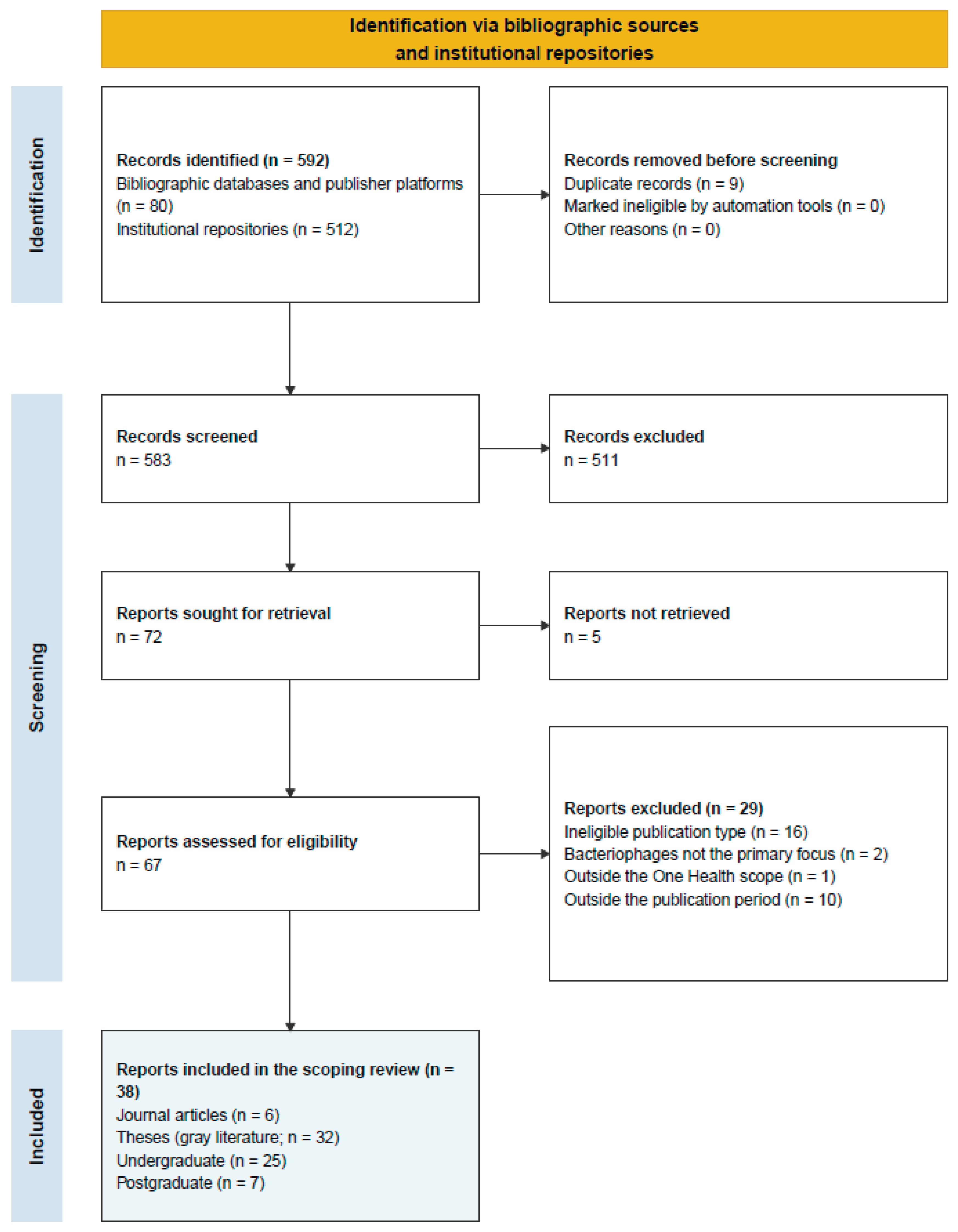
Figure 2.
Relationship Between Publication Year, Document Source, and One Health Research Area.
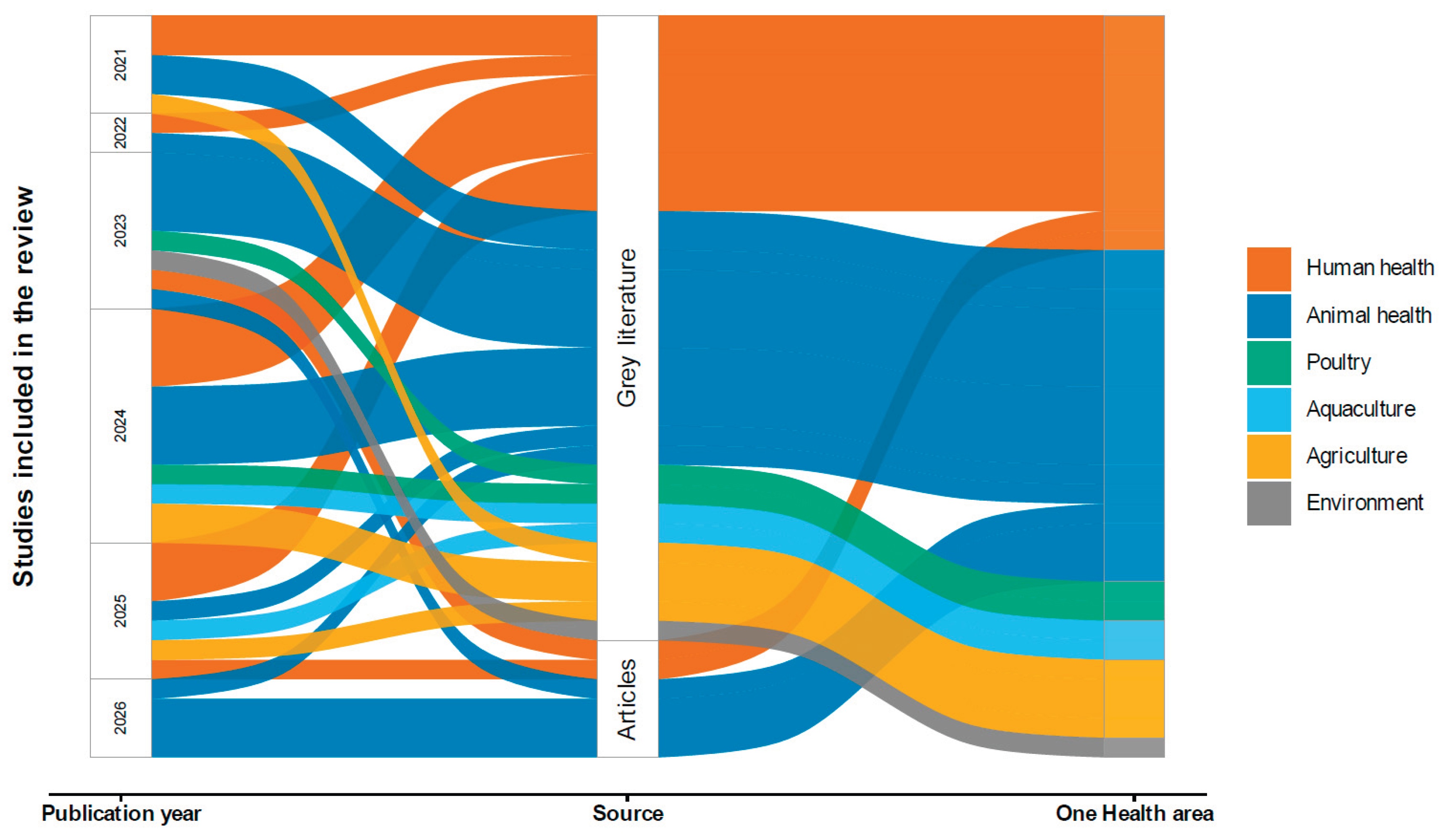
Figure 3.
Distribution of Included Documents Across One Health Research Areas.
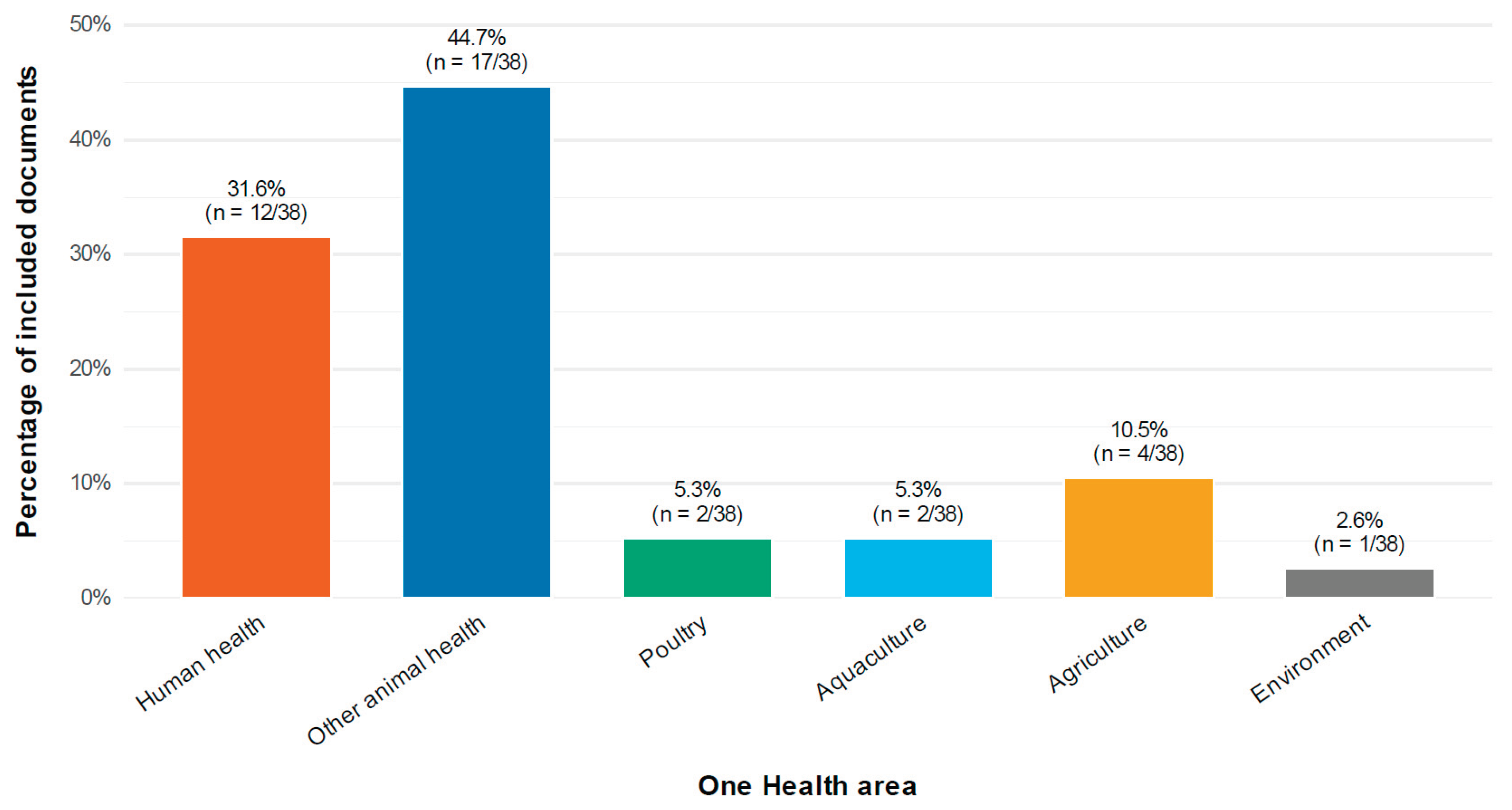
Figure 4.
Annual Frequency of Bacteriophage Publications in Ecuador by Document Source.
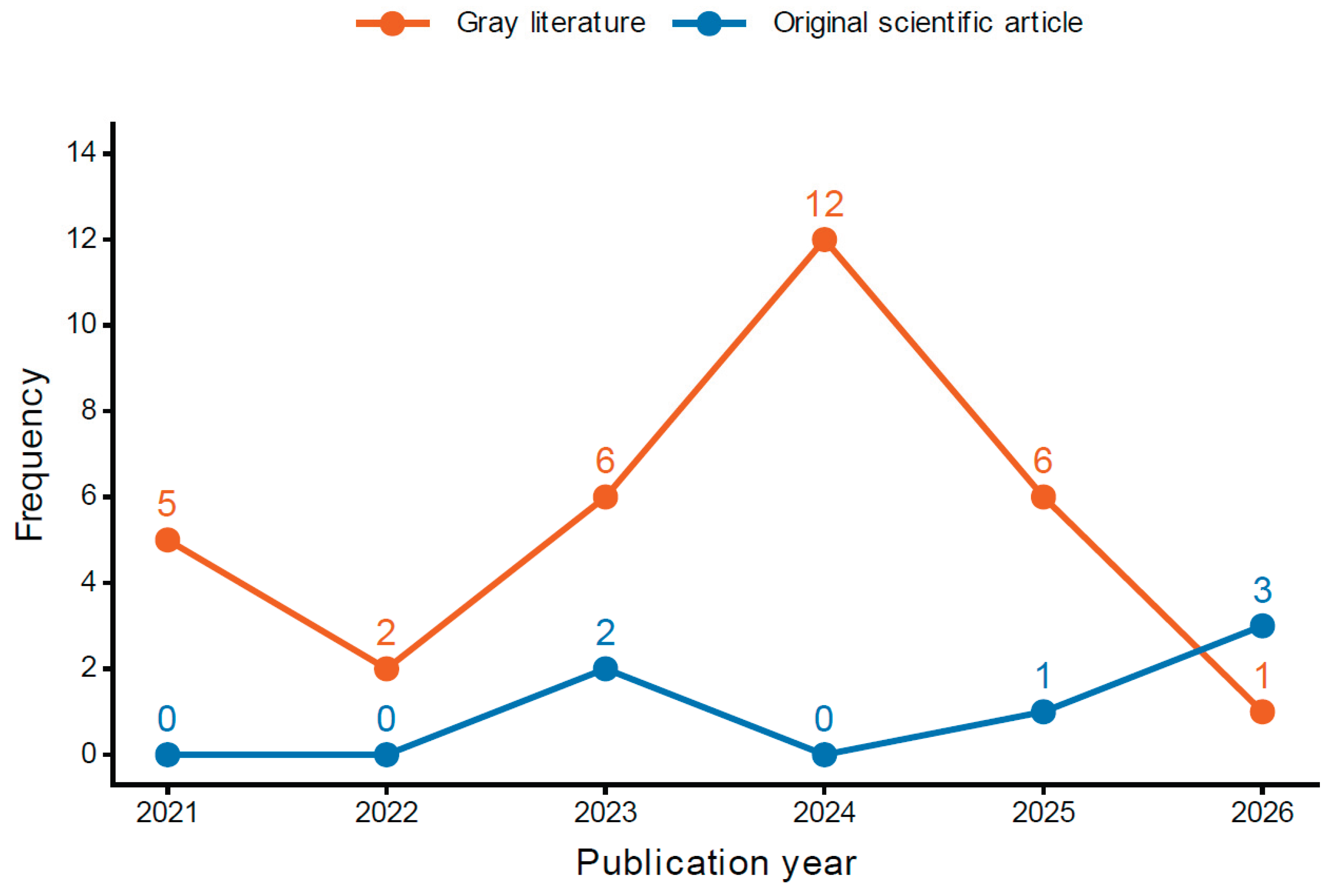
Figure 5.
Geographic Distribution of Bacteriophage Institutional Research Production.
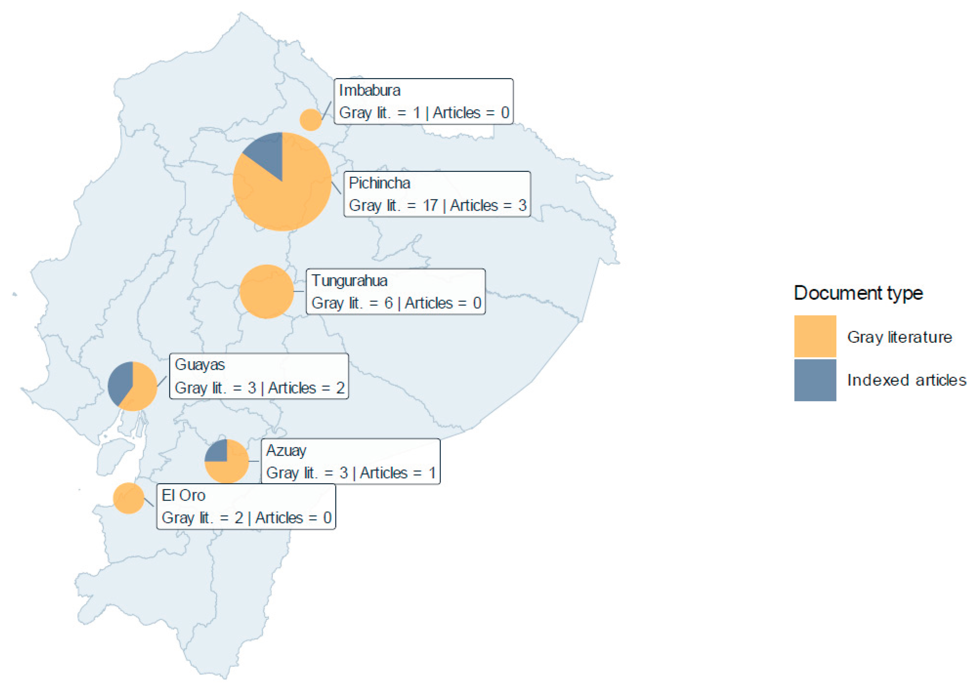
Figure 6.
Distribution of Included Documents by Province and Contributing University.
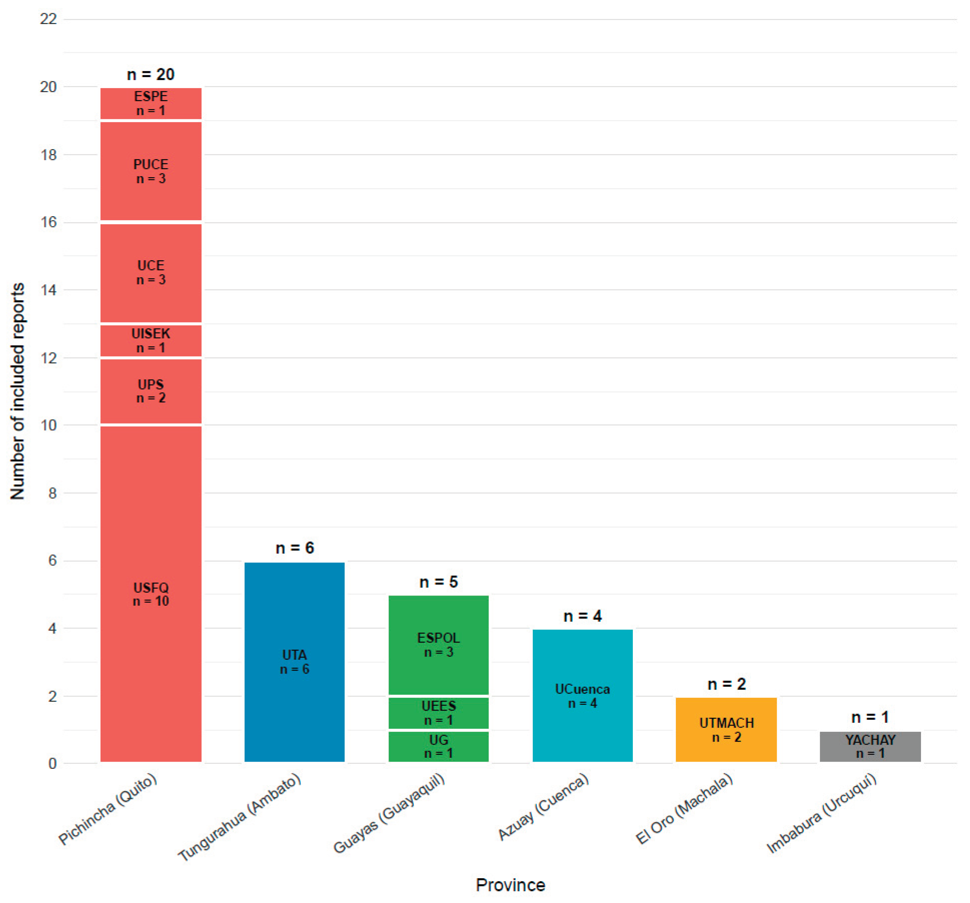
Figure 7.
The figure summarizes 38 reports assigned to six One Health-related application sectors. Sector labels indicate the number of reports assigned to each sector. Analytical approaches are not mutually exclusive: a report contributes one ribbon to each approach documented in Supplementary Table S1. In the chord diagram, ribbon width represents the number of report–approach occurrences, rather than the number of independent studies. Heatmap cells show the number of reports using a given approach divided by the total number of reports in that sector; navy indicates a lower proportion, and orange a higher proportion. Phenotypic includes observable laboratory measurements such as isolation, lysis, host range, efficiency of plating, growth or killing kinetics, stability, adsorption, and electron microscopy. Molecular refers to targeted assays or profiling, including PCR, restriction analysis, RFLP and plasmid profiling; these methods do not, by themselves, establish that a complete phage genome was obtained. Genomic includes sequencing, assembly, annotation, gene or prophage prediction, comparative sequence analysis and metagenomics. Assignment to this category does not imply that every report produced a complete phage genome. Applied includes functional evaluations directed toward an intended use, such as animal or farm interventions, biofilm assays, phage cocktails, carrier materials and testing of a recombinant phage-derived protein; it does not imply clinical validation. The approaches occur in 30, 6, 13, and 9 reports, respectively, giving 58 report–approach occurrences across all 38 reports. Preliminary lysis testing supports the phenotypic code for report 22; this does not imply extensive experimental characterization. Report 2 concerns bacterial antiphage defenses without direct phage characterization. Linked reports are identified in Supplementary Table S1 and are not independent replications. These categories describe reported methods and do not measure methodological quality or biological safety.
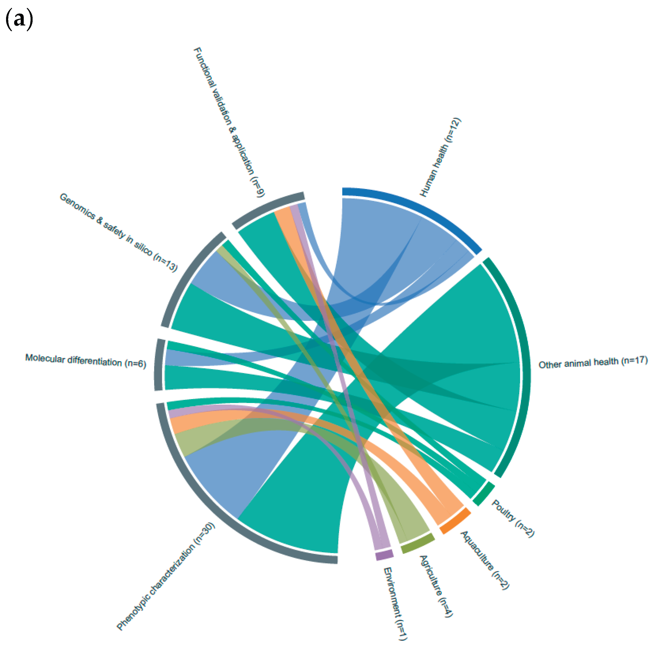
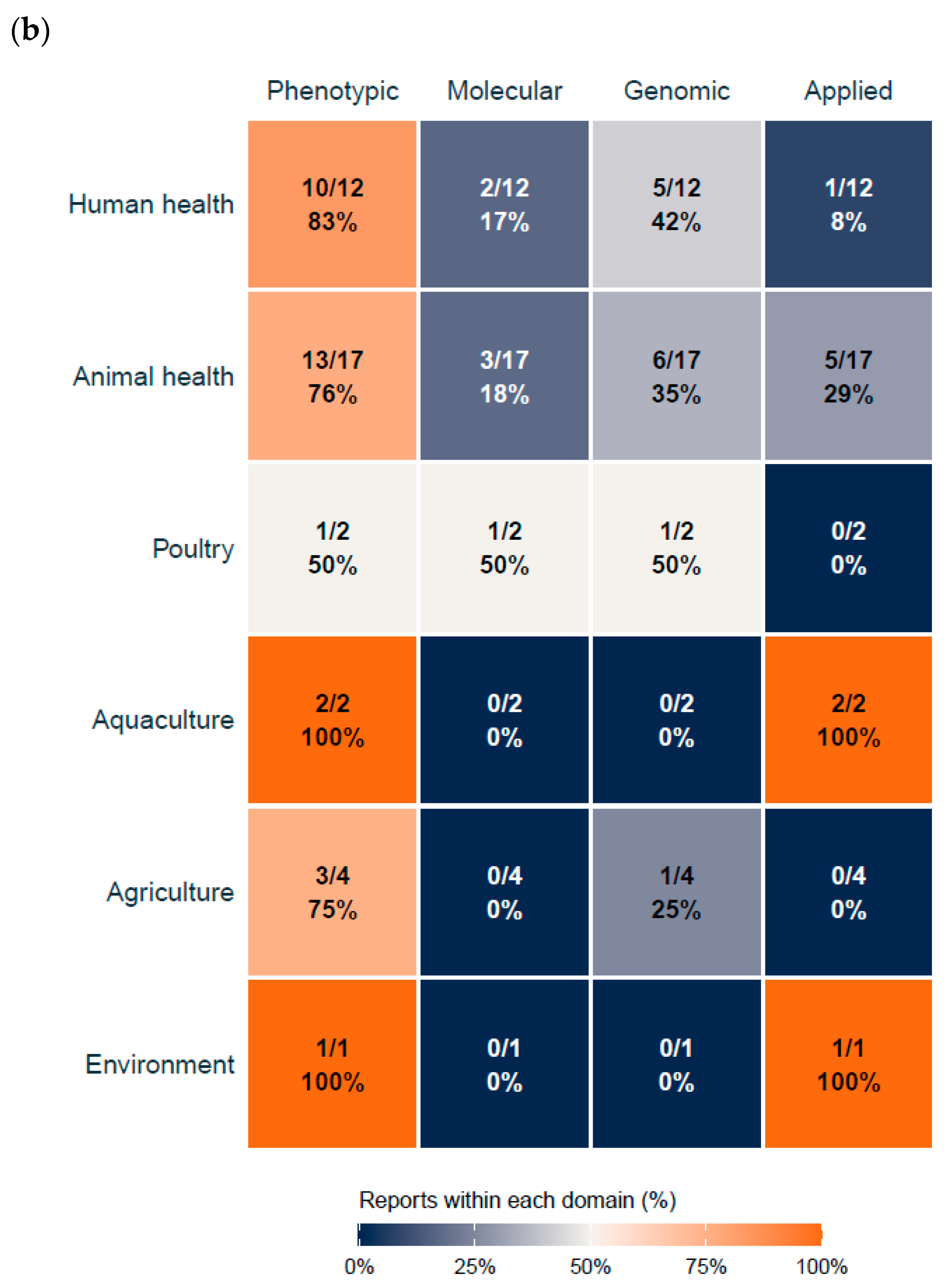
Figure 8.
The framework summarizes four complementary levels of evidence identified in Ecuadorian bacteriophage research: phenotypic characterization, molecular differentiation, whole-genome characterization, and functional and product validation. Progression across the framework represents increasing molecular resolution, evidential strength, and translational readiness, rather than a mandatory linear sequence or a validated scoring system. The warning statements indicate key inferential limitations: plaque clearing does not necessarily demonstrate productive infection; molecular fingerprints do not replace complete-genome analysis; failure to detect known hazardous genes does not establish absolute safety; and in vitro activity does not demonstrate causal efficacy in the intended application. EOP = efficiency of plating; PCR = polymerase chain reaction; RFLP = restriction fragment length polymorphism; rep-PCR = repetitive-element PCR; AMR = antimicrobial resistance; PFU/mL = plaque-forming units per milliliter.
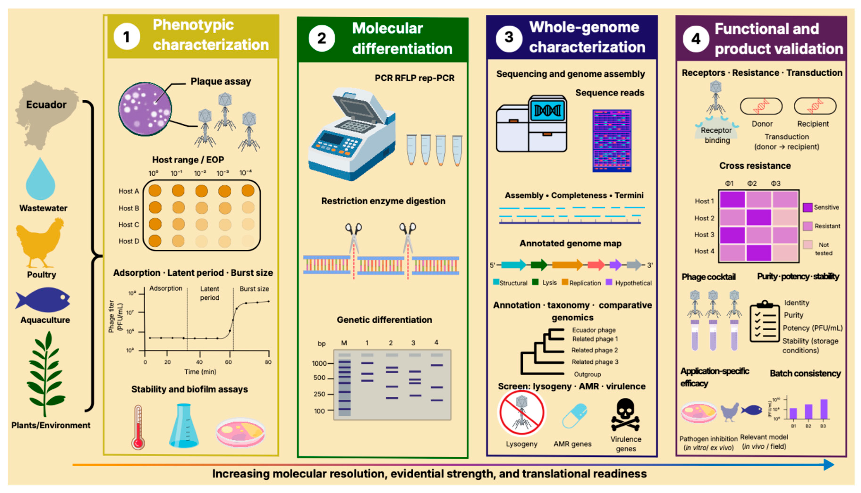
Disclaimer/Publisher’s Note: The statements, opinions and data contained in all publications are solely those of the individual author(s) and contributor(s) and not of MDPI and/or the editor(s). MDPI and/or the editor(s) disclaim responsibility for any injury to people or property resulting from any ideas, methods, instructions or products referred to in the content. |
© 2026 by the authors. Licensee MDPI, Basel, Switzerland. This article is an open access article distributed under the terms and conditions of the Creative Commons Attribution (CC BY) license (http://creativecommons.org/licenses/by/4.0/).
Copyright: This open access article is published under a Creative Commons CC BY 4.0 license, which permit the free download, distribution, and reuse, provided that the author and preprint are cited in any reuse.