

Submitted:
14 October 2026
Posted:
15 October 2026
You are already at the latest version
Abstract
Background/Objectives: Gadolinium-based contrast agents (GBCA) are widely used in magnetic resonance imaging and are generally considered to have a lower potential for nephrotoxicity compared to iodinated contrast agents. However, the actual prevalence of acute kidney injury (AKI) following the use of GBCA and the risk factors contributing to it remain a subject of debate today. The aim of this study is to determine the incidence of acute kidney injury following gadolinium administration (AKI-FGA) in patients undergoing contrast-enhanced magnetic resonance imaging (MRI) and to evaluate the independent risk factors contributing to it. Methods: The medical records of all patients who underwent contrast-enhanced MRI at our hospital between January 2021 and December 2022 were retrospectively reviewed. The patients’ demographic characteristics and serum creatinine levels before and after the contrast-enhanced MRI examination were evaluated. Contrast-induced acute kidney injury (AKI) was defined according to the Kidney Disease: Improving Global Outcomes (KDIGO) criteria as an increase in serum creatinine of ≥0.3 mg/dL within 48 hours following contrast agent administration or a rise to ≥1.5 times the baseline value within 7 days. Patients who met the KDIGO criteria were included in the AKI-FGA group. Results: A total of 503 patients who met the inclusion criteria were enrolled in the study. The mean age of the patients was 60.88 ± 16.83 years, and 60.4% of the patients (n=304) were male. Following GBCA administration, a statistically significant increase in serum urea and creatinine levels and a significant decrease in eGFR were observed (p=0.001). AKI-FGA was detected in 16.5% of the patients (n=83). In the AKI-FGA group, the number of patients with a history of coronary artery disease (CAD), diabetes mellitus (DM), hypertension (HT), and congestive heart failure (CHF) was statistically significantly higher than in the group without AKI-FGA (p<0.05). In the multivariate analysis, each 1-year increase in age increased the risk of AKI-FGA by a factor of 1.03, each 0.1 mg/dL increase in baseline serum creatinine increased it by a factor of approximately 4.96, and the presence of DM and CHF increased it by factors of 2.16 and 2.92, respectively. In contrast, no statistically significant association was found between the development of AKI-FGA and HT or CAD. Conclusions: Our findings demonstrate that acute kidney injury can develop following contrast-enhanced MRI even in patients without a history of kidney disease. In particular, for patients who are elderly or have DM, CHF, or elevated baseline serum creatinine levels, it is clinically important to perform an individualized risk assessment prior to contrast-enhanced MRI and to closely monitor kidney function following the procedure.
Keywords:
magnetic resonance imaging
; acute kidney injury
; gadolinium
; renal dysfunction
1. Introduction
Acute kidney injury (AKI) is defined as a sudden deterioration in kidney function characterized by a rapid increase in serum creatinine levels, a decrease in urine output, and accompanying fluid-electrolyte and acid-base imbalances [1]. Due to its significant impact on morbidity, mortality, length of hospital stay, and healthcare costs, AKI is recognized as a major public health issue. However, the fact that it can be prevented or reversed through early diagnosis and appropriate intervention underscores the importance of accurately identifying risk factors and detecting high-risk patients at an early stage [2].
Contrast-associated acute kidney injury (CA-AKI) is one of the major causes of AKI and continues to be clinically significant due to the increasing use of imaging and interventional procedures. In current terminology, the term “contrast-associated acute kidney injury (CA-AKI)” is increasingly being used in place of “contrast-induced nephropathy.” This approach emphasizes that renal dysfunction developing following contrast agent administration may not always be solely attributable to contrast exposure; comorbidities and other clinical factors may also play a role in this process [3,4]. According to the KDIGO definition, CA-AKI is characterized by an increase in serum creatinine of ≥0.3 mg/dL within 48 h following contrast agent exposure and/or an increase in serum creatinine to ≥1.5 times the baseline value within 7 days [1].
Gadolinium is a rare-earth metal in the lanthanide series that is used as an MRI contrast agent due to its strong paramagnetic properties. Since its free form is toxic, gadolinium is chelated with organic ligands in clinical use to form Gadolinium-based contrast agents (GBCA). This chelation reduces gadolinium’s toxicity while preserving its paramagnetic properties and ensures that GBCA are excreted by the kidneys primarily via glomerular filtration. Gadopentetate dimeglumine, the first gadolinium-based contrast medium, was introduced into clinical use in 1988, followed by new agents with different physicochemical properties. Today, GBCA in clinical use are classified as linear or macrocyclic based on their molecular structure, and as ionic or nonionic based on their electrical charge. It has been reported that macrocyclic agents, owing to the stronger chelation of the gadolinium ion within their rigid ring structure, possess higher thermodynamic and kinetic stability compared to linear agents and are considered safer in terms of the release of free gadolinium [4,5].
Although the pathophysiology of GBCA-related renal effects has not been fully elucidated, intrarenal vasoconstriction, medullary hypoxia, tubular cell damage, inflammatory response, and oxidative stress are thought to be among the underlying mechanisms. These findings suggest that the changes observed in renal function following GBCA administration may result from the interaction between the physicochemical properties of the contrast agent, such as osmolality and viscosity, and the administered dose, as well as patient-specific clinical risk factors [6]. In particular, pre-existing renal dysfunction, DM, advanced age, hypovolemia, and the use of nephrotoxic medications can increase the risk of renal damage [7,8,9].
Although kidney damage associated with iodinated contrast media has been studied in detail in the literature for many years, data on the effects of non-iodinated contrast media on kidney function are more limited and inconsistent. While current evidence suggests that non-iodinated contrast media have a lower potential for nephrotoxicity compared to iodinated contrast media, it also indicates that renal risk cannot be entirely ruled out, particularly in patients with impaired renal function. Therefore, it remains important to evaluate the renal safety profile of non-iodinated contrast media in different patient populations and to identify the risk factors contributing to the development of AKI following their use.
This study was conducted to determine the incidence of acute kidney injury following gadolinium administration (AKI-FGA) in patients undergoing contrast-enhanced MRI and to evaluate potential risk factors associated with its development. It is anticipated that the findings will contribute to patient-specific risk assessment prior to contrast-enhanced MRI, assist in identifying high-risk individuals, and shed light on the development of preventive clinical approaches.
2. Materials and Methods
This study was designed as a retrospective, single-center cohort study. The study included patients who underwent contrast-enhanced MRI using GBCA at our hospital between 1 January 2021, and 31 December 2022, for any clinical indication. A total of 503 patients who had baseline renal function tests prior to the contrast-enhanced MRI examination and whose serum creatinine levels were measured following IV contrast administration were evaluated. Patients under the age of 18; those with pregnancy, active malignancy, a diagnosis of sepsis, AKI prior to MRI, or chronic kidney disease (CKD); those admitted to the intensive care unit for any reason following MRI; and patients with incomplete clinical or laboratory data were excluded from the study (Figure 1).
During the contrast-enhanced MRI procedure, gadobutrol, a macrocyclic, nonionic contrast agent, was used as part of the hospital’s routine clinical practice. Gadobutrol is classified as a Group II GBCA according to the American College of Radiology (ACR) classification and was administered intravenously (IV) in accordance with clinical indications, the imaging protocol used, and the manufacturer’s recommendations. The gadobutrol dose administered varied depending on the imaging modality used.
The demographic characteristics of the patients included in the study, such as age, sex, and comorbidities, as well as their baseline renal function tests (RFT), biochemical, and hematological parameters prior to contrast-enhanced MRI were retrospectively retrieved from the hospital information management system.
According to the KDIGO 2012 diagnostic criteria, acute kidney injury (AKI) was defined as an increase in serum creatinine of ≥0.3 mg/dL within 48 h or a rise to ≥1.5 times the baseline value within 7 days.
Approval for our study was granted by our university’s ethics committee on 2 May 2023 (No. E-71522473-050.01.04-241679-125).
The demographic and clinical characteristics of the patients included in the study were summarized using descriptive statistics. Continuous variables that followed a normal distribution were reported as mean ± standard deviation; continuous variables that did not follow a normal distribution were reported as median (25th–75th percentile); and categorical variables were reported as counts and percentages.
The normality of the data distribution was assessed using appropriate tests. Differences between laboratory parameters before (baseline) and after (follow-up) MRI scans were analyzed using the paired t-test for normally distributed data and the Wilcoxon signed-rank test for non-normally distributed data. To identify independent risk factors associated with the development of AKI, univariate and then multivariate logistic regression analyses were performed. The results were reported as odds ratios (OR) and 95% confidence intervals (95% CI). In all statistical analyses, a p-value of <0.05 was considered statistically significant.
3. Results
3.1. Demographic Characteristics of the Participants
The 503 patients who participated in the study had a mean age of 60.88 ± 16.83 years, and 60.4% (n = 304) were male. The most common comorbidity was HT (38.9%), followed by DM (29.6%), cerebrovascular disease (CVD) (21.0%), CAD (14.5%), and CHF (4.0%). Upon evaluation of the laboratory test results, the baseline creatinine level prior to gadolinium administration was 0.76 ± 0.30 mg/dL, and the post-administration level was 0.91 ± 0.43 mg/dL; the baseline eGFR was 96.5 ± 50.5 mL/min/1.73 m2, and the post-administration value was 78.0 ± 18.0 mL/min/1.73 m2 (Table 1). When baseline and follow-up laboratory findings were compared, urea and creatinine levels were statistically significantly higher in the follow-up measurements (p = 0.001) (Figure 2), while the eGFR value was significantly lower (p = 0.001).
3.2. Evaluation of Patients with Acute Kidney Injury
AKI-FGA was detected in 83 (16.5%) of the 503 patients included in the study (Figure 3). In patients with AKI-FGA, the baseline serum creatinine level was 0.78 (0.61–0.95) mg/dL, whereas at the follow-up measurement, this value was found to have increased to 1.27 (1.09–1.51) mg/dL (p < 0.001). Similarly, while the baseline eGFR was 89.1 (75.6–101.4) mL/min/1.73 m2, it decreased to 50.94 (39.26–64.23) mL/min/1.73 m2 at the follow-up measurement (p < 0.001). None of the patients who developed AKI-FGA required dialysis. The mean age of the group that developed AKI-FGA was statistically significantly higher than that of the group that did not develop AKI-FGA (p < 0.001) (Table 2). In addition, a history of CAD, DM, HT, and CHF was significantly more common in the group that developed AKI-FGA than in the group that did not (p < 0.05 for all comparisons).
3.3. Analysis of Variables Associated with the Development of Acute Kidney Injury Following Gadolinium Administration
In the univariate logistic regression analysis, each one-year increase in age was associated with an increased probability of developing AKI-FGA (OR: 1.035; 95% CI: 1.018–1.052; p = 0.001). In contrast, no statistically significant association was found between baseline serum creatinine levels and the development of AKI-FGA (OR: 1.13; 95% CI: 0.55–2.30; p = 0.730). DM (OR: 1.91; 95% CI: 1.18–3.08; p < 0.001), HT (OR: 1.69; 95% CI: 1.05–2.72; p = 0.001), CHF (OR: 3.15; 95% CI: 1.27–7.75; p = 0.009), and CAD (OR: 1.97; 95% CI: 1.10–3.46; p = 0.007) were found to be significantly associated with the development of AKI-FGA (Table 3).
In the multivariate logistic regression analysis, each 1-year increase in age increased the likelihood of developing AKI-FGA by approximately 1.03 times (OR: 1.03; 95% CI: 1.01–1.05; p = 0.001). It was also found that each 0.1 mg/dL increase in baseline serum creatinine levels was associated with an approximately 4.96-fold increase in the likelihood of developing AKI-FGA (OR: 4.964; 95% CI: 1.405–17.536; p = 0.013). Similarly, the presence of DM was identified as an independent factor that increased the likelihood of developing AKI-FGA by approximately 2.2 times (OR: 2.159; 95% CI: 1.238–3.768; p = 0.007), while the presence of CHF was identified as an independent factor that increased this likelihood by approximately 2.9 times (OR: 2.921; 95% CI: 1.060–8.047; p = 0.038). In contrast, HT (OR: 1.186; 95% CI: 0.671–2.099; p = 0.557) and CAD (OR: 1.506; 95% CI: 0.799–2.835; p = 0.206) were not independently associated with the development of AKI-FGA (p > 0.05).
4. Discussion
It is generally accepted that non-ionic contrast media have a lower potential for nephrotoxicity than iodinated contrast media, due to their lower viscosity and the fact that they are administered at lower doses. However, there are case reports and clinical studies indicating that AKI may develop following the use of GBCA, particularly in patients with pre-existing renal dysfunction [10]. For this reason, the renal safety of GBCA remains a topic of ongoing debate today.
In our study, we evaluated changes in renal function parameters and the development of AKI following contrast-enhanced MRI using GBCA. Statistically significant changes were observed in serum urea and creatinine levels and eGFR values following GBCA administration. The finding that the incidence of AKI-FGA in the study population was 16.5% indicates that clinically significant changes in renal function may occur following contrast-enhanced MRI. These findings highlight the importance of closely monitoring renal function following GBCA administration, particularly in patient groups at risk for renal injury.
The incidence of AKI-FGA identified in our study falls near the upper limit of the wide range of rates reported in the literature. In the prospective RESCUE study conducted by Deray et al. [11] in patients with stable stage 3–4 chronic kidney disease, the incidence of contrast-induced nephropathy following administration of a standard dose of gadoterate meglumine (Gd-DOTA) was reported as 1.4%, and no statistically significant difference was found between this rate and the incidence in the control group that underwent MRI without contrast [11]. Researchers have noted that the higher rates of nephrotoxicity reported in previous studies may be attributable to methodological factors such as the type and dose of the contrast agent used, the clinical heterogeneity of the study populations, and differences in the criteria adopted for defining contrast-induced AKI. However, the fact that no cases of nephrogenic systemic fibrosis (NSF) were observed during the study was considered a supportive finding regarding the renal safety profile of gadoterate meglumine, a macrocyclic contrast agent [11]. The marked difference in incidence between our study and the RESCUE study may be related to differences in the characteristics of the patient populations, the type and dose of GBCA used, the time intervals at which renal function was assessed, and the criteria used in the definition of AKI-FGA.
Similarly, a joint consensus report published by the American College of Radiology (ACR) and the National Kidney Foundation (NKF) states that Group II GBCA pose a very low risk of NSF and nephrotoxicity at recommended clinical doses. For this reason, in patients with AKI, advanced-stage CKD (stages 4–5, eGFR < 30 mL/min/1.73 m2), or those on dialysis (stage 5D), it is not recommended to postpone clinically necessary contrast-enhanced MRI examinations solely because of the potential risks associated with the use of GBCA. The report also emphasizes that routine kidney function screening is not mandatory for patients undergoing Group II contrast-enhanced imaging, and that, in general, no changes to the current dialysis regimen are necessary following contrast administration [4].
Likewise, according to the latest ACR and European Society of Urogenital Radiology (ESUR) recommendations published in 2025, clinically necessary contrast-enhanced MRI examinations in patients with advanced renal impairment should not be postponed solely because of a low eGFR. The report emphasizes that in cases where the use of GBCA is necessary, agents with high stability and very low NSF risk, particularly ACR Group II GBCAs, should be used at the lowest dose that ensures diagnostic adequacy [12].
In an earlier study involving 91 patients with stage 3–4 CKD, Ergün et al. [13] reported that 12.1% of patients developed AKI following MR angiography in which gadolinium was used as the sole contrast agent; they identified baseline eGFR, advanced age, diabetic nephropathy, and low baseline hemoglobin and albumin levels as risk factors, and in their regression analysis, they determined that baseline eGFR and diabetic nephropathy were independent predictors. In this study, an increase of at least 0.5 mg/dL in serum creatinine levels was adopted as the criterion for AKI; given that this threshold is more restrictive than the KDIGO criteria used in our study (an increase of ≥0.3 mg/dL within 48 h), it is conceivable that the reported rate of 12.1% could actually be even higher under a broader definition. However, the fact that the incidence in this study was calculated only for patients with advanced-stage CKD may limit the ability to make a direct comparison between the two studies, despite the similarity in the rates.
In our study, statistically significant changes were observed in serum urea, creatinine, and eGFR levels following contrast agent administration (p = 0.001). These findings are consistent with some previous studies that reported changes in renal function parameters following the administration of contrast media [13,14]. In contrast to these studies, in the prospective case-control study by Gok Oguz et al., no significant differences were observed between the contrast-enhanced and non-contrast-enhanced MRI groups in serum creatinine, eGFR, and early AKI biomarkers following contrast-enhanced MRI with gadopentetate dimeglumine, and it was reported that the use of gadopentetate dimeglumine does not cause significant nephrotoxicity even in high-risk patients [10]. This discrepancy may be related to differences in methodological design between the studies.
In the univariate analyses of our study, DM, HT, and CHF were found to be significantly associated with the development of AKI-FGA; in the multivariate logistic regression model, advanced age, DM, CHF, and high baseline serum creatinine levels were identified as risk factors independent of other variables. Each additional year of age was associated with a gradual increase in risk; the presence of DM was associated with an approximately twofold increase in the risk of AKI-FGA, while the presence of CHF was associated with approximately a threefold increase. Baseline serum creatinine level emerged as one of the strongest predictors in the model. In contrast, HT and CAD did not retain their independent significance in the multivariate model; this finding suggests that the effects of these variables are shaped by interactions with other clinical characteristics and comorbidities.
The fact that baseline creatinine levels are among the strongest predictors suggests that impaired kidney function increases susceptibility to AKI-FGA. Reduced renal reserve is one of the mechanisms that may explain this susceptibility by limiting the kidney’s ability to adapt to hemodynamic and metabolic stress. Indeed, James et al. [15] and Gansevoort et al. [16] also reported that low eGFR and high serum creatinine levels are among the most important predictors of AKI development. When considered in conjunction with the identification of DM and CHF as independent risk factors, these findings indicate that the development of AKI-FGA cannot be explained solely by the direct effect of the contrast agent; the patient’s burden of comorbidities and current kidney function also play a decisive role.
This situation highlights the clinical significance of subclinical kidney damage that may develop following GBCA exposure and the accumulation of gadolinium in tissues. AKI-FGA may present subclinically without significant changes in serum creatinine levels; an increased risk has been reported in cases of advanced kidney dysfunction, DM, high dose exposure, or repeated exposure. Reduced gadolinium elimination in patients with impaired kidney function may also lead to increased accumulation in brain and kidney tissues. However, the long-term clinical consequences of this accumulation and the prognostic value of subclinical damage have not been definitively established, and the clinical implications of the inflammatory and fibrotic effects reported in experimental data have not been fully elucidated [17]. Therefore, in patients scheduled for contrast-enhanced imaging, the pre-procedural assessment of kidney function is not merely a diagnostic step but a clinical necessity that directly affects patient safety. In particular, conducting individualized risk assessments in patients with reduced renal reserve and a high burden of comorbidities, avoiding unnecessary and repeated exposure, and ensuring close laboratory monitoring after the procedure will help detect developing renal dysfunction at an early stage.
The most clinically significant implication of these findings is the need for a risk-based, individualized assessment rather than a standard approach to the use of GBCA. In particular, it is recommended to avoid the use of GBCA as much as possible in patients with an eGFR below 30 mL/min/1.73 m2, or to use the lowest effective dose when absolutely necessary [18]. In addition, adequate pre-procedural hydration, review of current nephrotoxic medications, and close monitoring of renal function after the procedure should be part of preventive clinical strategies. The first 48-h period, in particular, is of critical importance for the early detection of renal injury. This approach will not only help reduce complications but also contribute to making contrast-enhanced imaging safer for high-risk patient groups.
The most significant strength of our study lies in the way patient selection criteria were established. The vast majority of studies in the literature evaluating contrast-induced kidney injury have been conducted on high-risk populations with stage 3–5 CKD or those undergoing interventional procedures. In contrast, our study excluded patients with a prior diagnosis of CKD or AKI; thus, post-contrast renal changes could be evaluated in a population with preserved baseline renal function. This design reduces the likelihood that observed changes in kidney function are attributable to pre-existing kidney dysfunction and ensures that the findings can be directly applied to this group, which constitutes the vast majority of patients undergoing contrast-enhanced MRI in daily radiology practice. The fact that a significant proportion of AKI-FGA cases were detected even in patients without known renal dysfunction suggests that this population, which is currently considered low risk in existing clinical approaches, should be re-evaluated in terms of post-procedural follow-up. Furthermore, the fact that the study was conducted at a single center and with a single macrocyclic agent (gadobutrol) eliminated variability that might arise from different types of GBCA, thereby facilitating the interpretation of the findings.
However, our study has some limitations. First, due to the retrospective design of the study, the data were evaluated solely based on existing patient records, which limited our ability to fully control for potential confounding factors. Furthermore, the lack of detailed data on patients’ use of angiotensin-converting enzyme (ACE) inhibitors, angiotensin receptor blockers (ARBs), metformin, and other concomitant medications prevented the potential effects of these drugs on kidney function from being included in the analyses. Similarly, factors that may influence AKI development, such as hospitalization status, the clinical indication for MRI, the dose of contrast agent administered, intravenous fluid therapy, diuretic use, nonsteroidal anti-inflammatory drugs (NSAIDs), antibiotic therapy, sepsis, hypotension, surgical procedures, exposure to iodinated contrast agents, a history of bleeding or transfusion, and oral fluid intake, were not evaluated. Failure to account for these factors may have introduced residual confounding into the observed changes in kidney function. In addition, the lack of long-term follow-up of kidney function in patients who developed AKI has limited the assessment of the persistence of renal effects following contrast agent administration, the recovery process of kidney function, and the long-term renal prognosis. Therefore, there is a need for prospective, multicenter studies that collect detailed clinical data and include long-term monitoring of kidney function in order to more comprehensively validate the findings.
The findings indicate that the development of AKI-FGA cannot be explained solely by contrast agent exposure or pre-existing renal dysfunction; patient-specific clinical characteristics also play a significant role in this process. For this reason, it is clinically important to perform an individualized risk assessment prior to the procedure in patients scheduled for contrast-enhanced MRI, to take the necessary protective measures, particularly in older patients and those with DM, CHF, or impaired baseline renal function, and to ensure appropriate renal follow-up after the procedure. However, due to the retrospective design of our study, it is not possible to interpret the associations observed with AKI-FGA in terms of causality. Therefore, we believe that prospective, multicenter studies encompassing different patient populations and involving long-term monitoring of kidney function will provide a more comprehensive understanding of the renal safety profile of GBCA and make significant contributions to clinical decision-making processes.
5. Conclusions
A high incidence of post-contrast AKI, defined according to the KDIGO creatinine criteria, was observed among patients who underwent contrast-enhanced MRI with gadobutrol. Advanced age, diabetes, heart failure, and impaired baseline renal function were independently associated with post-contrast AKI. Given the retrospective study design, the absence of an unexposed control group, and potential residual confounding factors, these findings suggest an association rather than gadolinium-induced nephrotoxicity.
Author Contributions
Conceptualization, B.K. and A.T.; methodology, B.K., A.T. and H.D.; formal analysis, H.D., M.P. and Z.E.; investigation, B.K., M.P., Z.E. and A.C.G.; data curation, H.D., M.P., Z.E. and M.İ.; writing—original draft preparation, B.K., A.C.G. and M.İ.; writing—review and editing, H.D., A.T. and M.İ.; supervision, H.D. and A.C.G. All authors have read and agreed to the published version of the manuscript.
Funding
This research received no external funding.
Institutional Review Board Statement
The study was conducted in accordance with the Declaration of Helsinki and approved by the Ethics Committee of [Sakarya University Faculty of Medicine] (protocol code E-71522473-050.01.04-241679-125; date of approval: 2 May 2023).
Informed Consent Statement
Patient consent was waived due to the retrospective design of the study.
Data Availability Statement
The data presented in this study are available on request from the corresponding author due to privacy and ethical restrictions.
Conflicts of Interest
The authors declare no conflicts of interest.
Abbreviations
The following abbreviations are used in this manuscript:
| ACE | Angiotensin-converting enzyme |
| ACR | American College of Radiology |
| AKI | Acute kidney injury |
| AKI-FGA | Acute kidney injury following gadolinium administration |
| ARB | Angiotensin receptor blocker |
| CA-AKI | Contrast-associated acute kidney injury |
| CAD | Coronary artery disease |
| CHF | Congestive heart failure |
| CI | Confidence interval |
| CKD | Chronic kidney disease |
| CVD | Cerebrovascular disease |
| DM | Diabetes mellitus |
| eGFR | Estimated glomerular filtration rate |
| ESUR | European Society of Urogenital Radiology |
| GBCA | Gadolinium-based contrast agent |
| Gd-DOTA | Gadoterate meglumine |
| HT | Hypertension |
| IV | Intravenous |
| KDIGO | Kidney Disease: Improving Global Outcomes |
| MRI | Magnetic resonance imaging |
| NKF | National Kidney Foundation |
| NSAID | Nonsteroidal anti-inflammatory drug |
| NSF | Nephrogenic systemic fibrosis |
| OR | Odds ratio |
| RFT | Renal function tests |
| SD | Standard deviation |
References
- Khwaja, A. KDIGO clinical practice guidelines for acute kidney injury. Nephron Clin. Pract. 2012, 120, c179–c184. [Google Scholar] [CrossRef] [PubMed]
- Koza, Y. Acute kidney injury: Current concepts and new insights. J. Inj. Violence Res. 2016, 8, 58–62. [Google Scholar] [CrossRef] [PubMed]
- Macdonald, D.B.; Hurrell, C.D.; Costa, A.F.; McInnes, M.D.F.; O’Malley, M.; Barrett, B.J.; Brown, P.A.; Clark, E.G.; Hadjivassiliou, A.; Kirkpatrick, I.D.C.; et al. Canadian Association of Radiologists guidance on contrast-associated acute kidney injury. Can. J. Kidney Health Dis. 2022, 9, 20543581221097455. [Google Scholar] [CrossRef] [PubMed]
- Weinreb, J.C.; Rodby, R.A.; Yee, J.; Wang, C.L.; Fine, D.; McDonald, R.J.; Perazella, M.A.; Dillman, J.R.; Davenport, M.S. Use of intravenous gadolinium-based contrast media in patients with kidney disease: Consensus statements from the American College of Radiology and the National Kidney Foundation. Radiology 2021, 298, 28–35. [Google Scholar] [CrossRef] [PubMed]
- Rogosnitzky, M.; Branch, S. Gadolinium-based contrast agent toxicity: A review of known and proposed mechanisms. Biometals 2016, 29, 365–376. [Google Scholar] [CrossRef] [PubMed]
- Elmståhl, B.; Leander, P.; Grant, D.; Doughty, R.W.; Chai, C.M.; Björk, J.; Almén, T.; Nyman, U. Histomorphological changes after renal X-ray arteriography using iodine and gadolinium contrast media in an ischemic porcine model. Acta Radiol. 2007, 48, 1109–1119. [Google Scholar] [CrossRef] [PubMed]
- Detrenis, S.; Meschi, M.; Musini, S.; Savazzi, G. Lights and shadows on the pathogenesis of contrast-induced nephropathy: State of the art. Nephrol. Dial. Transplant. 2005, 20, 1542–1550. [Google Scholar] [CrossRef] [PubMed]
- Bucher, A.M.; De Cecco, C.N.; Schoepf, U.J.; Meinel, F.G.; Krazinski, A.W.; Spearman, J.V.; McQuiston, A.D.; Wang, R.; Bucher, J.; Vogl, T.J.; et al. Is contrast medium osmolality a causal factor for contrast-induced nephropathy? BioMed Res. Int. 2014, 2014, 931413. [Google Scholar] [CrossRef] [PubMed]
- Seeliger, E.; Lenhard, D.C.; Persson, P.B. Contrast media viscosity versus osmolality in kidney injury: Lessons from animal studies. BioMed Res. Int. 2014, 2014, 358136. [Google Scholar] [CrossRef] [PubMed]
- Gok Oguz, E.; Kiykim, A.; Turgutalp, K.; Olmaz, R.; Ozhan, O.; Muslu, N.; Horoz, M.; Bardak, S.; Sungur, M.A. Lack of nephrotoxicity of gadopentetate dimeglumine-enhanced non-vascular MRI and MRI without contrast agent in patients at high-risk for acute kidney injury. Med. Sci. Monit. 2013, 19, 942–948. [Google Scholar] [CrossRef] [PubMed]
- Deray, G.; Rouviere, O.; Bacigalupo, L.; Maes, B.; Hannedouche, T.; Vrtovsnik, F.; Rigothier, C.; Billiouw, J.-M.; Campioni, P.; Ferreiros, J.; et al. Safety of meglumine gadoterate (Gd-DOTA)-enhanced MRI compared to unenhanced MRI in patients with chronic kidney disease (RESCUE study). Eur. Radiol. 2013, 23, 1250–1259. [Google Scholar] [CrossRef] [PubMed]
- van der Molen, A.J.; van de Ven, A.A.J.M.; Vega, F.; Dekkers, I.A.; Geenen, R.W.F.; Bellin, M.-F.; Bertolotto, M.; Brismar, T.B.; Clément, O.; Correas, J.-M.; et al. Hypersensitivity reactions to contrast media: Part 1. Management of immediate and non-immediate hypersensitivity reactions in adults. Updated guidelines by the ESUR Contrast Media Safety Committee. Eur. Radiol. 2025, 35, 6798–6810. [Google Scholar] [CrossRef] [PubMed]
- Ergün, I.; Keven, K.; Uruç, I.; Ekmekçi, Y.; Canbakan, B.; Erden, I.; Karatan, O. The safety of gadolinium in patients with stage 3 and 4 renal failure. Nephrol. Dial. Transplant. 2006, 21, 697–700. [Google Scholar] [CrossRef] [PubMed]
- Akgun, H.; Gonlusen, G.; Cartwright, J.; Suki, W.N.; Truong, L.D. Are gadolinium-based contrast media nephrotoxic? A renal biopsy study. Arch. Pathol. Lab. Med. 2006, 130, 1354–1357. [Google Scholar] [CrossRef] [PubMed]
- James, M.T.; Grams, M.E.; Woodward, M.; Elley, C.R.; Green, J.A.; Wheeler, D.C.; de Jong, P.; Gansevoort, R.T.; Levey, A.S.; Warnock, D.G.; et al. A meta-analysis of the association of estimated GFR, albuminuria, diabetes mellitus, and hypertension with AKI. Am. J. Kidney Dis. 2015, 66, 602–612. [Google Scholar] [CrossRef] [PubMed]
- Gansevoort, R.T.; Matsushita, K.; van der Velde, M.; Astor, B.C.; Woodward, M.; Levey, A.S.; de Jong, P.E.; Coresh, J. Chronic Kidney Disease Prognosis Consortium. Lower estimated GFR and higher albuminuria are associated with adverse kidney outcomes. A collaborative meta-analysis of general and high-risk population cohorts. Kidney Int. 2011, 80, 93–104. [Google Scholar] [CrossRef] [PubMed]
- Vanmassenhove, J.; Van Biesen, W.; Vanholder, R.; Lameire, N. Subclinical AKI: Ready for primetime in clinical practice? J. Nephrol. 2019, 32, 9–16. [Google Scholar] [CrossRef] [PubMed]
- Haustein, J.; Niendorf, H.P.; Krestin, G.; Louton, T.; Schuhmann-Giampieri, G.; Clauss, W.; Junge, W. Renal tolerance of gadolinium-DTPA/dimeglumine in patients with chronic renal failure. Investig. Radiol. 1992, 27, 153–156. [Google Scholar] [CrossRef] [PubMed]
Figure 1.
Formation of the study population and patient selection process.
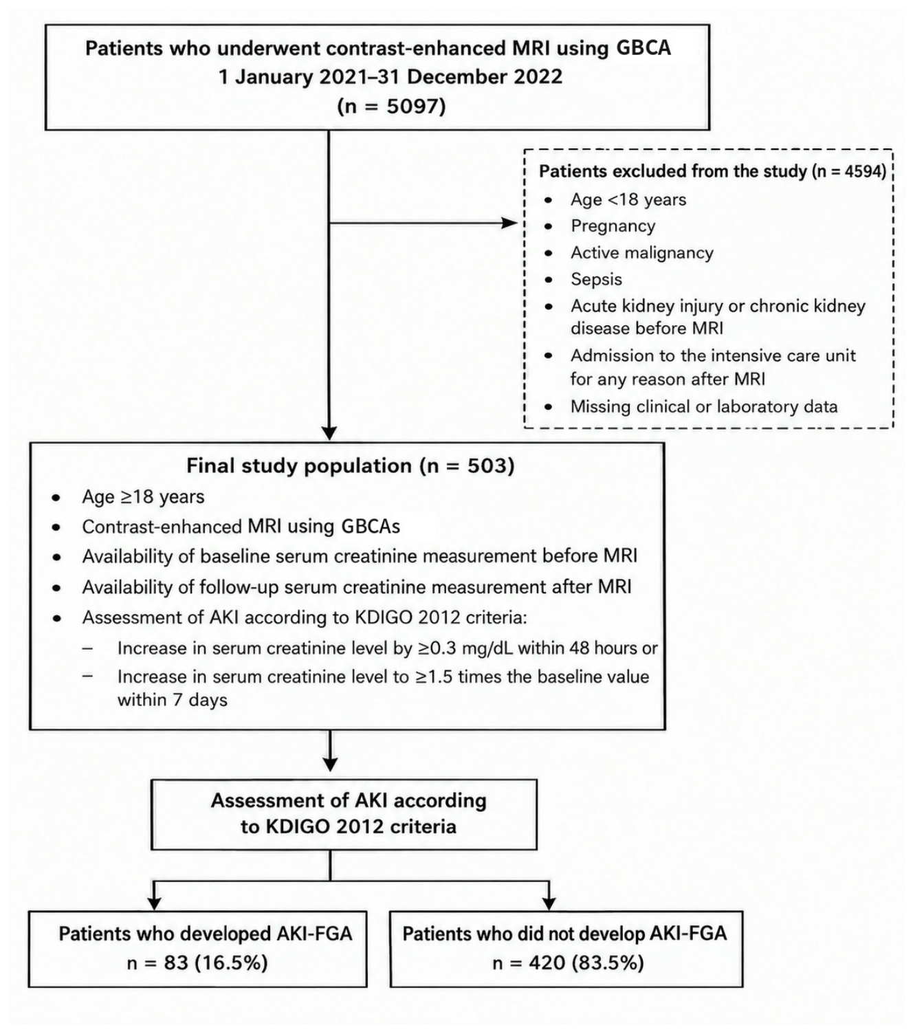
Figure 2.
Comparison of baseline and follow-up values for urea, creatinine, and eGFR.
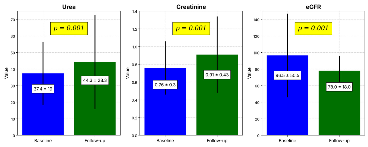
Figure 3.
Distribution of patients with and without AKI-FGA.
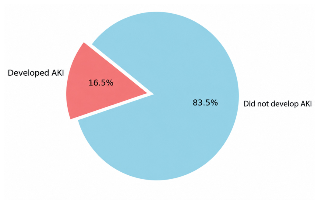

Table 1.
Comparison of laboratory tests before and after gadolinium administration.
| Parameter | Baseline Value (Mean ± SD) (n = 503) |
Follow-Up Value (Mean ± SD) (n = 503) |
p |
|---|---|---|---|
| Urea (mg/dL) | 37.4 ± 19 | 44.3 ± 28.3 | 0.001 |
| Creatinine (mg/dL) | 0.76 ± 0.3 | 0.91 ± 0.43 | 0.001 |
| eGFR (mL/min/1.73 m2) | 96.5 ± 50.5 | 78.0 ± 18.0 | 0.001 |
| Sodium (mmol/L) | 137.3 ± 9.7 | 138 ± 4.1 | 0.070 |
| Potassium (mmol/L) | 4.0 ± 0.4 | 4.2 ± 0.5 | 0.409 |
| Calcium (mg/dL) | 9.4 ± 0.7 | 9.6 ± 0.7 | 0.091 |
| Magnesium (mg/dL) | 1.8 ± 0.3 | 1.8 ± 0.3 | 0.927 |
| Phosphorus (mg/dL) | 3.2 ± 0.7 | 3.3 ± 0.8 | 0.303 |
| Albumin (g/dL) | 3.5 ± 0.7 | 3.5 ± 0.6 | 0.921 |
SD, standard deviation; eGFR, estimated glomerular filtration rate. Bold values indicate statistical significance (p < 0.05).
Table 2.
Comparison of age, sex, and comorbidities in patients with and without AKI-FGA.
| Variable | AKI-FGA Developed (n = 83) |
AKI-FGA Not Developed (n = 420) |
p |
|---|---|---|---|
| Age (years) | 67.99 ± 14.5 | 59.5 ± 16.91 | <0.001 |
| Sex | Male: 45 (54.2%) Female: 38 (45.8%) |
Male: 259 (61.7%) Female: 161 (38.3%) |
0.250 |
| Coronary artery disease | 20 (24.1%) | 53 (12.6%) | 0.007 |
| Hypertension | 46 (55.4%) | 150 (35.7%) | 0.001 |
| Diabetes mellitus | 39 (47.0%) | 110 (26.2%) | <0.001 |
| Congestive heart failure | 8 (9.6%) | 12 (2.9%) | 0.009 |
AKI-FGA, acute kidney injury following gadolinium administration. Bold values indicate statistical significance (p < 0.05).
Table 3.
Univariate and multivariate logistic regression analysis of variables associated with the development of AKI-FGA.
Table 3.
Univariate and multivariate logistic regression analysis of variables associated with the development of AKI-FGA.
| Variable | Univariate OR (95% CI) |
p | B | Multivariate OR (95% CI) |
p |
|---|---|---|---|---|---|
| Age (per 1-year increase) | 1.035 (1.018–1.052) | 0.001 | 0.030 | 1.03 (1.01–1.05) | 0.001 |
| Baseline creatinine (per 0.1 mg/dL increase) | 1.13 (0.55–2.30) | 0.730 | 1.602 | 4.964 (1.405–17.536) | 0.013 |
| Diabetes mellitus | 1.91 (1.18–3.08) | <0.001 | 0.770 | 2.159 (1.238–3.768) | 0.007 |
| Hypertension | 1.69 (1.05–2.72) | 0.001 | 0.171 | 1.186 (0.671–2.099) | 0.557 |
| Congestive heart failure | 3.15 (1.27–7.75) | 0.009 | 1.072 | 2.921 (1.060–8.047) | 0.038 |
| Coronary artery disease | 1.97 (1.10–3.46) | 0.007 | 0.409 | 1.506 (0.799–2.835) | 0.206 |
OR, odds ratio; CI, confidence interval; B, regression coefficient. Bold values indicate statistical significance (p < 0.05).
Disclaimer/Publisher’s Note: The statements, opinions and data contained in all publications are solely those of the individual author(s) and contributor(s) and not of MDPI and/or the editor(s). MDPI and/or the editor(s) disclaim responsibility for any injury to people or property resulting from any ideas, methods, instructions or products referred to in the content. |
© 2026 by the authors. Licensee MDPI, Basel, Switzerland. This article is an open access article distributed under the terms and conditions of the Creative Commons Attribution (CC BY) license (http://creativecommons.org/licenses/by/4.0/).
Copyright: This open access article is published under a Creative Commons CC BY 4.0 license, which permit the free download, distribution, and reuse, provided that the author and preprint are cited in any reuse.