

Submitted:
14 October 2026
Posted:
15 October 2026
You are already at the latest version
Abstract
Fabry disease (FD) is a rare, X-linked, lysosomal storage multisystemic disorder which may lead to kidney failure and require kidney transplantation (KT). Data on long-term kidney allograft outcomes and the evolution of systemic FD under dis-ease-specific therapy after KT remain limited. We retrospectively evaluated six kidney transplant recipients (KTRs) with FD followed in our center for median tine after KT of 7 years (range, 4-13). Five patients (83.3%) were males and five had a classic Fabry phe-notype. At last follow-up patient survival was 100% and five out of six KTR had a func-tional kidney graft. Among KTRs with a functioning graft, the median change is esti-mated glomerular filtration rate from 1-year to last follow-up was +3.3 ml/min/1.73m2 (range, -50.7 to +10.5), while proteinuria remained low. One patient developed late an-tibody-mediated rejection with de novo donor-specific antibodies and graft loss at 8 years post-transplantation. Five KTRs were receiving enzyme replacement therapy at the time of KT. Three patients were subsequently switched to migalastat and two later returned to enzyme replacement therapy. Lyso-Gb3 and cardiac structural parameters showed heterogenous longitudinal trajectories despite generally preserved kidney graft function. No cardiovascular events occurred during follow-up. These findings support KT as an effective renal replacement therapy in FD and highlight the need for contin-uous Fabry-specific therapy and multidisciplinary surveillance after transplantation.
Keywords:
Fabry
; kidney transplantation
; graft
; outcome
; cardiovascular
; migalastat
1. Introduction
Fabry disease (FD) is a rare X-linked lysosomal storage disorder characterized by progressive intracellular accumulation of glycosphingolipids in many tissues, leading to multisystemic, life-threating complications, including cardiac, nervous system, and kidney diseases [1]. Kidney impairment is an important cause of morbidity and mortality in patients with Fabry disease and has heterogeneous manifestations [1,2]. In male patients, the classic presentation involves reduced or absent α-galactosidase A (α-Gal A) activity, often accompanied by the presence of albuminuria and proteinuria, often starting in the patients’ twenties or thirties [1]. In a study involving 106 male Fabry patients, which was conducted prior to the introduction of enzyme replacement therapy (ERT), all those who survived to the age of 56 developed end-stage kidney disease; no patient survived beyond the age of 60. This highlights these patients’ reduced life expectancy and increased risk of developing kidney failure [3]. Late-onset male Fabry patients have residual α-Gal A activity, with clinical manifestation in adulthood, and could have a cardiac, renal or cerebrovascular phenotype [3]. While kidney failure is less common in females with FD than in males, when it develops, the median age for both male and female patients is approximately the same [4]. An analysis of the Fabry registry showed that 13% heterozygous females suffered moderate-to-severe kidney involvement [5]. Fabry disease-specific therapy could stabilize or reduce kidney-involvement progression, but better outcomes may be observed for patients who start treatment at an earlier age, and prior to the development of renal fibrosis and sclerosis [6,7,8]. For FD patients who progress to kidney failure, the therapeutic goal would be to provide optimal renal replacement therapy, and to maintain ERT in order to avoid other organ damage [9]. Kidney transplantation (KT) is the preferred renal replacement therapy, and ideally it should be performed before initiating dialysis to protect the other organs [9]. The first kidney transplant in a patient with Fabry disease was performed in 1967. Though the patient experienced a short survival of only 17 months due to sepsis, the procedure demonstrated the absence of lipid deposits in the renal allograft and showed that Fabry disease is not necessarily a contraindication for KT [10]. After the first experience, the development of immunosuppressive therapy improved KT outcomes. Also, enzyme replacement therapy as specific treatment for Fabry disease was introduced to clinical practice in 2001. Evidence on the outcomes of KT in patients with Fabry disease is growing and shows improvements in both patient and graft survival [11,12,13,14,15,16,17,18,19,20,21,22,23]. In 2009, a retrospective analysis of 197 kidney transplant recipients (KTRs) with FD reported a superior graft survival and similar patient survival rate in KTRs with FD compared to other causes of kidney failure [19]. The retrospective study with the longest follow-up of 25 years and a median follow-up of 11.5 years of Fabry kidney transplant grafts showed graft survival of 92% at 10 years and 22% at 25 years, and similar graft survival rates were found for non-Fabry disease patients [21]. A recent meta-analysis indicates that allograft and patient survival in patients with FD are comparable to those reported in patients without FD [23]. Regarding the follow-up of kidney-transplanted patients who have been treated with different FD-specific therapies, the data is still scarce [23,24,25,26,27].
In this study, we present a case series of six KTRs with FD. Our main aim was to report the long-term outcomes of the patients and kidney grafts and to describe their post-transplant management and evolution.
2. Materials and Methods
2.1. Study Participants
Our retrospective study includes 6 KTRs with Fabry disease, 4 classic male patients, 1 late-onset male patient, and 1 classic female. Between July 2013 and September2026, the patients were evaluated and monitored at the Expert Center for Rare Diseases of Reno-Urinary System, Fundeni Clinical Institute, Bucharest, which includes the department of Nephrology and the department of Urology and Kidney Transplantation. Kidney-transplanted patients with Fabry disease are managed in every phase of the transplant process by the Urology and Kidney Transplantation department. A multidisciplinary team in the Nephrology department manages the specific treatment for Fabry disease and the evolution of Fabry multisystem disease.
This study conformed to current clinical practice and anonymized patient data were analyzed. The study was conducted in accordance with the principles of the Helsinki Declaration and was approved by the Institutional Ethics Committee of the Fundeni Clinical Institute (No. 50539/2026).
2.2. Clinical Assessments
The patients were comprehensively evaluated to assess their kidney function, kidney graft function, cardiac involvement, neurological involvement and other extrarenal manifestations of Fabry disease. We collected data regarding demographics, Fabry disease phenotype, molecular data, history of kidney involvement, comorbidities, FD-specific treatment, clinical events, overall graft and patient survival and patient evolution during a follow-up period of up to 13 years. The patients were monitored every 6 months for Fabry-specific therapy response and FD evolution and every 1 month to every 6 months, according to kidney transplantation center protocol, for graft follow-up. Patient 4 received FD-specific treatment in his territorial hospital and follow-up data regarding FD evolution have been partially collected.
Evaluating kidney involvement involved measuring serum creatinine and the estimated glomerular filtration rate (eGFR) (with eGFR being estimated using the 2021 CKD Epidemiology Collaboration creatinine equation [28]. Chronic kidney disease (CKD) was classified based on the GFR category [29]. The α-Gal A activity was measured in leucocytes in two laboratories (Center of Biochemistry Diagnosis, Cluj University of Medicine and Pharmacy, Romania, and Biochemistry Lab, Mainz University, Germany) or from dried blood spots at ARCHIMED Life Science GmbH laboratory (Vienna, Austria). Globotriaosylsphingosine (lyso-Gb-3) levels and genetic analysis were determined via dried blood spots on filter paper at the medical laboratories of ARCHIMED Life Science GmbH. Patients were screened for anti-agalsidase beta IgG antibodies through a testing program supported by Sanofi Rare Disease Program. IgG antibodies status was assessed in two laboratories: Sanofi Genzyme Clinical Specialty Laboratory (Framingham, MA, USA), or Labcorp Esoterix Inc. (Calabasas Hills, CA, USA), using enzyme-linked immunosorbent assay and confirmatory radioimmunoprecipitation assays in the event of a positive result.
2.3. Statistical Analysis
Given the small sample size, the statistical analyses were mainly descriptive. Continuous variables were presented as a median and range or mean with standard deviation, whereas categorical variables were reported as counts and percentages. For kidney graft function, the change in the eGFR was calculated as a difference between the last available and 1-year eGFR. The average annualized eGFR change was calculated by dividing the absolute change in eGFR by the corresponding follow-up interval. Statistical analyses were performed using IBM SPSS, version 26 (IBM Corp., NY, USA).
3. Results
3.1. Pretransplant Characteristics and Fabry Phenotype
Individual demographic, genetic, renal, and systemic characteristics are summarized in Table 1. Five of the six patients (83.3%) were male. The median age at Fabry disease diagnosis was 33 years (range, 17–43). Five patients (83.3%) had a classic Fabry phenotype, whereas one patient had a late-onset cardiac phenotype. A distinct GLA variant was identified in each recipient, and all male patients were index cases. The cohort showed a broad spectrum of systemic Fabry involvement. Hypertrophic cardiomyopathy was present in four patients (66.7%), whereas angiokeratomas, cornea verticillata, and neuropathic pain were each present in five (83.3%). Hypohidrosis was documented in four patients (66.7%), cerebral involvement in three (50%), and hypertension in five (83.3%).
Renal involvement was already substantial at the time of Fabry disease diagnosis. Three patients (50%) had CKD stage G5, one (16.7%) had stage G4, and two (33.3%) had stage G3b; thus, four of six patients (66.7%) had CKD stage G4–G5 at diagnosis. Native kidney biopsy was performed in three patients, and all biopsies showed Fabry-associated intracellular deposits involving podocytes, tubular cells, and capillary/endothelial structures (Supplementary Table S1). All patients required hemodialysis before transplantation. The median age at hemodialysis initiation was 34 years (range, 18–48). The median dialysis duration before transplantation was 11.5 months (range, 2–36). Notably, Patient 3 was diagnosed with Fabry disease 6 years after KT.
3.2. Kidney Transplant Characteristics and Outcomes
Kidney transplant characteristics and outcomes are presented in Table 2. The median age at KT was 36 years (range, 18–48). Three patients (50%) received a kidney graft from a living donor, including two living-related and one living-unrelated transplantation. None of the recipients had detectable pretransplant anti-HLA antibodies. Induction immunosuppression consisted of antithymocyte globulin in two recipients (33.3%) and basiliximab in four (66.7%). All patients received maintenance immunosuppression with tacrolimus, mycophenolate, and prednisone. The median post-transplant follow-up was 7 years (range, 4–13).
One recipient (16.7%; Patient 1) developed de novo HLA donor-specific antibodies and experienced late rejection 8 years after transplantation, followed by graft loss. The graft biopsy showed chronic active T-cell-mediated rejection associated with acute/subacute thrombotic microangiopathy, early glomerular double contours, chronic allograft dysfunction, and calcineurin inhibitor arteriolopathy. No glycosphingolipid deposits were identified in the kidney allograft. The remaining five grafts were functioning at last follow-up, and the patient survival rate was 100%. No cardiovascular events or malignancies were recorded after KT. Post-transplant diabetes mellitus occurred in one patient (16.7%), obesity was recorded in two (33.3%), and infectious complications occurred in three recipients (50%), accounting for 11 documented infectious episodes.
3.3. Longitudinal Kidney Graft Function: Cardiac and Neurological Evolution
Individual longitudinal eGFR trajectories are shown in Figure 1. At 1 year after transplantation, the median eGFR was 69.7 mL/min/1.73 m² (range, 41.8–128.5). The median proteinuria was 0.05 g/24 h (range, 0–0.16), and all recipients had urinary protein excretion <0.3 g/24 h.
At last follow-up, five out of six recipients had a functioning kidney allograft. Among these patients, the median eGFR was 71.8 mL/min/1.73 m² (range, 32.0–77.8), and the median change in eGFR from 1 year after transplantation to last follow-up was +3.3 mL/min/1.73 m² (range, −50.7 to +10.5). eGFR increased in three recipients and decreased in two. The median proteinuria remained low at 0.10 g/24 h (range, 0–0.38). Patient 3 showed the largest decline in kidney function, with their eGFR decreasing from 128.5 to 77.8 mL/min/1.73 m² over 12 years. This represented an absolute decrease of 50.7 mL/min/1.73 m² (39.5%) and an average annualized decline of approximately 4.2 mL/min/1.73 m²/year.
Longitudinal cardiac structural parameters showed marked interpatient variability (Figure 2B–D). Median LVMI increased from 99.0 g/m² (range, 65.0–165.7) at baseline to 116.3 g/m² (range, 69.4–124.6) at last follow-up. LVMI decreased in two patients (33.3%) and increased in four (66.7%). The largest reduction, from 165.7 to 115.3 g/m², was observed in Patient 4, whereas Patient 5 showed the largest increase (from 82.0 to 117.3 g/m²). The median interventricular septal thickness was 11 mm (range, 7–15) at baseline and 12.5 mm (range, 9–15) at last follow-up. IVS decreased in three patients (50%), remained unchanged in one (16.7%), and increased in two (33.3%). The median posterior wall thickness was 11 mm (range, 9–14) at baseline and 10 mm (range, 8–13) at last follow-up. PW decreased in four patients (66.7%) and remained unchanged in two (33.3%); no patient showed an increase between baseline and the last available measurement.
Regarding neurological involvement, Patient 2 showed an increase in cerebral white matter lesions on magnetic resonance imaging during follow-up. Neurological findings remained stable in the other patients with available longitudinal assessments, whereas follow-up neurological data were unavailable for Patient 4.
3.4. Fabry Disease-Specific Therapy
At the time of KT, five recipients were receiving ERT with agalsidase beta (1 mg/kg intravenously every 2 weeks), whereas Patient 3 was diagnosed with FD 6 years after transplantation and subsequently started agalsidase beta treatment. Four patients carried migalastat-amenable GLA variants; three of them (Patients 1, 3, and 6) switched from ERT to migalastat after KT. The median interval from initiation of Fabry-specific therapy to the switch to migalastat was 52 months (range, 40–82). Patients 1 and 3 subsequently switched back to ERT. Patient 1 resumed ERT after graft loss and return to hemodialysis, whereas Patient 3 switched back after experiencing a marked increase in Lyso-Gb3, which was interpreted as in vivo non-amenability to migalastat. At last follow-up, five patients were receiving agalsidase alfa and Patient 6 remained on migalastat. The median exposure to Fabry-specific therapy was 1.5 years (range, 0–7) before KT and 6 years (range, 4–11) after transplantation. The median baseline Lyso-Gb3 concentration was 30.3 ng/mL (range, 5.8–99.2), whereas the median concentration at last follow-up was 15.5 ng/mL (range, 4.4–25.4). Measurements from the final follow-up appointment were available in five patients, three of whom had lower Lyso-Gb3 concentrations than at baseline. Individual Lyso-Gb3 trajectories are shown in Figure 2A.
Anti-agalsidase beta antibodies were documented in two patients. Patient 2 developed a high anti-drug antibodies (ADAs) titer (3200) associated with infusion-related reactions. Following a single rituximab administration, the ADA titer decreased to 1600 and was accompanied by a reduction in the frequency and intensity of infusion reactions, although the titer subsequently increased again to 3200. Patient 6 had a low positive ADA titer of 100 before transplantation. Patients 1, 3, and 5 tested negative for ADAs during the available assessments.

Table 3.
Fabry-specific therapy and biochemical response before and after kidney transplantation.
| Variable | Patient 1 | Patient 2 | Patient 3 | Patient 4 | Patient 5 | Patient 6 |
| Fabry-specific therapy | ||||||
| Fabry-specific therapy at KT | Agalsidase beta | Agalsidase beta | None | Agalsidase beta | Agalsidase beta | Agalsidase beta |
| Migalastat-amenable GLA variant | Yes | No | Yes | Yes | No | Yes |
| Switch from ERT to migalastat | Yes | No | Yes | No | No | Yes |
| Time from therapy initiation to switch, mo | 82 | — | 40 | — | — | 52 |
| Switch back from migalastat to ERT | Yes | — | Yes | — | — | No |
| Reason for switch back to ERT | Graft loss / return to HD | — | Lyso-Gb3 increase; in vivo non-amenability | — | — | — |
| Fabry-specific therapy at LFU | Agalsidase beta | Agalsidase beta | Agalsidase beta | Agalsidase beta | Agalsidase beta | Migalastat |
| Biochemical response and treatment-related events | ||||||
| Baseline Lyso-Gb3, ng/mL | 12.6 | 25.9 | 99.2 | 35.4 | 5.8 | 34.6 |
| Lyso-Gb3 at LFU, ng/mL | 25.4 | 19.7 | 15.5 | NA | 6.2 | 4.4 |
| Anti-agalsidase beta antibodies | Negative (pre/post-KT) | Positive | Negative | Negative | Negative (pre/post-KT) | Positive pre-KT (titer 100) |
| Infusion-associated reactions | NR | Yes | No | NA | Yes | NR |
Abbreviations: KT—kidney transplantation; ERT—enzyme replacement therapy; HD—hemodialysis; LFU—last follow-up; NA—not available; NR—not reported.
4. Discussion
Kidney failure is an important cause of morbidity and mortality in FD patients. A decline in function is associated with severe proteinuria and glomerulosclerosis, as well as with the late initiation of FD-specific therapy, in advanced stages of CKD [30]. In kidney failure patients, the European expert consensus statement recommends renal replacement therapy via dialysis or KT, with the latter being recommended as the first course of action, to prevent other organs from being affected [9]. Patients with FD who underwent dialysis had poor survival outcomes compared to patients with other non-diabetic nephropathies. The three-year survival of Fabry patients from the United Stated who started dialysis prior to the ERT era was significantly lower than that of non-diabetic dialysis patients: 63% versus 74% [31]. In the European Registry, Fabry patient survival for patients receiving dialysis was 41% at 5 years, compared to 68% in patients with primary nephropathies [12]. Most studies found that transplantation is associated with significantly lower mortality; that the risk of cardiovascular events is significantly lower among KTRs compared with dialysis patients; and that the quality of life is substantially better among KTRs [32]. Thus, KT remains the first-choice renal replacement therapy for patients with FD.
In this work, we focused on a cohort of six KTRs with FD with a median follow-up period of 7 (4-13) years, who are all alive at the time of writing; five out of the six patients had functioning grafts at the last follow-up. Our observations on the case series regarding the kidney graft survival and patient survival are similar to those reported in the literature. Previous studies reported similar patient and graft survival periods for both FD and non-FD patients [13,15,19,21]. One retrospective study, with a median post-transplant follow-up of 11.5 (0.8-25.5) years, reported a graft survival rate of 92% at 10 years of follow-up. Graft survival rates were similar in FD recipients compared to non-FD patients, but FD patients more frequently died with functioning grafts due to extrarenal causes, especially cardiovascular events [21]. A meta-analysis of 11 studies, involving 424 KTRs with FD, identified no significant differences in the risks of all-cause graft failure or mortality among FD vs. non-FD recipients. An analysis which exclusively focused on recent studies (published in 2001 or later, after ERT became available) showed a slight improvement in all-cause graft failure rate, graft failure before death, and allograft rejection rate compared to all studies from the pre-ERT and post-ERT era combined [23].
Although KT offers a survival advantage compared to dialysis, the main cause of mortality in KTRs with FD is cardiovascular events [13,15,19,21]. This underlines the importance of cardiological surveillance and cardiovascular protection; FD-specific treatment should take place concurrently with adjuvant cardiovascular treatments to prevent organ damage, manage symptoms, and lower the risk of major adverse cardiovascular events. There is limited data regarding the effect of ERT on other-organ involvement after KT [25]. In 2004, Mignani et al. published a pilot study on three KTRs with FD, examining the safety and efficacy of ERT, and showed an improvement in extrarenal symptoms and stable kidney function [33]. Mignani et al. also conducted a nationwide collaborative study in Italy which showed no change in renal function after 4 years of ERT and no significant change in Fabry cardiomyopathy for the entire group, but the cardiac response to ERT was not uniform [34]. FD patients from our reported case series continued to receive FD-specific therapy after KT for cardiovascular and other-organ protection. The patients were monitored for cardiovascular and cerebrovascular involvement, as most of the patients presented multiorgan involvement prior to KT. None of our patients experienced cardiovascular or cerebrovascular events.
We will also briefly describe our experience with FD-specific therapy in KTRs. FD patients were all treated with agalsidase beta before KT and continued to receive FD-specific therapy after the procedure. While migalastat is not recommended for patients with advanced CKD or those who are on dialysis, after KT, kidney function will no longer be a contraindication as long as the patient has a GFR > 30 mL/min/1.73 m2. Three patients with amenable GLA variants were switched to migalastat after KT. Patient 1, with classic FD, was treated with migalastat for 36 months and then had to be switched to agalsidase beta due to graft loss. Patient 3 was treated with migalastat for approximately one year, but we observed an in vivo non-amenability to migalastat. Thus, we underline the importance of treatment response surveillance based on biomarkers and multidisciplinary assessment. Patient 6 was switched to migalastat after KT, and had already undergone 42 months of migalastat treatment at the last follow-up; thus, he became the patient with the longest period of migalastat treatment after KT reported in the literature, with normal kidney graft function, stable cardiac parameters and stable lyso-Gb3 observed during the treatment period. Few cases of KT in patients with the GLA p.N215S variant have been reported in the literature [26,35,36], and only two KTRs—one with GLA gene variant D313Y and one patient with N215S variant—were treated with migalastat [26,27].
ERT could be associated with the development of ADAs, as determined by the patient’s immune response to continuous exposure to ERT [37]. Males with a nonsense variant are at greater risk of developing ADAs and IARs. ADA formation following ERT has been associated with a reduction in ERT efficacy [37,38]. Although immunosuppression, which is used to prevent transplant rejection, can reduce the ADA titer, this effect decreases when the doses of immunosuppressive drugs are tapered [39]. In our cohort, Patient 2 tested positive for ADAs and presented IARs. The ADA titer and incidence of IARs were variable and seemed to be related to the doses of immunosuppressive drugs that the patient received. The ADA titer also decreased after Rituximab administration, and this had a positive impact on the occurrence of IARs. Lendes et al. showed that ERT-naive patients undergoing maintenance immunosuppression did not develop ERT inhibition after ERT initiation [39]. This was the case for Patient 3, who did not develop ADAs under ERT. For KTRs with amenable GLA variant, chaperone therapy could be a treatment option, as it does not induce the formation of ADAs.
A specific challenge involving KT in FD is exemplified by a recipient who was diagnosed with FD 6 years after KT. This is not unusual, as an increasing number of patients with kidney failure of unknown etiology have undergone KT, and genetic testing is not part of the routine pretransplant workup. A meta-analysis of 55 studies including 84.062 patients with advanced CKD assessed the prevalence of pathogenic GLA variants. The prevalence of FD was as follows: 0.10% among dialysis patients, 0.28% among KTRs, and 0.17% among those who had not received kidney replacement therapy [40]. A single-center study on 301 KTRs showed an FD prevalence of 0.33% [41]. Although FD is rare among KTRs, genetic testing of patients with CKD and KT may help to diagnose new FD patients.
Another specific challenge was the phenotypic variability of FD. Only one of our KTRs is female, suggesting that females may also be significantly affected by FD, as previously noted in our Romanian cohort [42]. This patient had a predominant kidney phenotype, with unspecific nephrotic-range proteinuria, which required a kidney biopsy to facilitate differential diagnosis of renal impairment. Among FD patients that started dialysis in the United States and Europe, 12% were females [12,31], highlighting the importance of diagnosis and early treatment in females with FD. Also, in Patient 6, we noticed a lack of genotype–phenotype correlation. The GLA variant c.644A>G, p.N215S identified in his case is associated with late-onset presentation with hypertrophic cardiomyopathy, but the patient had severe kidney involvement, and the initial FD diagnosis was based on a kidney biopsy.
FD recurrence after KT was histologically described in 11.1% of allografts [23], but this did not seem to affect the graft outcomes [22]. In our case series, the only graft biopsy came from the patient with graft rejection and no glycosphingolipids deposits were identified.
The strengths of this study are the long follow-up period, regular multidisciplinary patient assessment, and the use of migalastat treatment in KTRs. Its limitations are the small patient sample, retrospective observational design, and its status as a single-center study.
5. Conclusions
Our study, which involved six KTRs with FD, supported previous findings from the literature that KT is considered safe and remains the optimal replacement therapy in patients for whom FD diagnosis and the initiation of FD-specific treatment are performed late, and who progress to kidney failure [9,22,23]. Although few cases of KTRs with FD treated with agalsidase beta and migalastat have been reported in the literature, our case series showed patient and graft survival rates similar to those previously reported, proving that specific therapy for FD should be continued after KT. This report provides valuable insights into the longitudinal evolution of kidney graft function and cardiac involvement under agalsidase beta and migalastat as a specific treatment for Fabry disease in six kidney transplant recipients. After KT, continuing the FD-specific therapy and the monitoring of the cardiovascular involvement became as important as the kidney graft follow-up. The data regarding migalastat treatment in KTRs is encouraging, taking into account our experience with three KTRs, including the longest reported example of a migalastat-treated patient. Careful monitoring of patients’ response to FD-specific therapy and the evolution of kidney graft, together with other target organs affected by FD, is mandatory. The management of such cases is complex and requires a multidisciplinary team. For further data on the efficacy of Fabry disease specific therapy in KTRs, more cases need to be reported.
Supplementary Materials
The following supporting information can be downloaded at the website of this paper posted on Preprints.org.
Author Contributions
Elena Emanuela Rusu and Teodor Căluşi contributed equally to this paper as the first authors. Conceptualization—E.E.R., B.M.S., G.I.; Data curation—E.E.R., S.B., B.S.; Methodology—E.E.R., B.M.S., G.I.; Investigation—G.I., E.E.R., D.B.T., C.T., B.M.S., R.O.J, R.A., A.M., M.G., V.S., P.V., C.G., C.B., S.M.; Writing—original draft—E.E.R., B.D.T, T.C., B.O., D.B.T, F.; Writing—review; Writing review and editing E.E.R., T.C., B.M.S., D.S.Z., G.I.; Formal analysis and software—E.E.R., T.C., and B.M.S.; Supervision—B.M.S and G.I.. All authors have read and agreed to the published version of the manuscript.
Funding
This research received no external funding.
Institutional Review Board Statement
This study was conducted in accordance with the Declaration of Helsinki and approved by the Institutional Ethics Committee of Fundeni Clinical Institute (Registration number: 50539; approval date 16 September 2026).
Informed Consent Statement
Informed consent was obtained from all subjects involved in the study.
Data Availability Statement
The data presented in this study are available on request from the corresponding author.
Conflicts of Interest
The authors declare no conflicts of interest.
References
- Germain, D.P. Fabry disease. Orphanet J Rare Dis. 2010, 5, 30. [CrossRef]
- Branton, M.; Schiffmann, R.; Kopp, J.B. Natural history and treatment of renal involvement in Fabry disease. J Am Soc Nephrol. 2002, 13, S139-43.
- Arends, M.; Wanner, C.; Hughes, D.; Mehta, A.; Oder, D.; Watkinson, O.T.; Elliott, P.M.; Linthorst, G.E.; Wijburg, F.A.; Biegstraaten, M.; Hollak, C.E. Characterization of Classical and Nonclassical Fabry Disease: A Multicenter Study. J Am Soc Nephrol. 2017, 28,16311641. [CrossRef]
- Wilcox, W.R.; Oliveira, J.P.; Hopkin, R.J.; Ortiz, A.; Banikazemi, M.; Feldt-Rasmussen, U.; Sims, K.; Waldek, S.; Pastores, G.M.; Lee, P.; Eng CM, Marodi L, Stanford KE, Breunig F, Wanner C, Warnock DG, Lemay RM, Germain DP; Fabry Registry. Females with Fabry disease frequently have major organ involvement: lessons from the Fabry Registry. Mol Genet Metab. 2008, 93, 112-128. [CrossRef]
- Ortiz, A.; Oliveira, J.P.; Waldek, S.; Warnock, D.G.; Cianciaruso, B.; Wanner, C.; on behalf of the Fabry Registry, Nephropathy in males and females with Fabry disease: cross-sectional description of patients before treatment with enzyme replacement therapy, Nephrol Dial Transplant, 2008, 23, 1600–1607. [CrossRef]
- Wanner, C.; Oliveira, J.P.; Ortiz, A.; Mauer, M.; Germain, D.P.; Linthorst, G.E.; Serra, A.L.; Maródi, L.; Mignani, R.; Cianciaruso, B.; Vujkovac, B.; Lemay, R.; Beitner-Johnson, D.; Waldek, S.; Warnock, D.G. Prognostic indicators of renal disease progression in adults with Fabry disease: natural history data from the Fabry Registry. Clin J Am Soc Nephrol. 2010, 5, 2220-2228. [CrossRef]
- Ortiz, A.; Germain, D.P.; Desnick, R.J.; Politei, J.; Mauer, M.; Burlina, A.; Eng, C.; Hopkin, R.J.; Laney, D.; Linhart, A.; Waldek, S.; Wallace, E.; Weidemann, F.; Wilcox, W.R. Fabry disease revisited: Management and treatment recommendations for adult patients. Mol. Genet. Metab. 2018, 123, 416–427. [CrossRef]
- Waldek, S.; Feriozzi, S. Fabry nephropathy: a review - how can we optimize the management of Fabry nephropathy? BMC Nephrol. 2014, 15, 72. [CrossRef]
- Wanner, C.; Arad, M.; Baron, R.; Burlina, A.; Elliott, P.M.; Feldt-Rasmussen, U.; Fomin, V.V.; Germain, D.P.; Hughes, D.A.; Jovanovic, A.; Kantola, I.; Linhart, A.; Mignani, R.; Monserrat, L.; Namdar, M.; Nowak, A.; Oliveira, J.P.; Ortiz, A.; Pieroni, M.; Spada, M.; Tylki-Szymańska, A.; Tøndel, C.; Viana-Baptista, M.; Weidemann, F.; Hilz, M.J. European expert consensus statement on therapeutic goals in Fabry disease. Mol Genet Metab. 2018, 124, 189-203. [CrossRef]
- Bühler, F. R.; Thiel, G.; Dubach, U. C.; Enderlin, F.; Gloor, F.; Thölen. H. “Kidney transplantation in Fabry’s disease.” Br Med J 1973, 3, 28-29. [CrossRef]
- Maizel, S. E.; Simmons, R. L.; Kjellstrand, C.; Fryd, D. S. Ten-year experience in renal transplantation for Fabry’s disease. Transplant Proc. 1981, 13, 57-59. https://www.ncbi.nlm.nih.gov/pubmed/6791331.
- Tsakiris, D.; Simpson, H.K.; Jones, E.H.; Briggs, J.D.; Elinder, C.G.; Mendel, S.; Piccoli, G.; dos Santos, J.P.; Tognoni, G.; Vanrenterghem, Y.; Valderrabano, F. Report on management of renale failure in Europe, XXVI, 1995. Rare diseases in renal replacement therapy in the ERA-EDTA Registry. Nephrol Dial Transplant. 1996, 11, 4-20. [CrossRef]
- Inderbitzin, D.; Avital, I.; Largiadèr, F.; Vogt, B.; Candinas, D. Kidney transplantation improves survival and is indicated in Fabry’s disease. Transplant Proc. 2005, 37, 4211- 4214. [CrossRef]
- Erten, Y.; Ozdemir, F. N.; Demirhan, B.; Karakayali, H.; Demirağ, A.; Akkoç, H. A case of Fabry’s disease with normal kidney function at 10 years after successful renal transplantation. Transplant Proc. 1998, 30, 842-843. [CrossRef]
- Ojo, A.; Meier-Kriesche, H.U.; Friedman, G.; Hanson, J.; Cibrik, D.; Leichtman, A.; Kaplan, B. Excellent outcome of renal transplantation in patients with Fabry’s disease. Transplantation 2000, 69, 2337-2339. [CrossRef]
- Mignani, R.; Gerra, D.; Maldini, L.; Bignardi, L.; Casanova, S.; Cambi, V.; Cagnoli, L. Long-term survival of patients with renal transplantation in Fabry’s disease. Contrib Nephrol. 2001, 136, 229-233. [CrossRef]
- Cybulla, M.; Walter, K.N.; Schwarting, A.; Divito, R.; Feriozzi, S.; Sunder-Plassmann, G. and European FOS Investigators Group. Kidney transplantation in patients with Fabry disease. Transpl Int. 2009, 22, 475-481. [CrossRef]
- Kleinert, J.; Kotanko, P.; Spada, M.; Pagliardini, S.; Paschke, E.; Paul, K.; Voigtländer, T.; Wallner, M.; Kramar, R.; Stummvoll, H. K.; Schwarz, C.; Horn, S.; Holzer, H.; Födinger, M.; Sunder-Plassmann, G. Anderson-Fabry disease: a case-finding study among male kidney transplant recipients in Austria. Transpl Int. 2009, 22, 287-92. [CrossRef]
- Shah, T.; Gill, J.; Malhotra, N.; Takemoto, S. K.; Bunnapradist, S. Kidney transplant outcomes in patients with Fabry disease. Transplantation 2009, 87, 280-285. [CrossRef]
- Aasebø, W.; Strøm, E. H.; Hovig, T.; Undset, L. H.; Heiberg, A.; Jenssen, T. Fabry disease in donor kidneys with 3- and 12-year follow-up after transplantation. NDT Plus 2010, 3, 303-305. [CrossRef]
- Ersözlü, S.; Desnick, R. J.; Huynh-Do, U.; Canaan-Kühl, S.; Barbey, F.; Genitsch, V.; Mueller, T. F.; Cheetham, M.; Flammer, A. J.; Schaub, S.; A. Nowak. Long-term Outcomes of Kidney Transplantation in Fabry Disease. Transplantation 2018, 102, 1924-1933. [CrossRef]
- Capelli, I.; Aiello, V.; Gasperoni, L.; Comai, G.; Corradetti, V.; Ravaioli, M.; Biagini, E.; Graziano, C.; La Manna, G. Kidney Transplant in Fabry Disease: A Revision of the Literature. Medicina (Kaunas) 2020, 56 (6). [CrossRef]
- Suarez, M. L. G.; Thongprayoon, C.; Hansrivijit, P.; Medaura, J.; Vaitla, P.;. Mao, M.A.; Bathini, T.; Boonpheng, B.; Kanduri, S. R.; Kovvuru, K.; Basu, A.; Cheungpasitporn, W. Outcomes of Kidney Transplantation in Fabry Disease: A Meta-Analysis. Diseases 2020, 9 (1). [CrossRef]
- Cybulla, M.; Kurschat, C.; West, M.; Nicholls, K.; Torras, J.; Sunder-Plassmann, G.; Feriozzi, S. Kidney transplantation and enzyme replacement therapy in patients with Fabry disease. J Nephrol. 2013, 26 (4): 645-51. [CrossRef]
- Mignani, R.; Feriozzi, S.; Schaefer, R. M.; Breunig, F.; Oliveira, J. P.; Ruggenenti, P.; Sunder-Plassmann, G. Dialysis and transplantation in Fabry disease: indications for enzyme replacement therapy. Clin J Am Soc Nephrol. 2010, 5, 379-385. [CrossRef]
- Di Stefano, V.; Mancarella, M.; Camporeale, A.; Regalia, A.; Ferraresi, M.; Pisaniello, M.; Cassinerio, E.; Pieruzzi, F.; Motta, I. Migalastat Treatment in a Kidney-Transplanted Patient with Fabry Disease and N215S Mutation: The First Case Report. Pharmaceuticals (Basel) 2021, 14 (12). [CrossRef]
- Eleftheriadis, T.; Divani, M.; Poulianiti, C.; Polyzou-Konsta, M. A.; Lykotsetas, E.; Balatsouka, A.; Ioannidis, I.; Stefanidis, I. A Kidney Transplant Recipient Treated with Migalastat for Fabry Disease for 33 Months. Exp Clin Transplant. 2025, 23, 383-387. [CrossRef]
- Inker, L.A.; Eneanya, N.D.; Coresh, J.; Tighiouart, H.; Wang, D.; Sang, Y.; Crews, D.C.; Doria, A.; Estrella, M.M.; Froissart, M.; et al. New Creatinine- and Cystatin C–Based Equations to Estimate GFR without Race. N. Engl. J. Med. 2021, 385, 1737–1749.
- Kidney Disease: Improving Global Outcomes (KDIGO) CKD Work Group. KDIGO 2024 Clinical Practice Guideline for the Evaluation and Management of Chronic Kidney Disease. Kidney Int. 2024, 105, S117-S314. [CrossRef]
- Germain, D.P.; Charrow, J.; Desnick, R.J.; Guffon, N.; Kempf, J.; Lachmann, R.H.; Lemay, R.; Linthorst, G.E.; Packman, S.; Scott C.R.; Waldek, S.; Warnock, D.G.; Weinreb, N.J.; Wilcox, W.R. Ten-year outcome of enzyme replacement therapy with agalsidase beta in patients with Fabry disease. J Med Genet. 2015, 52, 353-358. [CrossRef]
- Thadhani, R.; Wolf, M.; West, M.L.; Tonelli, M.; Ruthazer, R.; Pastores, G.M.; Obrador, G.T. Patients with Fabry disease on dialysis in the United States. Kidney Int. 2002, 61, 249-55. [CrossRef]
- Tonelli, M.; Wiebe, N.; Knoll, G.; Bello, A.; Browne, S.; Jadhav, D.; Klarenbach, S.; Gill, J. Systematic review: kidney transplantation compared with dialysis in clinically relevant outcomes. Am J Transplant. 2011, 11, 2093-2109. [CrossRef]
- Mignani, R.; Panichi, V.; Giudicissi, A.; Taccola, D.; Boscaro, F.; Feletti, C.; Moneti, G.; Cagnoli, L. Enzyme replacement therapy with agalsidase beta in kidney transplant patients with Fabry disease: a pilot study. Kidney Int 2004, 65, 1381-1385. [CrossRef]
- Mignani, R.; Feriozzi, S.; Pisani, A.; Cioni, A.; Comotti, C.; Cossu, M.; Foschi, A.; Giudicissi, A.; Gotti, E.; Lozupone, V.A.; Marchini, F.; Martinelli, F.; Bianco, F.; Panichi, V.; Procaccini, D.A.; Ragazzoni, E.; Serra, A.; Soliani, F.; Spinelli, L.; Torti, G.; Veroux, M.; Cianciaruso, B.; Cagnoli, L. Agalsidase therapy in patients with Fabry disease on renal replacement therapy: a nationwide study in Italy, Nephrol Dial Transplant, 2008, 23, 1628–1635. [CrossRef]
- Germain, D.P.; Brand, E.; Burlina, A.; Cecchi, F.; Garman, S.C.; Kempf, J.; Laney, D.A.; Linhart, A.; Maródi, L.; Nicholls, K.; Ortiz, A.; Pieruzzi, F.; Shankar, S.P.; Waldek, S.; Wanner, C.; Jovanovic, A. Phenotypic characteristics of the p.Asn215Ser (p.N215S) GLA mutation in male and female patients with Fabry disease: A multicenter Fabry Registry study. Mol Genet Genomic Med. 2018, 6, 492-503. [CrossRef]
- Sugarman, M.; Choudhury, J.; Jovanovic, A. An atypical p.N215S variant of Fabry disease with end-stage renal failure, Mol. Genet. Metab. Rep., 2018, 15, 43-45. [CrossRef]
- Lenders, M.; Brand, E. Effects of enzyme replacement therapy and antidrug antibodies in patients with fabry disease. J Am Soc Nephrol. 2018, 29, 2265–2278. [CrossRef]
- Matucci, A.; Feriozzi, S.; Biagini, E.; Mangeri, M.; Accinno, M.; Diomiaiuti, M.; Ditaranto, R.; Chimenti, C.; Cirami, C.; Graziani, F.; et al. Immunogenicity in Fabry Disease: Current Issues, Coping Strategies, and Future Directions. Biomedicines 2026, 14, 343. [CrossRef]
- Lenders, M.; Oder, D.; Nowak, A.; Canaan-Kühl, S.; Arashps, L.; Drechsler, C.; Schmitz, B.; Nordbeck, P.; Hennermann, J.B.; Kampmann, C.; Reuter, S.; Brand, S.M.; Wanner, C.; Brand, E. Impact of immunosuppressive therapy on therapy-neutralizing antibodies in transplanted patients with Fabry disease. J Intern Med. 2017, 282, 241–253. [CrossRef]
- Linares, D.; Luna, B.; Loayza, E.; Taboada, G.; Ramaswami, U. Prevalence of Fabry disease in patients with chronic kidney disease: A systematic review and meta-analysis. Mol. Genet. Metab. 2023, 140, 107714. [CrossRef]
- Erdogmus, S.; Kutlay, S.; Kumru, G.; Ors Sendogan, D.; Erturk, S.; Keven, K.; Ceylaner, G.; Sengul, S. Fabry Disease Screening in Patients with Kidney Transplant: A Single-Center Study in Turkey. Exp Clin Transplant. 2020, 18, 444-449. [CrossRef]
- Mursă, A.; Militaru, S.; Rusu, E.; Onciul, S.; Neculae, G.; Adam, R.; Ciobotaru, L.; Stefănescu, V.; Dulămea, A.; Rădoi, V.; Popescu, B.A.; Ismail, G.; Jurcuţ, R. Fabry disease phenotyping in women from the complete Romanian cohort - time for early diagnostic awareness. Rom J Intern Med. 2024, 62, 414-429. [CrossRef]
Figure 1.
Evolution of kidney graft function during the follow-up period.
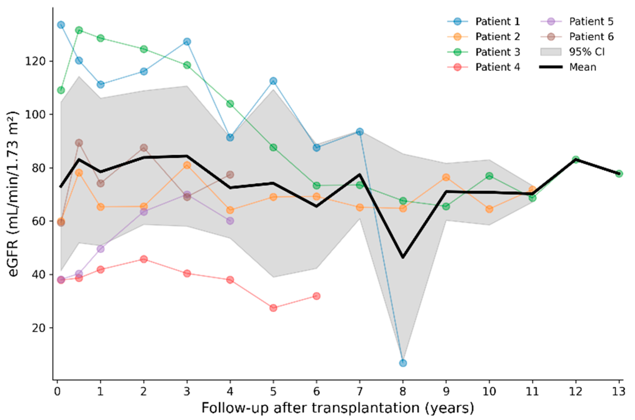
Figure 2.
Longitudinal biochemical and cardiac trajectories during Fabry-specific therapy relative to kidney transplantation. (A) Lyso-Gb3 concentration; (B) left ventricular mass index (LVMI); (C) interventricular septal thickness (IVS); and (D) posterior wall thickness (PW). Kidney transplantation is designated as time 0. Negative values indicate measurements obtained before transplantation and positive values indicate measurements obtained after transplantation.
Figure 2.
Longitudinal biochemical and cardiac trajectories during Fabry-specific therapy relative to kidney transplantation. (A) Lyso-Gb3 concentration; (B) left ventricular mass index (LVMI); (C) interventricular septal thickness (IVS); and (D) posterior wall thickness (PW). Kidney transplantation is designated as time 0. Negative values indicate measurements obtained before transplantation and positive values indicate measurements obtained after transplantation.
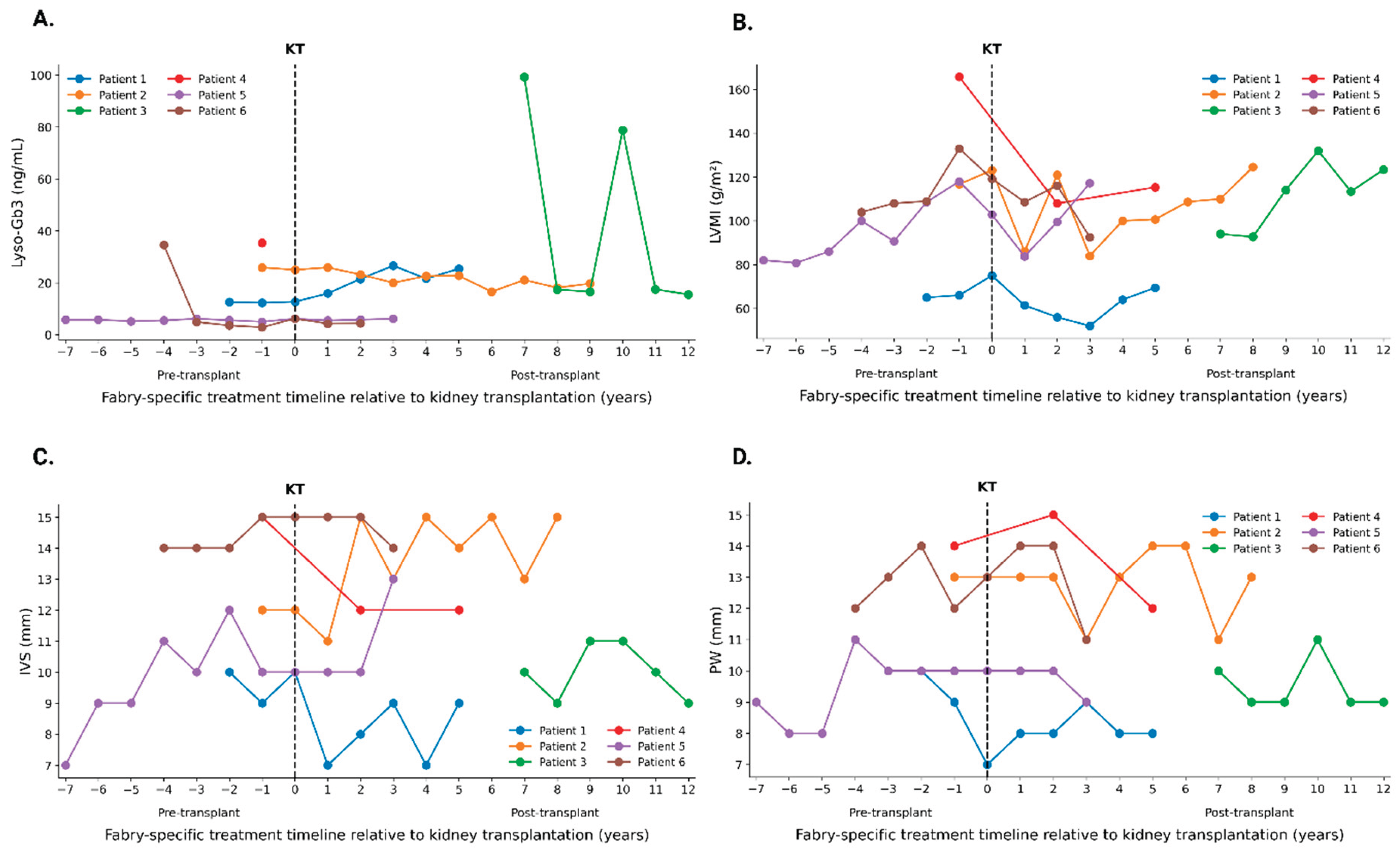
Table 1.
Fabry disease phenotype and pretransplant characteristics.
| Variable | Patient 1 | Patient 2 | Patient 3 | Patient 4 | Patient 5 | Patient 6 |
| Demographic and Fabry disease characteristics | ||||||
| Sex | Male | Male | Male | Male | Female | Male |
| Age at symptom onset, y | 13 | 12 | 8 | 25 | 27 | 43 |
| Age at Fabry disease diagnosis, y | 17 | 36 | 28 | 31 | 35 | 43 |
| Fabry phenotype | Classic | Classic | Classic | Classic | Classic | Late-onset cardiac variant |
| GLA variant | c.779G>A | c.141G>A | c.981A>C | c.955A>T | c.485G>A | c.644A>G |
| Protein change | p.G260E | p.W47* | p.Gln327His | p.Ile319Phe | p.W162* | p.(Asn215Ser) |
| α-Gal A activity | 12.54ᵃ nmol/h/mg | 35ᵃ nmol/h/mg | 0ᵇ µmol/L/h | 0ᵇ µmol/L/h | 0.39ᶜ mU/mg | 0.4ᵇ µmol/L/h |
| Renal involvement before transplantation | ||||||
| Age at kidney impairment diagnosis, y | 16 | 35 | 20 | 31 | 35 | 43 |
| CKD stage at Fabry disease diagnosis | G3b | G5 | G5 | G5 | G3b | G4 |
| Native kidney biopsy | Yes | No | No | No | Yes | Yes |
| Age at hemodialysis initiation, y | 18 | 37 | 20 | 31 | 41 | 48 |
| Hemodialysis duration, mo | 12 | 36 | 18 | 11 | 10 | 2 |
| Systemic involvement and comorbidities | ||||||
| Hypertrophic cardiomyopathy | No | Yes | Yes | Yes | No | Yes |
| Hypohidrosis | Yes | Yes | Yes | Yes | No | No |
| Angiokeratomas | Yes | Yes | Yes | Yes | Yes | No |
| Cornea verticillata | Yes | Yes | Yes | Yes | Yes | No |
| Neuropathic pain | Yes | Yes | Yes | Yes | Yes | No |
| Cerebral involvement | No | Yes | Yes | Yes | No | No |
| Hypertension | Yes | Yes | No | Yes | Yes | Yes |
| Obesity | No | No | No | No | Yes | No |
Note: α-Gal A activity was measured using different validated assays: ᵃ Leukocyte assay, reference range 160–320 nmol/h/mg protein; ᵇ dried blood spot assay, cut-off >2.8 µmol/L/h; ᶜ leukocyte assay, reference range 0.36–0.80 mU/mg. Abbreviations: CKD—chronic kidney disease; FD—Fabry disease; α-Gal A—α-galactosidase A.
Table 2.
Kidney transplantation characteristics and long-term outcomes.
| Variable | Patient 1 | Patient 2 | Patient 3 | Patient 4 | Patient 5 | Patient 6 |
| Transplant characteristics | ||||||
| Age at KT, y | 18 | 40 | 22 | 32 | 42 | 48 |
| Donor type | LR | DBD | DBD | LR | DBD | LUR |
| Follow-up after KT, y | 8 | 11 | 13 | 6 | 4 | 4 |
| Kidney allograft outcomes | ||||||
| eGFR at 1 y, mL/min/1.73 m² | 111.2 | 65.3 | 128.5 | 41.8 | 49.6 | 74.1 |
| eGFR at LFU, mL/min/1.73 m² | 6.9 | 71.8 | 77.8 | 32.0 | 60.1 | 77.4 |
| Proteinuria at 1 y, g/24 h | 0.00 | 0.00 | 0.10 | 0.16 | 0.00 | 0.12 |
| Proteinuria at LFU, g/24 h | 0.00 | 0.00 | 0.26 | 0.38 | 0.00 | 0.10 |
| Rejection | Yes | No | No | No | No | No |
| Time to rejection, y | 8 | — | — | — | — | — |
| De novo HLA-DSA | Yes | No | No | No | No | No |
| Graft status at LFU | Loss | Functioning | Functioning | Functioning | Functioning | Functioning |
| Time to graft loss, y | 8 | — | — | — | — | — |
| Post-transplant clinical events | ||||||
| PTDM | No | No | No | No | No | Yes |
| Obesity | No | No | No | No | Yes | Yes |
| Infectious episodes, n | 7 | 2 | 2 | 0 | 0 | 0 |
Abbreviations: DBD—donation after brain death; DSA—donor-specific antibody; eGFR—estimated glomerular filtration rate; KT—kidney transplantation; LFU—last follow-up; LR—living-related donor; LUR—living-unrelated donor; PTDM—post-transplant diabetes mellitus.
Disclaimer/Publisher’s Note: The statements, opinions and data contained in all publications are solely those of the individual author(s) and contributor(s) and not of MDPI and/or the editor(s). MDPI and/or the editor(s) disclaim responsibility for any injury to people or property resulting from any ideas, methods, instructions or products referred to in the content. |
© 2026 by the authors. Licensee MDPI, Basel, Switzerland. This article is an open access article distributed under the terms and conditions of the Creative Commons Attribution (CC BY) license (http://creativecommons.org/licenses/by/4.0/).
Copyright: This open access article is published under a Creative Commons CC BY 4.0 license, which permit the free download, distribution, and reuse, provided that the author and preprint are cited in any reuse.