
Submitted:
13 October 2026
Posted:
15 October 2026
You are already at the latest version
Abstract
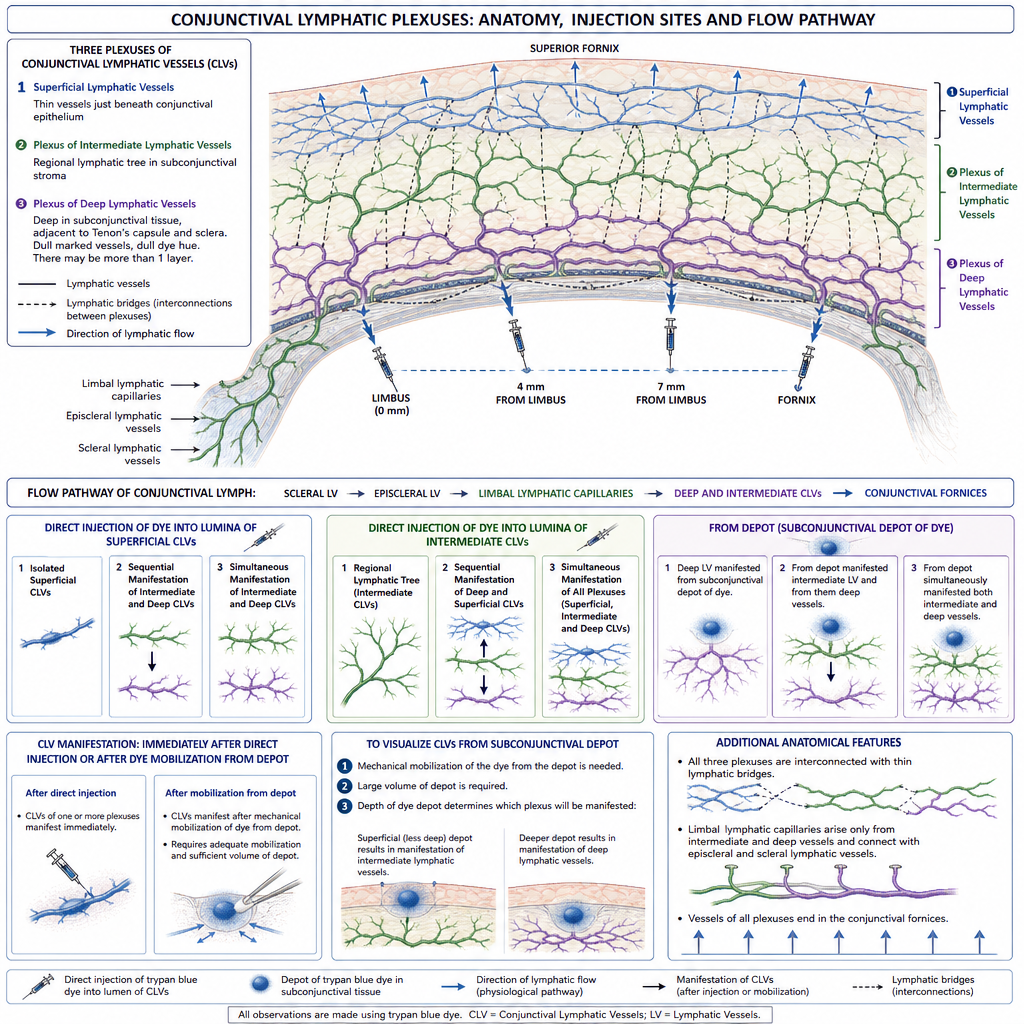
The conjunctival lymphatic network is critical for ocular surface immunity, aqueous humor drainage after glauco-ma surgery, and subconjunctival drug delivery, yet its in vivo architecture in humans remains incompletely char-acterized. This study aimed to visualize and describe the in vivo architecture and functional dynamics of conjunc-tival lymphatic vessels using a modified trypan blue injection technique. Ninety eyes of 78 patients undergoing rou-tine cataract surgery were enrolled. A modified intratissue injection technique using a 34 gauge needle, a silicone tube adapter, and 0.08 % trypan blue was applied at standardized locations (limbus, 4 mm, 7 mm, and fornix), with real time video recording under the operating microscope in all cases. A total of 249 investigations were performed, with successful lymphatic visualization in 143 (57.4 %). Three distinct lymphatic plexuses were identified: superfi-cial, intermediate, and deep. The superficial plexus showed a valve like “pearl beads” morphology with blind ending segments; the intermediate plexus consisted of larger collectors draining toward the fornix; and the deep plexus—previously undescribed—comprised flat, interconnected vessels without visible valves, filled exclu-sively via inter plexus lymphatic bridges. Limbal lymphatic capillaries originated only from the intermediate and deep plexuses. Drainage followed a consistent nasal to temporal and posterior to fornix pattern, and blood ele-ments were observed replacing trypan blue within lymphatic vessels during and after surgical manipulation. This study provides the first in vivo description of a three plexus conjunctival lymphatic architecture in humans and identifies inter plexus bridges as a novel anatomical feature. The modified trypan blue technique proved effective for mapping both structure and dynamics, even when subconjunctival dye depots formed, confirming its utility for in vivo lymphatic visualization.
Keywords:
conjunctival lymphatic vessels
; trypan blue
; lymphangiography
; lymphatic plexus
; in vivo imag-ing
; conjunctival anatomy
; glaucoma surgery
; ocular surface
Copyright: This open access article is published under a Creative Commons CC BY 4.0 license, which permit the free download, distribution, and reuse, provided that the author and preprint are cited in any reuse.