

Submitted:
12 October 2026
Posted:
14 October 2026
You are already at the latest version
Abstract
One of the biggest problems for researchers working in the field of idiopathic Parkinson's disease and patients is the lack of success in clinical trials attempting to develop new drugs that can slow or halt the progression of the disease. Enormous human and financial resources have been spent on preclinical research that has not led to the discovery of new drugs; however, a serious discussion about the causes of these failures is lacking. In our opinion, the problem lies in the fact that the preclinical studies on which the failed clinical trials were used an exogenous neurotoxin that induce a rapid and expansive degenerative process, which contrasts with the extremely slow degenerative process of idiopathic Parkinson's disease, which takes years. We have proposed that the neurodegenerative process of the nigrostriatal system, which culminates in the loss of dopaminergic neurons containing neuromelanin, is governed by a single-neuron neurodegeneration model. This degenerative process is triggered by the formation of aminochrome within neurons that synthesize neuromelanin, inducing a neurotoxic process without expansive effects on other neurons. This implies that the neurotoxic effects of aminochrome are restricted to a single neuron and that the loss of a single neuron accumulates over time until motor symptoms appear.
Keywords:
dopamine
; neuromelanin
; neurodegeneration
; neuroprotection
; parkinson’s disease
; clinical studies
; glutathione transferase M2-2
1. Idiopathic Parkinson Disease
Parkinson's disease is the second most common neurodegenerative disorder. As people live longer, the number of Parkinson's cases continues to rise. When we talk about Parkinson's disease, we're including patients with parkinsonism, those with genetic forms of the disease, and patients with idiopathic Parkinson's—meaning the cause is unknown. Making these distinctions is important for getting a clearer picture of idiopathic Parkinson's in particular. Patients with parkinsonism are those whose symptoms have a known underlying cause (1, 2). This group includes people exposed to certain environmental toxins, like the pesticide paraquat or metals such as manganese and copper, which can lead to progressive and permanent parkinsonism (3-5). But it also includes patients whose symptoms are triggered by medications—and in most of these cases, the symptoms are reversible once the drug is stopped. Common culprits include antipsychotics like risperidone and haloperidol, as well as antiemetics like metoclopramide (6-11). Other known causes of parkinsonism also fall into this category (12). Patients with genetic Parkinson's disease, on the other hand, have a mutation in a specific gene—such as alpha-synuclein, parkin, or LRRK2, among others (13-24).
The identity of the neurotoxin that triggers the neurodegenerative mechanisms leading to idiopathic Parkinson's disease remains unknown. The condition typically emerges after age 50 to 60, and its prevalence rises with age. Increased life expectancy has contributed to a growing prevalence of the disease. Diagnosis is usually based on the onset of motor symptoms, although some studies have reported the presence of premotor symptoms as well (25–27). It is estimated that by the time motor symptoms appear, approximately 60% of the neuromelanin-containing dopaminergic neurons have already been lost, and the disease progresses over a period of 10 to 20 years before death. Clinical diagnosis is typically established following the onset of motor symptoms; however, emerging evidence suggests that premotor features may precede these manifestations (25–27).
Several mechanisms explaining the degenerative process of the nigrostriatal systemin idiopathic Parkinson’s disease have been reported, such as: (i) Mitochondrial dysfunction (28-31). Healthy, functional mitochondria produce ATP which is key to neuronal function—such as maintaining the neuronal membrane potential, which is essential for firing electrical impulses via the Na⁺/K⁺-ATPase (pumping potassium in and calcium out); for synaptic neurotransmission (e.g., the release of dopamine into the synaptic cleft) and axonal transport of vesicles, organelles; proteins along the axon, among other. Oxidative stress and alpha-synuclein aggregation to neurotoxic oligomers play an important role in mitochondrial dysfunction in neurons (32.34); (ii) Alpha-synuclein aggregation to neurotoxic oligomers. Alpha-synuclein, under certain conditions, can aggregate to form fibrils that accumulate in Lewy bodies, which can be observed in post-mortem tissues of patients with Parkinson's disease. On the other hand, alpha-synuclein can aggregate to form neurotoxic oligomers that would be responsible for the neurotoxic effects of alpha-synuclein. It has been reported that alfa-synuclein induces a disruption of lysosomal protein degradation by affecting the acidification of lysosomes (35). Alpha-synuclein is also capable of inducing multiple neurotoxic mechanisms such as mitochondrial dysfunction, oxidative stress, synaptic dysfunction, mitochondrial dysfunction, microglia activation and disruption of calcium homeostasis (33, 36-39) ; (iii) Dysfunction of protein degradation systems (lysosomal and proteasomal). Autophagy dysfunction has been associated with Parkinson's disease (40-43). Parkin and PTEN-induced putative kinase 1 play an important role in mitochondrial degradation via the lysosomal system (43, 44) . Other genes associated with Parkinson's disease have also been linked to lysosomal protein degradation, such as GBA and LRRK2 (45). Proteasomal degradation of protein dysfunction also plays an important role in Parkinson's disease by regulating protein clearance, including the ubiquitin proteasome system (UPS) (46,47); (iv) Another important mechanism involved in the degenerative process of Parkinson's disease is endoplasmic reticulum stress. The endoplasmic reticulum plays an important role in the generation of new proteins, quality control, and their degradation. During endoplasmic reticulum stress, protein-folding capacity is overwhelmed, leading to an accumulation of unfolded or misfolded proteins that induce neurotoxicity in neurons. Cells activate a response mechanism (the Unfolded Protein Response) to restore balance, but under these severe conditions of endoplasmic reticulum stress, the result can be the degeneration of the nigrostriatal system in Parkinson's disease (48-51); (v) The brain uses a significant portion of oxygen of the body to generate ATP, which is necessary for normal neuronal function. Oxidative stress is one of the mechanisms associated with neurodegeneration of the nigrostriatal system in Parkinson's disease (52, 53). Reactive oxygen species are normally formed within the mitochondrial electron transport chain, where an electron is transferred to ubiquinone, generating a ubiquinone semiquinone; this intermediate can reduce oxygen to superoxide in a reaction catalyzed by NADH-ubiquinone reductase (Complex I)(54). Electron transfer from ubiquinol to cytochrome c proceeds via two pathways: ubiquinol transfers an electron to the Rieske iron-sulfur (Fe-S) protein—which subsequently passes it to cytochrome c. Simultaneously ubiquinol undergoing oxidation to ubiquinone through the formation of a semiquinone that can alternatively reduce oxygen to superoxide. Superoxide ultimately leads to the formation of hydrogen peroxide, which can generate hydroxy radicals in the presence of iron and finally oxidative stress (55, 56); (vi) Microglia are macrophages present in the brain that become activated in response to neurotoxins, alpha-synuclein misfolding, oxidative stress, mitochondrial dysfunction, or bacterial infections (57-59). Free neuromelanin induces neuroinflammation by activating microglia. Upon exposure to these stimuli, microglia become activated and release proinflammatory factors such as tumor necrosis factor-alpha, interleukin-1 beta, interleukin-6, and interleukin-12 (61). It has been proposed that the microbiome-gut-brain axis plays a role in the degenerative process of Parkinson's disease. An imbalance in the microorganisms inhabiting the digestive tract—characterized by a reduction in beneficial bacteria—promotes neuroinflammation. Consequently, intestinal changes may be associated with neurodegeneration via microbiome-gut-brain signaling (62) (Table I).

Table 1.
In this review, we will focus on the potential role of aminochrome in idiopathic Parkinson's disease.
Table 1.
In this review, we will focus on the potential role of aminochrome in idiopathic Parkinson's disease.
| Mechanism | Effect | References |
| Mitochondrial dysfunction |
|
28-34 |
| Alpha-synuclein aggregation to neurotoxic oligomers |
|
35-39 |
| Dysfunction of protein degradation systems (lysosomal and proteasomal) |
|
40-47 |
| Endoplasmic reticulum stress |
|
48-51 |
| Oxidative stress |
|
52-56 |
| Neuroinflammation |
|
57-61 |
2. Clinical Studies
L-dopa was introduced as a treatment for Parkinson’s disease 59 years ago and remains the gold standard in palliative care. While its benefits are undeniable, chronic use—typically after four to six years—often leads to severe side effects, including dyskinesias, which significantly impair patients’ quality of life. Despite extensive efforts to find drugs that can slow down or stop the disease from progressing, no new therapies have emerged. This remains one of the field’s greatest challenges. Promising preclinical results have consistently failed to translate into successful clinical trials (99-102). Understanding why these disease-modifying drugs have fallen short is a major barrier to advancing new treatments. Several factors may explain these failures, including patient selection criteria, the advanced stage of neurodegeneration at the time of treatment (which may be too late for reversal), a lack of reliable biomarkers to measure outcomes, the short duration of clinical trials, and compromised axonal transport that prevents the study drug from reaching its target (99).
Our explanation for the failure of clinical trials in Parkinson’s disease is that these studies have been based on exogenous neurotoxins that do not reflect what happens in idiopathic Parkinson’s disease. The exogenous neurotoxins used in preclinical studies are primarily MPTP and 6-hydroxydopamine. Injecting these neurotoxins into a specific brain region induces massive neurodegeneration that affects all exposed neurons. These toxins cause very rapid and widespread neurodegeneration, whereas the neurodegenerative process in dopaminergic neurons that contain neuromelanin is extremely slow, lasting years before motor symptoms appear and continuing throughout disease progression until the patient’s death. It has been suggested that pre-motor symptoms can emerge up to 20 years before motor symptoms (25), and furthermore, disease progression after diagnosis can extend from 10 to 20 years. This vast difference between the speed of neurodegeneration in preclinical models using MPTP and 6-hydroxydopamine and the extreme slowness of neurodegeneration in idiopathic Parkinson’s disease may explain why clinical studies based on successful results with these exogenous neurotoxins ultimately fail. It is important to note that MPTP induces symptoms of severe parkinsonism in just 3 days in drug users who used synthetic drugs contaminated with MPTP (103), suggesting that it is impossible for an exogenous neurotoxin to produce a neurodegenerative process as slow as that seen in patients with idiopathic Parkinson’s disease.
Generally, the scientific community agrees that motor symptoms in idiopathic Parkinson’s disease don’t appear until about 60% of the neuromelanin-containing dopaminergic neurons have been lost. This means that by the time motor symptoms emerge, the patient has somewhere between 320,000 and 400,000 of these neurons left, since it’s been estimated that a person typically has between 800,000 and 1,000,000 dopaminergic neurons in the substantia nigra pars compacta (104). If a patient survives 15 years after the onset of motor symptoms, they lose roughly 58 to 73 neurons per day until death. This gradual decline makes it very difficult to detect a meaningful treatment effect in clinical trials using the MDS-UPDRS, the standard rating scale for Parkinson’s motor function. For that reason, it seems unlikely that a drug developed using a fast-acting preclinical model would produce any measurable benefit in a patient with idiopathic Parkinson’s, whose disease progresses so slowly.
A neurodegenerative model for idiopathic Parkinson's disease, referred to as the single-neuron degeneration model, has been proposed based on the exceptionally slow progression of the neurodegenerative process in this condition (195). This model suggests that: (i) the degeneration of neuromelanin-containing dopaminergic neurons in the nigrostriatal system is non-expansive, meaning that the degeneration of one neuron does not spread to or affect its neighboring neurons—this endogenous neurotoxin cannot be released into or taken up by adjacent cells; (ii) the neurotoxin responsible for triggering the degenerative process, which ultimately leads to the loss of that individual neuron, is generated internally within that same neuron; and (iii) the progressive loss of neuromelanin-containing dopaminergic neurons occurs through a slow, neuron-to-neuron accumulation, which accounts for the extremely gradual nature of the overall neurodegenerative process.
3. Neuromelanin Synthesis
Neuromelanin is a pigment formed through the oxidation of the catechol group in free dopamine. Under normal conditions, dopamine remains stable and resistant to catechol oxidation when stored within monoaminergic vesicles in the terminals of dopaminergic neurons. The transport of dopamine into these vesicles via VMAT-2 is driven by an H⁺-ATP-dependent proton pump located in the vesicular membrane (106-107). The resulting accumulation of protons inside the vesicles creates a mildly acidic environment that keeps dopamine heavily protonated, thereby preventing oxidation of its catechol group.
During neurotransmission, dopamine is released and subsequently taken back up through the dopamine transporter (DAT) on the extracellular membrane of dopaminergic neurons. It has been suggested that the intracellular portion of DAT associates with synaptogyrin-3 and VMAT-2, which helps keep dopamine from entering the cytosol and undergoing oxidation (108). Once reuptaken, dopamine is transported directly into monoaminergic vesicles, where the slightly acidic pH keeps it fully stable and available for reuse in subsequent neurotransmission.
Another potential source of free dopamine arises during its synthesis from tyrosine. However, it has been proposed that aromatic amino acid decarboxylase and tyrosine hydroxylase form a complex with VMAT-2, thereby preventing the accumulation of free dopamine in the cytosol (109). The presence of neuromelanin appears to correlate with the level of VMAT-2 expression, which limits the availability of free dopamine and thus reduces the risk of catechol group oxidation (110).
Neuromelanin synthesis requires the oxidation of dopamine's catechol group, which generates three ortho-quinones as transient intermediates. The process begins with the sequential oxidation of dopamine to dopamine ortho-quinone, which then cyclizes to aminochrome, followed by 5,6-indolequinone, and finally neuromelanin (111). Dopamine ortho-quinone is stable only at a pH below 2.0; at physiological pH, it undergoes rapid cyclization at a constant rate of 0.15 s⁻¹ to form aminochrome (112, 113).
In in vitro experiments using the enzyme tyrosinase, ¹⁴C-dopamine and isolated mitochondria form adducts with several mitochondrial proteins, including creatine kinase, ubiquinol-cytochrome c reductase core protein 1, mitophilin, chaperonin, ubiquitin carboxy-terminal hydrolase L1 (UCH-L1), glucose-regulated protein 75/mtHSP70/mortalin, and DJ-1. However, in experiments with SH-SY5Y cells exposed to dopamine, adducts were only observed with DJ-1 and UCH-L1 (114). This discrepancy likely arises because, in the isolated mitochondria experiments, dopamine is oxidized by tyrosinase and forms adducts immediately, without interference from the hundreds of biomolecules present in the SH-SY5Y cell cytosol. In cell-based experiments, tyrosinase cannot be used to oxidize dopamine, as the resulting dopamine ortho-quinone would rapidly react with extracellular membrane proteins. Instead, dopamine must be transported into the cytosol, where it is oxidized to dopamine ortho-quinone in a biomolecule-rich environment.
Thus, it is reasonable to assume that the adducts formed intracellularly with dopamine are not due to dopamine ortho-quinone itself, but rather to aminochrome, which is more stable and converts to 5,6-dihydroxyindole—the precursor to 5,6-indolequinone—at a constant rate of 0.06 min⁻¹ (113). Other reports have suggested that dopamine ortho-quinone forms adducts with tyrosine hydroxylase, mitochondrial glutathione peroxidase, the human dopamine transporter, and parkin (115-117). However, given the inherent instability of dopamine ortho-quinone, it remains unclear whether these adducts were actually formed with dopamine ortho-quinone or with aminochrome.
5,6-Indolequinone is the direct precursor to neuromelanin and serves as the basic building block of the neuromelanin polymer (118-120) . In vitro studies have shown that it can form adducts with alpha-synuclein, which inhibits neuromelanin formation (121) . However, there is currently no evidence that the formation of 5,6-indolequinone–alpha-synuclein oligomers are neurotoxic.
Aminochrome is the most stable transient ortho-quinone generated during neuromelanin synthesis. In vitro NMR experiments show that the aminochrome peak is detected for up to 20 minutes, whereas the 5,6-dihydroxyindole peak—the precursor of 5,6-indolequinone—appears at 40 minutes (122). However, within a dopaminergic neuron containing neuromelanin, where the environment is crowded with biomolecules, aminochrome formation results in a much shorter stability period before it is metabolized.
Aminochrome can form adducts with various proteins, including mitochondrial complex I (123) (88), alpha-synuclein(124), actin, and alpha- and beta-tubulin (125) , among others. Additionally, aminochrome participates in redox reactions in which it can be reduced by flavoenzymes that transfer one or two electrons using NADH in the cytosol. This one-electron reduction of aminochrome produces leukoaminochrome ortho-semiquinone, which is highly reactive with oxygen. Leukoaminochrome ortho-semiquinone reduces dioxygen to a superoxide radical, and in the process, the leukoaminochrome ortho-semiquinone radical is oxidized back to aminochrome (119).
In the presence of NADH and molecular oxygen in the cytosol, aminochrome is reduced back to leukoaminochrome ortho-semiquinone, which is then oxidized in the presence of oxygen, generating a continuous redox cycle that persists until NADH and oxygen are depleted. This redox cycle impairs ATP synthesis via the respiratory chain coupled to oxidative phosphorylation and promotes oxidative stress (126). Because aminochrome can form adducts with proteins and participate in this redox cycle, it is not excreted from dopaminergic neurons that contain neuromelanin. This prevents a spreading effect that could otherwise impact neighboring neurons.
All three ortho-quinones formed during neuromelanin synthesis can be neurotoxic. However, because the constant rate of conversion of aminochrome to 5,6-indolequinone (0.06 min⁻¹) is much slower than that of dopamine to ortho-quinone to aminochrome (0.15 s⁻¹) (113), there will be a tendency for aminochrome to accumulate although transiently.
Aminochrome can be neurotoxic by triggering neuroinflammation, dysfunction of both lysosomal and mitochondrial protein degradation systems, endoplasmic reticulum stress, mitochondrial dysfunction, formation of neurotoxic alpha-synuclein oligomers, and oxidative stress in dopaminergic neurons containing neuromelanin (123-138).
Neuromelanin has been reported to accumulate in specific organelles, which keeps it from coming into contact with biomolecules in the cytosol. Neuromelanin also acts as a scavenger by accumulating iron and other metals and preventing them from participating in redox reactions that could generate oxidative stress. These neuromelanin-containing organelles have also been reported to accumulate lipids and certain lysosomal proteins. The buildup of neuromelanin in these organelles plays an important role in preventing the catechol groups of dopamine—which may have been released during neuromelanin polymerization—from taking part in redox reactions in the presence of iron. It has been reported that free neuromelanin released from these organelles can activate microglia, triggering neuroinflammation that leads to the production of pro-inflammatory factors, reactive nitrogen and oxygen species, and ultimately, neuronal death (139-144).
The formation of neurotoxic ortho-quinones—particularly aminochrome—raises important questions about the role of neuromelanin synthesis in idiopathic Parkinson's disease. Evidence from postmortem studies of healthy older adults shows that dopaminergic neurons containing neuromelanin in the substantia nigra remain intact, strongly suggesting that neuromelanin synthesis is a normal and harmless process. This synthesis occurs naturally and gradually accumulates with age in humans (145). However, the fact that its formation requires the transient production of potentially neurotoxic compounds like aminochrome seems at odds with the view that neuromelanin synthesis is benign. This paradox is resolved by the presence of two enzymes that protect against aminochrome's neurotoxic effects: DT-diaphorase and glutathione transferase M2-2 (146).(Fig.1).
Figure 1.
The neurotoxic effect of aminochrome during neuromelanin synthesis depends on its overproduction exceeding the Km of the neuroprotective enzymes DT-diaphorase and glutathione transferase M2-2. At these high aminochrome concentrations, these enzymes cannot fully prevent its neurotoxic effects. The fraction that surpasses their protective capacity triggers neurotoxic outcomes that ultimately lead to the degeneration of individual neurons.
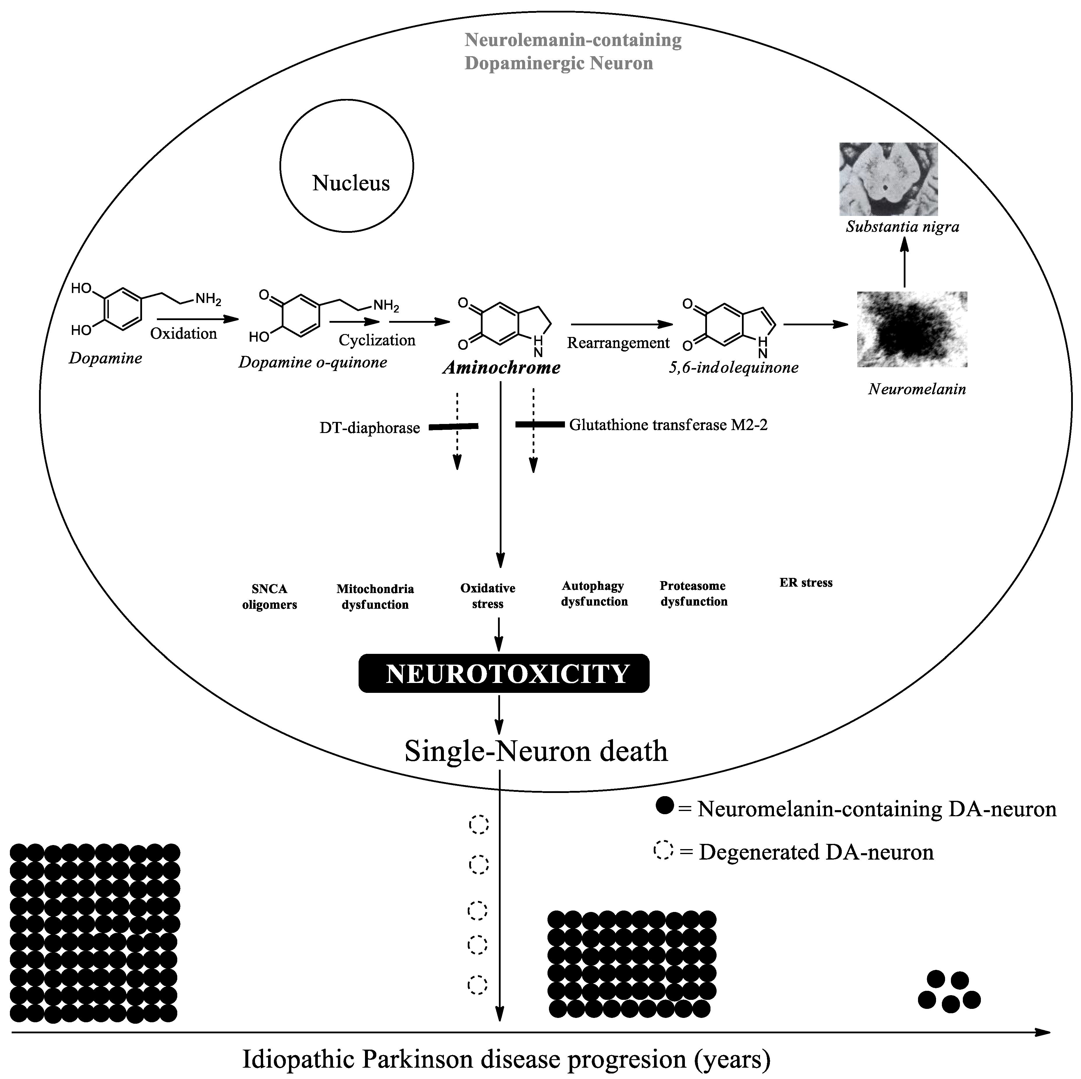
Dopamine does not oxidize within monoaminergic vesicles, as their environment is mildly acidic. However, proper dopamine storage in these vesicles relies on the presence of VMAT-2. An inverse relationship has been reported between neuromelanin levels and VMAT-2 expression. Increased VMAT-2 expression reduces cytosolic dopamine levels, thereby decreasing neuromelanin formation. Conversely, reduced VMAT-2 expression promotes oxidation of dopamine's catechol group, leading to increased aminochrome and neuromelanin production beyond the protective capacity of DT-diaphorase and glutathione transferase M2-2. Because aminochrome cannot be secreted outside the neuron where it exerts its toxic effects, it does not cause widespread damage to neighboring cells. Instead, the loss of individual neuromelanin-containing dopaminergic neurons accumulates gradually over years until motor symptoms eventually emerge. DA = Dopamine; SNCA = Alpha-synuclein; ER-stress = Endoplasmic reticulum stress.
4. Neuroprotective Enzymes
DT-diaphorase (NAD(P)H:quinone oxidoreductase, NQO1) is a dimeric flavoenzyme that contains two flavin adenine dinucleotide molecules and utilizes both NADH and NADPH as electron donors. It is the only flavoenzyme that reduces quinones via a two-electron transfer to produce a hydroquinone; other flavoenzymes typically catalyze one-electron reduction, generating semiquinones that are highly reactive with oxygen and can induce oxidative stress. DT-diaphorase thus competes with these one-electron quinone reductases. The enzyme is expressed in a variety of organs; in the rat brain, it has been detected in the substantia nigra, striatum, hypothalamus, cerebellum, hippocampus, and frontal cortex. At the cellular level, it is found in both dopaminergic neurons and astrocytes. Notably, DT-diaphorase accounts for approximately 97% of the total quinone reductase activity in the rat substantia nigra (147).
DT-diaphorase prevents the reduction of aminochrome by flavoenzymes that transfer a single electron, thereby blocking the formation of the leukoaminochrome ortho-semiquinone radical. To detect these radicals, we used electron spin resonance (ESR) stabilization, in which the radicals were trapped as complexes with Zn²⁺ to allow for spectral measurement. Using this approach, we attempted to measure the formation of the leukoaminochrome ortho-semiquinone radical when aminochrome was reduced by NADPH–cytochrome P450 reductase, a one-electron–transferring flavoenzyme. However, the leukoaminochrome ortho-semiquinone radical proved to be so highly reactive with oxygen that the Zn²⁺–radical complex could not be detected (148).
The autoxidation of leukoaminochrome ortho-semiquinone in the presence of oxygen cannot be inhibited, and it generates superoxide radicals and aminochrome, thereby creating a redox cycling process as mentioned earlier. In contrast, the two-electron reduction of aminochrome catalyzed by DT-diaphorase produces leukoaminochrome, which also undergoes autoxidation, but at a much slower rate than the radical form. Most importantly, the superoxide dismutase enzyme present in the cytosol inhibits this oxidation by converting superoxide radicals into oxygen, suggesting that the DT-diaphorase–mediated reduction of aminochrome serves as a neuroprotective reaction (126).
DT-diaphorase prevents the neurotoxic effects of aminochrome (149) by inhibiting aminochrome-induced mitochondrial dysfunction, the formation of neurotoxic alpha-synuclein oligomers, cytoskeleton disruption, dysfunction of both lysosomal and proteasomal protein degradation systems, and oxidative stress (125-127, 149-151) .
Glutathione transferase M2-2 is expressed in astrocytes within the substantia nigra, where it conjugates aminochrome (152, 153) and its precursor, dopamine ortho-quinone, with glutathione to form 4-S-glutathionyl-5,6-dihydroxyindoline and 5-S-glutathionyl-dopamine, respectively (154). The 4-S-glutathionyl-5,6-dihydroxyindoline product is resistant to oxidizing agents such as dioxygen, superoxide, and hydrogen peroxide, suggesting it represents an end product. Meanwhile, 5-S-glutathionyl-dopamine is metabolized to 5-cysteinyl-dopamine, a compound that has been detected in both human cerebrospinal fluid and neuromelanin (155, 156).
Astrocytes are capable of taking up dopamine, which can then be oxidized to aminochrome. However, glutathione transferase M2-2 protects these cells by conjugating aminochrome with glutathione to form 4-S-glutathionyl-5,6-dihydroxyindoline. Moreover, astrocytes secrete glutathione transferase M2-2 via exosomes, which are taken up by dopaminergic neurons, allowing the enzyme to be released into their cytosol (157, 158).
As a result, DT-diaphorase and glutathione transferase M2-2 work synergistically to prevent the neurotoxic effects of aminochrome in dopaminergic neurons (157, 158). This cooperative action of the two neuroprotective enzymes effectively neutralizes aminochrome toxicity during neuromelanin synthesis, accounting for the otherwise benign nature of this process.
Figure 2.
Neuroprotective Role of DT-Diaphorases and Glutathione Transferase M2-2 in Neuromelanin Synthesis.
Figure 2.
Neuroprotective Role of DT-Diaphorases and Glutathione Transferase M2-2 in Neuromelanin Synthesis.
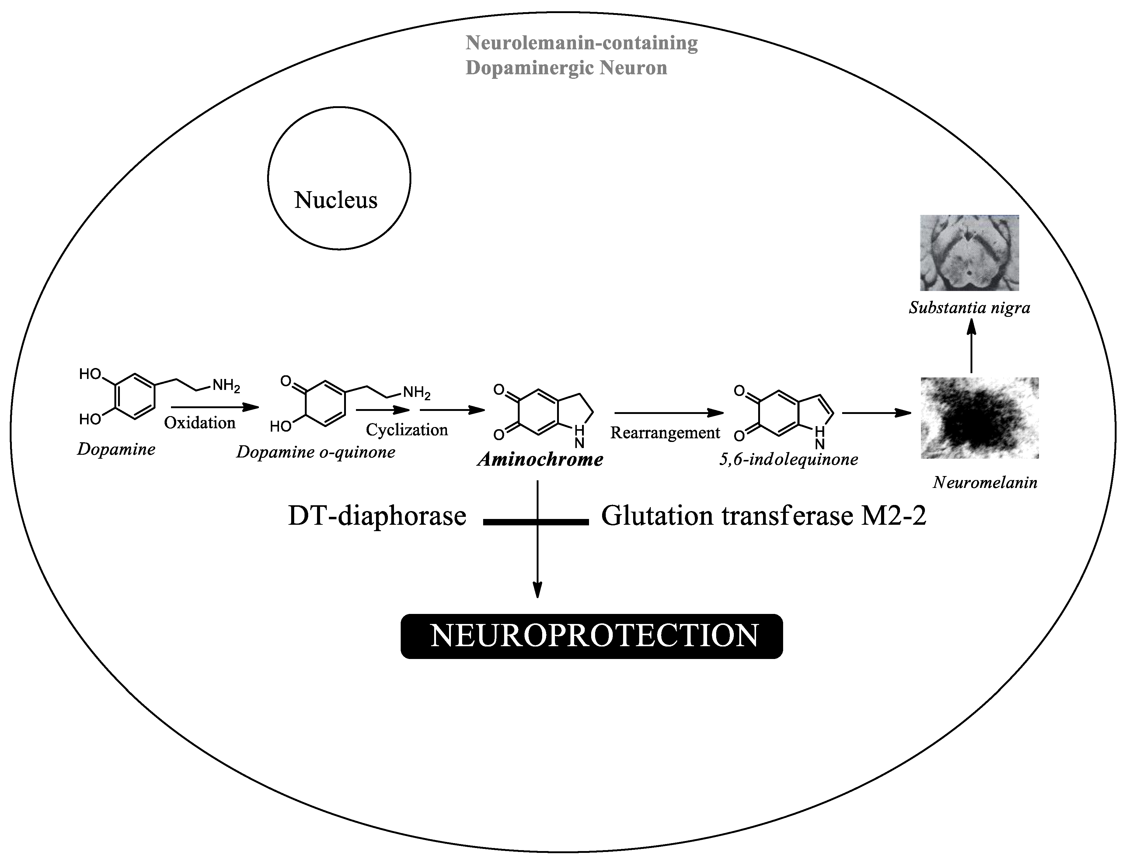
DT-diaphorases and glutathione transferase M2-2 protect against the neurotoxic effects of aminochrome by blocking several harmful processes, including the formation of neurotoxic alpha-synuclein oligomers, impairment of both lysosomal and proteasomal protein degradation systems, oxidative stress, neuroinflammation, mitochondrial dysfunction, and endoplasmic reticulum stress. This protective activity explains why neuromelanin synthesis is inherently harmless.
5. The Possible Role of Aminochrome in Idiopathic Parkinson´s Disease
The unusually slow progression of neurodegeneration in idiopathic Parkinson’s disease suggests several key points. First, the neurotoxin that initiates the neurodegenerative process must come from within the body—that is, it must be endogenous. More specifically, it has to be generated inside the very dopaminergic neurons that are degenerating, particularly those that contain neuromelanin. Second, this internally produced neurotoxin cannot have a widespread or expansive effect; It must induce neurotoxicity only in the neurons where it was generated, without inducing neurotoxicity in neighboring neurons. Third, the neurotoxin must trigger a cascade of harmful events, including mitochondrial dysfunction, formation of neurotoxic alpha-synuclein oligomers, neuroinflammation, oxidative stress, impaired protein degradation via both the proteasomal and lysosomal systems, and endoplasmic reticulum stress. Fourth and finally, the endogenous neurotoxin must be short-lived, with a brief half-life (105).
Aminochrome is an endogenous neurotoxin produced within dopaminergic neurons during neuromelanin synthesis. It does not have a spreading effect because it is a transient neurotoxin with a very short half-life. In the cytosol, which is crowded with biomolecules, aminochrome rapidly forms adducts with proteins or gets reduced by flavoenzymes.
7. Conclusions
The reasons why clinical trials based on successful preclinical studies using exogenous neurotoxins like MPTP or 6-hydroxydopamine have failed remain unclear. What is most striking is the extremely slow progression of neurodegeneration in idiopathic Parkinson's disease, which stands in stark contrast to the rapid neurodegeneration seen in preclinical models that rely on these same exogenous toxins.
One hypothesis suggests that the slow course of idiopathic Parkinson's may be explained by a single-neuron neurodegeneration model. In this model, each neurotoxic event causes the loss of just one neuron, and these losses accumulate over time until motor symptoms eventually emerge.
Unlike other endogenous neurotoxins—such as alpha-synuclein oligomers, which are not formed exclusively within neuromelanin-containing dopaminergic neurons and can spread to neighboring cells upon release (159-164)—aminochrome behaves differently. Aminochrome is generated specifically inside dopaminergic neurons that produce neuromelanin. It is not secreted, so it does not exert an expansive effect on surrounding neurons. However, it does trigger all the neurotoxic mechanisms implicated in nigrostriatal degeneration, including mitochondrial dysfunction, formation of neurotoxic alpha-synuclein oligomers, neuroinflammation, oxidative stress, and impairment of both lysosomal and proteasomal protein degradation systems, as well as endoplasmic reticulum stress. Fifty-nine years after L-dopa was introduced for the palliative treatment of Parkinson's disease—and after repeated clinical trial failures based on preclinical models using endogenous neurotoxins—there is a clear need to open a discussion about why these clinical studies keep failing.
Author Contributions
The authors contribution is the following: Conceptualization, IP, SH. and JSA.; writing—original draft preparation, JSA.; writing—review and editing, IP, SH.; literature revision, IP, SH and JSA. All authors have read and agreed to the published version of the manuscript.
Funding
not applicable.
Institutional Review Board Statement
Not applicable.
Informed Consent Statement
Not applicable.
Data Availability Statement
Not applicable.
Acknowledgments
Not applicable.
Conflicts of Interest
The authors declare no conflict of interest.
References
- Brigo, F.; Erro, R.; Marangi, A.; Bhatia, K.; Tinazzi, M. Differentiating drug-induced parkinsonism from Parkinson's disease: an update on non-motor symptoms and investigations. Park. Relat. Disord. 2014, 20(8), 808–14. [Google Scholar] [CrossRef]
- Segura-Aguilar, J. (Ed.) Clinical Studies and Therapies in Parkinson’s Disease: Translations from Preclinical Models. In Parkinson’s disease; Elsevier: Cambridge, MA, USA, 2021; pp. 1–171. [Google Scholar]
- Lucchini, R.; Tieu, K. Manganese-Induced Parkinsonism: Evidence from Epidemiological and Experimental Studies. Biomolecules 2023, 13(8), 1190. [Google Scholar] [CrossRef]
- McKnight, S.; Hack, N. Toxin-Induced Parkinsonism. Neurol. Clin. 2020, 38(4), 853–865. [Google Scholar] [CrossRef]
- Tong, T.; Duan, W.; Xu, Y.; Hong, H.; Xu, J.; Fu, G.; Wang, X.; Yang, L.; Deng, P.; Zhang, J.; He, H.; Mao, G.; Lu, Y.; Lin, X.; Yu, Z.; Pi, H.; Cheng, Y.; Xu, S.; Zhou, Z. Paraquat exposure induces Parkinsonism by altering lipid profile and evoking neuroinflammation in the midbrain. Environ. Int. 2022, 169, 107512. [Google Scholar] [CrossRef]
- Pacheco-Paez, T.; Montastruc, F.; Rousseau, V.; Chebane, L.; Lapeyre-Mestre, M.; Renoux, C.; Montastruc, J.L. Parkinsonism associated with gabapentinoid drugs: A pharmacoepidemiologic study. Mov. Disord. 2020, 35(1), 176–180. [Google Scholar] [CrossRef]
- Druschky, K.; Bleich, S.; Grohmann, R.; Engel, R.R.; Toto, S.; Neyazi, A.; Däubl, B.; Stübner, S. Severe parkinsonism under treatment with antipsychotic drugs. Eur. Arch. Psychiatry Clin. Neurosci. 2020, 270(1), 35–47. [Google Scholar] [CrossRef]
- Shuaib, U.A.; Rajput, A.H.; Robinson, C.A.; Rajput, A. Neuroleptic-induced Parkinsonism: Clinicopathological study. Mov. Disord. Epub. 2016, 31(3), 360–5. [Google Scholar] [CrossRef]
- Brigo, F.; Erro, R.; Marangi, A.; Bhatia, K.; Tinazzi, M. Differentiating drug-induced parkinsonism from Parkinson's disease: an update on non-motor symptoms and investigations. Park. Relat. Disord. 2014, 20(8), 808–14. [Google Scholar] [CrossRef]
- Jeong, S.; Cho, H.; Kim, Y.J.; Ma, H.I.; Jang, S. Drug-induced Parkinsonism: A strong predictor of idiopathic Parkinson's disease. PLoS ONE 2021, 16(3), e0247354. [Google Scholar] [CrossRef]
- Conn, H.; Jankovic, J. Drug-induced parkinsonism: diagnosis and treatment. Expert Opin. Drug Saf. 2024, 23(12), 1503–1513. [Google Scholar] [CrossRef]
- Holm, H.; Gundersen, V.; Dietrichs, E. Vascular parkinsonism; Tidsskr Nor Laegeforen: English, Norwegian, 4 May 2023; 7, p. 143. [Google Scholar] [CrossRef]
- Shadrina, M.I.; Slominsky, P.A. Genetic Architecture of Parkinson's Disease. Biochemistry (Mosc) 2023, 88(3), 417–433. [Google Scholar] [CrossRef] [PubMed]
- Cherian A, K P D, Vijayaraghavan A. Parkinson's disease - genetic cause. Curr Opin Neurol. 2023 Aug 1;36(4):292-301. [CrossRef]
- Blauwendraat, C.; Nalls, M.A.; Singleton, A.B. The genetic architecture of Parkinson's disease. Lancet Neurol. 2020, 19(2), 170–178. [Google Scholar] [CrossRef] [PubMed] [PubMed Central]
- Mnich, K.; Moghaddam, S.; Browne, P.; Counihan, T.; Fitzgerald, S.P.; Martin, K.; Richardson, C.; Samali, A.; Gorman, A.M. Endoplasmic Reticulum Stress-Regulated Chaperones as a Serum Biomarker Panel for Parkinson's Disease. Mol. Neurobiol. 2023, 60(3), 1476–1485. [Google Scholar] [CrossRef]
- Song, P.; Peng, W.; Sauve, V.; Fakih, R.; Xie, Z.; Ysselstein, D.; Krainc, T.; Wong, Y.C.; Mencacci, N.E.; Savas, J.N.; Surmeier, D.J.; Gehring, K.; Krainc, D. Parkinson's disease-linked parkin mutation disrupts recycling of synaptic vesicles in human dopaminergic neurons. Neuron 2023, 111(23), 3775–3788.e7. [Google Scholar] [CrossRef]
- Cyske, Z.; Gaffke, L.; Rintz, E.; Wiśniewska, K.; Węgrzyn, G.; Pierzynowska, K. Molecular mechanisms of the ambroxol action in Gaucher disease and GBA1 mutation-associated Parkinson disease. Neurochem Int. 2024, 178, 105774. [Google Scholar] [CrossRef] [PubMed]
- Nuytemans, K.; Theuns, J.; Cruts, M.; Van Broeckhoven, C. Genetic etiology of Parkinson disease associated with mutations in the SNCA, PARK2, PINK1, PARK7, and LRRK2 genes: a mutation update. Hum. Mutat. 2010, 31(7), 763–80. [Google Scholar] [CrossRef] [PubMed] [PubMed Central]
- Kumazawa, R.; Tomiyama, H.; Li, Y.; Imamichi, Y.; Funayama, M.; Yoshino, H.; Yokochi, F.; Fukusako, T.; Takehisa, Y.; Kashihara, K.; Kondo, T.; Elibol, B.; Bostantjopoulou, S.; Toda, T.; Takahashi, H.; Yoshii, F.; Mizuno, Y.; Hattori, N. Mutation analysis of the PINK1 gene in 391 patients with Parkinson disease. Arch. Neurol. 2008, 65(6), 802–8. [Google Scholar] [CrossRef] [PubMed]
- Kumazawa, R.; Tomiyama, H.; Li, Y.; Imamichi, Y.; Funayama, M.; Yoshino, H.; Yokochi, F.; Fukusako, T.; Takehisa, Y.; Kashihara, K.; Kondo, T.; Elibol, B.; Bostantjopoulou, S.; Toda, T.; Takahashi, H.; Yoshii, F.; Mizuno, Y.; Hattori, N. Mutation analysis of the PINK1 gene in 391 patients with Parkinson disease. Arch. Neurol. 2008, 65(6), 802–8. [Google Scholar] [CrossRef] [PubMed]
- Monroy-Jaramillo, N.; Guerrero-Camacho, J.L.; Rodríguez-Violante, M.; Boll-Woehrlen, M.C.; Yescas-Gómez, P.; Alonso-Vilatela, M.E.; López-López, M. Genetic mutations in early-onset Parkinson's disease Mexican patients: molecular testing implications. Am. J. Med. Genet B Neuropsychiatr. Genet. 2014, 165B(3), 235–44. [Google Scholar] [CrossRef] [PubMed]
- Macedo, M.G.; Verbaan, D.; Fang, Y.; van Rooden, S.M.; Visser, M.; Anar, B.; Uras, A.; Groen, J.L.; Rizzu, P.; van Hilten, J.J.; Heutink, P. Genotypic and phenotypic characteristics of Dutch patients with early onset Parkinson's disease. Mov. Disord. 2009, 24(2), 196–203. [Google Scholar] [CrossRef] [PubMed]
- Yonova-Doing, E.; Atadzhanov, M.; Quadri, M.; Kelly, P.; Shawa, N.; Musonda, S.T.; Simons, E.J.; Breedveld, G.J.; Oostra, B.A.; Bonifati, V. Analysis of LRRK2, SNCA, Parkin, PINK1, and DJ-1 in Zambian patients with Parkinson's disease. Park. Relat. Disord. Epub. 2012, 18(5), 567–71. [Google Scholar] [CrossRef] [PubMed]
- Leite Silva, A.B.R.; Gonçalves de Oliveira, R.W.; Diógenes, G.P.; de Castro Aguiar, M.F.; Sallem, C.C.; Lima, M.P.P.; de Albuquerque Filho, L.B.; Peixoto de Medeiros, S.D.; Penido de Mendonça, L.L.; de Santiago Filho, P.C. Premotor, nonmotor and motor symptoms of Parkinson's Disease: A new clinical state of the art. Ageing Res. Rev. 2023, 84, 101834. [Google Scholar] [CrossRef]
- Barber, T.R.; Klein, J.C.; Mackay, C.E.; Hu, M.T.M. Neuroimaging in pre-motor Parkinson's disease. Neuroimage Clin. 2017, 15, 215–227. [Google Scholar] [CrossRef]
- Reichmann, H. Premotor Diagnosis of Parkinson's Disease. Neurosci. Bull. 2017, 33(5), 526–534. [Google Scholar] [CrossRef]
- Moradi Vastegani, S.; Nasrolahi, A.; Ghaderi, S.; Belali, R.; Rashno, M.; Farzaneh, M.; Khoshnam, S.E. Mitochondrial Dysfunction and Parkinson's Disease: Pathogenesis and Therapeutic Strategies. Neurochem Res. 2023, 48(8), 2285–2308. [Google Scholar] [CrossRef]
- Hattori, N.; Sato, S. Mitochondrial dysfunction in Parkinson's disease. J. Neural Transm. 2024, 131(12), 1415–1428. [Google Scholar] [CrossRef]
- Lucchesi, M.; Biso, L.; Bonaso, M.; Longoni, B.; Buchignani, B.; Battini, R.; Santorelli, F.M.; Doccini, S.; Scarselli, M. Mitochondrial Dysfunction in Genetic and Non-Genetic Parkinson's Disease. Int. J. Mol. Sci. 2025, 26(9), 4451. [Google Scholar] [CrossRef] [PubMed] [PubMed Central]
- Blagov, A.; Postnov, A.; Sukhorukov, V.; Popov, M.; Uzokov, J.; Orekhov, A. Significance of Mitochondrial Dysfunction in the Pathogenesis of Parkinson's Disease. Front Biosci. (Landmark Ed) 2024, 29(1), 36. [Google Scholar] [CrossRef]
- Li, S.; Zhu, W.; Jiang, J.; Bu, Y.; Wang, Q.; Li, M.; Wang, D.; Gao, Y.; Wang, J.; Wang, T.; Hu, X.; Wan, C. Oxidative Damage Induced Disruption of Mitochondrial Quality Control Underlying Abnormal Energy Metabolism in Peripheral Leukocytes of Schizophrenia Patients. Schizophr. Bull. Open 2026, 7(1), sgag029. [Google Scholar] [CrossRef]
- Park, J.H.; Burgess, J.D.; Faroqi, A.H.; DeMeo, N.N.; Fiesel, F.C.; Springer, W.; Delenclos, M.; McLean, P.J. Alpha-synuclein-induced mitochondrial dysfunction is mediated via a sirtuin 3-dependent pathway. Mol. Neurodegener. 2020, 15(1), 5. [Google Scholar] [CrossRef]
- Morales-Martínez, A.; Martínez-Gómez, P.A.; Martinez-Fong, D.; Villegas-Rojas, M.M.; Pérez-Severiano, F.; Del Toro-Colín, M.A.; Delgado-Minjares, K.M.; Blanco-Alvarez, V.M.; Leon-Chavez, B.A.; Aparicio-Trejo, O.E.; Baéz-Cortés, M.T.; Cardenas-Aguayo, M.D.; Luna-Muñoz, J.; Pacheco-Herrero, M.; Angeles-López, Q.D.; Martínez-Dávila, I.A.; Salinas-Lara, C.; Romero-López, J.P.; Sánchez-Garibay, C.; Méndez-Cruz, A.R.; Soto-Rojas, L.O. Oxidative Stress and Mitochondrial Complex I Dysfunction Correlate with Neurodegeneration in an α-Synucleinopathy Animal Model. Int. J. Mol. Sci. 2022, 23(19), 11394. [Google Scholar] [CrossRef]
- Lo, C.H.; Ren, M.; Loi, G.W.Z.; Saipuljumri, E.N.; Indajang, J.; Lim, K.L.; Zeng, J. Lysosome-Acidifying Nanoparticles Rescue A30P α-Synuclein Induced Neuronal Death in Cellular and Drosophila Models of Parkinson's Disease. Adv. Healthc. Mater. 2026, 15(22), e02906. [Google Scholar] [CrossRef]
- Sohrabi, T.; Mirzaei-Behbahani, B.; Zadali, R.; Pirhaghi, M.; Morozova-Roche, L.A.; Meratan, A.A. Common Mechanisms Underlying α-Synuclein-Induced Mitochondrial Dysfunction in Parkinson's Disease. J. Mol. Biol. 2023, 435(12), 167992. [Google Scholar] [CrossRef]
- McCarthy, E.; Barron, A.; Morales-Prieto, N.; Mazzocchi, M.; McCarthy, C.M.; Collins, L.M.; Sullivan, A.M.; O'Keeffe, G.W. Gene Co-expression Analysis of the Human Substantia Nigra Identifies ZNHIT1 as an SNCA Co-expressed Gene that Protects Against α-Synuclein-Induced Impairments in Neurite Growth and Mitochondrial Dysfunction in SH-SY5Y Cells. Mol. Neurobiol. 2022, 59(5), 2745–2757. [Google Scholar] [CrossRef]
- Danyu, L.; Yanran, L.; Xiuna, J.; Ying, C.; Sudan, P.; Tianen, Z.; Zhifen, Z.; Dezhi, Z.; Kaixun, H.; Yingyu, X.; Enxiang, T. α-Synuclein induced mitochondrial dysfunction via cytochrome c oxidase subunit 2 in SH-SY5Y cells. Exp. Cell Res. 2019, 378(1), 57–65. [Google Scholar] [CrossRef]
- Ordonez, D.G.; Lee, M.K.; Feany, M.B. α-synuclein Induces Mitochondrial Dysfunction through Spectrin and the Actin Cytoskeleton. Neuron 2018, 97(1), 108–124.e6. [Google Scholar] [CrossRef]
- Papagiannakis, N.; Xilouri, M.; Koros, C.; Simitsi, A.M.; Stamelou, M.; Maniati, M.; Stefanis, L. Autophagy dysfunction in peripheral blood mononuclear cells of Parkinson's disease patients. Neurosci. Lett. 2019, 704, 112–115. [Google Scholar] [CrossRef]
- Cerri, S.; Blandini, F. Role of Autophagy in Parkinson's Disease. Curr. Med. Chem. 2019, 26(20), 3702–3718. [Google Scholar] [CrossRef]
- Karabiyik, C.; Lee, M.J.; Rubinsztein, D.C. Autophagy impairment in Parkinson's disease. Essays Biochem. 2017, 61(6), 711–720. [Google Scholar] [CrossRef]
- Basak, B.; Riley, J.F.; Nataraj, N.M.; Holzbaur, E.L. Mitophagy in neuronal health and disease: from mechanisms to neurodegeneration. J. Clin. Invest. 2026, 136(17), e199847. [Google Scholar] [CrossRef]
- Malpartida, A.B.; Williamson, M.; Narendra, D.P.; Wade-Martins, R.; Ryan, B.J. Mitochondrial Dysfunction and Mitophagy in Parkinson's Disease: From Mechanism to Therapy. Trends Biochem Sci. 2021, 46(4), 329–343. [Google Scholar] [CrossRef]
- Senkevich, K.; Gan-Or, Z. Autophagy lysosomal pathway dysfunction in Parkinson's disease; evidence from human genetics. Park. Relat. Disord. 2020, 73, 60–71. [Google Scholar] [CrossRef]
- Zhao, Z.; Song, X.; Wang, Y.; Yu, L.; Huang, G.; Li, Y.; Zong, R.; Liu, T.; Ji, Q.; Zheng, Y.; Liu, B.; Zhu, Q.; Chen, L.; Gao, C.; Liu, H. E3 ubiquitin ligase TRIM31 alleviates dopaminergic neurodegeneration by promoting proteasomal degradation of VDAC1 in Parkinson's Disease model. Cell Death Differ. 2024, 31(11), 1410–1421. [Google Scholar] [CrossRef]
- Di Costanzo, A.; Fusco, C.; Camerlingo, R.; Serafini, S.; Minò, A.; Tiberio, C.; Palmieri, G.; Matrone, C.; Cocca, E.; Angiolillo, A. Proteasome Dysregulation in Parkinson's Disease: Insights from Blood-Based Analyses. Mol. Neurobiol. 2025, 63(1), 326. [Google Scholar] [CrossRef]
- Mou, Z.; Yuan, Y.H.; Zhang, Z.; Song, L.K.; Chen, N.H. Endoplasmic reticulum stress, an important factor in the development of Parkinson's disease. Toxicol. Lett. 2020, 324, 20–29. [Google Scholar] [CrossRef]
- Tsujii, S.; Ishisaka, M.; Hara, H. Modulation of endoplasmic reticulum stress in Parkinson's disease. Eur. J. Pharmacol. 2015, 765, 154–6. [Google Scholar] [CrossRef]
- Chen, L.; Liu, F.; Sun, B.; Deng, D.; Han, Y.; Wang, J.; Song, J.; Wang, C.; Sun, K.; Yu, H. Endoplasmic reticulum stress in Parkinson's disease: A pivotal role in cell fate and a therapeutic target. Ageing Res. Rev. 2026, 118, 103096. [Google Scholar] [CrossRef]
- Zeng, H.; Liu, Y.; Liu, X.; Li, J.; Lu, L.; Xue, C.; Wu, X.; Zhang, X.; Zheng, Z.; Lu, G. Interplay of α-Synuclein Oligomers and Endoplasmic Reticulum Stress in Parkinson'S Disease: Insights into Cellular Dysfunctions. Inflammation 2025, 48(4), 1590–1606. [Google Scholar] [CrossRef]
- Hemmati-Dinarvand, M.; Saedi, S.; Valilo, M.; Kalantary-Charvadeh, A.; Alizadeh Sani, M.; Kargar, R.; Safari, H.; Samadi, N. Oxidative stress and Parkinson's disease: conflict of oxidant-antioxidant systems. Neurosci. Lett. Epub. 2019, 709, 134296. [Google Scholar] [CrossRef] [PubMed]
- Trist, B.G.; Hare, D.J.; Double, K.L. Oxidative stress in the aging substantia nigra and the etiology of Parkinson's disease. Aging Cell 2019, 18(6), e13031. [Google Scholar] [CrossRef]
- Zhao, R.Z.; Jiang, S.; Zhang, L.; Yu, Z.B. Mitochondrial electron transport chain, ROS generation and uncoupling (Review). Int. J. Mol. Med. 2019, 44(1), 3–15. [Google Scholar] [CrossRef] [PubMed] [PubMed Central]
- Bochkova, Z.V.; Baizhumanov, A.A.; Yusipovich, A.I.; Morozova, K.I.; Nikelshparg, E.I.; Fedotova, A.A.; Tiaglik, A.B.; Xu, Y.; Brazhe, A.R.; Maksimov, G.V.; Bilan, D.S.; Khramova, Y.V.; Parshina, E.Y.; Brazhe, N.A. The flexible chain: regulation of structure and activity of ETC complexes defines rate of ATP synthesis and sites of superoxide generation. Biophys. Rev. 2025, 17(1), 55–88. [Google Scholar] [CrossRef]
- Cadenas, E.; Boveris, A.; Ian Ragan, C.; O M Stoppani, A. Reprint of: Production of Superoxide Radicals and Hydrogen Peroxide by NADH- Ubiquinone Reductase and Ubiquinol-Cytochrome c Reductase from Beef-Heart Mitochondria. Arch. Biochem Biophys. 2022, 726, 109231. [Google Scholar] [CrossRef] [PubMed]
- Isik, S.; Yeman Kiyak, B.; Akbayir, R.; Seyhali, R.; Arpaci, T. Microglia Mediated Neuroinflammation in Parkinson's Disease. Cells 2023, 12(7), 1012. [Google Scholar] [CrossRef] [PubMed] [PubMed Central]
- Chatterjee, K.; Roy, A.; Banerjee, R.; Choudhury, S.; Mondal, B.; Halder, S.; Basu, P.; Shubham, S.; Dey, S.; Kumar, H. Inflammasome and α-synuclein in Parkinson’s Disease: A Cross-Sectional Study. J. Neuroimmunol. 2020, 338, 577089. [Google Scholar] [CrossRef]
- Cui, H.; Kong, Y.; Zhang, H. Oxidative Stress, Mitochondrial Dysfunction, and Aging. J. Signal. Transduct. 2012, 2012, 646354. [Google Scholar] [CrossRef]
- Zhang, W.; Phillips, K.; Wielgus, A.R.; Liu, J.; Albertini, A.; Zucca, F.A.; Faust, R.; Qian, S.Y.; Miller, D.S.; Chignell, C.F.; Wilson, B.; Jackson-Lewis, V.; Przedborski, S.; Joset, D.; Loike, J.; Hong, J.S.; Sulzer, D.; Zecca, L. Neuromelanin activates microglia and induces degeneration of dopaminergic neurons: implications for progression of Parkinson's disease. Neurotox. Res. 2011, 19(1), 63–72. [Google Scholar] [CrossRef]
- Zhu, X.; Chen, S.; Xie, Y.; Cheng, Z.; Zhu, X.; Guo, Q. Role of M1/M2 macrophages in pain modulation. Zhong Nan Da Xue Xue Bao Yi Xue Ban. 2024, 49(7), 1155–1163. [Google Scholar] [CrossRef]
- Porwal, M.; Malviya, R.; Chandra, P.; Sridhar, S.B.; Shareef, J.; Wadhwa, T.; Tripathy, D.B.; Arockiam, D. Microbiome-gut-brain axis modulation for the management of Parkinson's disease: Emerging mechanisms and translational insights. J. Neuroimmunol. 2026, 421, 579099. [Google Scholar] [CrossRef]
- Raafat, M.A.; Al-Hasnaawei, S.; Mousa, H.M.; Hanumanthayya, M.; Sahoo, S.; Prathiba, S.; Singh, G.; Sinha, A. The central role of mitochondrial dysfunction in neurodegeneration: implications for therapy. Mol. Cell Biochem 2026. [Google Scholar] [CrossRef]
- Abubakar, M.D.; Alom, S.; Ali, F.; Ali, S.R.; Adhikary, K.; Sarika, S.; Gupta, P.K.; Puppala, E.R.; Murti, K.; Rai, S.N.; Kumar, N. Molecular Mechanisms of Dopaminergic Neuron Degeneration in Parkinson's disease: A Comprehensive Review. Curr. Neuropharmacol. 2026. [Google Scholar] [CrossRef]
- Zhang, S.; Wang, T.; Peng, Y.; Wang, Q.; Zhang, Z.; Chu, S.; Huang, H.; Chen, N. Parkinson's disease: pathogenesis and therapeutic strategies. Mol. Biomed. 2026, 7(1), 46. [Google Scholar] [CrossRef]
- Mohamed Ameer, J.; Alexander, S.M.; Navya, K.; Shenoi, R.A.; Chandra, G. Revitalizing mitochondrial quality control: targeting mitochondria-derived vesicles in Parkinson's disease. Biochem Pharmacol. 2026, 250 Pt 1, 117935. [Google Scholar] [CrossRef]
- Hattori, N.; Sato, S. Mitochondrial dysfunction in Parkinson's disease. J. Neural Transm. 2024, 131(12), 1415–1428. [Google Scholar] [CrossRef]
- Rocha, E.M.; De Miranda, B.; Sanders, L.H. Alpha-synuclein: Pathology, mitochondrial dysfunction and neuroinflammation in Parkinson's disease. Neurobiol. Dis. 2018, 109 Pt B, 249–257. [Google Scholar] [CrossRef]
- Blagov, A.; Postnov, A.; Sukhorukov, V.; Popov, M.; Uzokov, J.; Orekhov, A. Significance of Mitochondrial Dysfunction in the Pathogenesis of Parkinson's Disease. Front Biosci. (Landmark Ed) 2024, 29(1), 36. [Google Scholar] [CrossRef]
- Moradi Vastegani, S.; Nasrolahi, A.; Ghaderi, S.; Belali, R.; Rashno, M.; Farzaneh, M.; Khoshnam, S.E. Mitochondrial Dysfunction and Parkinson's Disease: Pathogenesis and Therapeutic Strategies. Neurochem Res. 2023, 48(8), 2285–2308. [Google Scholar] [CrossRef]
- Moradi Vastegani, S.; Nasrolahi, A.; Ghaderi, S.; Belali, R.; Rashno, M.; Farzaneh, M.; Khoshnam, S.E. Mitochondrial Dysfunction and Parkinson's Disease: Pathogenesis and Therapeutic Strategies. Neurochem Res. 2023, 48(8), 2285–2308. [Google Scholar] [CrossRef]
- Mehra, S.; Sahay, S.; Maji, S.K. α-Synuclein misfolding and aggregation: Implications in Parkinson's disease pathogenesis. Biochim Biophys. Acta Proteins Proteom. 2019, 1867(10), 890–908. [Google Scholar] [CrossRef]
- Du, X.Y.; Xie, X.X.; Liu, R.T. The Role of α-Synuclein Oligomers in Parkinson's Disease. Int. J. Mol. Sci. 2020, 21(22), 8645. [Google Scholar] [CrossRef]
- Vidović, M.; Rikalovic, M.G. Alpha-Synuclein Aggregation Pathway in Parkinson's Disease: Current Status and Novel Therapeutic Approaches. Cells 2022, 11(11), 1732. [Google Scholar] [CrossRef]
- Li, B.; Xiao, X.; Bi, M.; Jiao, Q.; Chen, X.; Yan, C.; Du, X.; Jiang, H. Modulating α-synuclein propagation and decomposition: Implications in Parkinson's disease therapy. Ageing Res. Rev. 2024, 98, 102319. [Google Scholar] [CrossRef]
- Jiao, J.; Gao, G.; Zhu, J.; Wang, C.; Liu, L.; Yang, H. Binding of α-synuclein to ACO2 promotes progressive mitochondrial dysfunction in Parkinson's disease models. Redox Biol. 2024, 77, 103399. [Google Scholar] [CrossRef]
- Fanning, S.; Haque, A.; Imberdis, T.; Baru, V.; Barrasa, M.I.; Nuber, S.; Termine, D.; Ramalingam, N.; Ho, G.P.H.; Noble, T.; Sandoe, J.; Lou, Y.; Landgraf, D.; Freyzon, Y.; et al. Lipidomic Analysis of α-Synuclein Neurotoxicity Identifies Stearoyl CoA Desaturase as a Target for Parkinson Treatment. Mol. Cell 2019, 73(5), 1001–1014.e8. [Google Scholar] [CrossRef]
- Pickrell, A.M.; Youle, R.J. The roles of PINK1, parkin, and mitochondrial fidelity in Parkinson's disease. Neuron 2015, 85(2), 257–73. [Google Scholar] [CrossRef]
- Malpartida, A.B.; Williamson, M.; Narendra, D.P.; Wade-Martins, R.; Ryan, B.J. Mitochondrial Dysfunction and Mitophagy in Parkinson's Disease: From Mechanism to Therapy. Trends Biochem Sci. 2021, 46(4), 329–343. [Google Scholar] [CrossRef]
- Lizama, B.N.; Chu, C.T. Neuronal autophagy and mitophagy in Parkinson's disease. Mol. Asp. Med. 2021, 82, 100972. [Google Scholar] [CrossRef]
- Picca, A.; Guerra, F.; Calvani, R.; Romano, R.; Coelho-Júnior, H.J.; Bucci, C.; Marzetti, E. Mitochondrial Dysfunction, Protein Misfolding and Neuroinflammation in Parkinson's Disease: Roads to Biomarker Discovery. Biomolecules 2021, 11(10), 1508. [Google Scholar] [CrossRef]
- Di Costanzo, A.; Fusco, C.; Camerlingo, R.; Serafini, S.; Minò, A.; Tiberio, C.; Palmieri, G.; Matrone, C.; Cocca, E.; Angiolillo, A. Proteasome Dysregulation in Parkinson's Disease: Insights from Blood-Based Analyses. Mol. Neurobiol. 2025, 63(1), 326. [Google Scholar] [CrossRef]
- Burbulla, L.F.; Song, P.; Mazzulli, J.R.; Zampese, E.; Wong, Y.C.; Jeon, S.; Santos, D.P.; Blanz, J.; Obermaier, C.D.; Strojny, C.; Savas, J.N.; Kiskinis, E.; Zhuang, X.; Krüger, R.; Surmeier, D.J.; Krainc, D. Dopamine oxidation mediates mitochondrial and lysosomal dysfunction in Parkinson's disease. Science 2017, 357(6357), 1255–1261. [Google Scholar] [CrossRef]
- Nguyen, M.; Wong, Y.C.; Ysselstein, D.; Severino, A.; Krainc, D. Synaptic, Mitochondrial, and Lysosomal Dysfunction in Parkinson's Disease. Trends Neurosci. 2019, 42(2), 140–149. [Google Scholar] [CrossRef]
- Minakaki, G.; Safren, N.; Bustos, B.I.; Lubbe, S.J.; Mencacci, N.E.; Krainc, D. Commander complex regulates lysosomal function and is implicated in Parkinson's disease risk. Science 2025, 388(6743), 204–211. [Google Scholar] [CrossRef]
- Senkevich, K.; Gan-Or, Z. Autophagy lysosomal pathway dysfunction in Parkinson's disease; evidence from human genetics. Park. Relat. Disord. 2020, 73, 60–71. [Google Scholar] [CrossRef]
- Wallings, R.L.; Humble, S.W.; Ward, M.E.; Wade-Martins, R. Lysosomal Dysfunction at the Centre of Parkinson's Disease and Frontotemporal Dementia/Amyotrophic Lateral Sclerosis. Trends Neurosci. 2019, 42(12), 899–912. [Google Scholar] [CrossRef]
- Brooker, S.M.; Naylor, G.E.; Krainc, D. Cell biology of Parkinson's disease: Mechanisms of synaptic, lysosomal, and mitochondrial dysfunction. Curr. Opin. Neurobiol. 2024, 85, 102841. [Google Scholar] [CrossRef]
- Trist, B.G.; Hare, D.J.; Double, K.L. Oxidative stress in the aging substantia nigra and the etiology of Parkinson's disease. Aging Cell 2019, 18(6), e13031. [Google Scholar] [CrossRef]
- Percário, S.; da Silva Barbosa, A.; Varela, E.L.P.; Gomes, A.R.Q.; Ferreira, M.E.S.; de Nazaré Araújo Moreira, T.; Dolabela, M.F. Oxidative Stress in Parkinson's Disease: Potential Benefits of Antioxidant Supplementation. Oxid. Med. Cell Longev. 2020, 2020, 2360872. [Google Scholar] [CrossRef]
- Hemmati-Dinarvand, M.; Saedi, S.; Valilo, M.; Kalantary-Charvadeh, A.; Alizadeh Sani, M.; Kargar, R.; Safari, H.; Samadi, N. Oxidative stress and Parkinson's disease: conflict of oxidant-antioxidant systems. Neurosci. Lett. 2019, 709, 134296. [Google Scholar] [CrossRef]
- Imbriani, P.; Martella, G.; Bonsi, P.; Pisani, A. Oxidative stress and synaptic dysfunction in rodent models of Parkinson's disease. Neurobiol. Dis. 2022, 173, 105851. [Google Scholar] [CrossRef]
- Isik, S.; Yeman Kiyak, B.; Akbayir, R.; Seyhali, R.; Arpaci, T. Microglia Mediated Neuroinflammation in Parkinson's Disease. Cells 2023, 12(7), 1012. [Google Scholar] [CrossRef]
- Liu, T.W.; Chen, C.M.; Chang, K.H. Biomarker of Neuroinflammation in Parkinson's Disease. Int. J. Mol. Sci. 2022, 23(8), 4148. [Google Scholar] [CrossRef]
- Araújo, B.; Caridade-Silva, R.; Soares-Guedes, C.; Martins-Macedo, J.; Gomes, E.D.; Monteiro, S.; Teixeira, F.G. Neuroinflammation and Parkinson's Disease-From Neurodegeneration to Therapeutic Opportunities. Cells 2022, 11(18), 2908. [Google Scholar] [CrossRef]
- Kovaleva, V.; Saarma, M. Endoplasmic Reticulum Stress Regulators: New Drug Targets for Parkinson's Disease. J. Park. Dis. 2021, 11(s2), S219–S228. [Google Scholar] [CrossRef]
- Mou, Z.; Yuan, Y.H.; Zhang, Z.; Song, L.K.; Chen, N.H. Endoplasmic reticulum stress, an important factor in the development of Parkinson's disease. Toxicol. Lett. 2020, 324, 20–29. [Google Scholar] [CrossRef]
- Mnich, K.; Moghaddam, S.; Browne, P.; Counihan, T.; Fitzgerald, S.P.; Martin, K.; Richardson, C.; Samali, A.; Gorman, A.M. Endoplasmic Reticulum Stress-Regulated Chaperones as a Serum Biomarker Panel for Parkinson's Disease. Mol. Neurobiol. 2023, 60(3), 1476–1485. [Google Scholar] [CrossRef]
- Athauda, D.; Foltynie, T. The ongoing pursuit of neuroprotective therapies in Parkinson disease. Nat. Rev. Neurol. 2015, 11(1), 25–40. [Google Scholar] [CrossRef]
- Teil, M.; Huot, P. The challenges of experimental pharmacology in identifying novel treatments for Parkinson's disease. Curr. Opin. Neurobiol. 2026, 97, 103164. [Google Scholar] [CrossRef]
- Lenka, A.; Jankovic, J. How should future clinical trials be designed in the search for disease-modifying therapies for Parkinson's disease? Expert Rev. Neurother. 2023, 23(2), 107–122. [Google Scholar] [CrossRef]
- Olanow, C.W.; Bartus, R.T.; Volpicelli-Daley, L.A.; Kordower, J.H. Trophic factors for Parkinson's disease: To live or let die. Mov. Disord. 2015, 30(13), 1715–24. [Google Scholar] [CrossRef]
- Williams, A. MPTP parkinsonism. Br. Med. J. 1984, 289, 1401–1402. [Google Scholar] [CrossRef]
- Ni, A.; Ernst, C. Evidence That Substantia Nigra Pars Compacta Dopaminergic Neurons Are Selectively Vulnerable to Oxidative Stress Because They Are Highly Metabolically Active. Front. Cell Neurosci. 2022, 16, 826193. [Google Scholar] [CrossRef]
- Huenchuguala, S.; Segura-Aguilar, J. Single-neuron neurodegeneration as a degenerative model for Parkinson's disease. Neural Regen. Res. 2024, 19(3), 529–535. [Google Scholar] [CrossRef]
- Segura-Aguilar; Sulzer, D.; Zucca, F.A.; Zecca, L. Overexpression of Vesicular Monoamine Transporter-2 may Block Neurotoxic Metabolites from Cytosolic Dopamine: a Potential Neuroprotective Therapy for Parkinson's Disease. Clin. Pharmacol. Transl. Med. 2019, 3(1), 143–148. [Google Scholar]
- Huenchuguala, S.; Segura-Aguilar, J. Targets to Search for New Pharmacological Treatment in Idiopathic Parkinson's Disease According to the Single-Neuron Degeneration Model. Biomolecules 2024, 14(6), 673. [Google Scholar] [CrossRef]
- Egaña, L.A.; Cuevas, R.A.; Baust, T.B.; Parra, L.A.; Leak, R.K.; Hochendoner, S.; Peña, K.; Quiroz, M.; Hong, W.C.; Dorostkar, M.M.; Janz, R.; Sitte, H.H.; Torres, G.E. Physical and functional interaction between the dopamine transporter and the synaptic vesicle protein synaptogyrin-3. J. Neurosci. 2009, 29(14), 4592–604. [Google Scholar] [CrossRef] [PubMed] [PubMed Central]
- Cartier, E.A.; Parra, L.A.; Baust, T.B.; Quiroz, M.; Salazar, G.; Faundez, V.; Egaña, L.; Torres, G.E. A biochemical and functional protein complex involving dopamine synthesis and transport into synaptic vesicles. J. Biol. Chem. 2010, 285(3), 1957–66. [Google Scholar] [CrossRef]
- Liang, C.L.; Nelson, O.; Yazdani, U.; Pasbakhsh, P.; German, D.C. Inverse relationship between the contents of neuromelanin pigment and the vesicular monoamine transporter-2: human midbrain dopamine neurons. J. Comp. Neurol. 2004, 473(1), 97–106. [Google Scholar] [CrossRef]
- Zhang, S.; Wang, R.; Wang, G. Impact of Dopamine Oxidation on Dopaminergic Neurodegeneration. ACS Chem. Neurosci. 2019, 10(2), 945–953. [Google Scholar] [CrossRef] [PubMed]
- Tse, D.C.; McCreery, R.L.; Adams, R.N. Potential oxidative pathways of brain catecholamines. J. Med. Chem. 1976, 19(1), 37–40. [Google Scholar] [CrossRef] [PubMed]
- Bisaglia, M.; Soriano, M.E.; Arduini, I.; Mammi, S.; Bubacco, L. Molecular characterization of dopamine-derived quinones reactivity toward NADH and glutathione: implications for mitochondrial dysfunction in Parkinson disease. Biochim Biophys. Acta 2010, 1802(9), 699–706. [Google Scholar] [CrossRef]
- Van Laar, V.S.; Mishizen, A.J.; Cascio, M.; Hastings, T.G. Proteomic identification of dopamine-conjugated proteins from isolated rat brain mitochondria and SH-SY5Y cells. Neurobiol. Dis. 2009, 34(3), 487–500. [Google Scholar] [CrossRef]
- Xu, Y.; Stokes, A.H.; Roskoski, R., Jr.; Vrana, K.E. Dopamine, in the presence of tyrosinase, covalently modifies and inactivates tyrosine hydroxylase. J. Neurosci. Res. 1998, 54(5), 691–7. [Google Scholar] [CrossRef] [PubMed]
- Whitehead, R.E.; Ferrer, J.V.; Javitch, J.A.; Justice, J.B. Reaction of oxidized dopamine with endogenous cysteine residues in the human dopamine transporter. J. Neurochem. 2001, 76(4), 1242–51. [Google Scholar] [CrossRef] [PubMed]
- LaVoie, M.J.; Ostaszewski, B.L.; Weihofen, A.; Schlossmacher, M.G.; Selkoe, D.J. Dopamine covalently modifies and functionally inactivates parkin. Nat. Med. 2005, 11(11), 1214–21. [Google Scholar] [CrossRef] [PubMed]
- Hauser, D.N.; Hastings, T.G. Mitochondrial dysfunction and oxidative stress in Parkinson's disease and monogenic parkinsonism. Neurobiol. Dis. 2013, 51, 35–42. [Google Scholar] [CrossRef] [PubMed] [PubMed Central]
- Segura-Aguilar, J.; Paris, I.; Muñoz, P.; Ferrari, E.; Zecca, L.; Zucca, F.A. Protective and toxic roles of dopamine in Parkinson's disease. J. Neurochem. 2014, 129(6), 898–915. [Google Scholar] [CrossRef]
- Napolitano, A.; Manini, P.; d'Ischia, M. Oxidation chemistry of catecholamines and neuronal degeneration: an update. Curr. Med. Chem. 2011, 18(12), 1832–45. [Google Scholar] [CrossRef]
- Zajac, G.W.; Gallas, J.M.; Cheng, J.; Eisner, M.; Moss, S.C.; Alvarado-Swaisgood, A.E. The fundamental unit of synthetic melanin: a verification by tunneling microscopy of X-ray scattering results. Biochim Biophys. Acta 1994, 1199(3), 271–8. [Google Scholar] [CrossRef]
- Bisaglia, M.; Mammi, S.; Bubacco, L. Kinetic and structural analysis of the early oxidation products of dopamine: analysis of the interactions with alpha-synuclein. J. Biol. Chem. 2007, 282(21), 15597–605. [Google Scholar] [CrossRef]
- Aguirre, P.; Urrutia, P.; Tapia, V.; Villa, M.; Paris, I.; Segura-Aguilar, J.; Núñez, M.T. The dopamine metabolite aminochrome inhibits mitochondrial complex I and modifies the expression of iron transporters DMT1 and FPN1. Biometals 2012, 25(4), 795–803. [Google Scholar] [CrossRef]
- Muñoz, P.; Cardenas, S.; Huenchuguala, S.; Briceño, A.; Couve, E.; Paris, I.; Segura-Aguilar, J. DT-Diaphorase Prevents Aminochrome-Induced Alpha-Synuclein Oligomer Formation and Neurotoxicity. Toxicol. Sci. 2015, 145(1), 37–47. [Google Scholar] [CrossRef]
- Paris, I.; Perez-Pastene, C.; Cardenas, S.; Iturriaga-Vasquez, P.; Muñoz, P.; Couve, E.; Caviedes, P.; Segura-Aguilar, J. Aminochrome induces disruption of actin, alpha-, and beta-tubulin cytoskeleton networks in substantia-nigra-derived cell line. Neurotox. Res. 2010, 18(1), 82–92. [Google Scholar] [CrossRef]
- Segura-Aguilar, J.; Muñoz, P.; Inzunza, J.; Varshney, M.; Nalvarte, I.; Mannervik, B. Neuroprotection against Aminochrome Neurotoxicity: Glutathione Transferase M2-2 and DT-Diaphorase. Antioxidants 2022, 11(2), 296. [Google Scholar] [CrossRef]
- Arriagada, C.; Paris, I.; Sanchez de las Matas, M.J.; Martinez-Alvarado, P.; Cardenas, S.; Castañeda, P.; Graumann, R.; Perez-Pastene, C.; Olea-Azar, C.; Couve, E.; et al. On the neurotoxicity mechanism of leukoaminochrome o-semiquinone radical derived from dopamine oxidation: Mitochondria damage, necrosis, and hydroxyl radical formation. Neurobiol. Dis. 2004, 16, 468–477. [Google Scholar] [CrossRef]
- Soares, É.N.; Bartolomeo, C.S.; Nicoliche, T.; Lima, I.S.; de Oliveira, L.M.G.; de Jesus Ferrolho, G.; Oliveira, R.B.; Stilhano, R.S.; Costa, S.L.; Tizabi, Y.; Ureshino, R.P.; da Silva, V.D.A. Nicotine protects astrocytes expressing alpha-synuclein against aminochrome cytotoxicity: Implications for Parkinson's disease. Res. Sq. [Preprint] 2026, rs.3.rs–9410157. [Google Scholar] [CrossRef]
- Teles-Souza, J.; Oliveira, J.V.R.; Araújo, F.M.; Sanches, F.S.; Jesus, L.B.; Souza, S.L.; Brito, A.D.P.S.; Oliveira Leite, C.M.; Ferrolho, G.J.; Fernandez-Villalba, E.; Vilas Bôas, D.S.; Santana, R.C.; Costa, S.L.; Herrero, M.T.; Silva, V.D.A. Striatal injection of aminochrome in Wistar rats induces early-stage behavioral changes of Parkinson's disease and dopaminergic degeneration in the caudal and medial regions of the nigra pars compacta. Neurochem Int. 2025, 191, 106083. [Google Scholar] [CrossRef]
- Chagraoui, A.; Anouar, Y.; De Deurwaerdere, P.; Arias, H.R. To what extent may aminochrome increase the vulnerability of dopaminergic neurons in the context of Parkinson's disease. Int. J. Biochem Cell Biol. Epub. 2024, 168, 106528. [Google Scholar] [CrossRef]
- Zhang, S.; Wang, R.; Wang, G. Impact of Dopamine Oxidation on Dopaminergic Neurodegeneration. ACS Chem. Neurosci. 2019, 10(2), 945–953. [Google Scholar] [CrossRef]
- Umek, N.; Geršak, B.; Vintar, N.; Šoštarič, M.; Mavri, J. Dopamine Autoxidation Is Controlled by Acidic pH. Front Mol. Neurosci. 2018, 11, 467. [Google Scholar] [CrossRef] [PubMed] [PubMed Central]
- Herrera, A.; Muñoz, P.; Paris, I.; Díaz-Veliz, G.; Mora, S.; Inzunza, J.; Hultenby, K.; Cardenas, C.; Jaña, F.; Raisman-Vozari, R.; et al. Aminochrome induces dopaminergic neuronal dysfunction: A new animal model for Parkinson’s disease. Cell. Mol. Life Sci. 2016, 73, 3583–3597. [Google Scholar] [CrossRef]
- Muñoz, P.; Cardenas, S.; Huenchuguala, S.; Briceño, A.; Couve, E.; Paris, I.; Segura-Aguilar, J. DT-Diaphorase Prevents Aminochrome-Induced Alpha-Synuclein Oligomer Formation and Neurotoxicity. Toxicol. Sci. 2015, 145, 37–47. [Google Scholar] [CrossRef]
- Muñoz, P.; Huenchuguala, S.; Paris, I.; Segura-Aguilar, J. Dopamine oxidation and autophagy. Park. Dis. 2012, 2012, 920953. [Google Scholar] [CrossRef]
- de Araújo, F.M.; Ferreira, R.S.; Souza, C.S.; Dos Santos, C.C.; Rodrigues, T.L.R.S.; e Silva, J.H.C.; Gasparotto, J.; Gelain, D.P.; El-Bachá, R.S.; Costa, M.d.F.D.; et al. Aminochrome decreases NGF, GDNF and induces neuroinflammation in organotypic midbrain slice cultures. Neurotoxicology 2018, 66, 98–106. [Google Scholar] [CrossRef]
- Zafar, K.S.; Inayat-Hussain, S.H.; Siegel, D.; Bao, A.; Shieh, B.; Ross, D. Overexpression of NQO1 protects human SK-N-MC neuroblastoma cells against dopamine-induced cell death. Toxicol. Lett. 2006, 166(3), 261–7. [Google Scholar] [CrossRef]
- Xiong, R.; Siegel, D.; Ross, D. Quinone-induced protein handling changes: Implications for major protein handling systems in quinone-mediated toxicity. Toxicol. Appl. Pharmacol. 2014, 280, 285–295. [Google Scholar] [CrossRef]
- Brooks, J.; Everett, J.; Lermyte, F.; Tjendana Tjhin, V.; Sadler, P.J.; Telling, N.; Collingwood, J.F. Analysis of neuronal iron deposits in Parkinson's disease brain tissue by synchrotron x-ray spectromicroscopy. J. Trace Elem. Med. Biol. 2020, 62, 126555. [Google Scholar] [CrossRef]
- Zucca, F.A.; Cassidy, C.M.; Sturini, M.; Tuominen, L.; Adorni, F.; Cheung, V.; Casella, L.; Sulzer, D.; Pezzoli, G.; Isaias, I.U.; Horga, G.; Zecca, L. Neuromelanin, iron and MRI measurements in midbrain tissues of Parkinson's and Alzheimer's subjects. Front Aging Neurosci. 2026, 18, 1672578. [Google Scholar] [CrossRef]
- Zucca, F.A.; Capucciati, A.; Bellei, C.; Sarna, M.; Sarna, T.; Monzani, E.; Casella, L.; Zecca, L. Neuromelanins in brain aging and Parkinson's disease: synthesis, structure, neuroinflammatory, and neurodegenerative role. IUBMB Life 2023, 75(1), 55–65. [Google Scholar] [CrossRef]
- Zucca, F.A.; Capucciati, A.; Bellei, C.; Sarna, M.; Sarna, T.; Monzani, E.; Casella, L.; Zecca, L. Neuromelanins in brain aging and Parkinson's disease: synthesis, structure, neuroinflammatory, and neurodegenerative role. IUBMB Life 2023, 75(1), 55–65. [Google Scholar] [CrossRef]
- Zhang, W.; Phillips, K.; Wielgus, A.R.; Liu, J.; Albertini, A.; Zucca, F.A.; Faust, R.; Qian, S.Y.; Miller, D.S.; Chignell, C.F.; Wilson, B.; Jackson-Lewis, V.; Przedborski, S.; Joset, D.; Loike, J.; Hong, J.S.; Sulzer, D.; Zecca, L. Neuromelanin activates microglia and induces degeneration of dopaminergic neurons: implications for progression of Parkinson's disease. Neurotox. Res. 2011, 19(1), 63–72. [Google Scholar] [CrossRef]
- Zucca, F.A.; Vanna, R.; Cupaioli, F.A.; Bellei, C.; De Palma, A.; Di Silvestre, D.; Mauri, P.; Grassi, S.; Prinetti, A.; Casella, L.; Sulzer, D.; Zecca, L. Neuromelanin organelles are specialized autolysosomes that accumulate undegraded proteins and lipids in aging human brain and are likely involved in Parkinson's disease. npj Park. Dis. 2018, 4, 17. [Google Scholar] [CrossRef]
- Zecca, L.; Fariello, R.; Riederer, P.; Sulzer, D.; Gatti, A.; Tampellini, D. The absolute concentration of nigral neuromelanin, assayed by a new sensitive method, increases throughout the life and is dramatically decreased in Parkinson's disease. FEBS Lett. 2002, 510, 216–220. [Google Scholar] [CrossRef]
- Latif, S.; Jahangeer, M.; Maknoon Razia, D.; Ashiq, M.; Ghaffar, A.; Akram, M.; El Allam, A.; Bouyahya, A.; Garipova, L.; Ali Shariati, M.; Thiruvengadam, M.; Azam Ansari, M. Dopamine in Parkinson's disease. Clin. Chim. Acta 2021, 522, 114–126. [Google Scholar] [CrossRef]
- Schultzberg, M.; Segura-Aguilar, J.; Lind, C. Distribution of DT diaphorase in the rat brain: biochemical and immunohistochemical studies. Neuroscience 1988, 27(3), 763–76. [Google Scholar] [CrossRef] [PubMed]
- Segura-Aguilar, J.; Metodiewa, D.; Welch, C.J. Metabolic activation of dopamine o-quinones to o-semiquinones by NADPH cytochrome P450 reductase may play an important role in oxidative stress and apoptotic effects. Biochim Biophys. Acta 1998, 1381(1), 1–6. [Google Scholar] [CrossRef]
- Lozano, J.; Muñoz, P.; Nore, B.F.; Ledoux, S.; Segura-Aguilar, J. Stable expression of short interfering RNA for DT-diaphorase induces neurotoxicity. Chem. Res. Toxicol. 2010, 23, 1492–1496. [Google Scholar] [CrossRef]
- Muñoz, P.S.; Segura-Aguilar, J. DT-diaphorase Protects Against Autophagy Induced by Aminochrome-Dependent Alpha-Synuclein Oligomers. Neurotox. Res. 2017, 32(3), 362–367. [Google Scholar] [CrossRef]
- Meléndez, C.; Muñoz, P.; Segura-Aguilar, J. DT-Diaphorase Prevents Aminochrome-Induced Lysosome Dysfunction in SH-SY5Y Cells. Neurotox. Res. 2019, 35(1), 255–259. [Google Scholar] [CrossRef] [PubMed]
- Segura-Aguilar, J.; Baez, S.; Widersten, M.; Welch, C.J.; Mannervik, B. Human class Mu glutathione transferases, in particular isoenzyme M2-2, catalyze detoxication of the dopamine metabolite aminochrome. J. Biol. Chem. 1997, 272(9), 5727–31. [Google Scholar] [CrossRef]
- Baez, S.; Segura-Aguilar, J.; Widersten, M.; Johansson, A.S.; Mannervik, B. Glutathione transferases catalyse the detoxication of oxidized metabolites (o-quinones) of catecholamines and may serve as an antioxidant system preventing degenerative cellular processes. Biochem J. 1997, 324 Pt 1)(Pt 1, 25–8. [Google Scholar] [CrossRef]
- Dagnino-Subiabre, A.; Cassels, B.K.; Baez, S.; Johansson, A.S.; Mannervik, B.; Segura-Aguilar, J. Glutathione transferase M2-2 catalyzes conjugation of dopamine and dopa o-quinones. Biochem Biophys. Res. Commun. 2000, 274(1), 32–6. [Google Scholar] [CrossRef] [PubMed]
- Cheng, F.C.; Kuo, J.S.; Chia, L.G.; Dryhurst, G. Elevated 5-S-cysteinyldopamine/homovanillic acid ratio and reduced homovanillic acid in cerebrospinal fluid: Possible markers for and potential insights into the pathoetiology of Parkinson’s disease. J. Neural. Transm. 1996, 103, 433–446. [Google Scholar] [CrossRef]
- Rosengren, E.; Linder-Eliasson, E.; Carlsson, A. Detection of 5-S-cysteinyldopamine in human brain. J. Neural. Transm. 1985, 63, 247–253. [Google Scholar] [CrossRef]
- Cuevas, C.; Huenchuguala, S.; Muñoz, P.; Villa, M.; Paris, I.; Mannervik, B.; Segura-Aguilar, J. Glutathione transferase-M2-2 secreted from glioblastoma cell protects SH-SY5Y cells from aminochrome neurotoxicity. Neurotox. Res. 2015, 27(3), 217–28. [Google Scholar] [CrossRef]
- Segura-Aguilar, J.; Mannervik, B.; Inzunza, J.; Varshney, M.; Nalvarte, I.; Muñoz, P. Astrocytes protect dopaminergic neurons against aminochrome neurotoxicity. Neural Regen. Res. 2022, 17(9), 1861–1866. [Google Scholar] [CrossRef] [PubMed]
- Pérez-Acuña, D.; Shin, S.J.; Rhee, K.H.; Kim, S.J.; Lee, S.J. α-Synuclein propagation leads to synaptic abnormalities in the cortex through microglial synapse phagocytosis. Mol. Brain 2023, 16(1), 72. [Google Scholar] [CrossRef]
- Henderson, M.X.; Cornblath, E.J.; Darwich, A.; Zhang, B.; Brown, H.; Gathagan, R.J.; Sandler, R.M.; Bassett, D.S.; Trojanowski, J.Q.; Lee, V.M.Y. Spread of α-synuclein pathology through the brain connectome is modulated by selective vulnerability and predicted by network analysis. Nat. Neurosci. 2019, 22, 1248–1257. [Google Scholar] [CrossRef]
- Kim, S.; Kwon, S.H.; Kam, T.I.; Panicker, N.; Karuppagounder, S.S.; Lee, S.; Lee, J.H.; Kim, W.R.; Kook, M.; Foss, C.A.; et al. Transneuronal Propagation of Pathologic α-Synuclein from the Gut to the Brain Models Parkinson’s Disease. Neuron DOI] [PMC free article] [PubMed] [Google Scholar. 2019, 103, 627–641. [Google Scholar] [CrossRef]
- Okuzumi, A.; Hatano, T.; Matsumoto, G.; Nojiri, S.; Ueno, S.I.; Imamichi-Tatano, Y.; Kimura, H.; Kakuta, S.; Kondo, A.; Fukuhara, T.; et al. Propagative α-synuclein seeds as serum biomarkers for synucleinopathies. Nat. Med. 2023, 29, 1448–1455. [Google Scholar] [CrossRef]
- Desplats, P.; Lee, H.J.; Bae, E.J.; Patrick, C.; Rockenstein, E.; Crews, L.; Spencer, B.; Masliah, E.; Lee, S.J. Inclusion formation and neuronal cell death through neuron-to-neuron transmission of alpha-synuclein. Proc. Natl. Acad. Sci. USA. 2009, 106, 13010–13015. [Google Scholar] [CrossRef]
- Guo, M.; Wang, J.; Zhao, Y.; Feng, Y.; Han, S.; Dong, Q.; Cui, M.; Tieu, K. Microglial exosomes facilitate α-synuclein transmission in Parkinson's disease. Brain 2020, 143(5), 1476–1497. [Google Scholar] [CrossRef]
Disclaimer/Publisher’s Note: The statements, opinions and data contained in all publications are solely those of the individual author(s) and contributor(s) and not of MDPI and/or the editor(s). MDPI and/or the editor(s) disclaim responsibility for any injury to people or property resulting from any ideas, methods, instructions or products referred to in the content. |
© 2026 by the authors. Licensee MDPI, Basel, Switzerland. This article is an open access article distributed under the terms and conditions of the Creative Commons Attribution (CC BY) license (http://creativecommons.org/licenses/by/4.0/).
Copyright: This open access article is published under a Creative Commons CC BY 4.0 license, which permit the free download, distribution, and reuse, provided that the author and preprint are cited in any reuse.