

Submitted:
13 October 2026
Posted:
14 October 2026
You are already at the latest version
Abstract
(+)-Nootkatone (NTK) is a bioactive sesquiterpene with significant therapeutic potential, but its application is hindered by low aqueous solubility and physical instability. Inclusion complexation with βcyclodextrin (βCD) offers an effective strategy to overcome these biopharmaceutical limitations. The present study investigated the antinociceptive effects of isolated nootkatone and its β-cyclodextrin inclusion complex (NTK/βCD) in murine models of acute nociception. The NYK/βCD complex was obtained by solvent co-evaporation method, and the drug content was quantified at 43.37% by GC-FID. Animals received NTK and NTK/βCD orally at 10 mg/kg. Central nervous system (CNS) effects were assessed using the rotarod and open-field tests, while antinociceptive activity was evaluated through the acetic acid-induced abdominal writhing test (1% acetic acid) and the formalin test (2.5% formalin). Additional assessment of antinociceptive effects was conducted using the hot plate and tail-flick tests. To elucidate the involvement of specific pain-signaling pathways, the opioid, nitric oxide, vanilloid, glutamatergic, cyclic guanosine monophosphate (cGMP), α1- and α2-adrenergic, dopaminergic, and serotonergic pathways were examined. Administration of NTK and NTK/βCD (10 mg/kg) did not produce significant effects on the CNS. In the acetic acid-induced abdominal writhing test, NTK and NTK/βCD reduced the number of writhes by 90.21% and 84.78%, respectively. In the formalin test, reductions of 51.81% and 47.58% were observed during the first phase, while reductions of 81.82% and 74.29% were observed during the second phase. In the hot plate test, NTK and NTK/βCD increased response latency by 116.72% and 121.66%, respectively. In the tail-flick test, response latency increased by 79.87% and 100.55%, respectively. Crucially, as NTK/βCD exhibited a drug content of 43.37%, the 10 mg/kg dose supplied an effective active payload of only 4.34 mg/kg and showed antinociceptive efficacy statistically equivalent to 10 mg/kg of uncomplexed NTK. The findings indicate that NTK and NTK/βCD may exert antinociceptive effects through the opioid, nitric oxide, vanilloid, glutamatergic, cGMP, and α2-adrenergic signaling pathways. Collectively these results demonstrate that that NTK and NTK/βCD possess significant antinociceptive potential and that β-cyclodextrin inclusion significantly enhances the dose efficiency of nootkatone, enabling equivalent pain management at less than half the active ingredient dose, rendering them promising candidates for the development of alternative therapeutic strategies for pain management.
Keywords:
nootkatone
; natural product
; nociception
; cyclodextrin
1. Introduction
Pain is an unpleasant sensory and emotional experience that alerts the organism to actual or potential tissue damage, playing a fundamental physiological protective role [1]. While acute pain serves a short-term warning mechanism to prevent further injury [2]. Chronic pain, in contrast, is a persistent condition of longer duration and is considered a major global health problem with biological, economic, psychological, and socioeconomic impacts [3,4].
In this context, medicinal plants and their special metabolites represent valuable sources for drug discovery [5,6]. Essential oils and isolated terpenes have demonstrated a broad spectrum of biological activities [7,8]. Among these, (+)-nootkatone (NTK) is a naturally occurring bicyclic sesquiterpene found in species of the Rutaceae family [9]. Beyond its known biological effects, NTK exhibits significant neuroprotective and anti-inflammatory properties. Specifically, NTK attenuates inflammatory responses by downregulating pro-inflammatory cytokines (IL-1β and TNF-α) and chemokines, suppressing nuclear factor-kappa B (NF-ΚB) signaling pathways [9,10]. Furthermore, NTK exerts antiedematous and antioxidant effects by reducing vascular permeability, inhibiting leukocyte recruitment and myeloperoxidase (MPO) activity, and modulating key enzymatic systems such as superoxide dismutase, cyclooxygenase-2 (COX-2), and nitric oxide synthase [10,11].
Despite its pharmacological potential, the practical application of NTK is limited by its physical instability, high volatility, and poor aqueous solubility [12]. To overcome these biopharmaceutical limitations, cyclodextrin (CD) complexation has emerged as an effective, low-cost strategy to form host–guest inclusion complexes, thereby enhancing the aqueous solubility, chemical stability, and bioavailability of hydrophobic compounds [12,13]. Therefore, the present study evaluated the antinociceptive activity of isolated NTK and its inclusion complex, NTK/CD, in murine models of acute nociception, while investigating the specific neurochemical signaling pathways involved in its pain-relieving effects.
2. Results
2.1. Characterization of the NTK/βCD Complex
2.1.1. Gas Chromatography with Flame Ionization Detection (GC/FID) Analysis of NTK/βCD
2.1.2. Scanning Electron Microscopy (SEM) of the NTK/βCD Complex
The scanning electron microscopy (SEM) images (Figure 2) revealed marked morphological differences between β-cyclodextrin (β-CD) and the NTK/β-CD inclusion complex. β-CD (red boxes) showed predominantly well-defined, irregularly shaped crystalline structures with compact, angular surfaces, consistent with the morphology typically observed for native cyclodextrins. In contrast, the NTK/β-CD complex (green boxes) displayed a substantially modified morphology, characterized by smaller, irregularly shaped, fragmented particles, a more heterogeneous surface, and an apparent loss of the well-defined crystalline structures observed for native β-CD. These morphological changes suggest altered β-CD crystal organization following NTK incorporation, supporting inclusion-complex formation. At higher magnification, the NTK/β-CD system showed a rough, heterogeneous particulate surface, suggesting altered crystal packing after NTK incorporation. Overall, consistent with the GC-FID results, the SEM findings provide morphological evidence of an interaction between NTK and β-CD and support the formation of the NTK/β-CD inclusion complex.
2.2. Assessment of NTK and NTK/βCD Effects on the Central Nervous System (CNS)
Treatment with NTK and NTK/βCD at 10 mg/kg did not change motor coordination or spontaneous locomotor activity in the rotarod and open-field tests, respectively, compared with the control group. The number of falls and time spent on the rotating rod were like those observed in the negative control group. In the open-field test, NTK and NTK/βCD treatment did not reduce rearing and grooming behaviors or the number of crossings. Thus, the animals showed no significant changes in behavioral responses compared with the control group.
2.3. Antinociceptive Effect Screening
2.3.1. Acetic Acid-Induced Abdominal Writhing
Treatment with NTK and NTK/βCD (10 mg/kg, p.o.) reduced the number of abdominal writhing responses by 90.21% and 84.78%, respectively, compared with the negative control group (Figure 3).
2.3.2. Formalin Test
The groups treated with NTK and NTK/βCD (10 mg/kg, p.o.) showed a significant reduction in paw-licking time during the first phase of the formalin test (neurogenic phase) compared with the negative control group. The reductions were 51.81% and 47.58%, respectively (Figure 4A). During the second phase (inflammatory phase), NTK and NTK/βCD reduced paw-licking time by 81.82% and 74.29%, respectively, compared with the negative control group (Figure 4B).
2.4. Evaluation of the Central and Peripheral Antinociceptive Effects of NTK and NTK/βCD
2.4.1. Hot Plate Test
Treatment with NTK and NTK/βCD (10 mg/kg, p.o.) significantly increased nociceptive response latency on the hot plate by 116.72% and 121.66%, respectively, compared with the negative control group, based on the mean area under the curve (AUC) (Figure 5).
2.4.2. Tail-Flick Test
Treatment with NTK and NTK/βCD (10 mg/kg, p.o.) increased tail-flick response latency by 79.87% and 100.55%, respectively, compared with the negative control group, demonstrating the antinociceptive effects of NTK and NTK/βCD in the tail-flick test (Figure 6A and B).
2.5. Pain Signaling Pathways Involved in the Antinociceptive Effects of NTK and NTK/βCD
The involvement of the opioid, nitric oxide, vanilloid, glutamatergic, cyclic guanosine monophosphate (cGMP), α1- and α2-adrenergic, dopaminergic, and serotonergic signaling pathways in the antinociceptive effects of NTK and NTK/βCD was investigated
2.5.1. Involvement of the Opioid System
Treatment with NTK, NTK/βCD, and morphine (an opioid agonist) significantly reduced nociceptive behavior by 66.30%, 73.54%, and 98.55%, respectively, compared with the negative control group. Pretreatment with naloxone, a non-selective opioid receptor antagonist, reversed the antinociceptive effects of NTK and NTK/βCD, suggesting that these compounds’ antinociceptive effects may involve the opioid system (Figure 7).
2.5.2. Involvement of the L-Arginine/Nitric Oxide/cGMP Pathway
Treatment with NTK, NTK/βCD, and L-NOARG (a nitric oxide synthase inhibitor) significantly reduced nociceptive behavior by 69.89%, 63.43%, and 52.58%, respectively, compared with the negative control group. Pretreatment of animals receiving NTK or NTK/βCD with L-arginine, the substrate of nitric oxide synthase (NOS), reversed the antinociceptive effects of both compounds. These findings suggest that the antinociceptive effects of NTK and NTK/βCD may involve the nitric oxide signaling pathway (Figure 8).
2.5.3. Involvement of the Vanilloid System
Treatment with NTK, NTK/βCD, and ruthenium red (a non-selective transient receptor potential [TRP] antagonist) significantly reduced paw-licking time following intraplantar administration of capsaicin, a TRPV1 receptor agonist, by 57.20%, 60.86%, and 100%, respectively, compared with the negative control group (Figure 9).
2.5.4. Involvement of the Glutamatergic System
Treatment with NTK, NTK/βCD, and ascorbic acid significantly reduced paw-licking time following intraplantar injection of glutamate, a pharmacological agent used to investigate glutamatergic nociceptive signaling, by 61.06%, 65.29%, and 86.35%, respectively, compared with the negative control group. These findings demonstrate a significant antinociceptive effect of NTK and NTK/βCD involving the glutamatergic system (Figure 10).
2.5.5. Involvement of the Cyclic Guanosine Monophosphate Pathway
Treatment with NTK, NTK/βCD, and methylene blue significantly reduced paw-licking time by 57.60%, 48.02%, and 96.64%, respectively, compared with the negative control group. When animals treated with NTK or NTK/βCD were pretreated with methylene blue, a guanylate cyclase inhibitor, the antinociceptive effect was maintained, with a possible additive or synergistic effect. These findings suggest that both free and complexed NTK may partially modulate the cyclic guanosine monophosphate (cGMP) pathway. This interpretation is consistent with the findings obtained for the nitric oxide pathway, given the functional relationship between nitric oxide signaling and cGMP production (Figure 11).
2.5.6. Involvement of the α1-Adrenergic System
Treatment with NTK, NTK/βCD, and phenylephrine significantly reduced paw-licking time following intraplantar formalin injection by 39.38%, 67.76%, and 90.00%, respectively, compared with the negative control group. These findings demonstrate significant antinociceptive effects in the formalin test. However, when NTK and NTK/βCD were administered in combination with prazosin, the compounds maintained their antinociceptive effects, suggesting that their effects may not involve the α1-adrenergic pathway (Figure 12).
2.5.7. Involvement of the α2-Adrenergic System
Treatment with NTK, NTK/βCD, and clonidine significantly reduced paw-licking time by 62.88%, 75.35%, and 99.00%, respectively, compared with the negative control group. When animals treated with NTK or NTK/βCD were pretreated with yohimbine, their antinociceptive effects were reversed, suggesting that these compounds may exert their effects through the α2-adrenergic system (Figure 13).
2.5.8. Involvement of the Dopaminergic System
Treatment with NTK and NTK/βCD significantly reduced paw-licking time by 71.79% and 68.90%, respectively, compared with the negative control group. However, pretreatment with haloperidol in animals receiving NTK or NTK/βCD did not alter the antinociceptive effects of either compound, suggesting that the evaluated substances may not exert their antinociceptive effects through the dopaminergic system (Figure 14).
2.5.9. Involvement of the Serotonergic System
Treatment with NTK and NTK/βCD significantly reduced paw-licking time by 65.29% and 86.35%, respectively, compared with the negative control group. When animals treated with NTK or NTK/βCD received pretreatment with p-chlorophenylalanine (PCPA; 100 mg/kg, i.p., once daily for four consecutive days), the antinociceptive effects of both compounds were maintained. These findings suggest that the antinociceptive effects of NTK and NTK/βCD may not involve the serotonergic system (Figure 15).
3. Discussion
This study presents the first evidence of antinociceptive activity of (+)-nootkatone (NTK) and its β-cyclodextrin inclusion complex (NTK/βCD) at 10 mg/kg (p.o.), with significant effects observed in experimental models of chemical and thermal nociception. Pharmacological investigations further indicate that opioid, nitric oxide, vanilloid, glutamatergic, cyclic guanosine monophosphate (cGMP), and α2-adrenergic signaling pathways contribute to the antinociceptive effects of these compounds.
The dose used in this study was selected based on previous findings from investigations of the toxicological and pharmacological properties of NTK. In those studies, NTK was evaluated at doses of 10, 100, and 300 mg/kg (p.o.), demonstrating a low toxicity profile as well as antiedematous and anti-inflammatory effects. Accordingly, we chose the lowest dose tested, 10 mg/kg, for the present investigation to assess antinociceptive effects at a dose previously associated with pharmacological activity while minimizing unnecessary exposure to higher doses.
Beyond pharmacological evaluation, the physicochemical and morphological characterization of the NTK/βCD inclusion complex provides key insights into its biopharmaceutical behavior. Scanning electron microscopy (SEM) revealed a distinct transition from the well-defined crystalline morphology of native β-CD to irregular, amorphous aggregates in the NTK/β-CD system, confirming true supramolecular host–guest complexation rather than a simple physical mixture [12,14]. GC-FID chromatography established an encapsulation efficiency of 43%. Based on the initial 1:1 molar ratio, this yields an active terpene loading of approximately 7.7–8.5% (w/w) in the final freeze-dried complex, establishing the quantitative baseline for its subsequent biopharmaceutical evaluation.
Essential oils and isolated terpenes, such as (+)-nootkatone, typically present significant drug-delivery challenges, including high volatility, poor aqueous solubility, and susceptibility to oxidative degradation [12]. Complexation within the hydrophobic cavity of β-CD overcomes these limitations by enhancing aqueous dissolution, protecting the terpene core, and increasing its effective concentration at the gastrointestinal absorptive surface following oral administration. Consequently, the preservation and/or enhancement of antinociceptive activity by NTK/βCD at 10 mg/kg is particularly notable. Given that the complex contains a lower mass proportion of active NTK per milligram than the pure terpene, its equivalent in vivo performance strongly suggests that β-CD complexation improved NTK oral bioavailability or stability, compensating for the lower absolute dosage of the active compound.
Prior to evaluating antinociceptive activity, we assessed the effects of NTK and NTK/βCD on motor coordination and spontaneous exploratory behavior using rotarod and open-field tests, respectively. Neither treatment significantly changed the number of falls or the duration spent on the rotating rod. Similarly, the open-field test revealed no significant alterations in the number of crossings, rearing, or grooming behaviors. These results suggest that the antinociceptive responses observed in subsequent experiments are unlikely to result from gross motor impairment or nonspecific behavioral suppression. This preliminary behavioral assessment matters because changes in locomotor activity can confound interpretation of nociceptive behavioral assays.
The antinociceptive activity of NTK and NTK/βCD was initially evaluated using the acetic acid-induced abdominal writhing test. This model is commonly used to screen compounds for potential peripheral antinociceptive effects, as intraperitoneal administration of acetic acid induces the release of inflammatory mediators and activates peripheral nociceptive pathways [15,16]. In the present study, NTK and NTK/βCD reduced the number of writhing responses by 90.21% and 84.78%, respectively. The pronounced inhibition with both treatments, particularly the greater than 90% reduction with NTK, demonstrates a significant antinociceptive effect in this model. These findings are consistent with previously documented anti-inflammatory and antiedematous effects of NTK. Yu et al. [17] demonstrated that 50% ethanol extracts of Alpinia oxyphylla Miquel (AOE), administered at 150, 300, and 600 mg/kg, containing 5.81 ± 0.21 mg/g of nootkatone, inhibited acetic acid-induced abdominal writhing by 53.3%, 52.9%, and 88.4%, respectively.
The formalin test was subsequently employed to differentiate potential neurogenic and inflammatory components of the antinociceptive response. This model produces a characteristic biphasic response. The first phase occurs within the initial 5 minutes following formalin administration and is primarily associated with direct activation of peripheral nociceptive fibers. The second phase, observed approximately 15 to 30 minutes after administration, is linked to inflammatory processes as well as peripheral and central sensitization [18,19]. NTK and NTK/βCD significantly reduced paw-licking time in both phases. In the first phase, inhibition reached 51.81% and 47.58%, respectively, while the second phase showed reductions of 81.82% and 74.29%. Corroborating these results, the study of Gai et al. [20] demonstrated that NKT mitigates asthmatic airway inflammation by inhibiting ROS-triggered NLRP3 activation. The greater inhibition during the inflammatory phase aligns with previously reported anti-inflammatory properties of NTK and suggests that peripheral inflammatory mechanisms play a substantial role in its antinociceptive activity. However, the significant reduction during the neurogenic phase indicates that NTK and NTK/βCD effects are not limited to inflammatory processes and may involve additional mechanisms related to nociceptive neurotransmission.
To determine whether the antinociceptive effects of NTK and NTK/βCD also involve central mechanisms, we conducted hot plate and tail-flick tests. The hot plate test is commonly employed to assess centrally mediated antinociception, as the thermal stimulus activates supraspinal nociceptive circuits responsible for the perception and modulation of pain [18,21]. An increase in response latency is generally interpreted as evidence of reduced responsiveness to a noxious thermal stimulus and modulation of central nociceptive pathways [22]. In this study, NTK and NTK/βCD significantly increased response latency by 116.72% and 121.66%, respectively, as measured by the area under the curve. These results indicate that both treatments can modulate centrally mediated nociceptive responses.
The tail-flick test provides additional information, as the thermal stimulus primarily elicits a spinal withdrawal reflex. In this model, increased response latency indicates reduced sensitivity to the noxious thermal stimulus [23]. NTK and NTK/βCD increased response latency by 79.87% and 100.55%, respectively, compared with the negative control group. When considered alongside the hot plate findings, these results suggest that the antinociceptive activity of NTK and NTK/βCD extends beyond chemically induced peripheral nociception and may involve both supraspinal and spinal mechanisms. The lack of significant changes in rotarod and open-field performance further supports the conclusion that increased nociceptive latencies were not attributable to generalized motor impairment.
The pharmacological mechanisms underlying the antinociceptive effects of NTK and NTK/βCD were subsequently investigated using selective pharmacological modulators. The opioid, nitric oxide, vanilloid, glutamatergic, cGMP, α1- and α2-adrenergic, dopaminergic, and serotonergic systems were evaluated. The findings showed involvement of several pathways, while others did not contribute clearly. These results suggest that NTK antinociceptive activity likely arises from modulating multiple signaling mechanisms rather than interacting with a single pathway.
The opioid system constitutes a major endogenous mechanism for pain modulation. Opioid receptors, which are G protein-coupled and primarily associated with Gi/o proteins, inhibit adenylyl cyclase activity upon activation, decrease intracellular cAMP levels, and reduce neuronal excitability and nociceptive neurotransmission [24,25,26]. In this study, NTK and NTK/βCD reduced nociceptive behavior by 66.30% and 73.54%, respectively, while morphine produced a 98.55% reduction. Pretreatment with naloxone reversed the antinociceptive effects of both NTK and NTK/βCD. This pharmacological antagonism indicates that opioid signaling contributes to the effects observed for both formulations [29]. However, since naloxone is a non-selective opioid receptor antagonist, these findings do not clarify the involvement of specific opioid receptor subtypes.
The L-arginine/nitric oxide (NO)/cGMP pathway was investigated due to the established role of NO signaling in regulating nociceptive processing. L-arginine serves as the substrate for nitric oxide synthase (NOS), producing NO. Subsequently, NO activates soluble guanylate cyclase, which increases intracellular cGMP levels and modulates nociceptive signaling [27,28]. In this study, NTK and NTK/βCD reduced nociceptive behavior by 69.89% and 63.43%, respectively. Pretreatment with L-arginine reversed the antinociceptive effects of both treatments, indicating that modulation of NO-dependent signaling may contribute to the observed responses. These findings gain further significance when considered alongside results from the cGMP pathway.
The vanilloid system was examined using a capsaicin-induced nociception model. TRPV1, a non-selective cation channel, is activated by both thermal and chemical stimuli, including capsaicin, and is critical for the activation and sensitization of nociceptive neurons [29,30]. NTK and NTK/βCD reduced paw-licking time after intraplantar capsaicin administration by 57.20% and 60.86%, respectively, while ruthenium red completely inhibited the nociceptive response. These results indicate that the antinociceptive effects of NTK and NTK/βCD may involve modulation of TRP-associated signaling pathways, indicating functional inhibition or desensitization of TRPV1-mediated pain cascades.
Nootkatone promotes modulatory properties by PD-L1/PD-1 signaling pathways [31], inhibiting inflammatory and neuropathic pain by modulating neuronal excitability mediated by transient receptor potential vanilloid 1 (TRPV1).[32]. The analgesic effect of nootkatone can be conceptually associated with modulation of the oxidative stress–mitochondrial dysfunction–NLRP3/ASC/caspase-1–IL-1β/IL-18–TRPV1/TRPA1–MAPK axis, ultimately reducing neuronal sensitization and pain perception. [20]
Glutamatergic signaling is central to nociceptive transmission. Glutamate, a primary excitatory neurotransmitter in the nervous system, contributes to nociceptor activation, synaptic transmission, and amplification of nociceptive signaling [33]. In this study, intraplantar glutamate administration elicited a nociceptive response that NTK and NTK/βCD significantly reduced, with inhibition rates of 61.06% and 65.29%, respectively. These results indicate that glutamatergic signaling may underlie the antinociceptive effects of both treatments. However, because the glutamate-induced model activates multiple glutamate-mediated mechanisms, further studies using receptor-selective pharmacological tools are needed to identify the specific receptor subtypes involved.
The cGMP pathway was further examined using methylene blue, a guanylate cyclase inhibitor. This pathway is closely linked to nitric oxide (NO) signaling, as NO activates soluble guanylate cyclase and increases intracellular cGMP levels [34]. NTK and NTK/βCD reduced paw-licking time by 57.60% and 48.02%, respectively, while methylene blue produced a 96.64% reduction. Notably, pretreatment with methylene blue did not eliminate the antinociceptive effects of NTK or NTK/βCD. These findings, while consistent with modulation of cGMP-associated signaling, indicate that the persistence of antinociceptive responses following guanylate cyclase inhibition suggests this pathway is not the sole mediator of the observed effects. Given the relationship between NO and cGMP signaling, the results suggest that NTK and NTK/βCD may interact with multiple components of this signaling network rather than acting exclusively through guanylate cyclase [35].
The α1- and α2-adrenergic systems were also examined, as adrenergic signaling modulates nociceptive transmission [36]. NTK, NTK/βCD, and phenylephrine significantly reduced nociceptive behavior in the formalin model. Prazosin pretreatment did not eliminate the antinociceptive effects of NTK or NTK/βCD, indicating that α1-adrenergic receptors are unlikely to be primary mediators of these effects. In contrast, yohimbine pretreatment reversed the antinociceptive effects of both NTK and NTK/βCD. This pharmacological reversal indicates that α2-adrenergic signaling may contribute to the antinociceptive effects of these compounds [37]. The distinct responses to α1- and α2-adrenergic antagonism further support the hypothesis that the adrenergic contribution to NTK-mediated antinociception is dependent on receptor subtype.
Unlike the opioid, nitric oxide, vanilloid, glutamatergic, cGMP, and α2-adrenergic systems, the dopaminergic and serotonergic pathways did not demonstrate a significant role under the experimental conditions. Pretreatment with haloperidol did not modify the antinociceptive effects of NTK or NTK/βCD, indicating that dopamine receptor signaling is not required for the observed response. Similarly, pretreatment with p-chlorophenylalanine, an inhibitor of serotonin synthesis, did not eliminate the antinociceptive effects of either treatment, indicating that serotonergic signaling is unlikely to be a primary mediator. However, the lack of pharmacological reversal should be interpreted with caution, as these experiments do not rule out the involvement of specific receptor subtypes or alternative components within these neurotransmitter systems.
Collectively, these results demonstrate that NTK and NTK/βCD elicit significant antinociceptive effects in models assessing chemical, inflammatory, thermal, spinal, and supraspinal nociceptive responses. The lack of significant changes in motor coordination and exploratory behavior supports the conclusion that these effects are not due to nonspecific motor suppression. Pharmacological experiments further suggest that opioid, nitric oxide, vanilloid, glutamatergic, cGMP-associated, and α2-adrenergic signaling pathways may contribute to the observed antinociceptive activity, while the α1-adrenergic, dopaminergic, and serotonergic systems did not show clear involvement under the tested conditions.
The comparable activity observed for NTK and NTK/βCD across multiple models indicates that complexation with β-cyclodextrin does not abolish NTK’s biological activity. However, the current experimental design does not permit a definitive conclusion regarding whether β-cyclodextrin complexation enhances NTK’s intrinsic antinociceptive potency. Further pharmacokinetic and pharmacodynamic studies are required to determine whether the inclusion complex alters NTK absorption, distribution, stability, bioavailability, or duration of action. These investigations may clarify whether the formulation offers pharmacological advantages beyond those demonstrated in the present acute nociception models.
Integrating quantitative drug loading with the in vivo biological outcomes underscores a remarkable enhancement in pharmacological efficiency afforded by β -cyclodextrin complexation. Because the freeze-dried complex contains an active terpene payload of only 7.7 – 8.5% (w/w), administering 10 mg/kg of the NTK/ βCD complex delivers less than 1 mg/kg (approximately 0.77 – 0.85 mg/kg) of actual active (+)-nootkatone to the animals. The fact that a sub-milligram effective dose of encapsulated NTK achieved antinociceptive efficacy equivalent to 10 mg/kg of uncomplexed NTK across chemical, inflammatory, and thermal models demonstrates an over 10-fold dose-sparing effect. This significant gain in apparent potency confirms that supramolecular inclusion optimizes oral bioavailability, increases aqueous dissolution, and protects the volatile terpene core from premature degradation, enabling maximum therapeutic efficacy with a substantially lower active pharmaceutical ingredient load [38].
In summary, these findings provide experimental evidence that NTK is a bioactive sesquiterpene with significant antinociceptive properties. Crucially, the complexation with β-cyclodextrin markedly enhances its relative pharmacological efficiency, achieving equivalent therapeutic output with an over 10-fold lower active terpene payload. The multi-target profile of NTK engaging opioid, NO/cGMP, vanilloid, glutamatergic, and -adrenergic signaling pathways offers a versatile mechanism for pain control. Future research should focus on receptor-specific binding kinetics, pharmacokinetic profiling, formal dose–response curves, and chronic pain models to fully unlock the translational potential of NTK and NTK/βCD.
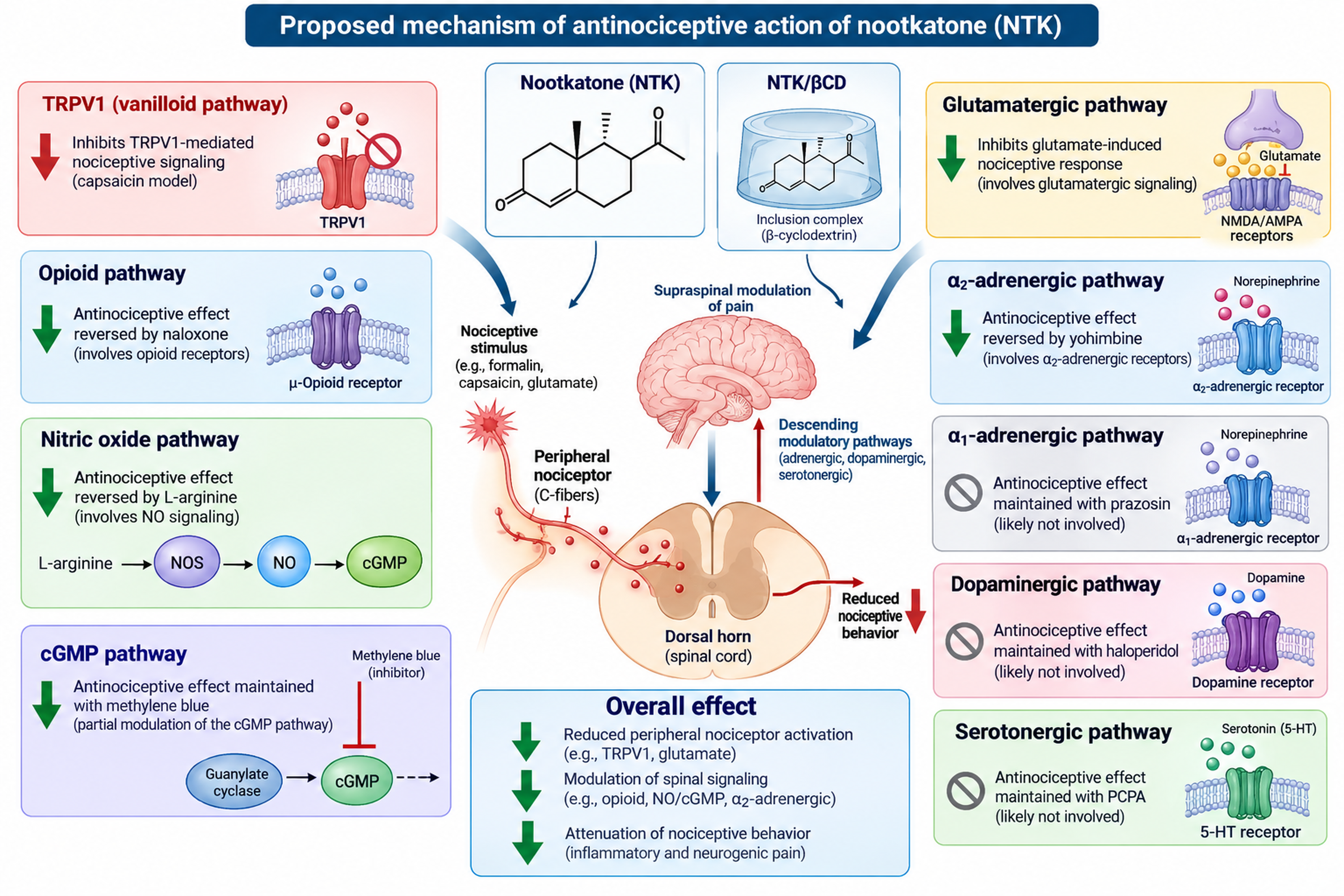
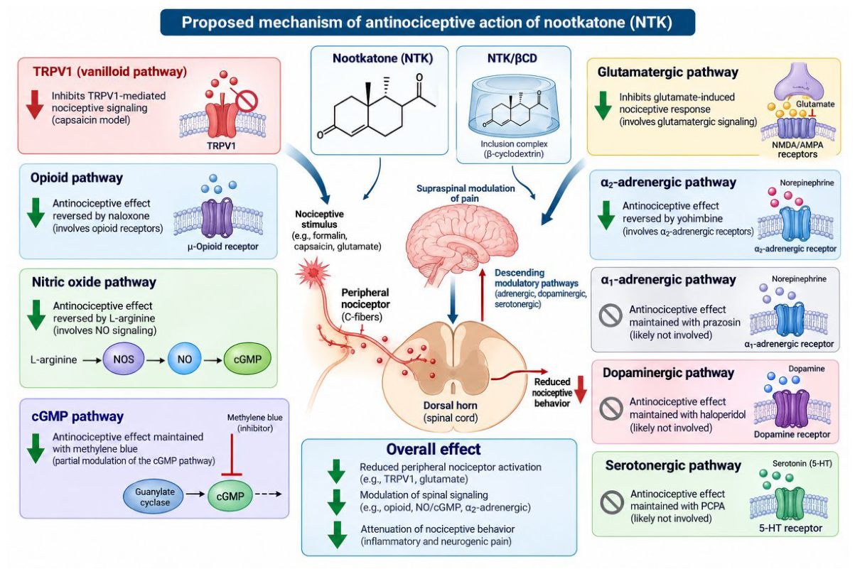
Collectively, these findings demonstrate that the antinociceptive activity of nootkatone (NTK) is mediated by a multimodal pharmacological mechanism involving multiple pain-signaling pathways. Pharmacological antagonism and agonist or substrate modulation reveal the involvement of opioid, nitric oxide (NO)/cyclic guanosine monophosphate (cGMP), vanilloid/TRPV1, glutamatergic, and α2-adrenergic systems in the antinociceptive effects of both free NTK and its β-cyclodextrin inclusion complex (NTK/βCD). In contrast, the persistence of the antinociceptive response after blockade of α1-adrenergic, dopaminergic, and serotonergic signaling indicates that these pathways are unlikely to contribute significantly to the observed effect. The combined modulation of peripheral nociceptor activation and central pain-processing pathways indicates that NTK acts through complementary mechanisms rather than a single molecular target [39]. This study provides the first comprehensive experimental evidence linking NTK-induced antinociception to the specific combination of opioid, NO/cGMP, TRPV1, glutamatergic, and α2-adrenergic mechanisms, thereby expanding the pharmacological understanding of this sesquiterpene. Furthermore, the comparable or enhanced activity observed with NTK/βCD supports cyclodextrin complexation as a promising strategy to improve the pharmacological performance of NTK. An integrated model of these mechanisms is presented in Figure 16.
4. Materials and Methods
4.1. Animals
Swiss mice (Mus musculus), weighing 20–30 g and of both sexes, were used in the study and monitored at the Experimental Animal Facility of the Regional University of Cariri (URCA). The animals were housed in polypropylene cages under controlled environmental conditions (24 ± 2 °C) with a 12-h light/dark cycle and free access to drinking water and standard rodent chow (Labina, Presence®). Before the experiments, the animals were fasted from solid food for 6–8 h. All procedures were performed in accordance with the guidelines for the use of laboratory animals. The study was submitted to and approved by the Ethics Committee on the Use of Animals of the Regional University of Cariri (CONCEA/MCTI/0301902026/PESQUISA/000005/2026).
4.2. Substances and Reagents
The substances used in this study were obtained from Sigma-Aldrich and included formalin (PubChem CID = 712), morphine (5 mg/kg, s.c.; PubChem CID = 5288826), L-NOARG (PubChem CID = 440005), ruthenium red (PubChem CID = 117587625), capsaicin (PubChem CID = 1548943), glutamate (PubChem CID = 23689119), methylene blue (PubChem CID = 6099), clonidine (PubChem CID = 20179), yohimbine (PubChem CID = 6169), L-arginine (PubChem CID: 6322), prazosin hydrochloride (PubChem CID: 4893), and p-chlorophenylalanine (PubChem CID = 46520). Methanol, acetonitrile (LiChrosolv®, ≥99.9%), and ammonium hydroxide (NH4OH) were purchased from Sigma-Aldrich (St. Louis, MO, USA).
The investigated substances, (+)-nootkatone (NTK; PubChem CID = 1268142) and (+)-valencene (PubChem CID = 9855795), as well as the β-cyclodextrin inclusion complex (PubChem CID = 444041), were administered orally at a dose of 10 mg/kg.
4.3. Preparation of the Nootkatone/β-Cyclodextrin Inclusion Complex (NTK/βCD)
The inclusion complex was prepared using the co-evaporation (CE) method according to Kfouri et al. [12]. β-Cyclodextrin (β-CD) and NTK were mechanically homogenized at a 1:1 NTK:β-CD molar ratio. Initially, β-CD (1 g) was dissolved in 25 mL of distilled water and maintained at 70 °C for 10 min. Subsequently, 0.218 g of NTK was dissolved in 10 mL of acetone under mechanical stirring for 3 min. The NTK solution was then gradually added to the β-CD solution and maintained under mechanical stirring at 400 rpm for 24 h at room temperature. After this process, the mixture was stored at −80 °C and then freeze-dried for 72 h. The entire procedure was performed in triplicate, and the resulting material was stored in airtight containers until further chemical analyses.
4.4. Characterization of the NTK/βCD Complex
4.4.1. Gas Chromatography with Flame Ionization Detection (GC/FID)
Nootkatone was diluted in 99.9% dichloromethane (UV/HPLC grade) to prepare a 5,000 ppm stock solution, which was then used to prepare solutions ranging from 100 to 1,000 ppm. The β-cyclodextrin–nootkatone complex was prepared at concentrations of 50 and 500 ppm for quantitative analysis.
Samples and standards were analyzed using a Shimadzu GC-2010 Plus gas chromatograph equipped with a flame ionization detector (FID; Shimadzu Scientific Instruments Inc., Columbia, MD, USA) and fitted with an SH-Rtx-5 fused-silica capillary column (30 m × 0.25 mm I.D.; 0.25 μm film thickness). The temperature program was as follows: 80–180 °C at 4 °C/min, then to 246 °C at 6.6 °C/min, and finally to 280 °C at 3.4 °C/min, with a final hold of 10 min, for a total analysis time of 30 min. Helium was used as the carrier gas at 1.5 mL/min. The injector was operated in split mode (1:15) at 220 °C. The injection volume was 1 μL of the sample or standard solution prepared in dichloromethane. The detector temperature was set at 300 °C.
The linear retention index was determined by injecting a mixture of C8–C40 n-alkanes under the same chromatographic conditions used for the samples, according to [40]. Compound identification was confirmed by comparing the retention indices and mass spectra with those reported in the literature [41]. Quantification was performed by peak integration using the external standard method. Compound concentrations were determined from analytical calibration curves generated using reference standards. The limit of detection (LOD) and limit of quantification (LOQ) were calculated based on the standard deviation of the response and the slope of the calibration curve using three independent analytical curves. LOD and LOQ were calculated as 3.3σ/S and 10σ/S, respectively, where σ is the standard deviation of the response and S is the slope of the calibration curve. Encapsulation efficiency (EE%) was calculated using the following equation:
4.4.2. Scanning Electron Microscopy (SEM)
The morphology of β-CD and the NTK/βCD inclusion complex (1:1) was examined using a scanning electron microscope (SEM; SU3500, Hitachi, Tokyo, Japan). The samples were mounted on aluminum stubs using carbon tape and analyzed under low-vacuum conditions at 50 Pa. The working distance was set to 5.5 mm, and images were acquired using a backscattered electron detector (BSE-3D) at an accelerating voltage of 15 kV.
4.5. In Vivo Models
4.5.1. Evaluation of the Effects of NTK and NTK/βCD on the Central Nervous System (CNS)
Rotarod Test
The apparatus consisted of a rotating rod measuring 2.5 cm in diameter, subdivided into five compartments and rotating at 16 rpm at a height of 25 cm above the base. Mice (n = 3/group) were treated with water for injection (H2O; 0.01 mL/g, p.o.), NTK (10 mg/kg, p.o.), or NTK/βCD (10 mg/kg, p.o.). One hour after treatment, the number of falls and the time spent on the rotating rod were recorded for each animal [42], with modifications.
Open-Field Test
Motor activity was evaluated using a square open-field apparatus measuring 30 cm on each side and divided into nine squares measuring 10 cm × 10 cm at the base. Animals (n = 3/group) were treated with water for injection (H2O; 0.01 mL/g, p.o.), NTK (10 mg/kg, p.o.), or NTK/βCD (10 mg/kg, p.o.). One hour after treatment, the animals were individually placed in the open-field apparatus, allowed to stay for 1 min, and subsequently observed for 5 min. The number of squares crossed was recorded as a measure of exploratory activity [43].
4.5.2. Screening of the Antinociceptive Effects of NTK and NTK/βCD
The antinociceptive effects of NTK and NTK/βCD were evaluated using the following experimental models: 1% acetic acid-induced abdominal writhing, 2.5% formalin test, hot plate test, and tail-flick test. The animals were divided into groups (n = 6) and treated with water for injection (H2O; 0.01 mL/g, p.o.), NTK (10 mg/kg, p.o.), or NTK/βCD (10 mg/kg, p.o.). To investigate the pain-signaling pathways involved in the antinociceptive response, we evaluated the opioid, nitric oxide, vanilloid, glutamatergic, cyclic guanosine monophosphate (cGMP), α1- and α2-adrenergic, dopaminergic, and serotonergic pathways.
Acetic Acid Induced Abdominal Writhing
Mice (n = 6/group) were randomly allocated and treated with water for injection (H2O; 0.01 mL/g, p.o.), NTK (10 mg/kg, p.o.), or NTK/βCD (10 mg/kg, p.o.) 1 h before the intraperitoneal (i.p.) administration of 1% acetic acid. Immediately after acetic acid administration, the number of abdominal writhing responses was recorded for each animal over a 30-min period [43].
Formalin Test
Mice (n = 6/group) were randomly allocated and subsequently treated with water for injection (H2O; 0.01 mL/g, p.o.), NTK (10 mg/kg, p.o.), or NTK/βCD (10 mg/kg, p.o.). One hour later, 2.5% formalin (20 μL) was administered by intraplantar injection into the ventral surface of the right hind paw. Nociceptive behavior, characterized by paw licking and/or biting, was recorded from 0–5 min and 15–30 min after formalin injection [18].
4.5.3. Evaluation of the Central and Peripheral Antinociceptive Effects of NTK and NTK/βCD
Hot Plate Test
Mice (n = 6/group) were subjected to thermal stimulation 24 h and 30 min before testing. Animals showing baseline response latencies outside the 5–15 s range were excluded. After baseline measurements, the animals were treated with water for injection (H2O; 0.01 mL/g, p.o.), NTK (10 mg/kg, p.o.), or NTK/βCD (10 mg/kg, p.o.). Nociceptive responses were evaluated by measuring the response latency (s) until the animals displayed a nociceptive response. Measurements were recorded at 30, 60, 90, 120, and 180 min after treatment [43].
Tail-Flick Test
Mice (n = 6/group) were treated orally with water for injection (H2O; 0.01 mL/g, p.o.), NTK (10 mg/kg, p.o.), or NTK/βCD (10 mg/kg, p.o.). Each animal was placed in an appropriate restrainer, with the dorsal surface of the tail positioned over the thermal stimulus. Heating was applied to the distal third of the tail by focusing a light beam set at 50 °C on the tail. The time, in seconds, required for the animal to withdraw its tail from the light beam was defined as the response latency and was automatically recorded by a timer connected to the apparatus. Latency was measured at baseline, 24 h and 30 min before treatment, and at 30, 60, 120, and 180 min after treatment. A cut-off latency of 10 s was applied to avoid thermal injury to the tail tissue [43].
4.5.4. Pain-Signaling Pathways Involved in the Antinociceptive Effects of NTK and NTK/βCD
Involvement of the Opioid System
Mice (n = 6/group) were divided into eight groups. The first four groups received water for injection (H2O; 0.01 mL/g, p.o.), morphine, an opioid agonist (5 mg/kg, s.c.), NTK (10 mg/kg, p.o.), or NTK/βCD (10 mg/kg, p.o.), respectively. The remaining four groups received naloxone, an opioid antagonist (4 mg/kg, i.p.), 15 min before treatment. One hour after oral administration or 30 min after subcutaneous administration, the animals were evaluated for nociceptive responses using the 2.5% formalin test (first phase: 0–5 min) [44].
Involvement of the L-Arginine/Nitric Oxide/cGMP Pathway
Mice (n = 6/group) were divided into eight groups. The first four groups were treated with water for injection (H2O; 0.01 mL/g, p.o.), L-NOARG, a nitric oxide synthase (NOS) inhibitor (75 mg/kg, i.p.), NTK (10 mg/kg, p.o.), or NTK/βCD (10 mg/kg, p.o.). The remaining four groups received L-arginine, the NOS substrate (600 mg/kg, i.p.), 15 min before treatment. One hour after oral administration or 30 min after intraperitoneal administration, the animals were evaluated using the 2.5% formalin test (first phase: 0–5 min) [28,33].
Involvement of the Vanilloid System
Mice (n = 6/group) were divided into four groups and treated with water for injection (H2O; 0.01 mL/g, p.o.), ruthenium red, a non-selective TRP antagonist (3 mg/kg, i.p.), NTK (10 mg/kg, p.o.), or NTK/βCD (10 mg/kg, p.o.). One hour after oral administration or 30 min after intraperitoneal administration, the animals were evaluated for nociception induced by intraplantar injection of capsaicin (20 μL, 5.2 nmol/paw), a TRPV1 receptor agonist. Nociceptive behavior was assessed for 5 min by recording the time spent licking, biting, or otherwise attending to the injected paw [45].
Involvement of the Glutamatergic System
Mice (n = 6/group) were divided into four groups and treated with water for injection (H2O; 0.01 mL/g, p.o.), ascorbic acid, a neuroprotective/redox modulator agent (100 mg/kg, i.p.), NTK (10 mg/kg, p.o.), or NTK/βCD (10 mg/kg, p.o.). One hour after oral administration or 30 min after intraperitoneal administration, the animals were evaluated for nociception induced by intraplantar injection of buffered glutamate (20 μL, 20 μmol/paw). Nociceptive behavior was assessed for 15 min by recording the time (s) spent licking, biting, or otherwise attending to the injected paw [46,47,48,49].
Involvement of the Cyclic Guanosine Monophosphate (cGMP) Pathway
Mice (n = 6/group) were divided into six groups. The first four groups received water for injection (H2O; 0.01 mL/g, p.o.), methylene blue, a guanylate cyclase inhibitor (20 mg/kg, i.p.), NTK (10 mg/kg, p.o.), or NTK/βCD (10 mg/kg, p.o.). The remaining groups receiving NTK (10 mg/kg, p.o.) or NTK/βCD (10 mg/kg, p.o.) were pretreated with methylene blue 15 min before treatment. One hour after oral administration or 30 min after intraperitoneal administration, the animals were evaluated for nociception induced by intraplantar injection of buffered glutamate (20 μL, 20 μmol/paw) and observed for 15 min. The time in seconds spent licking, biting, or otherwise attending to the injected paw was recorded [45].
Involvement of the α1-Adrenergic Receptor
Mice (n = 6/group) were divided into eight groups. The first four groups were treated with water for injection (H2O; 0.01 mL/g, p.o.), phenylephrine, an α1-adrenergic agonist (10 mg/kg, i.p.), NTK (10 mg/kg, p.o.), or NTK/βCD (10 mg/kg, p.o.). The remaining four groups received prazosin, an α1-adrenergic antagonist (0.15 mg/kg, i.p.), 15 min before treatment. One hour after oral administration or 30 min after intraperitoneal administration, the animals were evaluated using the 2.5% formalin test (first phase: 0–5 min) [50,51]
Involvement of the α2-Adrenergic Receptor
Mice (n = 6/group) were divided into eight groups. The first four groups were treated with water for injection (H2O; 0.01 mL/g, p.o.), clonidine, an α2-adrenergic agonist (0.1 mg/kg, i.p.), NTK (10 mg/kg, p.o.), or NTK/βCD (10 mg/kg, p.o.). The remaining four groups received yohimbine, an α2-adrenergic antagonist (0.15 mg/kg, i.p.), 15 min before treatment. One hour after oral administration or 30 min after intraperitoneal administration, the animals were evaluated using the 2.5% formalin test (first phase: 0–5 min) [52].
Involvement of the Dopaminergic System
Mice (n = 6/group) were divided into six groups. The first four groups received water for injection (H2O; 0.01 mL/g, p.o.), haloperidol (0.2 mg/kg, i.p.), NTK (10 mg/kg, p.o.), or NTK/βCD (10 mg/kg, p.o.). The remaining two groups received haloperidol 15 min before treatment. One hour after oral administration or 30 min after intraperitoneal administration, the animals were evaluated using the 2.5% formalin test (first phase: 0–5 min) [53,54].
Involvement of the Serotonergic Pathways
Mice (n = 6/group) were divided into six groups. The first three groups were treated with p-chlorophenylalanine (PCPA; 100 mg/kg, i.p.), a serotonin synthesis inhibitor, once daily for four consecutive days, whereas the remaining three groups received the negative control, water for injection (H2O; 0.01 mL/g, p.o.), once daily for four consecutive days. Thirty minutes after the final PCPA or control administration, the animals received water for injection (0.1 mL/10 g, p.o.), NTK (10 mg/kg, p.o.), or NTK/βCD (10 mg/kg, p.o.). One hour after treatment, the animals were evaluated using the 2.5% formalin test (first phase: 0–5 min) [55,56,57].
4.6. Statistical Analysis
Data were expressed as mean ± standard error of the mean (SEM). For experiments involving three or more groups with a single assessment, differences among groups were analyzed using one-way analysis of variance (ANOVA), followed by Tukey’s post hoc test. For experiments involving repeated measurements at multiple time points, two-way ANOVA followed by Dunnett’s or Tukey’s post hoc test was used. Statistical analyses were performed using GraphPad Prism version 11.0. Differences were considered statistically significant at p < 0.05.
5. Conclusions
In conclusion, (+)-nootkatone (NTK) and its β-cyclodextrin inclusion complex (NTK/βCD) exhibit significant central and peripheral antinociceptive properties, attenuating both neurogenic and inflammatory pain responses. Pharmacological evaluation indicates that these effects are mediated by a multi-target mechanism involving the opioid, L-arginine/NO/cGMP, glutamatergic, vanilloid (TRPV1) and α2-adrenergic signaling pathways. Crucially, the complexation markedly enhances the relative pharmacological efficiency of the terpene, achieving equivalent antinociceptive efficacy with a substantially lower effective dose of active NTK compared to the uncomplexed molecule. These findings demonstrate that β-cyclodextrin complexation represents an effective biopharmaceutical strategy to optimize oral delivery, highlighting NTKβ/CD as a promising candidate for further translational research in pain management.
Author Contributions
Conceptualization, I.S.A., I.R.A.M. and A.G.W.; methodology, I.S.A., R.T.P., L.Y.S.S., A.B.S., E.S.S., A.O.B.P.B.M., S.A.B., R.H.S.C., J.C.P.S., R.N.M., F.S.M.-R., P.I.D.M.M., A.G.P., F.S.T., I.R.A.M. and A.G.W.; investigation, I.S.A., R.T.P., L.Y.S.S., A.B.S., E.S.S., A.O.B.P.B.M., S.A.B., R.H.S.C., J.C.P.S., R.N.M., F.S.M.-R., P.I.D.M.M., A.G.P., F.S.T., formal analysis, I.S.A., F.S.M.-R., P.I.D.M.M., A.G.P., F.S.T., I.R.A.M. and A.G.W.; data curation, I.S.A. and R.H.S.C.; writing—original draft preparation, I.S.A., L.Y.S.S., R.N.M., F.S.M.-R., P.I.D.M.M., A.G.P., F.S.T., D.P.O., I.R.A.M. and A.G.W.; writing—review and editing, R.N.M., D.P.O., I.R.A.M. and A.G.W.; visualization, I.S.A., R.T.P., L.Y.S.S., A.B.S., E.S.S. and S.A.B.; supervision, R.N.M., I.R.A.M. and A.G.W.; project administration, I.S.A., I.R.A.M. and A.G.W.; funding acquisition, I.R.A.M. and A.G.W. All authors made substantial contributions to the conception, design, acquisition, analysis, or interpretation of data; drafted or critically revised the work for important intellectual content; approved the final version to be published; and agree to be accountable for all aspects of the work in ensuring that questions related to the accuracy or integrity of any part of the work are appropriately investigated and resolved.
Funding
This study receive the financial support from the Coordination for the Improvement of Higher Education Personnel (CAPES); the Cearense Foundation for Supporting Scientific and Technological Development (FUNCAP, finance code BPI); the National Council for Scientific and Technological Development (process 404780/2021-0 and 303438/2021-5 and 303733/2026-8) are research productivity fellows at the National Council for Scientific and Technological Development (CNPq), Brazil.); the National Institute of Science and Technology of the Health Economic–Industrial Complex (iCEIS); and the Financier of Studies and Projects (FINEP, Brazil).
Institutional Review Board Statement
The animal study protocol was approved by tthe Ethics Committee on the Use of Animals at URCA, under license number 00103/2021.1.
Informed Consent Statement
Not applicable.
Acknowledgments
The authors gratefully acknowledge the financial support provided by the Coordination for the Improvement of Higher Education Personnel (CAPES); the Cearense Foundation for Supporting Scientific and Technological Development (FUNCAP, finance code BPI); the National Council for Scientific and Technological Development (process 404780/2021-0 and 303438/2021-5 and 303733/2026-8) are research productivity fellows at the National Council for Scientific and Technological Development (CNPq), Brazil.). The authors would like to thank the financial support pro vided with contribution of National Institute of Science and Technology of the Health Industrial Economic Complex (INCT iCEIS); and the Financier of Studies and Projects (FINEP, Brazil). This work is also a contribution of the National Institute of Science and Technology—Ethnobiology, Bioprospecting, and Nature Conservation (INCT-CNPq/FACEPE), the Excitotoxicity and Neuroprotection (INCT-EN) for the support provided in carrying out this study.
Conflicts of Interest
The authors declare no conflicts of interest.
AI Assistance Disclosure: AI tools were used solely for English language editing [58]. This study complies with the CNPq Scientific Integrity Policy (Portaria No. 2,664/2026), ensuring transparent and responsible use of AI. No AI contributed to study design, data analysis, interpretation, or preparation of scientific content.
AI Assistance Disclosure
AI tools were used solely for English language editing [58]. This study complies with the CNPq Scientific Integrity Policy (Portaria No. 2,664/2026), ensuring transparent and responsible use of AI. No AI contributed to study design, data analysis, interpretation, or preparation of scientific content.
References
- Baral, P.; Udit, S.; Chiu, I.M. Pain and Immunity: Implications for Host Defence. Nat. Rev. Immunol. 2019, 19, 433–447. [Google Scholar] [CrossRef] [PubMed]
- Matsuda, M.; Huh, Y.; Ji, R.R. Roles of Inflammation, Neurogenic Inflammation, and Neuroinflammation in Pain. J. Anesth. 2019, 33, 131–139. [Google Scholar] [CrossRef] [PubMed]
- Arthur, J.; Bruera, E. Balancing Opioid Analgesia with the Risk of Nonmedical Opioid Use in Patients with Cancer. Nat. Rev. Clin. Oncol. 2019, 16, 213–226. [Google Scholar] [CrossRef] [PubMed]
- Cohen, S.P.; Vase, L.; Hooten, W.M. Chronic Pain: An Update on Burden, Best Practices, and New Advances. The Lancet 2021, 397, 2082–2097. [Google Scholar] [CrossRef] [PubMed]
- Atanasov, A.G.; Zotchev, S.B.; Dirsch, V.M.; Orhan, I.E.; Banach, M.; Rollinger, J.M.; Barreca, D.; Weckwerth, W.; Bauer, R.; Bayer, E.A.; et al. Natural Products in Drug Discovery: Advances and Opportunities. Nat. Rev. Drug Discov. 2021, 20, 200–216. [Google Scholar] [CrossRef] [PubMed]
- Newman, D.J.; Cragg, G.M. Natural Products as Sources of New Drugs over the Nearly Four Decades from 01/1981 to 09/2019. J. Nat. Prod. 2020, 83, 770–803. [Google Scholar] [CrossRef] [PubMed]
- Sharifi-Rad, J.; Sureda, A.; Tenore, G.C.; Daglia, M.; Sharifi-Rad, M.; Valussi, M.; Tundis, R.; Sharifi-Rad, M.; Loizzo, M.R.; Ademiluyi, A.O.; et al. Biological Activities of Essential Oils: From Plant Chemoecology to Traditional Healing Systems. Molecules 2017, 22. [Google Scholar] [CrossRef] [PubMed]
- Masyita, A.; Mustika Sari, R.; Dwi Astuti, A.; Yasir, B.; Rahma Rumata, N.; Emran, T. Bin; Nainu, F.; Simal-Gandara, J. Terpenes and Terpenoids as Main Bioactive Compounds of Essential Oils, Their Roles in Human Health and Potential Application as Natural Food Preservatives. Food Chem. X 2022, 13, 100217. [Google Scholar] [CrossRef] [PubMed]
- Park, J.-E.; Park, J.-S.; Leem, Y.-H.; Kim, D.-Y.; Kim, H.-S. NQO1 Mediates the Anti-Inflammatory Effects of Nootkatone in Lipopolysaccharide-Induced Neuroinflammation by Modulating the AMPK Signaling Pathway. Free Radic. Biol. Med. 2021, 164, 354–368. [Google Scholar] [CrossRef] [PubMed]
- Dai, C.; Zhang, X.; Lin, J.; Shen, J. Nootkatone Supplementation Ameliorates Carbon Tetrachloride-Induced Acute Liver Injury via the Inhibition of Oxidative Stress, NF-ΚB Pathways, and the Activation of Nrf2/HO-1 Pathway. Antioxidants 2023, 12, 194. [Google Scholar] [CrossRef] [PubMed]
- Wang, Y.; Wang, M.; Xu, M.; Li, T.; Fan, K.; Yan, T.; Xiao, F.; Bi, K.; Jia, Y. Nootkatone, a Neuroprotective Agent from Alpiniae Oxyphyllae Fructus, Improves Cognitive Impairment in Lipopolysaccharide-Induced Mouse Model of Alzheimer’s Disease. Int. Immunopharmacol. 2018, 62, 77–85. [Google Scholar] [CrossRef] [PubMed]
- Kfoury, M.; Landy, D.; Ruellan, S.; Auezova, L.; Greige-Gerges, H.; Fourmentin, S. Nootkatone Encapsulation by Cyclodextrins: Effect on Water Solubility and Photostability. Food Chem. 2017, 236, 41–48. [Google Scholar] [CrossRef] [PubMed]
- Jansook, P.; Ogawa, N.; Loftsson, T. Cyclodextrins: Structure, Physicochemical Properties and Pharmaceutical Applications. Int. J. Pharm. 2018, 535, 272–284. [Google Scholar] [CrossRef] [PubMed]
- Loftsson, T.; Brewster, M.E. Pharmaceutical Applications of Cyclodextrins: Basic Science and Product Development. J. Pharm. Pharmacol. 2010, 62, 1607–1621. [Google Scholar] [CrossRef] [PubMed]
- Deraedt, R.; Jouquey, S.; Delevallée, F.; Flahaut, M. Release of Prostaglandins E and F in an Algogenic Reaction and Its Inhibition. Eur. J. Pharmacol. 1980, 61, 17–24. [Google Scholar] [CrossRef] [PubMed]
- Le Bars, D.; Gozariu, M.; Cadden, S.W. Animal Models of Nociception. Pharmacol. Rev. 2001, 53, 597–652. [Google Scholar] [CrossRef]
- Yu, S.H.; Kim, H.J.; Jeon, S.Y.; Kim, M.R.; Lee, B.S.; Lee, J.J.; Kim, D.-S.; Lee, Y.C. Anti-Inflammatory and Anti-Nociceptive Activities of Alpinia Oxyphylla Miquel Extracts in Animal Models. J. Ethnopharmacol. 2020, 260, 112985. [Google Scholar] [CrossRef] [PubMed]
- Tjølsen, A.; Berge, O.-G.; Hunskaar, S.; Rosland, J.H.; Hole, K. The Formalin Test: An Evaluation of the Method. Pain 1992, 51, 5–17. [Google Scholar] [CrossRef] [PubMed]
- Ferreira, P.H.; Ferreira, M.L.; Hodges, P.W. Changes in Recruitment of the Abdominal Muscles in People With Low Back Pain. Spine (Phila. Pa. 1976). 2004, 29, 2560–2566. [Google Scholar] [CrossRef] [PubMed]
- Gai, Y.; Bai, C.; Zhang, W.; Xiao, H.; Xu, J.; Hou, J.; Ge, X. Nootkatone Attenuates Airway Inflammation in Asthmatic Mice through Repressing ROS-Induced NLRP3 Inflammasome Activation. Biochem. Cell Biol. 2023, 101, 513–522. [Google Scholar] [CrossRef] [PubMed]
- Rezaee-Asl, M.; Sabour, M.; Nikoui, V.; Ostadhadi, S.; Bakhtiarian, A. The Study of Analgesic Effects of Leonurus Cardiaca L. in Mice by Formalin, Tail Flick and Hot Plate Tests. Int. Sch. Res. Not. 2014, 2014, 1–5. [Google Scholar] [CrossRef] [PubMed]
- Turnaturi, R.; Piana, S.; Spoto, S.; Costanzo, G.; Reina, L.; Pasquinucci, L.; Parenti, C. From Plant to Chemistry: Sources of Antinociceptive Non-Opioid Active Principles for Medicinal Chemistry and Drug Design. Molecules 2024, 29, 815. [Google Scholar] [CrossRef] [PubMed]
- Ängeby Möller, K.; Johansson, B.; Berge, O.G. Assessing Mechanical Allodynia in the Rat Paw with a New Electronic Algometer. J. Neurosci. Methods 1998, 84, 41–47. [Google Scholar] [CrossRef] [PubMed]
- North, R.A. Opioid Receptor Types and Membrane Ion Channels. Trends Neurosci. 1986, 9, 114–117. [Google Scholar] [CrossRef]
- Mansour, A.; Fox, C.A.; Burke, S.; Meng, F.; Thompson, R.C.; Akil, H.; Watson, S.J. Mu, Delta, and Kappa Opioid Receptor MRNA Expression in the Rat CNS: An in Situ Hybridization Study. J. Comp. Neurol. 1994, 350, 412–438. [Google Scholar] [CrossRef] [PubMed]
- Dhawan, B.N.; Cesselin, F.; Raghubir, R.; Reisine, T.; Bradley, P.B.; Portoghese, P.S.; Hamon, M. International Union of Pharmacology. XII. Classification of Opioid Receptors. Pharmacol. Rev. 1996, 48, 567–592. [Google Scholar] [CrossRef]
- Meller, S.T.; Gebhart, G.F. Nitric Oxide (NO) and Nociceptive Processing in the Spinal Cord. Pain 1993, 52, 127–136. [Google Scholar] [CrossRef] [PubMed]
- Schmidtko, A.; Tegeder, I.; Geisslinger, G. No NO, No Pain? The Role of Nitric Oxide and CGMP in Spinal Pain Processing. Trends Neurosci. 2009, 32, 339–346. [Google Scholar] [CrossRef] [PubMed]
- Caterina, M.J.; Schumacher, M.A.; Tominaga, M.; Rosen, T.A.; Levine, J.D.; Julius, D. The Capsaicin Receptor: A Heat-Activated Ion Channel in the Pain Pathway. Nature 1997, 389, 816–824. [Google Scholar] [CrossRef] [PubMed]
- Tominaga, M.; Caterina, M.J.; Malmberg, A.B.; Rosen, T.A.; Gilbert, H.; Skinner, K.; Raumann, B.E.; Basbaum, A.I.; Julius, D. The Cloned Capsaicin Receptor Integrates Multiple Pain-Producing Stimuli. Neuron 1998, 21, 531–543. [Google Scholar] [CrossRef] [PubMed]
- Goel, K.; Singh, T.G.; Mujwar, S. Nootkatone as a Multi-Target Modulator of the PD-L1/PD-1 Signaling in Pancreatic Cancer: Network Pharmacology, Docking, Molecular Dynamics, and DFT Analysis. Naunyn. Schmiedebergs. Arch. Pharmacol. 2026, 399, 15629–15649. [Google Scholar] [CrossRef] [PubMed]
- Liu, B.-L.; Cao, Q.-L.; Zhao, X.; Liu, H.-Z.; Zhang, Y.-Q. Inhibition of TRPV1 by SHP-1 in Nociceptive Primary Sensory Neurons Is Critical in PD-L1 Analgesia. JCI Insight 2020, 5. [Google Scholar] [CrossRef] [PubMed]
- Meller, S.T.; Gebhart, G.F. Nitric Oxide (NO) and Nociceptive Processing in the Spinal Cord. Pain 1993, 52, 127–136. [Google Scholar] [CrossRef] [PubMed]
- Mayer, B.; Brunner, F.; Schmidt, K. Inhibition of Nitric Oxide Synthesis by Methylene Blue. Biochem. Pharmacol. 1993, 45, 367–374. [Google Scholar] [CrossRef] [PubMed]
- Soares, A.C.; Leite, R.; Tatsuo, M.A.K.F.; Duarte, I.D.G. Activation of ATP-Sensitive K+ Channels: Mechanism of Peripheral Antinociceptive Action of the Nitric Oxide Donor, Sodium Nitroprusside. Eur. J. Pharmacol. 2000, 400, 67–71. [Google Scholar] [CrossRef] [PubMed]
- Tanabe, M.; Takasu, K.; Kasuya, N.; Shimizu, S.; Honda, M.; Ono, H. Role of Descending Noradrenergic System and Spinal α 2 -adrenergic Receptors in the Effects of Gabapentin on Thermal and Mechanical Nociception after Partial Nerve Injury in the Mouse. Br. J. Pharmacol. 2005, 144, 703–714. [Google Scholar] [CrossRef] [PubMed]
- Guimarães, A.G.; Quintans, J.S.S.; Quintans-Júnior, L.J. Monoterpenes with Analgesic Activity—A Systematic Review. Phyther. Res. 2013, 27, 1–15. [Google Scholar] [CrossRef] [PubMed]
- Carneiro, S.; Costa Duarte, F.; Heimfarth, L.; Siqueira Quintans, J.; Quintans-Júnior, L.; Veiga Júnior, V.; Neves de Lima, Á. Cyclodextrin–Drug Inclusion Complexes: In Vivo and In Vitro Approaches. Int. J. Mol. Sci. 2019, 20, 642. [Google Scholar] [CrossRef] [PubMed]
- Assis, D.B.; Aragão Neto, H. de C.; da Fonsêca, D.V.; de Andrade, H.H.N.; Braga, R.M.; Badr, N.; Maia, M. dos S.; Castro, R.D.; Scotti, L.; Scotti, M.T.; et al. Antinociceptive Activity of Chemical Components of Essential Oils That Involves Docking Studies: A Review. Front. Pharmacol. 2020, 11. [Google Scholar] [CrossRef] [PubMed]
- van Den Dool, H.; Kratz, P. A Generalization of the Retention Index System Including Linear Temperature Programmed Gas—Liquid Partition Chromatography. J. Chromatogr. A 1963, 11, 463–471. [Google Scholar] [CrossRef] [PubMed]
- ROBERT P. ADAMS Identification of Essential Oil Components by Gas Chromatography/Quadrupole Mass Spectroscopy.; 4.1.; 2017;
- Dunham, N.W.; Miya, T.S. A Note on a Simple Apparatus for Detecting Neurological Deficit in Rats and Mice**College of Pharmacy, University of Nebraska, Lincoln 8. J. Am. Pharm. Assoc. (Scientific ed.) 1957, 46, 208–209. [Google Scholar] [CrossRef] [PubMed]
- Lapa, A.J.; Souccar, C.; Lima-Landman, M.T.R.; Castro, M.S.; Lima, T.C.M. Métodos de Avaliação Da Atividade Farmacológica de Plantas Medicinais. Soc. Bras. Plan. Med. 2003, 64–66. [Google Scholar]
- Stevenson, G.W.; Bilsky, E.J.; Negus, S.S. Targeting Pain-Suppressed Behaviors in Preclinical Assays of Pain and Analgesia: Effects of Morphine on Acetic Acid-Suppressed Feeding in C57BL/6J Mice. J. Pain 2006, 7, 408–416. [Google Scholar] [CrossRef] [PubMed]
- Beirith, A.; Santos, A.R.S.; Calixto, J.B. Mechanisms Underlying the Nociception and Paw Oedema Caused by Injection of Glutamate into the Mouse Paw. Brain Res. 2002, 924, 219–228. [Google Scholar] [CrossRef] [PubMed]
- Majewska, M.D.; Bell, J.A.; London, E.D. Regulation of the NMDA Receptor by Redox Phenomena: Inhibitory Role of Ascorbate. Brain Res. 1990, 537, 328–332. [Google Scholar] [CrossRef] [PubMed]
- Li, P.; Zhuo, M. Silent Glutamatergic Synapses and Nociception in Mammalian Spinal Cord. Nature 1998, 393, 695–698. [Google Scholar] [CrossRef] [PubMed]
- Lam, D.K.; Sessle, B.J.; Cairns, B.E.; Hu, J.W. Neural Mechanisms of Temporomandibular Joint and Masticatory Muscle Pain: A Possible Role for Peripheral Glutamate Receptor Mechanisms. Pain Res. Manag. 2005, 10, 145–152. [Google Scholar] [CrossRef] [PubMed]
- Zeraati, F.; Araghchian, M.; Farjoo, M.H. Ascorbic Acid Interaction With Analgesic Effect of Morphine and Tramadol in Mice. Anesth. Pain Med. 2014, 4. [Google Scholar] [CrossRef] [PubMed]
- Sato, J.; Perl, E.R. Adrenergic Excitation of Cutaneous Pain Receptors Induced by Peripheral Nerve Injury. Science (1979) 1991, 251, 1608–1610. [Google Scholar] [CrossRef] [PubMed]
- Choi, H.J.; Lee, J.H.; Jung, Y.S. (+)-Nootkatone Inhibits Tumor Necrosis Factor α/Interferon γ-Induced Production of Chemokines in HaCaT Cells. Biochem. Biophys. Res. Commun. 2014, 447, 278–284. [Google Scholar] [CrossRef] [PubMed]
- Kanui, T.I.; Tjølsen, A.; Lund, A.; Mjellem-Joly, N.; Hole, K. Antinociceptive Effects of Intrathecal Administration of α-Adrenoceptor Antagonists and Clonidine in the Formalin Test in the Mouse. Neuropharmacology 1993, 32, 367–371. [Google Scholar] [CrossRef] [PubMed]
- Wood, P.B. Mesolimbic Dopaminergic Mechanisms and Pain Control. Pain 2006, 120, 230–234. [Google Scholar] [CrossRef] [PubMed]
- Finan, P.H.; Smith, M.T. The Comorbidity of Insomnia, Chronic Pain, and Depression: Dopamine as a Putative Mechanism. Sleep Med. Rev. 2013, 17, 173–183. [Google Scholar] [CrossRef] [PubMed]
- Maleki, N.; Nayebi, A.M.; Garjani, A. Effects of Central and Peripheral Depletion of Serotonergic System on Carrageenan-Induced Paw Oedema. Int. Immunopharmacol. 2005, 5, 1723–1730. [Google Scholar] [CrossRef] [PubMed]
- Messing, R.B.; Lytle, L.D. Serotonin-Containing Neurons: Their Possible Role in Pain and Analgesia. Pain 1977, 4, 1–21. [Google Scholar] [CrossRef] [PubMed]
- Suzuki, R.; Rygh, L.J.; Dickenson, A.H. Bad News from the Brain: Descending 5-HT Pathways That Control Spinal Pain Processing. Trends Pharmacol. Sci. 2004, 25, 613–617. [Google Scholar] [CrossRef] [PubMed]
- Santos, C.M.D.; Gois, J.P. Embrace AI to Break down Barriers in Publishing for People Who Aren’t Fluent in English. Nature 2024, 627, 271–271. [Google Scholar] [CrossRef] [PubMed]
Figure 1.
GC/FID profile of the nootkatone standard (A) and the cyclodextrin–nootkatone inclusion complex (B). Nootkatone standard (retention time, RT: 16.365 min). Calibration curve: y = 1,179.1x − 59.616 (R2 = 0.9977).
Figure 1.
GC/FID profile of the nootkatone standard (A) and the cyclodextrin–nootkatone inclusion complex (B). Nootkatone standard (retention time, RT: 16.365 min). Calibration curve: y = 1,179.1x − 59.616 (R2 = 0.9977).
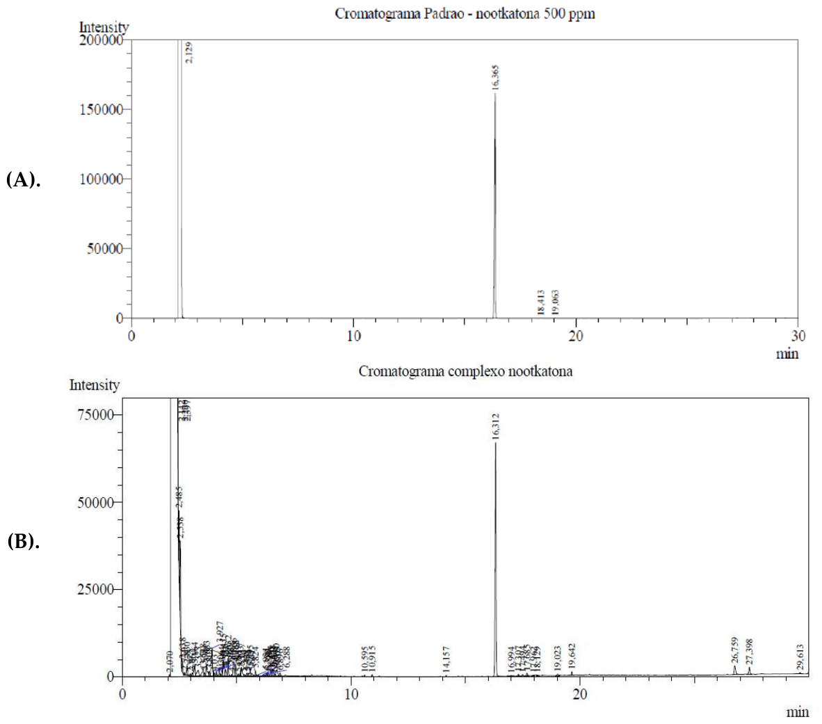
Figure 2.
Scanning electron microscopy (SEM) analysis of β-cyclodextrin (β-CD) and the NTK/β-CD inclusion complex. Red boxes indicate the crystalline morphology of β-CD, whereas green boxes highlight the morphology of the NTK/β-CD complex. Images were acquired at different magnifications, with scale bars corresponding to 500 and 100 μm.
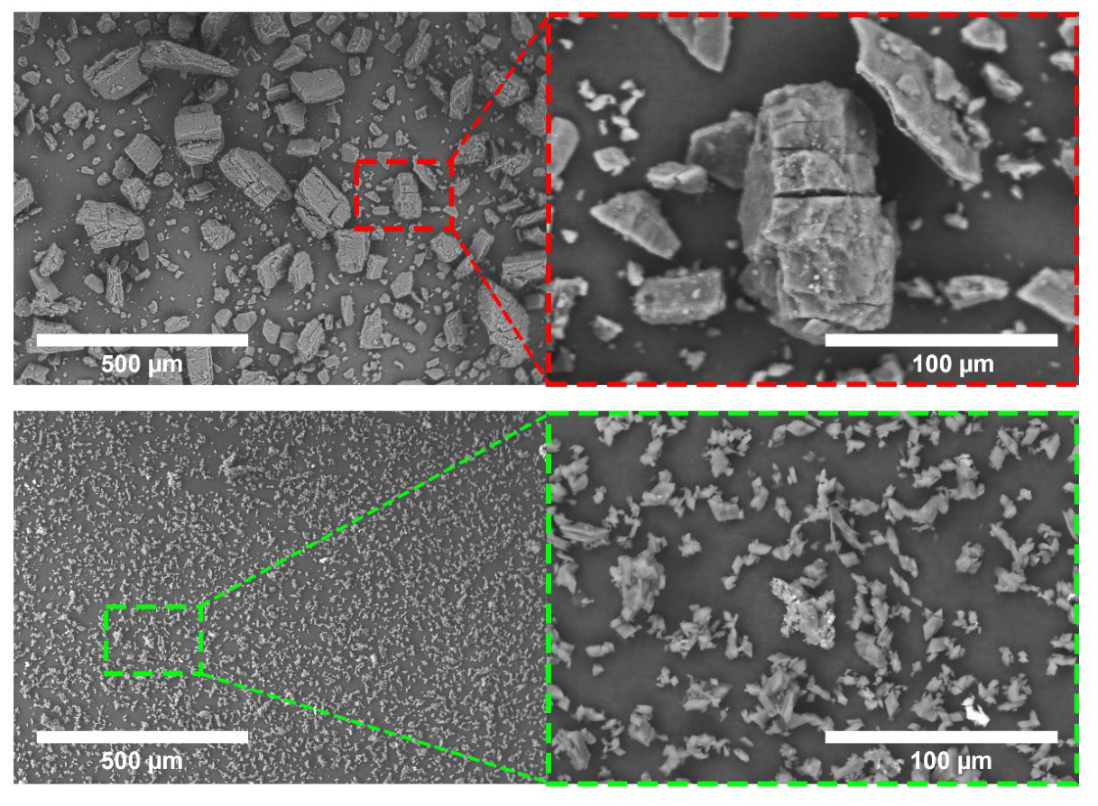
Figure 3.
Antinociceptive effects of NTK and NTK/βCD (10 mg/kg, p.o.) in the acetic acid-induced abdominal writhing test. Values represent the arithmetic mean ± standard error of the mean (SEM) (n = 6/group). One-way ANOVA followed by Tukey’s post hoc test. (A = *p < 0.5 compared with the negative control group).
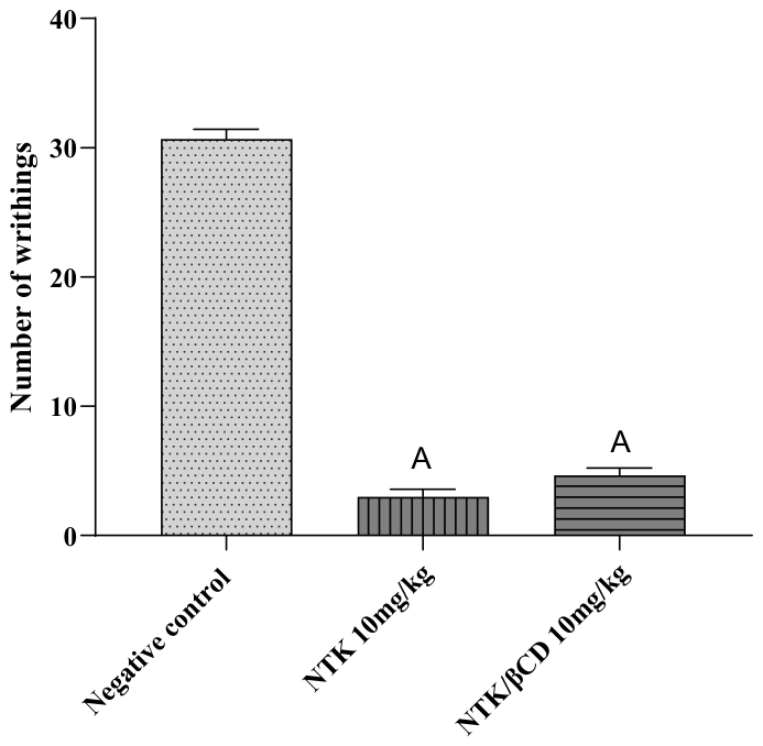
Figure 4.
Antinociceptive effects of NTK and NTK/βCD in the formalin test: A) first phase (neurogenic pain) and B) second phase (inflammatory pain). Values represent the mean ± standard error of the mean (SEM) (n = 6/group). One-way ANOVA followed by Tukey’s post hoc test (A = *p < 0.5 compared with the negative control group).
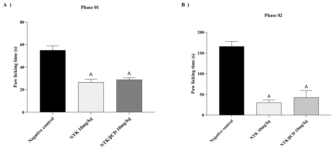
Figure 5.
Antinociceptive effects of NTK and NTK/βCD (10 mg/kg, p.o.) in the hot plate test. Values represent the mean ± standard error of the mean (SEM) (n = 6/group). Two-way ANOVA followed by Tukey’s post hoc test (A = *p < 0.5 compared with the negative control group).
Figure 5.
Antinociceptive effects of NTK and NTK/βCD (10 mg/kg, p.o.) in the hot plate test. Values represent the mean ± standard error of the mean (SEM) (n = 6/group). Two-way ANOVA followed by Tukey’s post hoc test (A = *p < 0.5 compared with the negative control group).
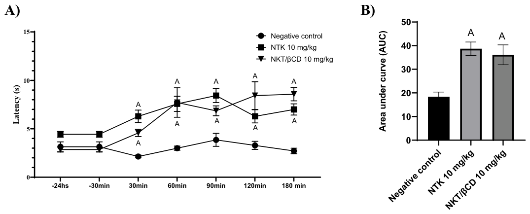
Figure 6.
Antinociceptive effects of NTK and NTK/βCD (10 mg/kg, p.o.) in the tail-flick test (A and B). Values represent the mean ± standard error of the mean (SEM) (n = 6/group). Two-way ANOVA followed by Tukey’s post hoc test (A = *p < 0.5 compared with the negative control group).
Figure 6.
Antinociceptive effects of NTK and NTK/βCD (10 mg/kg, p.o.) in the tail-flick test (A and B). Values represent the mean ± standard error of the mean (SEM) (n = 6/group). Two-way ANOVA followed by Tukey’s post hoc test (A = *p < 0.5 compared with the negative control group).
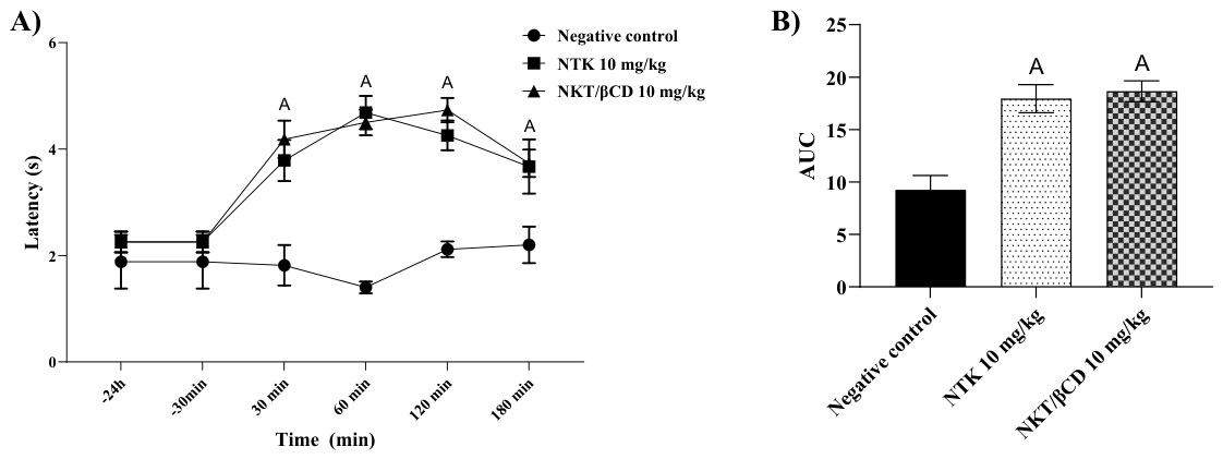
Figure 7.
Antinociceptive response of NTK and NTK/βCD (10 mg/kg, p.o.) mediated by the opioid pathway. Values represent the mean ± standard error of the mean (SEM) (n = 6/group). One-way ANOVA followed by Tukey’s post hoc test (A = *p < 0.5 compared with negative control group), (B = *p < 0.5 compared with naloxone+negative control group), (C = *p < 0.5 compared with Morphine group), (D = *p < 0.5 compared with naloxone+morphine group), (E = *p < 0.5 compared with NKT 10mg/kg group), (F = *p < 0.5 compared with naloxone+NKT group), (G = *p < 0.5 compared with NTK/βCD 10mg/kg group).
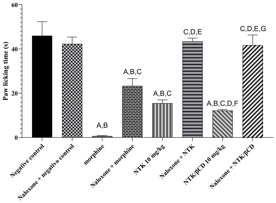
Figure 8.
Antinociceptive response of NTK and NTK/βCD (10 mg/kg, p.o.) involving the nitric oxide pathway. Values represent the mean ± standard error of the mean (SEM) (n = 6/group). One-way ANOVA followed by Tukey’s post hoc test (A = *p < 0.5 compared with negative control group), (B = *p < 0.5 compared with L-Arginine+negative control group), (C = *p < 0.5 compared with L-NOARG group), (D = *p < 0.5 compared with L-Arginine+L-NOARG), (E = *p < 0.5 compared with NKT 10mg/kg group), (F = *p < 0.5 compared with L-Arginine+NKT group), (G = *p < 0.5 compared with NTK/βCD 10mg/kg group).
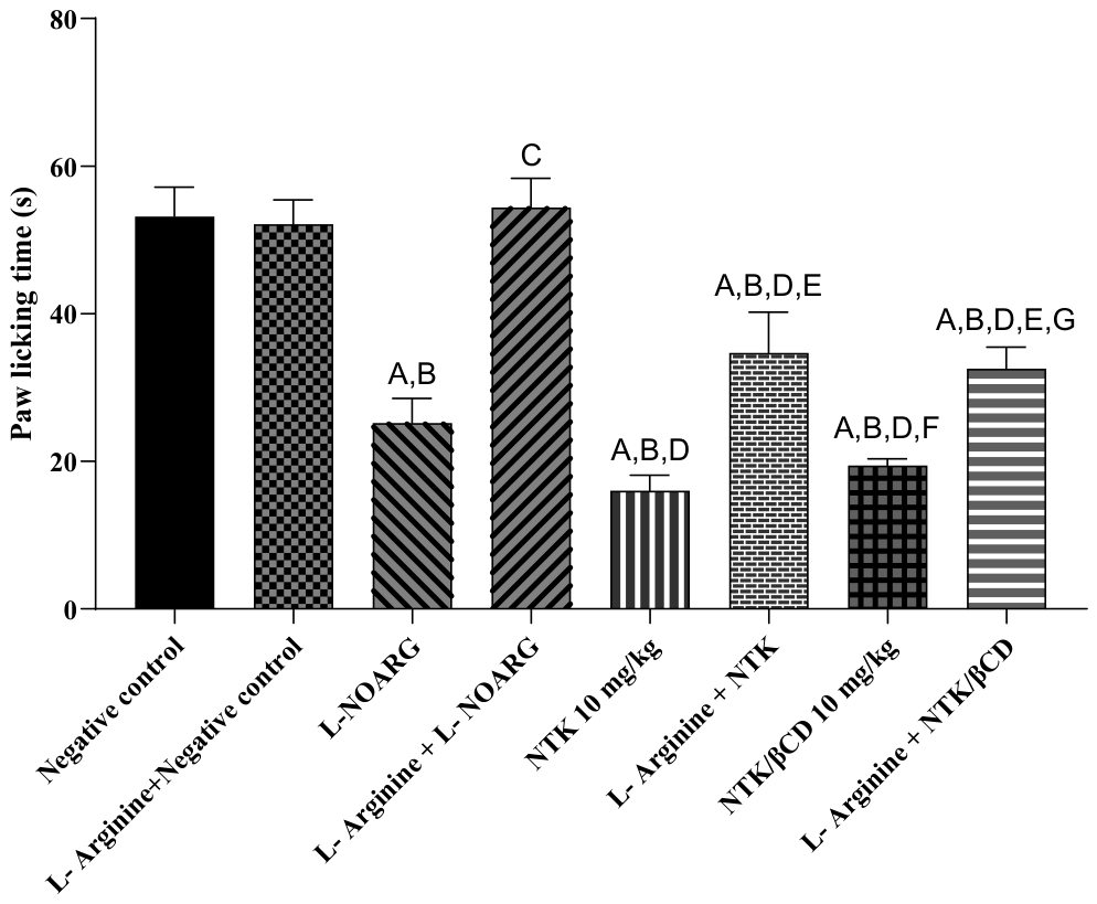
Figure 9.
Antinociceptive response of NTK and NTK/βCD (10 mg/kg, p.o.) involving the vanilloid pathway. Values represent the mean ± standard error of the mean (SEM) (n = 6/group). One-way ANOVA followed by Tukey’s post hoc test (A = *p < 0.5 compared with negative control group), (B = *p < 0.5 compared with Ruthenium red group).
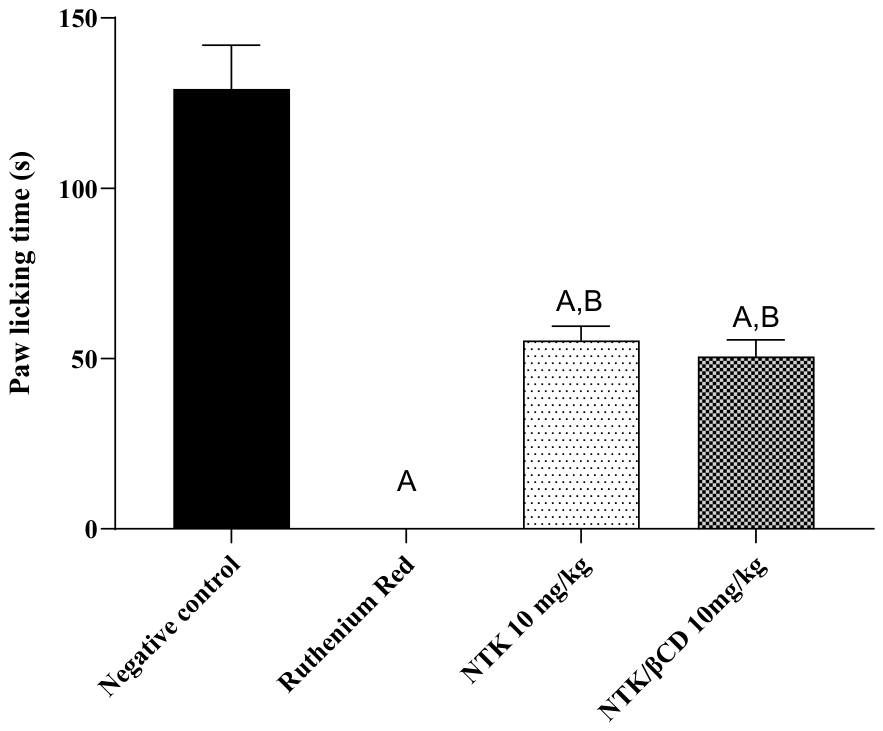
Figure 10.
Antinociceptive response of NTK and NTK/βCD (10 mg/kg, p.o.) involving the glutamatergic system. Values represent the mean ± standard error of the mean (SEM) (n = 6/group). One-way ANOVA followed by Tukey’s post hoc test (A = *p < 0.5 compared with negative control group), (B = *p < 0.5 compared with ascorbic acid group).
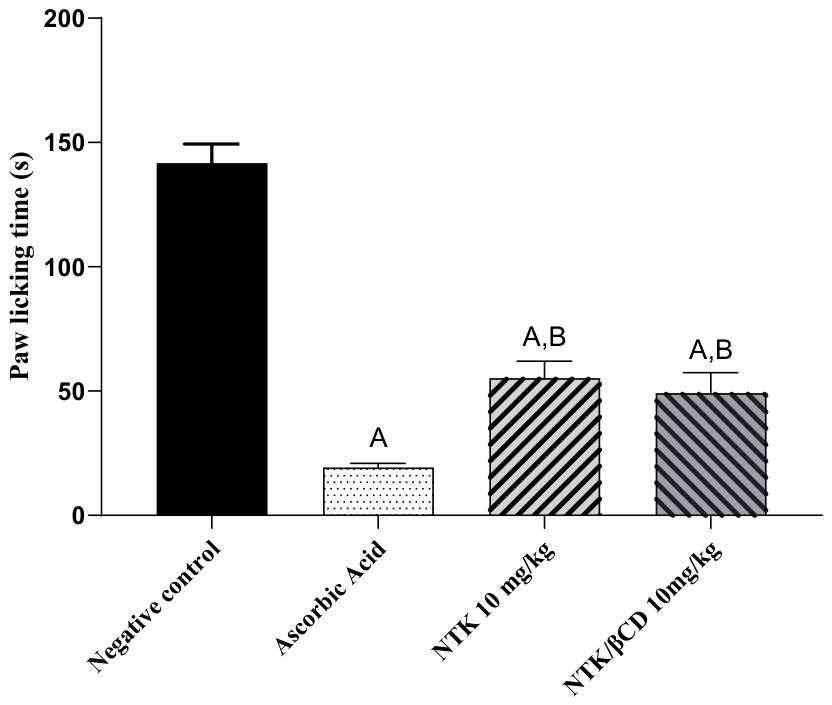
Figure 11.
Antinociceptive response of NTK and NTK/βCD (10 mg/kg, p.o.) involving the cyclic guanosine monophosphate (cGMP) pathway. Values represent the mean ± standard error of the mean (SEM) (n = 6/group). One-way ANOVA followed by Tukey’s post hoc test (A = *p < 0.5 compared with negative control group), (B = *p < 0.5 compared with Methylene blue group).
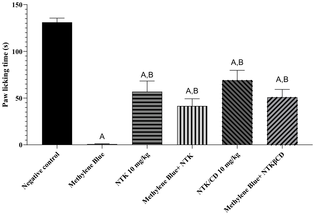
Figure 12.
Antinociceptive response of NTK and NTK/βCD (10 mg/kg, p.o.) involving the α1-adrenergic system. Values represent the mean ± standard error of the mean (SEM) (n = 6/group). One-way ANOVA followed by Tukey’s post hoc test (A = *p < 0.5 compared with negative control group), (B = *p < 0.5 compared with Prazosin+negative control group), (C = *p < 0.5 compared with Phenylephrine group), (D = *p < 0.5 compared with Prazosin+Phenylephrine group), (E = *p < 0.5 compared with NKT 10mg/kg group), (F = *p < 0.5 compared with Prazosin+NKT group), (G = *p < 0.5 compared with NTK/βCD 10mg/kg group).
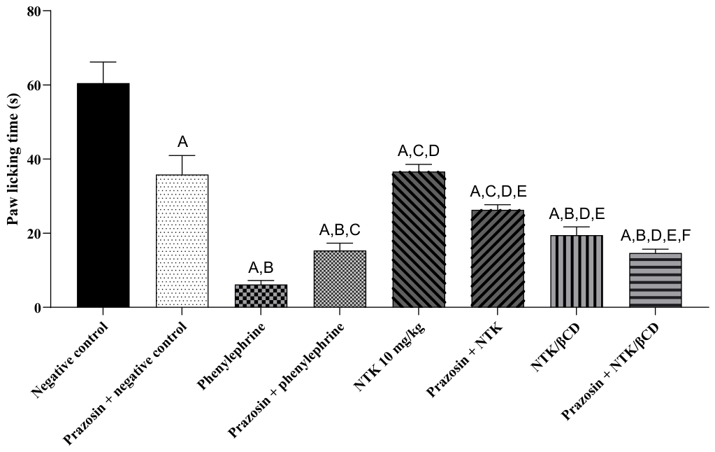
Figure 13.
Antinociceptive response of NTK and NTK/βCD (10 mg/kg, p.o.) involving the α2-adrenergic system. Values represent the mean ± standard error of the mean (SEM) (n = 6/group). One-way ANOVA followed by Tukey’s post hoc test (A = *p < 0.5 compared with negative control group), (B = *p < 0.5 compared with Yohimbine+negative control group), (C = *p < 0.5 compared with Clonidine group), (D = *p < 0.5 compared with Yohimbine+Clonidine group), (E = *p < 0.5 compared with NKT 10mg/kg group), (F = *p < 0.5 compared with Yohimbine+NKT group), (G = *p < 0.5 compared with NTK/βCD 10mg/kg group).
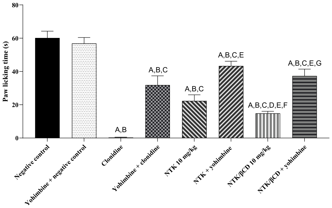
Figure 14.
Antinociceptive response of NTK and NTK/βCD (10 mg/kg, p.o.) involving the dopaminergic system. Values represent the mean ± standard error of the mean (SEM) (n = 6/group). One-way ANOVA followed by Tukey’s post hoc test (A = *p < 0.5 compared with negative control group).
Figure 14.
Antinociceptive response of NTK and NTK/βCD (10 mg/kg, p.o.) involving the dopaminergic system. Values represent the mean ± standard error of the mean (SEM) (n = 6/group). One-way ANOVA followed by Tukey’s post hoc test (A = *p < 0.5 compared with negative control group).
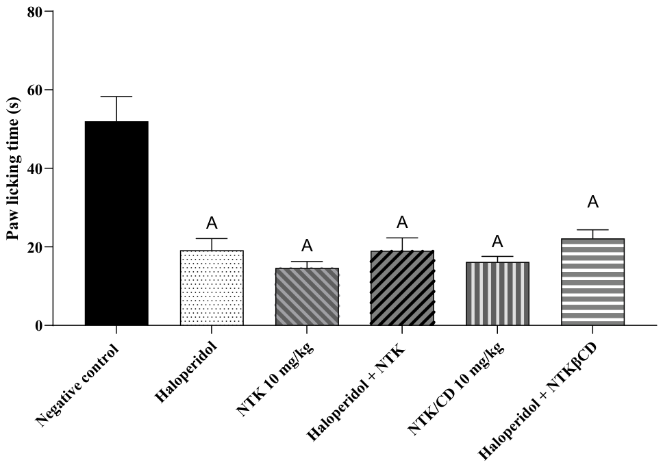
Figure 15.
Antinociceptive response of NTK and NTK/βCD (10 mg/kg, p.o.) involving the serotonergic system. Values represent the mean ± standard error of the mean (SEM) (n = 6/group). One-way ANOVA followed by Tukey’s post hoc test (A = *p < 0.5 compared with negative control group), (B = *p < 0.5 compared with PCPA+negative control group).
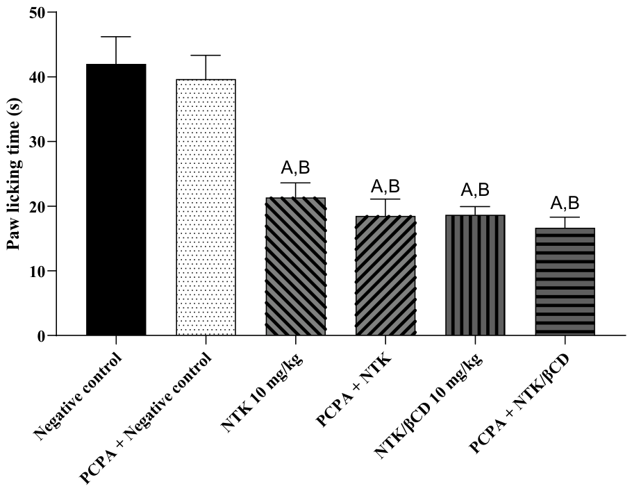
Figure 16.
Proposed multimodal mechanism underlying the antinociceptive effects of nootkatone (NTK) and its β-cyclodextrin inclusion complex (NTK/βCD). The experimental findings indicate the involvement of opioid, nitric oxide (NO)/cyclic guanosine monophosphate (cGMP), transient receptor potential vanilloid 1 (TRPV1), glutamatergic, and α2-adrenergic signaling pathways in NTK- and NTK/βCD-mediated antinociception. Opioid involvement was supported by reversal of the antinociceptive effect with naloxone, whereas modulation of the NO pathway was supported by reversal with L-arginine. Reduced nociceptive responses to capsaicin and glutamate indicate modulation of TRPV1-mediated and glutamatergic signaling, respectively. The maintenance of antinociception after guanylate cyclase inhibition suggests partial modulation of the cGMP pathway, potentially downstream of NO signaling. Reversal by yohimbine supports the involvement of α2-adrenergic receptors. Conversely, the persistence of the antinociceptive effect after α1-adrenergic, dopaminergic, or serotonergic blockade suggests that these pathways are unlikely to be primarily involved. Collectively, these findings support a multimodal mechanism through which NTK and NTK/βCD attenuate peripheral and spinal nociceptive signaling, ultimately reducing pain-related behavior.
Disclaimer/Publisher’s Note: The statements, opinions and data contained in all publications are solely those of the individual author(s) and contributor(s) and not of MDPI and/or the editor(s). MDPI and/or the editor(s) disclaim responsibility for any injury to people or property resulting from any ideas, methods, instructions or products referred to in the content. |
© 2026 by the authors. Licensee MDPI, Basel, Switzerland. This article is an open access article distributed under the terms and conditions of the Creative Commons Attribution (CC BY) license.
Copyright: This open access article is published under a Creative Commons CC BY 4.0 license, which permit the free download, distribution, and reuse, provided that the author and preprint are cited in any reuse.