

Submitted:
12 October 2026
Posted:
14 October 2026
You are already at the latest version
Abstract
Pulmonary alveolar microlithiasis (PAM) is a rare autosomal recessive lung disease in which calcium phosphate microliths fill the alveoli. Although the cause of it remained unknown for some time, more recently loss-of-function variants in SLC34A2, which encodes the sodium-phosphate cotransporter NPT2b, explain nearly all genetically tested cases; failure to clear phosphate from the alveolar lining fluid leads to microlith formation, and an epithelial Npt2b-knockout mouse reproduces the human phenotype. Diagnosis now rests mainly on characteristic high-resolution CT and, increasingly, on genotyping rather than lung biopsy. The protracted course has been described in some series of cases, with death from respiratory failure. No drug has proven efficacy; lung transplantation is the only established therapy, and no allograft recurrence has been documented. Preclinical data support phosphate restriction, EDTA lavage and gene-directed approaches. However, controlled trials with more genotype-phenotype correlation and prospective natural-history data are still needed.
Keywords:
pulmonary alveolar microlithiasis
; SLC34A2
; NPT2b
; calcium phosphate
; lung transplantation
; ul-tra-rare lung disease
Introduction
Pulmonary alveolar microlithiasis is a rare disease of unknown cause when Moran et al. reported their clinicopathologic and chemical analysis of seven cases in 1997 [1]. Their patients were six women and one man aged 19 to 70 years (mean 44.5). Every patient had diffused bilateral opacities on chest radiography, histology showed laminated calcospherites 250 to 750 µm in diameter filling the alveoli, and chemical analysis identified calcium and phosphorus as the principal components. Five patients died of respiratory failure 5 to 41 years after diagnosis, which established the disease as a protracted one [1].
That paper ended by calling for research into the etiology and for a prospective study. Both have since been partly answered. Homozygosity mapping and candidate-gene work identified biallelic mutations in SLC34A2 as the cause in 2006 and 2007 [3,4]. By December 2014, 1,022 cases had been reported from 65 countries [2]. A conditional knockout mouse now reproduces the human phenotype [11], and a growing body of case reports documents outcomes after lung transplantation [8].
The aim of this review is to reassess the previous findings against newer evidence, domain by domain, and to identify what remains unresolved. Because the disease is so rare, much of the evidence consists of case reports and small series, and the strength of each conclusion is stated accordingly.
Historical Background and Epidemiology
The earliest description is attributed to Malpighi, but Harbitz introduced the disease to the American literature in 1918 as “extensive calcification of the lungs” [5], and Puhr named it Mikrolithiasis alveolaris pulmonum in 1933 [6]. Sosman’s 1957 monograph on familial occurrence provided the first detailed description of the disease [7,12]. Moran et al. noted that earlier cases may have been grouped with pulmonary corpora amylacea [1].
The largest census of published cases, by Castellana and colleagues, counted 1,022 patients through December 2014 [2]. The disease has been reported on nearly every continent, without a consistent sex predilection, although Turkey has contributed the largest share of reported cases, followed by Italy and the United States in earlier published tallies. Consanguinity is common in reported families, in keeping with recessive inheritance [3]. A 2020 review emphasised that most new cases arise without a known family history [8].
Because many patients are asymptomatic and the diagnosis is often incidental, true prevalence is unknown. The 2023 systematic review of genetic variants concluded that PAM is probably under-reported because of poor recognition, misdiagnosis and mild presentations [9]. The 1997 series is consistent with this: two of its seven patients had a disease duration that could only be estimated, and the youngest, a 19-year-old woman, had probably been affected for years [1].
Genetics and Molecular Pathogenesis
The gene. In 2006, Corut et al. mapped the PAM locus to chromosome 4p15 by homozygosity mapping and identified SLC34A2 as the responsible gene, concluding that PAM is a recessive monogenic disease with full penetrance [3]. Huqun et al. confirmed the association in Japanese patients in 2007 [4]. SLC34A2 encodes NPT2b, a sodium-dependent phosphate cotransporter expressed on the apical surface of alveolar type II cells; it is also present in the small intestine, kidney and other tissues [8,9].
Variant spectrum. The 2023 systematic review found 34 allelic variants in at least 68 patients [9]. Most are homozygous, a few are compound heterozygous, and roughly half are nonsense or frameshift variants predicted to truncate the protein or trigger mRNA decay [9]. The 2020 review counted at least 27 known mutations, so the catalogue has grown by about a quarter in three years [8]. New variants continue to be reported, including a large deletion, c.524-18_559del, in an infant [13] and two novel compound heterozygous variants in a child in 2026 [14]. Available data do not support a genotype-phenotype correlation [2,15].
Functional evidence. Most reported variants have never been functionally tested. In a 2022 study, four variants were expressed in Xenopus laevis oocytes and phosphate uptake was measured [10]. Notably, the in-frame single-residue deletion Thr468del was synthesised and reached the oocyte membrane, yet its phosphate transport did not differ from control oocytes [10]. Full-length, correctly localised protein is therefore not proof of function, and non-truncating variants can be pathogenic.
Mechanism. The accepted model is that loss of NPT2b prevents alveolar type II cells from clearing inorganic phosphate from the alveolar lining fluid. Phosphate, which is released continuously as surfactant phospholipids are catabolised, accumulates, binds calcium and crystallises as hydroxyapatite microliths [8,15]. This resolves the etiologic hypotheses listed in 1997 (carbonic anhydrase defects, abnormal calcium or phosphorus metabolism, immune abnormalities) [1]: systemic calcium and phosphorus metabolism is normal, and the defect is local.
The mouse model. Saito et al. deleted Npt2b in the lung epithelium using a Shh-Cre driver [11]. The mice developed diffuse microliths, radiographic opacification, restrictive physiology, inflammation, fibrosis and an unexpected alveolar phospholipidosis [11]. Serum MCP-1 and SP-D were elevated in mice and confirmed in patient serum, identifying them as candidate biomarkers [11]. When microliths were transferred into the lungs of normal mice, they provoked macrophage-rich inflammation that resolved over a month as the stones cleared, suggesting that microliths can be removed [11]. Earlier human work had already shown that serum SP-A and SP-D track disease progression [12].
Clinical Presentation
Clinical-radiological dissociation. The hallmark of PAM is a striking gap between dramatic imaging and mild or absent symptoms [1,13]. Moran et al. made the same observation: their patients presented with shortness of breath or a gradual decline in exercise tolerance despite diffuse bilateral opacities [1]. Later reviews describe incremental exertional dyspnea, desaturation in young adulthood and respiratory insufficiency by late middle age [12]. Other reported features include dry cough, pleuritic chest pain, hemoptysis, crackles, clubbing and, late in the disease, pulmonary hypertension and cor pulmonale [8,15].
Age at onset. Most patients are diagnosed between birth and about 40 years, often incidentally [8,15]. Onset in infancy is rare. One reported infant had tachypnea, retractions, crackles and hypoxia from two months of age, and PAM was confirmed by lung biopsy and genotyping [13]. Children are often misattributed to recurrent pneumonia, which delays diagnosis [14].
Familial disease. The 1997 authors reviewed familial clusters, including four affected sisters in one Colombian family [16] and premature twins [17], and speculated about an inborn error of metabolism with variable penetrance [1]. Genetics has confirmed this in essence: sibling clusters, often from consanguineous marriages, reflect biallelic SLC34A2 variants [3]. The lack of affected parents in most pedigrees is what recessive inheritance predicts. Sporadic cases in the absence of family history are nonetheless the majority [8].
Extrapulmonary manifestations. Corut et al. reported an association with testicular microlithiasis [3]. Seminal vesicle microlithiasis with severe oligoasthenospermia has been described in a sporadic case [18]. Because NPT2b is expressed outside the lung, extrapulmonary calcification is biologically plausible. A study cited in the 2020 review found heterozygous SLC34A2 variants in two of 15 men with diffuse testicular microlithiasis, though their pathogenicity was unclear [8]. Coetzee’s 1970 report of microliths in the sympathetic ganglia and gonads, cited by Moran et al., is an early hint of the same phenomenon [1].
Imaging
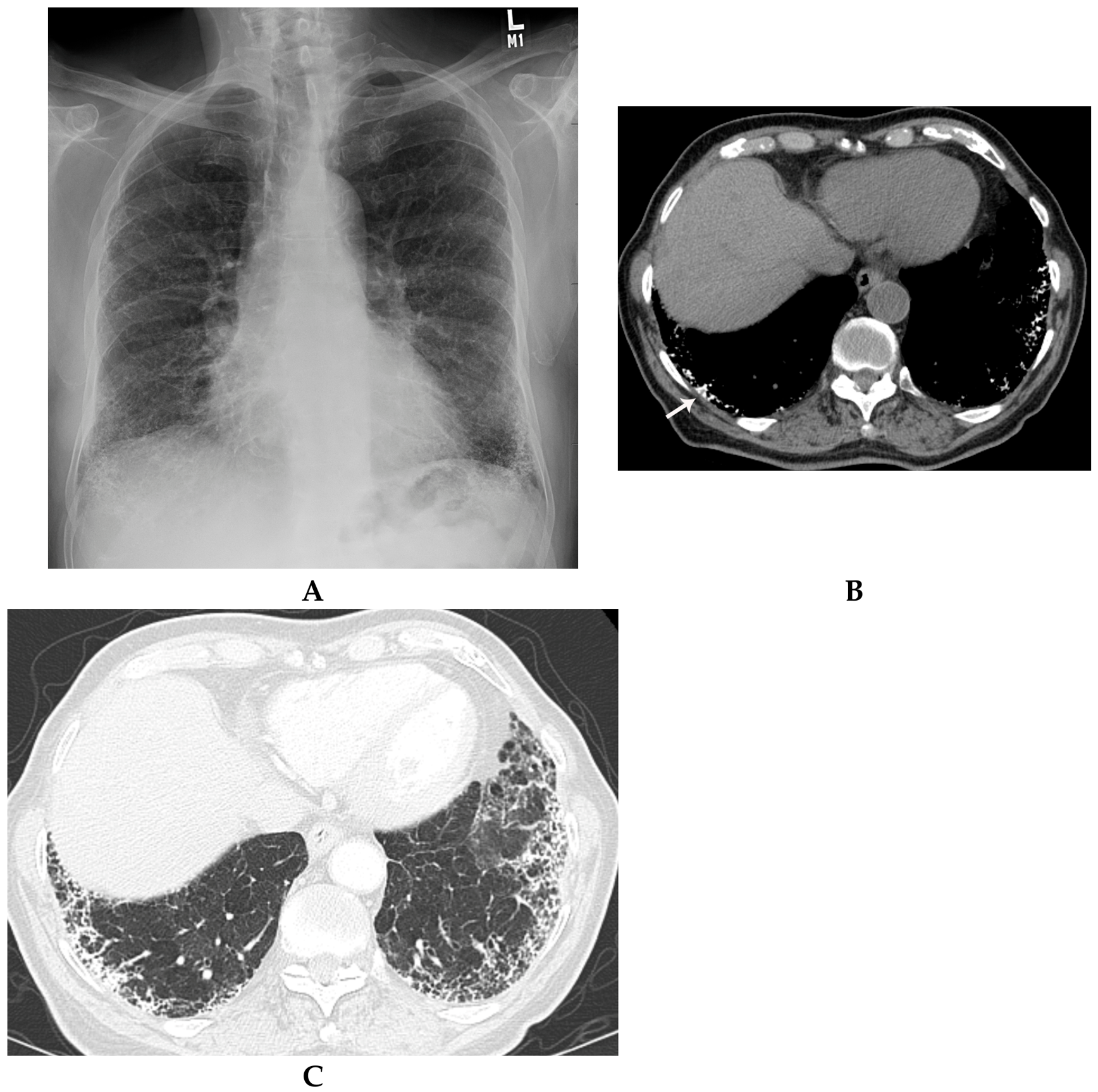
Chest radiography. The classic finding is a fine, sand-like micronodular pattern with basal predominance, described as a “sandstorm” [8]. Moran et al. found diffuse bilateral opacities in all seven patients, by radiology report in six and by direct review of the film in the seventh [1]. Micronodules are typically under 1 mm, and dense deposits can obscure the cardiac and diaphragmatic borders, leaving the lungs homogeneously hypotransparent on the film [19].
HRCT. High-resolution CT, which the 1997 series did not use, is now the central diagnostic test. Reported features include ground-glass opacity, small parenchymal and subpleural nodules, calcified interlobular septa, a crazy-paving pattern, subpleural cysts and linear subpleural calcification, dense consolidation, and fissural nodularity [19,20]. In one HRCT series, ground-glass opacity was present in every patient, subpleural nodules in 92.3%, subpleural cysts in 84.6% and crazy paving in 69.2%; disease was consistently worse in the lower thirds of the lungs [20]. Involvement is greatest posteriorly and medially, and ground-glass change predominates in children and early disease [19]. Calcified septa in the mediastinal window help separate PAM from pulmonary alveolar proteinosis, which also produces crazy paving [8].
The black pleural line. The radiolucent band seen between the ribs and the dense lung on radiographs was shown by HRCT and MRI to reflect a fat-dense layer between the ribs and calcified parenchyma, rather than subpleural cysts alone [21]. Subpleural cysts and paraseptal emphysema are separate findings that may coexist [8].
Other modalities. Technetium-99m bone scintigraphy showed avid uptake in early series and was used diagnostically; HRCT has largely made it unnecessary [8]. Chest ultrasound can show pleural irregularity and millimetre-sized echogenic subpleural foci without acoustic shadowing [8]. Progressive subpleural thickening may eventually calcify the pleura, but this is rarely present at diagnosis [8]. In advanced disease, HRCT shows extensive calcified nodules, confluent consolidation with air bronchograms and calcified septa and fissures, as in a 2025 case confirmed by cryobiopsy [22].
Figure 1.
A-C depict the most salient features in cases of PAM. A) Frontal chest radiograph shows fine, sand-like calcified micronodular pattern in the periphery of the bilateral lungs with predominance in the lower lobes. B) Axial non-contrast enhanced CT chest with mediastinal windows shows the calcified micronodules (arrow) consistent with microliths and calcified interlobular septae in the periphery of the lower lobe. C) Axial high resolution CT chest with lung windows shows the microliths in the subpleural location, interlobular septal thickening, and subpleural cysts.
Pathology and Microlith Composition
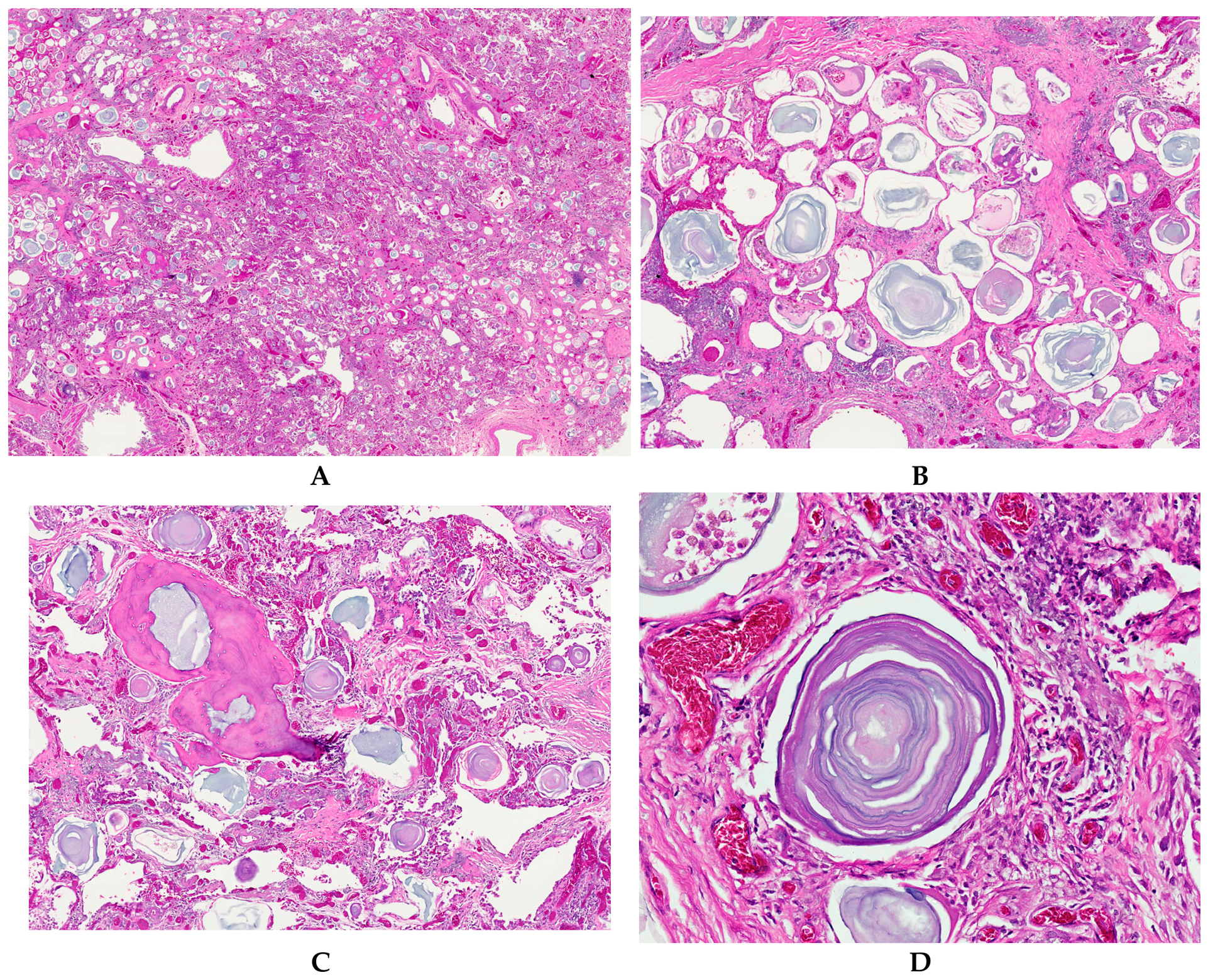
Gross and histologic findings. In the five autopsy cases of Moran et al., the lungs were heavy and large, with a hard, gritty cut surface caused by sandlike concretions that made sectioning difficult [1]. Microscopy showed calcospherites 250 to 750 µm across, concentrically laminated with an onionskin appearance, lying free in dilated alveolar spaces and obliterating them (Figure 2A-D); the interstitium showed variable fibrosis, and in one case macrophages surrounded some microliths [1]. These findings match the modern description of alveoli filled with spherical calcified bodies with concentric psammomatous calcification, as seen in a recent cryobiopsy specimen [22].
Composition. Scanning electron microscopy with energy-dispersive x-ray analysis in six of the 1997 cases showed calcium and phosphorus as the principal elements, with traces of potassium, iron, copper or magnesium in individual specimens [1]. The molecular work has since explained why: the crystals form from phosphate that the alveolar epithelium can no longer clear [8,11]. The mouse model added a finding that the human pathology had not emphasised, namely alveolar phospholipidosis [11].
Relationship to inflammation and fibrosis. The variable interstitial fibrosis in the 1997 series raised the question of whether fibrosis is primary or secondary. Moran et al. suggested that fibrosis in one patient with a 40-year history of fibrosing disease was probably secondary to microlithiasis [1]. Mouse data support this direction of causality: microliths introduced into normal lungs provoke macrophage-rich inflammation [11].
Diagnosis and Differential Diagnosis
From biopsy to imaging to genotype. Before 1980, more than two-thirds of cases were diagnosed by chest radiography alone. Between 1980 and 2015, diagnoses were made by radiography in 30.7%, bronchoalveolar lavage or transbronchial biopsy in 28%, open lung biopsy in 25.4%, CT in 12.5% and autopsy in 2.3% [15]. Invasive tissue sampling is now rarely needed. When imaging is typical, particularly with an affected sibling, the diagnosis can be made without biopsy [8,12,15].
Role of genetic testing. A 2020 diagnostic algorithm moves from least to most invasive: family history and genetic testing first, then HRCT, then lavage or biopsy only if the picture remains uncertain [8]. If a sibling or parent has a confirmed genotype and imaging is typical, the diagnosis is considered certain, and genotyping the index patient is the recommended step when available [8]. Genotyping is not required for diagnosis and does not currently guide management because there is no genotype-phenotype correlation, but it enables family screening [15]. The 2023 systematic review anticipated that genetic testing may in future replace invasive methods [9].
Supportive tests. Serum SP-A and SP-D are elevated and correlate with progression, so they may be useful for monitoring [12]. In mice and in patient serum, MCP-1 and SP-D were identified as biomarkers, but neither has been validated for routine clinical use [11]. Restrictive physiology develops with progressive fibrosis in advanced disease and was reproduced in the mouse model [8,11].
Differential diagnosis. Moran et al. distinguished PAM from three intrapulmonary bodies by size, structure and composition [1]. That framework remains useful histologically, and the imaging mimics are discussed below.
| Entity | Distinguishing features |
| Alveolar microliths (PAM) | 250 to 750 µm, concentrically laminated calcium phosphate, filling alveoli; sandstorm radiograph and calcified septa on HRCT [1,8] |
| Pulmonary corpora amylacea | Laminated rounded bodies, 30 to 200 µm, eosinophilic on H&E, do not calcify; seen in older patients and emphysematous lung [1] |
| Pulmonary blue bodies | Intra-alveolar, laminated, basophilic, 15 to 40 µm; calcium carbonate in a mucopolysaccharide matrix, so smaller and chemically different from microliths [1] |
| Pulmonary ossification | Bone particles deposited haphazardly through the parenchyma, including the interstitium [1] |
| Pulmonary alveolar proteinosis | Crazy paving on HRCT, but calcification is absent; in PAM, calcification traces the septa on mediastinal windows [8] |
| Other conditions causing diffuse micronodular opacities, such as miliary infection, may be considered clinically; one published case was treated as tuberculosis for 18 months despite negative microbiology before PAM was recognised [8]. Diagnostic delay is a particular problem in children [14]. A review published online in October 2025 still describes PAM as an ultra-rare disease for which no effective prevention or treatment has been established [23]. | |
Natural History and Prognosis
The most durable contribution of the 1997 series is its natural-history data. Five of seven patients had known disease for 5 to 41 years, and five died of respiratory failure in that interval; two were lost to follow-up [1].
| Case | Age / sex | Recorded course | Outcome |
| 1 | 33 / F | Bilateral lesions for 5 y; worked in a carbon plant with mica exposure | Lost to follow-up |
| 2 | 37 / F | Condition known for 8 y | Died 8 y after diagnosis, respiratory failure |
| 3 | 19 / F | Gradual decrease in exercise tolerance | Lost to follow-up |
| 4 | 70 / F | Pulmonary fibrosis for 40 y | Died 40 y after diagnosis |
| 5 | 42 / F | 14-y history of abnormal radiograph | Died 14 y after diagnosis |
| 6 | 32 / F | Condition known for 5 y | Died 5 y after diagnosis |
| 7 | 54 / M | Condition since age 13 | Died of respiratory failure 41 y after diagnosis |
The authors concluded that most patients follow a fatal course through complications of the microlithiasis and that patients should be monitored for treatable complications such as pneumonia [1]. Two features complicated the interpretation. In two patients the onset was ambiguous, and no sequential radiographs or pulmonary function tests were available to document progression [1].
Later data agree with this picture. A Japanese long-term follow-up of 53 patients, summarised in the 2020 review, found that respiratory insufficiency caused death in 34.1% of patients within 10 to 20 years of diagnosis, and in a further 42.9% of survivors within 20 to 49 years; overall survival extended to a mean age of 46.2 years [8]. Course is variable: some patients remain static, while others progress to pulmonary fibrosis, respiratory failure and cor pulmonale [8,15]. Early-onset and rapidly progressive forms are described but are uncommon [8,13].
Management
Supportive care. Moran et al. noted that no specific treatment existed and advised monitoring for treatable complications such as pneumonia [1]. That remains the basis of care: supplemental oxygen for hypoxemia, avoidance of smoking, prompt treatment of infection, and surveillance for pulmonary hypertension and right heart failure [8,15]. Corticosteroids have not been shown to help; one reported patient developed respiratory and right ventricular failure despite steroid treatment [26].
Bisphosphonates. Disodium etidronate is less potent as an anti-osteoclast agent than alendronate or risedronate, but it has the theoretical advantage of inhibiting hydroxyapatite crystal formation [8]. Benefit has been reported in children, including long-term follow-up [8,24]. All pharmacological evidence to date, however, comes from case reports of empiric therapy, and no organised trial has assessed safety or efficacy [8]. Etidronate should therefore be regarded as unproven.
Whole-lung lavage. Bronchoalveolar lavage has recovered microliths in individual patients but has not been shown to alter the course of the disease [8]. In mice, whole-lung EDTA lavage reduced stone burden [11]; this is discussed in the next section.
Lung transplantation. Transplantation is the only established treatment for end-stage disease [8,15]. Both single and bilateral lung transplants have been successful. The early concern that shunting through the native lung would compromise single-lung outcomes has not been borne out [8]. The calcified lungs create technical difficulty, including limited intraoperative deflation, dense pleural adhesions and a higher bleeding risk [8].
The largest single-centre experience is the Vienna series of five consecutive patients (four women, one man, mean age 46.3 years), transplanted between 1989 and 2013 [25]. ECMO was needed intraoperatively in four of the five. One early patient underwent re-transplantation on postoperative day 2 and died of sepsis on day 11; the other four were alive at follow-up, and the authors concluded that results were comparable to other indications [25]. A 2024 case report from Iran cited 19 transplanted PAM patients in the literature [26]. To date, no recurrence of microliths in the allograft has been documented [8,25,26,27].
Transplant recipients with PAM remain susceptible to standard complications. One 43-year-old man developed chronic lung allograft dysfunction with bronchiolitis obliterans syndrome two years after bilateral transplantation and died of COVID-19-related sepsis about three years after surgery, without calcific recurrence [27]. A familial case complicated by transplant rejection has also been described [28]. The absence of recurrence is consistent with the mechanism: donor lungs carry functional NPT2b, so the underlying defect is corrected in the graft. Evidence on recurrence remains limited to case reports and small series; in the Vienna series, the surviving patients were followed for up to 74 months without recurrence [8].
Emerging Therapies and Future Directions
Lowering alveolar phosphate. Knowledge of the mechanism points to a rational strategy: reduce the phosphate available to form crystals, or dissolve crystals that have formed [2,11]. In the epithelial Npt2b-deleted mouse, a low-phosphate diet prevented stone formation and whole-lung EDTA lavage reduced the stones already present [11]. These results support both prevention and clearance as therapeutic goals, and they suggest that treatment started early might matter more than treatment started late. Neither approach has been tested in patients, and systemic phosphate restriction has its own risks that a trial would have to address.
Gene-directed therapy. Because the disease is monogenic and its clinical burden is concentrated in the lung, with loss of NPT2b in alveolar type II cells, PAM is a plausible candidate for gene replacement or editing. A 2026 report states that, as of the end of 2025, no published preclinical study had demonstrated CRISPR-based gene correction in an animal model of PAM, although gene therapy is regarded as a promising direction [14].
Biomarkers and endpoints. Any trial will need measurable endpoints in a disease that progresses over decades. Serum SP-A and SP-D correlate with progression in patients [12], and MCP-1 and SP-D were identified in the mouse model and in patient serum [11]. Validation against imaging burden, pulmonary function and clinical events is still required.
Unresolved questions. The main gaps, in approximate order of importance:
The 2023 systematic review called for more standardised patient data, including genetic diagnosis, in larger international populations [9]. For a disease with roughly a thousand reported patients worldwide, an international registry with uniform genotyping and longitudinal imaging is the most practical route to answering these questions.
Conclusions
The 1997 series by Moran et al. correctly captured the essential features of pulmonary alveolar microlithiasis: diffuse radiographic opacities, laminated calcium phosphate microliths, and a course measured in decades that ends in respiratory failure for many patients [1]. What was missing was the cause. Since 2006, biallelic loss-of-function variants in SLC34A2 have been shown to abolish alveolar phosphate clearance through NPT2b, and an epithelial knockout mouse has confirmed the mechanism and opened the way to preclinical testing [3,4,11].
Clinically, diagnosis has shifted from biopsy to HRCT and genotyping. Management, however, has changed little: supportive care, unproven bisphosphonate therapy, and lung transplantation for end-stage disease, with no reported recurrence in the graft [8,25]. The priority now is to move from mechanism to trials, which will require validated biomarkers, standardised international data collection, and a prospective natural-history study.
Author Contributions
All authors contributed to the writing of this manuscript.
Funding
No funding was received in the elaboration of this manuscript.
Data Availability Statement
No new data were created; all sources are cited in the reference list.
Conflicts of Interest
None of the authors has any conflict of interest to declare.
References
- Moran CA, Hochholzer L, Hasleton PS, Johnson FB, Koss MN. Pulmonary alveolar microlithiasis: a clinicopathologic and chemical analysis of seven cases. Arch Pathol Lab Med. 1997;121:607-611.
- Castellana G, Castellana G, Gentile M, Castellana R, Resta O. Pulmonary alveolar microlithiasis: review of the 1022 cases reported worldwide. Eur Respir Rev. 2015;24(138):607-620. https://publications.ersnet.org/content/errev/24/138/607.full.pdf. [CrossRef]
- Corut A, Senyigit A, Ugur SA, et al. Mutations in SLC34A2 cause pulmonary alveolar microlithiasis and are possibly associated with testicular microlithiasis. Am J Hum Genet. 2006;79(4):650-656. https://pubmed.ncbi.nlm.nih.gov/16960801/. [CrossRef]
- Huqun, Izumi S, Miyazawa H, et al. Mutations in the SLC34A2 gene are associated with pulmonary alveolar microlithiasis. Am J Respir Crit Care Med. 2007;175(3):263-268. [CrossRef]
- Harbitz F. Extensive calcification of the lungs as a distinct disease. Arch Intern Med. 1918;21:139-146. [CrossRef]
- Puhr L. Mikrolithiasis alveolaris pulmonum. Virchows Arch Pathol Anat. 1933;290:156-160. [CrossRef]
- Sosman MC, Dodd GD, Jones WD, Pillmore GU. The familial occurrence of pulmonary alveolar microlithiasis. Am J Roentgenol. 1957;77:947-1012.
- Kosciuk P, et al. Pulmonary alveolar microlithiasis. Eur Respir Rev. 2020;29(158):200024. https://publications.ersnet.org/content/errev/29/158/200024. [CrossRef]
- Jönsson ÅLM, Simonsen U, Christensen JH, et al. New insights in the genetic variant spectrum of SLC34A2 in pulmonary alveolar microlithiasis; a systematic review. Orphanet J Rare Dis. 2023. https://ojrd.biomedcentral.com/articles/10.1186/s13023-023-02712-7.
- Impaired phosphate transport in SLC34A2 variants in patients with pulmonary alveolar microlithiasis. Hum Genomics. 2022. https://link.springer.com/article/10.1186/s40246-022-00387-y.
- Saito A, Nikolaidis NM, Amlal H, et al. Modeling pulmonary alveolar microlithiasis by epithelial deletion of the Npt2b sodium phosphate cotransporter reveals putative biomarkers and strategies for treatment. Sci Transl Med. 2015;7(313):313ra181. https://www.science.org/doi/10.1126/scitranslmed.aac8577.
- Tachibana T, Hagiwara K, Johkoh T. Pulmonary alveolar microlithiasis: review and management. Curr Opin Pulm Med. 2009. https://pubmed.ncbi.nlm.nih.gov/19617834/.
- Pulmonary alveolar microlithiasis: a case report with a novel mutation in the SLC34A2 gene and review of the literature. Respir Med Case Rep. 2018. https://www.sciencedirect.com/science/article/pii/S221433001830004X.
- Case report: novel compound heterozygous mutations in SLC34A2 gene: a case of pulmonary alveolar microlithiasis in a child. 2026. https://pmc.ncbi.nlm.nih.gov/articles/PMC13006609/.
- Shaw BM, Shaw SD, McCormack FX. Pulmonary alveolar microlithiasis. Semin Respir Crit Care Med. 2020;41:280-287. https://pmc.ncbi.nlm.nih.gov/articles/PMC12169814/. [CrossRef]
- Esguerra Gomez G, Lichtemberger E, Santamaria A, et al. Familial pulmonary alveolar microlithiasis: four cases from Colombia, S.A. Is microlithiasis also an environmental disease? Radiology. 1959;72:550-561.
- Caffrey PR, Altman RS. Pulmonary alveolar microlithiasis occurring in premature twins. J Pediatr. 1965;66:758-763. [CrossRef]
- Castellana G, Carone D, Castellana M. Microlithiasis of seminal vesicles and severe oligoasthenospermia in pulmonary alveolar microlithiasis (PAM): report of an unusual sporadic case. Int J Fertil Steril. 2015;9(1):137-140. [CrossRef]
- Ferreira Francisco FA, Pereira e Silva JL, Hochhegger B, Zanetti G, Marchiori E. Pulmonary alveolar microlithiasis. State-of-the-art review. Respir Med. 2013;107(1):1-9. https://www.sciencedirect.com/science/article/pii/S0954611112003976. [CrossRef]
- Can chest high-resolution computed tomography findings diagnose pulmonary alveolar microlithiasis? https://www.ncbi.nlm.nih.gov/pmc/articles/PMC4567357/.
- Hoshino H, Koba H, Inomata S, et al. Pulmonary alveolar microlithiasis: high-resolution CT and MR findings. J Comput Assist Tomogr. 1998;22:245-248. https://pubmed.ncbi.nlm.nih.gov/9530388/. [CrossRef]
- Pulmonary alveolar microlithiasis: case report (cryobiopsy). 2025. https://pubmed.ncbi.nlm.nih.gov/40949746/.
- Altınışık G, Yiğit N, Çetin N. Pulmonary alveolar microlithiasis: a review of a rare disease through rarely discussed perspectives. Semin Respir Crit Care Med. 2026;47(4):396-408. https://pubmed.ncbi.nlm.nih.gov/41161329/. [CrossRef]
- Ozcelik U, Yalcin E, Ariyurek M, et al. Long-term results of disodium etidronate treatment in pulmonary alveolar microlithiasis. Pediatr Pulmonol. 2010;45:514-517. [CrossRef]
- Klikovits T, Slama A, Hoetzenecker K, et al. A rare indication for lung transplantation: pulmonary alveolar microlithiasis, institutional experience of five consecutive cases. Clin Transplant. 2016;30(4):429-434. https://pubmed.ncbi.nlm.nih.gov/26841075/. [CrossRef]
- Successful bilateral lung transplantation in pulmonary alveolar microlithiasis: a case report and review of literature. 2024. https://pmc.ncbi.nlm.nih.gov/articles/PMC11082537/.
- Outcome of lung transplantation in patients with pulmonary alveolar microlithiasis in the era of COVID-19 infection. J Surg Case Rep. 2024;2024(4):rjae211. https://academic.oup.com/jscr/article/2024/4/rjae211/7643383. [CrossRef]
- Pulmonary alveolar microlithiasis: a unique case of familial PAM complicated by transplant rejection. https://www.ncbi.nlm.nih.gov/pmc/articles/PMC8041554/.
Figure 2.
A) Low power view of PAM, note the presence of numerous microlith filling alveolar spaces; B) PAM showing numerous microliths, also note the surrounding pulmonary parenchyma with fibrosis; C) Microliths of PAM in association with ossification, note the differences in both elements; D) higher magnification of PAM showing the characteristic onionskin of a microlith.
Disclaimer/Publisher’s Note: The statements, opinions and data contained in all publications are solely those of the individual author(s) and contributor(s) and not of MDPI and/or the editor(s). MDPI and/or the editor(s) disclaim responsibility for any injury to people or property resulting from any ideas, methods, instructions or products referred to in the content. |
© 2026 by the authors. Licensee MDPI, Basel, Switzerland. This article is an open access article distributed under the terms and conditions of the Creative Commons Attribution (CC BY) license (http://creativecommons.org/licenses/by/4.0/).
Copyright: This open access article is published under a Creative Commons CC BY 4.0 license, which permit the free download, distribution, and reuse, provided that the author and preprint are cited in any reuse.