
Submitted:
13 October 2026
Posted:
14 October 2026
You are already at the latest version
Abstract
Background: Mycotic Aortic aneurysms (MAA) are saccular aneurysms of the infected aortic wall. Mycobacterium, compared with other bacteria, is less likely to cause MAA; most cases follow, but it is a rare, late, systemic, and serious complication of intravesical Bacillus Calmette-Guérin (BCG) instillation for bladder cancer. Case Presentation: A 65-year-old male with a history of hypertension, hyperlipidemia, diabetes mellitus, diabetic foot ulcer with methicillin-sensitive Staphylococcus aureus (MSSA) bacteremia, status post partial toe amputation, and papillary urothelial carcinoma treated with transurethral resection and intravesical BCG instillation, recently underwent a computed tomography (CT) abdomen 2 weeks ago for abdominal pain, which revealed colonic stool without other pathological findings, who currently presented with worsening abdominal pain, and the CT scan identified a new 2.5-centimeter pseudoaneurysm of the infrarenal aorta, associated with a periaortic and aortocaval mass, likely a hematoma, infectious mycotic aneurysm, or neoplastic process. The patient received intravenous piperacillin-tazobactam for presumed MSSA, a potential cause of a mycotic aneurysm, which was later discontinued after negative blood cultures. Vascular surgery consultation led to endovascular aortic stenting. Infectious disease recommended a Karius test; despite no warning signs, it returned positive for the Mycobacterium tuberculosis complex, and a diagnosis of a BCG-induced aneurysm was made. The patient was treated with isoniazid, rifampin, ethambutol, and pyridoxine, then continued isoniazid and rifampin for 9 months. Conclusion: We presented a rare case of a BCG-induced mycotic aortic aneurysm that developed within 2 weeks. Therefore, we need a low threshold for MAA in patients with a history of intravesical BCG instillations who present with worsening abdominal pain, even if prior imaging 2 weeks ago showed no warning signs and instillations were given 2 years ago. Testing and diagnosing a BCG-induced aneurysm are necessary despite endovascular intervention, because it can further infect the stent and form an abscess if untreated.
Keywords:
Bacillus Calmette-Guérin
; Mycotic Aortic aneurysms
; bladder cancer
; intravesical BCG
1. Introduction
Mycotic aortic aneurysms (MAA) are infected aortic wall saccular aneurysms and are very uncommon, accounting for only 0.6 to 1% of all aortic aneurysms; the most commonly isolated organisms are Staphylococcus, Streptococcus, and Salmonella species [1]. In contrast, mycobacterial-associated mycotic aneurysms are rare, with the most occurring after intravesical Bacillus Calmette-Guérin (BCG) therapy; they are one of the systemic complications of intravesical BCG, with only 60 cases reported from 1900 to 2021 [2,3].
BCG is a live attenuated vaccine made from the Mycobacterium bovis strain against tuberculosis, developed in 1921 and licensed since then; it is used not only against TB but also to treat bladder cancer and fungal and parasitic infections [4]. Intravesical BCG is an adjuvant treatment for bladder cancer that has been used since the 1970s. The FDA approved its use in 1990 for the treatment of non-muscle-invasive bladder cancer, and it reduces the recurrence rate by about 30 to 40% [5]. The adverse effects of intravesical BCG instillation are categorized into self-limiting effects, which occur in the first 48 hours of instillation and are either local genitourinary symptoms or general constitutional symptoms, and systemic complications, which occur from a few hours to years after the last instillation and are more severe, requiring hospitalization [6].
Our aim is to present this case to the medical community because of the rare presentation and rapid progression of a mycotic aneurysm secondary to intravesical BCG instillation. We want clinicians to consider mycotic aneurysm in their differential diagnosis and maintain a low threshold for it in patients presenting with worsening abdominal pain and a history of BCG instillation, even if imaging was normal weeks ago.
2. Case Presentation
A 65-year-old man with a history of hypertension, hyperlipidemia, type 2 diabetes mellitus, and a diabetic foot ulcer complicated by methicillin-sensitive Staphylococcus aureus (MSSA) bacteremia in 2022, with subsequent partial amputation of the left first toe in February 2026, and papillary urothelial carcinoma treated with transurethral resection of the bladder tumor followed by six weekly intravesical Bacillus Calmette-Guérin (BCG) instillations in 2024, presented with worsening abdominal and right flank pain.
Two weeks before the current presentation, he presented to the emergency department with right lower back and abdominal pain, vomiting, and severe constipation. He reported only three bowel movements over the preceding two months and requested an enema. Computed tomography (CT) of the abdomen and pelvis without contrast at that time showed a large colonic stool burden without an acute intra-abdominal process (Figure 1a and Figure 1b). Importantly, the major abdominal vessels were unremarkable, with no evidence of an aortic aneurysm or periaortic abnormality. He was discharged on polyethylene glycol 17 mg daily.
Two weeks later, he returned with worsening lower abdominal pain radiating to the right flank and upper chest. On presentation, his heart rate was 113 beats/min, blood pressure was 146/96 mmHg, and temperature was 97.6 °F. Physical examination was notable for tenderness at the right flank and costovertebral angle. Laboratory work: white blood cells 9100 cell/mm3, hemoglobin 14.7 mg/dL, platelet count 158 K/mm3, urea nitrogen 17 mg/dL, creatinine 0.7 mg/dL. CT of the chest, abdomen, and pelvis with contrast demonstrated a new 2.5-cm pseudoaneurysm of the infrarenal aorta associated with a 5.7 × 5.4 × 10-cm periaortic and aortocaval mass. The findings were concerning for a contained rupture with surrounding hematoma; however, an infectious mycotic aneurysm or neoplastic process could not be excluded (Figure 2a and Figure 2b).
The patient had blood cultures drawn and was started on intravenous vancomycin one gram and IV piperacillin-tazobactam 4.5 grams every eight hours, as the CT suggested a mycotic aneurysm and history of MSSA bacteremia. Vascular surgery was consulted, and the patient had an emergent endovascular aortic stenting with a physician-modified endograft with two-vessel renal laser fenestrations with bilateral renal artery stenting. The patient was transferred to the intensive care unit for post-operative management, especially urine output monitoring, as the patient was at risk of developing acute kidney injury due to decreased intraoperative blood supply to the left and right kidneys for 25 minutes and 45 minutes, respectively.
Infectious Disease (ID) was consulted; they reported that an active MSSA infection was considered less likely, as the patient lacked significant leukocytosis, hypotension, or other findings suggestive of severe bacterial sepsis. Blood cultures remained negative; therefore, we discontinued broad-spectrum antibiotics. ID also initially considered BCG-associated infection less likely because mycotic aneurysm following intravesical BCG therapy typically has an indolent course, but CT imaging obtained only two weeks earlier showed no aortic or periaortic abnormality and no active pulmonary lesions on chest CT. The patient’s history of constitutional symptoms was inconsistent; he reported several weeks of fever and night sweats to the vascular surgery team but later denied these symptoms during the ID evaluation.
Given the unclear etiology, a plasma microbial cell-free DNA next-generation sequencing assay (Karius test) was performed to rule out possible causes, and it detected Mycobacterium tuberculosis complex. Given the patient’s prior intravesical BCG therapy, no travel outside the United States, and no known exposure to tuberculosis, the positive result was likely related to his prior BCG therapy, as molecular assays targeting the M. tuberculosis complex may not distinguish M. bovis from other members of the complex. ID discussed the case with physicians at Karius, and all agreed that the positive test is most likely due to active Mycobacterium bovis causing a mycotic aortic aneurysm.
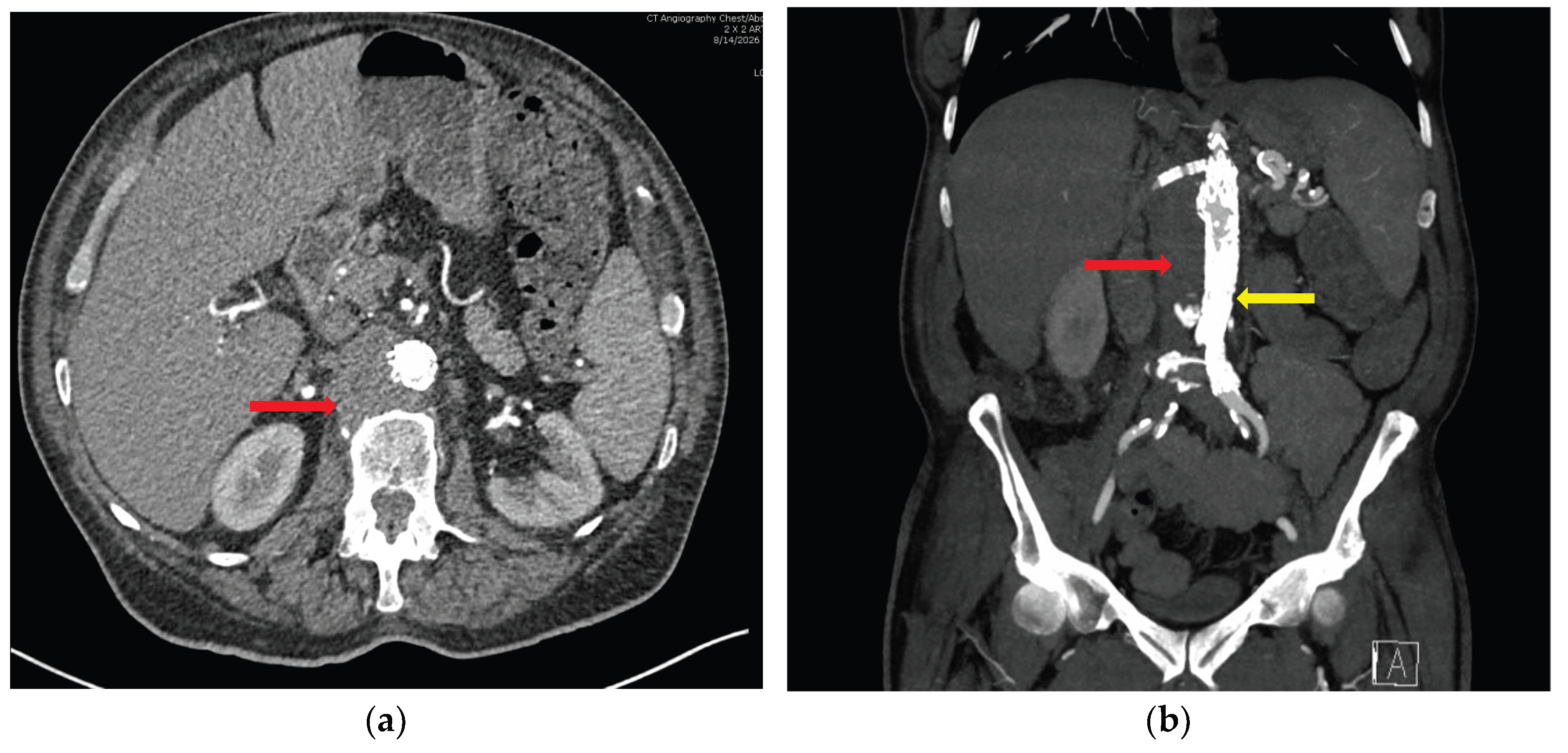
Treatment was started with isoniazid 300 mg daily, rifampin 600 mg daily, ethambutol 1200 mg daily, and pyridoxine 50 mg daily for two months, followed by isoniazid 300 mg daily and rifampin 600 mg daily for nine months. The patient followed up with the vascular surgeon at the clinic during the one-month follow-up and underwent CT angiography of the chest, abdomen, and pelvis, which showed postsurgical changes status post aortoiliac stent placement for the infrarenal abdominal aortic aneurysm. Interval decrease in the surrounding hypoattenuating fluid collection, now measuring 3.2 x 3 cm, previously 5.7 x 5.4 cm, with no extraluminal contrast opacification within the surrounding fluid collection to suggest a leak (Figure 3a and Figure 3b).
3. Discussion
We present a case of acute-onset mycotic aortic aneurysm that developed two years after receiving intravesical BCG for bladder cancer, but with no imaging warnings present two weeks prior to the current presentation. Our case is unique in two ways: it presents with an MAA, which is rare among all aortic aneurysms, and mycobacterial MAA is rare among MAAs; presentation after intravesical BCG is even rarer. Another unique feature of this case is its rapid development.
BCG attaches to and is internalized by urothelial and bladder cancer cells, which initiates local and systemic immunological reactions by activating innate and adaptive immunity [6]. Systemic complications are rare, less than 5%, mostly associated with traumatic catheterization during instillation; they include prostatitis, balanitis, epididymo-orchitis, granulomatous prostatitis, pneumonitis, hepatitis, and others like rheumatologic and vascular complications [7]. Moreover, three mechanisms have been proposed for vascular infection with Mycobacterium bovis: hematogenous dissemination with direct arterial seeding, metastatic implantation through the vasa vasorum, and contiguous spread from adjacent infected tissues, such as lymphadenitis or psoas abscesses [2]
Colonna et al. conducted a systematic review of post-intravesical BCG mycotic aneurysms and found that 70.4% were abdominal aortic aneurysms, followed by thoracic aortic, peripheral arterial, and carotid aneurysms at 12.1, 12.1, and 5.2%, respectively; the median time from the last instillation to development is 12 months. Pain is the most common symptom, followed by fever, weight loss, generalized malaise, and pulsatile mass. [8]. Our case reported abdominal pain as the main symptom but was not consistent with constitutional symptoms like fever or night sweats.
Infected aneurysms typically present as saccular or multilobulated arterial outpouchings, associated with perivascular soft-tissue thickening, inflammatory stranding, adjacent lymphadenopathy, and rapid interval progression [9]. We conducted a literature search using the Boolean search strategy “((Intravesical) AND (BCG)) AND (aneurysm),” filtered to case reports published within the preceding 10 years. The search yielded 46 articles; after careful review, we identified three case reports in which patients had no aneurysm on prior CT of the abdomen and pelvis but developed one on a subsequent CT performed approximately 2 months later. Although these cases differ from ours in terms of the timeline of aneurysm development, they represent the most closely comparable cases identified in the literature [10,11,12]. Of these three case reports, one showed aortitis [12], one showed lymphadenopathy [11], and the third showed no findings [10] on prior CT but eventually developed an aneurysm on a subsequent CT performed 2 months later. Godard et al.[3] reported a case in which a non-contrast CT abdomen showed new retroperitoneal lymphadenopathy with a suspected soft-tissue mass partially surrounding the aorta; the subsequent CT with contrast described this as an abdominal MAA measuring 5.8 cm, thereby indicating that the previously identified mass was probably the aneurysm and not a new aneurysm that had developed over the two weeks in between. Compared with these case reports, our case had no warning signs such as aortitis or lymphadenopathy and developed an aneurysm within 2 weeks.
Diagnosing mycobacterial MAA and initiating antitubercular treatment immediately are paramount to prevent potentially fatal complications. Diagnostic modalities for specific diagnosis of mycobacterial MAA include cultures, PCR, and emerging tests such as microbial cell-free DNA sequencing [13]. King et al. reported a BCG-induced MAA in which the patient’s initial culture results were sterile; growth of the Mycobacterium tuberculosis complex was identified 25 days later [14]. Nishizawa et al. presented a case in which a patient with MAA underwent endovascular stent placement, was started on IV antibiotics, and later discontinued them after negative initial cultures. However, after six weeks, cultures later identified M. bovis, confirmed by PCR, and anti-tuberculosis therapy was initiated. Despite this, the patient still developed stent infection with a psoas abscess, requiring open surgery and necessitating a 6-month hospital stay [12]. This highlights that an initially negative culture does not exclude Mycobacterium infection, and the importance of early consideration and investigation of BCG infection using a plasma microbial cell-free DNA sequencing assay in patients with a history of intravesical BCG therapy, as delayed recognition may allow progression to stent infection with other complications, as avoided in our case.
4. Conclusions
We conclude by presenting our case that mycotic aortic aneurysm should be included in the differential diagnosis of worsening abdominal pain in patients who received intravesical BCG for bladder cancer. We should maintain a low threshold for evaluating BCG-induced MAA and repeat imaging in patients who present with worsening abdominal pain, even if imaging performed two weeks ago showed no warning signs and even if intravesical BCG instillation was done two years ago. Prompt diagnosis and treatment of BCG-induced MAA with anti-mycobacterial agents is obligatory, because endovascular stenting can worsen the condition by promoting stent infection and the formation of an abscess or other complications.
Author Contributions
SP conceptualized the manuscript and prepared the initial draft. RS, AC, and SP participated in the patient’s clinical management during hospitalization. KB served as the infectious disease consultant and provided clinical guidance for patient management and manuscript development. SP and AC critically reviewed the manuscript throughout its preparation. ZMR conducted the literature review and contributed relevant evidence to support the manuscript. KB guided the development of the manuscript. All authors contributed to the review, revision, and editing of the manuscript and approved the final version for publication.
Funding
This case report received no funding.
Institutional Review Board Statement
Not applicable. This manuscript is a case report and does not involve research on human participants requiring Institutional Review Board (IRB) approval.
Informed Consent Statement
Obtained.
Data Availability Statement
All raw data mentioned in the manuscript were obtained from the patients’ electronic medical records. Not applicable. No custom code or software applications were developed or used for the analysis presented in this case report.
Acknowledgments
We thank our program director, Dr. Salman, under whom Dr. Rahmatullah is doing an observership. Grammarly is the AI platform used only for spell checks and grammar corrections.
Conflicts of Interest
None of the co-authors has declared any conflict of interest.
References
- Somers, T.; Nies, H.M.J.M.; Kouijzer, I.J.E.; Lee, P.Y.; Morshuis, W.J.; Geuzebroek, G.S.C. Mycotic aortic aneurysms: characteristic macroscopic findings in a case series. Scand. Cardiovasc. J. 2024, 58(1), 2341696. [Google Scholar] [CrossRef]
- Buerger, M.; Kapahnke, S.; Omran, S.; Schomaker, M.; Rief, M.; Greiner, A.; et al. Aortic aneurysm and aortic graft infection related to Mycobacterium bovis after intravesical Bacille Calmette–Guérin therapy—a case series. BMC Surg. 2021, 21(1), 138. [Google Scholar] [CrossRef]
- Godard, L.; Lee, L.; McGuinness, B.; Shirsat, H.; Hoag, N. Mycotic Abdominal Aortic Aneurysm Secondary to BCG Therapy for Non-muscle Invasive Urothelial Carcinoma of the Bladder. Vasc. Endovasc. Surg. 2023, 57(7), 776–80. [Google Scholar] [CrossRef]
- Shaji, A.; Verma, A.; Bhaskar, A.; Dwivedi, V.P. BCG and beyond: unlocking new frontiers in TB vaccine development. Front Immunol. 2025, 16, 1608104. [Google Scholar] [CrossRef]
- Ansari Djafari, A.; Javanmard, B.; Razzaghi, M.; Hojjati, S.A.; Razzaghi, Z.; Faraji, S.; et al. Intravesical Gemcitabine versus Intravesical Bacillus Calmette-Guerin for the Treatment of Intermediate-Risk Non-Muscle Invasive Bladder Cancer: A Randomized Controlled Trial. Urol. J. 2022, (Instant 2022), 7194. [Google Scholar] [CrossRef]
- Liatsos, G.D.; Mariolis, I.; Hadziyannis, E.; Bamias, A.; Vassilopoulos, D. Review of BCG immunotherapy for bladder cancer. In Clin Microbiol Rev.; Forrest, G.N., Ed.; 13 Mar 2025; Volume 38, 1, p. e00194-23. [Google Scholar] [CrossRef]
- Oliveira, A.A.; Morais, J.; Ribeiro, J.; Gouveia, P.F. Systemic infection following intravesical therapy with BCG. BMJ Case Rep. 2021, 14(11), e243641. [Google Scholar] [CrossRef]
- Colonna, G.; Liesker, D.; Wouthuyzen-Bakker, M.; Taurino, M.; Zeebregts, C.J.; Saleem, B.R. Clinical Characteristics and Management of Infective Native Arterial Aneurysms Associated with BCG Therapy for Bladder Cancer. Ann. Vasc. Surg. 2026, 123, 222–32. [Google Scholar] [CrossRef]
- Kumar, N.; Sarin, S. A multidisciplinary FEVAR and stent graft repair to exclude mycotic aneurysms by interventional radiology and vascular surgery: Case report. Radiol. Case Rep. 2026, 21(10), 4453–8. [Google Scholar] [CrossRef]
- Thia, I.; Yuminaga, Y. BCG mycotic aneurysm: Case report on a rare entity. Urol. Case Rep. 2023, 50, 102459. [Google Scholar] [CrossRef]
- Plymoth, M.; C., C.W.; Harvey, R.; Crighton, T.; Sandaradura, I. Disseminated Bacillus Calmette-Guerin infection following concurrent intravesical BCG therapy and immune checkpoint inhibitior therapy: a case report. BMC Infect. Dis. 2025, 25(1), 1575. [Google Scholar] [CrossRef]
- Nishizawa, M.; Kudo, T.; Kijima, T.; Fujii, Y. Failed endovascular abdominal aortic aneurysm repair due to Mycobacterium bovis infection following intravesical bacillus Calmette-Guérin therapy. J. Vasc. Surg. Cases Innov. Tech. 2022, 8(4), 807–12. [Google Scholar] [CrossRef]
- Bonni, S.; Ramnath, N.W.M.; Selten, K.; Moza, A. Bacillus Calmette-Guérin–Related Mycotic Pseudoaneurysm in the Aortic Arch: Clinical and Operative Challenges. Interdiscip. Cardiovasc. Thorac. Surg. 2026, 41(5), ivag120. [Google Scholar] [CrossRef]
- King, B.; Singh, D.; Rathore, A.; Flenner, R.; Flemmer, M. Case Report with Systematic Literature Review on Vascular Complications of BCG Intravesical Therapy for Bladder Cancer. JCM 2022, 11(20), 6226. [Google Scholar] [CrossRef]
Figure 1.
Computed tomography without contrast of the abdomen and pelvis two weeks prior to the current admission: a) Axial section, b) Coronal section.
Figure 1.
Computed tomography without contrast of the abdomen and pelvis two weeks prior to the current admission: a) Axial section, b) Coronal section.
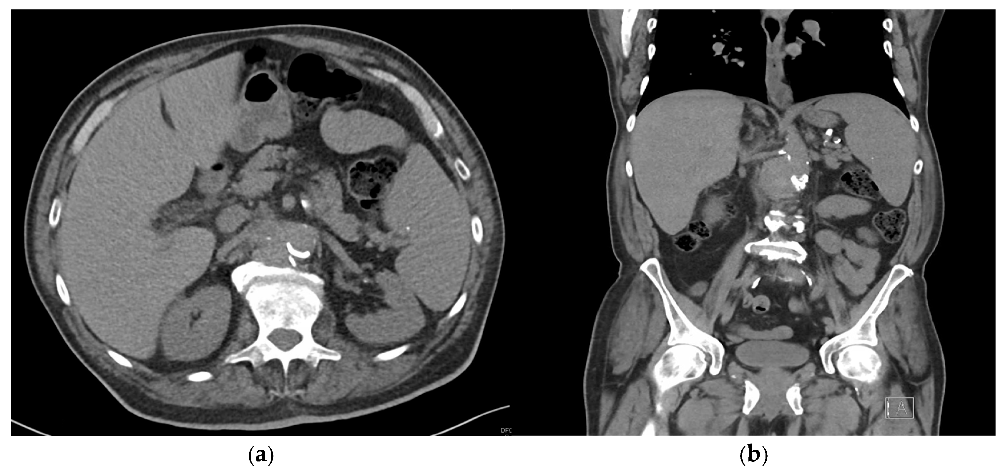
Figure 2.
Computed tomography with contrast of the chest, abdomen and pelvis at the current admission: a) Axial section, b) Coronal section. Red arrow: soft tissue swelling versus hematoma; blue arrow: Pseudoaneurysm.
Figure 2.
Computed tomography with contrast of the chest, abdomen and pelvis at the current admission: a) Axial section, b) Coronal section. Red arrow: soft tissue swelling versus hematoma; blue arrow: Pseudoaneurysm.
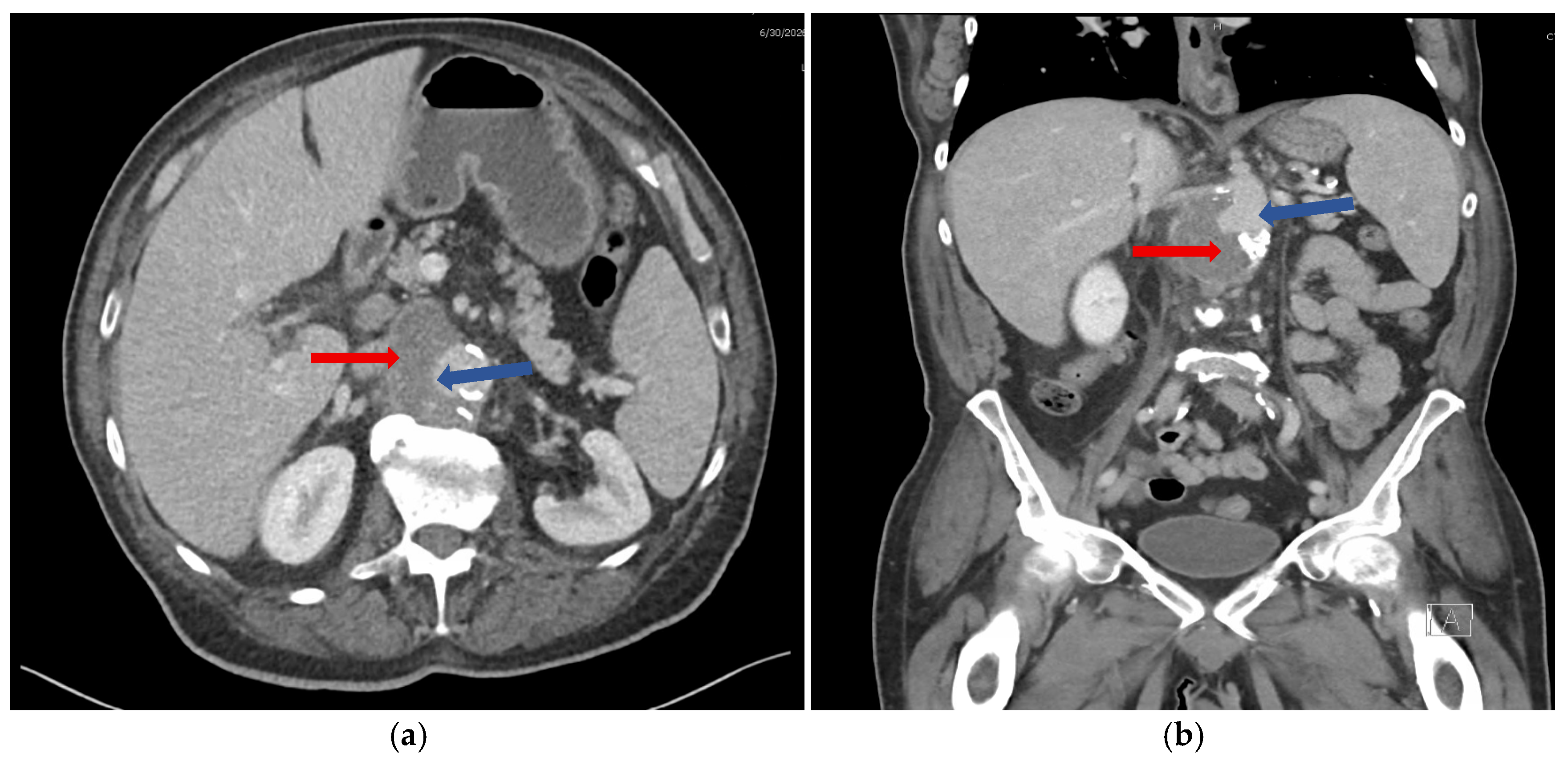
Figure 3.
Computed tomography angiogram of the chest, abdomen, and pelvis at the two-week follow-up: a) Axial section, b) Coronal section. Red arrow: improved soft tissue; yellow arrow: aortic stenting.
Figure 3.
Computed tomography angiogram of the chest, abdomen, and pelvis at the two-week follow-up: a) Axial section, b) Coronal section. Red arrow: improved soft tissue; yellow arrow: aortic stenting.
Disclaimer/Publisher’s Note: The statements, opinions and data contained in all publications are solely those of the individual author(s) and contributor(s) and not of MDPI and/or the editor(s). MDPI and/or the editor(s) disclaim responsibility for any injury to people or property resulting from any ideas, methods, instructions or products referred to in the content. |
© 2026 by the authors. Licensee MDPI, Basel, Switzerland. This article is an open access article distributed under the terms and conditions of the Creative Commons Attribution (CC BY) license (http://creativecommons.org/licenses/by/4.0/).
Copyright: This open access article is published under a Creative Commons CC BY 4.0 license, which permit the free download, distribution, and reuse, provided that the author and preprint are cited in any reuse.