

Submitted:
12 October 2026
Posted:
13 October 2026
You are already at the latest version
Abstract
Background/Objectives: Immune thrombocytopenia (ITP) is an autoimmune disease characterized by increased platelet destruction and insufficient platelet production. Accurate diagnosis, prognostic assessment, and treatment selection remain challenging. Long non-coding RNAs (lncRNAs) regulate immune responses and may contribute to ITP pathogenesis. This review examines lncRNA expression in ITP and its potential diagnostic, prognostic, and therapeutic relevance. Methods: We reviewed English-language literature published between 1994 and 2026 using PubMed, Scopus, and Google Scholar. We examined studies of specific lncRNAs in ITP, focusing on their expression differences and potential diagnostic and therapeutic implications. Re-sults: The studies examined HCP5, NEAT1, lnc-DC, IFNG-AS1, MEG3, GAS5, MALAT1, and THRIL. Findings suggest these lncRNAs may contribute to ITP pathogenesis and serve as candidate diagnostic and prognostic biomarkers. Some were reported as independent predictors of poor prognosis or were associated with glucocorticoid responsiveness. Potential applications include distinguishing primary from secondary ITP, informing patient selection for splenectomy, and identifying therapeutic targets. These applications remain investigational. Conclusions: Research on lncRNAs provides insight into autoimmune mechanisms underlying ITP and may support improvements in diagnosis, prognostic assessment, and individualized treatment. Although several lncRNAs show promise as biomarkers and therapeutic targets, further research, clinical validation, and methodological standardization are needed before routine clinical implementation.
Keywords:
long non-coding RNAs
; immune thrombocytopenia
; gene regulation
; targeted therapies
; diagnos-tic biomarkers
; autoimmunity
; T cells
; B cells
; platelets
1. Introduction
Immune thrombocytopenia (ITP) is an autoimmune disorder characterized by increased reticuloendothelial clearance of platelets and insufficient compensatory platelet production. It affects both children and adults and frequently follows a chronic course in adults. The reported annual incidence in adults is approximately 3.3 cases per 100,000 individuals [1]. Despite advances in understanding the underlying mechanisms of ITP and the availability of established treatment approaches, uncertainties remain regarding its pathogenesis, assessment of disease severity, and treatment selection.
Long non-coding RNAs (lncRNAs) have attracted interest as potential regulators of autoimmune processes. These transcripts are generally longer than 200 nucleotides and have limited protein-coding potential, although some can encode small peptides with biological functions [2]. LncRNAs are implicated in cancer, cardiovascular disorders, and neurological diseases. They also regulate cellular and humoral immune responses, supporting investigation of their contribution to autoimmune disorders such as ITP [3,4].
Studies of lncRNA expression in ITP suggest that these transcripts may provide insights into disease pathogenesis and serve as diagnostic and prognostic biomarkers. Their possible associations with treatment responsiveness have also prompted interest in their potential to inform therapeutic decisions [5,6]. These applications, however, require further investigation and clinical validation.
ITP is classified as primary when no underlying cause or associated condition is identified and as secondary when associated with conditions or exposures such as other autoimmune diseases, medications, or viral infections [7]. Diagnosis relies on excluding alternative causes of thrombocytopenia because no definitive diagnostic test is available [8]. The rate of misdiagnosis remains high, although it is difficult to estimate. This diagnostic challenge highlights the need for reliable biomarkers.
This narrative review examines lncRNA expression and function in ITP, focusing on HCP5, NEAT1, lnc-DC, IFNG-AS1, MEG3, GAS5, MALAT1, and THRIL. It evaluates their potential roles in diagnosis, prognostic assessment, prediction of treatment response, and targeted therapy. Available evidence suggests that these lncRNAs contribute to immune dysregulation and may have clinical value, although findings on expression patterns and regulatory effects are not always consistent. Further validation and methodological standardization are required before clinical implementation.
2. Materials and Methods
We reviewed English-language literature published between 1994 and 2026 using Google Scholar, PubMed, and Scopus. Search terms included “immune thrombocytopenia”, “long non-coding RNA”, “lncRNA”, “idiopathic thrombocytopenic purpura”, “ITP”, “autoimmune diseases”, “ITP treatment”, and “targeted therapies”. Duplicate articles, publications unrelated to the review topic were excluded.
The review examined ITP pathogenesis, treatment efficacy, and current standards of care, with emphasis on recent publications. Particular attention was given to variability in disease course, duration of remission, differences in clinical presentation in children and adults, and evidence that could inform treatment selection. Publications defining lncRNAs and describing their roles in autoimmune regulation were also examined. The principal focus was the involvement of specific lncRNAs in ITP pathogenesis and disease course, including their potential to influence disease progression and serve as therapeutic targets. The findings were synthesized to summarize current knowledge and advances relevant to the development of lncRNA-based therapies for ITP.
3. Immune Thrombocytopenia (ITP)
This section may be divided by subheadings. It should provide a concise and precise description of the experimental results, their interpretation, and the experimental conclusions that can be drawn.
3.1. Etiology and Pathogenesis
ITP affects both children and adults, with a higher prevalence in females [9]. Its etiology and clinical course differ between children and adults; in children, ITP often follows a viral infection and resolves spontaneously [10]. The annual incidence in adults is approximately 3.3 cases per 100,000 individuals [1]. Owing to its predominantly chronic course, ITP has a higher prevalence in adults, estimated at 9.5–12.1 cases per 100,000 individuals [11].
ITP is classified according to disease duration as newly diagnosed during the first 3 months after diagnosis, persistent between 3 and 12 months, and chronic beyond 12 months [12]. During the newly diagnosed phase, diagnostic testing focuses on confirming the diagnosis. Historically, clinicians used the term “acute ITP” to describe childhood disease with a short, self-limiting course; adults more frequently develop chronic disease.
The pathogenesis of ITP involves several interacting mechanisms that are not yet fully understood. A central feature is the loss of immune tolerance, leading to the production of autoantibodies against platelets [13]. These antibodies opsonize platelets, facilitating their clearance by the reticuloendothelial system. Antibody-mediated mechanisms and T-cell activity can also promote megakaryocyte apoptosis and impair platelet production [14]. T-cell dysfunction, including abnormalities in cytotoxic T cells, regulatory T cells (Tregs), and the balance between T-helper 1 (Th1) and T-helper 2 (Th2) responses, further contributes to impaired self-tolerance and platelet destruction [15].
Genetic factors may also contribute to susceptibility to ITP. Reported polymorphisms involve Fcγ receptors, chemokines, cytokines, transcription factors, and genes within the major histocompatibility complex (MHC) [16]. Altered gene expression has also been implicated in immune dysregulation [6]. Elevated levels of proinflammatory cytokines, particularly interleukin (IL)-17, IL-2, and interferon-γ (IFN-γ), promote autoantibody formation. Reduced levels of IL-4, IL-10, and transforming growth factor-β (TGF-β) have also been observed. These changes may favor an inflammatory environment that supports autoreactive immune responses.
Dendritic cells (DCs) are the most effective antigen-presenting cells; they process and present antigens to T cells and help regulate immune responses [17]. Dysregulated presentation of self-antigens can contribute to the breakdown of immune tolerance and the development of autoimmune diseases [18]. Viral infections may disrupt these processes and have been associated with the onset of ITP, particularly in children. Influenza has been reported as a potential trigger, and thrombocytopenia has also been described in association with COVID-19 [19,20].
In addition to increased platelet destruction, impaired platelet production contributes to thrombocytopenia in ITP. Bone marrow megakaryocyte numbers are often increased but may be within the normal range. Abnormalities in megakaryocyte maturation can limit effective platelet production [21]. The resulting thrombocytopenia may manifest as mucocutaneous bleeding. Although the conventional lower limit of the normal platelet count is approximately 150 × 10⁹/L, bleeding manifestations most often occur at counts below 15 × 10⁹/L [22].
3.2. Clinical Presentation
ITP is characterized clinically by an increased tendency to bleed, although bleeding does not correlate linearly with platelet count [23]. The clinical course varies, with most children achieving spontaneous resolution within a year [24]. Mortality among patients with ITP is approximately 1.5 times higher than that in the general population and varies with disease severity. Deaths among patients with ITP are most often due to hemorrhagic complications, particularly severe organ hemorrhage. Treatment-related adverse effects, especially those associated with glucocorticoid therapy, also contribute to mortality [25].
3.3. Treatment
Treatment aims to prevent clinically significant bleeding by maintaining a platelet count sufficient for adequate hemostasis while minimizing adverse effects. The target platelet count depends on age, bleeding, comorbidities, medications and planned procedures; a response threshold should not be equated with remission or normal hemostasis [7,8].
Patients with ITP undergoing surgery usually require treatment to increase their platelet count immediately before the procedure. The choice and timing of treatment depend on the baseline platelet count, the bleeding risk associated with the procedure, and its urgency. Treatment options include intravenous immunoglobulin and platelet transfusion.
Treatment guidelines for ITP are based on consensus informed by many years of clinical observation, with bleeding severity and platelet count guiding treatment decisions. Treatment is indicated for patients with active bleeding, regardless of platelet count, as well as those with platelet counts below 10 × 10⁹/L and most patients with counts between 10 and 15 × 10⁹/L [7,8].
3.3.1. First-Line Treatment
Glucocorticoids are the most commonly used first-line treatment for ITP. Regimens include dexamethasone at 40 mg/day for 4 days or prednisone at 1 mg/kg of body weight, with the prednisone dose tapered once the platelet count increases [26]. This treatment aims to reduce autoantibody-mediated platelet sequestration through Fcγ receptors [27]. Changes in Treg numbers have been reported with rituximab-based treatment [28]. In one study, low-dose rituximab combined with short-term glucocorticoids increased Treg levels; this combination study should not be treated as evidence for glucocorticoids alone [29].
Intravenous immunoglobulin (IVIg) is used when a rapid increase in platelet count is necessary, such as in patients with extremely low platelet counts or bleeding manifestations. It is administered at 1 g/kg of body weight for 1–2 days and is considered the safest therapy for ITP during pregnancy. [7,30]. Its mechanisms of action are not fully understood. Proposed mechanisms include blockade of macrophage Fc receptors to reduce antibody-mediated platelet clearance and receptor saturation to promote autoantibody clearance. IVIg also affects dendritic cells and modulates T-cell responses, increasing Tregs and reducing Th17 cells [31,32]. Additional effects include inhibition of pro-inflammatory cytokine production, such as IFN-γ, and the suppression of complement activation [33,34].
Polyclonal anti-D immunoglobulin may also be considered as first-line treatment in Rh-positive patients with an intact spleen. It offers an alternative to IVIg when a rapid, but often temporary, increase in platelet count is required. Its use is limited, partly by availability [7], and carries a risk of hemolysis. Like IVIg, its mechanisms of action remain incompletely understood. Effects on B lymphocytes, including reduced autoantibody production, have been reported [35].
Responses to first-line treatment are often transient. Approximately 50% of patients treated with dexamethasone or prednisone maintained a response at 6 months [36]. IVIg produces the most rapid increase in platelet count, usually within 1–3 days. Approximately 70-90% of patients respond, although the response is generally short-lived [37].
3.3.2. Treatment of Refractory/Relapsing ITP
Historically, splenectomy has been the most radical and effective treatment for ITP. The spleen is the primary site of activation of T and B cells involved in antiplatelet antibody production and immune-mediated platelet destruction [38]. Splenectomy carries a high perioperative risk, however, including surgical and vascular complications, even when performed laparoscopically. It also increases susceptibility to infections, necessitating vaccination, particularly against pneumococcal disease. Approximately 60% of patients have normal platelet counts 5 years after splenectomy [39]. Conversely, approximately 20% fail to respond or experience an early recurrence of thrombocytopenia [7]. Splenectomy is not currently recommended until at least 12 months after diagnosis and may be considered when drug therapy has been ineffective [8].
Rituximab is a chimeric monoclonal antibody directed against CD20 on B cells. Its mechanisms include antibody-dependent cellular cytotoxicity, complement-dependent cytotoxicity and effects on B-cell survival [40,41,42]. The resulting reduction in B-cell numbers decreases antiplatelet antibody production and improves T-cell function [42]. Approximately 50% of patients with refractory ITP respond to rituximab, although responses are often transient, with only approximately 30% maintaining a response at 6 months [43,44].
Thrombopoietin receptor agonists (TPO-RAs), including eltrombopag, avatrombopag, and romiplostim, address impaired platelet production by activating TPO receptors on megakaryocytes and stimulating JAK2/STAT5 signaling [45]. TPO-RAs are also reported to normalize Treg cell levels in chronic ITP [46]. Eltrombopag and avatrombopag are oral medications intended for chronic use, whereas romiplostim is administered subcutaneously once weekly. Reported response rates range from 40% to 60%, with platelet counts typically increasing within 1–2 weeks of treatment initiation. Approximately 30% of patients maintain a sustained response after several months of continuous romiplostim therapy [47]. The most serious adverse effect of eltrombopag is hepatotoxicity. Romiplostim may cause muscle pain and bone marrow fibrosis. TPO-RAs are an important treatment option for refractory or relapsing ITP and may allow sustained responses, including treatment-free responses in selected patients.
Fostamatinib is an oral spleen tyrosine kinase (Syk) inhibitor approved by the United States Food and Drug Administration in 2018 for patients with an insufficient response to previous treatment. In randomized trials, 18% of patients achieved a stable response, defined as platelet counts >50 × 10⁹/L, with a median time to response of 15 days. The most common adverse effects, reported in 30% of treated patients, included diarrhea, nausea, hypertension, and hepatitis [48].
A 2024 observational study conducted at 42 Spanish centers evaluated fostamatinib in adults with primary or secondary ITP who had experienced failure of at least one previous line of treatment [49]. A total of 138 patients aged >18 years were treated [49]. Primary ITP was defined as a platelet count below 100 × 10⁹/L after excluding secondary causes of thrombocytopenia [12]. Treatment efficacy was assessed by an increase in platelet count or resolution of bleeding. A complete response was defined as a platelet count ≥100 × 10⁹/L, a partial response as a platelet count >30 × 10⁹/L, and nonresponsive as a platelet count <30 × 10⁹/L or less than twice the baseline value [12]. A platelet count of at least 30 × 10⁹/L was achieved in 109 patients (79%), with a median time to platelet count increase of 11 days. Fostamatinib was generally well tolerated. Grade 3 or 4 adverse events occurred in 13 patients (9,4%); diarrhea and increased blood pressure were the most commonly reported adverse events [49].
Rilzabrutinib is an oral, reversible covalent inhibitor of Bruton’s tyrosine kinase. Its actions include reducing Fcγ receptor-mediated platelet destruction by macrophages and limiting pathogenic autoantibody production. In the registration study, 64% of participants experienced an increase in platelet count during the first 12 weeks of treatment, with a median time to first response of 36 days [50]. The cited phase 3 trial evaluated persistent or chronic ITP and does not establish a perioperative bridging indication [50].
4. Long Non-Coding RNA (lncRNA)
This section may be divided by subheadings. It should provide a concise and precise description of the experimental results, their interpretation, and the experimental conclusions that can be drawn.
LncRNAs are transcripts generally longer than 200 nucleotides with limited protein-coding potential, although some can encode small peptides that exhibit diverse biological functions [2]. Their presence in both the nucleus and cytoplasm contributes to their broad range of functions. Compared with protein-coding mRNAs, lncRNAs are generally less stable and expressed at approximately 10-fold lower levels, often in a cell- or tissue-specific manner [51]. Compared with protein-coding genes, lncRNA genes are generally transcribed less efficiently, which contributes to their reduced expression and more cell-specific expression patterns [52].
LncRNAs participate in gene regulation, including post-transcriptional regulation, and play important roles in cellular and humoral immune responses [3,4]. They contribute to numerous physiological processes and are implicated in cancer, cardiovascular disorders, neurological disease, and autoimmune disorders. In both innate and adaptive immunity, lncRNAs regulate gene expression and influence immune-cell function and homeostasis [53].
Advances in transcript identification have expanded the catalog of human lncRNAs. A study published in 2023 identified approximately 95,243 lncRNA genes and 323,950 lncRNA transcripts [54]. The LncR-NADisease v3.0 database contains 25,440 experimentally supported entries describing disease associations covering 6066 lncRNAs, 10,732 circular RNAs (circRNAs), and 566 diseases [55]. Most lncRNAs are transcribed by RNA polymerase II and undergo processing, including polyadenylation, splicing, and capping [56].
4.1. Classification of lncRNAs
LncRNAs are a highly diverse class of transcripts with distinct molecular mechanisms and functions. Their low sequence conservation complicates sequence-based classification, prompting the development of alternative approaches based on genomic context and molecular characteristics [57]. A widely used approach classifies lncRNAs according to their genomic location relative to protein-coding genes. These categories include intronic, intergenic, antisense, bidirectional, and sense RNAs (Figure 1) [58].
4.2. Antisense lncRNAs
Antisense RNAs are transcribed from the DNA strand opposite that of a protein-coding gene and often overlap its intronic or exonic regions. They can influence gene expression through transcriptional interference, chromatin remodeling, and RNA–RNA duplex formation. Their dysregulation contributes to the development of cardiovascular diseases, cancers, and neurological disorders [59,60].
4.3. Intron-Derived lncRNAs
Intron-derived lncRNAs originate from the intronic regions of protein-coding genes, either through processing of intronic RNA sequences or transcription from independent units within introns. They participate in alternative splicing, transcriptional regulation, and chromatin remodeling [61]. Their dysregulation contributes to the development of metabolic diseases and cancers. [61].
4.4. Sense lncRNAs
Sense lncRNAs originate from the DNA gene strand that also serves as the template for protein-coding transcripts. They influence gene expression through RNA-RNA interactions, modulation of RNA splicing, and transcriptional interference. Dysregulation of these lncRNAs leads to the development of various diseases, particularly cancer [62].
4.5. Bidirectional lncRNAs
Bidirectional lncRNAs originate near the promoter regions of protein-coding genes. They act as gene enhancers, increasing gene expression through cis- and trans-regulatory effects, particularly in cell differentiation and proliferation. Their dysregulation is associated with neurodegenerative diseases, cancer, and cardiovascular diseases [52].
4.6. Intergenic lncRNAs
Intergenic lncRNAs originate in the genomic intervals separating protein-coding genes and do not overlap annotated protein-coding loci [63]. They regulate gene expression by modulating RNA stability and influencing chromatin-modifying complexes. Their tissue-specific expression makes them potential biomarkers for guiding targeted therapy [56].
5. LncRNAs in Autoimmune Diseases
Autoimmune diseases result from immune responses directed against the body’s own tissues and organs, causing inflammation and tissue damage. Autoimmune diseases affect nearly 10% of the global population [64]. Their development and clinical course reflect interactions among immune dysregulation, environmental factors, abnormal antigen presentation, and genetic predisposition. Genetic factors offer opportunities to identify new diagnostic and prognostic markers [65]. LncRNAs play important roles in cellular and humoral immune responses [3]. They contribute to both innate and adaptive immunity by regulating the homeostasis of immune cells [4].
5.1. HLA Complex P5 (HCP5)
HCP5 is a hybrid HLA class I endogenous retroviral gene within the MHC region and has reported associations with immune-mediated diseases [66]. HCP5 regulates Treg function in allergic rhinitis through the miR-16/ATXN2L axis [67]. Increased HCP5 expression in rheumatoid arthritis synovial tissue has been associated with immune-cell infiltration [68]. Genetic and epigenetic associations with systemic lupus erythematosus have also been reported; these findings should be distinguished from evidence validating circulating HCP5 as a disease-activity biomarker [66].
5.2. Exosomal Non-Coding RNAs
Exosomes contribute to the regulation and pathogenesis of immune-mediated diseases [69,70]. These extracellular vesicles, measuring approximately 30–150 nm in diameter, transport RNAs, proteins, and lipids between cells. By transferring their cargo, exosomes can influence antigen presentation and activate T cells by stimulating cytokine production. These mechanisms activate inflammatory pathways that contribute to autoimmune disease [69]. Through intercellular communication, exosomes influence both the innate and adaptive immune responses. Their cargo includes non-coding RNAs, such as microRNAs (miRNAs), circRNAs, and lncRNAs [71]. Evidence indicates that exosomal non-coding RNAs contribute to the development of various diseases, including autoimmune disorders, with lncRNAs playing a role in the regulation of autoimmune processes [70].
5.3. Systemic Lupus Erythematosus
Systemic lupus erythematosus (SLE) is an autoimmune disease characterized by autoreactive lymphocyte activation, autoantibody production, and immune complex deposition in multiple organs. Immune-complex-mediated nephritis is the most serious complication of SLE [72]. Dysregulated B-cell activation contributes to immune complex formation. Activated exosomes derived from basophils carry the lncRNA ENST00000537616, which binds to miR-330-5p and activates KRAS signaling. This process enhances B-cell activation and promotes immune complex deposition [73]. Increased NEAT1 expression also plays an important role in exacerbating the inflammatory process in SLE by promoting Th2 cell activation through STAT6 signaling [74].
5.4. Psoriasis
Psoriasis is chronic, recurrent systemic autoimmune disease in which dysregulated immune responses trigger inflammatory processes, resulting in skin lesions and joint inflammation [75]. Exosomes derived from the human skin keratinocyte cell line HaCaT carry the lncRNA AGAP2-AS1, which influences Th1- and Th17-cell differentiation and modulates inflammatory responses [76].
5.5. Rheumatoid Arthritis
Rheumatoid arthritis is a chronic autoimmune disease characterized primarily by synovial inflammation that leads to cartilage destruction and bone damage. Progressive joint damage can result in permanent disability [77]. Fibroblast-like synoviocytes (FLS) play a key role in the pathogenesis of rheumatoid arthritis. Their abnormal activation promotes the secretion of pro-inflammatory mediators such as IL-6, IL-15, and CXCL8. This increases fibronectin production and adhesion molecule expression, promoting leukocyte recruitment to the synovial membrane and inflammation [77,78]. The lncRNA TRAFD1-4:1, carried in FLS cell exosomes, increases CXCL1 expression, leading to chondrocyte dysfunction and extracellular matrix damage [79]. NEAT1 also promotes inflammation by enhancing the differentiation of CD4+ T cells into Th17 cells [74].
6. Role of LncRNAs in the Pathogenesis of ITP
6.1. HCP5
The study of Liu et al. evaluated 40 patients with immune thrombocytopenia (ITP) and 40 healthy controls to investigate the prognostic value of HCP5 expression [5]. The authors monitored the patients over a one-year treatment period, classifying them into favorable or poor-prognosis groups based on disease recurrence, bleeding complications, and platelet counts. Patients with hemorrhagic complications were classified according to bleeding severity as mild, moderate, or severe. Patients received treatment according to ITP standards of care. First-line therapy included glucocorticoids and immunoglobulins, while TPO-RAs or rituximab were used for refractory or relapsing disease. After 1 year of treatment, patients were classified into favorable and poor-prognosis groups. Criteria for poor prognosis included disease recurrence within 12 months, hemorrhagic complications, and platelet count <100 × 10⁹/L. HCP5 expression was significantly lower in patients with ITP than in controls (p < 0.001) [5]. The authors showed that HCP5 expression was significantly reduced in ITP patients and identified low HCP5 levels as an independent risk factor for a poor prognosis. These results are promising but should be considered preliminary because the study population was relatively small.
6.2. NEAT1
NEAT1 plays important roles in gene regulation, chromatin remodeling, immune responses, and the development of cancer [80]. It regulates the activity of various immune cells (Figure 2) [74]. A study at the Pediatric Department of Fayoum University Hospital in El Fayoum, Egypt, investigated 60 children with ITP, including 42 girls (70%) and 18 boys (30%). The median age of the children was 5 years (range: 1–13 years). NEAT1 expression was elevated in these patients [81]. Other studies suggest that NEAT1 may contribute to ITP progression through its effects on inflammatory biomarkers [6]. Viral infections can directly induce NEAT1 expression, promoting antiviral immune responses but potentially contributing to acute thrombocytopenia. This mechanism may play an important role in infection-associated acute ITP, including in patients with HIV-1 or SARS-CoV-2 infection [74].
6.3. Lnc-DC
Expressed in dendritic cells, lnc-DC promotes STAT3 phosphorylation at tyrosine 705, supporting dendritic-cell maturation and differentiation [82]. Increased lnc-DC expression promotes monocyte differentiation into dendritic cells and subsequent T-cell activation. Dendritic cells play a significant role in the pathogenesis of ITP by presenting platelet antigens to T cells [83]. Prednisone treatment reduces dendritic cell numbers, which contributes significantly to the increase in platelet count. The study at the Pediatric Department of Fayoum University Hospital in El Fayoum, Egypt, examined lnc-DC expression in the same pediatric cohort described above for NEAT1 [81]. Relative serum lnc-DC expression was significantly higher in patients with the non-chronic form of ITP than in controls [81]. These findings are consistent with those of Zhou and colleagues, who reported dendritic-cell activation in patients with this form of ITP [84].
6.4. MEG3
MEG3 has been widely described as a causative factor in numerous cancers [85], and its role in ITP pathogenesis is under investigation. MEG3 expression is significantly higher in patients with ITP than in healthy controls [86]. By inhibiting miR-125a-5p, MEG3 disrupts the Treg/Th17 balance and promotes Th17 differentiation, contributing to immune dysregulation in ITP. Li and colleagues investigated MEG3 expression in CD4+ T cells from patients with ITP and healthy controls. MEG3 expression was increased in patients with ITP. Dexamethasone reduced MEG3 expression in cultured CD4+ T cells in vitro; this experiment does not establish that MEG3 mediates clinical glucocorticoid responsiveness [86]. MEG3 is also reported to activate NF-κB signaling through a miRNA-mediated mechanism [87].
In 2023, Naguib and colleagues reported a study involving 45 adults with newly diagnosed ITP and 45 healthy controls [88]. Patients were diagnosed in a hospital or outpatient clinic. Exclusion criteria included HCV infection, other autoimmune diseases, thyroid disease, drug-induced thrombocytopenia, liver cirrhosis, cancer, and chronic kidney disease. The mean age of patients with ITP was 34.58 ± 12.43 years, and 84% were women. The mean platelet count increased from 13.77 ± 12.24 × 10⁹/L before treatment to 123.96 ± 91.86 × 10⁹/L after treatment. Ten patients (22.2%) had organ hemorrhage, while 35 patients (77.8%) had mucocutaneous hemorrhage. Platelet transfusions were required in 29 patients (64.4%). Glucocorticoids were used in 44.4% of patients; other treatments included rituximab and splenectomy. MEG3 levels were significantly higher in patients with ITP than in controls (6.79 ± 1.98 vs 1.04 ± 0.07; p < 0.001) and decreased after treatment. Higher levels were associated with severe bleeding and the need for platelet transfusions. Patients with severe bleeding had higher MEG3 levels than those with mucocutaneous hemorrhage, whereas patients who responded to glucocorticoids had lower levels than the other treated patients [88].
6.5. GAS5
GAS5 is located on chromosome 1q25 and encodes an approximately 630-nucleotide transcript with a 5'-terminal oligopyrimidine tract. The gene comprises 12 exons [89]. Initially identified for its growth-inhibitory role in several cancers, GAS5 also participates in the regulation of autoimmune diseases [90]. GAS5 accumulation is linked to mTOR (mammalian target of rapamycin) inhibition. In human T cells, GAS5 is required for the antiproliferative effects of mTOR antagonists [91]. Its effects on Th17 differentiation in ITP were investigated separately [92].
Given its role in T-cell regulation, GAS5 may also contribute to ITP pathogenesis. The role of mutations in the GAS5 promoter region in ITP development, however, remains unclear. Between June 2023 and June 2024, researchers at Quzhou People’s Hospital in China investigated the association between GAS5 polymorphisms and susceptibility to ITP [93]. The study included 302 adults with ITP (157 men and 145 women) and 300 healthy controls. The mean age was 33 years in both groups. Patients met the diagnostic criteria for ITP, and those with potential secondary causes of thrombocytopenia were excluded. The deletion (D) allele was significantly less frequent in patients with ITP than in controls (23.0% vs 31.17%) and was associated with a significantly lower risk of developing ITP. The combined frequency of the ID and DD genotypes was also significantly lower in patients than in controls (40.7% vs 53.3%). Serum GAS5 levels were significantly reduced in the ITP group [93].
6.6. MALAT1
MALAT1 is a nuclear non-coding transcript originally identified in lung adenocarcinoma and subsequently found to be overexpressed in other malignancies [94]. Also known as NEAT2, MALAT1 was originally described as being responsible for the metastatic potential of cancer cells [94]. The transcript is 8000 nucleotides long, and its gene is located on chromosome 11q13.1 [94]. MALAT1 is also implicated in autoimmune diseases [78,95].
A nearly 2-year study at the Pediatric Clinic of Fayoum University Hospital in Egypt from July 2017 to May 2019 investigated 90 patients younger than 18 years with ITP and an age-matched control group [96]. Patients had platelet counts below 100 × 10⁹/L, and secondary causes of thrombocytopenia were excluded. Sixty patients (66.6%) had newly diagnosed ITP, while the remaining patients had chronic disease. MALAT1 expression was significantly higher in patients with ITP than in controls and correlated positively with lymphocyte count. MALAT1 RNA localizes to hundreds of genomic sites in human cells [97]. Yang and colleagues reported that increased MALAT1 expression may induce SIRT1 expression, which participates in innate immune responses in the THP-1 monocyte cell line [98]. MALAT1 also regulates expression of the neighboring serum amyloid A3 gene in a mouse model [99]; serum amyloid A3 can activate MD-2-dependent p38 and NF-κB signaling [100]. These effects on inflammatory mediators suggest a potential mechanism through which MALAT1 may contribute to ITP pathogenesis.
6.7. IFNG-AS1
IFNG-AS1, also known as TMEVPG1 or NeST, is a long non-coding RNA located near the IFNG locus and participates in the regulation of IFN-γ expression. Its involvement in autoimmune diseases has been reviewed, including disease- and cell-specific differences in expression [101]. IFN-γ and Th1-related responses are implicated in ITP [102,103]. Importantly, IFNG-AS1 findings differ between adult PBMC studies and childhood serum studies and should not be treated as interchangeable evidence [104,105].
Li and colleagues reported significantly lower TMEVPG1/IFNG-AS1 expression in PBMCs from patients with active primary ITP than in healthy controls [104]. In vitro, expression of TMEVPG1 and IFNG changed with stimulation duration: both increased after short stimulation, whereas TMEVPG1 decreased and IFNG increased after 24 hours. The authors proposed a possible feedback-regulatory role. In contrast, Ali and colleagues found increased serum IFNG-AS1 and GAS5 expression in children with ITP, with higher levels in persistent or chronic disease and in patients with partial or absent treatment responses [105]. Differences in age, disease phase, sample type and treatment may contribute to these contrasting observations.
Earlier experimental work linked Tmevpg1 to IFN-γ regulation [106], while subsequent studies demonstrated that the magnitude of IFN-γ responses depends on both Ifng-as1 transcription and local DNA architecture [107]. Together, these findings support investigation of IFNG-AS1 regulation but do not establish that increasing its expression is an effective treatment for ITP.
6.8. THRIL
THRIL, an immunoregulatory lncRNA associated with TNF-α and heterogeneous nuclear ribonucleoprotein L (hnRNPL), regulates TNF-α expression and thereby modulates innate immunity. TNF-α is the major pro-inflammatory and immunoregulatory cytokine that acts through TNFR1 and TNFR2 receptors, which are found on most cells. It promotes leukocyte recruitment, stimulates the production of other cytokines, and amplifies acute-phase responses, while also influencing immune tolerance [108].
Following TLR2 signaling in THP-1 macrophages, THRIL binds to hnRNPL to form a complex that associates with the TNF-α gene promoter and induces transcription. Conversely, reduced THRIL expression decreases TNF-α expression in macrophages [109]. These findings suggest a link between THRIL and autoimmune processes [110].
A study conducted at the Pediatric Clinic of Fayoum University Hospital in Egypt examined THRIL alongside MALAT1 [96]. THRIL expression was significantly higher in patients with ITP than in controls. First-line glucocorticoid treatment reduced the expression of both MALAT1 and THRIL.
Li and colleagues further demonstrated that THRIL regulates inflammatory responses through its interaction with hnRNPL and its effects on TNF-α transcription. It also influences the expression of CXCL10, CCL1, CSF1, and IL-8 [109]. Through these effects on TNF-α regulation, THRIL contributes to ITP pathophysiology [96,111].
7. Role of LncRNAs in the Diagnosis and Treatment of ITP
Li and colleagues reported lower TMEVPG1/IFNG-AS1 expression in patients with active primary ITP than in healthy controls [104]. A separate microarray study by Li and colleagues identified 1177 upregulated and 632 downregulated lncRNAs in primary ITP [6]. Fan and colleagues analyzed datasets GSE43177 and GSE43178 and identified 468 upregulated and 272 downregulated mRNAs, 134 upregulated and 23 downregulated lncRNAs, and 29 upregulated and 39 downregulated miRNAs. Their ceRNA-network analysis implicated pathways including cell adhesion, proteasomal degradation and mRNA degradation [112]. These exploratory expression signatures require independent validation before diagnostic use. The broader role of miRNAs in ITP has also been reviewed [113].
Persistent thrombocytopenia or failure to respond to treatment should prompt reassessment of the diagnosis and consideration of alternative causes, including myelodysplastic syndromes, drug-induced thrombocytopenia, bone marrow failure and inherited thrombocytopenia. Definitions of refractory or treatment-resistant ITP vary, and assessment should consider bleeding, platelet count, treatment history and patient-specific factors [7,8]. These challenges underscore the need for validated predictors of disease course and treatment response.
A study at Qilu Hospital of Shandong University in Jinan, China, included 102 participants: 41 adults with primary ITP, 20 patients with hematologic malignancies who had received chemotherapy, and 41 healthy controls. Exclusion criteria included autoimmune, respiratory, cardiovascular, and infectious diseases [111]. The mean age of patients with ITP was 40 years. The study compared lnc-DC and THRIL expression across the three groups. First described by Wang and colleagues, lnc-DC is strongly expressed in the cytoplasm of dendritic cells and cells of the monocyte lineage. It binds directly to STAT3 and promotes phosphorylation at tyrosine 705, supporting dendritic-cell differentiation [82]. THRIL regulates TNF-α expression at the transcriptional level. Evidence from studies to date supports the involvement of lnc-DC and THRIL in ITP pathogenesis through their effects on dendritic cells and TNF-α.
Jiang and colleagues found significantly lower plasma lnc-DC expression in adults with primary ITP than in healthy controls (p = 0.012) and patients with post-chemotherapy thrombocytopenia (p = 0.035) [111]. In contrast, Chen and colleagues reported elevated plasma lnc-DC in primary Sjögren’s syndrome, with particularly high expression in patients with associated ITP [114]. These findings arose from separate cohorts; they suggest context-dependent expression but do not establish the diagnostic accuracy of lnc-DC for directly distinguishing primary from secondary ITP.
In the adult study by Jiang and colleagues, plasma THRIL expression was higher in primary ITP than in healthy controls (p = 0.0005) and post-chemotherapy thrombocytopenia (p < 0.0001). The combined lnc-DC/THRIL model had an area under the ROC curve of 0.869 (95% confidence interval, 0.795–0.943) for distinguishing ITP from healthy controls [111]. This finding supports further evaluation of these markers rather than routine diagnostic implementation.
Between January 2017 and June 2018, a study at Soochow University Hospital in China enrolled 20 adults with ITP (13 women, 7 men) and a sex- and age-matched control group [92]. The study examined GAS5 expression in PBMCs and its role in Th17-cell differentiation to identify potential therapeutic targets for ITP. Flow cytometry revealed a higher proportion of Th17 cells and a lower proportion of Tregs in patients with ITP than in controls. Th17 cells primarily produce IL-17. PBMCs from patients with ITP also showed lower GAS5 expression and higher STAT3 levels [92]. Experimental GAS5 overexpression promoted STAT3 ubiquitination and inhibited Th17 differentiation, without affecting Treg differentiation. GAS5 also alleviated disease in an experimental mouse model; these findings support further preclinical investigation and do not establish therapeutic efficacy in patients [92].
MEG3, implicated in cancer biology, also shows increased expression in CD4+ T cells from patients with ITP compared with healthy controls [86]. MEG3 participates in miRNA regulation, consistent with the broader role of lncRNAs in regulating miRNA expression [56]. Li and colleagues demonstrated that MEG3 disrupts the Treg/Th17 balance by inhibiting miR-125a-5p expression [86], supporting its involvement in ITP pathogenesis. As discussed in Section 6.4, serum MEG3 levels were significantly higher in patients with ITP than in healthy controls [88].
Receiver operating characteristic curve analysis was performed using Stata 16 to evaluate the predictive value of serum MEG3 levels for bleeding severity and response to glucocorticoid therapy. Statistical significance was defined as p ≤ 0.05. Higher MEG3 levels were associated with a greater risk of severe bleeding, whereas patients with sustained responses to glucocorticoid therapy had significantly lower levels than non-responders. [88]
In the study by Naguib and colleagues, serum MEG3 showed exploratory associations with organ bleeding, glucocorticoid response and subsequent splenectomy. The corresponding ROC areas were 0.805, 0.853 and 0.750, respectively [88]. These results require external validation and do not justify an earlier splenectomy solely on the basis of MEG3 expression.
The analysis of LncRNA expression may become an important tool for the diagnosis and monitoring of ITP treatment in the future. However, standardizing the analysis of individual gene expression remains a challenge. A variety of biological samples are used in these studies, such as plasma, whole venous blood, and peripheral blood mononuclear cells which makes comparing them difficult. (Table 1).
8. Discussion
Primary ITP is a rare autoimmune disease, with an estimated annual incidence in adults of 3.3 cases per 100,000 individuals [1]. Loss of immune tolerance and autoreactivity against platelets are central to its pathogenesis, although the mechanisms underlying thrombocytopenia remain incompletely understood and are likely multifactorial. Although severe hemorrhagic complications are rare, identifying patients at risk is a major clinical challenge that informs treatment. Neunert and colleagues reported severe bleeding in 9.5% of adults and 20.2% of children with ITP [23]. Critical bleeding carries a risk of death or disability and includes intracranial, intraspinal, intraocular, pericardial, or retroperitoneal hemorrhage, or intramuscular hemorrhage with compartment syndrome. Ongoing bleeding that causes hemodynamic instability or respiratory compromise also meets this definition [115]. Among patients treated for ITP in the United States between 2007 and 2016, intracranial hemorrhage occurred in 1.1% of adults and 0.7% in children and was associated with a mortality rate of 26.7% [116].
The studies reviewed suggest that lncRNA expression may help identify patients at increased risk of hemorrhagic complications. Liu and colleagues found that low HCP5 expression was associated with bleeding severity and independently predicted poor prognosis in adults with ITP [5]. In children, Ali and colleagues reported increased serum IFNG-AS1 and GAS5 expression, with higher levels associated with persistent or chronic disease and partial or absent treatment responses [105]. The prognostic relevance of serum IFNG-AS1 for bleeding remains to be established.
Naguib and colleagues reported higher serum MEG3 levels in patients with organ bleeding and an association with platelet transfusion requirements [88]. Jiang and colleagues identified plasma THRIL and lnc-DC as candidate diagnostic markers in adults with primary ITP [111]. Further studies are needed to validate whether these markers improve bleeding-risk prediction beyond clinical assessment and platelet count.
Distinguishing primary ITP from secondary immune-mediated thrombocytopenia remains an important diagnostic challenge. Jiang and colleagues reported lower plasma lnc-DC expression in adults with primary ITP than in healthy controls [111], whereas Chen and colleagues found higher expression in primary Sjögren’s syndrome, particularly with associated ITP [114]. Because these were separate studies rather than a direct comparative validation cohort, the ability of lnc-DC to distinguish primary from secondary ITP remains unproven.
Glucocorticoids remain the most commonly used first-line treatment for ITP, but only approximately 50% of patients maintain a response at 6 months [36]. Identifying markers of glucocorticoid responsiveness could therefore help guide treatment selection. Lower MEG3 levels have been associated with sustained responses to glucocorticoid therapy, suggesting potential predictive value [88].
Splenectomy remains a treatment option for selected patients with ITP after consideration of medical alternatives, disease duration, bleeding, patient preference and infection risk [7,8]. Naguib and colleagues reported an association between serum MEG3 and subsequent splenectomy in a small cohort [88]. This exploratory observation does not establish MEG3-guided indications or timing for surgery.
Studies linking altered lncRNA expression to ITP pathogenesis support investigation of therapeutic targets. GAS5 has experimental support from cell and mouse studies [92]. IFNG-AS1 is a candidate for further mechanistic research, but differences between adult PBMC and childhood serum findings must be resolved before therapeutic manipulation can be proposed [104,105].
This review has several limitations. A major challenge may be comparing studies based on diverse patient groups. Furthermore, due to the rarity of ITP, the study groups described in the cited articles were often small. Developing new diagnostic and prognostic guidelines for ITP based on the epigenetic markers described requires further research involving larger patient groups.
As understanding of lncRNAs in autoimmune diseases advances, these findings may inform the development of targeted therapies aimed at achieving sustained remission in ITP. Further analysis, validation, and standardization are needed before these approaches can be translated into clinical practice.
Supplementary Materials
The following supporting information can be downloaded at the website of this paper posted on Preprints.org, Figure 1. Genomic origins and biogenesis of lncRNAs; Figure 2. Schematic overview of immune cell populations associated with NEAT1; Table 1. lncRNA expression in ITP varies according to the patient population and biological sample.
Author Contributions
Conceptualization, W.H. and R.M.; methodology, W.H.; resources, W.H.; I.Z.; data curation, D.S. and A.P.; figures W.H.; writing—original draft preparation, W.H.; D.S.; A.P.; K.H.; writing—review and editing, R.M.; N.P.-W. and S.P.; supervision, I.Z.; project administration, W.H; D.S..; funding acquisition, W.H. All authors have read and agreed to the published version of the manuscript.
Funding
This research was funded by Pomeranian University in Slupsk, grant name “The significance of genetic polymorphisms and epigenetic changes in immune-mediated hematological diseases”.
Institutional Review Board Statement
Not applicable.
Informed Consent Statement
Not applicable.
Data Availability Statement
No new data were created or analyzed in this study. Data sharing is not applicable to this article.
Conflicts of Interest
The authors declare no conflicts of interest. The funders had no role in the design of the study; in the collection, analyses, or interpretation of data; in the writing of the manuscript; or in the decision to publish the results.
Abbreviations
| ATXN2L | ataxin-2-like protein |
| BTK | Bruton tyrosine kinase |
| CCL | C-C motif chemokine ligand |
| ceRNA | competing endogenous RNA |
| circRNA | circular RNA |
| COVID-19 | coronavirus disease 2019 |
| CSF1 | colony-stimulating factor 1 |
| CXCL | C-X-C motif chemokine ligand |
| DC | dendritic cell |
| FLS | fibroblast-like synoviocyte |
| GAS5 | growth arrest-specific transcript 5 |
| HCP5 | HLA complex P5 |
| HCV | hepatitis C virus |
| HIV-1 | human immunodeficiency virus type 1 |
| hnRNPL | heterogeneous nuclear ribonucleoprotein L |
| IFN-γ | interferon gamma |
| IFNG-AS1 | IFNG antisense RNA 1 |
| IL | interleukin |
| ITP | immune thrombocytopenia |
| IVIg | intravenous immunoglobulin |
| JAK2 | Janus kinase 2 |
| lnc-DC | long non-coding RNA in dendritic cells |
| lncRNA | long non-coding RNA |
| MALAT1 | metastasis-associated lung adenocarcinoma transcript 1 |
| MEG3 | maternally expressed gene 3 |
| MHC | major histocompatibility complex |
| miRNA | microRNA |
| mRNA | messenger RNA |
| mTOR | mammalian target of rapamycin |
| NEAT1 | nuclear paraspeckle assembly transcript 1 |
| NF-κB | nuclear factor kappa B |
| NK | natural killer |
| PBMC | peripheral blood mononuclear cell |
| RA | rheumatoid arthritis |
| ROC | receiver operating characteristic |
| SIRT1 | sirtuin 1 |
| SLE | systemic lupus erythematosus |
| STAT | signal transducer and activator of transcription |
| Syk | spleen tyrosine kinase |
| TGF-β | transforming growth factor beta |
| Th | T helper |
| THRIL | TNF and hnRNPL-related immunoregulatory lncRNA |
| TLR2 | Toll-like receptor 2 |
| TMEVPG1 | Theiler’s murine encephalomyelitis virus persistence candidate gene 1 |
| TNF-α | tumor necrosis factor alpha |
| TNFR | tumor necrosis factor receptor |
| TPO-RA | thrombopoietin receptor agonist |
| Treg | regulatory T cell |
References
- Abrahamson, P.E.; Hall, S.A.; Feudjo-Tepie, M.; Mitrani-Gold, F.S.; Logie, J. The incidence of idiopathic thrombocytopenic purpura among adults: a population-based study and literature review. Eur. J. Haematol. 2009, 83, 83–89. [Google Scholar] [CrossRef]
- Yi, Q.; Feng, J.; Lan, W.; Shi, H.; Sun, W.; Sun, W. CircRNA and lncRNA-encoded peptide in diseases, an update review. Mol. Cancer 2024, 23, 214. [Google Scholar] [CrossRef]
- Zeni, P.F.; Mraz, M. LncRNAs in adaptive immunity: role in physiological and pathological conditions. RNA Biol. 2021, 18, 619–632. [Google Scholar] [CrossRef]
- Chen, J.; Ao, L.; Yang, J. Long non-coding RNAs in diseases related to inflammation and immunity. Ann. Transl. Med. 2019, 7, 494. [Google Scholar] [CrossRef]
- Liu, Y.; Wang, D.; Zhang, H.; Du, J.; Liao, Y. Analysis of diagnostic value of lncRNA HCP5 in immune thrombocytopenia and its predictive value for disease progression. Expert Rev. Hematol. 2026, 19, 89–95. [Google Scholar] [CrossRef]
- Li, T.; Gu, M.; Liu, P.; Liu, Y.; Guo, J.; Zhang, W.; Deng, A.; Qian, C. Abnormal Expression of Long Noncoding RNAs in Primary Immune Thrombocytopenia: A Microarray Related Study. Cell Physiol. Biochem. 2018, 48, 618–632. [Google Scholar] [CrossRef]
- Provan, D.; Arnold, D.M.; Bussel, J.B.; Chong, B.H.; Cooper, N.; Gernsheimer, T.; Ghanima, W.; Godeau, B.; González-López, T.J.; Grainger, J.; et al. Updated international consensus report on the investigation and management of primary immune thrombocytopenia. Blood Adv. 2019, 3, 3780–3817. [Google Scholar] [CrossRef]
- Neunert, C.; Terrell, D.R.; Arnold, D.M.; Buchanan, G.; Cines, D.B.; Cooper, N.; Cuker, A.; Despotovic, J.M.; George, J.N.; Grace, R.F.; et al. American Society of Hematology 2019 guidelines for immune thrombocytopenia. Blood Adv. 2019, 3, 3829–3866. [Google Scholar] [CrossRef]
- Neylon, A.J.; Saunders, P.W.; Howard, M.R.; Proctor, S.J.; Taylor, P.R. Northern Region Haematology Group. Clinically significant newly presenting autoimmune thrombocytopenic purpura in adults: a prospective study of a population-based cohort of 245 patients. Br. J. Haematol. 2003, 122, 966–974. [Google Scholar] [CrossRef]
- Provan, D.; Newland, A.C. Current Management of Primary Immune Thrombocytopenia. Adv. Ther. 2015, 32, 875–887. [Google Scholar] [CrossRef]
- Lambert, M.P.; Gernsheimer, T.B. Clinical updates in adult immune thrombocytopenia. Blood 2017, 129, 2829–2835. [Google Scholar] [CrossRef]
- Rodeghiero, F.; Stasi, R.; Gernsheimer, T.; Michel, M.; Provan, D.; Arnold, D.M.; Bussel, J.B.; Cines, D.B.; Chong, B.H.; Cooper, N.; et al. Standardization of terminology, definitions and outcome criteria in immune thrombocytopenic purpura of adults and children: report from an international working group. Blood 2009, 113, 2386–2393. [Google Scholar] [CrossRef]
- Zhou, Z.H.; Zhuang, L.; Li, X.Y.; Li, J.; Luo, S.K. The role of B cell-activating factor secreted by peripheral blood monocyte-derived dendritic cell in chronic idiopathic thrombocytopenic purpura. Zhonghua Xue Ye Xue Za Zhi 2010, 31, 599–602. [Google Scholar]
- McMillan, R.; Wang, L.; Tomer, A.; Nichol, J.; Pistillo, J. Suppression of in vitro megakaryocyte production by antiplatelet autoantibodies from adult patients with chronic ITP. Blood 2004, 103, 1364–1369. [Google Scholar] [CrossRef]
- Olsson, B.; Andersson, P.O.; Jernås, M.; Jacobsson, S.; Carlsson, B.; Carlsson, L.M.; Wadenvik, H. T-cell-mediated cytotoxicity toward platelets in chronic idiopathic thrombocytopenic purpura. Nat. Med. 2003, 9, 1123–1124. [Google Scholar] [CrossRef]
- Audia, S.; Mahévas, M.; Samson, M.; Godeau, B.; Bonnotte, B. Pathogenesis of immune thrombocytopenia. Autoimmun. Rev. 2017, 16, 620–632. [Google Scholar] [CrossRef]
- Amodio, G.; Gregori, S. Dendritic cells a double-edge sword in autoimmune responses. Front. Immunol. 2012, 3, 233. [Google Scholar] [CrossRef]
- Saito, A.; Yokohama, A.; Osaki, Y.; Ogawa, Y.; Nakahashi, H.; Toyama, K.; Mitsui, T.; Hashimoto, Y.; Koiso, H.; Uchiumi, H.; et al. Circulating plasmacytoid dendritic cells in patients with primary and Helicobacter pylori-associated immune thrombocytopenia. Eur. J. Haematol. 2012, 88, 340–349. [Google Scholar] [CrossRef]
- Ceglie, G.; De Ioris, M.A.; Mercadante, S.; Olivini, N.; Del Bufalo, F.; Marchesani, S.; Cocca, F.; Monteferrario, E.; Boccieri, E.; Pianese, J.; et al. Immune thrombocytopenia in a child with COVID-19: Is it the calm after the (cytokine) storm? Pediatr. Blood Cancer 2022, 69, e29326. [Google Scholar] [CrossRef]
- Rand, M.L.; Wright, J.F. Virus-associated idiopathic thrombocytopenic purpura. Transfus. Sci. 1998, 19, 253–259. [Google Scholar] [CrossRef]
- Mititelu, A.; Onisâi, M.C.; Roșca, A.; Vlădăreanu, A.M. Current Understanding of Immune Thrombocytopenia: A Review of Pathogenesis and Treatment Options. Int. J. Mol. Sci. 2024, 25, 2163. [Google Scholar] [CrossRef]
- Hato, T.; Shimada, N.; Kurata, Y.; Kuwana, M.; Fujimura, K.; Kashiwagi, H.; Takafuta, T.; Murata, M.; Tomiyama, Y. Risk factors for skin, mucosal, and organ bleeding in adults with primary ITP: a nationwide study in Japan. Blood Adv. 2020, 4, 1648–1655. [Google Scholar] [CrossRef]
- Neunert, C.; Noroozi, N.; Norman, G.; Buchanan, G.R.; Goy, J.; Nazi, I.; Kelton, J.G.; Arnold, D.M. Severe bleeding events in adults and children with primary immune thrombocytopenia: a systematic review. J. Thromb. Haemost. 2015, 13, 457–464. [Google Scholar] [CrossRef]
- Bennett, C.M.; Neunert, C.; Grace, R.F.; Buchanan, G.; Imbach, P.; Vesely, S.K.; Kuhne, T. Predictors of remission in children with newly diagnosed immune thrombocytopenia: Data from the Intercontinental Cooperative ITP Study Group Registry II participants. Pediatr. Blood Cancer 2018, 65, e26736. [Google Scholar] [CrossRef]
- Frederiksen, H.; Maegbaek, M.L.; Nørgaard, M. Twenty-year mortality of adult patients with primary immune thrombocytopenia: a Danish population-based cohort study. Br. J. Haematol. 2014, 166, 260–267. [Google Scholar] [CrossRef]
- Mithoowani, S.; Gregory-Miller, K.; Goy, J.; Miller, M.C.; Wang, G.; Noroozi, N.; Kelton, J.G.; Arnold, D.M. High-dose dexamethasone compared with prednisone for previously untreated primary immune thrombocytopenia: a systematic review and meta-analysis. Lancet Haematol. 2016, 3, e489–e496. [Google Scholar] [CrossRef]
- Cines, D.B.; Cuker, A.; Semple, J.W. Pathogenesis of immune thrombocytopenia. Press. Med. 2014, 43, e49–e59. [Google Scholar] [CrossRef]
- Stasi, R.; Cooper, N.; Del Poeta, G.; Stipa, E.; Evangelista, M.L.; Abruzzese, E.; Amadori, S. Analysis of regulatory T-cell changes in patients with idiopathic thrombocytopenic purpura receiving B cell-depleting therapy with rituximab. Blood 2008, 112, 1147–1150. [Google Scholar] [CrossRef]
- Li, Z.; Mou, W.; Lu, G.; Cao, J.; He, X.; Pan, X.; Xu, K. Low-dose rituximab combined with short-term glucocorticoids up-regulates Treg cell levels in patients with immune thrombocytopenia. Int. J. Hematol. 2011, 93, 91–98. [Google Scholar] [CrossRef]
- Nicolescu, A.; Vladareanu, A.M.; Voican, I.; Onisai, M.; Vladareanu, R. Therapeutic Options for Immune Thrombocytopenia (ITP) During Pregnancy. Maedica (Bucur.) 2013, 8, 182–188. [Google Scholar]
- Clynes, R. Immune complexes as therapy for autoimmunity. J. Clin. Invest. 2005, 115, 25–27. [Google Scholar] [CrossRef]
- Gilardin, L.; Bayry, J.; Kaveri, S.V. Intravenous immunoglobulin as clinical immune-modulating therapy. CMAJ 2015, 187, 257–264. [Google Scholar] [CrossRef]
- Park-Min, K.H.; Serbina, N.V.; Yang, W.; Ma, X.; Krystal, G.; Neel, B.G.; Nutt, S.L.; Hu, X.; Ivashkiv, L.B. FcgammaRIII-dependent inhibition of interferon-gamma responses mediates suppressive effects of intravenous immune globulin. Immunity 2007, 26, 67–78. [Google Scholar] [CrossRef]
- Pang, S.J.; Lazarus, A.H. Mechanisms of platelet recovery in ITP associated with therapy. Ann. Hematol. 2010, 89 Suppl 1, 31–35. [Google Scholar] [CrossRef]
- Boughton, B.J.; Cooke, R.M.; Smith, N.A.; Simpson, A.W. Autoimmune thrombocytopenia: anti-glycoprotein IIb/IIIa auto antibodies are reduced after human anti-D immunoglobulin treatment. Autoimmunity 1994, 18, 141–144. [Google Scholar] [CrossRef]
- Song, F.; Al-Samkari, H. Management of Adult Patients with Immune Thrombocytopenia (ITP): A Review on Current Guidance and Experience from Clinical Practice. J. Blood Med. 2021, 12, 653–664. [Google Scholar] [CrossRef]
- Khan, A.M.; Mydra, H.; Nevarez, A. Clinical Practice Updates in the Management Of Immune Thrombocytopenia. P T 2017, 42, 756–763. [Google Scholar]
- Kuwana, M.; Okazaki, Y.; Kaburaki, J.; Kawakami, Y.; Ikeda, Y. Spleen is a primary site for activation of platelet-reactive T and B cells in patients with immune thrombocytopenic purpura. J. Immunol. 2002, 168, 3675–3682. [Google Scholar] [CrossRef]
- Kojouri, K.; Vesely, S.K.; Terrell, D.R.; George, J.N. Splenectomy for adult patients with idiopathic thrombocytopenic purpura: a systematic review to assess long-term platelet count responses, prediction of response, and surgical complications. Blood 2004, 104, 2623–2634. [Google Scholar] [CrossRef]
- Golay, J.; Manganini, M.; Facchinetti, V.; Gramigna, R.; Broady, R.; Borleri, G.; Rambaldi, A.; Introna, M. Rituximab-mediated antibody-dependent cellular cytotoxicity against neoplastic B cells is stimulated strongly by interleukin-2. Haematologica 2003, 88, 1002–1012. [Google Scholar]
- Rudnicka, D.; Oszmiana, A.; Finch, D.K.; Strickland, I.; Schofield, D.J.; Lowe, D.C.; Sleeman, M.A.; Davis, D.M. Rituximab causes a polarization of B cells that augments its therapeutic function in NK-cell-mediated antibody-dependent cellular cytotoxicity. Blood 2013, 121, 4694–4702. [Google Scholar] [CrossRef]
- Stasi, R.; Del Poeta, G.; Stipa, E.; Evangelista, M.L.; Trawinska, M.M.; Cooper, N.; Amadori, S. Response to B-cell depleting therapy with rituximab reverts the abnormalities of T-cell subsets in patients with idiopathic thrombocytopenic purpura. Blood 2007, 110, 2924–2930. [Google Scholar] [CrossRef]
- Marangon, M.; Vianelli, N.; Palandri, F.; Mazzucconi, M.G.; Santoro, C.; Barcellini, W.; Fattizzo, B.; Volpetti, S.; Lucchini, E.; Polverelli, N.; et al. Rituximab in immune thrombocytopenia: gender, age, and response as predictors of long-term response. Eur. J. Haematol. 2017, 98, 371–377. [Google Scholar] [CrossRef]
- Reboursiere, E.; Fouques, H.; Maigne, G.; Johnson, H.; Chantepie, S.; Gac, A.C.; Reman, O.; Macro, M.; Benabed, K.; Troussard, X.; et al. Rituximab salvage therapy in adults with immune thrombocytopenia: retrospective study on efficacy and safety profiles. Int. J. Hematol. 2016, 104, 85–91. [Google Scholar] [CrossRef]
- Cohn, C.S.; Bussel, J.B. Romiplostim: a second-generation thrombopoietin agonist. Drugs Today (Barc.) 2009, 45, 175–188. [Google Scholar] [CrossRef]
- Bao, W.; Bussel, J.B.; Heck, S.; He, W.; Karpoff, M.; Boulad, N.; Yazdanbakhsh, K. Improved regulatory T-cell activity in patients with chronic immune thrombocytopenia treated with thrombopoietic agents. Blood 2010, 116, 4639–4645. [Google Scholar] [CrossRef]
- Cooper, N.; Ghanima, W. Immune Thrombocytopenia. N Engl. J. Med. 2019, 381, 945–955. [Google Scholar] [CrossRef]
- Bussel, J.; Arnold, D.M.; Grossbard, E.; Mayer, J.; Treliński, J.; Homenda, W.; Hellmann, A.; Windyga, J.; Sivcheva, L.; Khalafallah, A.A.; et al. Fostamatinib for the treatment of adult persistent and chronic immune thrombocytopenia: Results of two phase 3, randomized, placebo-controlled trials. Am. J. Hematol. 2018, 93, 921–930. [Google Scholar] [CrossRef]
- González-López, T.J.; Bermejo-Vega, N.; Cardesa-Cabrera, R.; Martínez-Robles, V.; Aguilar-Monserrate, G.; Pérez-Segura, G.; Domingo, A.; Luis-Navarro, J.; Lakhwani, S.; Acedo, N.; et al. Fostamatinib effectiveness and safety for immune thrombocytopenia in clinical practice. Blood 2024, 144, 646–656. [Google Scholar] [CrossRef]
- Kuter, D.J.; Ghanima, W.; Cooper, N.; Liebman, H.A.; Zhang, L.; Hu, Y.; Miyakawa, Y.; Homenda, W.; Galindo, L.E.M.; Basquiera, A.L.; et al. Safety and efficacy of rilzabrutinib vs placebo in adults with immune thrombocytopenia: the phase 3 LUNA3 study. Blood 2025, 145, 2914–2926. [Google Scholar] [CrossRef]
- Goñi, E.; Mas, A.M.; Gonzalez, J.; Abad, A.; Santisteban, M.; Fortes, P.; Huarte, M.; Hernaez, M. Uncovering functional lncRNAs by scRNA-seq with ELATUS. Nat. Commun. 2024, 15, 9709. [Google Scholar] [CrossRef]
- Cabili, M.N.; Trapnell, C.; Goff, L.; Koziol, M.; Tazon-Vega, B.; Regev, A.; Rinn, J.L. Integrative annotation of human large intergenic noncoding RNAs reveals global properties and specific subclasses. Genes Dev. 2011, 25, 1915–1927. [Google Scholar] [CrossRef]
- Li, Z.; Zhou, P.; Kwon, E.; Fitzgerald, K.A.; Weng, Z.; Zhou, C. Flnc: Machine Learning Improves the Identification of Novel Long Noncoding RNAs from Stand-Alone RNA-Seq Data. Non-Coding RNA 2022, 8, 70. [Google Scholar] [CrossRef]
- Li, Z.; Liu, L.; Feng, C.; Qin, Y.; Xiao, J.; Zhang, Z.; Ma, L. LncBook 2.0: integrating human long non-coding RNAs with multi-omics annotations. Nucleic Acids Res. 2023, 51, D186–D191. [Google Scholar] [CrossRef]
- Lin, X.; Lu, Y.; Zhang, C.; Cui, Q.; Tang, Y.D.; Ji, X.; Cui, C. LncRNADisease v3.0: an updated database of long non-coding RNA-associated diseases. Nucleic Acids Res. 2024, 52, D1365–D1369. [Google Scholar] [CrossRef]
- Statello, L.; Guo, C.J.; Chen, L.L.; Huarte, M. Gene regulation by long non-coding RNAs and its biological functions. Nat. Rev. Mol. Cell Biol. 2021, 22, 96–118. [Google Scholar] [CrossRef]
- Quinn, J.J.; Chang, H.Y. Unique features of long non-coding RNA biogenesis and function. Nat. Rev. Genet. 2016, 17, 47–62. [Google Scholar] [CrossRef]
- Ulitsky, I.; Bartel, D.P. lincRNAs: genomics, evolution, and mechanisms. Cell 2013, 154, 26–46. [Google Scholar] [CrossRef]
- Faghihi, M.A.; Wahlestedt, C. Regulatory roles of natural antisense transcripts. Nat. Rev. Mol. Cell Biol. 2009, 10, 637–643. [Google Scholar] [CrossRef]
- Lamping, J.P.; Krebber, H. The hidden power of antisense long non-coding RNAs: a dive into a novel regulatory layer mediated by double-stranded RNA formation. RNA Biol. 2025, 22, 1–16. [Google Scholar] [CrossRef]
- Zhang, Y.; Zhang, X.O.; Chen, T.; Xiang, J.F.; Yin, Q.F.; Xing, Y.H.; Zhu, S.; Yang, L.; Chen, L.L. Circular intronic long noncoding RNAs. Mol. Cell 2013, 51, 792–806. [Google Scholar] [CrossRef]
- Ma, L.; Bajic, V.B.; Zhang, Z. On the classification of long non-coding RNAs. RNA Biol. 2013, 10, 925–933. [Google Scholar] [CrossRef]
- Ransohoff, J.D.; Wei, Y.; Khavari, P.A. The functions and unique features of long intergenic non-coding RNA. Nat. Rev. Mol. Cell Biol. 2018, 19, 143–157. [Google Scholar] [CrossRef]
- Wen, X.; Li, B. A population-based study on autoimmune disease. Lancet 2023, 401, 1829–1831. [Google Scholar] [CrossRef]
- Pisetsky, D.S. Pathogenesis of autoimmune disease. Nat. Rev. Nephrol. 2023, 19, 509–524. [Google Scholar] [CrossRef]
- Kulski, J.K. Long Noncoding RNA HCP5, a Hybrid HLA Class I Endogenous Retroviral Gene: Structure, Expression, and Disease Associations. Cells 2019, 8, 480. [Google Scholar] [CrossRef]
- Yang, C.; Shangguan, C.; Cai, C.; Xu, J.; Qian, X. LncRNA HCP5 Participates in the Tregs Functions in Allergic Rhinitis and Drives Airway Mucosal Inflammatory Response in the Nasal Epithelial Cells. Inflammation 2022, 45, 1281–1297. [Google Scholar] [CrossRef]
- Xiao, J.; Cai, X.; Huang, X.; Guo, F.; Chen, X.; Hong, Y.; Yin, Z.; Ye, Z. The expression of long non-coding RNA human leukocyte antigen complex P5(lncRNA HCP5) in synovial tissue of patients with rheumatoid arthritis is up-regulated and correlated with immune cell infiltration]. Xi Bao Yu Fen Zi Mian Yi Xue Za Zhi 2023, 39, 445–450. [Google Scholar]
- Li, C.; Ni, Y.Q.; Xu, H.; Xiang, Q.Y.; Zhao, Y.; Zhan, J.K.; He, J.Y.; Li, S.; Liu, Y.S. Roles and mechanisms of exosomal non-coding RNAs in human health and diseases. Signal Transduct. Target. Ther. 2021, 6, 383. [Google Scholar] [CrossRef]
- Saadh, M.J.; Allela, O.Q.B.; Al-Hussainy, A.F.; Baldaniya, L.; Rekha, M.M.; Nathiya, D.; Kaur, P.; Aminov, Z.; Sameer, H.N.; Hameed, H.G.; et al. Exosomal non-coding RNAs: gatekeepers of inflammation in autoimmune disease. J. Inflamm. (Lond.) 2025, 22, 18. [Google Scholar] [CrossRef]
- Waqas, M.Y.; Javid, M.A.; Nazir, M.M.; Niaz, N.; Nisar, M.F.; Manzoor, Z.; Bhatti, S.A.; Hameed, S.; Khaliq, M.H. Extracellular vesicles and exosome: insight from physiological regulatory perspectives. J. Physiol. Biochem. 2022, 78, 573–580. [Google Scholar] [CrossRef]
- Rahbar Saadat, Y.; Hejazian, M.; Bastami, M.; Hosseinian Khatibi, S.M.; Ardalan, M.; Zununi Vahed, S. The role of microbiota in the pathogenesis of lupus: Dose it impact lupus nephritis? Pharmacol. Res. 2019, 139, 191–198. [Google Scholar] [CrossRef]
- Chen, J.; Chen, S.; Shen, K.; Wang, S.; Liu, X.; Lun, J.; Xu, X.; Lu, L.; Li, T.; Lu, J.; et al. Basophilic exosomes promote SLE development via exacerbating B cell activation through the lncRNA ENST00000537616/miR-330-5p/KRAS axis. J. Adv. Res. 2026, 81, 593–611. [Google Scholar] [CrossRef]
- Liu, X.; Haugh, W.; Zhang, Z.; Huang, J. Emerging Role of Long, Non-Coding RNA Nuclear-Enriched Abundant Transcript 1 in Stress- and Immune-Related Diseases. Int. J. Mol. Sci. 2025, 26, 4413. [Google Scholar] [CrossRef]
- Reich, K. The concept of psoriasis as a systemic inflammation: implications for disease management. J. Eur. Acad. Dermatol. Venereol. 2012, 26 Suppl. 2, 3–11. [Google Scholar] [CrossRef]
- Yuan, Z.; Zeng, X.; Zhang, X.; Xia, C.; Peng, X. Exosomes containing long non-coding RNA AGAP2-AS1 promote the differentiation of CD4+ T cells through the miR-424-5p/SGK1 axis in psoriasis. Front. Genet. 2025, 16, 1521470. [Google Scholar] [CrossRef]
- Zhang, S.; Duan, Z.; Liu, F.; Wu, Q.; Sun, X.; Ma, H. The impact of exosomes derived from distinct sources on rheumatoid arthritis. Front. Immunol. 2023, 14, 1240747. [Google Scholar] [CrossRef]
- Gao, Y.; Li, S.; Zhang, Z.; Yu, X.; Zheng, J. The Role of Long Non-coding RNAs in the Pathogenesis of RA, SLE, and SS. Front. Med. 2018, 5, 193. [Google Scholar] [CrossRef]
- Ren, J.; Zhang, F.; Zhu, S.; Zhang, W.; Hou, J.; He, R.; Wang, K.; Wang, Z.; Liang, T. Exosomal long non-coding RNA TRAFD1-4:1 derived from fibroblast-like synoviocytes suppresses chondrocyte proliferation and migration by degrading cartilage extracellular matrix in rheumatoid arthritis. Exp. Cell Res. 2023, 422, 113441. [Google Scholar] [CrossRef]
- Sunwoo, H.; Dinger, M.E.; Wilusz, J.E.; Amaral, P.P.; Mattick, J.S.; Spector, D.L. MEN epsilon/beta nuclear-retained non-coding RNAs are up-regulated upon muscle differentiation and are essential components of paraspeckles. Genome Res. 2009, 19, 347–359. [Google Scholar] [CrossRef]
- Hamdy, S.M.; Ali, M.S.; Abd El-Hmid, R.G.; Abdelghaffar, N.K.; Abdelaleem, O.O. Role of Long non Coding RNAs, NEAT1 and Lnc-DC Expression in Pediatric Immune Thrombocytopenic Purpura. Rep. Biochem. Mol. Biol. 2023, 11, 635–643. [Google Scholar] [CrossRef]
- Wang, P.; Xue, Y.; Han, Y.; Lin, L.; Wu, C.; Xu, S.; Jiang, Z.; Xu, J.; Liu, Q.; Cao, X. The STAT3-binding long noncoding RNA lnc-DC controls human dendritic cell differentiation. Science 2014, 344, 310–313. [Google Scholar] [CrossRef]
- Catani, L.; Fagioli, M.E.; Tazzari, P.L.; Ricci, F.; Curti, A.; Rovito, M.; Preda, P.; Chirumbolo, G.; Amabile, M.; Lemoli, R.M.; et al. Dendritic cells of immune thrombocytopenic purpura (ITP) show increased capacity to present apoptotic platelets to T lymphocytes. Exp. Hematol. 2006, 34, 879–887. [Google Scholar] [CrossRef]
- Zhou, Z.H.; Li, X.Y.; Pan, Q.Y.; Zhou, Y.Y.; Su, C.; Li, J. Distribution and activation of dendritic cells in immune thrombocytopenia patients. Zhongguo Shi Yan Xue Ye Xue Za Zhi 2013, 21, 1513–1516. [Google Scholar] [CrossRef]
- Xue, H.; Gao, H.; Xia, H.; Li, S.; Li, N.; Duan, Y.; Ren, Y.; Zhang, H.; Liu, J.; Gao, W. Prognostic significance of long non coding maternally expressed gene 3 in pediatric acute myeloid leukemia. Medicine 2021, 100, e26959. [Google Scholar] [CrossRef]
- Li, J.Q.; Hu, S.Y.; Wang, Z.Y.; Lin, J.; Jian, S.; Dong, Y.C.; Wu, X.F.; Dai-Lan; Cao, L.J. Long non-coding RNA MEG3 inhibits microRNA-125a-5p expression and induces immune imbalance of Treg/Th17 in immune thrombocytopenic purpura. Biomed. Pharmacother. 2016, 83, 905–911. [Google Scholar] [CrossRef]
- Li, J.; Bai, J.; Tuerdi, N.; Liu, K. Long non-coding RNA MEG3 promotes tumor necrosis factor-alpha induced oxidative stress and apoptosis in interstitial cells of cajal via targeting the microRNA-21 /I-kappa-B-kinase beta axis. Bioengineered 2022, 13, 8676–8688. [Google Scholar] [CrossRef]
- Naguib, M.; El Sawy, S.; Rashed, L.; AlHelf, M.; Abdelgwad, M. Long non-coding RNA maternally expressed gene 3, miR-125a-5p, CXCL13, and NF-kB in patients with immune thrombocytopenia. Genes Immun. 2023, 24, 108–115. [Google Scholar] [CrossRef]
- Schneider, C.; King, R.M.; Philipson, L. Genes specifically expressed at growth arrest of mammalian cells. Cell 1988, 54, 787–793. [Google Scholar] [CrossRef]
- Wu, G.C.; Li, J.; Leng, R.X.; Li, X.P.; Li, X.M.; Wang, D.G.; Pan, H.F.; Ye, D.Q. Identification of long non-coding RNAs GAS5, linc0597 and lnc-DC in plasma as novel biomarkers for systemic lupus erythematosus. Oncotarget 2017, 8, 23650–23663. [Google Scholar] [CrossRef]
- Mourtada-Maarabouni, M.; Hasan, A.M.; Farzaneh, F.; Williams, G.T. Inhibition of human T-cell proliferation by mammalian target of rapamycin (mTOR) antagonists requires noncoding RNA growth-arrest-specific transcript 5 (GAS5). Mol. Pharmacol. 2010, 78, 19–28. [Google Scholar] [CrossRef]
- Li, J.; Tian, J.; Lu, J.; Wang, Z.; Ling, J.; Wu, X.; Yang, F.; Xia, Y. LncRNA GAS5 inhibits Th17 differentiation and alleviates immune thrombocytopenia via promoting the ubiquitination of STAT3. Int. Immunopharmacol. 2020, 80, 106127. [Google Scholar] [CrossRef]
- Liang, T.; Yi, H.; Guo, H.; Xie, Y.; Hu, J.; Chen, Y. lncRNA GAS 5 rs145204276 as a risk factor for primary immune thrombocytopenia in the Chinese Han population. Expert Rev. Hematol. 2025, 18, 283–289. [Google Scholar] [CrossRef]
- Ji, P.; Diederichs, S.; Wang, W.; Böing, S.; Metzger, R.; Schneider, P.M.; Tidow, N.; Brandt, B.; Buerger, H.; Bulk, E.; et al. MALAT-1, a novel noncoding RNA, and thymosin beta4 predict metastasis and survival in early-stage non-small cell lung cancer. Oncogene 2003, 22, 8031–8041. [Google Scholar] [CrossRef]
- Shaker, O.G.; Mahmoud, R.H.; Abdelaleem, O.O.; Ibrahem, E.G.; Mohamed, A.A.; Zaki, O.M.; Abdelghaffar, N.K.; Ahmed, T.I.; Hemeda, N.F.; Ahmed, N.A.; et al. LncRNAs, MALAT1 and lnc-DC as potential biomarkers for multiple sclerosis diagnosis. Biosci. Rep. 2019, 39, BSR20181335. [Google Scholar] [CrossRef]
- Ayoub, S.E.; Hefzy, E.M.; Abd El-Hmid, R.G.; Ahmed, N.A.; Khalefa, A.A.; Ali, D.Y.; Ali, M.A. Analysis of the expression profile of long non-coding RNAs MALAT1 and THRIL in children with immune thrombocytopenia. IUBMB Life 2020, 72, 1941–1950. [Google Scholar] [CrossRef]
- West, J.A.; Davis, C.P.; Sunwoo, H.; Simon, M.D.; Sadreyev, R.I.; Wang, P.I.; Tolstorukov, M.Y.; Kingston, R.E. The long noncoding RNAs NEAT1 and MALAT1 bind active chromatin sites. Mol. Cell 2014, 55, 791–802. [Google Scholar] [CrossRef]
- Yang, H.; Liang, N.; Wang, M.; Fei, Y.; Sun, J.; Li, Z.; Xu, Y.; Guo, C.; Cao, Z.; Li, S.; et al. Long noncoding RNA MALAT-1 is a novel inflammatory regulator in human systemic lupus erythematosus. Oncotarget 2017, 8, 77400–77406. [Google Scholar] [CrossRef]
- Zhang, B.; Arun, G.; Mao, Y.S.; Lazar, Z.; Hung, G.; Bhattacharjee, G.; Xiao, X.; Booth, C.J.; Wu, J.; Zhang, C.; et al. The lncRNA Malat1 is dispensable for mouse development but its transcription plays a cis-regulatory role in the adult. Cell Rep. 2012, 2, 111–123. [Google Scholar] [CrossRef]
- Deguchi, A.; Tomita, T.; Omori, T.; Komatsu, A.; Ohto, U.; Takahashi, S.; Tanimura, N.; Akashi-Takamura, S.; Miyake, K.; Maru, Y. Serum amyloid A3 binds MD-2 to activate p38 and NF-κB pathways in a MyD88-dependent manner. J. Immunol. 2013, 191, 1856–1864. [Google Scholar] [CrossRef]
- Zhao, J.; Gui, Y.; Wu, W.; Li, X.; Wang, L.; Wang, H.; Luo, Y.; Zhou, G.; Yuan, C. The function of long non-coding RNA IFNG-AS1 in autoimmune diseases. Hum. Cell 2024, 37, 1325–1335. [Google Scholar] [CrossRef]
- Semple, J.W.; Milev, Y.; Cosgrave, D.; Mody, M.; Hornstein, A.; Blanchette, V.; Freedman, J. Differences in serum cytokine levels in acute and chronic autoimmune thrombocytopenic purpura: relationship to platelet phenotype and antiplatelet T-cell reactivity. Blood 1996, 87, 4245–4254. [Google Scholar] [CrossRef]
- Zhang, J.; Ma, D.; Zhu, X.; Qu, X.; Ji, C.; Hou, M. Elevated profile of Th17, Th1 and Tc1 cells in patients with immune thrombocytopenic purpura. Haematologica 2009, 94, 1326–1329. [Google Scholar] [CrossRef]
- Li, H.; Hao, Y.; Zhang, D.; Fu, R.; Liu, W.; Zhang, X.; Xue, F.; Yang, R. Aberrant expression of long noncoding RNA TMEVPG1 in patients with primary immune thrombocytopenia. Autoimmunity 2016, 49, 496–502. [Google Scholar] [CrossRef]
- Ali, M.A.; Hussein, S.K.; Khalifa, A.A.; El Amin Ali, A.M.; Farhan, M.S.; Ibrahim Amin, A.A.; Mohamed, E.A. The Ifng antisense RNA 1 (IFNG-AS1) and growth arrest-specific transcript 5 (GAS5) are novel diagnostic and prognostic markers involved in childhood ITP. Front. Mol. Biosci. 2022, 9, 1007347. [Google Scholar] [CrossRef]
- Vigneau, S.; Rohrlich, P.S.; Brahic, M.; Bureau, J.F. Tmevpg1, a candidate gene for the control of Theiler's virus persistence, could be implicated in the regulation of gamma interferon. J. Virol. 2003, 77, 5632–5638. [Google Scholar] [CrossRef]
- Petermann, F.; Pękowska, A.; Johnson, C.A.; Jankovic, D.; Shih, H.Y.; Jiang, K.; Hudson, W.H.; Brooks, S.R.; Sun, H.W.; Villarino, A.V.; et al. The Magnitude of IFN-γ Responses Is Fine-Tuned by DNA Architecture and the Non-coding Transcript of Ifng-as1. Mol. Cell 2019, 75, 1229–1242.e5. [Google Scholar] [CrossRef]
- Idriss, H.T.; Naismith, J.H. TNF alpha and the TNF receptor superfamily: structure-function relationship(s). Microsc. Res. Tech. 2000, 50, 184–195. [Google Scholar] [CrossRef]
- Li, Z.; Chao, T.C.; Chang, K.Y.; Lin, N.; Patil, V.S.; Shimizu, C.; Head, S.R.; Burns, J.C.; Rana, T.M. The long noncoding RNA THRIL regulates TNFα expression through its interaction with hnRNPL. Proc. Natl. Acad. Sci. USA 2014, 111, 1002–1007. [Google Scholar] [CrossRef]
- Hur, K.; Kim, S.H.; Kim, J.M. Potential Implications of Long Noncoding RNAs in Autoimmune Diseases. Immune Netw. 2019, 19, e4. [Google Scholar] [CrossRef]
- Jiang, N.; Wang, Y.; Feng, Q.; Wang, S.; Leng, S.; Zhang, X.; Liu, Q.; Peng, J.; Li, X. Plasma long noncoding RNAs lncDC and THRIL as potential diagnostic markers of adult primary immune thrombocytopenia. Int. J. Lab Hematol. 2023, 45, 481–488. [Google Scholar] [CrossRef]
- Fan, Z.; Wang, X.; Li, P.; Mei, C.; Zhang, M.; Zhao, C.; Song, Y. Systematic Identification of lncRNA-Associated ceRNA Networks in Immune Thrombocytopenia. Comput Math. Methods Med. 2020, 2020, 6193593. [Google Scholar] [CrossRef]
- Zhao, Y.; Cui, S.; Wang, Y.; Xu, R. The Extensive Regulation of MicroRNA in Immune Thrombocytopenia. Clin. Appl. Thromb. Hemost. 2022, 28, 10760296221093595. [Google Scholar] [CrossRef]
- Chen, Y.; Chen, Y.; Zu, B.; Liu, J.; Sun, L.; Ding, C.; Wang, D.; Cheng, X.; Yang, D.; Niu, G. Identification of Long Noncoding RNAs lnc-DC in Plasma as a New Biomarker for Primary Sjögren's Syndrome. J. Immunol. Res. 2020, 2020, 9236234. [Google Scholar] [CrossRef]
- Sirotich, E.; Guyatt, G.; Gabe, C.; Ye, Z.; Beck, C.E.; Breakey, V.; Cooper, N.; Cuker, A.; Charness, J.; de Wit, K.; et al. Definition of a critical bleed in patients with immune thrombocytopenia: Communication from the ISTH SSC Subcommittee on Platelet Immunology. J. Thromb. Haemost. 2021, 19, 2082–2088. [Google Scholar] [CrossRef]
- Bhatt, P.; Yagnik, P.J.; Ayensu, M.; Khan, A.W.; Adjei, A.; Parmar, N.; Bhal, K.; Donda, K.; Dapaah-Siakwan, F.; Bhatt, N.S. Temporal Trends of Intracranial Hemorrhage Among Immune Thrombocytopenia Hospitalizations in the United States. Cureus 2020, 12, e9427. [Google Scholar] [CrossRef]
Figure 1.
Genomic origins and biogenesis of lncRNAs. All classes are transcribed by RNA polymerase II and undergo 5′ capping, splicing, and 3′ polyadenylation. The individual arrows indicate the direction of transcription; the blue and orange bands denote protein-coding exons and lncRNA exons, respectively. Conceptual content adapted from reference [58].
Figure 1.
Genomic origins and biogenesis of lncRNAs. All classes are transcribed by RNA polymerase II and undergo 5′ capping, splicing, and 3′ polyadenylation. The individual arrows indicate the direction of transcription; the blue and orange bands denote protein-coding exons and lncRNA exons, respectively. Conceptual content adapted from reference [58].
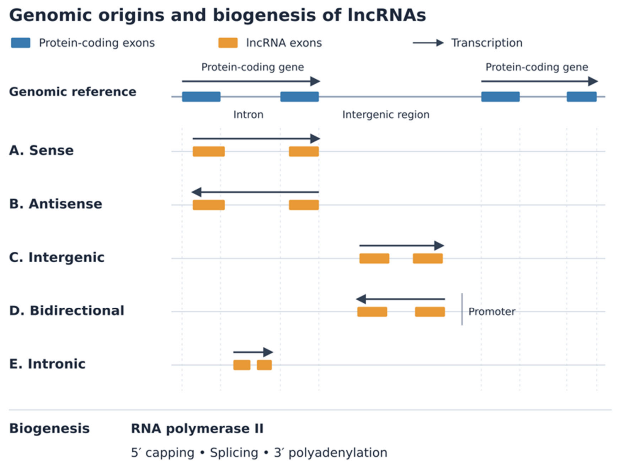
Figure 2.
Schematic overview of immune cell populations associated with NEAT1, reorganized into innate, adaptive, and other immune cell categories. Conceptual content adapted from reference [74].
Figure 2.
Schematic overview of immune cell populations associated with NEAT1, reorganized into innate, adaptive, and other immune cell categories. Conceptual content adapted from reference [74].
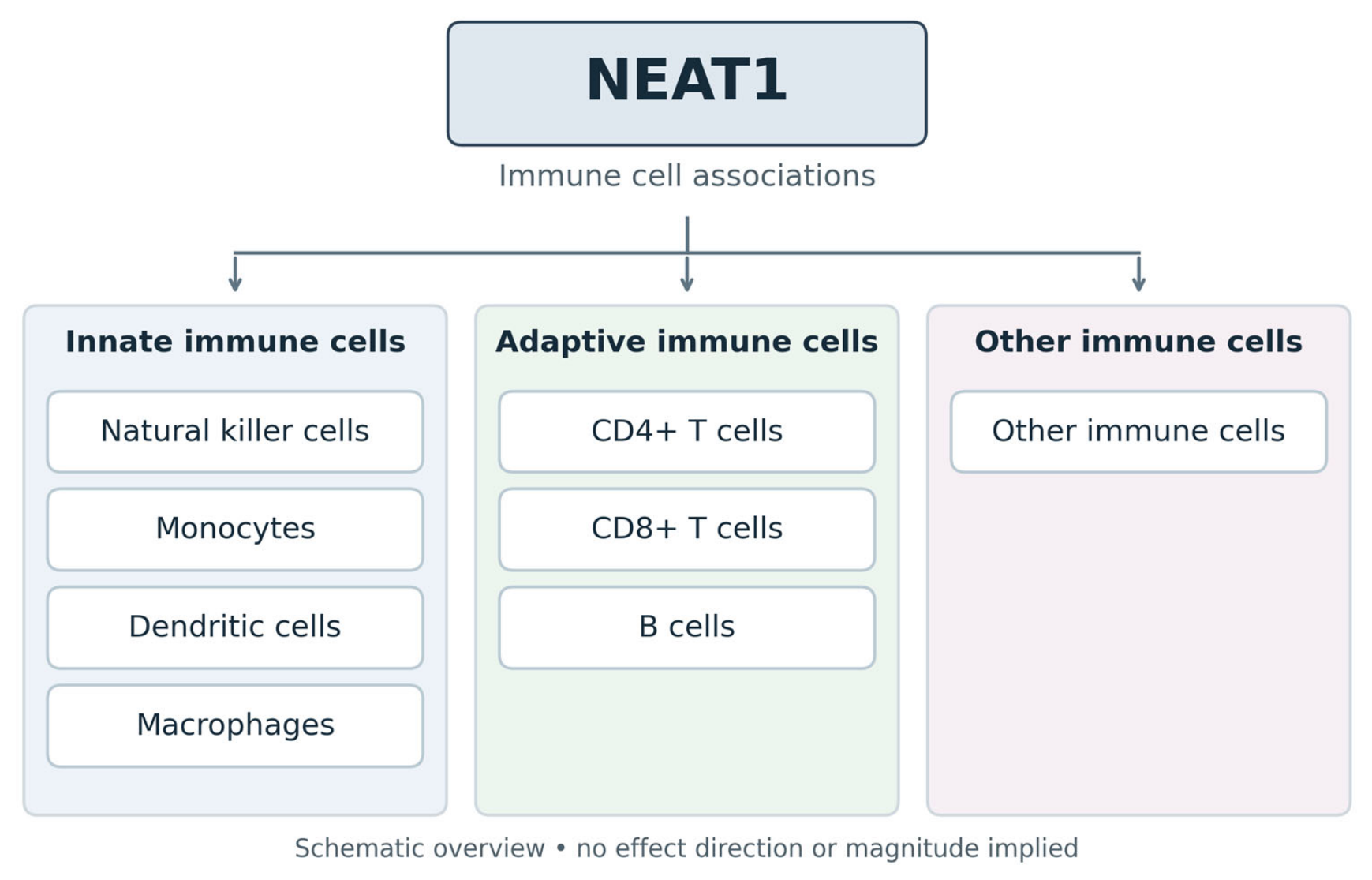

Table 1.
lncRNA expression in ITP varies according to the patient population and biological sample.
| Title 1 | Title 2 | Title 3 |
| Long non-coding RNA | Expression compared with healthy controls | Biological sample |
| HCP5 [5] | Decreased in adults with ITP | Peripheral venous whole blood |
| NEAT1 [81] | Increased in children with ITP | Serum |
| lnc-DC [81] | Increased in children with non-chronic ITP | Serum |
| lnc-DC [111] | Decreased in adults with primary ITP | Plasma |
| MEG3 [86] | Increased in patients with ITP | Isolated CD4+ T cells |
| MEG3 [88] | Increased in adults with ITP | Serum |
| GAS5 [92] | Decreased in adults with ITP | Peripheral blood mononuclear cells (PBMCs) |
| GAS5 [93] | Decreased in adults with ITP | Serum |
| GAS5 [105] | Increased in children with ITP | Serum |
| MALAT1 [96] | Increased in children with ITP | Serum |
| IFNG-AS1 / TMEVPG1 [104] | Decreased in adults with active primary ITP | PBMCs |
| IFNG-AS1 [105] | Increased in children with ITP | Serum |
| THRIL [96] | Increased in children with ITP | Serum |
| THRIL [111] | Increased in adults with primary ITP | Plasma |
Disclaimer/Publisher’s Note: The statements, opinions and data contained in all publications are solely those of the individual author(s) and contributor(s) and not of MDPI and/or the editor(s). MDPI and/or the editor(s) disclaim responsibility for any injury to people or property resulting from any ideas, methods, instructions or products referred to in the content. |
© 2026 by the authors. Licensee MDPI, Basel, Switzerland. This article is an open access article distributed under the terms and conditions of the Creative Commons Attribution (CC BY) license.
Copyright: This open access article is published under a Creative Commons CC BY 4.0 license, which permit the free download, distribution, and reuse, provided that the author and preprint are cited in any reuse.