

Submitted:
11 October 2026
Posted:
13 October 2026
You are already at the latest version
Abstract
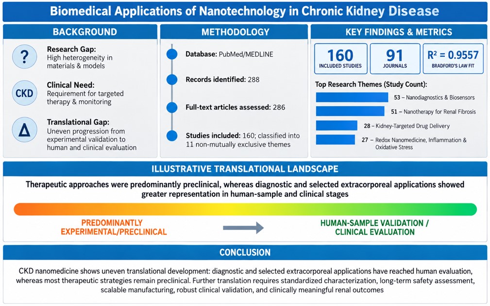
Background: Nanotechnology has expanded biomedical applications in chronic kidney disease (CKD), including diagnostics, monitoring, targeted therapy, and extracorporeal management. However, heterogeneity in materials, experimental models, and translational development complicates assessment of clinical progress. This review characterized major CKD nanotechnology applications and their documented translational stages, supported by complementary bibliometric analysis. Methods: English-language original studies indexed in PubMed/MEDLINE between July 1, 2021, and July 1, 2026, were organized into 11 overlapping thematic categories and classified according to their highest documented translational stage. Bibliometrix/Biblioshiny characterized publication, journal, institutional, and geographic patterns, while VOSviewer analyzed MeSH term co-occurrence. Results: Of 288 records identified, 160 studies across 91 journals were included. The most frequent categories were nanodiagnostics and biosensors (53 studies), nanotherapy for renal fibrosis (51), kidney-targeted drug delivery (28), and redox nanomedicine, inflammation, and oxidative stress (27). Diagnostic platforms more frequently reached human-sample validation or early clinical evaluation, whereas therapeutic approaches remained predominantly preclinical. Extracorporeal nanotechnologies were largely experimental, with limited human-sample evaluation. The 10 most productive journals accounted for 55 studies (34.4%), illustrating the concentration of publications within a multidisciplinary field. Conclusions: CKD nanomedicine exhibits substantial technological diversity but uneven translational development. Progress toward clinical implementation requires standardized physicochemical characterization, reproducibility, independent validation, long-term safety assessment, manufacturing scalability, and rigorous comparisons with established standards of care using clinically meaningful outcomes.
Keywords:
chronic kidney disease
; nanotechnology
; translational research
1. Introduction
Chronic kidney disease (CKD) is defined by abnormalities of kidney structure or function persisting for at least three months with implications for health [1]. It is identified by a sustained estimated glomerular filtration rate (eGFR) below 60 mL/min/1.73 m² or persistent markers of kidney damage, including albuminuria, urinary sediment abnormalities, histopathological findings, or structural abnormalities, even when glomerular filtration is preserved [1]. Classification according to cause, GFR category (G1–G5), and albuminuria category (A1–A3), following the cause–GFR–albuminuria (CGA) framework, supports prognostic assessment and risk stratification for disease progression, cardiovascular complications, and kidney failure [1].
CKD affects more than 780 million people worldwide, corresponding to approximately 10% of the adult population [1]. Prevalence increases substantially with age, diabetes, and hypertension and may exceed 30% in high-risk populations [1]. Frequently asymptomatic in its early stages, CKD remains underdiagnosed and contributes substantially to global morbidity and mortality [1,2,3,4]. Its burden is particularly pronounced in socioeconomically vulnerable populations with limited access to healthcare [1,2,3,4]. Despite advances in nephroprotective strategies, early detection, individualized risk assessment, and longitudinal monitoring remain major clinical challenges [2,3,4].
CKD progression involves interconnected mechanisms, including persistent inflammation, oxidative stress, mitochondrial dysfunction, tubular injury, and tubulointerstitial fibrosis [2,3]. Their relative contributions vary across etiologies, creating biological and molecular heterogeneity that complicates early diagnosis, cell-specific targeting, and selective modulation of pathways driving progressive kidney dysfunction [2,3,4,5,6]. These challenges have stimulated interest in technologies capable of improving molecular detection and enabling more precise therapeutic interventions.
Nanotechnology has emerged as a versatile approach to CKD diagnosis, monitoring, and treatment [7,8,9]. Nanoparticles, nanocarriers, biosensors, nanozymes, microenvironment-responsive systems, and extracellular vesicle-based platforms have been investigated to enhance analytical sensitivity, facilitate point-of-care monitoring, and improve kidney-targeted therapeutic delivery [7,8,9]. Depending on their design, nanostructured systems may improve drug stability, pharmacokinetics, cellular uptake, and tissue selectivity while enabling controlled release or integrating diagnostic and therapeutic functions [9].
Nevertheless, the expanding literature encompasses heterogeneous materials, experimental models, biological targets, and translational stages [9,10,11,12]. This diversity complicates comparisons across studies and the distinction between experimental proof-of-concept and technologies evaluated in human samples or clinical settings [9,10,11,12]. Moreover, publication growth does not necessarily reflect clinical advancement, as diagnostic, therapeutic, regenerative, and extracorporeal technologies face distinct translational requirements. Integrating thematic synthesis with translational-stage assessment and complementary bibliometric analysis may therefore help distinguish research activity from documented progress toward clinical application.
Accordingly, this review aimed to characterize the principal biomedical applications of nanotechnology in CKD and their highest documented translational stages in recent PubMed/MEDLINE-indexed literature. Complementary bibliometric analyses were used to contextualize publication patterns and research organization. By integrating these perspectives, this review sought to identify dominant research directions, differences in translational development, and persistent barriers to clinical implementation. The translational-stage classification is descriptive and should not be interpreted as a validated measure of evidence quality or clinical readiness.
2. Materials and Methods
This thematic and translational review, supported by complementary bibliometric analysis, searched PubMed/MEDLINE on July 1, 2026, for publications from July 1, 2021, to July 1, 2026. The search combined Medical Subject Headings (MeSH) with free-text terms in titles and abstracts (Table 1).
The PubMed/MEDLINE search identified 288 records, which were exported to Rayyan [13] for duplicate removal and eligibility assessment. After removing two duplicates, the titles and abstracts of 286 records were screened, followed by full-text eligibility assessment of all 286 articles. Eligible original studies investigated nanotechnologies, nanomaterials, or nanostructured systems for CKD diagnosis, monitoring, treatment, or management, including CKD-related complications, across in vitro, ex vivo, preclinical in vivo, human-sample, observational, and interventional designs. Exclusion criteria comprised studies unrelated to CKD, restricted to acute kidney injury without assessment of progression to CKD, lacking a defined nanotechnology component, or published as reviews, editorials, letters, commentaries, conference abstracts, protocols, preprints, or non-English articles. Eligibility was jointly assessed by the authors, with decisions reached by consensus. Of the 286 full-text articles assessed, 126 were excluded, yielding 160 studies for thematic synthesis, translational assessment, and complementary bibliometric analysis.
The included studies were assigned to 11 thematic categories developed iteratively by consensus, based on study objectives, nanotechnology applications, and the clinical or pathophysiological context of CKD. Categories were pragmatic, overlapping, and not hierarchically equivalent, reflecting the multiple roles of nanotechnologies as therapeutic agents, drug-delivery or targeting systems, diagnostic substrates, experimental probes, and extracorporeal components. Accordingly, individual studies could contribute to multiple categories, and cumulative thematic frequencies exceeded the number of articles included.
Translational stages were assessed descriptively within each category, using the corresponding number of studies as the percentage denominator. Each study was classified according to the highest documented stage of the investigated technology: (i) in vitro/ex vivo experimental, involving acellular systems, cell cultures, tissues, organoids, or artificial, simulated, or modified biological matrices without in vivo assessment or direct evaluation in human participants; (ii) preclinical in vivo, involving animal models, regardless of additional experimental or ancillary analyses of human-derived samples; (iii) human-sample validation, involving direct testing of biological material from human participants without clinical evaluation of the technology in those participants; (iv) clinical observational, involving evaluation of the technology in human participants without prospective intervention; (v) clinical interventional, involving prospective administration or assignment of the investigated intervention; and (vi) established clinical use, reflecting documented clinical application for CKD or related conditions.
This operational framework was descriptive rather than formally validated or strictly linear. Studies encompassing multiple stages were assigned to the highest stage at which the technology was directly evaluated, except for established clinical use, which was determined from documented clinical practice.
Complementary bibliometric analyses were conducted using VOSviewer (version 1.6.20) and the Bibliometrix package [14], including its Biblioshiny interface, in R (version 4.6.1). Bibliographic records of the 160 included studies were exported from PubMed/MEDLINE. VOSviewer was used to analyze MeSH term co-occurrence using full counting, without an additional thesaurus file. Of 455 identified MeSH terms, 57 met the minimum occurrence threshold of five and were included in the network.
Bibliometrix/Biblioshiny was used to assess publication trends, journal productivity, institutional affiliations, and geographic distribution of research output. Journal concentration was evaluated using Bradford’s Law, with journals ranked by publication count and divided into three zones containing approximately equal numbers of articles. The empirical distribution was compared with the theoretical Bradford model using regression analysis and the Kolmogorov–Smirnov test. The Bradford multiplier was estimated from the fitted model rather than from observed journal-count ratios between zones, while the coefficient of determination (R²) was used to assess model fit. Figure 1 summarizes the methodological workflow, including study selection, thematic classification, translational-stage assessment, and complementary bibliometric analyses.
3. Thematic Review and Translational-Stage Assessment
3.1. Thematic Structure and MeSH Term Co-Occurrence Analysis
Figure 2 presents the MeSH term co-occurrence network generated using VOSviewer. Cluster colors were assigned algorithmically, and thematic interpretations were based on the predominant and highly connected terms. The red cluster primarily reflected nanodiagnostics and analytical technologies, including biomarkers, metal nanoparticles, electrochemical techniques, Raman spectroscopy, urea, cystatin C, and quantum dots. The green cluster predominantly represented preclinical nanotherapeutic research, encompassing animal models, renal fibrosis, inflammation, oxidative stress, mitochondrial dysfunction, and drug delivery systems.
The blue cluster connected chronic renal insufficiency with extracellular vesicles, mesenchymal stem cells, vascular calcification, and calcium-related processes. The smaller yellow cluster contained heterogeneous mechanistic terms, including adenine and transforming growth factor beta, limiting its interpretation as a distinct research domain. Overall, the network highlighted two interconnected research domains: diagnostic and analytical technologies, and preclinical therapeutic approaches.
The connectivity among terms related to humans, chronic renal insufficiency, animals, and nanoparticles reflected overlapping research interests rather than direct evidence of clinical translation. Consistent with these bibliometric patterns, the thematic distribution (Figure 3) was dominated by nanodiagnostics and biosensors (53 studies) and nanotherapy for renal fibrosis (51), followed by kidney-targeted drug delivery (28) and redox nanomedicine, inflammation, and oxidative stress (27). The remaining seven categories comprised 4–14 studies each. Because thematic categories were not mutually exclusive, the 160 included studies accounted for 240 thematic assignments.
3.2. Nanodiagnostics and Biosensors
Creatinine was the most frequently investigated biomarker. Fluorescent nanosensors incorporating carbon dots, graphene oxide, copper nanocomposites, or gold nanoparticles were evaluated primarily in human urine [15,16,17], whereas electrochemical platforms based on MXenes, metallic nanoparticles, molecularly imprinted polymers, and carbon-based electrodes targeted creatinine in serum, urine, and saliva, including potential point-of-care applications [18,19,20,21]. Several platforms demonstrated high analytical sensitivity and selectivity, with some showing agreement with the Jaffé method in human samples [15,20]. Additional nanozyme-based and silver nanoparticle colorimetric assays were evaluated in animal, artificial, or spiked samples [22,23]. Nanostructured sensors also targeted urinary proteins and uremic toxins [24,25]. Representative strategies are illustrated in Figure 4.
Multianalyte sensing expanded CKD-related biomarker monitoring through paper-based electrochemical devices detecting ammonium, urea, and creatinine in human urine [26], molecularly imprinted polymer–carbon nanotube sensors enabling albumin-to-creatinine ratio determination [27], and smartphone-integrated electrochemical platforms evaluated in urine from patients with diabetes [28]. Additional approaches included electrochemical detection of creatinine and uric acid and surface-enhanced Raman spectroscopy (SERS) for creatinine and urea in artificial blood, urine, and sweat [21,29]. Wearable gold nanoparticle-containing silk patches enabled urea detection in artificial sweat [30], while graphene oxide–cellulose nanofiber hydrogels incorporating polyacrylic acid served as colorimetric sensors and substrates for mass spectrometric analysis [31].
Other nanostructured platforms targeted additional CKD-related analytes. Enzyme-mimetic nanocrystals incorporated into wood membranes enabled electrochemical detection of salivary uric acid without sample pretreatment [32]. A gold nanoparticle-based microfluidic aptasensor measured potassium in whole blood from patients with CKD, with results compared against ion-selective electrode measurements [33]. Portable laser-induced graphene sensors incorporating printed nanomaterials enabled non-invasive monitoring of potassium, creatinine, and lactate in sweat [34], while an ion-imprinted nanocomposite sensor was evaluated for calcium monitoring in dialysate samples from patients with CKD [35].
Protein-related biomarkers were investigated using nanostructured sensing platforms. Gold- and silver-nanoparticle-based SERS differentiated individuals with proteinuria from healthy controls in 111 human urine samples [36]. Europium nanoparticle-based lateral-flow immunoassays enabled urinary cystatin C detection, with results compared against ELISA [37], while paper-based microfluidic devices incorporating molecularly imprinted Mn–ZnS quantum dots measured the urinary albumin-to-creatinine ratio [38]. A zwitterionic nanohydrogel containing gold nanoparticles and peptide receptors enabled electrochemical sclerostin detection in samples from healthy individuals and patients with osteoporosis or CKD [39].
SERS applications further included silver-coated gold nanostars for creatinine detection in human saliva and sweat without prior separation [40], and silver nanoparticle-containing cellulose aerogel–hydrogel systems combining filtration, analyte enrichment, and detection in urine from healthy individuals and patients with stage 5 CKD [41]. Silver nanoparticle-on-PDMS substrates enabled multiplexed detection of uremic metabolites, including adenine, with selected analytes evaluated in serum from patients with kidney failure [42]. Label-free SERS was also applied to post-dialysis effluent for β2-microglobulin and leptin detection [43]. Integrating Raman spectroscopy with computational analysis, deep-learning algorithms classified serum spectra from patients with diabetic nephropathy or primary Sjögren syndrome and healthy controls using silver nanoparticle substrates [44]. Raman signatures of urinary extracellular vesicles were also associated with CKD stage and estimated glomerular filtration rate in patients with type 2 diabetes [45].
Fluorescent and colorimetric nanosensors expanded CKD-related biomarker detection. Smartphone-readable carbon-dot fluorescence systems quantified cystatin C in healthy individuals and patients with CKD [46], while gold nanoparticle-based lateral-flow immunoassays measured serum cystatin C against a laboratory reference method [47]. Ce–Zr–O nanozymes with phosphatase-like activity were also employed for fluorometric detection of urinary phosphate [48].
Electrochemical approaches included g-C₃N₄ nanozymes combined with molecularly imprinted polythiophene for creatinine detection in spiked human saliva [49]. Cystatin C was measured using laser-induced graphene sensors incorporating gold nanoparticles and manganese oxide [50], and MoS₂ quantum dot-modified paper electrodes evaluated in human whole blood and plasma [51]. Molecularly imprinted sensors detected hippuric acid in spiked urine [52], whereas chitosan-based nanocomposite immunosensors targeted indoxyl sulfate in fortified urinary samples [53].
Decentralized testing was further explored using a paper-based electrochemical biosensor integrated into a urine collection container, enabling simultaneous pH and glucose measurements in human urine, with results compared against commercial methods [54]. Collectively, these approaches illustrate the integration of nanomaterials with portable sensing platforms, miniaturized electronics, and digital readout technologies for potential decentralized renal biomarker monitoring.
Beyond conventional biosensors, a solid-state nanopore system combined with a sandwich immunoassay quantified urinary RBP4 and differentiated patients with CKD from healthy controls [55]. Breath-based ammonia detection was investigated using polypyrrole and gallium nitride nanostructure arrays coupled with exploratory neural-network analysis for CKD screening [56], and wearable CeO₂@polyaniline sensors evaluated in patients with uremia [57]. At the preclinical stage, a microneedle biosensor incorporating nanostructured bioelectrodes enabled longitudinal monitoring of pharmacokinetic clearance across different degrees of renal dysfunction and during therapeutic intervention in rats [58]. These approaches highlight the potential of minimally invasive and continuous monitoring, although clinical validation remains limited.
Nanotechnology also supported experimental renal imaging. Biodegradable black phosphorus quantum dots with renal clearance properties enabled photoacoustic detection of acute kidney injury and CKD with greater sensitivity than conventional serum indices in mice [59]. Gadolinium- and zinc-containing inulin nanoparticles combined renal targeting, rapid clearance, and magnetic resonance imaging contrast with experimental antifibrotic activity in HK-2 cells and mice subjected to unilateral ureteral obstruction [60]. Methemoglobin-containing liposomes enhanced T1-weighted magnetic resonance signals as intravascular contrast agents in mice with chronic renal insufficiency, with limited adverse biological responses [61]. Mechanistic studies further demonstrated that phosphate exposure increased hyaluronic acid content in vascular smooth muscle cells and extracellular vesicles, enhancing their interactions with ultrasmall superparamagnetic iron oxide particles, with similar findings in plasma vesicles from uremic rats [62].
Additional nanotechnology-based platforms extended biomarker monitoring to sweat and urinary extracellular vesicles. A portable, flexible electrochemical biosensor incorporating ternary NiCoMOF electrodes, multiwalled carbon nanotubes, and nitrogen-doped carbon dots simultaneously detected urea, creatinine, and uric acid in human sweat [63]. In a veterinary CKD study, urinary extracellular vesicle profiling identified five differentially expressed proteins between normotensive and hypertensive cats with CKD, although no differences were detected between healthy animals and the combined CKD population [64].
Advanced analytical platforms also targeted uremic toxins and proteinuria. Aminofunctionalized fibrous silica–graphene oxide nanocomposites enabled dispersive solid-phase extraction, preconcentration, and fluorometric determination of p-cresyl sulfate and were evaluated in spiked samples and plasma from five patients with CKD [65]. A fluorescent sensor combining aminofunctionalized gold nanoparticles and DNA exploited differences in urinary protein size and charge; integrated with principal component analysis and a PSO-BP neural network, it was used to classify renal injury progression using data from animal models and 62 urine samples from patients with CKD [66]. Finally, an electrochemical nanozyme based on ABTS-functionalized carbon nanotubes enabled voltammetric detection of urinary albumin without sample pretreatment, supporting microalbuminuria assessment [67].
3.3. Nanotherapy for Renal Fibrosis
Strategies targeting matrix-producing cells and the fibrotic interstitium demonstrated promising effects in preclinical models. PDGFRβ-directed CAR-T cells, administered by adoptive transfer or generated in vivo using lipid nanoparticles, reduced renal and cardiovascular fibrosis in murine CKD models, with antifibrotic effects also observed in human kidney organoids. In this approach, lipid nanoparticles served as delivery vehicles for CAR-T-cell generation rather than direct therapeutic effectors [68]. Collagen IV-binding nanoparticles enhanced distribution within the fibrotic interstitium and fibroblast uptake, although therapeutic efficacy was not demonstrated [69]. ROS-responsive, CD44-targeted nanoparticles improved celastrol delivery to activated myofibroblasts, reducing fibrotic markers and systemic toxicity [70], while myofibroblast-targeted sorafenib delivery reduced collagen deposition and improved renal plasma flow in a unilateral ureteral obstruction model [71].
Inorganic nanomaterials with intrinsic antioxidant activity also showed antifibrotic potential. Gold–platinum nanoparticles exhibiting catalase- and peroxidase-like activities reduced apoptosis, inflammatory cytokine release, inflammasome formation, and long-term fibrosis following renal ischemia–reperfusion [72]. Ultrasmall ceria nanoclusters (~1.2 nm) combined potent ROS-scavenging activity with lower organ accumulation and faster elimination than larger particles, attenuating oxidative stress, inflammation, apoptosis, and fibrosis in CKD mice [73]. Lactoferrin functionalization enhanced renal targeting and antifibrotic efficacy of cerium oxide nanoparticles compared with non-functionalized particles in unilateral ureteral obstruction [74]. PEG-coated nanoceria further reduced epithelial-to-mesenchymal transition and fibrosis, restored mitochondrial membrane potential, and promoted oxidative phosphorylation over aerobic glycolysis in HK-2 cells and murine renal fibrosis models [75].
Carbon-based nanomaterials also demonstrated intrinsic antioxidant and antifibrotic activity. Graphene quantum dots attenuated inflammation, apoptosis, and fibrosis in adriamycin nephropathy and subtotal nephrectomy models, while restoring podocyte cytoskeletal organization and mitochondrial function through modulation of TRPC5 activity, intracellular calcium, and mitochondrial membrane potential [76]. In unilateral ureteral obstruction, graphene quantum dots reduced ROS generation, pro-inflammatory cytokines, apoptosis, and TGF-β1/Smad signaling while suppressing tubular epithelial-to-mesenchymal transition and fibrosis [77]. Mulberry-derived carbon dots improved renal function and attenuated fibrosis in experimental diabetic nephropathy through direct interaction with the PI3K complex and inhibition of its kinase activity [78].
Additional antifibrotic strategies integrated redox modulation, metabolic signaling, and responsive drug delivery. In adenine-induced CKD, zinc oxide nanoparticles enhanced the nephroprotective effects of spironolactone, with activation of Nrf2/HO-1 and suppression of inflammatory and profibrotic mediators associated with TGF-β1 and Wnt/β-catenin signaling [79]. Dietary magnesium hydride nanoparticles provided sustained hydrogen release and reduced renal dysfunction and fibrosis in high-fat diet-fed mice, with effects associated with PTEN modulation and inhibition of TGF-β/Smad2/3 and AKT/mTOR signaling [80]. In renal ischemia–reperfusion, hyaluronic acid-conjugated reduced graphene oxide nanoparticles loaded with paricalcitol combined CD44-mediated renal targeting, ROS scavenging, and oxidative stress-responsive drug release, attenuating tubular injury, inflammation, apoptosis, and fibrosis during AKI-to-CKD transition [81].
Mitochondrial preservation emerged as a key therapeutic strategy. PEG- and triphenylphosphine-functionalized cobalt oxide nanoparticles accumulated in proximal tubular cells and induced BNIP3-mediated mitophagy, attenuating chronic injury progression in murine ischemia models and gentamicin-exposed zebrafish [82]. A cascade-targeting system combining neutrophil-mediated glomerular transport, MMP-2/9-responsive release, and KIM-1 recognition delivered nicotinamide mononucleotide to injured tubular cells, activating the NAD⁺–SIRT3 axis, restoring mitochondrial function, and reducing fibrosis during AKI-to-CKD transition [83]. Similarly, layered double hydroxide nanocarriers enabled pH-responsive butylphthalide delivery, attenuating renal dysfunction and fibrosis through PINK1–Parkin-dependent mitophagy [84].
Additional strategies targeted NAD⁺ metabolism and oxidative injury. Ultrasmall gallic acid–NAD⁺ nanoparticles crossed the glomerular filtration barrier, increased renal NAD⁺ availability, and restored mitochondrial and immune homeostasis through NAD⁺–SIRT1 signaling, suppressing TGF-β activity and AKI-to-CKD progression [85]. Dual ROS- and MMP-2-responsive nanoparticles delivered vitamin E to injured renal cells, reducing oxidative stress, ferroptosis, and CKD progression [86]. Positively charged cerium-doped polypyrrole nanoparticles enhanced proximal tubular uptake and mitochondrial localization, exerting intrinsic antioxidant effects that reduced mitochondrial dysfunction, apoptosis, and AKI-to-CKD transition through involvement of the Mpv17l pathway [87].
Ferroptosis-targeted nanoplatforms also demonstrated antifibrotic potential. Self-assembled peptide nanoparticles containing a p38-inhibitory domain preferentially accumulated in tubular epithelial cells, combining ROS scavenging and p38 inhibition to suppress ferroptosis through SLC7A11/GPX4 activation and attenuate AKI-to-CKD transition after ischemia–reperfusion [88]. K3 peptide-functionalized hollow polydopamine nanoparticles loaded with rosiglitazone exhibited prolonged retention in fibrotic kidneys, combining targeted delivery and iron chelation to reduce ferroptosis, oxidative stress, inflammation, and fibrosis, alongside ACSL4 downregulation and preservation of mitochondrial membrane potential [89].
Autophagy, senescence, and immune infiltration were addressed through complementary strategies. RDYH58 nanoparticles delivered RGS19-targeting siRNA, restoring autophagic flux, suppressing mTOR activation, reducing CXCL9/CXCL10 production and CD8⁺ T-cell infiltration, and attenuating collagen deposition in murine renal fibrosis models [90]. Glutaminase-responsive nanocarriers preferentially released rapamycin in senescent cells, restoring autophagy and reducing senescence, inflammation, and fibrosis in experimental CKD [91].
Tubular metabolic dysfunction represented another therapeutic target. Reduced MAGL expression promoted 2-arachidonoylglycerol accumulation, impaired fatty acid oxidation, and activated CB2/β-catenin signaling. However, 2-AG-loaded nanoparticles served as experimental tools to exacerbate lipotoxicity, whereas therapeutic benefits resulted from restoring MAGL activity [92]. In contrast, KIM-1-targeted nanoparticles delivered a Klotho plasmid to injured tubular cells, sustaining Klotho expression, enhancing PPARα-mediated fatty acid β-oxidation, and reducing lipid accumulation and fibrosis during AKI-to-CKD transition [93]. Phosphatidylserine-functionalized black phosphorus nanocomposites carrying the HDAC inhibitor FK228 targeted renal macrophages, modulated oxidative and inflammatory pathways, improved fatty acid metabolism, and attenuated fibrosis [94].
Renal-targeted nanocarriers enhanced the delivery of conventional and repurposed therapeutics. Biomimetic HDL nanoparticles exploited KIM-1-mediated uptake to deliver triptolide and nintedanib to injured tubular cells, reducing inflammatory cytokines, immune infiltration, myofibroblast activation, and triptolide-related toxicity in murine CKD models [95]. Acid-responsive chitosan nanoparticles carrying linagliptin accumulated in fibrotic rat kidneys and reduced creatinine, urea, TGF-β1, collagen I, and extracellular matrix deposition [96]. Chrysophanol encapsulation in N-octyl-O-sulfate chitosan increased bioavailability 2.57-fold and enhanced nephroprotective and antifibrotic effects [97], whereas nanoparticle-formulated quercetin achieved antifibrotic efficacy comparable to higher doses of its conventional formulation in adenine-induced CKD [98]. PLGA nanoparticles prolonged renal retention of eleutheroside B for up to seven days in obstructed kidneys, reducing α-SMA and collagen I through a Smad3-dependent mechanism [99].
Oral nanotherapies additionally targeted the gut–kidney axis. Deoxycholic acid-conjugated, chitosan-coated liposomes enhanced emodin absorption and, combined with an in situ colonic gel, reduced renal fibrosis and partially corrected dysbiosis in unilateral ureteral obstruction [100]. Thiolated chitosan–alginate nanoparticles delivering emodin improved renal function, intestinal barrier integrity, and beneficial bacterial abundance while reducing systemic inflammation and fibrosis in murine CKD [101]. An oral microgel incorporating self-assembled emodin–asiatic acid nanoparticles, yeast-wall particles, and Lactobacillus casei enhanced renal accumulation of therapeutic compounds, modulated gut microbiota, and attenuated fibrosis through TGF-β/Smad signaling [102].
Additional nanoformulations demonstrated nephroprotective and antifibrotic potential. Furosemide and tadalafil nanoformulations enhanced nephroprotection compared with conventional formulations, particularly in combination, reducing KIM-1, NGAL, caspase-3, IL-1β, and renal injury markers in adenine-induced chronic renal failure [103]. Chitosan nanoparticles containing bromelain-treated pea protein hydrolysate improved solubility, encapsulation, and sustained release, although robust in vivo evidence of antifibrotic efficacy was lacking [104]. In cisplatin nephrotoxicity, crab chitosan nanoparticles reduced creatinine, urea, KIM-1, oxidative stress, and apoptosis, with renal improvements reported as comparable or superior to mesenchymal stem cell treatment, although comparisons involved distinct experimental groups [105]. Oral poly(lipoic acid trisulfide) nanomedicine enabled sustained H₂S and lipoic acid release, reducing blood pressure, albuminuria, urea, and creatinine in hypertensive CKD mice while modulating the renin–angiotensin system and endothelial function [106].
Biomimetic and nucleic acid-based nanotherapies further expanded antifibrotic strategies. M2 macrophage membrane-coated nanoparticles delivered ADAM9-targeting siRNA to inflamed kidneys, suppressing AKT/NF-κB signaling, RAC1 and TRAF6 expression, macrophage infiltration, and M1 polarization, thereby attenuating inflammation and fibrosis [107]. Platelet membrane-camouflaged PLGA nanoparticles delivered TGF-β1-targeting siRNA, reducing inflammation and fibrosis through TGF-β1/Smad3 inhibition in unilateral ureteral obstruction and ischemia–reperfusion models [108]. Renal aptamer-functionalized phi29 three-way-junction RNA nanoparticles delivered anti-miR-34a, suppressing TGF-β, FGF2, and Wnt/β-catenin signaling while increasing Klotho, SMAD7, and SIRT1 expression in adenine-induced CKD [109]. MMP-9-responsive HDL nanoparticles co-delivering progranulin-targeting siRNA and quercetin restored PPARα signaling, inhibited NF-κB, modulated macrophage polarization, and reduced apoptosis, inflammation, and fibrosis more effectively than the corresponding free agents [110].
Extracellular vesicle-based therapies were also investigated. Mesenchymal stem cell-derived vesicles modified with superparamagnetic iron oxide nanoparticles enabled magnetic targeting to injured kidneys and CHIP delivery to tubular cells, promoting Smad2/3 degradation and reducing fibrosis and collagen deposition in experimental models [111]. Bone marrow mesenchymal stem cell-derived exosomes delivered Nedd4L, restored autophagy, and attenuated TGF-β1-induced epithelial-to-mesenchymal transition in HK-2 cells. Ceria nanoparticle pretreatment increased exosome secretion and biological activity, although antifibrotic efficacy remained unvalidated in vivo [112]. Vesicles derived from mesenchymal stromal cells of trained animals, enriched in miR-296, reduced collagen accumulation and increased angiogenesis- and hypoxia-related markers in rats subjected to unilateral ureteral obstruction [113].
Liposomal and multifunctional nanoplatforms further targeted inflammation, metabolism, and fibrosis. A single administration of JQ1-loaded liposomes after renal ischemia–reperfusion enhanced renal drug delivery, reduced neutrophil and monocyte infiltration, IL-6, CCL2, CCL5, and extracellular matrix protein production, and improved renal functional parameters during AKI-to-CKD transition [114]. KIM-1-targeted nanoliposomes delivered stiripentol to injured tubular cells, inhibiting LDHA activity, lactate production, histone lactylation, TGF-β1/Smad signaling, epithelial-to-mesenchymal transition, and fibrosis [115]. Ultrasmall gadolinium- and zinc-containing inulin nanoparticles combined renal targeting, T1-weighted magnetic resonance contrast, rapid clearance, and low experimental toxicity with ROS reduction and antifibrotic activity through TGF-β1 modulation in HK-2 cells and unilateral ureteral obstruction [60].
Regenerative and tissue-remodeling strategies yielded heterogeneous outcomes. A multifunctional scaffold incorporating PLGA, magnesium hydroxide, extracellular matrix components, zinc oxide, lactoferrin, nicotinamide-loaded liposomes, and extracellular vesicle-hybrid nanoparticles reduced inflammation, oxidative stress, apoptosis, and fibrosis in a 5/6 nephrectomy model, alongside increased PI3K–AKT and SIRT1 signaling and markers of podocyte and tubular recovery [116]. Conversely, VEGF-D- or VEGF-C C156S-based nanoparticles promoted renal lymphangiogenesis without improving established CKD and worsened renal function, inflammation, and fibrosis in one experimental model [117]. Sub-10-nm folic acid-conjugated gold nanoparticles crossed the glomerular filtration barrier, preferentially accumulated in fibrotic kidneys, and directly inhibited p38α MAPK, reducing tissue degeneration, extracellular matrix expression, and fibrosis after a single administration, with greater antifibrotic efficacy than captopril in a unilateral ureteral obstruction model [118]. Representative targeting strategies, therapeutic mechanisms, and nanoplatforms are summarized in Figure 5.
3.4. Kidney-Targeted Drug Delivery
Renal targeting was investigated at anatomical, cellular, and subcellular levels. Mesoscale nanoparticles delivered the renalase agonist RP81 to proximal tubules, reducing regulated necrosis, inflammation, and progression of cisplatin-induced renal injury toward CKD [119]. Peptide-functionalized lipid nanoparticles targeted celastrol to glomerular endothelial cells and podocytes, attenuating inflammation, endothelial injury, and systemic toxicity [120]. Beyond the renal parenchyma, antibody-functionalized albumin nanoparticles delivered EDTA to degraded elastin in calcified arteries, promoting reversal of CKD-associated vascular calcification [121]. At the subcellular level, PLGA–PEG–TPP nanoparticles delivered coenzyme Q10 to mitochondria and improved mitochondrial function in COQ8B-deficient HK-2 cells, although evidence remained limited to in vitro experiments [122]. Chitobionic acid-functionalized gene carriers also demonstrated selective tubular uptake through vimentin-mediated targeting in an Alport syndrome model, without definitive therapeutic efficacy [123].
The injured renal microenvironment provided additional opportunities for selective accumulation and responsive drug release. Collagen IV-targeted nanoparticles enhanced fibrotic interstitial retention and fibroblast uptake without demonstrated therapeutic efficacy [69]. ROS-responsive, CD44-targeted systems improved celastrol delivery to activated myofibroblasts [70], whereas dual ROS- and MMP-2-responsive nanoparticles delivered vitamin E to injured renal cells, reducing ferroptosis and AKI-to-CKD progression [86]. Glutaminase-responsive carriers selectively released rapamycin in senescent cells to restore autophagy [91], while MMP-9-responsive nanoparticles co-delivered progranulin-targeting siRNA and quercetin to fibrotic kidneys [110]. KIM-1-targeted nanoliposomes delivered stiripentol to injured tubular cells, attenuating lactate production, histone lactylation, and profibrotic signaling [115].
Biomimetic nanoplatforms exploited cellular membranes and adhesion mechanisms to enhance delivery to inflamed renal tissue. M2 macrophage membrane-coated nanoparticles delivered ADAM9-targeting siRNA, attenuating inflammation and fibrosis through AKT/NF-κB inhibition [107]. Platelet membrane-camouflaged nanoparticles similarly delivered TGF-β1-targeting siRNA, reducing renal injury in unilateral ureteral obstruction and ischemia–reperfusion models [108]. GSNO-loaded Cu/ZIF-8 nanoparticles enabled nitric oxide release and were associated with prevention of CKD-related hypertension in young rats, although active renal targeting was not demonstrated [124]. E-selectin-targeted polymer–peptide conjugates reduced post-ischemic inflammation but showed limited efficacy in adenine-induced CKD, accompanied by compensatory P-selectin and VCAM-1 expression [125]. Neutrophil membrane-coated black phosphorus nanosheets delivered DNase I, reducing ROS and NETosis in experimental calcium oxalate nephrocalcinosis [126], whereas activated mesangial cell membrane-coated nanoparticles enhanced glomerular accumulation and attenuated proliferative mesangial glomerulonephritis [127].
Subcellular and molecular targeting further expanded renal delivery strategies. PEG- and triphenylphosphine-functionalized cobalt oxide nanoparticles preferentially accumulated in proximal tubular cells and localized to mitochondria, inducing BNIP3-mediated mitophagy and attenuating AKI-to-CKD transition in ischemia and gentamicin nephrotoxicity models [82]. Renal aptamer-functionalized phi29 three-way-junction RNA nanoparticles delivered anti-miR-34a, suppressing profibrotic pathways and increasing antifibrotic mediators in murine CKD [109]. Iron oxide-modified extracellular vesicles enabled magnetic renal targeting and CHIP delivery to tubular cells, promoting Smad2/3 degradation [111]. PLGA nanoparticles prolonged renal retention of eleutheroside B and attenuated fibrosis through a Smad3-dependent mechanism [99], whereas linagliptin-loaded chitosan nanoparticles combined acid-responsive release with renal accumulation, improving functional and fibrotic markers [96].
Oral and multifunctional nanoplatforms further expanded CKD-targeted delivery strategies. An oral microgel incorporating self-assembled emodin–asiatic acid nanoparticles, yeast-wall particles, and Lactobacillus casei enhanced therapeutic compound delivery and renal accumulation, modulated gut microbiota, and attenuated fibrosis through TGF-β/Smad signaling [102]. Oral poly(lipoic acid trisulfide) formulations crossed mucosal and cellular barriers, enabling sustained H₂S and lipoic acid release and reducing blood pressure and renal injury in hypertensive CKD [106]. Ultrasmall gadolinium- and zinc-containing inulin nanoparticles combined renal targeting, rapid clearance, magnetic resonance contrast, and experimental antifibrotic activity [60]. A bioinstructive hybrid scaffold incorporating biomaterials, liposomes, and extracellular vesicles promoted renal repair-associated responses involving PI3K–AKT and SIRT1 signaling [116]. Conversely, nanoparticle-induced renal lymphangiogenesis failed to improve established CKD and exacerbated disease-related parameters in one experimental model [117].
Targeted delivery also addressed CKD-associated vascular complications. EDTA-loaded albumin nanoparticles functionalized with antibodies against degraded elastin selectively targeted calcified vascular walls and were associated with reduced senescent cell burden, NLRP3 expression, and components of the senescence-associated secretory phenotype in experimental CKD-associated vascular calcification, suggesting senomorphic activity alongside calcium chelation [128].
3.5. Redox Nanomedicine, Inflammation, and Oxidative Stress
Metal-based nanozymes and cerium-containing nanomaterials were investigated for their ability to modulate oxidative stress and inflammation. Gold–platinum nanoparticles with catalase- and peroxidase-like activities reduced apoptosis, inflammatory cytokine release, inflammasome activation, and fibrosis following renal ischemia–reperfusion [72]. Ultrasmall ceria nanoclusters (~1.2 nm) exhibited regenerative ROS-scavenging activity, lower organ retention, and faster elimination than larger particles, attenuating oxidative stress, inflammation, apoptosis, and CKD progression in mice [73]. Dietary nanoceria also attenuated structural renal alterations in adenine-induced CKD, although reductions in creatinine, urea, and phosphorus were reported primarily as trends toward improvement [129]. Conversely, prolonged nanoceria administration in healthy animals altered systemic iron homeostasis, increasing serum iron and hepatic transferrin while reducing hepatic iron stores [130].
Multifunctional Mn₃O₄ nanozymes further combined antioxidant activity with therapeutic delivery. Citrate-functionalized Mn₃O₄ nanoparticles loaded with tacrolimus integrated ROS scavenging, sustained immunosuppressive drug release, mitochondrial protection, and reduced inflammation in doxorubicin-induced CKD [131]. The same platform without tacrolimus restored redox balance and mitochondrial function in cisplatin-induced CKD, reducing glomerulosclerosis and fibrosis while partially preserving physiological ROS signaling [132]. Positively charged cerium-doped polypyrrole nanoparticles enhanced proximal tubular uptake and mitochondrial localization, reducing oxidative stress, mitochondrial dysfunction, and apoptosis while attenuating AKI-to-CKD transition through involvement of the Mpv17l pathway [87].
Mitochondrial homeostasis was another therapeutic target. PEG- and triphenylphosphine-functionalized cobalt oxide nanoparticles accumulated in proximal tubular mitochondria, inducing BNIP3-dependent mitophagy and attenuating ischemic renal injury and AKI-to-CKD progression [82]. Graphene quantum dots also exhibited intrinsic antioxidant activity, reducing ROS generation, apoptosis, pro-inflammatory cytokines, and TGF-β1/Smad signaling in unilateral ureteral obstruction [77].
Ferroptosis-targeted nanotherapies included self-assembled peptide nanoparticles with p38-inhibitory activity that preferentially accumulated in tubular epithelial cells through albumin-mediated protein corona interactions, suppressing ferroptosis via SLC7A11/GPX4 signaling and attenuating AKI-to-CKD transition [88]. In hypertensive nephropathy, SJB3-019A@MIL-PEGTK nanoparticles delivered the USP1 inhibitor SJB3-019A, reducing oxidative stress, lipid ROS, and ferroptosis-related markers in angiotensin II-exposed renal cells and in vivo models [133]. Dual ROS- and MMP-2-responsive nanocarriers enhanced vitamin E delivery to injured renal cells, reducing lipid peroxidation, ferroptosis, and AKI-to-CKD progression after ischemia–reperfusion [86].
Cellular senescence was also targeted using glutaminase-responsive poly-γ-glutamic acid nanocarriers that preferentially degraded in senescent cells, releasing rapamycin to restore autophagy and attenuate inflammation and fibrosis in experimental CKD [91]. Beyond the renal parenchyma, elastin-targeted albumin nanoparticles delivered EDTA to calcified vascular tissue, reducing senescence markers and NLRP3 expression in CKD-associated vascular calcification [128].
Inflammatory modulation was investigated using nanoplatforms targeting macrophages and activated renal cells. M2 macrophage membrane-coated nanoparticles delivered ADAM9-targeting siRNA to inflamed kidneys, suppressing AKT/NF-κB signaling, RAC1 and TRAF6 expression, macrophage infiltration, and M1 polarization, thereby attenuating inflammation and fibrosis [107]. MMP-9-responsive HDL nanoparticles co-delivered progranulin-targeting siRNA and quercetin, restoring PPARα signaling, inhibiting NF-κB, modulating macrophage polarization, and reducing apoptosis, inflammation, and fibrosis in unilateral ureteral obstruction [110].
Microenvironment-responsive targeting was further explored using celastrol-loaded chondroitin sulfate–bilirubin nanoparticles, which combined CD44-mediated myofibroblast uptake, ROS-responsive drug release, and intrinsic antioxidant activity. Enhanced accumulation in fibrotic kidneys was associated with reduced α-SMA and collagen I expression, improved renal functional parameters, and lower systemic toxicity than free celastrol [70]. E-selectin-targeted HPMA polymer–peptide conjugates reduced post-ischemic inflammation but showed limited efficacy in adenine-induced CKD, accompanied by compensatory P-selectin and VCAM-1 expression [125].
Additional approaches combined redox and immunological modulation. S-nitrosoglutathione-loaded Cu/ZIF-8 nanoparticles provided nitric oxide release, reduced renal oxidative stress, modulated renin–angiotensin system components, and prevented hypertension and renal dysfunction in juvenile rats with adenine-induced CKD [124]. Crab-derived chitosan nanoparticles exhibited intrinsic biological activity in cisplatin nephrotoxicity, increasing glutathione and albumin levels while reducing KIM-1, malondialdehyde, creatinine, urea, and caspase-3. However, comparisons with mesenchymal stem cells involved distinct acute and chronic injury groups, limiting direct interpretation of their relative efficacy [105].
Biomimetic nanoplatforms were also investigated in disease-specific inflammatory conditions. In proliferative mesangial glomerulonephritis, self-assembled Paotianxiong polysaccharide–α-lipoic acid nanoparticles coated with activated mesangial cell membranes enhanced glomerular accumulation and reduced ROS generation, inflammation, M1 macrophage polarization, and mesangial proliferation [127]. In calcium oxalate nephrocalcinosis, DNase I-loaded black phosphorus nanosheets coated with CD44-targeted neutrophil membranes combined biomimetic targeting with reductions in ROS production, neutrophil extracellular trap formation, and tubular injury [126].
Other strategies acted predominantly through systemic or extrarenal mechanisms. Oral poly(lipoic acid trisulfide) nanotherapy provided sustained H₂S and lipoic acid release, reducing blood pressure, albuminuria, urea, and creatinine while modulating renin–angiotensin system-related genes and endothelial function in hypertensive CKD [106]. Dietary magnesium hydride nanoparticles enabled sustained hydrogen release, attenuating inflammation, oxidative stress, and fibrosis in high-fat diet-induced CKD through mechanisms associated with PTEN modulation and inhibition of TGF-β/Smad2/3 and AKT/mTOR signaling [80].
Gut–kidney axis modulation was explored using emodin-loaded thiolated chitosan–alginate nanoparticles, which improved renal function, intestinal barrier integrity, and gut microbiota composition while reducing systemic inflammation and fibrosis. In Caco-2 cells, the formulation also attenuated oxidative stress and apoptosis compared with free emodin [101]. A multifunctional regenerative scaffold promoted PI3K–AKT-associated survival signaling and, in a 5/6 nephrectomy model, reduced inflammation, oxidative stress, apoptosis, and fibrosis while improving renal functional parameters and markers of podocyte and tubular recovery [116].
Safety findings underscored potential adverse effects of nanomaterials. Beyond altered systemic iron metabolism following prolonged nanoceria exposure [130], hydroxyapatite nanoparticles induced Ca²⁺ overload, mitochondrial dysfunction, oxidative stress, mitochondrial DNA damage, and NLRP3–caspase-1–gasdermin D-mediated pyroptosis. Renal particle accumulation was also associated with kidney injury markers in mice [134].
3.6. CKD Complications and Dialysis Support
Nanotechnology-based approaches to CKD-related dialysis access complications focused on arteriovenous fistula (AVF) maturation and vascular remodeling. Gold nanoparticles incorporated into bioresorbable polycaprolactone perivascular wraps provided radiopacity for longitudinal imaging of mesenchymal stem cell-loaded constructs without compromising their beneficial effects on AVF maturation [135]. Bismuth-containing electrospun wraps enabled local delivery of rosuvastatin or rapamycin, reducing vascular stenosis in CKD rats; rosuvastatin primarily attenuated endothelial dysfunction and hypoxia, whereas rapamycin additionally modulated inflammation and cell proliferation [136]. Quercetin-loaded PLGA nanoparticles immobilized on decellularized tissue-engineered vascular grafts provided sustained drug release, suppressed M1 macrophage polarization, promoted M2 polarization, and improved early graft recellularization and neovascularization [137].
Mechanistic studies further characterized nanoparticle-assisted vascular access support. Transcriptomic analysis of bismuth-containing polycaprolactone wraps revealed downregulation of pathways associated with vascular smooth muscle cells, endothelial processes, and cell adhesion, alongside increased phagocytic and lysosomal activity. Importantly, bismuth incorporation did not substantially alter the scaffold's biological effects [138]. In experimental CKD, a light-responsive nanoparticle system enabled local verteporfin delivery to inhibit YAP1, implicated in myostatin-associated muscle wasting and AVF failure, reducing fistula stiffening and neointimal formation [139]. Bismuth-containing polycaprolactone wraps also combined sustained mesenchymal stem cell delivery with radiopaque imaging, attenuating neointimal hyperplasia, endothelial dysfunction, hypoxia, and vascular inflammation in experimental AVFs [140].
Nanomedicine was also investigated for CKD-associated hypertension. In juvenile rats with adenine-induced CKD, polymeric nanoparticles enabled sustained sodium thiosulfate delivery, reducing systolic and diastolic blood pressure while modulating H₂S-generating enzymes, nitric oxide signaling, and the renin–angiotensin system [141]. GSNO-loaded Cu/ZIF-8 nanoparticles provided sustained nitric oxide release, preventing hypertension and reducing renal oxidative stress in young CKD rats [124]. A MIL-100-based nanocarrier delivered the USP1 inhibitor SJB3-019A, attenuating oxidative stress and ferroptosis in hypertensive nephropathy [133]. Oral poly(lipoic acid trisulfide) nanotherapy enabled sustained H₂S and lipoic acid release, reducing blood pressure, albuminuria, urea, and creatinine in experimental hypertensive CKD [106]. These findings highlight potential applications in systemic cardiovascular complications of CKD.
Mineral and electrolyte disturbances were addressed through nano-enabled and nanoparticle-related approaches. Oxylanthanum carbonate, incorporating nanoparticle-based lanthanum delivery, exhibited lower medication volume and required less volume per gram of phosphate bound than comparator phosphate binders [142]. In hemodialysis patients, high-dose sucroferric oxyhydroxide reduced primary and secondary calciprotein particles by 62% and 38%, respectively, alongside decreases in inflammatory mediators and serum pro-calcific activity, suggesting modulation of endogenous mineral–protein nanoparticles [143]. For hyperkalemia, a nanoporous ammonium-form merlinoite zeolite identified through machine-learning-assisted screening selectively bound potassium and reduced blood potassium without sodium release in experimental models [144].
Nanostructured formulations were also explored for CKD-associated pruritus. A lipid nanoparticle-based self-microemulsifying system enhanced oral exposure to difelikefalin acetate, a treatment for moderate-to-severe pruritus in adults undergoing hemodialysis, and demonstrated pharmacodynamic activity in experimental models, supporting further investigation as an alternative to injectable administration [145].
3.7. Artificial Kidney and Nanomaterial-Assisted Uremic Toxin Removal
Nanotechnology-based dialysis strategies aimed to improve the clearance of poorly removed uremic toxins, enhance hemocompatibility, and support the development of compact extracorporeal systems. Figure 6 schematically illustrates a nanoporous adsorbent concept for uremic toxin removal. A tetrahedral protein nanoparticle displaying 12 β2-microglobulin-binding nanobodies demonstrated nanomolar affinity and increased the target protein’s effective size by more than 50-fold, potentially facilitating removal through filtration or matrix capture. Despite selective binding, the platform has not been evaluated in integrated dialysis circuits or patients [146].
Cyclodextrin-coated magnetic nanoparticles were investigated as magnetically recoverable extracorporeal adsorbents. In human plasma and blood, α-, β-, and γ-cyclodextrin coatings reduced nonspecific protein adsorption per unit surface area without relevant alterations in hemolysis, platelet activation, or hemostatic parameters [147]. Under simulated uremic conditions, protein corona composition varied, particularly in complement, fibrinogen, fibronectin, immunoglobulin, and coagulation-related protein adsorption, without clear evidence of impaired hemocompatibility [148]. All three formulations captured uremic metabolites, with adsorption efficiency dependent on surface chemistry, contact time, and solution composition [149].
For hemoperfusion, amyloid-like nanocoatings enhanced activated carbon adsorption, increasing urea and creatinine capture nearly fourfold while improving adsorption of other compounds and reducing carbon debris, cellular damage, nonspecific protein adsorption, and platelet adhesion. Although evaluated in animal models of CKD, clinical hemoperfusion validation remains lacking [150].
Nanostructured filtration systems were also developed for wearable artificial kidneys. A dual-layer electrospun filter combined urease-mediated urea hydrolysis with ammonium adsorption, achieving complete urea hydrolysis within 1 hour and approximately 90% reduction in ammonium accumulation under continuous-flow conditions at blood-relevant urea concentrations [151]. Chitosan-derived porous carbon nanofibers incorporating nitrogen-doped zeolites increased creatinine removal approximately 1.8-fold compared with unmodified nanofibers and showed favorable cytotoxicity and hemolysis profiles, although validation remained limited to laboratory experiments [152].
Albumin coatings were investigated to improve sorbent hemocompatibility, reducing adsorption of proteins involved in complement activation, coagulation, and fibrinolysis without fully eliminating interactions associated with the uremic milieu [153]. Nylon 6 nonwoven materials incorporating modified carbon black exhibited increased hydrophilicity and removed approximately 80–85% of inulin and 80–90% of urea, although evaluation in dialysis circuits, human blood, or animal models remains lacking [154]. Amino-functionalized and molecularly imprinted mesoporous silica nanoparticles demonstrated differential adsorption of urea, creatinine, and uric acid, with greater urea affinity in imprinted materials and preferential creatinine and uric acid adsorption in amino-functionalized formulations, although testing was restricted to simulated media [155].
Nanoporous poly(methyl methacrylate) fibers with 12–15 nm pores selectively adsorbed β2-microglobulin and other middle-molecular-weight proteins while limiting albumin penetration. Cross-shaped fiber geometries increased blood-contact surface area and exhibited lower predicted hydraulic resistance than bead-packed systems [156]. A nanobiocomposite incorporating sodium alginate, carrageenan, UiO-66-NH₂, and immobilized urease combined enzymatic urea hydrolysis with material-based retention, preserving 67.38% of enzyme activity after 24 hours of continuous reaction and achieving 75.05% urea removal during the fifth perfusion cycle. Despite favorable experimental biocompatibility, clinical safety and efficacy in peritoneal dialysis remain unestablished [157].
Related applications extended beyond extracorporeal toxin removal. In hemodialysis patients, high-dose sucroferric oxyhydroxide reduced circulating calciprotein particles, inflammatory mediators, and serum-associated pro-calcific activity, linking phosphate control to modulation of endogenous mineral–protein nanoparticles [143]. For dialysis monitoring, a label-free SERS platform incorporating silver nanoparticles and gold substrates on zirconia enabled sensitive and reproducible detection of β2-microglobulin and leptin in spent dialysate, supporting analytical monitoring rather than direct toxin removal [43].
3.8. Vascular Calcification and CKD–Mineral and Bone Disorder
Nanotechnology applications in CKD–mineral and bone disorder primarily targeted vascular calcification, mineral deposits, and calciprotein particles. Ultrasmall Cu₂₋ₓSe nanoparticles with intrinsic nanozyme-like activity attenuated vascular calcification in cellular, ex vivo, and animal models, reducing oxidative stress, mitochondrial dysfunction, and NF-κB/NLRP3 signaling [158]. Curcumin-loaded degradable polysuccinimide nanoparticles reduced aortic calcium deposition through calcium chelation, antioxidant activity, and inhibition of osteogenic transdifferentiation, without detectable bone alterations or significant organ toxicity under experimental conditions [159].
Targeted calcium removal was investigated using EDTA-loaded albumin nanoparticles functionalized with antibodies against degraded elastin. In experimental CKD-associated vascular calcification, this platform promoted selective accumulation in calcified arteries, reduced mineral deposition and BMP2/RUNX2 expression, and enabled calcium removal from aortic rings and calcified human femoral arteries ex vivo [121]. The same formulation also exhibited senomorphic effects, reducing vascular senescence markers, components of the senescence-associated secretory phenotype, and NLRP3 expression [128]. Tetracycline-functionalized PEG–PLA nanoparticles demonstrated hydroxyapatite-targeting feasibility with limited whole-body accumulation, although vitamin K encapsulation altered pharmacokinetics and systemic exposure, and therapeutic reversal of calcification was not established [160].
Calciprotein particles were additionally investigated as mediators of mineral toxicity and potential biomarkers. In a randomized crossover study involving 28 hemodialysis patients, high-dose sucroferric oxyhydroxide reduced primary and secondary calciprotein particles, inflammatory cytokines, and the pro-calcific and vascular-activating effects of patient serum [143]. In 52 cats with spontaneous azotemic CKD, plasma calciprotein particle concentrations were associated with FGF-23, parathyroid hormone, phosphate, and longitudinal changes in ionized calcium [161].
In human endothelial cells, these particles induced cytosolic and mitochondrial Ca²⁺ overload, impaired oxidative phosphorylation, reduced mitochondrial membrane potential and antioxidant capacity, and promoted endothelial activation and cell death. Partial attenuation by cyclosporine A suggested involvement of the mitochondrial permeability transition pore [162].
Mechanistic studies showed that inorganic phosphate increased hyaluronic acid content in vascular smooth muscle cells and extracellular vesicles, enhancing interactions with ultrasmall superparamagnetic iron oxide nanoparticles, with similar findings in plasma vesicles from uremic rats [62]. Calciprotein particles also induced epithelial-to-mesenchymal transition-like changes in a three-dimensional proximal tubular model [163]. A peptide receptor-functionalized zwitterionic nanohydrogel incorporating gold nanoparticles enabled electrochemical sclerostin detection with picomolar sensitivity and differentiated samples from healthy individuals and patients with CKD, supporting further investigation for CKD–mineral and bone disorder monitoring [39].
Nanotechnology was also explored for vitamin D delivery. A pilot, double-blind, randomized clinical trial of vitamin D₃ dispersed in soy protein nanoparticles provided preliminary feasibility evidence in adults with CKD, although interpretation was limited by the small sample size and short follow-up [164]. Conversely, hydroxyapatite nanoparticles exhibited intrinsic nephrotoxic potential, inducing Ca²⁺ overload, mitochondrial dysfunction, oxidative stress, mitochondrial DNA damage, and NLRP3–caspase-1–gasdermin D-mediated pyroptosis following lysosomal internalization. Renal accumulation was also observed in mice, accompanied by markers of kidney injury [134].
3.9. Molecular, Regenerative, and Systemic Therapies
Renal-targeted gene delivery was investigated using synthetic and biomimetic nanocarriers. Chitobionic acid-modified sorbitol–polyethylenimine copolymers exhibited low cytotoxicity, improved transfection efficiency, and vimentin-mediated proximal tubular targeting, with selective uptake by HK-2 cells and renal accumulation in Alport syndrome mice. However, therapeutic gene-mediated functional correction was not demonstrated [123]. Renal aptamer-functionalized phi29 three-way-junction RNA nanoparticles delivered anti-miR-34a in adenine-induced CKD mice, reducing miR-34a expression, modulating profibrotic pathways, and increasing Klotho, SMAD7, and SIRT1 expression, consistent with experimental antifibrotic activity [109].
Biomimetic siRNA delivery strategies included platelet membrane-coated PLGA nanoparticles targeting TGF-β1, which preferentially accumulated in injured kidneys and reduced TGF-β1 expression, Smad3 activation, inflammation, and extracellular matrix deposition in unilateral ureteral obstruction and ischemia–reperfusion models [108]. M2 macrophage membrane-coated nanoparticles enhanced ADAM9-targeting siRNA delivery to inflamed renal tissue, suppressing AKT/NF-κB signaling, RAC1 and TRAF6 expression, macrophage infiltration, and M1 polarization, thereby attenuating inflammation and fibrosis [107]. MMP-9-responsive HDL-like nanoparticles co-delivered progranulin-targeting siRNA and quercetin, promoting localized release in fibrotic tissue, modulating the PGRN–PPARα–NF-κB axis, and reducing apoptosis, inflammation, and fibrosis while increasing markers associated with M2 macrophage polarization [110].
Oral multifunctional delivery was also explored using chitosan–alginate microgels incorporating yeast-wall-encapsulated emodin–asiatic acid nanoparticles and Lactobacillus casei. This system prolonged intestinal probiotic retention, enhanced renal accumulation of therapeutic compounds, and attenuated TGF-β/Smad signaling and fibrosis [102]. Oral emodin delivery was enhanced using deoxycholic acid–chitosan-coated liposomes combined with a thermosensitive colonic gel, increasing intestinal absorption and bioavailability, modulating dysbiosis, and attenuating fibrosis in unilateral ureteral obstruction [100]. Thiolated chitosan–alginate nanoparticles similarly improved renal function, restored intestinal junction proteins, increased beneficial bacterial abundance, and reduced systemic inflammation and fibrosis [101].
Cellular senescence was targeted using glutaminase-responsive nanocarriers that preferentially released rapamycin in senescent cells, restoring autophagy and attenuating senescence, inflammation, and fibrosis in CKD mice [91]. In vascular calcification, degraded elastin-targeted albumin nanoparticles delivered EDTA to calcified tissue, reducing mineral deposition, inflammation, NLRP3 expression, and components of the senescence-associated secretory phenotype, indicating senomorphic activity without selective senescent cell elimination [128]. Extracellular vesicles derived from mesenchymal stem cells of trained animals and enriched in miR-296 reduced collagen deposition and increased CD34, HIF-1α, and Nrf2 expression in rats subjected to unilateral ureteral obstruction [113].
Regenerative strategies included a multifunctional scaffold incorporating PLGA, magnesium hydroxide, extracellular matrix components, zinc oxide, lactoferrin, nicotinamide-loaded liposomes, and extracellular vesicle-hybrid nanoparticles. In a 5/6 nephrectomy model, the scaffold reduced inflammation, oxidative stress, apoptosis, and fibrosis, alongside increased PI3K–AKT signaling, enhanced markers of podocyte and tubular recovery, and improved renal functional parameters [116]. Conversely, VEGF-D- or VEGF-C C156S-based nanoparticle strategies promoting renal lymphangiogenesis failed to improve renal function or fibrosis in established CKD and exacerbated inflammation, macrophage infiltration, and fibrotic progression in one experimental model [117].
3.10. Cell-Derived Nanosystems and Molecular Applications
Cell-derived nanosystems were investigated for molecular delivery, regeneration, intercellular communication, and diagnostic applications. M2 macrophage-derived exosomes carrying Stattic targeted parathyroid cells and macrophages in CKD-associated secondary hyperparathyroidism, inhibiting STAT3/mTOR signaling, restoring autophagy, suppressing parathyroid cell proliferation, and promoting anti-inflammatory macrophage polarization. These effects were demonstrated in primary human cells, macrophage organoids, and mice, with reduced inflammation and parathyroid hormone secretion [165]. Podocyte-derived exosomes loaded with siRNA or pre-miRNA retained characteristic vesicular properties, underwent time-dependent uptake by cultured podocytes, and modulated gene expression, reducing filamin A and increasing mature miR-21 levels [166].
Extracellular vesicles containing functional polycystin-1 reduced cyst growth in vitro and decreased kidney size and cystic index while increasing polycystin-1 levels in Pkd1-deficient mice, without relevant treatment-associated toxicity [167]. In adenine-induced CKD, extracellular vesicles derived from mesenchymal stem cells or expanded CD133⁺ cells improved renal functional parameters, although distinct histological responses suggested source-dependent biological effects [168].
Mesenchymal stem cell-derived exosomes also demonstrated experimental antifibrotic activity. Nedd4L-containing exosomes promoted autophagy and attenuated TGF-β1-induced epithelial-to-mesenchymal transition in HK-2 cells. Ceria nanoparticle pretreatment of donor cells increased exosome production and Nedd4L delivery, enhancing these in vitro effects; thus, nanoceria acted indirectly by modulating vesicle production and function [112]. In another approach, superparamagnetic iron oxide nanoparticle-modified extracellular vesicles carrying CHIP enabled magnetic targeting to injured kidneys, promoting Smad2/3 degradation and attenuating fibrosis-related changes and collagen deposition in experimental models [111].
Extracellular vesicles from bone marrow mesenchymal stromal cells of trained animals, enriched in miR-296, reduced collagen deposition and increased CD34, HIF-1α, and Nrf2 expression in unilateral ureteral obstruction, suggesting nephroprotective and pro-regenerative effects [113]. Cell-derived vesicles were also integrated into a multifunctional scaffold comprising PLGA, magnesium hydroxide, extracellular matrix components, zinc oxide, lactoferrin, nicotinamide-loaded liposomes, and extracellular vesicle-hybrid nanoparticles. In a 5/6 nephrectomy model, this regenerative platform reduced inflammation, oxidative stress, apoptosis, and fibrosis while improving renal functional parameters and markers of podocyte and tubular recovery [116].
Extracellular vesicles were further implicated in systemic and intercellular signaling. In adenine-induced CKD, the Niaodukang mixture increased exosome-associated miR-146a, attenuated TLR4/TRAF6/NF-κB signaling, and reduced pro-inflammatory cytokines [169]. Proteomic analysis of endothelial cells exposed to indoxyl sulfate revealed alterations in cellular and extracellular vesicle proteins involved in inflammation, extracellular matrix regulation, adipogenesis, and lipid metabolism, suggesting a role in CKD-associated endothelial dysfunction and intercellular communication [170].
Urinary extracellular vesicles were also investigated as diagnostic biomarkers. Raman spectroscopy revealed differences in protein- and lipid-associated spectral signatures between healthy individuals and patients with type 2 diabetes across CKD stages, with tryptophan- and amide III-related features associated with estimated glomerular filtration rate [45]. In cats with spontaneous CKD, ultrafiltration combined with size-exclusion chromatography improved vesicle preparation purity. Although vesicle size and concentration did not differ significantly between healthy and CKD animals, five proteins distinguished normotensive from hypertensive CKD, suggesting potential biomarker applications [64]. For molecular imaging, inorganic phosphate increased hyaluronic acid content in vascular smooth muscle cells and their extracellular vesicles, enhancing interactions with ultrasmall superparamagnetic iron oxide nanoparticles. Similar findings in plasma-derived vesicles from uremic rats suggested potential applications in CKD-associated vascular imaging [62].
3.11. Diagnostics, Monitoring, and Nanotheranostics
Surface-enhanced Raman spectroscopy (SERS) emerged as a prominent nanodiagnostic approach. Silver nanoparticle-coated polydimethylsiloxane (PDMS) substrates enabled reproducible multiplexed detection of uremic metabolites with submillimolar detection limits and quantification of selected analytes in serum from patients with kidney failure [42]. Silver nanoparticle-containing cellulose nanofiber–alginate aerogel–hydrogel composites combined filtration, analyte enrichment, and SERS detection, achieving high urine-sample classification accuracy with machine-learning analysis [41]. Flexible PDMS/ZIF-67@Ag substrates enabled simultaneous creatinine and urea monitoring in artificial blood, urine, and sweat using different sampling and enrichment modes [29]. For dialysis monitoring, a SERS platform combining silver nanoparticles with gold nanoparticle substrates on zirconia detected β2-microglobulin and leptin in spent dialysate [43]. Mercaptopropionic acid-capped, silver-coated gold nanostars enabled hydrogen-bond-mediated creatinine enrichment, achieving picomolar detection limits in solution and subnanomolar limits in human saliva and sweat [40].
Longitudinal functional monitoring was explored using a nanostructured microneedle biosensor. In animal models, serial drug measurements over several days yielded pharmacokinetic parameters comparable to blood-based measurements, detected renal dysfunction earlier than conventional biomarkers, and enabled monitoring of functional recovery during treatment [58].
Nanostructured contrast agents were investigated for renal and vascular imaging. Methemoglobin-encapsulating liposomes served as intravascular T1-weighted magnetic resonance imaging (MRI) contrast agents, increasing longitudinal relaxivity and enhancing vascular signals, particularly in venous structures, with limited adverse biological responses in mice with chronic renal failure [61]. Ultrasmall superparamagnetic iron oxide particles were also investigated as imaging probes interacting with extracellular vesicles. Inorganic phosphate increased hyaluronic acid content in vascular smooth muscle cells and their vesicles, enhancing magnetic particle interactions, with similar findings in plasma-derived vesicles from uremic rats [62].
Iron-based nanoplatforms further integrated therapeutic delivery and imaging. In anemic CKD mice, the iron oxide nanoparticle MPB-1523 enabled sustained iron delivery and MRI monitoring, increasing hematocrit and iron-storage parameters over time. MRI, histology, and electron microscopy indicated predominantly hepatic localization, without significant inflammatory or oxidative stress responses under the experimental conditions evaluated [171]. Formulation-dependent nano–bio interactions were investigated using clinically established intravenous iron–carbohydrate nanomedicines. Iron sucrose, characterized by a diffuse and dynamic carbohydrate shell, interacted significantly with human serum albumin and fibrinogen, forming nanoparticle–protein clusters. In contrast, ferric carboxymaltose exhibited a more localized carbohydrate shell and significant interactions with fibrinogen, but not albumin, under the experimental conditions. These findings highlight the influence of carbohydrate shell architecture on nanoparticle–protein interactions, although their implications for clinical outcomes remain uncertain [172].
3.12. Specific Kidney Diseases and Emerging Therapies
Disease-specific nanotechnologies addressed less frequently studied renal conditions. In COQ8B deficiency, triphenylphosphonium-functionalized PLGA-b-PEG nanoparticles delivered CoQ₁₀ to mitochondria, restoring mitochondrial metabolism more effectively than free CoQ₁₀ without detectable cytotoxicity in HK-2 cells [122]. In autosomal dominant polycystic kidney disease, kidney-targeted micelles co-delivering BRD4 and BMI1 inhibitors synergistically reduced patient-derived cell proliferation and three-dimensional cyst growth [173]. Emerging approaches included intravenous oxyhydrogen nanobubbles evaluated in three patients with CKD, with modest changes in serum creatinine, estimated glomerular filtration rate, and clinical symptoms. However, the absence of a control group, very small sample size, and clinical heterogeneity precluded conclusions regarding therapeutic efficacy [174]. In calcium oxalate nephrocalcinosis, DNase I-loaded black phosphorus nanosheets coated with neutrophil membranes reduced reactive oxygen species, neutrophil extracellular trap formation, crystal deposition, and renal injury in preclinical models [126].
3.13. Highest Documented Translational Stage Across Thematic Categories
Figure 7 illustrates differences in the highest documented translational stages across thematic categories. Nanodiagnostics and biosensors exhibited the broadest distribution: of 53 studies, 17 (32.1%) were in vitro/ex vivo experimental, 6 (11.3%) preclinical in vivo, 28 (52.8%) involved human-sample validation, and 2 (3.8%) were clinical observational [32,57]. None reached clinical interventional or established clinical-use stages. In contrast, nanotherapy for renal fibrosis (50/51; 98.0%) [112] and kidney-targeted drug delivery (27/28; 96.4%) [122] were predominantly preclinical in vivo, with one in vitro/ex vivo study in each category. All 27 studies on redox nanomedicine, inflammation, and oxidative stress were classified as preclinical in vivo.
CKD complications and dialysis support were predominantly preclinical in vivo (12/14 studies), with one in vitro/ex vivo study [142] and one clinical interventional study [143]. Artificial kidney and nanomaterial-assisted uremic toxin removal included 10 in vitro/ex vivo studies, one preclinical in vivo study [150], two human-sample validation studies [43,147], and one clinical interventional study [143]. Vascular calcification and CKD–mineral and bone disorder comprised eight preclinical in vivo studies, two in vitro/ex vivo studies [162,163], one human-sample validation study [39], and two clinical interventional studies [143,164]. Notably, study [143] evaluated the effects of phosphate-binding therapy on endogenous calciprotein nanoparticles in hemodialysis patients rather than an extracorporeal toxin-removal device, reflecting the overlapping and application-related nature of the thematic classification.
All 13 studies on molecular, regenerative, and systemic therapies were preclinical in vivo. Cell-derived nanosystems and molecular applications included nine preclinical in vivo studies, three in vitro/ex vivo studies [112,166,170], and one human-sample validation study [45]. Diagnostics, monitoring, and nanotheranostics included one in vitro/ex vivo study [29], four preclinical in vivo studies, four human-sample validation studies, and one classified as established clinical use [172]. The latter designation reflected the established clinical use of intravenous iron–carbohydrate nanomedicines, although the included study evaluated their physicochemical properties and protein interactions rather than clinical outcomes.
Specific kidney diseases and emerging therapies comprised one study at each of four stages (25.0% each): in vitro/ex vivo [122], preclinical in vivo [126], human-sample validation [173], and clinical interventional [174].
Overall, translational-stage distributions differed substantially across thematic categories. Therapeutic nanotechnologies remained concentrated at preclinical stages, particularly in renal fibrosis, kidney-targeted drug delivery, redox nanomedicine, and molecular or regenerative therapies. Conversely, nanodiagnostics showed greater representation at the human-sample validation stage, whereas clinical interventional studies were uncommon. One study was classified as established clinical use because it investigated intravenous iron–carbohydrate nanomedicines used for CKD-associated anemia, although its focus was physicochemical characterization rather than clinical evaluation [172]. These findings highlight an uneven translational landscape, although overlapping thematic assignments and the descriptive staging framework should be considered when interpreting differences in technological maturity.
3.14. Bibliometric Overview of the Literature
Figure 8A presents journal productivity, with the 10 most productive journals accounting for 55 of the 160 articles (34.4%). Journal of Nanobiotechnology led with 10 articles, followed by Analytica Chimica Acta (8), Biosensors & Bioelectronics and International Journal of Biological Macromolecules (6 each), and International Journal of Molecular Sciences and Talanta (5 each). ACS Applied Bio Materials, ACS Applied Materials & Interfaces, and Materials Today Bio contributed four articles each, while ACS Nano contributed three. Figure 8B illustrates the distribution of the 91 journals according to Bradford’s Law. The core zone comprised 10 journals and 55 articles (34.4%), Zone 2 included 28 journals and 52 articles (32.5%), and Zone 3 comprised 53 journals and 53 articles (33.1%). The fitted Bradford distribution yielded a coefficient of determination of R² = 0.9557 and a Bradford multiplier of 2.35. The Kolmogorov–Smirnov test detected no statistically significant difference between the observed and theoretical Bradford distributions (p = 0.232).
Figure 9 illustrates the geographic distribution of scientific output, showing broad international participation but a marked concentration in Asia. China contributed the largest number of publications, followed by the United States, with additional contributions from Canada, Japan, South Korea, India, Australia, Brazil, and several European countries. These findings highlight the global scope of CKD nanotechnology research and substantial geographic disparities in publication output.
Figure 10 presents the most frequently reported institutional affiliations in the analyzed literature. The University of Texas MD Anderson Cancer Center (United States) had the highest affiliation frequency (49), followed by Sichuan University (China; 41), Central South University (China; 36), Southern Medical University (China; 35), University of California (United States; 35), Zhejiang University (China; 34), University of Southern California (United States; 33), Fudan University (China; 32), University of Alberta (Canada; 32), and Army Medical University (China; 26). Although MD Anderson ranked first individually, Chinese institutions predominated among the ten leading affiliations, followed by institutions from the United States and Canada.
4. Discussion
This thematic and translational review highlights the diversity of nanotechnology applications in CKD and their uneven translational development. Despite advances in diagnostic, therapeutic, extracorporeal, vascular, and regenerative technologies, clinical translation remains limited. Nanodiagnostic platforms more frequently reached human-sample validation, whereas therapeutic approaches remained predominantly preclinical. Importantly, analytical validation does not establish clinical utility, nor does preclinical efficacy demonstrate safety or effectiveness in patients. This distinction between technological innovation and clinical readiness represents a central finding of the review.
Bibliometric analyses provided complementary context. The 10 most productive journals accounted for 55 of 160 studies (34.4%), with the remainder distributed across 81 journals, reflecting the multidisciplinary nature of the field and a pattern consistent with Bradford’s Law. Scientific output was internationally distributed but concentrated in China, with prominent institutional contributions from China and the United States. However, publication volume reflects research activity rather than evidence quality or clinical impact.
MeSH term co-occurrence analysis identified two interconnected domains: diagnostic technologies centered on biomarkers and biosensors, and predominantly preclinical therapeutic research addressing fibrosis, oxidative stress, inflammation, mitochondrial dysfunction, and targeted drug delivery. These patterns broadly aligned with the thematic synthesis but did not independently establish clinical maturity. Together, the findings distinguish areas of intense scientific activity from those with more advanced translational evaluation.
Translational differences may partly reflect fundamental distinctions between diagnostic and therapeutic nanotechnologies. Diagnostic platforms can often be validated in biological samples without systemic nanomaterial administration, whereas therapeutic nanoplatforms face additional challenges involving biodistribution, renal handling, cellular uptake, degradation, immunological interactions, dose optimization, and long-term safety [9,12]. These challenges are particularly relevant in CKD, where altered renal architecture, perfusion, filtration, and cellular composition may affect nanoparticle distribution, retention, clearance, and biological interactions [9,12]. Such differences may explain the greater representation of diagnostic technologies at human-sample validation stages, while most therapeutic platforms remain preclinical. Some dialysis-related applications also reached human-sample or clinical evaluation, although extracorporeal technologies remained largely experimental.
Translational-stage classification provides a cross-sectional assessment of the highest documented stage rather than the complete developmental trajectory of each technology. More advanced classifications do not necessarily indicate regulatory readiness, clinical effectiveness, or proximity to routine implementation. Moreover, preclinical in vivo studies encompassed heterogeneous obstructive, toxic, ischemic, metabolic, hypertensive, and surgical models, differing substantially in biological complexity, translational relevance, and methodological robustness. These distinctions should be considered when comparing translational stage across thematic categories.
Despite their technological diversity, therapeutic nanoplatforms frequently converged on common pathogenic pathways, acting either through intrinsic nanomaterial activity or therapeutic cargo delivery. These included TGF-β/Smad signaling [77,99,108,115], oxidative stress and mitochondrial dysfunction [75,82,87,132], ferroptosis [88,89,133], autophagy [90,91], cellular senescence [91], and macrophage activation, polarization, and metabolic or inflammatory reprogramming [94,107,110]. This convergence suggests that much of the innovation in CKD nanomedicine involves improving the selectivity, duration, and spatial control of established therapeutic targets rather than identifying entirely new mechanisms. Whether these advances translate into superior efficacy or safety requires appropriately controlled comparisons across platforms.
An important distinction concerns the functional role of nanotechnology. Nanomaterials served as active therapeutic agents, drug carriers, targeting systems, analytical substrates, experimental probes, or extracorporeal components, representing different levels and types of evidence. Stimulus-responsive platforms exploited ROS [70,86], MMP-2/9 or MMP-9 activity [83,86,110], pH changes [84,96], and increased glutaminase activity in senescent cells [91]. Targeting strategies involved KIM-1 [83,93,115], CD44 [70], collagen IV [69], inflammatory-cell-mediated transport or targeting [83,94], and mitochondrial localization [82]. Although these approaches may enhance tissue selectivity and controlled drug release, their translational value depends on demonstrable improvements in efficacy and safety that justify the additional challenges of physicochemical characterization, reproducibility, manufacturing scalability, and clinical development [10,12].
Safety findings and negative results are critical for assessing translational potential. Repeated nanoceria administration disrupted systemic iron homeostasis in mice, increasing serum iron and hepatic transferrin while reducing hepatic iron stores [130]. Hydroxyapatite nanoparticles induced Ca²⁺ overload, mitochondrial dysfunction, oxidative stress, mitochondrial DNA damage, and NLRP3–caspase-1–gasdermin D-mediated pyroptotic signaling, with renal accumulation and injury in vivo [134]. Nanoparticle-mediated therapeutic lymphangiogenesis failed to improve established CKD and worsened disease-related parameters in one experimental model [117]. Similarly, E-selectin-targeted polymer–peptide conjugates reduced post-ischemic inflammation but showed limited efficacy in adenine-induced CKD, accompanied by compensatory P-selectin and VCAM-1 expression [125]. These findings underscore the importance of material-related toxicity, disease-specific responses, and compensatory mechanisms in evaluating therapeutic efficacy and translational feasibility.
Extracorporeal nanotechnologies may follow a distinct translational pathway because adsorbents, filters, and enzyme-containing systems operate largely outside the body, potentially limiting systemic exposure. Nevertheless, laboratory toxin-removal efficiency does not ensure performance in integrated dialysis circuits. Clinical translation requires rigorous evaluation of hemocompatibility, nonspecific protein adsorption, platelet and coagulation interactions, and preservation of essential plasma proteins, including albumin [147,148,150,153,156], alongside sustained toxin-removal efficiency under continuous-flow or repeated-perfusion conditions [151,157]. Ultimately, clinical feasibility depends on integrating nanomaterial functionality with device safety, selectivity, reproducibility, and operational stability.
Across applications, insufficient standardization of physicochemical characterization and experimental reporting remains a major barrier to reproducibility and clinical translation. Variability in particle size, morphology, surface chemistry, functionalization, protein corona formation, dose, and experimental models can influence biological responses and hinder cross-study comparisons [10]. These challenges are particularly relevant in CKD, where impaired renal function and altered tissue architecture may affect nanoparticle distribution, retention, clearance, and toxicity [9,12].
Advancing CKD nanomedicine beyond experimental proof-of-concept requires reproducible performance under clinically relevant conditions. Key priorities include standardized characterization, independent validation, long-term safety assessment, manufacturing scalability, and rigorous comparisons with established standards of care using clinically meaningful outcomes [10,12]. Figure 11 integrates current applications, major translational barriers, and a proposed pathway encompassing reproducibility, safety, scalability, quality control, and clinical validation to guide the progression of CKD nanotechnologies toward clinical implementation.
Limitations of the Study
This review was restricted to English-language original studies indexed in PubMed/MEDLINE between July 1, 2021, and July 1, 2026, potentially excluding relevant publications from broader multidisciplinary databases and earlier developments in the field. The overlapping thematic categories and descriptive translational-stage classification involved interpretative judgments and limit direct comparisons across research domains. Furthermore, translational stage should not be interpreted as a validated measure of evidence quality, therapeutic efficacy, regulatory readiness, or likelihood of clinical adoption. The absence of a formal risk-of-bias assessment further limits conclusions regarding the robustness of the underlying evidence. Finally, publication coverage for 2026 was incomplete because the search was conducted on July 1.
5. Conclusions
This review characterized the major biomedical applications of nanotechnology in chronic kidney disease and their highest documented translational stages in recent PubMed/MEDLINE-indexed literature. Despite substantial technological diversity, translational development remained uneven: therapeutic approaches were predominantly preclinical, whereas nanodiagnostic platforms more frequently reached human-sample validation or early clinical evaluation. Most extracorporeal technologies remained experimental, although some underwent human-sample validation, while selected dialysis-related applications reached clinical evaluation. Complementary bibliometric analyses provided context on publication and research patterns without establishing clinical maturity. Major translational barriers included insufficient standardization of physicochemical characterization, limited independent validation and reproducibility, incomplete long-term safety assessment, manufacturing scalability challenges, and inadequate comparison with established standards of care. CKD nanomedicine represents a promising field with substantial potential to improve renal diagnostics and targeted therapies. However, its translation into clinical practice will require rigorous, application-specific validation demonstrating safety, reproducible performance, and clinically meaningful benefits.
Author Contributions
Conceptualization, M.B.C. and F.T.B.; methodology, A.F.S., M.B.C. and F.T.B.; software, R.G.P.M. and M.B.C.; validation, M.B.C. and F.T.B.; formal analysis, M.B.C. and F.T.B.; investigation, M.B.C. and F.T.B.; resources, M.B.C. and F.T.B.; data curation, R.G.P.M., and M.B.C.; writing—original draft preparation, M.B.C.; writing—review and editing, M.B.C. and F.T.B.; visualization, M.B.C. and F.T.B.; supervision, F.T.B.; project administration, F.T.B. All authors have read and agreed to the published version of the manuscript.
Funding
This research received no external funding.
Data Availability Statement
The data analyzed in this study were derived from publicly available bibliographic records indexed in PubMed/MEDLINE. Bibliometric analyses were performed using Bibliometrix/Biblioshiny and VOSviewer. The search strategy and the resulting thematic, translational-stage, and bibliometric findings are presented in the article.
Acknowledgments
The authors acknowledge the support of the São Paulo Research Foundation (FAPESP), the Brazilian National Council for Scientific and Technological Development (CNPq), Funding for Studies and Projects (FINEP), the Oswaldo Ramos Foundation (FOR), and the Coordination for the Improvement of Higher Education Personnel (CAPES). During the preparation of this manuscript, the authors used Grammarly (Version: v.1.2.291.1949) and Ginger (version 3.7.158) exclusively for grammar checking and language refinement.
Conflicts of Interest
The authors declare no conflicts of interest.
References
- Kidney Disease: Improving Global Outcomes (KDIGO) CKD Work Group. KDIGO 2024 Clinical Practice Guideline for the Evaluation and Management of Chronic Kidney Disease. Kidney Int. 2024, 105(4S), S117–S314. [CrossRef] [PubMed]
- Herrington, W. G.; Judge, P. K.; Grams, M. E.; Wanner, C. Chronic Kidney Disease. Lancet 2026, 407, 90–104. [Google Scholar] [CrossRef] [PubMed]
- Romagnani, P.; Agarwal, R.; Chan, J. C. N.; Levin, A.; Kalyesubula, R.; Karam, S.; Nangaku, M.; Rodríguez-Iturbe, B.; Anders, H. J. Chronic Kidney Disease. Nat. Rev. Dis. Primers 2025, 11, 8. [Google Scholar] [CrossRef] [PubMed]
- Ortiz, A.; Arreola Guerra, J. M.; Chan, J. C. N.; Jha, V.; Kramer, H.; Nicholas, S. B.; Pavkov, M. E.; Wanner, C.; Wong, L. P.; Cheung, M.; King, J. M.; Grams, M. E.; Jadoul, M.; Tuttle, K. R.; Conference Participants. Preventing Chronic Kidney Disease and Maintaining Kidney Health: Conclusions from a Kidney Disease: Improving Global Outcomes (KDIGO) Controversies Conference. Kidney Int. 2025, 108, 555–571. [Google Scholar] [CrossRef] [PubMed]
- Delrue, C.; Delanghe, J. R.; Speeckaert, M. M. Special Issue: “Molecular Mechanisms and Regulation in Chronic Kidney Diseases”. Int. J. Mol. Sci. 2026, 27, 1500. [Google Scholar] [CrossRef] [PubMed]
- Khan, S.; Zobairi, A.; Rehan, E.; Mohammed, A. H. Novel Biomarkers for CKD Risk Stratification: A Literature Review. J. Bras. Nefrol. 2026, 48, e20250109. [Google Scholar] [CrossRef] [PubMed]
- Merlin, J. P. J.; Li, X. Role of Nanotechnology and Their Perspectives in the Treatment of Kidney Diseases. Front. Genet. 2022, 12, 817974. [Google Scholar] [CrossRef] [PubMed]
- Convento, M. B.; Borges, F. T. A Comprehensive Review of Engineered Bone Marrow Mesenchymal Stem Cell-Derived Exosomes as Nanotheranostic Platforms for Acute and Chronic Kidney Diseases. J. Nanotheranostics 2026, 7, 4. [Google Scholar] [CrossRef]
- Wang, Z.; Zhang, C. Nanomaterials for Targeted Therapy of Kidney Diseases: Strategies and Advances. Mater. Today Bio 2025, 31, 101534. [Google Scholar] [CrossRef] [PubMed]
- Sharifi, S.; Mahmoud, N. N.; Voke, E.; Landry, M. P.; Mahmoudi, M. Importance of Standardizing Analytical Characterization Methodology for Improved Reliability of the Nanomedicine Literature. Nano-Micro Lett. 2022, 14, 172. [Google Scholar] [CrossRef] [PubMed]
- Yao, F.; Gao, W.; Li, L.; Huang, Y.; Sang, W.; Zhang, R. Nanomedical Strategies for Kidney Disease: Diagnostic Innovations and Therapeutic Advancements. Adv. Healthc. Mater. 2025, 14, e2500657. [Google Scholar] [CrossRef] [PubMed]
- Schoales, Z.; Ghosh, P.; Vasylaki, A.; Jaimes, E. A.; Williams, R. Pathways to Translation for Nanomedicine in Nephrology. Clin. Kidney J. 2025, 18, sfaf192. [Google Scholar] [CrossRef] [PubMed]
- Ouzzani, M.; Hammady, H.; Fedorowicz, Z.; Elmagarmid, A. Rayyan—A Web and Mobile App for Systematic Reviews. Syst. Rev. 2016, 5, 210. [Google Scholar] [CrossRef] [PubMed]
- Passas, I. Bibliometric Analysis: The Main Steps. Encyclopedia 2024, 4, 1014–1025. [Google Scholar] [CrossRef]
- Bhatt, P.; Kukkar, D.; Yadav, A. K. Carbon Dot–Graphene Oxide-Based Luminescent Nanosensor for Creatinine Detection in Human Urine. Mikrochim. Acta 2024, 191, 745. [Google Scholar] [CrossRef] [PubMed]
- Bhatt, P.; Kukkar, D.; Yadav, A. K.; Kim, K.-H. Carbon Dot–Copper Nanocomposite-Based Fluorescent Sensor for Detection of Creatinine in Urine Samples of CKD Patients. Spectrochim. Acta Part A 2024, 307, 123666. [Google Scholar] [CrossRef] [PubMed]
- Chhillar, M.; Kukkar, D.; Kumar Yadav, A.; Kim, K.-H. Nitrogen-Doped Carbon Dots and Gold Nanoparticles Mediated FRET for the Detection of Creatinine in Human Urine Samples. Spectrochim. Acta Part A 2024, 321, 124752. [Google Scholar] [CrossRef] [PubMed]
- Li, Y.; Hang, Y.; Gopali, R.; Xu, X.; Chen, G.; Guan, X.; Bao, N.; Liu, Y. Point-of-Care Testing Device Platform for the Determination of Creatinine on an Enzyme@CS/PB/MXene@AuNP-Based Screen-Printed Carbon Electrode. Mikrochim. Acta 2024, 191, 534. [Google Scholar] [CrossRef] [PubMed]
- Divya; Nandi, I.; Mendki, N.; Chandra, P. Software-Integrated Personalized Biosensing Device for Serum Creatinine Detection Based on Boron-Doped MXene Nanohybrid. ACS Biomater. Sci. Eng. 2024, 10, 6603–6614. [Google Scholar] [CrossRef] [PubMed]
- Khursheed, S.; Sarwar, S.; Hussain, D.; Shah, M. R.; Barek, J.; Malik, M. I. Electrochemical Detection of Creatinine at Picomolar Scale with an Extended Linear Dynamic Range in Human Body Fluids for Diagnosis of Kidney Dysfunction. Anal. Chim. Acta 2025, 1353, 343978. [Google Scholar] [CrossRef] [PubMed]
- Saddique, Z.; Shahzad, N.; Saeed, M.; Afzal, A. Biomimetic Polythiophene:g-C3N4 Nanotube Composites with Induced Creatinine and Uric Acid Specificity for Portable CKD and Gout Detection. ACS Appl. Bio Mater. 2025, 8, 4262–4271. [Google Scholar] [CrossRef] [PubMed]
- Manikandan, R.; Kim, M.-J.; Jang, H.-G.; Mugunthan, A.; Kim, C.-S.; Yoon, J.-H.; Lee, J.; Chung, K. W.; Chang, S.-C. Fabrication of Bio-Mimic Nanozyme Based on MXene@AuNPs and Molecularly Imprinted Poly(thionine) Films for Creatinine Detection. Biosens. Bioelectron. 2025, 271, 117075. [Google Scholar] [CrossRef] [PubMed]
- Huang, J.; Sokolikova, M.; Ruiz-Gonzalez, A.; Kong, Y.; Wang, Y.; Liu, Y.; Xu, L.; Wang, M.; Mattevi, C.; Davenport, A.; Lee, T.-C.; Li, B. Ultrasensitive Colorimetric Detection of Creatinine via Its Dual Binding Affinity for Silver Nanoparticles and Silver Ions. RSC Adv. 2024, 14, 9114–9121. [Google Scholar] [CrossRef] [PubMed]
- Zhou, Q.; Xu, W.; Wang, L.; Wang, S.; Pan, J.; Fang, Y.; Liang, H.; Pan, C.; Xia, F. Triboelectric Nanogenerator-Based Self-Powered Urinary Protein Detection Utilizing Triboelectric Material with Colorimetric Function. ACS Nano 2025, 19, 1566–1576. [Google Scholar] [CrossRef] [PubMed]
- Yan, X.; Kanike, C.; Lu, Q.; Li, Y.; Wu, H.; Niestanak, V. D.; Maeda, N.; Atta, A.; Unsworth, L. D.; Zhang, X. Streamlined Flow Synthesis of Plasmonic Nanoparticles and SERS Detection of Uremic Toxins with Trace-Level Liquid Volumes in a Microchamber. ACS Appl. Mater. Interfaces 2024, 16, 63268–63283. [Google Scholar] [CrossRef] [PubMed]
- Wang, D.; Mao, X.; Liang, Y.; Cai, Y.; Tu, T.; Zhang, S.; Li, T.; Fang, L.; Zhou, Y.; Wang, Z.; Jiang, Y.; Ye, X.; Liang, B. Multi-Parameter Detection of Urine Based on Electropolymerized PANI:PSS/AuNPs/SPCE. Biosensors 2023, 13, 272. [Google Scholar] [CrossRef] [PubMed]
- Indah Wardani, N.; Kanatharana, P.; Thavarungkul, P.; Limbut, W. Molecularly Imprinted Polymer Dual Electrochemical Sensor for the One-Step Determination of Albuminuria to Creatinine Ratio (ACR). Talanta 2023, 265, 124769. [Google Scholar] [CrossRef] [PubMed]
- Lee, S.-Y.; Ciou, D.-S.; Lee, H.-Y.; Chen, J.-Y.; Wei, Y.-C.; Shieh, M.-D. Portable Electrochemical System and Platform with Point-of-Care Determination of Urine Albumin-to-Creatinine Ratio to Evaluate Chronic Kidney Disease and Cardiorenal Syndrome. Biosensors 2024, 14, 463. [Google Scholar] [CrossRef] [PubMed]
- Liu, M.; Fu, S.; Xu, J.; Yuan, C.; Ma, J.; Gu, C.; Jiang, T.; Bian, X. Simultaneous Monitoring of Creatinine and Urea in Diversified Clinical Samples Based on SERS-Active PDMS-Flower-Like ZIF-67@Ag Nanoparticles. Anal. Chim. Acta 2025, 1349, 343829. [Google Scholar] [CrossRef] [PubMed]
- Promphet, N.; Phamonpon, W.; Karintrithip, W.; Rattanawaleedirojn, P.; Saengkiettiyut, K.; Boonyongmaneerat, Y.; Rodthongkum, N. Carbonization of Self-Reduced AuNPs on Silk as Wearable Skin Patches for Non-Invasive Sweat Urea Detection. Int. J. Biol. Macromol. 2023, 242, 124757. [Google Scholar] [CrossRef] [PubMed]
- Passornraprasit, N.; Siripongpreda, T.; Ninlapruk, S.; Rodthongkum, N.; Potiyaraj, P. γ-Irradiation Crosslinking of Graphene Oxide/Cellulose Nanofiber/Poly(acrylic acid) Hydrogel as a Urea Sensing Patch. Int. J. Biol. Macromol. 2022, 213, 1037–1046. [Google Scholar] [CrossRef] [PubMed]
- Zhao, J.; Guo, J.; Li, J.; Zhao, C.; Gao, Z.; Song, Y.-Y.; Li, X. Wood Membrane: A Sustainable Electrochemical Platform for Enzyme-Free and Pretreatment-Free Monitoring Uric Acid in Bodily Fluids. Anal. Chim. Acta 2025, 1336, 343522. [Google Scholar] [CrossRef] [PubMed]
- Tseng, C.-C.; Lu, S.-Y.; Chen, S.-J.; Wang, J.-M.; Fu, L.-M.; Wu, Y.-H. Microfluidic Aptasensor POC Device for Determination of Whole Blood Potassium. Anal. Chim. Acta 2022, 1203, 339722. [Google Scholar] [CrossRef] [PubMed]
- Huang, Y.; Zhong, H.; Yang, R.; Pan, Y.; Lin, J.; Lee, C. K. W.; Chen, S.; Tan, M.; Lu, X.; Poon, W. Y.; Yuan, Q.; Li, M. G. Multifunctional Laser-Induced Graphene Circuits and Laser-Printed Nanomaterials toward Non-Invasive Human Kidney Function Monitoring. Biosens. Bioelectron. 2024, 259, 116386. [Google Scholar] [CrossRef] [PubMed]
- Ruiz-Gonzalez, A.; Chhabra, R.; Cao, X.; Huang, Y.; Davenport, A.; Choy, K.-L. Calcium Ion Sensors with Unrivaled Stability and Selectivity Using a Bilayer Approach with Ionically Imprinted Nanocomposites. Nanomaterials 2025, 15, 741. [Google Scholar] [CrossRef] [PubMed]
- Aitekenov, S.; Sultangaziyev, A.; Boranova, A.; Dyussupova, A.; Ilyas, A.; Gaipov, A.; Bukasov, R. SERS for Detection of Proteinuria: A Comparison of Gold, Silver, Al Tape, and Silicon Substrates for Identification of Elevated Protein Concentration in Urine. Sensors 2023, 23, 1605. [Google Scholar] [CrossRef] [PubMed]
- Natarajan, S.; Saatçi, E.; Joseph, J. Development and Evaluation of Europium-Based Quantitative Lateral Flow Immunoassay for the Chronic Kidney Disease Marker Cystatin-C. J. Fluoresc. 2022, 32, 419–426. [Google Scholar] [CrossRef] [PubMed]
- Goman, O.; Chunta, S.; Chairam, S.; Chaiyasith, W. C.; Amatatongchai, M. An Origami Microfluidic Paper-Based Analytical Device for Colorimetric Determination of the Albumin–Creatinine Ratio Using Mn–ZnS Quantum Dots Coated with Molecularly Imprinted Polymers. Talanta 2026, 300, 129244. [Google Scholar] [CrossRef] [PubMed]
- Yang, H. J.; Shin, J. H.; Jo, M.; Lee, C.; Yi, H.; Ali, A.-M. B. H.; Park, J. P. Peptide Receptor-Functionalized AuNP-Embedded Zwitterionic Biopolymeric Nanohydrogel for Electrochemical Sclerostin Sensing. Adv. Sci. 2026, 13, e74575. [Google Scholar] [CrossRef] [PubMed]
- Atta, S.; Vo-Dinh, T. Improved Solution-Based SERS Detection of Creatinine by Inducing Hydrogen-Bonding Interaction for Effective Analyte Capture. Talanta 2024, 278, 126373. [Google Scholar] [CrossRef] [PubMed]
- Wang, H.; Xu, P.; Chen, Y.; Wang, C.; Chen, S.; Ren, J.; Lu, Y.; Chen, J.; Zhang, L.; Liu, Y.; You, R. “Partner” Cellulose Gel with “Dialysis” Function: Achieve the Integration of Filtration–Enrichment–SERS Detection. Biosens. Bioelectron. 2025, 267, 116775. [Google Scholar] [CrossRef] [PubMed]
- Kianpoor, Z.; Tiwari, A.; Rashidi, M.; Wu, H.; Niestanak, V. D.; Tonelli, M.; Unsworth, L. D.; Zhang, X. High-Throughput Surface-Enhanced Raman Spectroscopy Platform for Multiplex Detection of Uremic Metabolites Relevant to Chronic Kidney Disease. Langmuir 2026, 42, 17569–17578. [Google Scholar] [CrossRef] [PubMed]
- Lee, H.; Liao, J.-D.; Guo, Y.-S.; Wang, C.-Y.; Liu, K.-H.; Lai, S.-H.; Lin, B.-S. Enhanced Label-Free SERS Detection of Uremic Toxins in Clinical Spent Dialysate. Anal. Chim. Acta 2026, 1409, 345601. [Google Scholar] [CrossRef] [PubMed]
- Yang, J.; Chen, X.; Luo, C.; Li, Z.; Chen, C.; Han, S.; Lv, X.; Wu, L.; Chen, C. Application of Serum SERS Technology Combined with Deep Learning Algorithm in the Rapid Diagnosis of Immune Diseases and Chronic Kidney Disease. Sci. Rep. 2023, 13, 15719. [Google Scholar] [CrossRef] [PubMed]
- Kamińska, A.; Roman, M.; Wróbel, A.; Gala-Błądzińska, A.; Małecki, M. T.; Paluszkiewicz, C.; Stępień, E. Ł. Raman Spectroscopy of Urinary Extracellular Vesicles to Stratify Patients with Chronic Kidney Disease in Type 2 Diabetes. Nanomedicine 2022, 39, 102468. [Google Scholar] [CrossRef] [PubMed]
- Sun, M.; Liang, M.; Kong, R.; Guo, L.; Xia, L.; Qu, F. FRET-Based Dual-Color Carbon Dot Ratiometric Fluorescent Sensor Enables the Smartphone-Integrated Device for Noninvasive and Portable Diagnosis of Chronic Kidney Disease. Anal. Chem. 2024, 96, 17907–17913. [Google Scholar] [CrossRef] [PubMed]
- Zhang, X.; Fishlock, S.; Sharpe, P.; McLaughlin, J. Performance of Colorimetric Lateral Flow Immunoassays for Renal Function Evaluation with Human Serum Cystatin C. Biosensors 2025, 15, 445. [Google Scholar] [CrossRef] [PubMed]
- Kauno, J. G.; Wei, H.; Xia, S.; Gao, W.; Saddique, Z.; Li, F.; Wu, F.; Xu, G.; Hsu, H.-Y.; Rehman, Z.; Tian, Y.; Niu, W. Defect-Rich Ce–Zr–O Solid-Solution Nanozymes with Phosphatase Activity for Selective Phosphate Sensing. Anal. Chim. Acta 2026, 1400, 345332. [Google Scholar] [CrossRef] [PubMed]
- Saeed, M.; Saddique, Z.; Mujahid, A.; Afzal, A. Discerning Biomimetic Nanozyme Electrodes Based on g-C₃N₄ Nanosheets and Molecularly Imprinted Polythiophene Nanofibers for Detecting Creatinine in Microliter Droplets of Human Saliva. Biosens. Bioelectron. 2024, 247, 115899. [Google Scholar] [CrossRef] [PubMed]
- Yu, W.; Chen, X.; Li, X.; Wei, Z.; Tang, J.; Zhang, M. Laser-Induced Multi-Doped Graphene Extended-Gate Field-Effect Transistor Sensor for Enhanced Detection of Cystatin C. Talanta 2025, 282, 127039. [Google Scholar] [CrossRef] [PubMed]
- Li, Y.; Bian, Y.; McMath, R.; Davis, J.; Papakonstantinou, P.; Shao, Y.; Li, M. Construction of Flexible Electrochemical Sensing Interfaces Based on Paper/PEDOT:PSS for the Detection of Cystatin C. Anal. Chem. 2025, 97, 14426–14434. [Google Scholar] [CrossRef] [PubMed]
- Archana; Kumar, A.; Solanki, P. R. Chitosan-Supported Molecularly Imprinted Polypyrrole@MoSe₂ Nanosheet-Based Bioinspired Electrochemical Sensor for Improved Quantification of Gut Microbiota-Derived Hippuric Acid. ACS Appl. Bio Mater. 2025, 8, 4961–4972. [Google Scholar] [CrossRef] [PubMed]
- Archana; Rana, P.; Teotia, A.; Singh, R.; Kumar, S.; Solanki, P. R.; Kumar, A. A Low-Cost Disposable Label-Free Immunosensor for High-Performance Detection of Indoxyl Sulfate Using Chitosan-Based Tertiary Nanocomposite. Int. J. Biol. Macromol. 2026, 362, 152024. [Google Scholar] [CrossRef] [PubMed]
- Li, Y.; Kong, Y.; Hu, Y.; Li, Y.; Asrosa, R.; Zhang, W.; Deka Boruah, B.; Yetisen, A. K.; Davenport, A.; Lee, T.-C.; Li, B. A Paper-Based Dual Functional Biosensor for Safe and User-Friendly Point-of-Care Urine Analysis. Lab Chip 2024, 24, 2454–2467. [Google Scholar] [CrossRef] [PubMed]
- Chen, D.; Zhu, Z.; Guo, W.; Wang, Y.; Yu, Z.; Zhu, B.; Lu, J.; Zan, J. Enhancing RBP4 Protein Detection in Clinical Urine Samples with Solid-State Nanopores through Optimized Sandwich Immunoassay Techniques. Biosens. Bioelectron. 2025, 278, 117318. [Google Scholar] [CrossRef] [PubMed]
- Jia, Z.; Wang, W.; Han, D.; Yan, L.; Guang, J.; Duan, Q.; Wang, Y.; Cheng, Z.; Wang, G.; Wang, W.; Sang, S. RBFNN-Based PPy/GaN Sensor Array with Wide Dynamic Range and Sub-ppb Detection for Accurate Ammonia Identification in Human Exhaled Breath. Microsyst. Nanoeng. 2026, 12, 3. [Google Scholar] [CrossRef] [PubMed]
- Wang, C.; Cai, Y.; Zhou, W.; Chen, P.; Xu, L.; Han, T.; Hu, Y.; Xu, X.; Liu, B.; Yu, X. A Wearable Respiration Sensor for Real-Time Monitoring of Chronic Kidney Disease. ACS Appl. Mater. Interfaces 2022, 14, 12630–12639. [Google Scholar] [CrossRef] [PubMed]
- Zhu, J.; Cheng, X.; Bahramian, M.; Yao, K.; Li, Z.; Hu, B.; Wu, T.-Y.; Sabet, K. A.; Cui, J.; Tan, J.; Fang, J.; Li, Y.; Ho, C.; Ng, J.; Sung, A.; Romero, I.; Lin, S.; Zhao, Y.; Zhang, K.; Chaiyakul, R.; Yousefi, H.; Flynn, C. D.; Das, J.; Jelinek, D.; Voisin, L.; Ambrus, A.; Zhang, A.; Chi, Y.; Chen, Y.; Liu, C.; Coller, H. A.; Wu, B. M.; Suthana, N.; Kelley, S. O.; Milla, C.; Kurtz, I.; Emaminejad, S. Resilient Nanostructured Bioanalytic Microneedle Longitudinally Monitors Preclinical Renal and Hepatic Drug Clearance and Dysfunction. Sci. Transl. Med. 2026, 18, eadr5493. [Google Scholar] [CrossRef] [PubMed]
- Zhang, W.; Shen, Z.; Wu, Y.; Zhang, W.; Zhang, T.; Yu, B.-Y.; Zheng, X.; Tian, J. Renal-Clearable and Biodegradable Black Phosphorus Quantum Dots for Photoacoustic Imaging of Kidney Dysfunction. Anal. Chim. Acta 2022, 1204, 339737. [Google Scholar] [CrossRef] [PubMed]
- Wang, D.; Zhang, W.; Chen, L.; Mai, Y.; Wang, H.; Miao, Z.; Zhao, Y. Renal-Targeted Inulin-Based Gd/Zn Hybrids for Safe MRI Contrast and Early Fibrosis Mitigation. Int. J. Nanomed. 2026, 21, 564432. [Google Scholar] [CrossRef] [PubMed]
- Taguchi, K.; Sumiyoshi, A.; Suzuki, Y.; Ozawa, Y.; Iiyama, M.; Gao, S.; Sakai, H.; Osada, K.; Matsumoto, K.; Aoki, I. Methemoglobin-Encapsulating Liposome: A Low-Risk Intravascular Contrast Agent for Magnetic Resonance Imaging. ACS Appl. Bio Mater. 2025, 8, 2838–2846. [Google Scholar] [CrossRef] [PubMed]
- Freise, C.; Biskup, K.; Blanchard, V.; Schnorr, J.; Taupitz, M. Inorganic Phosphate-Induced Extracellular Vesicles from Vascular Smooth Muscle Cells Contain Elevated Levels of Hyaluronic Acid, Which Enhance Their Interaction with Very Small Superparamagnetic Iron Oxide Particles. Int. J. Mol. Sci. 2024, 25, 2571. [Google Scholar] [CrossRef] [PubMed]
- Gao, P.; Liu, Y.; Sun, J.; Chang, Q.; Li, L.; Su, Z. A Portable Sweat Biosensor for Multiple Chronic Kidney Diseases Biomarkers Detection. Talanta 2025, 294, 128227. [Google Scholar] [CrossRef] [PubMed]
- Lawson, J. S.; Syme, H. M.; Antrobus, P. R.; Karttunen, J. M.; Stewart, S. E.; Karet Frankl, F. E.; Williams, T. L. Urinary Extracellular Vesicles as a Source of Protein-Based Biomarkers in Feline Chronic Kidney Disease and Hypertension. J. Small Anim. Pract. 2023, 64, 3–11. [Google Scholar] [CrossRef] [PubMed]
- Moradi, M.; Soleymani, J.; Jouyban, A. A Combination of Amino-Functionalized Fibrous Silica (KCC-1-NH₂)/Effectively and Efficiently Oxidized Graphene Oxide (EEGO) Nanocomposite for Dispersive Solid-Phase Extraction, Pre-Concentration and Fluorescence Determination of Total Para-Cresol in Plasma Samples of Chronic Kidney Disease Patients. J. Pharm. Biomed. Anal. 2022, 214, 114746. [Google Scholar] [CrossRef] [PubMed]
- Zeng, D.; Wang, B.; Guo, Y.; Wang, Q.; Tang, X.; Xiao, Z.; Yao, X.; Huang, C.; Guo, W.; Li, M.; Wang, P.; Feng, Q.; Yu, X.-A.; Dai, Y. Rapid and Non-Invasive Renal Injury Diagnosis Unlocked by a Glimpse into Urinary Protein Particle Size and Charge. Biosens. Bioelectron. 2025, 271, 116994. [Google Scholar] [CrossRef] [PubMed]
- Lin, S.-Y.; Lin, C.-Y. Electrochemically-Functionalized CNT/ABTS Nanozyme Enabling Sensitive and Selective Voltammetric Detection of Microalbuminuria. Anal. Chim. Acta 2022, 1197, 339517. [Google Scholar] [CrossRef] [PubMed]
- Zhao, S.; Li, R.; Xia, Y.; Wang, X.; Liu, Z.; Chu, Q.; He, J.; Zhang, J.; Guo, Y.; Wang, Y.; Wu, J.; Zhang, Y.; Wang, Z.; Zhang, Z.; Zeng, R.; Zhang, C.; Lv, J.; Sun, J.; Tang, W.; Yi, F. Targeting ECM-Producing Cells with CAR-T Therapy Alleviates Fibrosis in Chronic Kidney Disease. Cell Stem Cell 2025, 32(9), 1390–1402.e9. [Google Scholar] [CrossRef] [PubMed]
- Nakamura, Y.; Togami, K.; Chono, S. Type IV Collagen-Targeting Nanoparticles for Efficient Delivery to the Renal Interstitium in Fibrotic Kidneys. Curr. Drug Deliv. 2025. [Google Scholar] [CrossRef] [PubMed]
- Zhao, W.; Zhou, J.; Xia, Y.; Li, J.; Chen, J.; Ji, X.; Wang, L.; Zhang, J.; Qiu, S.; Xiong, Y.; Liu, L.; He, J.; Li, Y. Chondroitin Sulfate-Based ROS-Responsive Nanoparticles Targeting Activated Myofibroblasts via CD44 Receptors for the Renal Fibrosis Therapy. Mol. Pharm. 2026, 23(3), 1785–1800. [Google Scholar] [CrossRef] [PubMed]
- Cheng, H.-T.; Huang, H.-C.; Lee, T.-Y.; Liao, Y.-H.; Sheng, Y.-H.; Jin, P.-R.; Huang, K.-W.; Chen, L.-H.; Chen, Y.-T.; Liu, Z.-Y.; Lin, T.-C.; Wang, H.-C.; Chao, C.-H.; Juang, I. P.; Su, C.-T.; Huang, K.-H.; Lin, S.-L.; Wang, J.; Sung, Y.-C.; Chen, Y. Delivery of Sorafenib by Myofibroblast-Targeted Nanoparticles for the Treatment of Renal Fibrosis. J. Control. Release 2022, 346, 169–179. [Google Scholar] [CrossRef] [PubMed]
- Feng, S.; Qu, Y.; Chu, B.; Chen, X.; Yang, Z.; Li, P.; Wang, P.; He, Q.; He, Y.; Lin, T.; Huang, Z.; Qian, Z. Novel Gold–Platinum Nanoparticles Serve as Broad-Spectrum Antioxidants for Attenuating Ischemia Reperfusion Injury of the Kidney. Kidney Int. 2022, 102(5), 1057–1072. [Google Scholar] [CrossRef] [PubMed]
- Wu, Q.; Yang, L.; Zou, L.; Yang, W.; Liu, Q.; Zhang, A.; Cao, J.; Shi, G.; He, J.; Yang, X. Small Ceria Nanoclusters with High ROS Scavenging Activity and Favorable Pharmacokinetic Parameters for the Amelioration of Chronic Kidney Disease. Adv. Healthc. Mater. 2023, 12(24), e2300632. [Google Scholar] [CrossRef] [PubMed]
- Aslam Saifi, M.; Hirawat, R.; Godugu, C. Lactoferrin-Decorated Cerium Oxide Nanoparticles Prevent Renal Injury and Fibrosis. Biol. Trace Elem. Res. 2023, 201(4), 1837–1845. [Google Scholar] [CrossRef] [PubMed]
- Wang, M.; Zeng, F.; Ning, F.; Wang, Y.; Zhou, S.; He, J.; Li, C.; Wang, C.; Sun, X.; Zhang, D.; Xiao, J.; Hu, P.; Reilly, S.; Xin, H.; Xu, X.; Zhang, X. Ceria Nanoparticles Ameliorate Renal Fibrosis by Modulating the Balance between Oxidative Phosphorylation and Aerobic Glycolysis. J. Nanobiotechnol. 2022, 20(1), 3. [Google Scholar] [CrossRef] [PubMed]
- Kim, K. H.; Park, J. B.; An, J. N.; Bae, G.; Kim, K. H.; Park, S. J.; Jung, Y.; Kim, Y. C.; Lee, J. P.; Lee, J. W.; Kim, D. K.; Kim, Y. S.; Hong, B. H.; Yang, S. H. Effects of Graphene Quantum Dots on Renal Fibrosis through Alleviating Oxidative Stress and Restoring Mitochondrial Membrane Potential. Adv. Sci. 2025, 12(10), e2410747. [Google Scholar] [CrossRef] [PubMed]
- Li, L.; Jin, W.; Kim, J.; Bae, G.; Yang, S. H.; Cho, B.; Han, S. H.; Lee, J.; Kim, D.; Kim, D. K.; Lim, C. S.; Hong, B. H.; Lee, J. P. Graphene Quantum Dots as Antifibrotic Therapy for Kidney Disease. ACS Appl. Bio Mater. 2025, 8(2), 980–991. [Google Scholar] [CrossRef] [PubMed]
- Liu, T.; Wu, Y.; Wang, C.; Zhou, T.; Yang, R.; Zhang, X.; Yan, W.; Fan, Q.; Lu, Z. Mulberry-Derived Carbon Dots Attenuate Kidney Fibrosis through Direct Inhibition of PI3K. Mater. Today Bio 2025, 35, 102626. [Google Scholar] [CrossRef] [PubMed]
- Awadalla, A.; Hamam, E. T.; El-Senduny, F. F.; Omar, N. M.; Mahdi, M. R.; Barakat, N.; Ammar, O. A.; Hussein, A. M.; Shokeir, A. A.; Khirallah, S. M. Zinc Oxide Nanoparticles and Spironolactone-Enhanced Nrf2/HO-1 Pathway and Inhibited Wnt/β-Catenin Pathway in Adenine-Induced Nephrotoxicity in Rats. Redox Rep. 2022, 27(1), 249–258. [Google Scholar] [CrossRef] [PubMed]
- Lu, H.; Chen, W.; Ying, Y.; Gu, D.; Li, R.; Li, X.; Cheng, J.; Sun, X.; Zhang, Y.; Liu, W.; Shen, H. Dietary Addition of Magnesium Hydride Nanoparticles: A Breakthrough in Combating High-Fat Diet-Induced Chronic Kidney Disease. Med. Gas. Res. 2025, 15(3), 374–382. [Google Scholar] [CrossRef] [PubMed]
- Lee, S.; Suh, S. H.; Ma, S. K.; Kim, J.; Park, S.; Kim, S. W.; Lee, J. Y. ROS-Responsive Graphene–Hyaluronic Acid Nanomedicine for Targeted Therapy in Renal Ischemia/Reperfusion Injury. Theranostics 2026, 16(2), 618–636. [Google Scholar] [CrossRef] [PubMed]
- Qin, S.; Liu, C.; Chen, Y.; Yao, M.; Liao, S.; Xin, W.; Gong, S.; Guan, X.; Li, Y.; Xiong, J.; Chen, J.; Shen, Y.; Liu, Y.; Zhao, J.; Huang, Y. Cobaltosic Oxide–Polyethylene Glycol–Triphenylphosphine Nanoparticles Ameliorate the Acute-to-Chronic Kidney Disease Transition by Inducing BNIP3-Mediated Mitophagy. Kidney Int. 2023, 103(5), 903–916. [Google Scholar] [CrossRef] [PubMed]
- Ma, H.; Li, Y.; Lin, S.; Chu, F.; Ren, G.; Mao, Y.; Wu, M.; Ma, Y.; Ji, Q.; Chen, Z.; Ji, J.; Sun, M.; Xu, Y.; Sun, X.; Tang, L.; Zhou, H. Neutrophil-Driven Cascade-Targeted Nanocarriers Restore Mitochondrial Homeostasis to Ameliorate Renal Ischemia–Reperfusion Injury. Adv. Sci. 2026, 13(30), e20940. [Google Scholar] [CrossRef] [PubMed]
- Wang, K.; Zhu, X.-C.; Xia, Y.; Yan, Y.; Ma, J.-J.; Xia, J.-Y.; Xu, Y.; He, M.-H.; Guo, F.-L.; Wang, W.-R.; Ren, L.; Zhang, C.-L. Layered Double Hydroxide Nanocarriers Loaded with Butylphthalide Attenuate the AKI–CKD Transition by Regulating Mitophagy. Mater. Today Bio 2026, 39, 103393. [Google Scholar] [CrossRef] [PubMed]
- Kong, Y.; Chen, X.; Liu, F.; Tang, J.; Zhang, Y.; Zhang, X.; Zhang, L.; Zhang, T.; Wang, Y.; Su, M.; Zhang, Q.; Chen, H.; Zhou, D.; Yi, F.; Liu, H.; Fu, Y. Ultrasmall Polyphenol–NAD⁺ Nanoparticle-Mediated Renal Delivery for Mitochondrial Repair and Anti-Inflammatory Treatment of AKI-to-CKD Progression. Adv. Mater. 2024, 36(30), e2310731. [Google Scholar] [CrossRef] [PubMed]
- Zhang, J.; Ren, X.; Nie, Z.; You, Y.; Zhu, Y.; Chen, H.; Yu, H.; Mo, G. P.; Su, L.; Peng, Z.; Tang, M.-C. Dual-Responsive Renal Injury Cell-Targeting Nanoparticles for Vitamin E Delivery to Treat Ischemia–Reperfusion-Induced Acute Kidney Injury. J. Nanobiotechnol. 2024, 22(1), 626. [Google Scholar] [CrossRef] [PubMed]
- Huang, M.; Liu, Y.; Xiao, T.; Qi, P.; Shen, W.; Huo, Y.; Fu, B.; Hao, J.; Ao, Q.; Huo, Y.; Cai, G.; Chen, X.; Hong, Q.; Wu, M.; Feng, Z. Charge-Dependent Mitochondrial Targeting of Cerium-Doped Polypyrrole Nanoparticles to Injured Renal Tubules for Acute Kidney Injury Therapy. J. Nanobiotechnol. 2025, 24(1), 86. [Google Scholar] [CrossRef] [PubMed]
- Xin, W.; Gong, S.; Chen, Y.; Yao, M.; Qin, S.; Chen, J.; Zhang, A.; Yu, W.; Zhou, S.; Zhang, B.; Gu, J.; Zhao, J.; Huang, Y. Self-Assembling P38 Peptide Inhibitor Nanoparticles Ameliorate the Transition from Acute to Chronic Kidney Disease by Suppressing Ferroptosis. Adv. Healthc. Mater. 2024, 13(23), e2400441. [Google Scholar] [CrossRef] [PubMed]
- Xiao, Z.; Luo, P.; Li, G.; Tan, H.; Zhou, C.; Zeng, D.; Chen, Y.; Li, Y. HPDA-Based Tubule-Targeting Nanoplatform Alleviates Oxidative Stress and Ferroptosis for the Reversal of Renal Interstitial Fibrosis. J. Nanobiotechnol. 2025, 24(1), 57. [Google Scholar] [CrossRef] [PubMed]
- Niu, X.; Zhao, Y.; Li, L.; Xu, X.; Xu, C.; Wu, J.; Yu, X.; Chen, Y.; Rong, R.; Xu, B. Targeted Inhibition of RGS19 Alleviates Renal Fibrosis by Restoring Autophagy and Modulating Immune Cell Infiltration. J. Nanobiotechnol. 2026, 24(1), 315. [Google Scholar] [CrossRef] [PubMed]
- Zhou, W.; Yu, C.; Meng, T.; Jiang, Q.; Yu, F.; Yuan, H. Glutaminase-Responsive Nano-Carrier for Precise Rejuvenation of Senescent Cells by Restoring Autophagy in Chronic Kidney Disease Treatment. Int. J. Pharm. 2025, 674, 125469. [Google Scholar] [CrossRef] [PubMed]
- Zhou, S.; Ling, X.; Zhu, J.; Liang, Y.; Feng, Q.; Xie, C.; Li, J.; Chen, Q.; Chen, S.; Miao, J.; Zhang, M.; Li, Z.; Shen, W.; Li, X.; Wu, Q.; Wang, X.; Liu, R.; Wang, C.; Hou, F. F.; Kong, Y.; Liu, Y.; Zhou, L. MAGL Protects against Renal Fibrosis through Inhibiting Tubular Cell Lipotoxicity. Theranostics 2024, 14(4), 1583–1601. [Google Scholar] [CrossRef] [PubMed]
- Li, H.; Ouyang, Y.; Lv, H.; Liang, H.; Luo, S.; Zhang, Y.; Mao, H.; Chen, T.; Chen, W.; Zhou, Y.; Liu, Q. Nanoparticle-Mediated Klotho Gene Therapy Prevents Acute Kidney Injury to Chronic Kidney Disease Transition through Regulating PPARα Signaling in Renal Tubular Epithelial Cells. Biomaterials 2025, 315, 122926. [Google Scholar] [CrossRef] [PubMed]
- Yang, M.; Wang, Z.; Song, Y.; Xie, Y.; Hu, M.; Huang, W.; Zhang, C. Macrophage-Targeted Black Phosphorus Nanocomposites Inhibit Renal Fibrosis by Disrupting the Oxidative Stress–Inflammation Cycle and Improving Fatty Acid Metabolism. Biomater. Adv. 2026, 179, 214520. [Google Scholar] [CrossRef] [PubMed]
- He, S.; Li, X.; He, Y.; Guo, L.; Dong, Y.; Wang, L.; Yang, L.; Li, L.; Huang, S.; Fu, J.; Lin, Q.; Zhang, Z.; Zhang, L. High-Density Lipoprotein Nanoparticles Spontaneously Target Damaged Renal Tubules and Alleviate Renal Fibrosis by Remodeling the Fibrotic Niches. Nat. Commun. 2025, 16(1), 1061. [Google Scholar] [CrossRef] [PubMed]
- Wu, Z.; He, Y.; Deng, H.; Tan, Y.; Gao, W.; Yin, J.; Zeng, Z.; Sun, J.; Zhang, L.; Li, Y.; Li, S. Linagliptin-Loaded Chitosan Nanoparticles for the Efficient Therapy of Renal Fibrosis. Front. Pharmacol. 2026, 17, 1773015. [Google Scholar] [CrossRef] [PubMed]
- Wei, Q.; Gao, F.; Gao, L.; Gao, H.; Zhang, J.; Bao, R.; Zhang, H.; Wang, J.; Shen, Q.; Gu, M. Construction of Chrysophanol-Loaded Nanoparticles with N-Octyl-O-Sulfate Chitosan for Enhanced Nephroprotective Effect. Eur. J. Pharm. Sci. 2024, 193, 106685. [Google Scholar] [CrossRef] [PubMed]
- Sánchez-Jaramillo, E. A.; Gasca-Lozano, L. E.; Vera-Cruz, J. M.; Hernández-Ortega, L. D.; Gurrola-Díaz, C. M.; Bastidas-Ramírez, B. E.; Vargas-Guerrero, B.; Mena-Enríquez, M.; Martínez-Limón, F. J.; Salazar-Montes, A. M. Nanoparticle Formulation Improves the Antifibrogenic Effect of Quercetin in an Adenine-Induced Model of Chronic Kidney Disease. Int. J. Mol. Sci. 2022, 23(10), 5392. [Google Scholar] [CrossRef] [PubMed]
- Chen, X.; Liu, Q.; Yang, J.; Kan, M.; Jin, R.; Pu, T.; Yang, Y.; Xing, T.; Meng, X.; Zang, H. Eleutheroside B-Loaded Poly(lactic-co-glycolic acid) Nanoparticles Protect against Renal Fibrosis via a Smad3-Dependent Mechanism. Phytother. Res. 2021, 35(11), 6401–6416. [Google Scholar] [CrossRef] [PubMed]
- Xu, Z.; Hou, Y.; Sun, J.; Zhu, L.; Zhang, Q.; Yao, W.; Fan, X.; Zhang, K.; Piao, J.-G.; Wei, Y. Deoxycholic Acid–Chitosan-Coated Liposomes Combined with an In Situ Colonic Gel Enhance Emodin Therapy for Renal Fibrosis. Phytomedicine 2022, 101, 154110. [Google Scholar] [CrossRef] [PubMed]
- Liu, W.; Zhang, Y.; Gu, S.; Zhang, L.; Wu, Y.; Huang, L.; Lu, F.; Liu, X.; Lu, Z. Thiolated Chitosan/Alginate/Emodin Nanoparticles Alleviate Chronic Kidney Disease via Gut Microbiota Regulation and Intestinal Barrier Protection. Microbiol. Spectr. 2026, 14(5), e0236025. [Google Scholar] [CrossRef] [PubMed]
- Hou, Y.; Zhu, L.; Ye, X.; Ke, Q.; Zhang, Q.; Xie, X.; Piao, J.-G.; Wei, Y. Integrated Oral Microgel System Ameliorates Renal Fibrosis by Hitchhiking Co-Delivery and Targeted Gut Flora Modulation. J. Nanobiotechnol. 2024, 22(1), 305. [Google Scholar] [CrossRef] [PubMed]
- Hamdy, M. M.; Abdel-Rahman, M. S.; Badary, D. M.; Sabra, M. S. Effects of Furosemide and Tadalafil in Both Conventional and Nanoforms against Adenine-Induced Chronic Renal Failure in Rats. Eur. J. Med. Res. 2022, 27(1), 117. [Google Scholar] [CrossRef] [PubMed]
- Hidayat, M.; Hasan, K.; Yusuf, M.; Sriwidodo, S.; Panatarani, C.; Joni, I. M. Nano Delivery Chitosan–Protein/Hydrolysate of Green Peas Bromelain Synthesized by Colloidal-Spray Drying Method. Polymers 2023, 15(11), 2546. [Google Scholar] [CrossRef] [PubMed]
- Shati, A. A.; Alkabli, J.; Alfaifi, M. Y.; Elbehairi, S. E. I.; Elshaarawy, R. F. M.; Serag, W. M.; Hassan, Y. A. Comparison of the Ameliorative Roles of Crab Chitosan Nanoparticles and Mesenchymal Stem Cells against Cisplatin-Triggered Nephrotoxicity. Int. J. Biol. Macromol. 2023, 242, 124985. [Google Scholar] [CrossRef] [PubMed]
- Shao, Z.; Li, K.; Guo, H.; Cheng, Y.; Zhang, S. An Oral H₂S Nanotherapeutic for Hypertensive Chronic Kidney Disease via Synergistic Antihypertensive and Renoprotective Activities. J. Control. Release 2026, 389, 114419. [Google Scholar] [CrossRef] [PubMed]
- Li, Q.; Liu, Y.; Zhou, K.; Song, X.; Yue, K.; Zhou, B.; Fei, S.; Wang, X.; Zhou, Y.; Guan, Y.; Huang, X. M2 Macrophage Membrane-Mediated Biomimetic Nanoparticles Carrying ADAM9 siRNA Alleviate Renal Inflammation and Fibrosis via the AKT/NF-κB Pathway. J. Nanobiotechnol. 2026, 24(1), 623. [Google Scholar] [CrossRef] [PubMed]
- Fei, S.; Ma, Y.; Zhou, B.; Chen, X.; Zhang, Y.; Yue, K.; Li, Q.; Gui, Y.; Xiang, T.; Liu, J.; Yang, B.; Wang, L.; Huang, X. Platelet Membrane Biomimetic Nanoparticle-Targeted Delivery of TGF-β1 siRNA Attenuates Renal Inflammation and Fibrosis. Int. J. Pharm. 2024, 659, 124261. [Google Scholar] [CrossRef] [PubMed]
- Alsenousy, A. H. A.; Sharker, S. A.; Gowayed, M. A.; Elblehi, S. S.; Kamel, M. A. Aptamer-Targeted Anti-miR RNA Construct Based on 3WJ as a New Approach for the Treatment of Chronic Kidney Disease in an Experimental Model. Gene Ther. 2025, 32(4), 359–375. [Google Scholar] [CrossRef] [PubMed]
- Tu, W.; Yan, H.; He, Y.; Zhao, Y. Degradable MMP9-Responsive High-Density Lipoprotein Nanoparticles Enable Efficient siRNA-PGRN and Quercetin Delivery to Remodel the Fibrotic Microenvironment and Attenuate Renal Fibrosis. J. Nanobiotechnol. 2026. [Google Scholar] [CrossRef] [PubMed]
- Ji, C.; Zhang, J.; Shi, L.; Shi, H.; Xu, W.; Jin, J.; Qian, H. Engineered Extracellular Vesicle-Encapsulated CHIP as Novel Nanotherapeutics for Treatment of Renal Fibrosis. npj Regen. Med. 2024, 9(1), 3. [Google Scholar] [CrossRef] [PubMed]
- Yin, S.; Zhou, S.; Ren, D.; Zhang, J.; Xin, H.; He, X.; Gao, H.; Hou, J.; Zeng, F.; Lu, Y.; Zhang, X.; Fan, M. Mesenchymal Stem Cell-Derived Exosomes Attenuate Epithelial–Mesenchymal Transition of HK-2 Cells. Tissue Eng. Part A 2022, 28(13–14), 651–659. [Google Scholar] [CrossRef] [PubMed]
- Luiz, R. da S.; Rampaso, R. R.; Dos Santos, A. A. C.; Convento, M. B.; Barbosa, D. A.; da Fonseca, C. D.; de Oliveira, A. S.; Caires, A.; Furlan, A.; Schor, N.; Borges, F. T. BM-MSC-Derived Small Extracellular Vesicles from Trained Animals Presented Nephroprotective Potential in a Unilateral Ureteral Obstruction Model. J. Venom. Anim. Toxins Incl. Trop. Dis. 2021, 27, e20200187. [Google Scholar] [CrossRef] [PubMed]
- Saiz, M. L.; Lozano-Chamizo, L.; Florez, A. B.; Marciello, M.; Diaz-Bulnes, P.; Corte-Iglesias, V.; Bernet, C. R.; Rodrigues-Diez, R. R.; Martin-Martin, C.; Rodriguez-Santamaria, M.; Fernandez-Vega, I.; Rodriguez, R. M.; Diaz-Corte, C.; Suarez-Alvarez, B.; Filice, M.; Lopez-Larrea, C. BET Inhibitor Nanotherapy Halts Kidney Damage and Reduces Chronic Kidney Disease Progression after Ischemia–Reperfusion Injury. Biomed. Pharmacother. 2024, 174, 116492. [Google Scholar] [CrossRef] [PubMed]
- Zhao, P.; Hu, X.; Liu, Q.; Wang, Y.; Sun, J.; Li, S.; Sun, D. Inhibiting Lactylation for Efficient Treatment of Renal Fibrosis through Targeting Nanomedicine of Renal Lactate Accumulation in Obstructed Kidney. J. Nanobiotechnol. 2026, 24(1), 572. [Google Scholar] [CrossRef] [PubMed]
- Park, J. M.; Kim, J. Y.; Lee, S. Y.; Song, D. H.; Kim, B.; Lee, E. H.; Rhim, W.-K.; Lee, J. E.; Rho, S.; Ahn, T.-K.; Kim, B. S.; Lee, D. R.; Han, D. K. Bioinstructive Hybrid Scaffold Integrating Phosphoinositide 3-Kinase–Akt and Complementary Survival Pathways for Kidney Regeneration. ACS Nano 2026. [Google Scholar] [CrossRef] [PubMed]
- Kannan, S.; Phan, T. T.; Creed, H. A.; Reyna, A. J.; Baranwal, G.; Rich, A. L.; Weiss, D. L.; Rutkowski, J. M. Therapeutically Induced Lymphangiogenesis Is Ineffective in Resolving Established Kidney Disease in Mice. Kidney360 2025, 6(4), 509–520. [Google Scholar] [CrossRef]
- Chan, C. K. W.; Szeto, C. C.; Lee, L. K. C.; Xiao, Y.; Yin, B.; Ding, X.; Lee, T. W. Y.; Lau, J. Y. W.; Choi, C. H. J. A Sub-10-nm, Folic Acid-Conjugated Gold Nanoparticle as Self-Therapeutic Treatment of Tubulointerstitial Fibrosis. Proc. Natl. Acad. Sci. U.S.A. 2023, 120(43), e2305662120. [Google Scholar] [CrossRef] [PubMed]
- Guo, X.; Xu, L.; Velazquez, H.; Chen, T.-M.; Williams, R. M.; Heller, D. A.; Burtness, B.; Safirstein, R.; Desir, G. V. Kidney-Targeted Renalase Agonist Prevents Cisplatin-Induced Chronic Kidney Disease by Inhibiting Regulated Necrosis and Inflammation. J. Am. Soc. Nephrol. 2022, 33(2), 342–356. [Google Scholar] [CrossRef] [PubMed]
- Wu, Q.; Wang, J.; Wang, Y.; Xiang, L.; Tan, Y.; Feng, J.; Zhang, Z.; Zhang, L. Targeted Delivery of Celastrol to Glomerular Endothelium and Podocytes for Chronic Kidney Disease Treatment. Nano Res. 2022, 15(4), 3556–3568. [Google Scholar] [CrossRef] [PubMed]
- Zohora, F. T.; Arora, S.; Swiss, A.; Vyavahare, N. Reversal of Heavy Arterial Calcification in a Rat Model of Chronic Kidney Disease Using Targeted Ethylene Diamine Tetraacetic Acid-Loaded Albumin Nanoparticles. Cardiovasc. Diagn. Ther. 2024, 14(4), 489–508. [Google Scholar] [CrossRef] [PubMed]
- Ozbay, H. S.; Yabanoglu-Ciftci, S.; Baysal, I.; Gultekinoglu, M.; Eylem, C. C.; Ulubayram, K.; Nemutlu, E.; Topaloglu, R.; Ozaltin, F. Mitochondria-Targeted CoQ10-Loaded PLGA-b-PEG-TPP Nanoparticles: Their Effects on Mitochondrial Functions of COQ8B−/− HK-2 Cells. Eur. J. Pharm. Biopharm. 2022, 173, 22–33. [Google Scholar] [CrossRef] [PubMed]
- Mathew, A. P.; Uthaman, S.; Bae, E. H.; Lee, J. Y.; Park, I.-K. Vimentin-Targeted Nano-Gene Carrier for Treatment of Renal Diseases. J. Korean Med. Sci. 2021, 36(49), e333. [Google Scholar] [CrossRef] [PubMed]
- Tain, Y.-L.; Yang, H.-W.; Hou, C.-Y.; Chang-Chien, G.-P.; Lin, S.; Hsu, C.-N. Anti-Hypertensive Property of an NO Nanoparticle in an Adenine-Induced Chronic Kidney Disease Young Rat Model. Antioxidants 2023, 12(2), 513. [Google Scholar] [CrossRef] [PubMed]
- Milošević, N.; Rütter, M.; Ventura, Y.; Feinshtein, V.; David, A. Targeted Polymer–Peptide Conjugates for E-Selectin Blockade in Renal Injury. Pharmaceutics 2025, 17(1), 82. [Google Scholar] [CrossRef] [PubMed]
- He, Y.; Yang, X.; Bai, Y.; Ba, X.; Ye, T.; Zhong, Z.; Chen, Z.; Li, H.; Tang, K. Bionic Black Phosphorus Nanosheets Confer Dual Protection against Nephrocalcinosis-Induced Kidney Injury via ROS Scavenging and NETosis Inhibition. Mater. Today Bio 2026, 39, 103367. [Google Scholar] [CrossRef] [PubMed]
- Ren, Q.; Liang, X.; Jiang, J.; Wen, X.; Lin, Y.; Zhang, Q.; Wang, J.; Huang, Q. Biomimetic Self-Assembled Nanosoldiers of Paotianxiong Polysaccharides and α-Lipoic Acid: Highly Specific Targeted Therapy for Mesangial Proliferative Glomerulonephritis. Mater. Today Bio 2026, 38, 103313. [Google Scholar] [CrossRef] [PubMed]
- Arora, S.; Halsey, G.; Zohora, F. T.; Swiss, A.; Vyavahare, N. Targeted Chelation Therapy Decreases NLRP3 Expression by Vascular Cells and Acts as Senomorphic in Chronic Kidney Disorder-Induced Vascular Calcification. Int. J. Immunopathol. Pharmacol. 2025, 39, 3946320251391142. [Google Scholar] [CrossRef] [PubMed]
- Kanome, Y.; Gao, J.; Hashimoto, A.; Ogawa, Y.; Nakatsu, M.; Kohno, M.; Fukui, K. Effects of Cerium Oxide Nanoparticles on Adenine-Induced Chronic Kidney Disease Model Rats. Geriatr. Gerontol. Int. 2024, 24 (Suppl. 1), 88–95. [Google Scholar] [CrossRef] [PubMed]
- Kanome, Y.; Ohtomo, S.; Nakatsu, M.; Kohno, M.; Fukui, K. Effect of Cerium Oxide on Iron Metabolism in Mice. J. Clin. Biochem. Nutr. 2024, 75(3), 190–196. [Google Scholar] [CrossRef] [PubMed]
- Wang, Q.; Nie, X.; Song, Y.; Qiu, H.; Chen, L.; Zhu, H.; Zhang, X.; Yang, M.; Xu, X.; Chen, P.; Zhang, C.; Xu, J.; Ren, Y.; Shang, W. Redox Nanodrugs Alleviate Chronic Kidney Disease by Reducing Inflammation and Regulating ROS. Biomater. Sci. 2024, 12(24), 6403–6415. [Google Scholar] [CrossRef] [PubMed]
- Adhikari, A.; Mondal, S.; Chatterjee, T.; Das, M.; Biswas, P.; Ghosh, R.; Darbar, S.; Alessa, H.; Althakafy, J. T.; Sayqal, A.; Ahmed, S. A.; Das, A. K.; Bhattacharyya, M.; Pal, S. K. Redox Nanomedicine Ameliorates Chronic Kidney Disease (CKD) by Mitochondrial Reconditioning in Mice. Commun. Biol. 2021, 4(1), 1013. [Google Scholar] [CrossRef] [PubMed]
- Hao, F.; Li, Y.; Zhang, Y.; Han, Y.; Shang, J.; Gan, L.; Zheng, J.; Zhang, C. Inhibition of USP1 Ameliorates Hypertensive Nephropathy through Regulating Oxidative Stress and Ferroptosis: A Precise Treatment via SJB3-019A Nanodelivery. Eur. J. Pharm. Biopharm. 2023, 193, 187–197. [Google Scholar] [CrossRef] [PubMed]
- Xia, Y.; Li, B.; Zhang, F.; Wu, Q.; Wen, S.; Jiang, N.; Liu, D.; Huang, C.; Liu, S. Hydroxyapatite Nanoparticles Promote Mitochondrial-Based Pyroptosis via Activating Calcium Homeostasis and Redox Imbalance in Vascular Smooth Muscle Cells. Nanotechnology 2022, 33(27). [Google Scholar] [CrossRef] [PubMed]
- Barcena, A. J. R.; Perez, J. V. D.; Damasco, J. A.; Bernardino, M. R.; San Valentin, E. M. D.; Klusman, C.; Martin, B.; Cortes, A.; Canlas, G. M.; Del Mundo, H. C.; Heralde, F. M., 3rd; Avritscher, R.; Fowlkes, N.; Bouchard, R. R.; Cheng, J.; Huang, S. Y.; Melancon, M. P. Gold Nanoparticles for Monitoring of Mesenchymal Stem-Cell-Loaded Bioresorbable Polymeric Wraps for Arteriovenous Fistula Maturation. Int. J. Mol. Sci. 2023, 24(14), 11754. [Google Scholar] [CrossRef] [PubMed]
- Barcena, A. J. R.; Perez, J. V. D.; Bernardino, M. R.; San Valentin, E. M. D.; Damasco, J. A.; Klusman, C.; Martin, B.; Court, K. A.; Godin, B.; Canlas, G.; Fowlkes, N.; Bouchard, R. R.; Cheng, J.; Huang, S. Y.; Melancon, M. P. Controlled Delivery of Rosuvastatin or Rapamycin through Electrospun Bismuth Nanoparticle-Infused Perivascular Wraps Promotes Arteriovenous Fistula Maturation. ACS Appl. Mater. Interfaces 2024, 16(26), 33159–33168. [Google Scholar] [CrossRef] [PubMed]
- Wang, T.; He, Z.; Lu, P.; Liao, S.; Cheng, S.; Wang, T.; An, Y.; Cheng, Z.; Shu, C. Quercetin-Loaded Nanoparticle-Modified Decellularized Tissue-Engineered Vascular Graft Regulates Macrophage Polarization and Promotes In Vivo Graft Remodeling. Int. J. Nanomed. 2025, 20, 2761–2778. [Google Scholar] [CrossRef] [PubMed]
- Barcena, A. J. R.; Bernardino, M. R.; Bolinas, D. K. M.; Marco, K. P. D.; Marks, A. E.; Manalastas, T. M. S.; Mishra, A.; Fowlkes, N.; Bouchard, R. R.; Cheng, J.; Huang, S. Y.; Melancon, M. P. Transcriptomic Profiling of Radiopaque Nanoparticle-Loaded, Bioresorbable Polymeric Perivascular Wrap for Arteriovenous Fistula Maturation. Biomed. Microdevices 2026, 28(2), 34. [Google Scholar] [CrossRef] [PubMed]
- Wu, Y.; Lee, T. H.; Cheng, O. H.; Peden, E. K.; Li, Q.; Wang, J.; Huang, F.; Melancon, M. P.; Sheikh-Hamad, D.; Wang, T. Interplay between Skeletal Muscle Catabolism and Remodeling of Arteriovenous Fistula by Yes-Associated Protein 1 (YAP1) Signaling. J. Am. Soc. Nephrol. 2025, 36(5), 845–858. [Google Scholar] [CrossRef] [PubMed]
- Barcena, A. J. R.; Perez, J. V. D.; Bernardino, M. R.; Damasco, J. A.; San Valentin, E. M. D.; Klusman, C.; Martin, B.; Canlas, G. M.; Heralde, F. M., 3rd; Fowlkes, N.; Bouchard, R. R.; Cheng, J.; Huang, S. Y.; Melancon, M. P. Bismuth-Infused Perivascular Wrap Facilitates Delivery of Mesenchymal Stem Cells and Attenuation of Neointimal Hyperplasia in Rat Arteriovenous Fistulas. Biomater. Adv. 2025, 166, 214052. [Google Scholar] [CrossRef] [PubMed]
- Tain, Y.-L.; Hsu, C.-N.; Hou, C.-Y.; Chen, C.-K. Antihypertensive Effects of a Sodium Thiosulfate-Loaded Nanoparticle in a Juvenile Chronic Kidney Disease Rat Model. Antioxidants 2024, 13(12), 1574. [Google Scholar] [CrossRef] [PubMed]
- Sprague, S. M.; Reddy, G.; Jermasek, D.; Gupta, P. High Phosphate-Binding Capacity of Oxylanthanum Carbonate with a Low Medication Volume: Comparison with Commercially Available Phosphate Binders. Am. J. Nephrol. 2023, 54(5–6), 219–223. [Google Scholar] [CrossRef] [PubMed]
- Thiem, U.; Hewitson, T. D.; Toussaint, N. D.; Holt, S. G.; Haller, M. C.; Pasch, A.; Cejka, D.; Smith, E. R. Effect of the Phosphate Binder Sucroferric Oxyhydroxide in Dialysis Patients on Endogenous Calciprotein Particles, Inflammation, and Vascular Cells. Nephrol. Dial. Transplant. 2023, 38(5), 1282–1296. [Google Scholar] [CrossRef]
- Xiang, L.; Chen, J.; Zhao, X.; Hu, J.; Yu, J.; Zeng, X.; Liu, T.; Ren, J.; Zhang, S. Synergistic Machine Learning Accelerated Discovery of Nanoporous Inorganic Crystals as Non-Absorbable Oral Drugs. Adv. Mater. 2024, 36(32), e2404688. [Google Scholar] [CrossRef] [PubMed]
- Ming, C.; Li, Z.; Zhou, W. Development of a SMEDDS for Oral Delivery of Hydrophilic Peptide Difelikefalin Acetate: Preparation, Characterization, and PK/PD Evaluation. J. Pharm. Sci. 2026, 115(8), 104356. [Google Scholar] [CrossRef] [PubMed]
- Miller, J. E.; Castells-Graells, R.; Arbing, M. A.; Munoz, A.; Jiang, Y.-X.; Espinoza, C. T.; Nguyen, B.; Moroz, P.; Yeates, T. O. Design of Beta-2 Microglobulin Adsorbent Protein Nanoparticles. Biomolecules 2023, 13(7), 1122. [Google Scholar] [CrossRef] [PubMed]
- Ghaffari Sharaf, M.; Li, S.; Rowe, E. M.; Devine, D. V.; Unsworth, L. D. Characterization and Hemocompatibility of α, β, and γ Cyclodextrin-Modified Magnetic Nano-Adsorbents. Int. J. Mol. Sci. 2024, 25(19), 10710. [Google Scholar] [CrossRef] [PubMed]
- Ghaffari Sharaf, M.; Li, S.; Rowe, E. M.; Ali-Mohamad, N.; Shew, M.; Devine, D. V.; Unsworth, L. D. Hemocompatibility of Cyclodextrin-Coated Magnetic Nanoparticles under Uremic Conditions. Carbohydr. Polym. 2025, 366, 123836. [Google Scholar] [CrossRef] [PubMed]
- Ghaffari Sharaf, M.; Li, S.; Tonelli, M.; Unsworth, L. D. Adsorption Dynamics of Uremic Toxins to Cyclodextrin-Coated Magnetic Nano-Adsorbents. Carbohydr. Polym. 2025, 347, 122573. [Google Scholar] [CrossRef] [PubMed]
- Zhou, X.; Yang, C.; Zhao, J.; Xia, Y.; Fu, C.; Hu, B.; Wei, S.; Cui, W.; Luo, Y.; Li, K.; Yang, P. Amyloid-Like Nanocoatings for Enhanced Hemoperfusion Materials. Adv. Mater. 2026, 38(36), e73480. [Google Scholar] [CrossRef] [PubMed]
- Sasaki, M.; Szabó, L.; Uto, K.; Ebara, M. Double-Layered Electrospun Nanofiber Filter for the Simultaneous Removal of Urea and Ammonium from Blood. ACS Appl. Mater. Interfaces 2024, 16(49), 67399–67410. [Google Scholar] [CrossRef] [PubMed]
- Yang, I.-H.; Szabó, L.; Sasaki, M.; Uto, K.; Henzie, J.; Lin, F.-H.; Samitsu, S.; Ebara, M. Biobased Chitosan-Derived Self-Nitrogen-Doped Porous Carbon Nanofibers Containing Nitrogen-Doped Zeolites for Efficient Removal of Uremic Toxins during Hemodialysis. Int. J. Biol. Macromol. 2023, 253, 126880. [Google Scholar] [CrossRef] [PubMed]
- Sharma, I.; Sharaf, M. G.; Pawar, A.; Lockwood, E.; Unsworth, L. D. Albumin Coatings for Counteracting Uremic Metabolites Effects on Host Responses to Biomaterials. Front. Bioeng. Biotechnol. 2025, 13, 1704352. [Google Scholar] [CrossRef] [PubMed]
- Andrade-Guel, M.; Reyes-Rodríguez, P. Y.; Cabello-Alvarado, C. J.; Cadenas-Pliego, G.; Ávila-Orta, C. A. Influence of Modified Carbon Black on Nylon 6 Nonwoven Fabric and Performance as Adsorbent Material. Nanomaterials 2022, 12(23), 4247. [Google Scholar] [CrossRef] [PubMed]
- Miardan, L. N.; Rezaii, E.; Mahkam, M.; Khosroshahi, H. T. Uremic Toxins Elimination from Simulated Intestinal Serum with Mesoporous Silica Nanoparticles Coupled with Molecularly Imprinted Polymers and Amino Linker. Curr. Org. Synth. 2025, 22(2), 243–252. [Google Scholar] [CrossRef] [PubMed]
- Fujieda, H.; Osuka, T.; Nakada, M.; Kanda, J.; Kishikawa, T.; Sakaguchi, H.; Fujita, M.; Osabe, M.; Sugata, T.; Sugaya, H.; Ueno, Y. Cross-Shaped Nanoporous Poly(methyl methacrylate) Fibers for Selective Adsorption of β2-Microglobulin and Other Middle-Molecular-Weight Proteins. Int. J. Artif. Organs 2026, 49(3), 167–175. [Google Scholar] [CrossRef] [PubMed]
- Wang, Z.; Wang, B.; Yang, L.; Guo, X.; Liu, Z.; Chai, Y.; Du, Y.; Tian, Z.; Yang, J.; Zhu, C.; Cao, X.; Ou, L. Sodium Alginate-Based Hydrogel Combined with MOF-Dynamic Defect Generation Strategy: A Nanobiocomposite for Synergistic Enhancement of Urease Immobilization. Int. J. Biol. Macromol. 2026, 338 Pt 1, 149579. [Google Scholar] [CrossRef] [PubMed]
- Liu, D.; Ye, Y.; Lan, Z.; Chen, A.; Zhou, X.; Li, Z.; Wen, X.; Xiao, J.; Ouyang, F.; Yan, J. Ultrasmall Cu₂₋ₓSe Nanoparticles Alleviate Vascular Calcification through Inhibiting Oxidative Stress and NF-κB/NLRP3-Mediated Inflammation. Redox Biol. 2026, 89, 103961. [Google Scholar] [CrossRef] [PubMed]
- Adelnia, H.; Moonshi, S. S.; Wu, Y.; Bulmer, A. C.; Mckinnon, R.; Fastier-Wooller, J. W.; Blakey, I.; Ta, H. T. A Bioactive Disintegrable Polymer Nanoparticle for Synergistic Vascular Anticalcification. ACS Nano 2023, 17(19), 18775–18791. [Google Scholar] [CrossRef] [PubMed]
- Maio, V. D. P.; Ung, R.-V.; Greffard, K.; Gaudreault, N.; Picard, S.; Mac-Way, F.; Bertrand, N. Effect of Kidney Disease and Vascular Calcification on the Circulation and Distribution of Tetracycline-Functionalized Polymer Nanoparticles. Nanoscale 2025, 17(27), 16239–16255. [Google Scholar] [CrossRef] [PubMed]
- Tang, P.-K.; Kuro-O, M.; Tsuchida, M.; et al. Calciprotein Particles in Cats with Naturally Occurring Chronic Kidney Disease. J. Vet. Intern. Med. 2026, 40(2), aalag037. [Google Scholar] [CrossRef] [PubMed]
- Feenstra, L.; Chatre, L.; Bernay, B.; Pontin, J.; Mastik, M. F.; van Buiten, A.; Nakládal, D.; Star, B. S.; Hillebrands, J.-L.; Krenning, G. Calciprotein Particle-Induced Calcium Overload Triggers Mitochondrial Dysfunction in Endothelial Cells. J. Physiol. 2026, 604(11), 4186–4207. [Google Scholar] [CrossRef] [PubMed]
- Yoon, Y. M.; Woo, S. M.; Guim, H.; Kim, J. A. Effects of Calciprotein Particles on EMT Induction in an In Vitro 3D-Cultured Proximal Tubule Epithelial Cell Model of CKD. BioFactors 2025, 51(2), e70009. [Google Scholar] [CrossRef] [PubMed]
- Kuo, L. Y.; Casta Neda, B. N.; Luo, Y. E.; Aslan Ceylan, J.; Andrade, J. E.; Andrade, J. M. Impact of Almond Milk Fortified with Vitamin D3-Loaded Soy Protein Nanoparticles on Serum 25(OH)D3 Concentrations in Adults with CKD: A Pilot Double-Blind Randomized Controlled Trial. J. Nephrol. 2025, 38(6), 1629–1638. [Google Scholar] [CrossRef] [PubMed]
- Li, P.; Liao, R.; Li, J.; Chen, S.; Diao, W.; Shi, K.; Zuo, C.; Tan, S.; Li, J.; Jiang, Z.; Hu, Y.; Wu, W.; Zhou, J. Stattic Engineering M2 Macrophage-Derived Exosomes Mediate Autophagy and Immune Reprogramming for Secondary Hyperparathyroidism. J. Adv. Res. 2026, 81, 781–796. [Google Scholar] [CrossRef] [PubMed]
- Lange, T.; Maron, L.; Weber, C.; Biedenweg, D.; Schlüter, R.; Endlich, N. Efficient Delivery of Small RNAs to Podocytes In Vitro by Direct Exosome Transfection. J. Nanobiotechnol. 2025, 23(1), 373. [Google Scholar] [CrossRef] [PubMed]
- Huang, Y.; Osouli, A.; Li, H.; Dudaney, M.; Pham, J.; Mancino, V.; Khan, T.; Chaudhuri, B.; Pastor-Soler, N. M.; Hallows, K. R.; Chung, E. J. Therapeutic Potential of Urinary Extracellular Vesicles in Delivering Functional Proteins and Modulating Gene Expression for Genetic Kidney Disease. Biomaterials 2025, 321, 123296. [Google Scholar] [CrossRef] [PubMed]
- Miyasaki, D. M.; Senegaglia, A. C.; de Moura, S. A. B.; Leitolis, A.; Capriglione, L. G. A.; Fracaro, L.; Boldrini Leite, L. M.; Utumi, P. H.; Fragoso, F. Y. I.; Meyer, F.; Correa, A.; Brofman, P. R. S. Treatment of Chronic Kidney Disease with Extracellular Vesicles from Mesenchymal Stem Cells and CD133⁺ Expanded Cells: A Comparative Preclinical Analysis. Int. J. Mol. Sci. 2022, 23(5), 2521. [Google Scholar] [CrossRef] [PubMed]
- Liu, L.; Wang, L.; Wang, H.; Jiang, Q.; Zhang, T.; Xu, J.; Yuan, L.; Cui, X.; Chen, G.; Kang, L.; Pan, Y.; Li, Y. Niaodukang Mixture Inhibits Micro-Inflammation in CKD Rats by Enhancing miR-146a Levels in Enterogenous Exosomes. J. Ethnopharmacol. 2024, 332, 118318. [Google Scholar] [CrossRef] [PubMed]
- Figuer, A.; Santos, F. M.; Ciordia, S.; Valera, G.; Martín-Jouve, B.; Hernández-Fonseca, J. P.; Bodega, G.; Ceprián, N.; Ramírez, R.; Carracedo, J.; Alique, M. Proteomic Analysis of Endothelial Cells and Extracellular Vesicles in Response to Indoxyl Sulfate: Mechanisms of Endothelial Dysfunction in Chronic Kidney Disease. Life Sci. 2024, 351, 122810. [Google Scholar] [CrossRef] [PubMed]
- Hsiao, J.-K.; Chen, C.-L.; Hsieh, W.-Y.; Kuo, K.-L. Theranostic Role of Iron Oxide Nanoparticle for Treating Renal Anemia: Evidence of Efficacy and Significance by MRI, Histology and Biomarkers. Pharmaceutics 2023, 15(6), 1714. [Google Scholar] [CrossRef] [PubMed]
- Krupnik, L.; Avaro, J.; Liebi, M.; Anaraki, N. I.; Kohlbrecher, J.; Sologubenko, A.; Handschin, S.; Rzepiela, A. J.; Appel, C.; Totu, T.; Blanchet, C. E.; Alston, A. E. B.; Digigow, R.; Philipp, E.; Flühmann, B.; Silva, B. F. B.; Neels, A.; Wick, P. Iron-Carbohydrate Complexes Treating Iron Anaemia: Understanding the Nano-Structure and Interactions with Proteins through Orthogonal Characterisation. J. Control. Release 2024, 368, 566–579. [Google Scholar] [CrossRef] [PubMed]
- Giblin, J.; Simon, R.; Zarate-Diaz, J.; Lee, B.; Chung, E. J. Targeting Dysregulated Epigenetic Modifiers with Kidney-Targeted Nanotherapeutics for Polycystic Kidney Disease. J. Biomed. Mater. Res. Part A 2025, 113(4), e37909. [Google Scholar] [CrossRef] [PubMed]
- Indrajani, O.; Hernowo, A. T.; Ekasari, C. P.; Yudhanata, R.; Sumitro, S. B. The Effect of Intravenous Oxyhydrogen Nanobubble on Chronic Kidney Disease: Case Series. Clin. Ther. 2025, 47(9), e13–e17. [Google Scholar] [CrossRef]
Figure 1.
Methodological workflow of study selection and analysis. PubMed/MEDLINE records were screened using Rayyan. The 160 included studies were assigned to 11 overlapping thematic categories and classified according to their highest documented translational stage. Complementary bibliometric analyses were performed using VOSviewer and Bibliometrix/Biblioshiny.
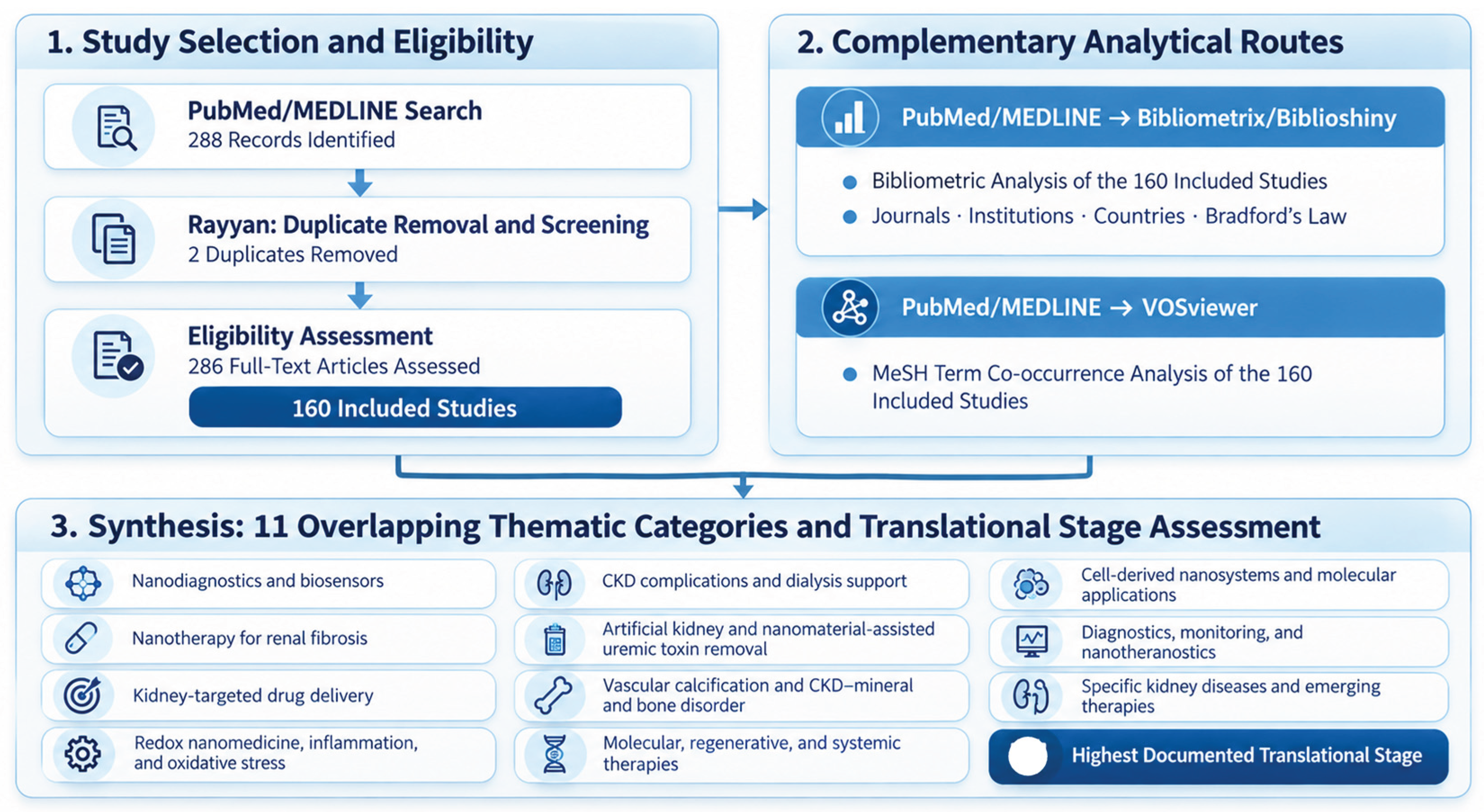
Figure 2.
MeSH term co-occurrence network of the 160 included studies on nanotechnology and chronic kidney disease. Node size indicates term frequency, links represent co-occurrence, and colors distinguish clusters identified by VOSviewer. Cluster colors have no intrinsic semantic meaning; thematic interpretations were based on the predominant and highly connected MeSH terms within each cluster. Source: VOSviewer (version 1.6.20), using PubMed/MEDLINE records from the included studies.
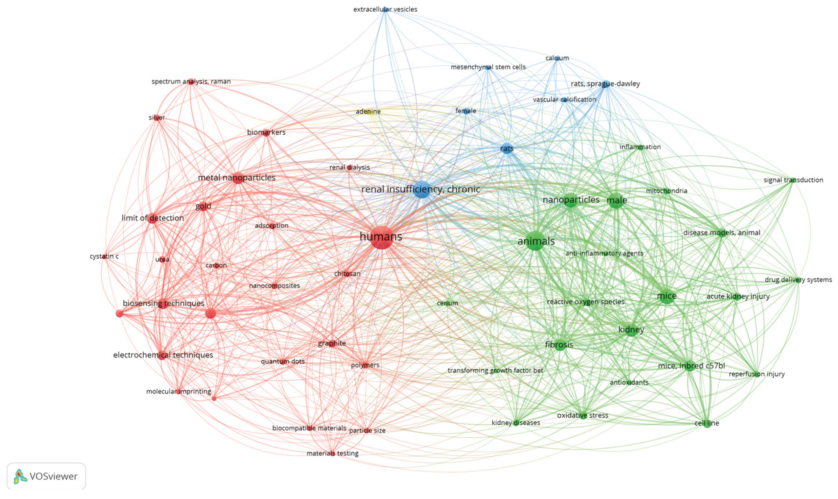
Figure 3.
Distribution of studies across 11 thematic categories of nanotechnology in chronic kidney disease. Color intensity indicates study frequency, with numerical counts displayed for each category. Because categories were not mutually exclusive, the total number of thematic assignments (n = 240) exceeds the 160 included studies. Source: Authors’ thematic classification of PubMed/MEDLINE studies; visualization created using Datawrapper.
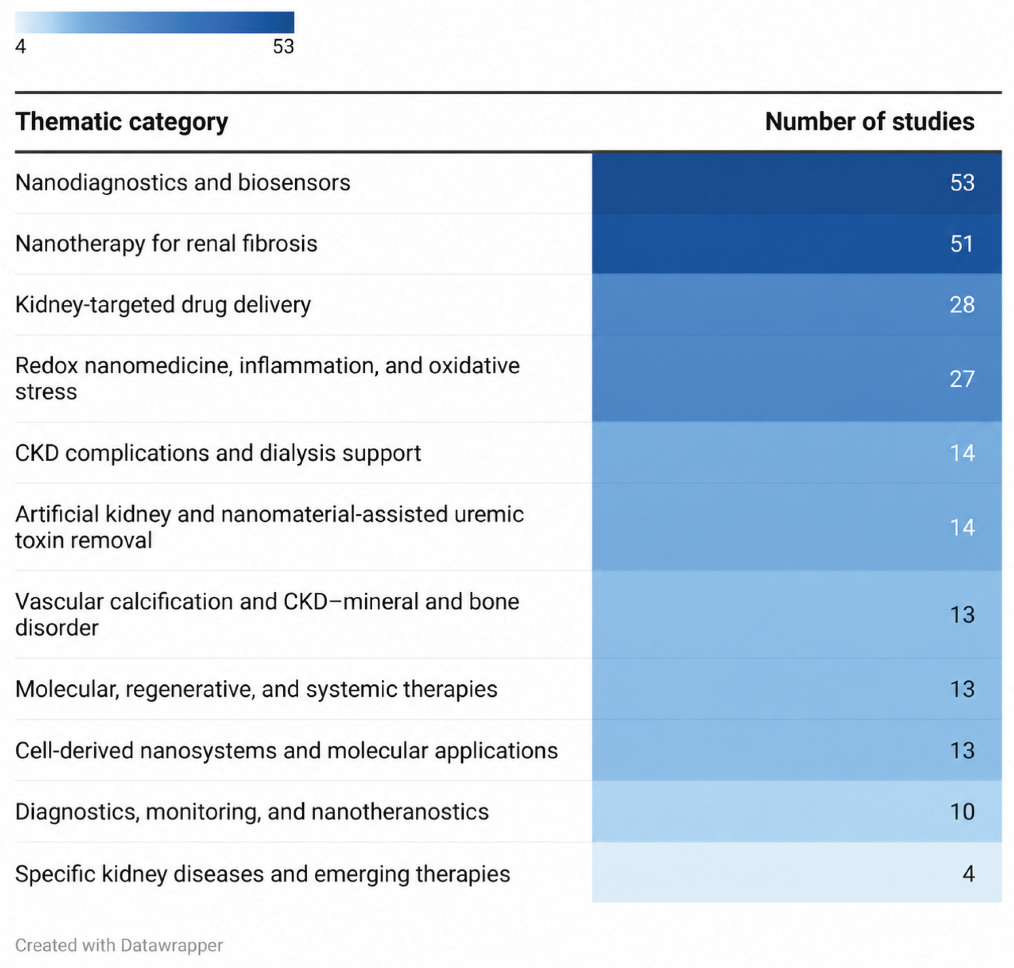
Figure 4.
Schematic illustration of nanodiagnostic strategies for chronic kidney disease. Nanostructured sensing platforms incorporating gold nanoparticles and carbon nanotubes enable the analytical detection of kidney-related biomarkers and uremic toxins in biological samples. The illustration represents potential diagnostic applications rather than clinically validated early CKD detection.
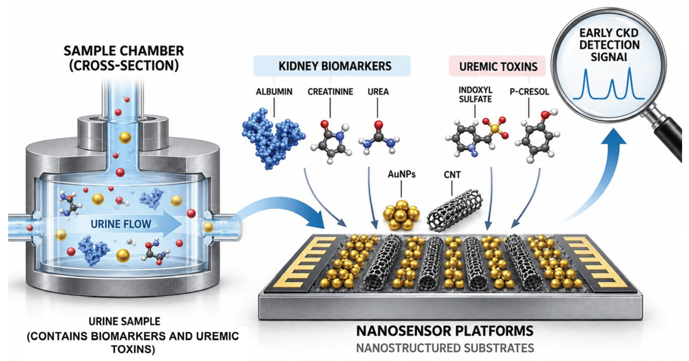
Figure 5.
Representative nanomedicine strategies, therapeutic mechanisms, and nanoplatforms investigated for renal fibrosis in chronic kidney disease. A. Kidney-targeting and responsive delivery strategies involving KIM-1, CD44, collagen IV, and therapeutic cargoes. B. Major therapeutic mechanisms, including ROS scavenging, mitochondrial protection, ferroptosis inhibition, autophagy restoration, and modulation of TGF-β/Smad signaling. C. Representative nanoplatforms, including lipid nanoparticles, PLGA-based systems, gold nanoparticles, ceria nanozymes, graphene quantum dots, black phosphorus-based materials, and extracellular vesicles. The schematic integrates approaches investigated across different studies.
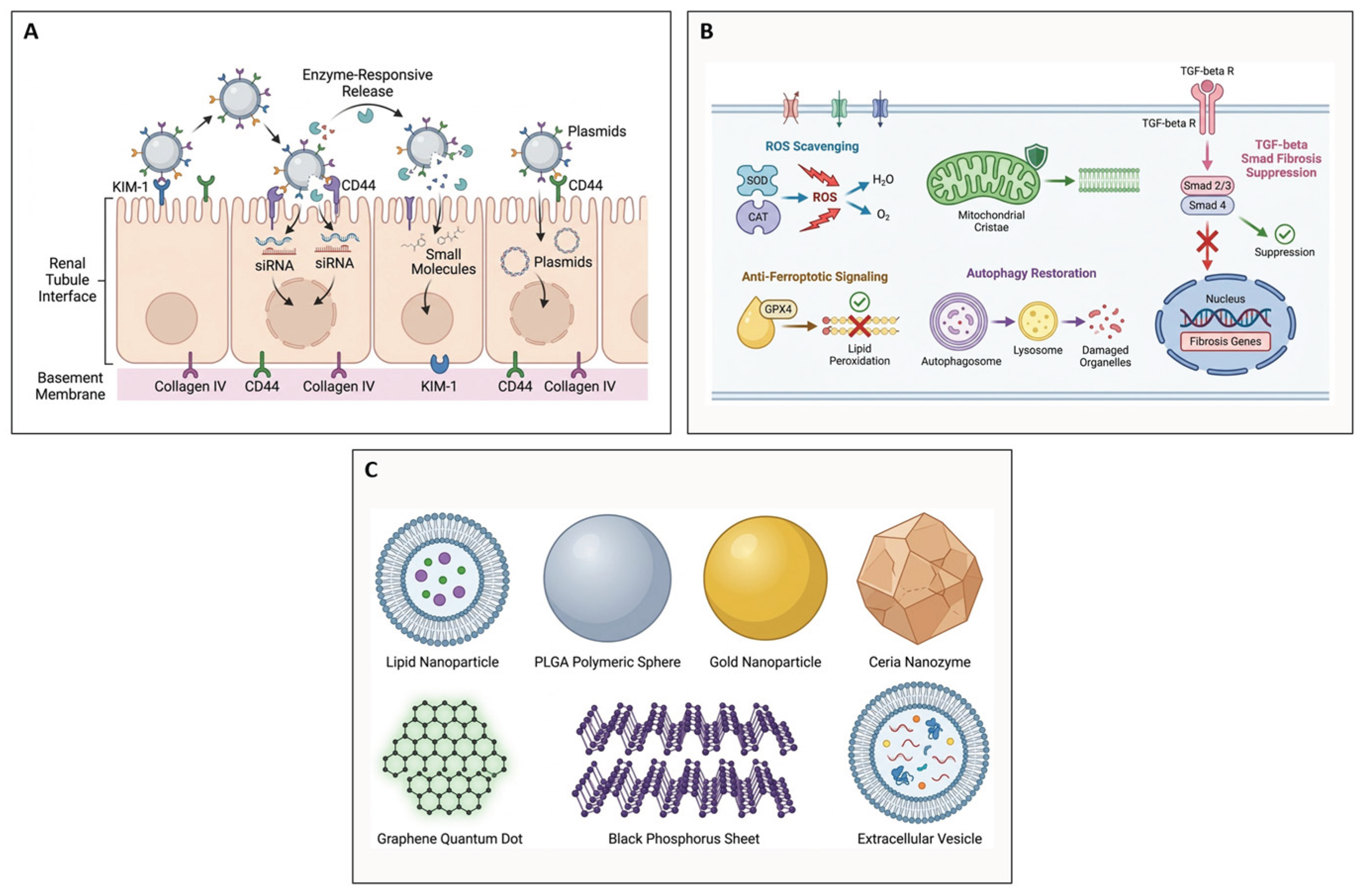
Figure 6.
Schematic representation of a nanoporous adsorbent system for extracorporeal uremic toxin removal. The illustration depicts the diffusion of uremic solutes from the blood compartment into a high-surface-area nanoporous matrix, where adsorption enables toxin retention, while cellular blood components remain within the blood compartment.
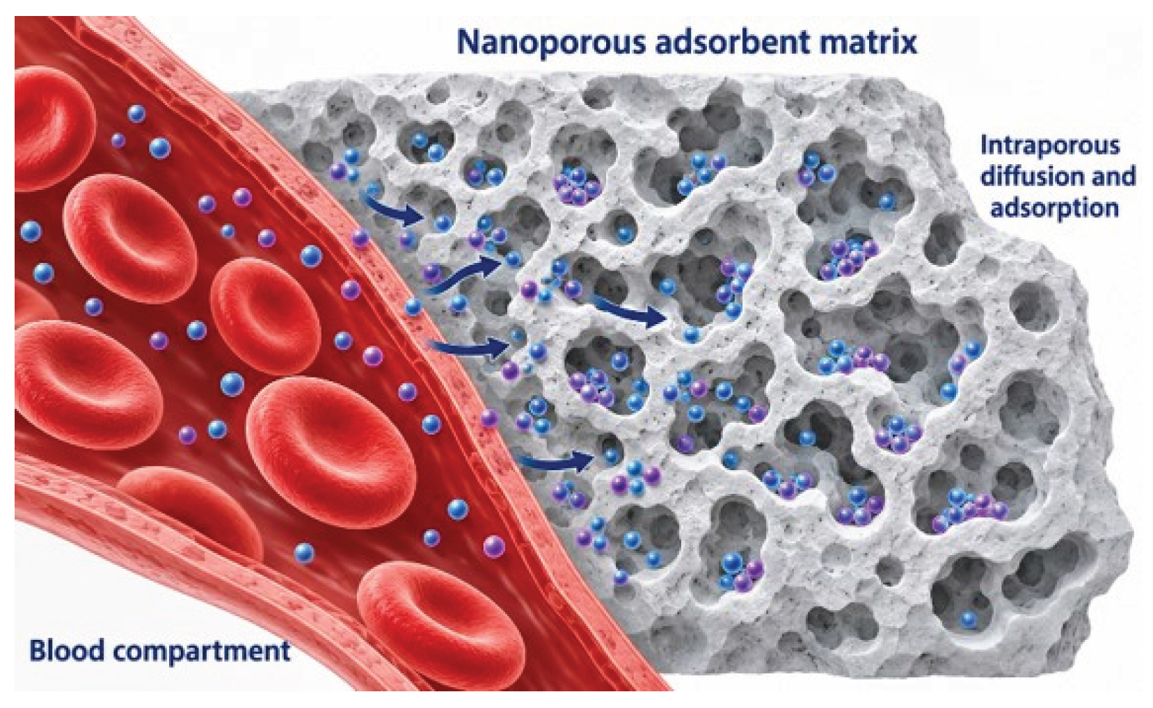
Figure 7.
Distribution of translational stages across thematic categories of nanotechnology research in chronic kidney disease. Values indicate study counts for each category–stage combination, with color intensity reflecting their frequency. Percentages reported in the text were calculated using the total number of studies within each category as the denominator. Studies could belong to multiple thematic categories but were assigned only one highest documented translational stage within each category. Source: Authors’ classification of the included PubMed/MEDLINE studies; visualization created using Datawrapper.
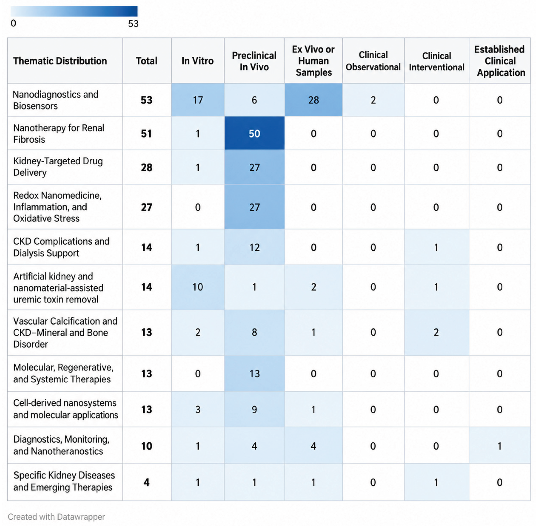
Figure 8.
Journal productivity and Bradford’s Law distribution in the analyzed PubMed/MEDLINE literature. A. Ten most productive journals among the 160 included studies. B. Bradford’s Law distribution across the 91 journals. Source: Generated using Bibliometrix/Biblioshiny from the PubMed/MEDLINE bibliographic records of the included studies and edited by the authors.
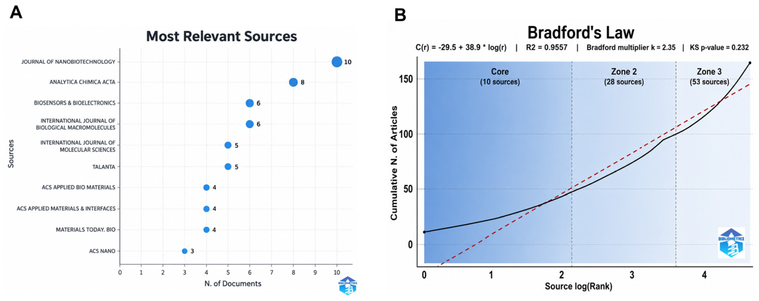
Figure 9.
Geographic distribution of scientific output by country. Darker shades indicate higher publication frequency. Source: Generated using Bibliometrix/Biblioshiny from the PubMed/MEDLINE records of the included studies and edited by the authors.
Figure 9.
Geographic distribution of scientific output by country. Darker shades indicate higher publication frequency. Source: Generated using Bibliometrix/Biblioshiny from the PubMed/MEDLINE records of the included studies and edited by the authors.
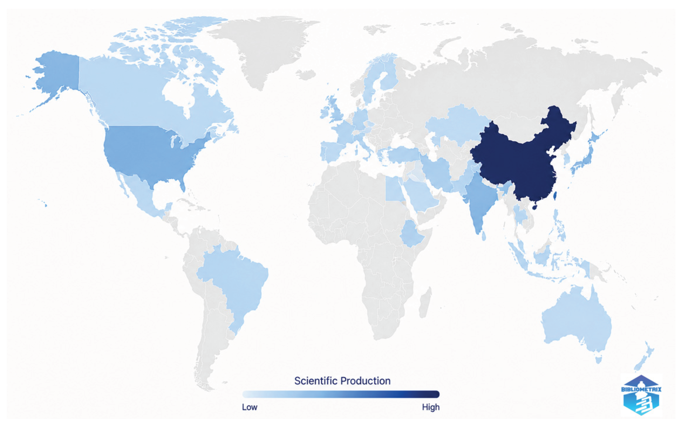
Figure 10.
Most frequent institutional affiliations in the analyzed literature. Values represent institutional affiliation frequencies identified in the bibliographic records. Source: generated from the PubMed/MEDLINE bibliographic records using Bibliometrix/Biblioshiny and visualized with Datawrapper.
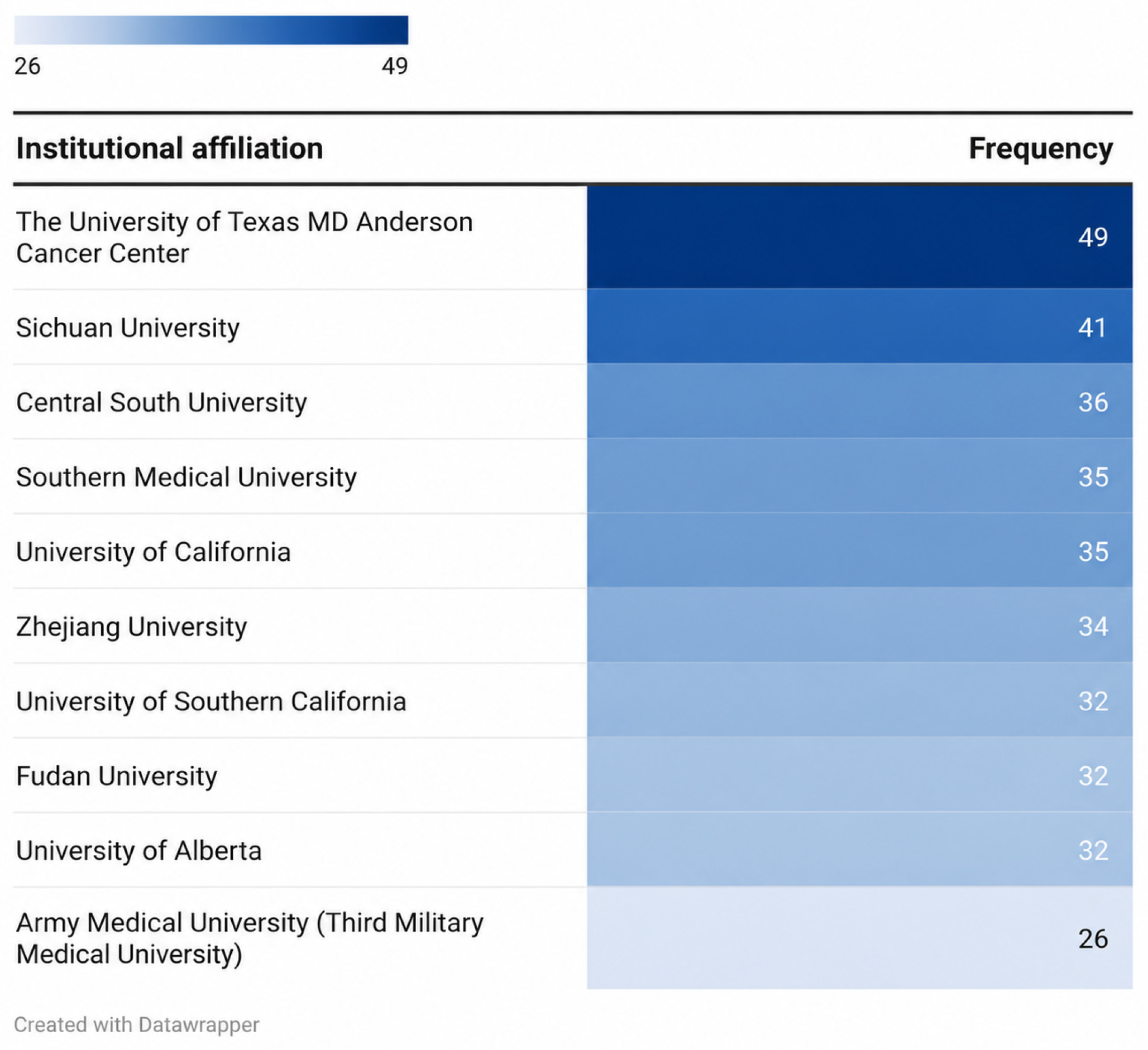
Figure 11.
Conceptual framework for the clinical translation of nanotechnology in chronic kidney disease. A. Major applications of CKD nanomedicine. B. Key translational barriers. C. Proposed pathway from experimental proof-of-concept to clinically relevant implementation. The framework is illustrative and does not represent a formally validated or strictly linear model.
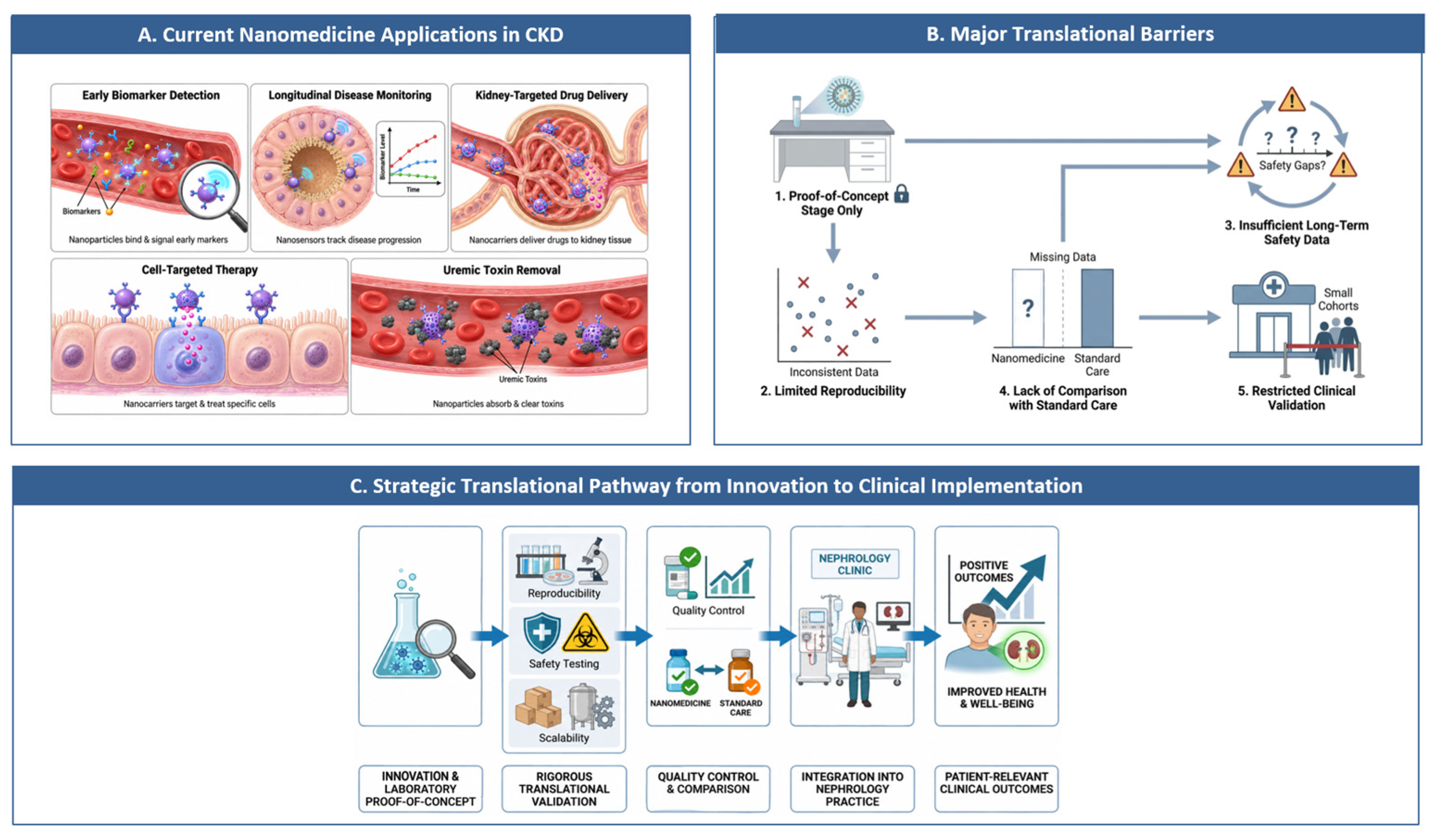

Table 1.
PubMed/MEDLINE search strategy for nanotechnology in chronic kidney disease.
| Search block | Search terms |
|---|---|
| Chronic kidney disease | (“Kidney Failure, Chronic”[Mesh] OR “chronic kidney disease”[Title/Abstract] OR “chronic renal disease”[Title/Abstract] OR “chronic renal failure”[Title/Abstract] OR CKD[Title/Abstract]) |
| Nanotechnology | (“Nanotechnology”[Mesh] OR “Nanomedicine”[Mesh] OR “Nanoparticles”[Mesh] OR “Nanostructures”[Mesh] OR nanotechnolog*[Title/Abstract] OR nanomedicin*[Title/Abstract] OR nanoparticle*[Title/Abstract] OR nanostructure*[Title/Abstract] OR nanomaterial*[Title/Abstract] OR nanocarrier*[Title/Abstract] OR nanotheranostic*[Title/Abstract] OR nanobiosensor*[Title/Abstract] OR nanosensor*[Title/Abstract] OR nanoformulation*[Title/Abstract]) |
| Combined strategy | Chronic kidney disease block AND Nanotechnology block |
Disclaimer/Publisher’s Note: The statements, opinions and data contained in all publications are solely those of the individual author(s) and contributor(s) and not of MDPI and/or the editor(s). MDPI and/or the editor(s) disclaim responsibility for any injury to people or property resulting from any ideas, methods, instructions or products referred to in the content. |
© 2026 by the authors. Licensee MDPI, Basel, Switzerland. This article is an open access article distributed under the terms and conditions of the Creative Commons Attribution (CC BY) license (http://creativecommons.org/licenses/by/4.0/).
Copyright: This open access article is published under a Creative Commons CC BY 4.0 license, which permit the free download, distribution, and reuse, provided that the author and preprint are cited in any reuse.