
Submitted:
09 October 2026
Posted:
13 October 2026
You are already at the latest version
Abstract
Staphylococci are the major pathogens associated with bovine mastitis, and the antimicrobial resistance, biofilm formation, and virulence factors may promote infection persistence. Forty-two Staphylococcus spp. isolates ob-tained from the milk of cows with clinical and subclinical mastitis were analyzed from farms in the Almaty, Zhetysu, and Kyzylorda regions of the Republic of Kazakhstan. The isolates were identified using MALDI-TOF MS, after which their hemolytic and DNase activities, ability to form biofilms, and susceptibility to antimicrobial agents were determined. PCR was used to detect the presence of antibiotic resistance genes (blaZ, mecA, tetM, tetK, ermB, aac(6′)/aph(2″)), as well as genes associated with adhesion and biofilm formation (clfA, icaA, icaD, aap, fnbpA) and virulence (hla, hlb, sea, seb, tst). S. aureus was detected in 17 (40.5%) cases, and S. epidermidis in 12 (28.6%). Bio-film formation was detected in 22 (52.4%) isolates. Resistance to penicillin G and ampicillin was detected in 41 (97.6%) isolates, and the most common genetic determinants were clfA (73.8%), blaZ (47.6%), and mecA (35.7%). Overall, these findings show that resistance- and persistence-related traits are common among Staphylococcus spp. isolates associated with bovine mastitis.
Keywords:
bovine mastitis
; Staphylococcus spp.
; antimicrobial resistance
; coagulase-negative staphylococci
; biofilm
; virulence factors
; Kazakhstan
1. Introduction
Mastitis is a major economic concern in modern dairy farming because it reduces milk yield and quality, increases diagnostic and treatment costs, and raises the risk of premature culling. Current estimates indicate that the economic burden of mastitis is substantial at both the individual-animal and industry levels, with reduced milk production accounting for much of the overall loss [1,2,3,4]. According to a global economic analysis published in 2024 covering 183 milk-producing countries, annual global losses from clinical mastitis are estimated at approximately $13 billion, and from subclinical mastitis at an additional $9 billion, for a combined total of about $22 billion annually [5].
This issue is especially relevant in Kazakhstan, where dairy farming is an important component of the livestock sector. According to data from the National Statistics Bureau of the Republic of Kazakhstan, 2.304 million metric tons of cow’s milk were produced in the country from January through July 2026, which is 2.2% more than during the same period in 2025. Despite the growth in dairy production, data on the prevalence of mastitis and its etiological profile in Kazakhstan remain limited, as existing studies cover only certain regions of the country. Studies conducted in Eastern and Northern Kazakhstan demonstrated a significant prevalence of clinical and subclinical mastitis and identified the involvement of various bacterial pathogens, including Staphylococcus aureus, Streptococcus agalactiae, and Escherichia coli. Several studies have also characterized S. aureus isolates from cows with subclinical mastitis, including their antimicrobial resistance and biofilm-forming ability [6,7]. Subsequent work in the Kostanay Region identified S. aureus in milk and dairy products and characterized its toxigenic properties and antimicrobial resistance profile [8]. Collectively, studies from Kazakhstan confirm the occurrence of mastitis across several dairy-producing regions and the involvement of both Gram-positive and Gram-negative bacterial pathogens, with recent work focusing particularly on S. aureus and antimicrobial resistance. However, the available studies are predominantly regional in nature, and current data on the species composition of mastitis pathogens and their susceptibility profiles to antimicrobial agents remain limited.
Antimicrobial resistance (AMR) among mastitis pathogens can compromise the effectiveness of treatment and disease control. Staphylococci, including S. aureus and coagulase-negative staphylococci (CoNS), are of particular concern, as resistance to various classes of antimicrobial agents has been reported for these organisms. In Kazakhstan, antibiotic resistance among staphylococcal mastitis pathogens has been identified in studies conducted in various regions of the country, including the Northern and Almaty regions. S. aureus and CoNS isolates from cows with clinical and subclinical mastitis have shown resistance to β-lactams, tetracyclines, and macrolides; specific genetic determinants of resistance and methicillin-resistant isolates have also been identified [7,8,9]. A global systematic review and meta-analysis of 155 studies also demonstrated the widespread prevalence of antibiotic resistance in S. aureus isolated from cattle with mastitis [10]. Biofilm formation may further contribute to the persistence of staphylococcal infection. A meta-analysis of 22 studies revealed a high prevalence of genes associated with adhesion and biofilm formation in S. aureus , including clfB, icaD, icaA, and fnbA [11].
Antimicrobial resistance in mastitis extends beyond veterinary medicine and is increasingly addressed within the One Health framework, which links animal, human, and environmental health. The use of antimicrobials in dairy farming creates selective pressure that contributes to the emergence and spread of resistant microorganisms and resistance genes. In this context, milk and dairy products, animals, farm workers, and the farm environment form interconnected pathways through which resistant microorganisms may circulate [12]. Current evidence supports integrated surveillance of antimicrobial resistance in mastitis pathogens that considers the links among animals, humans, and the environment [13,14]. This approach aligns with international priorities for combating antibiotic resistance, which call for the development of integrated epidemiological surveillance, the rational use of antimicrobials, infection prevention, and intersectoral collaboration. In Kazakhstan, these areas are reflected in the national “Roadmap of Measures to Curb Antimicrobial Resistance in the Republic of Kazakhstan for 2023–2027,” which calls for strengthening AMR surveillance, developing laboratory diagnostics, promoting the rational use of antimicrobials, and implementing measures to curb their consumption.
Despite these studies, data on staphylococcal mastitis pathogens in Kazakhstan remain fragmented and largely regional. Most published studies focus on S. aureus, whereas comparable data on the species composition of staphylococci, including CoNS, their susceptibility to antimicrobials, biofilm-forming ability, and the prevalence of genetic resistance determinants remain limited.
The aim of this study was to comprehensively characterize Staphylococcus spp. isolates obtained from cows with clinical and subclinical mastitis at nine dairy farms in Kazakhstan. The analysis included species identification, antimicrobial susceptibility testing, assessment of biofilm formation, hemolytic and DNase activities, and detection of genes associated with antimicrobial resistance, adhesion, biofilm formation, and virulence, with consideration of their relevance to AMR surveillance within the One Health framework.
2. Materials and Methods
2.1. Sample Collection
The study was conducted at the laboratory of the Department of Microbiology, Virology, and Immunology at the Kazakh National Agrarian Research University and at the scientific and diagnostic laboratory of the Kazakhstan-Japan Innovation Center. Milk samples from cows with clinical and subclinical mastitis served as the study material. Samples were collected from nine dairy farms located in the Almaty, Zhetysu, and Kyzylorda regions of the Republic of Kazakhstan.
Samples were collected under aseptic conditions in accordance with GOST 31449-2013. Prior to sampling, the teats were treated with a 70% ethyl alcohol solution; the first streams of milk were discarded, after which 10–15 mL of milk were collected from each affected udder quarter into sterile test tubes. The samples were transported to the laboratory in thermal containers at a temperature of approximately 4 °C and analyzed within 4 hours of collection.
2.2. Isolation and Identification of Staphylococci
Milk samples were inoculated onto blood agar and incubated at 37 °C for 24–48 hours. After incubation, colony morphology was assessed based on size, shape, color, surface characteristics, consistency, and hemolysis. Colonies with morphology consistent with the genus Staphylococcus were selected for further analysis. For preliminary identification of the isolates, Gram-stained preparations were examined microscopically to assess Gram-positivity, cell morphology, and arrangement. The selected isolates were additionally inoculated onto salt agar and incubated at 37 °C for 24–48 hours. Growth on the selective medium and colony morphology served as additional criteria for preliminary identification of staphylococci.
Isolates that met the morphological and cultural characteristics of Staphylococcus spp. underwent definitive species identification using matrix-assisted laser desorption/ionization time-of-flight mass spectrometry (MALDI-TOF MS) using the Bruker MALDI Biotyper Sirius (Bruker Daltonics, Bremen, Germany). Pure cultures were used for analysis. The resulting mass spectra were compared with reference profiles in the MALDI Biotyper database to identify the isolates to species level. Species-level identification was considered reliable when the MALDI Biotyper score was >2.0.
2.3. Phenotypic Characterization of Isolates
The hemolytic activity of Staphylococcus spp. isolates was determined by streaking pure cultures onto commercial blood agar containing 5% defibrinated sheep blood. The plates were incubated under aerobic conditions at 37 °C for 24–48 h. The type and extent of hemolysis (alpha-, beta-, gamma-, or delta-hemolysis) were assessed visually in transmitted light based on the presence and transparency of clearing zones around the colonies.
The ability to produce extracellular deoxyribonuclease (DNase) was studied using DNase Test Agar (HiMedia Laboratories, India), a selective solid medium. One-day-old pure cultures of isolates were plated onto the surface of the medium and incubated at 37 °C for 24 hours. To visualize the zones of DNA hydrolysis, the agar surface was stained with a 0.1% solution of toluidine blue. DNase activity was recorded when persistent bright pink or clear hydrolysis zones were observed around the colonies against the blue-green background of the medium.
The ability of Staphylococcus spp. isolates to form biofilms was determined using the phenotypic Congo Red Agar (CRA) method according to a previously described procedure [15] . CRA was prepared using Brain Heart Infusion Agar (BHI Agar, TM 361), sucrose (Code 1307), and Congo Red (CAS 573-58-0) (TM Media, India). The isolates under study were streaked onto the surface of the prepared medium and incubated at 37 °C for 24 hours, after which they were left overnight at room temperature. The ability to form biofilms was assessed visually based on the color and morphology of the colonies. Red colonies were classified as non-biofilm-forming, dark-colored colonies as weak biofilm producers, and black colonies as strong biofilm producers.
2.4. Determination of Antibiotic Resistance
The susceptibility of the isolated Staphylococcus spp. isolates to antimicrobial agents was determined using the disk diffusion method. Fresh 24-hour cultures were used for testing. A bacterial suspension was prepared in sterile saline to a density corresponding to 0.5 on the McFarland scale, after which it was evenly spread onto the surface of Mueller–Hinton agar using a sterile swab. Discs containing the appropriate antimicrobial agents were placed on the surface of the inoculated medium. The plates were incubated at 37 °C for 18–24 hours. The antimicrobial disks included penicillin (P), ampicillin (Amp), tetracycline (tet), furazolidone (Fx), gentamicin (gen), erythromycin (erm), kanamycin (kan), chloramphenicol (C), norfloxacin (10 µg), cefoxitin (fox), cefoperazone/sulbactam (CES), and rifampin (rif).
After incubation, the diameters of the zones of growth inhibition around the discs were measured in millimeters. The results were interpreted in accordance with current EUCAST criteria for staphylococci. Isolates were classified as susceptible (S), susceptible with extended exposure (I), or resistant (R) in accordance with established breakpoints [16].
2.5. PCR Analysis of Antibiotic Resistance, Biofilm Formation, and Virulence Genes
Genomic DNA was extracted from the Staphylococcus spp. isolates under study using the CAMOMILE-NKMagPCR kit in accordance with the manufacturer’s instructions (Diamed Asia Test LLP). The extracted DNA was used as a template for polymerase chain reaction (PCR). Molecular genetic analysis was performed to identify genes associated with antibiotic resistance, adhesion, biofilm formation, and virulence in Staphylococcus spp.
To determine the genetic determinants of antibiotic resistance, the mecA, blaZ, tetK, tetM, ermB, and aac(6′)/aph(2″) genes were examined. To assess genetic factors associated with adhesion and biofilm formation, the presence of the clfA, icaA, icaD, aap, and fnbpA genes was determined. The hla, hlb, tst, sea, and seb genes were screened as virulence-associated determinants. The sequences of the primers used, the sizes of the expected amplicons, and the annealing temperatures are presented in Table 1.
Following amplification, the PCR products were separated by horizontal agarose gel electrophoresis. The electropherograms were visualized using the Infinity VX2 3026, WL/LC/26M XPress system from Vilber Lourmat (USA). Target genes were considered present when an amplicon of the expected size was detected, as specified in Table 1.
2.6. Statistical Analysis of Data
Statistical analysis of the results was performed using GraphPad Prism 11.0 software (GraphPad Software, San Diego, CA, USA). To describe the results, absolute counts (n) and percentages (%) were calculated. Fisher’s exact test was used to assess differences and associations between categorical variables. Fisher’s exact test was selected because of the small sample size and low expected frequencies in some contingency-table cells. The statistical analysis included an assessment of the association between genotypic markers and corresponding phenotypic traits, as well as a comparison of the distribution of antibiotic resistance, phenotypic characteristics, and genetic determinants among isolates obtained from cows with clinical (n = 18) and subclinical (n = 24) mastitis. All statistical tests were two-tailed. Differences were considered statistically significant at p < 0.05.
3. Results
3.1. Species composition and taxonomic diversity of Staphylococcus isolates
A total of 42 Staphylococcus isolates were recovered from milk samples collected from cows with clinical and subclinical mastitis at nine farms in the Almaty, Zhetysu, and Kyzylorda regions of the Republic of Kazakhstan. The most common species was Staphylococcus aureus, which accounted for 40.5% (17/42) of the total number of isolates. In the clinical form of mastitis, S. aureus accounted for 50.0% (9/18) of the isolates, whereas in the subclinical form, it accounted for 33.3% (8/24). The second most frequently detected species was S. epidermidis, which accounted for 28.6% (12/42) of all isolates. Its proportion among isolates from cows with subclinical mastitis was 33.3% (8/24), whereas in clinical mastitis it accounted for 22.2% (4/18). S. borealis, S. chromogenes, and S. cohni occurred with equal frequency—7.1% (3/42) each. In addition, four other species of coagulase-negative staphylococci were identified: S. auricularis, S. lugdunensis, S. urealyticus, and S. capitis, each represented by a single isolate (2.4%; 1/42). For subsequent comparative analysis of the phenotypic and genotypic characteristics of S. cohni and the four species mentioned above, they were grouped into the “Other CoNS” category (n = 7). The numerical and percentage distribution of the identified Staphylococcus species (n = 42) is presented in Figure 1.
3.2. Phenotypic characteristics of Staphylococcus spp. isolates
The phenotypic characteristics of Staphylococcus spp. isolates obtained from milk samples of cows with clinical and subclinical forms of mastitis are presented in Figure 2. The study included an assessment of the isolates’ ability to form biofilms, their DNase activity, and their hemolytic properties.
3.2.1. Biofilm formation
Biofilm formation was detected across several Staphylococcus species and was observed in 22 of 42 isolates (52.38%). This phenotype was most frequently observed among the dominant species: in 76.47% (13/17) of S. aureus isolates (including 8 cultures from clinical mastitis and 5 from subclinical mastitis) and in 58.33% (7/12) of S. epidermidis strains (4 in clinical and 3 in subclinical forms). Biofilm formation was also noted in individual representatives of coagulase-negative staphylococci (CoNS), including one isolate each of S. borealis, S. cohni, and S. chromogenes in the respective groups (Figure 2A). When analyzing the distribution of this trait between the clinical and subclinical groups, taking into account the taxonomic groups studied, no statistically significant differences were found (Fisher’s exact test, p = 0.9230).
3.2.2. DNase activity
DNase activity was predominantly detected among S. aureus isolates, whereas this trait was significantly less common among other CoNS representatives. The distribution of DNase-positive isolates differed significantly among the studied taxonomic groups and forms of mastitis (Fisher’s exact test, p < 0.0001), indicating marked heterogeneity of this phenotypic trait among the studied isolates.
3.2.3. Hemolytic activity
Hemolytic activity was observed mainly in S.aureus and was also detected in individual CoNS representatives. However, no statistically significant differences in the distribution of the hemolytic phenotype were found among the studied groups (Fisher’s exact test, p = 0.5338).
Among the phenotypic traits assessed, DNase activity showed the greatest statistical heterogeneity (p < 0.0001). At the same time, the distribution of biofilm formation ability and hemolytic activity did not reach statistical significance (p = 0.9230 and p = 0.5338, respectively).
3.3. Antimicrobial Susceptibility Profiles of Staphylococcus spp. Isolates
Antimicrobial susceptibility profiles differed among the Staphylococcus species examined (Table 2).
The highest resistance rates were observed for penicillin G and ampicillin. Resistance to both drugs was 100% among S. aureus, S. epidermidis, S. chromogenes, and the “Other CoNS” group, whereas among S. borealis it was 66.7%. A high level of resistance to tetracycline was also noted: 88.2% of S. aureus isolates, 91.7% of S. epidermidis, 100% of S. borealis, 66.7% of S. chromogenes, and 85.7% of Other CoNS were classified as resistant. Meanwhile, 11.8%, 8.3%, 0%, 33.3%, and 14.3% of the isolates remained susceptible to tetracycline, respectively.
Resistance to cefoxitin was 47.1% for S. aureus and 50.0% for S. epidermidis. Among S. borealis and other CoNS, this rate was 33.3% and 42.9%, respectively, whereas all S. chromogenes isolates were susceptible. For cefoperazone/sulbactam, the majority of isolates remained susceptible: 82.4% of S. aureus, 75.0% of S. epidermidis, 100% of S. borealis, 66.7% of S. chromogenes, and 71.4% of Other CoNS. Intermediate susceptibility was observed in 11.8%, 25.0%, 0%, 33.3%, and 28.6% of isolates, respectively. Resistance to gentamicin ranged from 8.3% in S. epidermidis to 66.7% in S. borealis and S. chromogenes. At the same time, 70.6% of S. aureus , 91.7% of S. epidermidis, 33.3% of S. borealis, 33.3% of S. chromogenes, and 42.9% of other CoNS were susceptible. For kanamycin, susceptible isolates predominated among S. aureus (70.6%), S. epidermidis (66.7%), and Other CoNS (71.4%). Among S. borealis, 33.3% were resistant, 33.3% were intermediate, and 33.3% were susceptible. Resistance to erythromycin was most pronounced among S. chromogenes (66.7%) and S. epidermidis (41.7%), whereas among S. aureus it was 17.6%. The majority of S. aureus (82.4%) and Other CoNS (71.4%) isolates remained susceptible. Resistance to rifampin was 23.5% among S. aureus, 8.3% among S. epidermidis, and 42.9% among other CoNS. All S. borealis and S. chromogenes isolates tested were resistant to this drug. All S. aureus, S. epidermidis, and other CoNS isolates were susceptible to chloramphenicol. Among S. borealis, resistance was 66.7%, while among S. chromogenes, 33.3% of isolates exhibited an intermediate susceptibility profile. For norfloxacin, the majority of isolates were classified as susceptible: 88.2% of S. aureus and 100% of isolates from the other groups. A resistant profile was detected in only 11.8% of S. aureus. For norfloxacin, it should be noted that the initial criteria used a screening assessment; therefore, the results are presented in the table in the standardized R/I/S format.
Overall, the highest resistance among staphylococcal isolates was observed against β-lactam antibiotics (penicillin G and ampicillin) and tetracycline. At the same time, a relatively high proportion of susceptible isolates was observed for cefoperazone/sulbactam, gentamicin, kanamycin, chloramphenicol, and norfloxacin. Resistance profiles varied among Staphylococcus species; however, the results for species with a small number of isolates (S. borealis and S. chromogenes, n=3) should be interpreted with caution.
A comparison of phenotypic resistance between clinical and subclinical mastitis revealed significant differences for selected antimicrobials. Thus, in the subclinical form of mastitis, resistance to furazolidone was completely absent (0.0%), whereas in the clinical mastitis group, it was detected in 33.3% (6/18) of isolates and this difference was statistically significant (Fisher’s exact test, p = 0.0035; p < 0.01). A similar pattern was observed for rifampicin, where the frequency of resistant strains in the clinical form of mastitis was 50.0% (9/18), which significantly exceeded the rate observed in the subclinical form—16.7% (4/24) (p = 0.0410; p < 0.05). For the remaining antimicrobials studied, staphylococcal resistance levels remained consistently high in both groups of cows, with no statistically significant differences (p > 0.05).
3.3.1. Analysis of Combined Antibiotic Resistance Profiles
Analysis of the distribution of combined antibiotic resistance profiles among the isolated staphylococci revealed species-specific differences in their prevalence (Table 3).
Combined resistance to penicillin G and ampicillin (Pen + Amp) was detected in 41 of the 42 isolates studied (97.6%). This resistance profile was observed in all isolates of S. aureus (17/17; 100.0%), S. epidermidis (12/12; 100.0%), S. chromogenes (3/3; 100.0%), and the “Other CoNS” group (7/7; 100.0%). Among S. borealis isolates, combined resistance to Pen + Amp was detected in 2 out of 3 isolates (66.7%). When tetracycline was included in the resistance profile (Pen + Amp + Tet), this combination was detected in 36 of 42 isolates (85.7%). The highest frequency of this profile was observed among S. epidermidis—11/12 (91.7%), S. aureus —15/17 (88.2%)—and the “Other CoNS” group—6/7 (85.7%). Among S. borealis and S. chromogenes, this profile was detected in 2 out of 3 isolates (66.7%) in each group.
A combination of resistance to penicillin G, cefoxitin, and gentamicin (Pen + Cefox + Gen) was detected in 5 of the 42 isolates studied (11.9%). This resistance profile was detected in 1 S. aureus isolate (5.9%), 1 S. epidermidis isolate (8.3%), and 3 isolates from the Other CoNS group (42.9%). This combination of resistance was not detected among S. borealis and S. chromogenes.
A resistance profile to kanamycin, erythromycin, and tetracycline (Kan + Erm + Tet) was detected in 4 of 42 isolates (9.5%). It was found in 3 S. epidermidis isolates (25.0%) and 1 S. chromogenes isolate (33.3%), whereas this profile was not detected among S. aureus , S. borealis, and the “Other CoNS” group.
Thus, the most common resistance combination in the study sample was the Pen + Amp profile, detected in 97.6% of isolates, followed by the Pen + Amp + Tet profile at 85.7%. The combinations Pen + Cefox + Gen and Kan + Erm + Tet were significantly less common—found in 11.9% and 9.5% of isolates, respectively.
3.4. Distribution of Antimicrobial Resistance, Adhesion, Biofilm-Associated and Virulence Genes in Staphylococcus spp.
PCR screening was performed to characterize genetic determinants associated with antimicrobial resistance, adhesion, biofilm formation, and virulence in the Staphylococcus spp. isolates (Table 4). The target loci showed a heterogeneous distribution within S. aureus and across the coagulase-negative staphylococcal (CoNS) species. PCR analysis revealed differences in the prevalence of genes for antibiotic resistance, adhesion, and biofilm formation, as well as virulence factors and toxins, among the studied Staphylococcus spp. isolates.
The blaZ gene was most frequently detected among S. aureus isolates – in 11 out of 17 (64.7%). Among S. epidermidis isolates, it was detected in 5 out of 12 (41.7%); among S. chromogenes isolates, in 1 out of 3 (33.3%); and in the “Other CoNS” group, in 3 out of 7 isolates (42.9%). Overall, the blaZ gene was detected in 20 of the 42 isolates studied (47.6%), making it the most prevalent antibiotic resistance gene in the study sample. Among the antibiotic resistance genes associated with tetracycline resistance, the tetM gene was detected in 10 of the 42 isolates studied (23.8%), whereas tetK was detected significantly less frequently – in 3 isolates (7.1%). The tetM gene was detected in 5 of 17 S. aureus isolates (29.4%), 3 of 12 S. epidermidis isolates (25.0%), and 2 of 7 isolates from the “Other CoNS” group (28.6%); this gene was not detected among S. borealis and S. chromogenes. The tetK gene was detected in 2 of 12 S. epidermidis isolates (16.7%) and in 1 of 7 isolates from the “Other CoNS” group (14.3%), whereas it was not detected among S. aureus, S. borealis, and S. chromogenes. Thus, tetM was more than three times as common as tetK (23.8% versus 7.1%).
Among the genes associated with adhesion and biofilm formation, clfA was the most prevalent, detected in 31 of 42 isolates (73.8%). The highest frequency of clfA detection was observed in S. borealis – 100% (3/3), S. epidermidis– 75.0% (9/12), Other CoNS – 71.4% (5/7), S. aureus – 70.6% (12/17), and S. chromogenes – 66.7% (2/3). The icaA and aap genes were detected in 8 isolates each (19.0% each), while the icaD and fnbpA genes were detected in 7 isolates each (16.7% each).
Virulence factor and toxin genes were detected predominantly among S. aureus isolates. The hla gene was detected in 6 isolates (14.3%), and the hlb gene in 5 (11.9%). The sea enterotoxin gene was detected in 3 isolates (7.1%), while the seb and tst genes were each detected in 2 isolates (4.8% each). In the sample, these genes were not detected in S. epidermidis, S. borealis, S. chromogenes, or Other CoNS.
Overall, among the isolates studied, the most common genetic determinants were clfA (73.8%), blaZ (47.6%), and mecA (35.7%). Among virulence genes and toxins, hla was the most prevalent (14.3%), while seb and tst were detected least frequently – at 4.8% each.
The association between mecA carriage, detected by PCR, and phenotypic cefoxitin resistance, determined by disk diffusion, was assessed as a marker of methicillin resistance. Fisher’s exact test showed a statistically significant association between mecA carriage and cefoxitin resistance (p < 0.0001). The mecA gene was detected in 15 of 42 isolates (35.7%), and all 15 mecA-positive isolates exhibited phenotypic resistance to cefoxitin. Overall, cefoxitin resistance was detected in 18 of 42 isolates (42.9%). A discrepancy between genotype and phenotype was observed in 3 isolates (7.1%), which were resistant to cefoxitin despite the absence of the mecA gene. Thus, in the studied sample, the presence of mecA was highly correlated with phenotypic resistance to cefoxitin; however, the identification of three mecA-negative cefoxitin-resistant isolates suggests the possible existence of additional resistance mechanisms that were not investigated in this study.
4. Discussion
In the present dataset, S. aureus was the predominant staphylococcal species, while the substantial proportion of CoNS suggests that coagulase-negative staphylococci also contribute to intramammary infection. Recent reviews identify CoNS as one of the most frequently isolated groups in bovine mammary gland infections and may be associated with both subclinical and clinical forms of mastitis. However, different CoNS species vary significantly in terms of epidemiology, ability to persist, virulence, and impact on the inflammatory response in the mammary gland[25,26]. The higher proportion of S. aureus in clinical cases and the more frequent detection of some CoNS in subclinical cases are broadly consistent with reported differences in the epidemiology of these organisms. S. aureus is one of the major pathogens of cattle and is capable of causing both clinical and persistent intramammary infections. CoNS, in turn, are particularly often associated with subclinical mastitis, although certain species, including S. chromogenes, S. epidermidis, and S. haemolyticus, may also be associated with clinical forms of the disease[27]. Among the isolated coagulase-negative staphylococci (CoNS), the presence of Staphylococcus borealis isolates is noteworthy, accounting for 7.14% of the total number of cultures detected. This species was reclassified and established as a separate taxon relatively recently – in 2020 – and its role in the etiology of intramammary infections in cattle has been described in a number of international publications over the past three years[28,29]. Because S. borealis shares several biochemical features with S. haemolyticus, routine diagnostic methods may misidentify it, potentially leading to underestimation of its prevalence. MALDI-TOF MS enabled species-level differentiation and identified S. borealis among isolates obtained from dairy farms in Kazakhstan.
A study from Northern Kazakhstan likewise reported S. aureus as an important staphylococcal species isolated from cows with mastitis[30]. At the same time, differences in the composition of the staphylococcal microbiota between individual studies may reflect regional characteristics, differences between farms, animal housing and milking conditions, as well as the specifics of sample collection and identification.
Published data on S. aureus in Kazakhstan remain largely limited to individual regions and farms. By including nine farms in the Almaty, Zhetysu, and Kyzylorda regions, the present study broadens the available data on the species composition of staphylococci associated with bovine mastitis in Kazakhstan.
The predominance of S. aureus together with the substantial presence of CoNS highlights the heterogeneous staphylococcal etiology of mastitis in the surveyed farms. These findings add to existing knowledge on the species diversity of staphylococci isolated from cow’s milk in Kazakhstan and provide a basis for further study of the characteristics of individual species associated with various forms of mastitis.
The marked phenotypic heterogeneity among the Staphylococcus spp. isolates underscores that the clinical behavior of staphylococcal mastitis cannot be inferred from species identity alone and may also depend on the combination of virulence-associated traits. For S. aureus, key mechanisms of pathogenesis include adhesion, biofilm formation, production of cytolytic toxins, and the ability to persist in mammary gland tissues, whereas among CoNS, the expression of these properties can vary significantly between species and individual strains.
The ability to form biofilms, observed in isolates of various species, is of particular importance for staphylococcal intramammary infections. Biofilms create a protective extracellular matrix that may promote bacterial persistence in the mammary gland by limiting immune clearance and antimicrobial activity. For S. aureus , biofilm formation is considered one of the mechanisms underlying the chronic course of infection; however, the extent of this property varies significantly among isolates[31]. The absence of a statistically significant association between biofilm formation and the form of mastitis in the present study is consistent with the results of other studies, which also failed to identify an association between the ability of isolates to form biofilms and the clinical or subclinical course of mastitis. In particular, a study of S. aureus isolates from cows with clinical and subclinical mastitis found no differences in biofilm-forming ability depending on the form of the disease[32]. Therefore, the absence of a statistical association with the clinical form of the disease does not rule out the potential significance of biofilms in pathogen persistence; however, their role in the pathogenesis of mastitis should be interpreted with consideration of strain-specific characteristics and study conditions.
Hemolytic activity was also widely observed among the staphylococcal isolates studied; however, no statistically significant differences in its distribution were found among the groups analyzed (p=0.5338). In this dataset, the hemolytic phenotype was not associated with a specific taxonomic group or with the clinical form of mastitis. At the same time, hemolytic activity is one of the known phenotypic manifestations of staphylococcal virulence and is associated with the production of hemolysins capable of damaging host cells. For S. aureus, α- and β-hemolysins are the most extensively studied; however, their expression may vary among strains. Therefore, the absence of statistically significant differences in this study does not rule out the biological significance of hemolytic activity but rather indicates that this phenotype alone did not allow for the differentiation of the studied groups of isolates.
DNase activity varied markedly among the staphylococcal species examined. The most pronounced DNase activity was observed in S. aureus, whereas this trait was significantly less common among certain CoNS species. The statistically significant association (p < 0.0001) indicates heterogeneity in the distribution of DNase activity among the studied groups and underscores differences in the phenotypic characteristics of the isolated staphylococci.
DNase activity is traditionally used as one of the phenotypic markers employed in the identification of S. aureus. However, the presence of this trait should not be considered an independent indicator of virulence or the severity of mastitis. A complex of factors is involved in the pathogenesis of staphylococcal infection, including adhesion, biofilm formation, and the production of hemolysins and other virulence factors. Therefore, the high prevalence of DNase activity among S. aureus strains identified in this study should be interpreted in conjunction with other phenotypic and genotypic characteristics of the isolates.
Phenotypic susceptibility profiling revealed high antimicrobial resistance among the isolated Staphylococcus spp. strains to several drugs commonly used in empirical mastitis therapy. The most pronounced resistance across the entire studied population (n = 42) was observed against β-lactam antibiotics—penicillin G and ampicillin (100% among S. aureus, S. epidermidis, S. chromogenes, and the “Other CoNS” group, and 66.7% among S. borealis). The high prevalence of resistance to unprotected penicillins may reflect prolonged selective pressure associated with intramammary antimicrobial use. These findings are consistent with the study by Baymenov et al. in Northern Kazakhstan, which also reported 100% resistance of bovine S. aureus isolates to β-lactams [7]. A global meta-analysis by Molineri et al. similarly documented widespread penicillin resistance among mastitis-associated S. aureus isolates [10]. High tetracycline resistance was also observed, with rates ranging from 66.7% (S. chromogenes) to 88.2% (S. aureus), 91.7% (S. epidermidis), and 100% (S. borealis). The widespread prevalence of tetracycline resistance among isolates from cow’s milk in Kazakhstan was previously highlighted in a study by Rychshanova et al., where resistance to tetracyclines was one of the leading phenotypic profiles [7]. These high resistance rates were also reflected in the combined resistance profiles observed on the surveyed farms. Thus, the Pen + Amp profile was detected in 41 of the 42 isolates studied (97.6%), and the three-component Pen + Amp + Tet profile was found in 36 isolates (85.7%). Among individual species, this profile was detected in 91.7% of S. epidermidis isolates, 88.2% of S. aureus isolates, and 85.7% of isolates from the “Other CoNS” group. The results indicate a widespread prevalence of combined resistance to β-lactam antibiotics and tetracycline among the staphylococci studied. The high frequency of combined resistance is consistent with reports of multidrug-resistant staphylococci in dairy cattle [33,34]. Recent surveillance studies have also documented increasing multidrug resistance among mastitis-associated Staphylococcus isolates [35]. Recent studies indicate substantial geographical and farm-level variation in the prevalence of MDR mastitis isolates [33,34]. Coagulase-negative staphylococci (CoNS) may also serve as reservoirs of antimicrobial resistance determinants [36]. The high frequency of the Pen + Amp + Tet profile in the surveyed farms (85.7%) therefore emphasizes the importance of considering multidrug resistance when interpreting susceptibility results and selecting therapy.
Comparison of clinical and subclinical isolates revealed differences in resistance frequencies for selected antimicrobials. Statistically significant associations were observed between the clinical form of mastitis and resistance to furazolidone and rifampicin. In cases of latent (subclinical) mastitis, resistance to furazolidone was completely absent (0.0%), whereas in the clinical form, it was detected in 33.3% of isolates (Fisher’s exact test, p = 0.0035; p < 0.01). For rifampicin, the frequency of resistant strains in the clinical form of mastitis was 50.0%, which significantly exceeded the rate observed in the subclinical form—16.7% (p = 0.04; p < 0.05). These differences may reflect variation in antimicrobial exposure or strain composition between clinical and subclinical cases; however, treatment histories were not assessed in this study. Despite high resistance to penicillin and tetracycline, several agents retained substantial in vitro activity. Most of the cultures tested remained susceptible to cefoperazone/sulbactam: ranging from 71.4% (Other CoNS) and 75.0% (S. epidermidis) to 82.4% (S. aureus) and 100% (S. borealis). High susceptibility was also observed for gentamicin (up to 91.7% of susceptible strains among S. epidermidis and 70.6% among S. aureus) and chloramphenicol (100% susceptibility in S. aureus, S. epidermidis, and Other CoNS). The high proportion of susceptible isolates to the inhibitor-protected β-lactam cefoperazone/sulbactam and the relatively high susceptibility to gentamicin and chloramphenicol are consistent with recent comprehensive data from Baimenov et al. [30]. In their study of staphylococci from raw milk and dairy products in Kazakhstan, the authors also reported retained in vitro activity of alternative antimicrobial classes despite high levels of baseline penicillin resistance and moderate prevalence of MDR phenotypes (20.2%). The high proportion of isolates susceptible to cefoperazone/sulbactam and gentamicin suggests that these agents retained activity against the studied staphylococci in vitro. The results obtained may be useful in the further selection of antimicrobial therapy; however, the clinical efficacy of the drugs requires evaluation in light of local treatment protocols and the results of susceptibility testing for specific isolates.
Molecular genetic screening (Table 4) provided additional insight into determinants associated with antimicrobial resistance and virulence in the studied isolates. A strong association was observed between mecA carriage and phenotypic cefoxitin resistance (Fisher’s exact test, p < 0.0001). All mecA-positive isolates were cefoxitin-resistant, supporting the close association between this genetic marker and the resistance phenotype in the study sample.
The blaZ gene was most frequently detected in S. aureus (64.7%) and, to a lesser extent, in coagulase-negative staphylococci. Published studies show marked geographical variation in the distribution of blaZ, which may reflect differences in antimicrobial use and veterinary control practices. For example, studies from Brazil reported blaZ in 95.7–100% of penicillin-resistant S. aureus isolates [37]. Significant prevalence of this determinant has also been documented on dairy farms in China, where laboratory screening identified the blaZ locus in S. aureus in 82.1% of mastitis strains [38].
Because phenotypic β-lactam resistance was more common than blaZ carriage, blaZ alone could not explain the full resistance phenotype. Nevertheless, blaZ was the most frequently detected β-lactam resistance determinant in the PCR panel and was present in 11 of 17 S. aureus isolates (64.7%; Table 4). A similar discrepancy between the frequency of blaZ and phenotypic resistance has previously been described for staphylococci, since phenotypic resistance may reflect the combined action of several mechanisms, and the expression of resistance genes is not always constant[39].
A similar genotype–phenotype discrepancy was observed for tetracycline resistance. The tetM and tetK genes were detected in 10 (23.8%) and 3 (7.1%) isolates, respectively—that is, a combined total of only 13 out of 42 cultures (31.0%). At the same time, phenotypic resistance to tetracycline was detected in 37 isolates (88.1%). This indicates that the tetM and tetK genes identified in this study do not fully account for the observed phenotype of tetracycline resistance. Possible explanations include the presence of other resistance determinants not included in the PCR panel used, differences in the expression of the corresponding genes, and other mechanisms of tetracycline resistance. It has also been previously shown that phenotypic and genotypic methods for determining staphylococcal resistance do not always show complete agreement, and the presence of a single marker cannot be considered a universal predictor of phenotypic resistance [39].
A similar trend was observed for macrolides and aminoglycosides. ermB was detected in 6 isolates (14.3%), while resistance to erythromycin was observed in 13 isolates (31.0%). In addition, the aac(6′)/aph(2″) gene was detected in 5 isolates (11.9%), whereas phenotypic resistance to gentamicin was observed in 14 isolates (33.3%). These results indicate that the genetic markers studied explain only a portion of the corresponding resistance phenotypes. A similar genotype-phenotype mismatch has previously been observed in coagulase-negative staphylococci: phenotypic resistance to gentamicin, erythromycin, and tetracyclines did not always correspond to the presence of specific resistance genes [40].
The genetic determinants of adhesion, biofilm formation, and virulence occurred at different frequencies among the studied Staphylococcus spp. isolates. At the same time, a comparison of molecular and phenotypic characteristics showed that the presence of individual genes was not always accompanied by a corresponding phenotypic manifestation. Such genotype–phenotype discrepancies are well recognized in staphylococci and reflect the complex regulation of virulence, in which genetic potential is not necessarily expressed under a given set of culture or infection conditions. The current literature also emphasizes that the virulence of S. aureus is determined not by individual genes, but by their combination, expression levels, regulatory systems, and environmental conditions [41].
In the present study, the ability to form a biofilm was phenotypically detected in 22 of 42 isolates (52.4%). Among the genes associated with adhesion and biofilm formation that were examined, clfA was detected most frequently – in 31 isolates (73.8%), while aap and icaA were detected in 8 isolates each (19.0%), and icaD and fnbpA were each detected in 7 isolates (16.7%). The presence of individual genetic determinants therefore did not directly predict the phenotypic ability to form biofilms, supporting a multifactorial basis for biofilm formation in the studied isolates. The icaADBC operon is involved in the synthesis of the polysaccharide intercellular adhesin PIA/PNAG; however, its presence is not a prerequisite for biofilm formation. In addition to the PIA-dependent mechanism, surface proteins – including fibronectin-binding proteins (FnBPs), clumping factors ClfA/ClfB, biofilm-associated protein, and other MSCRAMM factors – are involved in adhesion and matrix formation. Therefore, a negative PCR result for a single gene does not indicate that the isolate is incapable of forming a biofilm [42].
Hemolytic activity can be considered one of the factors potentially involved in mammary tissue damage. In this study, α-hemolysis was detected in 20 of 42 isolates (47.6%), β-hemolysis in 18 (42.9%), while 4 isolates (9.5%) showed no hemolytic activity. At the same time, the hla gene was detected in 6 of 42 isolates (14.3%), while the hlb gene was detected in 5 of 42 (11.9%).
Thus, phenotypic hemolytic activity was detected more frequently than the hla and hlb genes. This genotype–phenotype discrepancy may reflect contributions from hemolysins other than hla and hlb, as well as differences in the regulation of their expression. Studies of mastitis also demonstrate that the presence of hemolysin genes does not always directly correlate with the intensity of hemolytic activity [43].
Toxin genes represented an additional component of pathogenic potential. The tst gene was detected in 2 of 42 isolates (4.8%), the seb gene in 2 (4.8%), and the sea gene in 3 isolates (7.1%). The low prevalence of individual toxin determinants does not imply a lack of virulence potential in the studied strains, since the pathogenesis of mastitis is determined by a combination of factors. Detection of a toxin gene indicates genetic potential but does not confirm its expression or toxin production in vivo. Studies of S. aureus isolates obtained from cows with mastitis have also reported significant variability in the prevalence of enterotoxins and other toxin genes among geographic and clonal groups [44].
Overall, the isolates displayed a combination of traits that may favor persistence in the mammary gland. High phenotypic antibiotic resistance limits the effectiveness of antimicrobial therapy; adhesion factors facilitate bacterial attachment to tissues; biofilm formation creates conditions for long-term persistence; and hemolytic and toxin-producing factors may contribute to tissue damage and disruption of the local immune response. The frequency of individual traits varied across the isolate set, highlighting phenotypic and genotypic heterogeneity. This combination of traits is consistent with the current understanding of the pathogenesis of staphylococcal mastitis as a multistep process in which colonization, persistence, immune evasion, and tissue damage are interrelated [45].
5. Conclusion
This study provided a comprehensive phenotypic and genotypic characterization of 42 Staphylococcus spp. isolates obtained from cows with clinical and subclinical mastitis at nine dairy farms in the Almaty, Zhetysu, and Kyzylorda regions of Kazakhstan. S. aureus predominated among the isolated staphylococci (40.5%), but coagulase-negative staphylococci accounted for a significant proportion, indicating species diversity in the staphylococcal etiology of mastitis on the surveyed farms.
Biofilm formation was observed in 52.4% of the isolates. However, no statistically significant differences in the frequency of biofilm formation or hemolytic activity were found between the groups studied, whereas DNase activity was characterized by marked heterogeneity. Antimicrobial susceptibility testing showed particularly high resistance to penicillin G, ampicillin, and tetracycline. A combination of resistance to penicillin G and ampicillin was detected in 97.6% of isolates, and a combination of resistance to penicillin G, ampicillin, and tetracycline was detected in 85.7%.
Molecular genetic analysis revealed widespread prevalence of clfA (73.8%), blaZ (47.6%), and mecA (35.7%). All mecA-positive isolates were cefoxitin-resistant, and mecA carriage was significantly associated with phenotypic cefoxitin resistance. At the same time, the identification of three cefoxitin-resistant mecA-negative isolates suggests the possible existence of additional resistance mechanisms that were not investigated in this study.
Overall, the studied staphylococci combined high phenotypic antimicrobial resistance with biofilm-forming capacity and genetic determinants related to adhesion, resistance, and virulence. These findings expand current knowledge of staphylococci associated with mastitis in cows in Kazakhstan and underscore the need for further molecular-epidemiological monitoring of antibiotic resistance and the rational use of antimicrobial agents in dairy cattle farming.
Author Contributions
Investigation, Lyailya Bessembayeva, Arman Zhylkaidar and Assel Zholdasbekova; formal analysis, Gulnur Kuzembekova; validation, Zhumagul Kirkimbayeva and Svetlana Yermagambetova; writing - original draft preparation, Lyailya Bessembayeva; writing—review and editing, Zhumagul Kirkimbayeva and Svetlana Yermagambetova. All authors have read and agreed to the published version of the manuscript.
Funding
This research was funded by the Ministry of Science and Higher Education of the Republic of Kazakhstan under the “Zhas Galym” grant program for young scientists, project AP25794411, entitled “Development of drugs based on plant extracts against cow mastitis and study of their antimicrobial and antibiofilm action against antibiotic-resistant Staphylococcus spp.,” for the period 2025–2027.
Institutional Review Board Statement
The study was conducted in accordance with ethical principles for the humane treatment and use of animals and was approved by the Local Commission on Bioethics for Biomedical Studies on Humans and Animals of the NJSC “Kazakh National Agrarian Research University” (Protocol No. 1, September 11, 2024). The experimental procedures involving laboratory and farm animals were performed in accordance with applicable principles of biosafety and animal welfare, including the European Convention for the Protection of Vertebrate Animals Used for Experimental and Other Scientific Purposes (Strasbourg, 1987) and the World Organisation for Animal Health (WOAH) Terrestrial Animal Health Code.
Informed Consent Statement
Not applicable.
Data Availability Statement
he data presented in this study are available on request from the corresponding author.
Acknowledgments
The authors gratefully acknowledge the Kazakh National Agrarian Research University for providing laboratory facilities, institutional support, and the necessary infrastructure to conduct this study.
Conflicts of Interest
The authors declare no conflict of interest.
Abbreviations
The following abbreviations are used in this manuscript:
| AMR | Antimicrobial resistance |
| AST | Antimicrobial susceptibility testing |
| CoNS | Coagulase-negative staphylococci |
| DNA | Deoxyribonucleic acid |
| DNase | Deoxyribonuclease |
| EUCAST | European Committee on Antimicrobial Susceptibility Testing |
| MALDI-TOF MS | Matrix-assisted laser desorption/ionization–time of flight mass spectrometry |
| NAS | Non-aureus staphylococci |
| PCR | Polymerase chain reaction |
References
- Dhital, B.; Pudasaini, R.; Hsieh, J.-C.; Pudasaini, R.; Chen, Y.-T.; Chao, D.-Y.; Chiang, H.-I. One Health Perspective on Antimicrobial Resistance in Bovine Mastitis Pathogens—A Narrative Review. Antibiotics 2026, 15. [CrossRef]
- Halasa, T.; Huijps ,K.; Østerås ,O.; and Hogeveen, H. Economic Effects of Bovine Mastitis and Mastitis Management: A Review. Vet. Q. 2007, 29, 18–31. [CrossRef]
- Hogeveen, H.; Steeneveld, W.; Wolf, C.A. Production Diseases Reduce the Efficiency of Dairy Production: A Review of the Results, Methods, and Approaches Regarding the Economics of Mastitis. Annu. Rev. Resour. Econ. 2019, 11, 289–312. [CrossRef]
- Dyussekenova, N.; Nessipbayeva, A.; Buralkhiyev, B.; Khussainov, D.; Akimzhan, N.; Muslimova, Z.; Kalkayeva, D.; Turgumbekov, A.; Ussenbekov, Y. Methods for Diagnosing the DUMPS, BLAD, HY and OH1 Hereditary Abnormalities in Cattle and Determining Their Prevalence. Int. J. Vet. Sci. 2025, 14, 595–604. [CrossRef]
- Rasmussen, P.; Barkema, H.W.; Osei, P.P.; Taylor, J.; Shaw, A.P.; Conrady, B.; Chaters, G.; Muñoz, V.; Hall, D.C.; Apenteng, O.O.; et al. Global Losses Due to Dairy Cattle Diseases: A Comorbidity-Adjusted Economic Analysis. J. Dairy Sci. 2024, 107, 6945–6970. [CrossRef]
- Rychshanova, R.; Mendybayeva, A.; Miciński, B.; Mamiyev, N.; Shevchenko, P.; Bermukhametov, Z.; Orzechowski, B.; Miciński, J. Antibiotic Resistance and Biofilm Formation in Staphylococcus Aureus Isolated from Dairy Cows at the Stage of Subclinical Mastitis in Northern Kazakhstan. Arch. Anim. Breed. 2022, 65, 439–448. [CrossRef]
- Rychshanova, R.; Mendybayeva, A.; Miciński, B.; Mamiyev, N.; Shevchenko, P.; Bermukhametov, Z.; Orzechowski, B.; Miciński, J. Antibiotic Resistance and Biofilm Formation in Staphylococcus Aureus Isolated from Dairy Cows at the Stage of Subclinical Mastitis in Northern Kazakhstan. Arch Anim Breed 2022, 65, 439–448. [CrossRef]
- Baymenov, B.M.; Bulashev, A.K.; Chuzhebayeva, G.D.; Aliyeva, G.K.; Beishova, I.S.; Kokanov, S.K.; Raketsky, V.A. Phenotypic and Genotypic Resistance to Antibiotics in Staphylococcus Aureus Strains Isolated from Cattle Milk in Northern Kazakhstan. Vet. World 2023, 16, 1815–1820. [CrossRef]
- Bessembayeva, L.; Kirkimbayeva, Z.; Biyashev, B.; Zholdasbekova, A.; Sarybayeva, D.; Zhylkaidar, A.; Oryntaev, K.; Bakiyeva, F. Investigation of the Antibiotic Resistance and Biofilm-Forming Ability of Staphylococcus Species from Bovine Mastitis Cases in the Almaty Region, Kazakhstan. 2024.
- Molineri, A.I.; Camussone, C.; Zbrun, M.V.; Suárez Archilla, G.; Cristiani, M.; Neder, V.; Calvinho, L.; Signorini, M. Antimicrobial Resistance of Staphylococcus Aureus Isolated from Bovine Mastitis: Systematic Review and Meta-Analysis. Prev. Vet. Med. 2021, 188, 105261. [CrossRef]
- Sharifi, A.; Sobhani, K.; Mahmoudi, P. A Systematic Review and Meta-Analysis Revealed a High-Level Antibiotic Resistance of Bovine Mastitis Staphylococcus Aureus in Iran. Res. Vet. Sci. 2023, 161, 23–30. [CrossRef]
- Pandey, S.; Doo, H.; Keum, G.B.; Kim, E.S.; Kwak, J.; Ryu, S.; Choi, Y.; Kang, J.; Kim, S.; Lee, N.R.; et al. Antibiotic Resistance in Livestock, Environment and Humans: One Health Perspective. J. Anim. Sci. Technol. 2024, 66, 266–278. [CrossRef]
- Barlow, J. Antimicrobial Resistance of Mastitis Pathogens of Dairy Cattle. Vet. Clin. North Am. Food Anim. Pract. 2025, 41, 223–236. [CrossRef]
- Kazemzadeh, S.; Korneeva, O.; Shabunin, S.; Syromyatnikov, M. Antibiotic Resistance in Mastitis-Causing Bacteria: Exploring Antibiotic-Resistance Genes, Underlying Mechanisms, and Their Implications for Dairy Animal and Public Health. Open Vet. J. 2025, 15, 3980–4006. [CrossRef]
- Mariana, N.; Atshan, S.; Neela, V.; Sekawi, Z. Evaluation of Modified Congo Red Agar for Detection of Biofilm Produced by Clinical Isolates of Methicillin–Resistance Staphylococcus Aureus. Afr. J. Microbiol. Res. 2009, 3, 330–338.
- Eucast: AST of Bacteria Available online: https://www.eucast.org/ast_of_bacteria (accessed on 19 August 2025).
- Felipe, V.; Morgante, C.A.; Somale, P.S.; Varroni, F.; Zingaretti, M.L.; Bachetti, R.A.; Correa, S.G.; Porporatto, C. Evaluation of the Biofilm Forming Ability and Its Associated Genes in Staphylococcus Species Isolates from Bovine Mastitis in Argentinean Dairy Farms. Microb. Pathog. 2017, 104, 278–286. [CrossRef]
- Silva, V.O.; Soares, L.O.; Silva Júnior, A.; Mantovani, H.C.; Chang, Y.-F.; Moreira, M.A.S. Biofilm Formation on Biotic and Abiotic Surfaces in the Presence of Antimicrobials by Escherichia Coli Isolates from Cases of Bovine Mastitis. Appl. Environ. Microbiol. 2014, 80, 6136–6145. [CrossRef]
- Martineau, F.; Picard, F.J.; Grenier, L.; Roy, P.H.; Ouellette, M.; Bergeron, M.G. Multiplex PCR Assays for the Detection of Clinically Relevant Antibiotic Resistance Genes in Staphylococci Isolated from Patients Infected after Cardiac Surgery. The ESPRIT Trial. J. Antimicrob. Chemother. 2000, 46, 527–534. [CrossRef]
- Strommenger, B.; Kettlitz, C.; Werner, G.; Witte, W. Multiplex PCR Assay for Simultaneous Detection of Nine Clinically Relevant Antibiotic Resistance Genes in Staphylococcus Aureus. J. Clin. Microbiol. 2003, 41, 4089–4094. [CrossRef]
- Abdulmanea, A.A.; Alharbi, N.S.; Somily, A.M.; Khaled, J.M.; Algahtani, F.H. The Prevalence of the Virulence Genes of Staphylococcus Aureus in Sickle Cell Disease Patients at KSUMC, Riyadh, Saudi Arabia. Antibiotics 2023, 12, 1221. [CrossRef]
- Tremblay, Y.D.N.; Lamarche, D.; Chever, P.; Haine, D.; Messier, S.; Jacques, M. Characterization of the Ability of Coagulase-Negative Staphylococci Isolated from the Milk of Canadian Farms to Form Biofilms. J. Dairy Sci. 2013, 96, 234–246. [CrossRef]
- Moreira, L.H.; de Souza, J.C.P.; de Lima, C.J.; Salgado, M.A.C.; Fernandes, A.B.; Andreani, D.I.K.; Villaverde, A.B.; Zângaro, R.A. Use of Photodynamic Therapy in the Treatment of Bovine Subclinical Mastitis. Photodiagnosis Photodyn. Ther. 2018, 21, 246–251. [CrossRef]
- Moreno, J.; Diana, L.; Martínez, M.; Iribarnegaray, V.; Puentes, R. Comprehensive Analysis of Antimicrobial Resistance, Biofilm Formation and Virulence Factors of Staphylococci Isolated from Bovine Mastitis. Heliyon 2025, 11. [CrossRef]
- De Buck, J.; Ha, V.; Naushad, S.; Nobrega, D.B.; Luby, C.; Middleton, J.R.; De Vliegher, S.; Barkema, H.W. Non-Aureus Staphylococci and Bovine Udder Health: Current Understanding and Knowledge Gaps. Front. Vet. Sci. 2021, 8, 658031. [CrossRef]
- Condas, L.A.Z.; Buck, J.D.; Nobrega, D.B.; Carson, D.A.; Roy, J.-P.; Keefe, G.P.; DeVries, T.J.; Middleton, J.R.; Dufour, S.; Barkema, H.W. Distribution of Non-Aureus Staphylococci Species in Udder Quarters with Low and High Somatic Cell Count, and Clinical Mastitis. J. Dairy Sci. 2017, 100, 5613–5627. [CrossRef]
- Kerro Dego, O.; Vidlund, J. Staphylococcal Mastitis in Dairy Cows. Front. Vet. Sci. 2024, 11. [CrossRef]
- Król, J.; Wanecka, A.; Twardoń, J.; Florek, M.; Marynowska, M.; Banaszkiewicz, S.; Kaczmarek-Pieńczewska, A.; Pląskowska, E.; Brodala, M.; Chwirot, W.; et al. Staphylococcus Borealis - A Newly Identified Pathogen of Bovine Mammary Glands. Vet. Microbiol. 2023, 286, 109876. [CrossRef]
- Freu, G.; Gioia, G.; Gross, B.; Biscarini, F.; Virkler, P.; Watters, R.; Addis, M.F.; Franklin-Guild, R.J.; Runyan, J.; Masroure, A.J.; et al. Frequency of Non-Aureus Staphylococci and Mammaliicocci Species Isolated from Quarter Clinical Mastitis: A 6-Year Retrospective Study. J. Dairy Sci. 2024, 107, 3813–3823. [CrossRef]
- Baimenov, B.; Malikzada, K.; Dossova, A.; Bermukhametov, Z.; Shevchenko, P.; Gabitova, A.; Tomaruk, O.; Yskak, A.; Chuzhebayeva, G.; Rychshanova, R. Prevalence, Toxigenicity and Antimicrobial Resistance of Staphylococcus Aureus from Animal-Derived Food Products in the Kostanay Region, Kazakhstan: A Genotypic–Phenotypic Concordance Study. Vet. Sci. 2026, 13. [CrossRef]
- Campos, B.; Pickering, A.C.; Rocha, L.S.; Aguilar, A.P.; Fabres-Klein, M.H.; de Oliveira Mendes, T.A.; Fitzgerald, J.R.; de Oliveira Barros Ribon, A. Diversity and Pathogenesis of Staphylococcus Aureus from Bovine Mastitis: Current Understanding and Future Perspectives. BMC Vet. Res. 2022, 18, 115. [CrossRef]
- Fidelis, C.E.; Orsi, A.M.; Freu, G.; Gonçalves, J.L.; dos Santos, M.V. Biofilm Formation and Antimicrobial Resistance of Staphylococcus Aureus and Streptococcus Uberis Isolates from Bovine Mastitis. Vet. Sci. 2024, 11, 170. [CrossRef]
- Moradi, A.; Bidarian, B.; Mohammadian, F.; Akbarian, F.; Kalateh Rahmani, H.; Tashakkori, N.; Khoramian, B. Trends in Antimicrobial Resistance of Major Mastitis-Causing Pathogens: A Nine-Year Study. Vet. Med. Sci. 2025, 11, e70417. [CrossRef]
- Fikru, E.; Dulo, F.; Sombo, M. Multi-Drug Resistant Staphylococcus Aureus from Bovine Mastitis: Epidemiological and Molecular Insights from Central Ethiopia. BMC Vet. Res. 2026, 22, 429. [CrossRef]
- Bouari, C.M.; Nadăş, G.C.; Crăciun, S.; Fiț, N.I. Staphylococcus Aureus in Bovine Mastitis: Pathogenesis, Antimicrobial Resistance, and Emerging Control Strategies. Microorganisms 2026, 14. [CrossRef]
- Mlynarczyk-Bonikowska, B.; Kowalewski, C.; Krolak-Ulinska, A.; Marusza, W. Molecular Mechanisms of Drug Resistance in Staphylococcus Aureus. Int. J. Mol. Sci. 2022, 23, 8088. [CrossRef]
- Lucas, A.P.; Farias, A.R.B. de; Silva, E.C. da; Santoro, K.R.; Mendonça, M.; Silva, E.R. da Detection of β-Lactamase, blaZ and mecA in Penicillin-Resistant Staphylococcus Aureus Isolated from Bovine Mastitis in Garanhuns, Brazil. Acta Vet. Bras. 2021, 15, 140–145. [CrossRef]
- Xu, J.; Tan, X.; Zhang, X.; Xia, X.; Sun, H. The Diversities of Staphylococcal Species, Virulence and Antibiotic Resistance Genes in the Subclinical Mastitis Milk from a Single Chinese Cow Herd. Microb. Pathog. 2015, 88, 29–38. [CrossRef]
- Rasheed, H.; Ijaz, M.; Ahmed, A.; Javed, M.U.; Shah, S.F.A.; Anwaar, F. Discrepancies between Phenotypic and Genotypic Identification Methods of Antibiotic Resistant Genes Harboring Staphylococcus Aureus. Microb. Pathog. 2023, 184, 106342. [CrossRef]
- Duran, N.; Ozer, B.; Duran, G.G.; Onlen, Y.; Demir, C. Antibiotic Resistance Genes & Susceptibility Patterns in Staphylococci. Indian J. Med. Res. 2012, 135, 389–396.
- Jenul, C.; Horswill, A.R. Regulation of Staphylococcus Aureus Virulence. Microbiol. Spectr. 7, 10.1128/microbiolspec.gpp3-0031–2018. [CrossRef]
- Fitzpatrick, F.; Humphreys, H.; O’Gara, J.P. Evidence for icaADBC-Independent Biofilm Development Mechanism in Methicillin-Resistant Staphylococcus Aureus Clinical Isolates. J. Clin. Microbiol. 2005, 43, 1973–1976. [CrossRef]
- Neelam; Jain, V.K.; Singh, M.; Joshi, V.G.; Chhabra, R.; Singh, K.; Rana, Y.S. Virulence and Antimicrobial Resistance Gene Profiles of Staphylococcus Aureus Associated with Clinical Mastitis in Cattle. PLOS ONE 2022, 17, e0264762. [CrossRef]
- Pérez, V.K.C.; Custódio, D.A.C.; Silva, E.M.M.; de Oliveira, J.; Guimarães, A.S.; Brito, M.A.V.P.; Souza-Filho, A.F.; Heinemann, M.B.; Lage, A.P.; Dorneles, E.M.S. Virulence Factors and Antimicrobial Resistance in Staphylococcus Aureus Isolated from Bovine Mastitis in Brazil. Braz. J. Microbiol. 2020, 51, 2111–2122. [CrossRef]
- Li, H.; Wang, Z.; Barkema, H.W.; Li, X.; Song, D.; Ren, M.; Tong, J.; Liu, M.; Gao, J.; Cheng, J. Virulence Factors of Bovine Mastitis Pathogens: Distribution, Pathogenesis, and Emerging Vaccines Targeting Virulence Factors: A Literature Review. Front. Vet. Sci. 2026, 12. [CrossRef]
Figure 1.
Species composition and quantitative distribution of the studied Staphylococcus spp. isolates obtained from milk samples of cows with mastitis (n = 42).
Figure 1.
Species composition and quantitative distribution of the studied Staphylococcus spp. isolates obtained from milk samples of cows with mastitis (n = 42).
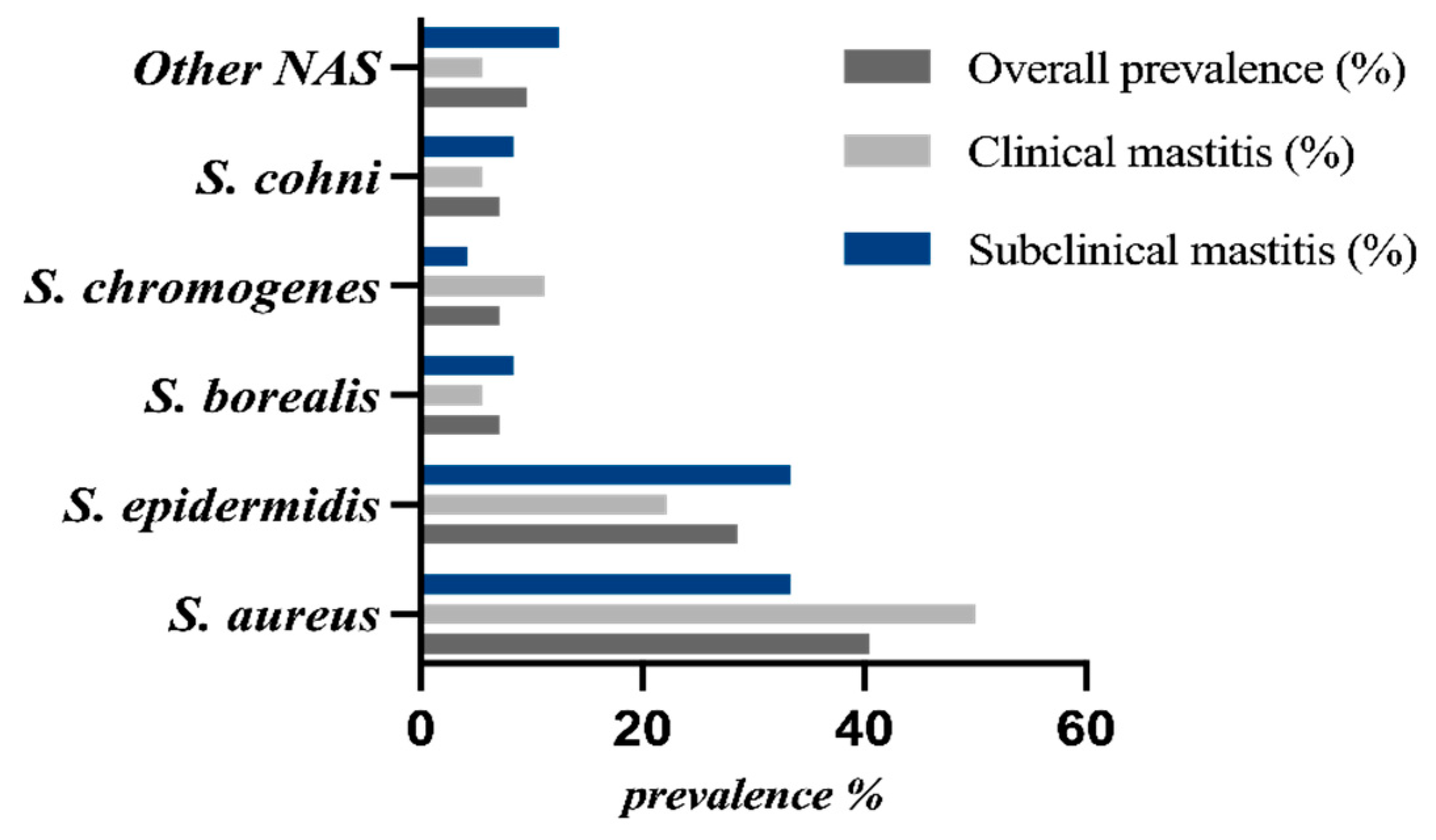
Figure 2.
Distribution of phenotypic characteristics among Staphylococcus spp. isolates from clinical and subclinical bovine mastitis: (A) biofilm formation; ns, p = 0.9230; (B) DNase activity; p < 0.0001; and (C) hemolytic activity; ns, p = 0.5338. Statistical significance was assessed using Fisher’s exact test.
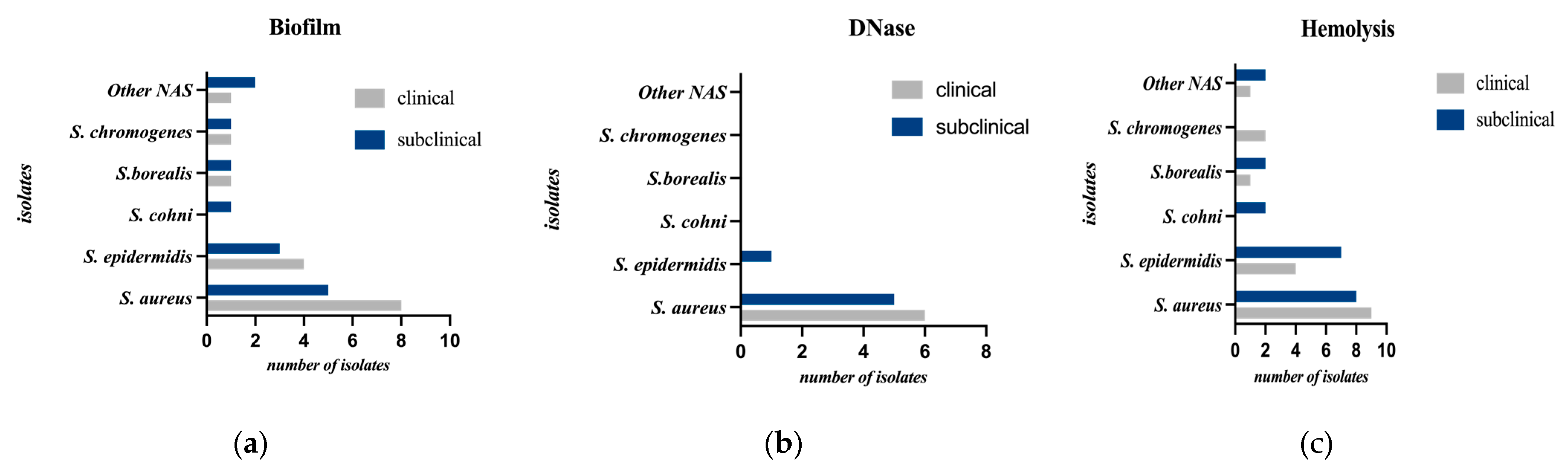

Table 1.
Primers used for PCR detection of antimicrobial resistance, adhesion, biofilm formation, and virulence-associated genes in Staphylococcus spp.
Table 1.
Primers used for PCR detection of antimicrobial resistance, adhesion, biofilm formation, and virulence-associated genes in Staphylococcus spp.
| No. | Gene | Primer Sequence (5′ → 3′) | Size | Annealing | Reference |
|---|---|---|---|---|---|
| 1 | clfA |
F: ATTGGCGTGGCTTCAGTGCT R: CGTTTCTTCCGTAGTTGCATTTG |
294 | 61°C | Felipe et al.[17] |
| 2 | mecA |
F: TGG TAT GTG GAA GTT AGA TTG GGA T R: CTA ATC TCA TAT GTG TTC CTG TAT TGG C |
155 | 52°C | Silva et al.[18] |
| 3 | ermB |
F: CTATCTGATTGTTGAAGAAGGATT R: GTTTACTCTTGGTTTAGGATGAAA |
142 | 55°C | F. Martineau et al.[19] |
| 4 | tetK |
F: GTAGCGACAATAGGTAATAGT R: GTAGTGACAATAAACCTCCTA |
360 | 52°C | Strommenger et al.[20] |
| 5 | tetM |
F: AGTGGAGCGATTACAGAA R: CATATGTCCTGGCGTGTCTA |
158 | 52°C | Strommenger et al.[20] |
| 6 | blaZ |
F: TGACCACTTTTATCAGCAACC R: ACTTCAACACCTGCTGCTTTC |
173 | 51°C | Abdulmanea et al.[21] |
| 7 | icaD |
F: AAA CGT AAG AGA GGT GG R: GGC AAT ATG ATC AAG ATA C |
199 | 55.5°C | Tremblay et al.[22] |
| 8 |
aac(6ʹ) /aph(2ʹʹ) |
F: TAA TCC AAG AGC AAT AAG GGC R: GCC ACA CTA TCA TAA CCA CTA |
227 | 55°C | Strommenger et al.[20] |
| 9 | hla |
F: CTGATTACTATCCAAGAAATTCGATTG R: CTTTCCAGCCTACTTTTTTATCAGT |
209 | 61°C | Moreno et al.[23] |
| 10 | hlb |
F: GTGCACTTACTGACAATAGTGC R: GTTGATGAGTAGCTACCTTCAGT |
309 | 61°C | Moreno et al.[24] |
| 11 | fnbpA |
F: CATAAATTGGGAGCAGCATCA R: ATCAGCAGCTGAATTCCCATT |
127 | 61°C | Felipe et al.[17] |
| 12 | tst |
F: ACCCCTGTTCCCTTATCATC R: TTTTCAGTATTTGTAACGCC |
326 | 55°C | Moreno et al.[24] |
| 13 | sea |
F: GGTTATCAATGTGCGGGTGG R: CGGCACTTTTTTCTCTTCGG |
102 | 55°C | Moreno et al.[24] |
| 14 | seb |
F: GTATGGTGGTGTAACTGAGC R: CCAAATAGTGACGAGTTAGG |
164 | 61°C | Moreno et al.[24] |
| 15 | aar |
F: GAAGCACCGAATGTTCCAACTATC R: AGTTGGCGGTATATCTATTGTA |
289 | 55°C | Tremblay et al.[22] |
Table 2.
Antimicrobial Susceptibility Profiles of the Identified Staphylococcus Species (n = 42).
| Antimicrobial agent |
S. aureus (n = 17) R/I/S (%) |
S. epidermidis (n = 12) R/I/S (%) | S. borealis (n = 3) R/I/S (%) | S. chromogenes (n = 3) R/I/S (%) | Other CoNS (n=7) R/I/S (%) |
|---|---|---|---|---|---|
| Penicillin G | 100/0/0 | 100/0/0 | 66.7/0/33.3 | 100/0/0 | 100/0/0 |
| Ampicillin | 100/0/0 | 100/0/0 | 66.7/0/33.3 | 100/0/0 | 100/0/0 |
| Cefoxitin | 47.1/0/52.9 | 50.0/0/50.0 | 33.3/0/66.7 | 0/0/100 | 42.9/0/57.1 |
| Cefoperazone/ sulbactam |
5.9/11.8/82.4 | 0/25.0/75.0 | 0/0/100 | 0/33.3/66.7 | 0/28.6/71.4 |
| Tetracycline | 88.2/0/11.8 | 91.7/0/8.3 | 100/0/0 | 66.7/0/33.3 | 85.7/0/14.3 |
| Gentamicin | 29.4/0/70.6 | 8.3/0/91.7 | 66.7/0/33.3 | 66.7/0/33.3 | 57.1/0/42.9 |
| Kanamycin | 0/29.4/70.6 | 25.0/8.3/66.7 | 33.3/33.3/33.3 | 33.3/0/66.7 | 0/28.6/71.4 |
| Erythromycin | 17.6/0/82.4 | 41.7/0/58.3 | 33.3/0/66.7 | 66.7/0/33.3 | 28.6/0/71.4 |
| Rifampicin | 23.5/0/76.5 | 8.3/0/91.7 | 100/0/0 | 100/0/0 | 42.9/0/57.1 |
| Chloramphenicol | 0/0/100 | 0/0/100 | 66.7/0/33.3 | 0/33.3/66.7 | 0/0/100 |
| Norfloxacin | 11.8/0/88.2 | 0/0/100 | 0/0/100 | 0/0/100 | 0/0/100 |
| Furazolidone | 5.9/0/94.1 | 0/0/100 | 33.3/0/66.7 | 33.3/0/66.7 | 33.3/0/66.7 |
Table 3.
Selected combinations of antimicrobial resistance among Staphylococcus spp. isolates.
| Species | Total isolates, n | Pen + Amp, n (%) | Pen + Amp + Tet, n (%) | Pen + Cefox + Gen, n (%) | Kan + Erm + Tet, n (%) |
|---|---|---|---|---|---|
| S. aureus | 17 | 17 (100.0%) | 15 (88.2%) | 1 (5.9%) | 0 (0.0%) |
| S. epidermidis | 12 | 12 (100.0%) | 11 (91.7%) | 1 (8.3%) | 3 (25.0%) |
| S. borealis | 3 | 2 (66.7%) | 2 (66.7%) | 0 (0.0%) | 0 (0.0%) |
| S. chromogenes | 3 | 3 (100.0%) | 2 (66.7%) | 0 (0.0%) | 1 (33.3%) |
| Other CoNS | 7 | 7 (100.0%) | 6 (85.7%) | 3 (42.9%) | 0 (0.0%) |
| Total | 42 | 41 (97.6%) | 36 (85.7%) | 5 (11.9%) | 4 (9.5%) |
Table 4.
Molecular-genetic characterization of antibiotic resistance, adhesion, and virulence genes in the studied Staphylococcus spp. isolates (n = 42).
Table 4.
Molecular-genetic characterization of antibiotic resistance, adhesion, and virulence genes in the studied Staphylococcus spp. isolates (n = 42).
| Group of genetic determinants |
Gene | S.aureus (n=17) n/ % |
S.epidemidis (n = 12) n/ % |
S.borealis (n =3) n/ % | S.chromogenes (n=3) n. / % |
Other CoNS (n=7) n/ % |
Total (n=42) n/ % |
| I. Antibiotic Resistance | blaZ | 11 / 64.7% | 5 / 41.7% | 0 / 0.0% | 1 / 33.3% | 3 / 42.9% | 20 / 47.6% |
| mecA | 5 / 29.4% | 6 / 50.0% | 0 / 0.0% | 1 / 33.3% | 3 / 42.9% | 15 / 35.7% | |
| tetM | 5 / 29.4% | 3 / 25.0% | 0 / 0.0% | 0 / 0.0% | 2 / 28.6% | 10 / 23.8% | |
| tetK | 0 / 0.0% | 2 / 16.7% | 0 / 0.0% | 0 / 0.0% | 1 / 14.3% | 3 / 7.1% | |
| aac(6ʹ)/ aph(2'') |
0 / 0.0% | 4 / 33.3% | 0 / 0.0% | 1 / 33.3% | 0 / 0.0% | 5 / 11.9% | |
| ermB | 3 / 17.6% | 2 / 16.7% | 0 / 0.0% | 1 / 33.3% | 0 / 0.0% | 6 / 14.3% | |
| II. Biofilm Formation | clfA | 12 / 70.6% | 9 / 75.0% | 3 / 100% | 2 / 66.7% | 5 / 71.4% | 31 / 73.8% |
| icaA | 4 / 23.5% | 2 / 16.7% | 1 / 33.3% | 0 / 0.0% | 1 / 14.3% | 8 / 19.0% | |
| icaD | 2 / 11.8% | 4 / 33.3% | 1 / 33.3% | 0 / 0.0% | 0 / 0.0% | 7 / 16.7% | |
| aap | 5 / 29.4% | 2 / 16.7% | 0 / 0.0% | 0 / 0.0% | 1 / 14.3% | 8 / 19.0% | |
| fnbpA | 4 / 23.5% | 2 / 16.7% | 0 / 0.0% | 0 / 0.0% | 1 / 14.3% | 7 / 16.7% | |
| III. Virulence | HLA | 6 / 35.3% | 0 / 0.0% | 0 / 0.0% | 0 / 0.0% | 0 / 0.0% | 6 / 14.3% |
| hlb | 5 / 29.4% | 0 / 0.0% | 0 / 0.0% | 0 / 0.0% | 0 / 0.0% | 5 / 11.9% | |
| sea | 3 / 17.6% | 0 / 0.0% | 0 / 0.0% | 0 / 0.0% | 0 / 0.0% | 3 / 7.1% | |
| seb | 2 / 11.8% | 0 / 0.0% | 0 / 0.0% | 0 / 0.0% | 0 / 0.0% | 2 / 4.8% | |
| tst | 2 / 11.8% | 0 / 0.0% | 0 / 0.0% | 0 / 0.0% | 0 / 0.0% | 2 / 4.8% |
Disclaimer/Publisher’s Note: The statements, opinions and data contained in all publications are solely those of the individual author(s) and contributor(s) and not of MDPI and/or the editor(s). MDPI and/or the editor(s) disclaim responsibility for any injury to people or property resulting from any ideas, methods, instructions or products referred to in the content. |
© 2026 by the authors. Licensee MDPI, Basel, Switzerland. This article is an open access article distributed under the terms and conditions of the Creative Commons Attribution (CC BY) license (http://creativecommons.org/licenses/by/4.0/).
Copyright: This open access article is published under a Creative Commons CC BY 4.0 license, which permit the free download, distribution, and reuse, provided that the author and preprint are cited in any reuse.