

Submitted:
11 October 2026
Posted:
13 October 2026
You are already at the latest version
Abstract
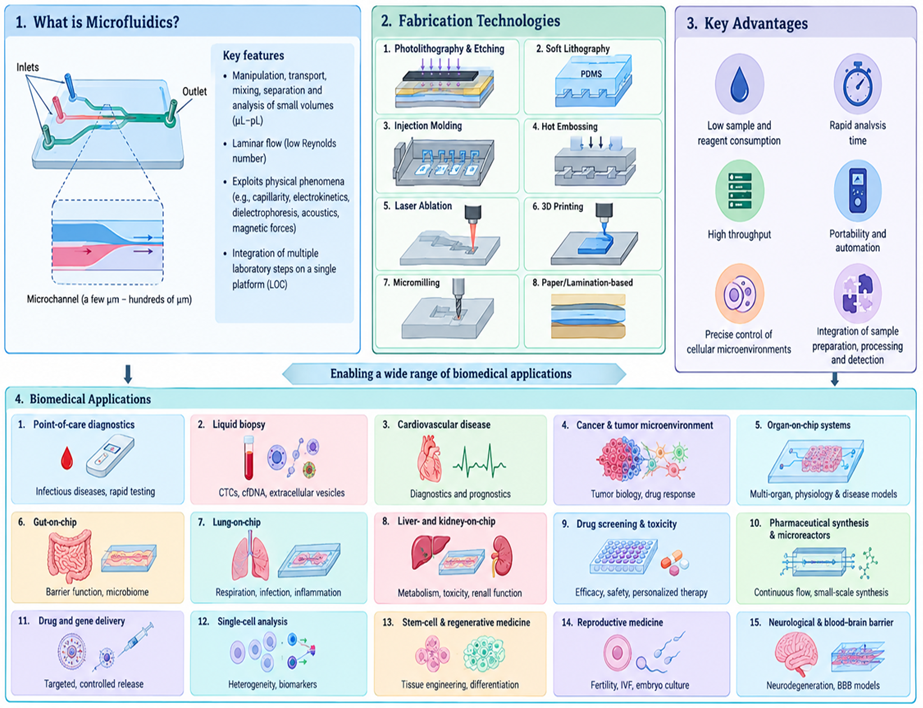
Microfluidics has emerged as an important platform for manipulating and analyzing small volumes of fluids, cells, biomolecules, and particles with high precision. Its ability to control microscale flows and integrate multiple analytical steps has enabled diverse biomedical technologies, including lab-on-a-chip, point-of-care diagnostics, organ-on-chip, and microphysiological systems. This review summarizes the fundamental principles of biomedical microfluidics, including microscale fluid dynamics, laminar flow, diffusion, and key dimensionless parameters, together with major platform formats such as continuous-flow, droplet-based, and digital microfluidics. Recent fabrication approaches, including photolithography, soft lithography, molding, laser-based methods, 3D printing, micromilling, and paper- and lamination-based techniques, are also discussed. The review highlights applications in cancer and liquid biopsy, infectious disease diagnostics, cardiovascular and neurological disorders, organ-on-chip models, drug screening and toxicity testing, pharmaceutical synthesis, drug delivery, single-cell analysis, and regenerative and reproductive medicine. Particular attention is given to the growing use of microfluidics for personalized and human-relevant biomedical research. The review further examines the major challenges associated with microfluidic biomedical systems, including fabrication reproducibility, material compatibility, biological complexity, standardization, scalability and manufacturing, clinical validation, regulatory requirements, and integration with advanced computational approaches such as artificial intelligence and machine learning. Finally, future perspectives are discussed, with particular emphasis on personalized medicine, decentralized diagnostics, human-relevant drug development, multi-organ microphysiological systems, wearable and implantable microfluidics, and automated biomedical platforms.
Keywords:
microfluidics
; point-of-care diagnostics
; liquid biopsy
; organ-on-chip
; drug screening
; microreactor
; single-cell analysis
; personalized medicine
1. Introduction
Microfluidics has emerged as an important interdisciplinary technology for controlling and analysing small volumes of fluids within microscale structures. By combining principles from fluid mechanics, engineering, chemistry, biology, and materials science, microfluidic systems enable controlled transport, mixing, separation, and reaction of fluids, cells, particles, and biomolecules. The field developed from advances in microfabrication, semiconductor processing, and microelectromechanical systems and subsequently evolved into lab-on-a-chip (LOC) platforms capable of integrating multiple laboratory functions within compact devices [1,2,3,4,5,6]. The physical behaviour of fluids at the microscale provides the basis for many of these capabilities. Microchannels generally operate under low Reynolds numbers, where viscous forces dominate and flow is predominantly laminar. Although limited turbulence reduces spontaneous mixing, short diffusion distances allow molecular transport to be efficiently controlled through channel geometry, flow conditions, and interfacial effects [1,2]. Other physical mechanisms, including capillary, inertial, electrical, magnetic, acoustic, and hydrodynamic forces, can be incorporated to manipulate biological and chemical components. These principles have enabled the development of continuous-flow, droplet-based, and digital microfluidic systems for increasingly complex analytical and biological tasks [2,4].
The integration of fluid handling, sample preparation, biochemical reactions, and detection has particularly advanced biomedical applications. Microfluidic platforms can reduce sample requirements and analysis time while enabling automated and multiplexed workflows, making them attractive for decentralized and point-of-care (POC) testing [3,4,5,6,7,8]. Such systems have been investigated for pathogen detection, nucleic-acid testing, immunoassays, and biomarker analysis. In cancer research, microfluidic approaches have also expanded the capabilities of liquid biopsy by enabling the enrichment and characterization of rare circulating tumor cells (CTCs), cell-free DNA, and extracellular vesicles from complex biological samples [6,7,8,9,10]. Another major advance has been the integration of microfluidics with tissue engineering and cell biology to create organ-on-chip (OoC) and microphysiological systems. These platforms combine living cells with controlled perfusion, extracellular matrices, tissue interfaces, and mechanical or biochemical cues to reproduce selected features of human physiology and disease [11,12,13]. Microfluidic organ models have been developed for tissues including the lung, intestine, liver, kidney, heart, brain, and tumor microenvironment, providing experimental systems for studying disease mechanisms, host–pathogen interactions, and therapeutic responses. Compared with conventional static cell cultures, OoC systems can better reproduce dynamic tissue environments and, in some applications, improve the physiological relevance of preclinical testing [12,13,14,15,16].
The scope of biomedical microfluidics now extends from molecular diagnostics and liquid biopsy to single-cell analysis, drug screening, tissue engineering, regenerative medicine, drug delivery, and human-relevant disease modelling. The convergence of microfluidics with organoids, three-dimensional bioprinting, advanced biomaterials, biosensors, automation, and computational approaches is further increasing the complexity and functionality of these platforms. Nevertheless, translation into routine biomedical and clinical use remains constrained by issues including fabrication reproducibility, material compatibility, biological complexity, standardization, scalability, and validation [10,11,12,13,14,15,16].
In this review, we summarize the fundamental principles underlying biomedical microfluidics and discuss major fabrication strategies and platform configurations. We then examine recent applications in diagnostics, liquid biopsy, cancer and tumor-microenvironment modelling, organ-on-chip systems, drug screening, pharmaceutical applications, single-cell analysis, regenerative and reproductive medicine, and neurological models. Finally, we discuss current technological and translational challenges and consider emerging opportunities for personalized medicine, decentralized diagnostics, automated platforms, and increasingly human-relevant biomedical models.
2. Fundamental Principles of Microfluidics
Microfluidic behaviour arises from the interplay of viscous, inertial, capillary, and diffusive forces at small length scales. Because microchannels typically have characteristic dimensions of only a few to several hundred micrometres, viscous and interfacial effects become particularly important, resulting in flow regimes that differ markedly from those encountered in conventional laboratory systems [17,18,19,20]. The Reynolds number (Re), which describes the ratio of inertial to viscous forces, is therefore a fundamental parameter for characterizing microchannel flow. Under the low-Reynolds-number conditions commonly encountered in microfluidics, flow is predominantly laminar and highly predictable, allowing stable fluid streams and well-defined interfaces to be generated (Figure 1). Other parameters, including the capillary number (Ca) and Péclet number (Pe), are important for describing interfacial behaviour and the relative contributions of convection and molecular diffusion, respectively [18,19,20,21,22]. The capillary number (Ca) describes the relative importance of viscous forces compared with surface-tension forces and is particularly relevant in systems involving droplets, multiphase flows, and fluid–fluid interfaces. The Péclet number (Pe) describes the relative contributions of convective transport and molecular diffusion and is important for understanding mixing and mass transport in microchannels. Transport and mixing in microfluidic devices are consequently governed largely by diffusion and controlled convection rather than turbulence. Although the short diffusion distances associated with microscale dimensions can promote rapid molecular transport, channel architecture can be deliberately engineered to improve mixing and mass transfer. Serpentine channels, flow-focusing geometries, herringbone structures, and other engineered interfaces have therefore been developed to regulate fluid contact and enhance molecular exchange (Figure 1) [20,21,22,23,24]. These approaches are particularly relevant to biochemical reactions, nucleic-acid amplification, drug formulation, and cell-based assays, where reproducible control of concentration and reaction conditions is required.
Beyond fluid transport, microfluidic platforms provide diverse mechanisms for manipulating cells, particles, and biomolecules. Hydrodynamic focusing enables particles or cells to be positioned within defined flow streams, whereas inertial migration and Dean-flow effects can facilitate passive separation. Electrical approaches, including electrophoresis and dielectrophoresis, exploit particle charge or polarizability, while magnetophoresis enables the manipulation of magnetically responsive particles and labelled cells. Acoustic and optical forces provide additional means of positioning, concentrating, sorting, or manipulating biological components with minimal physical contact (Figure 1) [23,24,25,26,27,28,29,30]. The combination of these mechanisms has enabled applications ranging from blood-cell and circulating tumor cell separation to single-cell analysis and pathogen enrichment.
Microfluidic platforms can also be distinguished according to how fluids are transported and controlled. Continuous-flow systems operate with uninterrupted streams and are widely used for mixing, separation, chemical synthesis, cell culture, and analytical processing [31,32,33]. Droplet-based microfluidics instead generates discrete, highly uniform droplets that can function as individual reaction compartments, enabling high-throughput screening, single-cell assays, enzymatic reactions, and digital molecular analysis [34,35,36,37,38]. Digital microfluidics provides programmable manipulation of individual droplets, commonly through electrowetting-on-dielectric (EWOD), allowing droplets to be dispensed, transported, merged, and divided on an electrode array (Figure 1) [39,40,41]. These different formats provide complementary approaches for controlling biological and chemical processes and can be selected according to sample characteristics and analytical requirements.
An important development in biomedical microfluidics has been the integration of fluid handling with sensing and detection technologies. Optical, electrochemical, electrical, thermal, and mass-spectrometric methods can be coupled with microfluidic architectures to detect biochemical reactions, cellular responses, and molecular targets (Figure 1). Fluorescence- and absorbance-based detection remains widely used for nucleic-acid amplification, immunoassays, and cell analysis, whereas electrochemical approaches enable compact detection based on changes in current, potential, impedance, or conductivity. Thermal sensing and mass spectrometry provide additional analytical capabilities for reaction monitoring and molecular characterization. Such integration allows sample preparation, separation, reaction, and detection to be connected within a single workflow, reducing manual handling and facilitating automated and portable analytical systems [42,43,44,45,46,47,48,49,50,51,52]. Collectively, these physical principles and operating configurations provide the foundation for the versatility of microfluidic technology in biomedical research. Precise regulation of flow, mass transport, interfacial phenomena, and external physical forces enables the controlled manipulation of biological materials while integration with sensing technologies permits rapid and often automated analysis (Figure 1). These capabilities underpin a broad range of applications, including point-of-care diagnostics, liquid biopsy, single-cell analysis, organ-on-chip models, drug screening, and other advanced biomedical platforms.
3. Fabrication Methods for Microfluidic Devices
Microfluidic devices can be fabricated using a wide range of subtractive, replication-based, additive, and low-cost manufacturing approaches. The choice of fabrication method depends on factors such as required feature size, channel geometry, substrate material, production scale, device complexity, and intended biomedical application [53,54]. Conventional approaches such as photolithography and soft lithography provide high-resolution structures, whereas replication techniques such as injection molding and hot embossing are better suited to larger-scale production. More recent approaches, including laser processing and three-dimensional (3D) printing, offer rapid prototyping and greater geometric flexibility, while paper- and lamination-based methods provide inexpensive alternatives for decentralized diagnostics [53,54,55]. (Figure 2).
3.1. Photolithography and Etching
Photolithography and etching remain fundamental techniques for producing high-resolution microfluidic structures. In photolithography, a photoresist-coated substrate is selectively exposed to ultraviolet light through a patterned photomask, followed by development to generate the desired microstructure (Figure 2). The pattern can subsequently be transferred into substrates such as silicon or glass using wet or dry etching [54,55]. This approach provides precise control over channel dimensions and is widely used to prepare master structures for subsequent polymer replication. Its established processing protocols and high reproducibility have made it an important foundation for microfluidic device fabrication, particularly when small and well-defined features are required [54,55,56,57].
3.2. Soft Lithography
Soft lithography is widely employed for the fabrication of polymeric microfluidic devices, particularly in biomedical research. A typical workflow involves producing a patterned master, casting an elastomer such as polydimethylsiloxane (PDMS) onto the master, curing and peeling the polymer, and subsequently bonding the patterned layer to glass or another substrate (Figure 2). The resulting devices can contain complex channel networks and are compatible with optical imaging and cell-based experiments [58,59]. PDMS is especially useful because of its optical transparency, flexibility, gas permeability, and ease of processing. Consequently, soft lithography has been extensively adopted for cell culture, organ-on-chip models, and rapid prototyping [58,59,60].
3.3. Injection Molding
Injection molding is a replication-based technique designed primarily for the scalable production of thermoplastic microfluidic devices. In this process, a heated polymer melt is injected into a microstructured mold under pressure and subsequently cooled to reproduce the desired channel architecture (Figure 2). Materials such as polycarbonate, polymethyl methacrylate (PMMA), and cyclic olefin copolymer (COC) can be processed using this approach [61,62]. High replication rates and consistent dimensional characteristics make injection molding particularly suitable for disposable diagnostic cartridges and other applications requiring large numbers of devices. However, fabrication of the initial mold can involve considerable cost and is generally more practical for established designs and higher production volumes [61,62,63,64].
3.4. Hot Embossing
Hot embossing is another replication strategy for manufacturing thermoplastic microfluidic structures. A patterned mold is pressed against a thermoplastic substrate maintained at an appropriate temperature, transferring the microstructured features into the polymer surface (Figure 2). After cooling, the embossed substrate can be separated from the mold and subsequently bonded to form enclosed channels [65,66]. The method is applicable to materials such as PMMA and polycarbonate and is useful for producing relatively detailed planar microstructures. Its suitability for replication of thermoplastic devices has supported applications in analytical, diagnostic, and biomedical microfluidics [65,66,67].
3.5. Laser Ablation and Xurography
Laser ablation and xurography provide relatively rapid alternatives to conventional photolithographic processing. In laser ablation, a focused laser directly removes material from a substrate to form channels, wells, or other microstructures [68,69]. Xurography uses computer-controlled cutting tools to pattern channels in polymer films and related materials [70,71]. Both approaches enable rapid modification of device designs and are therefore useful for prototyping and small-scale fabrication (Figure 2). They can be applied to a range of polymeric substrates without requiring a conventional photolithographic mask. Feature resolution, surface morphology, and heat-affected regions, however, depend strongly on the processing conditions and substrate material [68,69,70,71].
3.6. Three-Dimensional Printing
Three-dimensional printing has expanded the design space of microfluidic fabrication by enabling structures to be constructed directly from digital models. Techniques such as stereolithography (SLA), digital light processing (DLP), two-photon polymerization (2PP), and fused deposition modelling (FDM) can generate channels and integrated components with complex three-dimensional architectures [Figure 2]. SLA constructs 3D structures by using a laser to selectively photopolymerize a liquid resin, building the object layer-by-layer. This technique is valued for its high resolution, capable of achieving features around 20 µm, which is suitable for creating intricate microfluidic networks [72,73,74]. DLP operates on a similar photopolymerization principle as SLA but uses a digital micromirror device to project an entire cross-sectional image of a layer at once, enabling faster print speeds. It offers a balance of high resolution, with features approaching the single-micron scale, and practical printing dimensions for complex microfluidic devices [75,76]. 2PP is a high-resolution, laser-based technique where a near-infrared femtosecond laser induces polymerization only at the focal point, where two photons are absorbed simultaneously. This allows for the fabrication of extremely fine features, with positive structures as small as 5 µm or even sub-micrometer resolution scaffolds, making it the highest-resolution 3D printing method for microfluidics [77,78]. FDM builds objects by extruding a continuous filament of a melted thermoplastic material through a heated nozzle, which solidifies to form each layer. While well-suited for rapid prototyping, FDM’s resolution is fundamentally limited by the nozzle diameter, which typically restricts the fabrication of microchannels to widths around 100 µm or larger [79,80]. These high-resolution photopolymerization techniques are particularly promising for microscale biomedical structures, although material biocompatibility, surface quality, optical properties, and printing resolution remain important considerations.
3.7. Micromilling and Mechanical Micromachining
Micromilling employs computer-controlled miniature cutting tools to directly machine microchannels and other structures into materials such as plastics, metals, and composites. The technique is attractive for rapid prototyping because it does not require the cleanroom processing associated with conventional photolithography. It can also be used to fabricate molds, masters, manifolds, and relatively large microfluidic channels. However, the achievable feature size is constrained by tool dimensions, while machining may introduce surface roughness and debris. Thus, micromilling is particularly useful for prototypes and structures where extremely small features are not required (Figure 2) [81,82].
3.8. Paper-Based and Lamination-Based Fabrication
Paper-based and lamination-based approaches provide simple and economical routes for producing microfluidic devices, particularly for point-of-care and resource-limited applications. In paper microfluidics, liquid transport occurs primarily through capillary forces, while hydrophobic barriers define the pathways and reaction zones. Patterning can be achieved using wax printing, inkjet or screen printing, and laser-based methods. Lamination of patterned polymer or adhesive layers provides another straightforward strategy for constructing multilayer fluidic networks [83]. The low material cost, lightweight nature, disposability, and minimal instrumentation requirements of these approaches make them attractive for decentralized diagnostic testing (Figure 2) [83,84,85].
4. Biomedical Applications of Microfluidics
4.1. Point-of-Care Detection of Infectious Pathogens
One of the most clinically important applications of microfluidics is the rapid detection of infectious pathogens. Conventional pathogen diagnosis often requires centralized laboratories, trained personnel, lengthy culture procedures, and expensive analytical equipment. Microfluidic point-of-care (POC) systems integrate multiple diagnostic steps within a compact and portable platform [86,87]. The diagnostic workflow (Figure 3) can begin with minimally processed clinical specimens such as blood, nasopharyngeal swabs, urine, or stool. Sample preparation may involve filtration, cell lysis, purification, concentration, and removal of interfering substances. These processes improve downstream detection by enriching the target pathogen and reducing matrix-associated interference. The processed sample can subsequently undergo nucleic-acid extraction followed by molecular amplification within integrated microfluidic chambers. Polymerase chain reaction (PCR) and isothermal amplification methods, including loop-mediated isothermal amplification (LAMP) and recombinase polymerase amplification (RPA), can provide sensitive pathogen detection. Nucleic-acid hybridization and other molecular recognition strategies can also be incorporated for specific identification of microbial targets. In parallel, immunological approaches can detect pathogen-specific antigens or host antibodies using integrated immunoassays. Optical, fluorescence, electrochemical, and other biosensing technologies can then convert molecular recognition events into measurable signals [88,89]. Microfluidic platforms have been investigated for detecting a broad range of pathogens, including bacteria, viruses, parasites, fungi, and other infectious agents. Their ability to combine sample processing, concentration, extraction, amplification, and detection reduces the need for multiple separate laboratory instruments. The use of microlitre- to nanolitre-scale volumes reduces reagent consumption and can shorten analysis time compared with conventional laboratory workflows [90,91]. Compact device architectures also provide portability and support rapid testing in hospitals, emergency settings, remote locations, and resource-limited environments. Potential automation and closed-system operation can further reduce manual handling and improve the reproducibility of diagnostic procedures. These characteristics make microfluidic POC platforms particularly valuable for decentralized healthcare, early diagnosis, outbreak surveillance, and rapid public-health response. Despite these advantages, clinical implementation requires reliable sample preparation, effective contamination control, and robust operation across diverse clinical sample matrices. Standardized analytical performance, including sensitivity, specificity, reproducibility, and limit of detection, must also be established through rigorous clinical validation [92,93,94].
4.2. Liquid Biopsy and Circulating Tumor Cell Detection
Liquid biopsy is an important emerging application of microfluidics in precision oncology. Unlike conventional tissue biopsy, liquid biopsy can be performed repeatedly using blood or other body fluids. Important circulating biomarkers include circulating tumor cells (CTCs), circulating tumor DNA (ctDNA), cell-free RNA, exosomes, and tumor-derived proteins [10,12,13]. CTCs are extremely rare in peripheral blood, creating a major challenge for conventional analysis. Microfluidic systems can exploit differences in cell size, deformability, density, electrical properties, magnetic susceptibility, or surface biomarkers to enrich CTCs. Passive methods include deterministic lateral displacement, inertial focusing, filtration, and hydrodynamic separation, while active methods can employ magnetic, electrical, acoustic, or optical forces [95,96,97]. After enrichment, CTCs can be counted, imaged, genotyped, or subjected to downstream molecular analysis (Figure 4). Microfluidic liquid biopsy may therefore contribute to early cancer detection, prognosis, monitoring of treatment response, and characterization of therapeutic resistance. Microfluidic platforms can further integrate CTC enrichment with downstream molecular characterization, enabling analysis of genetic and phenotypic heterogeneity within individual tumor-cell populations. The isolation of viable CTCs can also support ex vivo drug-response testing and functional studies, providing opportunities for treatment selection and personalized therapy [98,99,100,101]. In addition to CTCs, microfluidic systems can facilitate the isolation and analysis of extracellular vesicles, including exosomes, which carry tumour-derived nucleic acids, proteins, and other molecular information. Microfluidic enrichment of ctDNA and other cell-free biomarkers can improve analytical sensitivity while requiring only small volumes of patient samples. Integration with multiplexed biosensors, digital PCR, sequencing, and automated imaging can further enhance the molecular resolution of liquid-biopsy platforms [102,103,104]. However, challenges including the extreme rarity and heterogeneity of CTCs, variable biomarker expression, nonspecific cell capture, and differences in clinical sample composition remain important barriers to standardization.
4.3. Cardiovascular Disease Diagnostics and Prognostics
Microfluidics has significant potential in cardiovascular medicine because cardiovascular physiology depends strongly on fluid flow, shear stress, endothelial function, platelet activation, and coagulation. Microfluidic devices can reproduce physiologically relevant blood-flow conditions and provide controlled environments for studying thrombosis, hemostasis, atherosclerosis, vascular inflammation, and cardiac disease (Figure 5A) [105,106]. Microfluidic thrombosis platforms allow platelet adhesion and coagulation to be investigated under controlled shear rates using very small blood volumes. This creates opportunities for patient-specific assessment of platelet function and anticoagulant or antiplatelet drug responses [107,108]. Vessel-on-chip platforms can additionally model endothelial dysfunction and atherosclerotic processes. Heart-on-chip systems have expanded this field by incorporating cardiomyocytes, endothelial cells, extracellular matrix, and mechanical stimulation. Such models can be used to investigate ischemia-reperfusion injury, cardiac hypertrophy, fibrosis, arrhythmia, and drug-induced cardiotoxicity [109,110] (Figure 5B). Microfluidic biosensors can detect circulating cardiovascular biomarkers, including cardiac troponins, natriuretic peptides, inflammatory mediators, and coagulation-related proteins, with high sensitivity and low sample volumes (Figure 5C). Such platforms enable rapid point-of-care assessment of myocardial injury, heart failure, vascular inflammation, and thrombotic risk [111,112] (Figure 5D). Microfluidic blood-analysis systems can also characterize red blood cell deformability, platelet activation, leukocyte behaviour, and plasma viscosity, providing functional information beyond conventional biochemical assays [113,114] (Figure 5E). Patient-derived blood samples can be incorporated into personalized microfluidic models to evaluate individual differences in thrombosis susceptibility and therapeutic responses (Figure 5F). These systems may support rapid screening of antiplatelet and anticoagulant therapies while reducing sample consumption and experimental time [115]. Vascular-on-chip platforms can further reproduce endothelial barrier function, inflammatory signaling, and interactions between blood cells and vessel walls under physiologically relevant flow conditions (Figure 5G). Integration with automated imaging, biosensors, and machine-learning-based analysis may improve quantitative interpretation and enable earlier detection of pathological changes (Figure 5H). Despite these advantages, challenges related to device standardization, biological variability, clinical validation, sample preparation, and integration into routine diagnostic workflows remain [105,106,107,108,109,110,111,112,113,114,115].
4.4. Cancer and Tumor-Microenvironment-on-a-Chip
Cancer is a highly heterogeneous disease involving interactions between malignant cells, immune cells, endothelial cells, fibroblasts, extracellular matrix, blood vessels, and biochemical gradients. Conventional two-dimensional cultures cannot reproduce many of these interactions. Microfluidic tumor chips provide a controllable platform for reconstructing selected features of the tumor microenvironment (Figure 6) [116,117]. Tumor-on-chip systems can establish gradients of oxygen, nutrients, growth factors, and chemotherapeutic agents. They can also model cancer-cell migration, invasion, angiogenesis, immune-cell infiltration, and metastatic dissemination. Patient-derived cells or tumor organoids can potentially be incorporated to investigate individual therapeutic responses. These platforms are particularly promising for precision oncology because drug responses can be examined in a patient-specific microenvironment rather than solely in conventional cell culture. Current challenges include reproducing tumor heterogeneity, vascularization, extracellular matrix complexity, immune interactions, and clinically relevant pharmacokinetics [118,119]. Tumor-on-chip platforms can further incorporate stromal components, including cancer-associated fibroblasts, pericytes, and extracellular-matrix proteins, to better mimic tumor architecture. Microvascular networks within these systems enable investigation of tumor angiogenesis and interactions between circulating tumor cells and endothelial barriers. Such models can be used to study epithelial–mesenchymal transition, intravasation, extravasation, and metastatic colonization under controlled conditions. Integration of immune cells allows evaluation of tumor–immune interactions and the effects of immunotherapeutic agents, including immune checkpoint inhibitors [120,121,122,123]. Microfluidic chips can also reproduce hypoxic and acidic tumor niches that contribute to therapeutic resistance and malignant progression. Real-time imaging enables continuous monitoring of cellular behavior, drug penetration, tumor growth, and treatment-induced responses. Multiplexed microfluidic platforms may allow simultaneous testing of multiple drugs or drug combinations using limited numbers of patient-derived cells. Integration with biosensors and molecular analysis can provide dynamic measurements of secreted cytokines, metabolites, circulating tumor markers, and other disease-associated biomarkers. Advances in organoid technology and three-dimensional bioprinting are further improving the structural and cellular complexity of tumor-on-chip models [124,125].
4.5. Organ-on-Chip Platforms
Organ-on-chip technology represents one of the most influential biomedical developments enabled by microfluidics. Organ chips use micro-engineered channels containing living human cells to reproduce selected structural, biochemical, mechanical, and physiological characteristics of organs [14]. Unlike conventional static cell cultures, organ chips can expose cells to continuous fluid flow, mechanical deformation, concentration gradients, and interactions with multiple cell types. Lung, liver, kidney, heart, intestine, brain, bone marrow, and vascular systems have all been modelled. The long-term objective is to develop interconnected multi-organ or human-on-chip systems capable of modelling systemic drug distribution and toxicity (Figure 7). Such platforms may eventually complement or reduce dependence on animal models, although standardization, reproducibility, scalability, and regulatory acceptance remain major challenges [14,126,127].
4.5.1. Gut-on-a-Chip
Gut-on-chip models are particularly useful for studying inflammatory bowel disease, infection, intestinal permeability, microbiome-associated disease, and oral drug absorption (Figure 7). They may also provide improved models for investigating how the gut influences systemic metabolism and pharmacokinetics [128,129]. The incorporation of intestinal stem-cell-derived organoids can further improve the physiological relevance of these models by providing differentiated epithelial cell populations, including enterocytes, goblet cells, Paneth cells, and enteroendocrine cells. Co-culture with immune cells enables investigation of inflammatory signaling, epithelial injury, immune-cell recruitment, and tissue repair. Endothelial channels can also be integrated to model interactions between the intestinal epithelium and the underlying microvasculature [130,131]. Microfluidic flow facilitates controlled delivery of nutrients, drugs, microbial metabolites, and inflammatory mediators while enabling continuous removal of metabolic waste. Mechanical deformation of the intestinal epithelium can reproduce peristalsis-like motions and provide important biomechanical cues that influence barrier integrity and cellular differentiation. These platforms can also be used to evaluate epithelial permeability and transepithelial transport using real-time imaging and molecular assays [132,133]. Advanced gut-on-chip systems may incorporate patient-derived intestinal cells to investigate disease-specific phenotypes and individualized responses to therapeutic interventions. Integration with biosensors and analytical technologies allows monitoring of cytokines, metabolites, microbial products, and other biomarkers in real time. Despite these advantages, challenges remain in maintaining long-term microbial stability, reproducing the full diversity of the human microbiome, standardizing chip designs, and establishing clinically validated experimental protocols. Overall, gut-on-chip technology provides a versatile platform for studying intestinal physiology and disease while supporting drug development, microbiome research, and personalized medicine [134,135].
4.5.2. Lung-on-a-Chip and Respiratory Disease
Lung-on-chip systems attempt to reproduce the alveolar–capillary interface and mechanical environment of the human lung (Figure 7). A landmark microfluidic lung model incorporated human alveolar epithelial cells and vascular endothelial cells separated by a porous membrane, while cyclic mechanical deformation reproduced breathing-like movements [136]. Such platforms can be used to investigate pulmonary inflammation, infection, edema, vascular leakage, toxic exposures, and drug responses. Lung chips have also been used to model interactions with inhaled particles and pathogens. The ability to introduce controlled mechanical forces is particularly important because breathing-induced deformation can substantially affect lung-cell behaviour. Lung-on-chip technology therefore represents a useful bridge between conventional cell culture and complex animal models [137,138]. More advanced lung-on-chip platforms incorporate multiple pulmonary cell types, including alveolar epithelial cells, endothelial cells, fibroblasts, macrophages, and other immune populations, to better reproduce the cellular complexity of the lung microenvironment. These systems can model inflammatory signaling and immune-cell recruitment during bacterial or viral respiratory infections. Controlled exposure to aerosols, nanoparticles, environmental pollutants, and airborne toxicants enables assessment of their effects on epithelial integrity and pulmonary inflammation [139,140]. Lung chips can also reproduce pathological conditions such as acute respiratory distress syndrome, pulmonary edema, fibrosis, and chronic airway inflammation. Real-time imaging allows continuous monitoring of barrier disruption, cellular injury, leukocyte migration, and tissue recovery. Integration of sensors can provide dynamic measurements of oxygen concentration, barrier permeability, inflammatory mediators, and other physiological parameters [136,137,138,139,140,141]. Patient-derived cells and induced pluripotent stem-cell-derived pulmonary cells may further enable disease-specific modeling and personalized drug-response testing. Integration with organoid technology and three-dimensional biomaterials can increase the structural complexity of lung models, while automated imaging and computational analysis can improve experimental throughput. Despite these advances, challenges remain in reproducing the full anatomical complexity of the lung, maintaining long-term tissue function, and standardizing mechanical and biochemical conditions across platforms. Overall, lung-on-chip technology provides a powerful approach for investigating respiratory physiology, disease mechanisms, environmental exposures, and therapeutic responses under controlled human-relevant conditions [142,143].
4.5.3. Liver-on-Chip and Kidney-on-Chip
The liver and kidney are central to drug metabolism, detoxification, transport, and elimination. Consequently, liver and kidney toxicity represents an important cause of drug-development failure [144]. Microfluidic liver-on-chip systems can provide controlled perfusion of hepatocytes, endothelial cells, and supporting cell types, allowing metabolic activity and drug-induced injury to be studied under more physiologically relevant conditions [145]. Liver-on-chip platforms can incorporate primary human hepatocytes, induced pluripotent stem-cell-derived hepatocytes, hepatic stellate cells, Kupffer cells, and endothelial cells to better reproduce the multicellular liver microenvironment. Continuous perfusion supports nutrient delivery, waste removal, and maintenance of hepatic metabolic functions over extended periods. These systems can be used to investigate cytochrome P450 activity, drug metabolism, oxidative stress, inflammation, and hepatotoxicity [146,147].
Kidney-on-chip models can reproduce aspects of glomerular filtration, tubular transport, epithelial function, and nephrotoxicity. These systems are especially valuable for evaluating compounds that may cause renal injury [148]. Kidney-on-chip platforms can similarly incorporate specialized epithelial and endothelial compartments to reproduce the physiological conditions of the nephron. Tubule-on-chip models enable investigation of solute transport, drug reabsorption, secretion, and epithelial injury. Glomerulus-on-chip systems can be used to study filtration-barrier integrity and interactions between podocytes and glomerular endothelial cells [149,150].
Mechanical and fluidic cues are also important because shear stress and pressure influence renal and hepatic cell function. Real-time imaging and integrated biosensors can enable monitoring of cell viability, barrier permeability, metabolic activity, and biomarkers of organ injury. Patient-derived cells may further support personalized assessment of drug metabolism and susceptibility to organ-specific toxicity. More advanced multi-organ platforms can connect liver and kidney compartments with other tissues, allowing investigation of pharmacokinetics, metabolite formation, and downstream toxic effects. Such systems may reduce reliance on animal models during early drug development and improve prediction of human-specific responses. However, challenges remain in maintaining long-term tissue maturity, reproducing complex organ architecture, and standardizing chip designs and analytical endpoints [151,152,153].
4.6. Neurological Disease and Blood–Brain-Barrier Models
The blood–brain barrier (BBB) is a highly selective neurovascular interface that regulates the exchange of ions, nutrients, metabolites, signaling molecules, and therapeutics between the systemic circulation and the central nervous system (CNS). Its barrier properties arise from the coordinated organization of brain microvascular endothelial cells, pericytes, astrocytes, basement membrane, and associated extracellular-matrix components, collectively forming the neurovascular unit. Brain endothelial cells exhibit specialized tight junctions, low paracellular permeability, selective transporters, and active efflux systems that restrict the entry of many circulating compounds into the brain. This structural and biochemical complexity makes conventional static in vitro models inadequate for reproducing the dynamic interactions that regulate BBB integrity and function [154,155,156]. Microfluidic BBB-on-chip systems address these limitations by arranging living human cells within perfusable microchannels, frequently using separate vascular and brain-side compartments connected by a porous membrane or an extracellular-matrix interface (Figure 8). Such architectures enable controlled fluid flow, physiological shear stress, compartmentalized molecular transport, and communication between endothelial and neural cells. The resulting platforms provide a more controllable representation of the neurovascular microenvironment than conventional monolayer cultures and can be adapted to investigate both normal BBB physiology and disease-associated barrier dysfunction [157]. The BBB-on-chip architecture shown in Figure 8 typically consists of a vascular channel containing endothelial cells and a brain-side compartment containing astrocytes, neurons, and extracellular matrix, with pericytes positioned near the endothelial barrier. The porous membrane or matrix interface permits diffusion of soluble factors while maintaining separation between the vascular and neural compartments. Endothelial cells form the principal physical barrier, whereas pericytes contribute to vascular stability, endothelial maturation, and regulation of barrier properties. Astrocytes provide structural and biochemical support through their interactions with endothelial cells and the surrounding matrix, while neurons contribute neural signaling and tissue-specific functional responses. The extracellular matrix provides adhesion sites and biochemical cues that influence cell organization, differentiation, and barrier maintenance. Under controlled perfusion, the vascular compartment can reproduce aspects of blood flow and shear stress, whereas the brain-side compartment allows investigation of neural responses to circulating molecules. This compartmentalized design is particularly valuable for distinguishing vascular exposure, barrier transport, and neural effects, which are difficult to separate in conventional well-plate assays [157,158].
Microfluidic BBB models can reproduce important features of the neurovascular unit, including tight-junction formation, endothelial barrier integrity, selective molecular transport, and cell–cell signaling. Tight-junction proteins such as claudins, occludin, and zonula occludens-1 contribute to the restriction of paracellular transport, while transporter-mediated mechanisms regulate the movement of nutrients and other molecules across the endothelial layer. Controlled perfusion provides physiologically relevant shear stress and enables investigation of how vascular conditions influence endothelial morphology, junctional organization, and barrier function. Microfluidic systems can also establish concentration gradients of oxygen, nutrients, inflammatory mediators, and therapeutic compounds, allowing researchers to examine how spatially varying conditions affect BBB permeability. Barrier integrity may be evaluated through permeability assays, fluorescent tracer transport, imaging of junctional proteins, and measurements of transendothelial electrical resistance where suitable electrodes are incorporated. These complementary readouts help distinguish changes in physical barrier integrity from alterations in active transport or cellular signaling [159,160,161].
The controlled microenvironment of BBB-on-chip platforms is particularly useful for investigating neuroinflammation and disease-associated barrier disruption. Inflammatory cytokines, oxidative stress, hypoxia, and other pathological stimuli can alter endothelial junctions, increase vascular permeability, and modify communication among endothelial cells, pericytes, astrocytes, and neurons. More advanced platforms incorporate immune cells, such as microglia-like cells, macrophages, or circulating lymphocytes, to study immune-cell recruitment, endothelial activation, and inflammatory signaling within the neurovascular unit. These models can be used to investigate how immune-mediated damage affects BBB integrity and how inflammatory responses influence CNS drug delivery [162,163,164]. In addition, microfluidic brain tumor-on-chip systems can incorporate tumor cells together with endothelial cells, astrocytes, pericytes, and extracellular matrix to reproduce selected features of the tumor–vascular interface. Such platforms enable investigation of tumor invasion, angiogenic signaling, vascular permeability, and the penetration of therapeutic agents into brain tumor tissue. They may also provide controlled models for studying how tumor-associated inflammation and altered vascular architecture contribute to treatment resistance [165,166,167]. Beyond BBB modeling, brain-on-chip systems can reproduce aspects of neuronal networks, glial interactions, and CNS tissue organization under dynamic culture conditions. Microfluidic compartments can guide the spatial arrangement of neurons, support axonal growth, and permit controlled exposure to drugs, toxins, or disease-associated factors. Integration with three-dimensional brain organoids can further increase cellular and structural complexity by incorporating multiple neural cell populations within a microengineered environment. Patient-derived cells and induced pluripotent stem-cell-derived neural populations may support disease-specific modeling of neurological conditions such as Alzheimer’s disease, Parkinson’s disease, epilepsy, and glioblastoma. These approaches are particularly valuable when disease mechanisms depend on human-specific cellular interactions or when conventional animal models do not fully reproduce patient-associated phenotypes. However, the extent to which organoid-based systems reproduce mature brain architecture, regional specialization, and long-term neuronal function remains dependent on the model design and experimental conditions [165,166,167].
Real-time imaging, biosensors, and molecular analysis further expand the analytical capabilities of BBB- and brain-on-chip platforms. Live-cell imaging can monitor endothelial morphology, barrier disruption, neuronal activity, immune-cell migration, tumor-cell invasion, and treatment-induced changes over time. Integrated biosensors may enable dynamic measurement of barrier integrity, oxygen levels, inflammatory mediators, and other biochemical signals, while molecular assays can characterize changes in gene expression, protein signaling, and secreted factors. These measurements provide temporal information that is often unavailable from endpoint-based conventional cultures. Microfluidic platforms can also support systematic screening of CNS therapeutics by evaluating compound permeability, transport, efficacy, and neurotoxicity within a human-relevant cellular environment. Such approaches may help identify compounds with improved BBB penetration while reducing the risk of selecting candidates that appear effective in simplified assays but fail because of inadequate CNS exposure or unexpected neural toxicity [168,169,170].
Despite their potential, several challenges limit the broader adoption of BBB-on-chip and brain-on-chip technologies. Reproducing the full cellular diversity, regional specialization, vascular architecture, and biochemical complexity of the human brain remains difficult. Long-term maintenance of mature neuronal and endothelial phenotypes, stable barrier function, and reproducible cell–cell interactions also require careful control of culture conditions. Differences in cell source, extracellular-matrix composition, channel geometry, flow parameters, membrane properties, and analytical methods can lead to substantial variation between platforms. Standardized protocols, validated permeability benchmarks, reproducible disease phenotypes, and clinically relevant pharmacokinetic conditions are therefore essential for meaningful comparison and regulatory translation.
4.7. Microfluidics in Pharmaceutical Synthesis, Drug Screening, and Drug Delivery
Microfluidic reactors provide highly controlled environments for pharmaceutical and chemical synthesis. Their small dimensions generate high surface-to-volume ratios and short diffusion distances, enabling efficient mass and heat transfer, rapid mixing, precise temperature control, and well-defined residence times compared with conventional batch reactors [171,172]. The continuous-flow architecture shown in Figure 9 enables reactants, solvents, and catalysts to be introduced through separate inlets and mixed within microscale channels, where reaction conditions can be precisely regulated. Microreactors have been investigated for the synthesis of pharmaceutical intermediates, nanoparticles, polymers, and other functional materials, as well as for enzymatic and photochemical reactions. Continuous-flow operation can facilitate process automation, improve reproducibility, and reduce batch-to-batch variability [173,174]. The high degree of process control also supports rapid optimization of reaction parameters, including flow rate, residence time, temperature, and reactant concentration, which are important for reproducible pharmaceutical manufacturing. An important biomedical application is the fabrication of drug-loaded nanoparticles and other delivery systems, in which microfluidic mixers enable rapid and controlled mixing of polymers, lipids, nucleic acids, and therapeutic compounds to generate particles with tailored size, composition, and encapsulation characteristics [175,176]. As illustrated in Figure 9, controlled mixing can produce delivery carriers with more uniform physicochemical properties, supporting subsequent drug-loading and therapeutic applications.
Microfluidic technologies can also significantly improve pharmaceutical drug screening and toxicity testing by reducing reagent consumption while increasing throughput and experimental control. Unlike conventional microplate-based screening, microfluidic platforms can generate concentration gradients, dose–response profiles, cell compartments, and multiplexed assays within small volumes [177,178]. The concentration-gradient and multiplexed-assay formats shown in Figure 9 allow multiple experimental conditions to be evaluated simultaneously while maintaining spatial control over drug exposure. Applications include enzyme assays, protein–drug interactions, antimicrobial screening, cancer-drug evaluation, toxicity assessment, and combination therapy [179,180]. Droplet microfluidics is particularly advantageous for generating thousands to millions of isolated reaction compartments, enabling high-throughput analysis of individual cells, molecules, or biochemical reactions. Each droplet can function as an independent microreactor, allowing controlled encapsulation of cells or biomolecules, compartmentalized reactions, and quantitative analysis of cellular or molecular responses. Furthermore, integration with organ-on-chip systems extends screening from molecular and cellular assays to tissue-level physiological models, allowing simultaneous assessment of therapeutic efficacy, toxicity, transport, and multicellular interactions under more physiologically relevant conditions [181,182,183]. These integrated platforms can combine dose-dependent exposure with real-time imaging, biomarker analysis, and tissue-level response measurements, thereby improving the physiological relevance of pharmaceutical screening.
Microfluidic platforms have become increasingly important for the preparation of advanced drug- and gene-delivery systems. Precise control over flow rates, mixing, solvent exchange, and reaction conditions enables the fabrication of nanoparticles, liposomes, lipid nanoparticles, polymeric particles, and other delivery carriers with relatively narrow size distributions [184,185,186]. The controlled-parameter framework illustrated in Figure 9 allows systematic adjustment of flow rates, mixing conditions, solvent exchange, and reaction composition to regulate particle formation and improve batch-to-batch consistency. Microfluidic droplet generation can produce monodisperse microparticles, whereas rapid mixing can facilitate the formation of drug- or nucleic-acid-loaded nanoparticles with controlled physicochemical properties. These approaches can improve encapsulation efficiency and provide greater control over particle size, surface charge, composition, morphology, and release characteristics [184,185,186,187]. The resulting carrier architectures, including polymeric particles, liposomes, and lipid nanoparticles, can be tailored for different therapeutic payloads and delivery requirements. The technology is particularly relevant to nucleic-acid therapeutics, including RNA- and gene-based delivery, where reproducible control of carrier properties is essential for effective cellular uptake and therapeutic activity [188,189,190]. As shown in Figure 9, microfluidic platforms can support the formation of nucleic-acid-loaded particles with controlled encapsulation and delivery characteristics, offering a scalable route toward more reproducible drug and gene-delivery formulations.
4.8. Precision biological Control: Microfluidics in Single-Cell Analysis, Regenerative Medicine, and Reproductive Technologies
Cells within the same tissue can exhibit substantial molecular, phenotypic, and functional heterogeneity that may be obscured by conventional bulk measurements. Microfluidic technologies enable precise manipulation and analysis of individual cells, including cell isolation, trapping, sorting, manipulation, and lysis, followed by downstream molecular characterization (Figure 10). Microfluidic single-cell platforms can be coupled with fluorescence imaging, sequencing, proteomics, transcriptomics, and secretome analysis, thereby enabling comprehensive characterization of cellular heterogeneity at single-cell resolution [191,192,193]. In particular, droplet-based microfluidics allows individual cells to be encapsulated within picolitre-scale compartments containing defined reaction components, facilitating high-throughput and parallel analysis of large cell populations. These capabilities are particularly relevant to cancer research, immunology, stem-cell biology, developmental biology, and precision medicine, where single-cell molecular profiling can identify rare cell populations, disease-associated phenotypes, and patient-specific molecular signatures (Figure 10) [194,195].
Microfluidics also provides highly controlled cellular microenvironments for stem-cell research, tissue engineering, and regenerative medicine (Figure 10). Microfluidic platforms can establish spatially and temporally controlled gradients of growth factors, oxygen, nutrients, extracellular-matrix components, and mechanical stimuli, enabling systematic investigation of stem-cell proliferation, migration, differentiation, and lineage commitment [196,197]. As illustrated in Figure 10, integration with biomaterials, three-dimensional culture, organoids, and bioprinting can facilitate the formation of organized tissue structures and vascular networks under controlled microenvironmental conditions. These systems provide opportunities for developing engineered tissues, cell-based therapies, and regenerative strategies while improving control over cellular organization and tissue maturation [198,199]. The ability to precisely regulate the cellular microenvironment may also enhance the reproducibility and functional maturation of organoids and bioprinted tissues, supporting the development of physiologically relevant models for regenerative medicine. Controlled perfusion within microfluidic constructs can additionally improve nutrient and oxygen transport while generating physiologically relevant shear and mechanical cues that influence cell behavior. Such dynamic microenvironments can promote cell–cell and cell–matrix interactions, thereby supporting vascularization, tissue remodeling, and the development of more functional engineered tissue architectures [200,201].
Reproductive medicine and assisted reproduction represent another emerging application of microfluidic technology (Figure 10). Conventional sperm-processing procedures can expose spermatozoa to mechanical and biochemical stresses, whereas microfluidic sperm-selection platforms can exploit differences in motility, size, morphology, and hydrodynamic behavior to enrich viable and functionally competent sperm. As depicted in Figure 10, microfluidic systems can further support sperm–oocyte interaction and fertilization under controlled microscale conditions [202,203]. Such platforms have also been investigated for embryo culture, imaging, developmental monitoring, and embryo selection, with the potential to provide a more controlled and reproducible environment throughout assisted reproductive procedures. Future integrated microfluidic systems may combine sperm selection, fertilization, embryo culture, imaging, and developmental assessment within automated platforms (Figure 10). The small-scale architecture of these devices can provide controlled fluidic conditions and reduce unnecessary mechanical manipulation of gametes and embryos during handling and transfer [204,205]. Integration with real-time imaging may further enable continuous assessment of sperm–oocyte interactions, embryo morphology, and developmental progression under standardized culture conditions. Nevertheless, rigorous clinical validation, reproducibility studies, ethical considerations, and comprehensive assessment of embryo and offspring safety remain essential before these technologies can achieve widespread clinical implementation [206].
As illustrated in Figure 10, Microfluidics provides a versatile platform for controlling biological systems across multiple scales, from single-cell isolation and molecular analysis to cellular microenvironment engineering, tissue regeneration, sperm selection, fertilization, and embryo culture. The integration of microfluidics with advanced analytical, organoid, biomaterial, and bioprinting technologies may therefore contribute to increasingly precise, personalized, and functionally relevant biomedical applications [202,203,204,205,206].
Advantages, Challenges, and Future Perspectives of Microfluidic Biomedical Platforms
Microfluidic biomedical platforms have been rapidly adopted because they offer low sample and reagent consumption, high throughput, precise microenvironmental control, automation and integration, portability, and compatibility with personalized medicine. They operate with extremely small liquid volumes, reducing the use of expensive reagents and enabling experiments with scarce biological samples, while parallel channels, droplets, microwells, and integrated arrays can process thousands to millions of individual reactions or cells. Flow rate, oxygen concentration, chemical gradients, shear stress, temperature, and cell–cell interactions can be carefully controlled, and sample preparation, separation, amplification, detection, and data analysis can potentially be integrated into a single cartridge. Miniaturized instruments can also be designed for use outside centralized laboratories, making microfluidics attractive for point-of-care applications, and patient-derived cells, blood samples, organoids, and tumor material can be directly incorporated into these systems.
Despite this progress, microfluidics faces several obstacles before widespread clinical adoption. Fabrication reproducibility is a major concern because small variations in channel dimensions, surface chemistry, bonding, and material properties can influence fluid flow and biological results, making standardized fabrication protocols essential. PDMS remains widely used in academic research but can absorb hydrophobic molecules and allow gas and water-vapour transport, complicating quantitative drug studies; thermoplastics, glass, cyclic olefin polymers, and newer biomaterials may offer better alternatives for specific applications. Biological complexity is another challenge, since human organs contain numerous interacting cell types, extracellular matrix components, immune cells, vascular networks, nerves, hormones, and dynamic mechanical signals, and no current chip completely reproduces a human organ. Scaling and manufacturing are difficult because a device that performs well in an academic laboratory may be expensive to manufacture millions of times, requiring design-for-manufacturing, quality control, packaging, and supply-chain considerations. Standardization is also needed because different laboratories may use different materials, cell sources, channel geometries, flow rates, and analytical endpoints, making study comparison difficult. Clinical validation must demonstrate sensitivity, specificity, accuracy, reproducibility, robustness, and clinical utility in appropriately large patient populations, while regulatory agencies require clear evidence that organ-on-chip and microphysiological systems produce reliable and clinically relevant information. Finally, integration with detection systems remains an important engineering challenge, since optical, electrical, electrochemical, mass-spectrometric, or smartphone-based readouts must be reliable and practical.
The future of biomedical microfluidics is likely to involve increasingly integrated systems rather than isolated microfluidic functions. AI-integrated microfluidics can analyse complex microscopy, biosensor, single-cell, and organ-chip data, and may optimize channel geometries, operating conditions, drug combinations, and diagnostic decisions. Personalized organ-on-chip systems using patient-derived cells, induced pluripotent stem cells, organoids, and tumor tissues may enable patient-specific drug testing and therapeutic selection, while multi-organ and body-on-chip platforms could connect liver, kidney, heart, intestine, lung, and tumor modules through a circulating fluid network to better represent drug absorption, metabolism, distribution, and toxicity; human organ chips are already being explored for disease modelling, drug development, personalized medicine, and host–microbiome interactions. Wearable and implantable microfluidics may continuously analyse sweat, interstitial fluid, saliva, or other biological fluids for longitudinal biomarker monitoring, and point-of-care molecular diagnostics are likely to integrate sample preparation, nucleic-acid extraction, amplification, detection, wireless communication, and automated interpretation into inexpensive disposable cartridges. Integration with 3D bioprinting could produce increasingly realistic vascularized tissues and disease models, while standardized microphysiological systems with defined cell sources, materials, operating parameters, and performance metrics will be required for widespread pharmaceutical and regulatory adoption. Ultimately, microfluidics could form the physical foundation of autonomous laboratories in which liquid handling, experimentation, imaging, data analysis, and optimization are performed with minimal human intervention, substantially accelerating biomedical discovery.
10. Conclusions
Microfluidics has emerged as a transformative biomedical technology that enables precise control of fluids, cells, biomolecules, particles, and tissues at the microscale. Microfluidics has grown from a miniaturization strategy for chemical analysis into a versatile platform with far-reaching applications in diagnostics, disease modelling, drug development, and personalized medicine. In the present review, we have discussed the fundamental principles of microfluidics, including laminar flow, diffusion, and key dimensionless parameters such as Reynolds, capillary, and Péclet numbers. We have described the main platform types such as continuous-flow, droplet-based, and digital microfluidics and examined how each offers distinct advantages for different biological and analytical tasks. We have reviewed the major fabrication technologies used to produce microfluidic devices, including photolithography, soft lithography, injection molding, hot embossing, laser ablation, 3D printing, micromilling, and paper-based methods. Each approach offers different combinations of resolution, cost, scalability, and material compatibility, and the choice of fabrication method is critical for translating a laboratory prototype into a reliable product. We have examined a broad range of biomedical applications. These include point-of-care detection of infectious pathogens, liquid biopsy and circulating tumor cell analysis, cardiovascular disease diagnostics, cancer and tumor-microenvironment modelling, and organ-on-chip systems for the gut, lung, liver, kidney, brain, and blood–brain barrier. We have also discussed drug synthesis, drug screening, drug delivery, single-cell analysis, regenerative medicine, and reproductive technologies. The advantages of microfluidic platforms are clear: low sample and reagent consumption, high throughput, precise control over cellular microenvironments, automation, portability, and compatibility with personalized medicine. However, significant challenges remain. Reproducibility, material limitations, biological complexity, standardization, manufacturing scale-up, clinical validation, regulatory acceptance, and integration with detection systems must all be addressed before widespread clinical adoption can occur.
Looking ahead, the future of biomedical microfluidics lies in increasingly integrated systems. Artificial intelligence, personalized organ-on-chip platforms, multi-organ and body-on-chip systems, wearable and implantable devices, point-of-care molecular diagnostics, 3D bioprinting, standardized microphysiological systems, and autonomous laboratories are all promising directions. The greatest impact of microfluidics will not come from miniaturizing a single laboratory procedure, but from integrating multiple biological and analytical functions into intelligent, automated, and clinically validated platforms. With continued collaboration across engineering, biology, medicine, and regulatory science, microfluidics can help drive the transition toward rapid diagnostics, human-relevant disease models, precision therapeutics, decentralized healthcare, and personalized medicine.
Conflicts of Interest
All authors declare that there are no personal and financial conflicts of interests to declare. All authors read and approved the manuscript to publish.
Data Availability Statement
No datasets were generated or analysed during the current study.
Author Contributions
Vasudeva Reddy Netala: Supervision, Conceptualization and Final Editing. Pragathi Duggina: Writing Original Draft-Editing, Katreddy Sankar Reddy: Formal Analysis and Methodology. Vijaya Tartte: Software validation. Zhijun Zhang: Formal analysis and Methodology.:
References
- Whitesides, G. The origins and the future of microfluidics. Nature 2006, 442, 368–373. [Google Scholar] [CrossRef] [PubMed]
- Squires, T.M.; Quake, S.R. Microfluidics: Fluid physics at the nanoliter scale. Rev. Mod. Phys. 2005, 77, 977–1026. [Google Scholar] [CrossRef]
- Bhatia, S. N.; Ingber, D. E. Microfluidic organs-on-chips. Nat. Biotechnol. 2014, 32, 760–772. [Google Scholar] [CrossRef] [PubMed]
- Low, L. A.; Mummery, C.; Berridge, B. R.; Austin, C. P.; Tagle, D. A. Organs-on-chips: into the next decade. Nat. Rev. Drug. Discov. 2021, 20, 345–361. [Google Scholar] [CrossRef] [PubMed]
- Dittrich, P.; Manz, A. Lab-on-a-chip: microfluidics in drug discovery. Nat. Rev. Drug Discov. 2006, 5, 210–218. [Google Scholar] [CrossRef] [PubMed]
- Sackmann, E.K.; Fulton, A.L.; Beebe, D.J. The present and future role of microfluidics in biomedical research. Nature 2014, 507, 181–189. [Google Scholar] [CrossRef] [PubMed]
- Khare, V.; Parihar, A. Microfluidic Point-of-Care Devices. In Microfluidics in Pharmaceutical Sciences. AAPS Introductions in the Pharmaceutical Sciences; Lamprou, D.A., Weaver, E., Eds.; Springer: Cham, 2024; vol 14. [Google Scholar] [CrossRef]
- Yang, S.M.; Lv, S.; Zhang, W.; Cui, Y. Microfluidic Point-of-Care (POC) Devices in Early Diagnosis: A Review of Opportunities and Challenges. Sensors 2022, 22(4), 1620. [Google Scholar] [CrossRef] [PubMed]
- Su, W.; Gao, X.; Jiang, L.; Qin, J. Microfluidic platform towards point-of-care diagnostics in infectious diseases. J. Chromatogr. A 2015, 1377, 13–26. [Google Scholar] [CrossRef] [PubMed]
- Warkiani, M.; Khoo, B.; Wu, L.; et al. Ultra-fast, label-free isolation of circulating tumor cells from blood using spiral microfluidics. Nat. Protoc. 2016, 11, 134–148. [Google Scholar] [CrossRef] [PubMed]
- Nagrath, S.; et al. Isolation of rare circulating tumor cells in cancer patients by microchip technology. Nature 2007, 450, 1235–1239. [Google Scholar] [CrossRef] [PubMed]
- Neoh, K.H.; Hassan, A.A.; Chen, A.; Sun, Y.; Liu, P.; Xu, K.F.; Wong, A.S.T.; Han, R.P.S. Rethinking liquid biopsy: Microfluidic assays for mobile tumor cells in human body fluids. Biomaterials 2018, 150, 112–124. [Google Scholar] [CrossRef] [PubMed]
- Ozkumur, E.; Shah, A.M.; Ciciliano, J.C.; et al. Inertial focusing for tumor cell enrichment and CTC analysis. Sci. Transl. Med. 2013, 5, 179ra47. [Google Scholar] [PubMed]
- Ingber, D.E. Human organs-on-chips for disease modelling, drug development and personalized medicine. Nat. Rev. Genet. 2022, 23, 467–491. [Google Scholar] [CrossRef] [PubMed]
- Huh, D.; Matthews, B.D.; Mammoto, A.; Montoya-Zavala, M.; Hsin, H.Y.; Ingber, D.E. Reconstituting organ-level lung functions on a chip. Science 2010, 328, 1662–1668. [Google Scholar] [CrossRef] [PubMed]
- Esch, E.W.; Bahinski, A.; Huh, D. Organs-on-chips at the frontiers of drug discovery. Nat. Rev. Drug Discov. 2015, 14, 248–260. [Google Scholar] [CrossRef] [PubMed]
- Chakraborty, D.; Chakraborty, S. Microfluidic Transport and Micro-scale Flow Physics: An Overview. In Microfluidics and Microfabrication; Chakraborty, S., Ed.; Springer: Boston, MA, 2010. [Google Scholar] [CrossRef]
- Fernandes, P. Basic Principles of Microfluidics. In Microfluidics in Pharmaceutical Sciences. AAPS Introductions in the Pharmaceutical Sciences; Lamprou, D.A., Weaver, E., Eds.; Springer: Cham, 2024; vol 14. [Google Scholar] [CrossRef]
- Zhou, J.; Papautsky, I. Viscoelastic microfluidics: progress and challenges. Microsyst. Nanoeng. 2020, 6, 113. [Google Scholar] [CrossRef] [PubMed]
- Jackson, J.D. The Reynolds number—A fundamental unifying parameter in fluid mechanics based on physical insight and experiment. In Proceedings of the Opening Lecture of the 4th European Thermal Sciences Conference, Birmingham, UK, 29–31 March 2004; National Exhibition Centre: Birmingham, UK, 2004. [Google Scholar]
- Saldana, M.; Gallegos, S.; Gálvez, E.; Castillo, J.; Salinas-Rodríguez, E.; Cerecedo-Sáenz, E.; Hernández-Ávila, J.; Navarra, A.; Toro, N. The Reynolds Number: A Journey from Its Origin to Modern Applications. Fluids 2024, 9(12), 299. [Google Scholar] [CrossRef]
- Panwar, J.; Roy, R. Modified capillary number to standardize droplet generation in suction-driven microfluidics. Microfluid Nanofluid 2024, 28, 23. [Google Scholar] [CrossRef]
- Guo, H.; Song, K.; Hilfer, R. A Brief Review of Capillary Number and its Use in Capillary Desaturation Curves. Transp. Porous Med. 2022, 144, 3–31. [Google Scholar] [CrossRef]
- Huysmans, M.; Dassargues, A. Review of the use of Péclet numbers to determine the relative importance of advection and diffusion in low permeability environments. Hydrogeol. J. 2005, 13, 895–904. [Google Scholar] [CrossRef]
- Rodd, L.E.; Scott, T.P.; Boger, D.V.; Cooper-White, J.J.; McKinley, G.H. The inertio-elastic planar entry flow of low-viscosity elastic fluids in micro-fabricated geometries. J. Non-Newton. Fluid Mech. 2005, 129, 1–22. [Google Scholar] [CrossRef]
- Rodd, L.; Cooper-White, J.; Boger, D.; McKinley, G.H. Role of the elasticity number in the entry flow of dilute polymer solutions in micro-fabricated contraction geometries. J. Non-Newton. Fluid Mech. 2007, 143, 170–191. [Google Scholar] [CrossRef]
- Nguyen, N.T.; Wu, Z. Micromixers—A review. J. Micromech. Microeng. 2004, 15, R1. [Google Scholar] [CrossRef]
- Lauricella, G.; Naderi, M.M.; Owen, B.; et al. Computational methods for inertial microfluidics: recent advances and future perspectives. Microsyst. Nanoeng. 2025, 11, 202. [Google Scholar] [CrossRef] [PubMed]
- Mukherjee, J.; Chaturvedi, D.; Mishra, S.; et al. Microfluidic technology for cell biology–related applications: a review. J. Biol. Phys. 2024, 50, 1–27. [Google Scholar] [CrossRef] [PubMed]
- Niculescu, A.G.; Chircov, C.; Bîrcă, A.C.; Grumezescu, A.M. Fabrication and Applications of Microfluidic Devices: A Review. Int. J. Mol. Sci. 2021, 22(4), 2011. [Google Scholar] [CrossRef] [PubMed]
- Ma, L.; Hou, J.; Luo, Z.; et al. Microfluidic continuous flow production of noble bimetallic nanoparticles stabilized on evolvable polymer microspheres for confined synergistic catalysis. Microsyst. Nanoeng. 2026, 12, 99. [Google Scholar] [CrossRef] [PubMed]
- Raby-Buck, S.E.; Devlin, J.; Gupta, P.; et al. Continuous flow chemistry for molecular synthesis. Nat. Rev. Methods Primers 2025, 5, 44. [Google Scholar] [CrossRef]
- Martel, J.; Smith, K.; Dlamini, M.; et al. Continuous Flow Microfluidic Bioparticle Concentrator. Sci. Rep. 2015, 5, 11300. [Google Scholar] [CrossRef] [PubMed]
- Moragues, T.; Arguijo, D.; Beneyton, T.; et al. Droplet-based microfluidics. Nat. Rev. Methods Primers 2023, 3, 32. [Google Scholar] [CrossRef]
- Ma, L.; Zhao, X.; Hou, J.; et al. Droplet microfluidics for biomedical applications: emerging trends and future developments. Microsyst. Nanoeng. 2026, 12, 53. [Google Scholar] [CrossRef] [PubMed]
- Trinh, T.N.D.; Do, H.D.K.; Nam, N.N.; Dan, T.T.; Trinh, K.T.L.; Lee, N.Y. Droplet-Based Microfluidics: Applications in Pharmaceuticals. Pharmaceuticals 2023, 16(7), 937. [Google Scholar] [CrossRef] [PubMed]
- Fergola, A.; Ballesio, A.; Frascella, F.; Napione, L.; Cocuzza, M.; Marasso, S.L. Droplet Generation and Manipulation in Microfluidics: A Comprehensive Overview of Passive and Active Strategies. Biosensors 2025, 15(6), 345. [Google Scholar] [CrossRef] [PubMed]
- Ding, Y.; Howes, P. D.; deMello, A. J. Recent advances in droplet microfluidics. Anal. Chem. 2020, 92, 132–149. [Google Scholar] [CrossRef] [PubMed]
- Wang, H.; Chen, L.; Sun, L. Digital microfluidics: A promising technique for biochemical applications. Front. Mech. Eng. 2017, 12, 510–525. [Google Scholar] [CrossRef]
- Li, C.W.; Yang, M. Digital Microfluidics. In Encyclopedia of Microfluidics and Nanofluidics; Li, D., Ed.; Springer: Boston, MA, 2008. [Google Scholar] [CrossRef]
- Xu, Xing; Cai, Linfeng; Liang, Shanshan; Zhang, Qiannan; Lin, Shiyan; Li, Mingying; Yang, Qizheng; Li, Chong; Han, Ziyan; Yang, Chaoyong. Digital microfluidics for biological analysis and applications. Lab Chip 2023, 23(5), 1169–1191. [Google Scholar] [CrossRef] [PubMed]
- Venkatesan, S.; Jerald, J.; Asokan, P.; Prabakaran, R. A Comprehensive Review on Microfluidics Technology and its Applications. In Recent Advances in Mechanical Engineering. Lecture Notes in Mechanical Engineering.; Kumar, H., Jain, P., Eds.; Springer: Singapore, 2020. [Google Scholar] [CrossRef]
- Yazhini, S.; Sreeja, B.S. Comprehensive overview of microfluidic flow sensors: principles, materials, and fabrication. Microsyst. Technol. 2025, 31, 1259–1288. [Google Scholar] [CrossRef]
- Behera, Prateechee Padma; Kumar, Natish; Kumari, Monika; Kumar, Sumit; Mondal, Pranab Kumar; Arun, Ravi Kumar. Integrated microfluidic devices for point-of-care detection of bio-analytes and disease. Sens. Diagn. 2023, 2(6), 1437–1459. [Google Scholar] [CrossRef]
- Cao, Q.; Chen, X. Microfluidics Detection Technologies and Applications. Advances in Analytical and Coordination Chemistry—Applications and Innovations; IntechOpen, 2025. [Google Scholar] [CrossRef]
- Didarian, R.; Azar, M.T. Microfluidic biosensors: revolutionizing detection in DNA analysis, cellular analysis, and pathogen detection. BioMed Microdevices 2025, 27, 10. [Google Scholar] [CrossRef] [PubMed]
- Sun, A.; Vopařilová, P.; Liu, X.; et al. An integrated microfluidic platform for nucleic acid testing. Microsyst. Nanoeng. 2024, 10, 66. [Google Scholar] [CrossRef] [PubMed]
- Creelman, J.J.; Luy, E.A.; Beland, G.C.H.; Sonnichsen, C.; Sieben, V.J. Simultaneous Absorbance and Fluorescence Measurements Using an Inlaid Microfluidic Approach. Sensors 2021, 21(18), 6250. [Google Scholar] [CrossRef] [PubMed]
- Nesakumar, N.; Kesavan, S.; Li, C.Z.; et al. Microfluidic Electrochemical Devices for Biosensing. J. Anal. Test. 2019, 3, 3–18. [Google Scholar] [CrossRef]
- Mazzaracchio, V.; Arduini, F. Smart microfluidic devices integrated in electrochemical point-of-care platforms for biomarker detection in biological fluids. Anal. Bioanal. Chem. 2026, 418, 459–481. [Google Scholar] [CrossRef] [PubMed]
- Gao, D.; Song, C.; Lin, J.M. Microfluidics-Mass Spectrometry Combination Systems for Single-Cell Analysis. In Microfluidics for Single-Cell Analysis. Integrated Analytical Systems.; Lin, J.M., Ed.; Springer: Singapore, 2019. [Google Scholar] [CrossRef]
- Lin, L.; Lin, J.M. Microfluidics-Mass Spectrometry for Cell Analysis. In Cell Analysis on Microfluidics. Integrated Analytical Systems.; Lin, J.M., Ed.; Springer: Singapore, 2018. [Google Scholar] [CrossRef]
- Liu, X.; Sun, A.; Brodský, J.; et al. Microfluidics chips fabrication techniques comparison. Sci. Rep. 2024, 14, 28793. [Google Scholar] [CrossRef] [PubMed]
- Scott, S.M.; Ali, Z. Fabrication Methods for Microfluidic Devices: An Overview. Micromachines 2021, 12(3), 319. [Google Scholar] [CrossRef] [PubMed]
- Gale, B.K.; Jafek, A.R.; Lambert, C.J.; Goenner, B.L.; Moghimifam, H.; Nze, U.C.; Kamarapu, S.K. A Review of Current Methods in Microfluidic Device Fabrication and Future Commercialization Prospects. Inventions 2018, 3(3), 60. [Google Scholar] [CrossRef]
- Madou, M.; Wang, C. Photolithography. In Encyclopedia of Nanotechnology; Bhushan, B., Ed.; Springer: Dordrecht, 2012. [Google Scholar] [CrossRef]
- Wu, J.; Gu, M. Microfluidic sensing: State of the art fabrication and detection techniques. J. Biomed. Opt. 2011, 16, 080901. [Google Scholar] [CrossRef] [PubMed]
- Mukherjee, P.; Nebuloni, F.; Gao, H.; Zhou, J.; Papautsky, I. Rapid Prototyping of Soft Lithography Masters for Microfluidic Devices Using Dry Film Photoresist in a Non-Cleanroom Setting. Micromachines 2019, 10(3), 192. [Google Scholar] [CrossRef] [PubMed]
- Kim, D.N.H.; Kim, K.T.; Kim, C.; et al. Soft lithography fabrication of index-matched microfluidic devices for reducing artifacts in fluorescence and quantitative phase imaging. Microfluid Nanofluid 2018, 22, 2. [Google Scholar] [CrossRef] [PubMed]
- Rein, C.; Toner, M.; Sevenler, D. Rapid prototyping for high-pressure microfluidics. Sci. Rep. 2023, 13, 1232. [Google Scholar] [CrossRef] [PubMed]
- Attia, U.M.; Marson, S.; Alcock, J.R. Micro-injection moulding of polymer microfluidic devices. Microfluid Nanofluid 2009, 7, 1–28. [Google Scholar] [CrossRef]
- Ma, X.; Li, R.; Jin, Z.; et al. Injection molding and characterization of PMMA-based microfluidic devices. Microsyst. Technol. 2020, 26, 1317–1324. [Google Scholar] [CrossRef]
- Woo, S.W.; Cheong, S.; Park, H.H.; et al. Scalable fabrication of plastic microfluidic chips via injection molding and ultrasonic welding for CTC isolation. Micro Nano Manuf. 2025, 1, 5. [Google Scholar] [CrossRef]
- Filion, R.; St-Georges-Robillard, A.; Chaudoir, H.; et al. Mass fabrication of PDMS microfluidic devices by injection molding and applications in sensitive 3D spheroid and explant culture. Sci. Rep. 2025, 15, 33218. [Google Scholar] [CrossRef] [PubMed]
- Yang, S.; DeVoe, D.L. Microfluidic Device Fabrication by Thermoplastic Hot-Embossing. In Microfluidic Diagnostics. Methods in Molecular Biology; Jenkins, G., Mansfield, C., Eds.; Humana Press: Totowa, NJ, 2013; vol 949. [Google Scholar] [CrossRef] [PubMed]
- Asif, M.; Tait, R.N.; Berini, P. Hot embossing of microfluidics in cyclic-olefin co-polymer using a wafer aligner-bonder. Microsyst. Technol. 2021, 27, 3899–3906. [Google Scholar] [CrossRef]
- Jeon, J.S.; Chung, S.; Kamm, R.D.; Charest, J.L. Hot embossing for fabrication of a microfluidic 3D cell culture platform. BioMed Microdevices 2011, 13(2), 325–33. [Google Scholar] [CrossRef] [PubMed]
- Waddell, E.A. Laser Ablation as a Fabrication Technique for Microfluidic Devices. In Microfluidic Techniques. Methods In Molecular Biology™; Minteer, S.D., Ed.; Humana Press, 2006; vol 321. [Google Scholar] [CrossRef] [PubMed]
- Carnero, B.; Gómez-Varela, A.I.; Bao-Varela, C.; Flores-Arias, M.T. Technologies for microfluidic devices fabrication: laser ablation vs stereolithography. InThe European Conference on Lasers and Electro-Optics 2021 Jun 21 (p. cl_p_3); 2021 Jun 21, Optica Publishing Group.
- Shahriari, S.; Patel, V.; Selvaganapathy, P.R. Xurography as a tool for fabrication of microfluidic devices. J. Micromechanics Microengineering 2023, 33(8), 083002. [Google Scholar] [CrossRef]
- Martínez-López, J.I.; Mojica, M.; Rodríguez, C.A.; Siller, H.R. Xurography as a Rapid Fabrication Alternative for Point-of-Care Devices: Assessment of Passive Micromixers. Sensors 2016, 16(5), 705. [Google Scholar] [CrossRef] [PubMed]
- Miner, D.S.; Viglione, M.S.; Hooper, K.; et al. Fast multi-resolution 3D printing of microfluidics: enabling 2 μm channels and ultra-compact mixers. Microsyst. Nanoeng. 2026, 12, 66. [Google Scholar] [CrossRef] [PubMed]
- Kuo, A.P.; Bhattacharjee, N.; Lee, Y.S.; Castro, K.; Kim, Y.T.; Folch, A. High-precision stereolithography of biomicrofluidic devices. Adv. Mater. Technol. 2019, 4(6), 1800395. [Google Scholar] [CrossRef] [PubMed]
- Au, A.K.; Lee, W.; Folch, A. Mail-order microfluidics: evaluation of stereolithography for the production of microfluidic devices. Lab A Chip 2014, 14(7), 1294–301. [Google Scholar] [CrossRef] [PubMed]
- Wang, X.; Liu, J.; Dong, R.; Gilchrist, M.D.; Zhang, N. High-precision digital light processing (DLP) printing of microstructures for microfluidics applications based on a machine learning approach. Virtual Phys. Prototyp. 2024, 19(1), e2318774. [Google Scholar] [CrossRef]
- Amini, A.; Guijt, R.M.; Themelis, T.; De Vos, J.; Eeltink, S. Recent developments in digital light processing 3D-printing techniques for microfluidic analytical devices. J. Chromatogr. A 2023, 1692, 463842. [Google Scholar] [CrossRef] [PubMed]
- El-Tamer, A.; Hinze, U.; Chichkov, B.N. Two-photon polymerization in optics, microfluidics, and biomedicine. In InHandbook of Laser Micro-and Nano-Engineering; Springer International Publishing: Cham, 2021; pp. 1691–1735. [Google Scholar]
- Faraji Rad, Z.; Prewett, P.D.; Davies, G.J. High-resolution two-photon polymerization: the most versatile technique for the fabrication of microneedle arrays. Microsyst. Nanoeng. 2021, 7, 71. [Google Scholar] [CrossRef] [PubMed]
- Cano-Vicent, A.; Tambuwala, M.M.; Hassan, S.S.; Barh, D.; Aljabali, A.A.; Birkett, M.; Arjunan, A.; Serrano-Aroca, Á. Fused deposition modelling: Current status, methodology, applications and future prospects. Addit. Manuf. 2021, 47, 102378. [Google Scholar] [CrossRef]
- MS, A. A review on advancements in applications of fused deposition modelling process. Rapid Prototyp. J. 2020, 26(4), 669–87. [Google Scholar] [CrossRef]
- Ullah, N.; Rehan, M.; Farooq, M.U.; et al. A comprehensive review of micro-milling: fundamental mechanics, challenges, and future prospective. Int. J. Adv. Manuf. Technol. 2025, 137, 4309–4351. [Google Scholar] [CrossRef]
- Balázs, B.Z.; Geier, N.; Takács, M.; et al. A review on micro-milling: recent advances and future trends. Int. J. Adv. Manuf. Technol. 2021, 112, 655–684. [Google Scholar] [CrossRef]
- Nishat, S.; Jafry, A.T.; Martinez, A.W.; Awan, F.R. based microfluidics: Simplified fabrication and assay methods. Sens. Actuators B Chem. 2021, 336, 129681. [Google Scholar] [CrossRef]
- Kumar, S.; Bhushan, P.; Bhattacharya, S. Fluid Transport Mechanisms in Paper-Based Microfluidic Devices. In Paper Microfluidics. Advanced Functional Materials and Sensors.; Bhattacharya, S., Kumar, S., Agarwal, A., Eds.; Springer: Singapore, 2019. [Google Scholar] [CrossRef]
- Sousa, L.R.; Velasco, L.G.; Vlachovsky, S.G.; Figueredo, F.; Cortón, E.; Coltro, W.K.T. Paper-Based Microfluidic Devices and Their Fabrication. In The Handbook of Paper-Based Sensors and Devices; Korotcenkov, G., Ed.; Springer: Cham, 2026. [Google Scholar] [CrossRef]
- Yang, S.M.; Lv, S.; Zhang, W.; Cui, Y. Microfluidic Point-of-Care (POC) Devices in Early Diagnosis: A Review of Opportunities and Challenges. Sensors 2022, 22(4), 1620. [Google Scholar] [CrossRef] [PubMed]
- Li, Y.; Xu, J.; Yi, X.; et al. Rapid culture-free diagnosis of clinical pathogens via integrated microfluidic-Raman micro-spectroscopy. Nat. Commun. 2026, 17, 283. [Google Scholar] [CrossRef] [PubMed]
- Plebani, M. The Laboratory Diagnostic Process. In Clinical and Laboratory Medicine Textbook; Ciaccio, M., Ed.; Springer: Cham, 2023. [Google Scholar] [CrossRef]
- Linscott, A.J.; Wang, H. Collection, Transport, and Manipulation of Clinical Specimens and Other Preanalytical Concerns. In ClinMicroNow. 2023. [CrossRef]
- Zhao, X.; Li, M.; Liu, Y. Microfluidic-Based Approaches for Foodborne Pathogen Detection. Microorganisms 2019, 7(10), 381. [Google Scholar] [CrossRef] [PubMed]
- Zhang, D.; Lv, X.; Jiang, H.; Fan, Y.; Liu, K.; Wang, H.; Deng, Y. AI-Enabled Microfluidics for Respiratory Pathogen Detection. Sensors 2025, 25(18), 5791. [Google Scholar] [CrossRef] [PubMed]
- Lehnert, Thomas; Gijs, Martin A. M. Microfluidic systems for infectious disease diagnostics. Lab Chip 2024, 24(5), 1441–1493. [Google Scholar] [CrossRef] [PubMed]
- Manmana, Y.; Yamada, K.; Citterio, D. Paper-Based Microfluidics for Point-of-Care Medical Diagnostics. In Applications of Microfluidic Systems in Biology and Medicine. Bioanalysis; Tokeshi, M., Ed.; Springer: Singapore, 2024; vol 13. [Google Scholar] [CrossRef]
- Mejia-Salazar, J.R.; Rodrigues Cruz, K.; Materon Vasques, E.M.; Novais de Oliveira, O., Jr. Microfluidic point-of-care devices: New trends and future prospects for ehealth diagnostics. Sensors 2020, 20(7), 1951. [Google Scholar] [CrossRef] [PubMed]
- Habli, Z.; AlChamaa, W.; Saab, R.; Kadara, H.; Khraiche, M.L. Circulating Tumor Cell Detection Technologies and Clinical Utility: Challenges and Opportunities. Cancers 2020, 12(7), 1930. [Google Scholar] [CrossRef] [PubMed]
- Lin, D.; Shen, L.; Luo, M.; et al. Circulating tumor cells: biology and clinical significance. Sig Transduct. Target Ther. 2021, 6, 404. [Google Scholar] [CrossRef] [PubMed]
- Li, C.; He, W.; Wang, N.; Xi, Z.; Deng, R.; Liu, X.; Kang, R.; Xie, L.; Liu, X. Application of Microfluidics in Detection of Circulating Tumor Cells. Front. Bioeng. Biotechnol. 2022, 10, 907232. [Google Scholar] [CrossRef] [PubMed]
- Zhang, J.; Ren, J.I.; LI, Z.; Gou, Y. Microfluidic platform for circulating tumor cells isolation and detection. Biocell 2023, 47(7). [Google Scholar] [CrossRef]
- Cho, Hyungseok; Kim, Jinho; Song, Hanjung; Sohn, Keun Yong; Jeon, MinHyon; Han, Ki-Ho. Microfluidic technologies for circulating tumor cell isolation. Analyst 2018, 143(13), 2936–2970. [Google Scholar] [CrossRef] [PubMed]
- Tian, T.; Ma, S.; Wang, Y.; Yin, H.; Dang, T.; Li, G.; Li, J.; Feng, W.; Tian, M.; Ma, J.; et al. A Novel Microfluidic Platform for Circulating Tumor Cell Identification in Non-Small-Cell Lung Cancer. Micromachines 2025, 16(10), 1136. [Google Scholar] [CrossRef] [PubMed]
- Sierra-Agudelo, J.; Rodriguez-Trujillo, R.; Samitier, J. Microfluidics for the Isolation and Detection of Circulating Tumor Cells. In Microfluidics and Biosensors in Cancer Research. Advances in Experimental Medicine and Biology; Caballero, D., Kundu, S.C., Reis, R.L., Eds.; Springer: Cham, 2022; vol 1379. [Google Scholar] [CrossRef] [PubMed]
- Su, W.; Li, H.; Chen, W.; Qin, J. Microfluidic strategies for label-free exosomes isolation and analysis. TrAC Trends Anal. Chem. 2019, 118, 686–698. [Google Scholar] [CrossRef]
- Contreras-Naranjo, J.C.; Wu, H.-J.; Ugaz, V.M. Microfluidics for exosome isolation and analysis: Enabling liquid biopsy for personalized medicine. Lab Chip 2017, 17, 3558–3577. [Google Scholar] [CrossRef] [PubMed]
- Naghib, S.M.; Rezaeian, Y. Integration of Biosensors and Microfluidics and Their Application. In Frontiers of Nanobiotechnology. Series in BioEngineering.; Springer: Cham, 2025. [Google Scholar] [CrossRef]
- Ma, Q.; Ma, H.; Xu, F.; Wang, X.; Sun, W. Microfluidics in cardiovascular disease research: state of the art and future outlook. Microsyst. Nanoeng. 2021, 7(1), 19. [Google Scholar] [CrossRef] [PubMed]
- Netala, V.R.; Hou, T.; Zhang, Z. Microfluidics in biomedicine: Heart-on-chip platforms for cardiovascular disease modeling and therapeutic innovation. J. Drug Deliv. Sci. Technol. 2025, 111, 107171. [Google Scholar] [CrossRef]
- Kang, M.; Bresette, C.A.; Ku, D.N. Advancing microfluidic point-of-care platelet function tests: opportunities and challenges from bench to market. Front. Bioeng. Biotechnol. 2024, 12, 1507972. [Google Scholar] [CrossRef] [PubMed]
- Akther, F.; Zhang, J.; Tran, H.D.; Fallahi, H.; Adelnia, H.; Phan, H.P.; Nguyen, N.T.; Ta, H.T. Atherothrombosis-on-Chip: a site-specific microfluidic model for thrombus formation and drug discovery. Adv. Biol. 2022, 6(7), 2101316. [Google Scholar] [CrossRef] [PubMed]
- Wang, Y.; Liu, A.; Zhang, X.; Lyu, Y.; Rong, X.; Niu, C.; Jia, C.; Sun, J.; Ye, F.; Shao, C.; Chu, M. Microfluidic organ-on-a-chip for modeling coronary artery disease: Recent applications, limitations and potential. J. Tissue Eng. 2025, 16, 20417314251394447. [Google Scholar] [CrossRef] [PubMed]
- Khosravi, R.; Radisic, M. Heart-on-a-chip and vasculature-on-a-chip platforms as models of cardiovascular disease. Nat. Rev. Cardiol. 2026, 1–18. [Google Scholar] [CrossRef] [PubMed]
- Zhang, W.; Mei, Z.; Guo, X.; Li, S.; Li, H.; Xia, F. Biosensors for Cardiovascular Health Monitoring. In Nano-bioelectronics for Precision Health Monitoring; Mahato, K., Chandra, P., Eds.; Springer: Singapore, 2025. [Google Scholar] [CrossRef]
- Zadeh, Zahra Beiram; Hosseini, Seyed Mohammad; Mohammadnejad, Javad; Tanhaei, Mohammad; Ramakrishna, Seeram. Label-Free Detection of Cardiac Biomarkers: A Review on Microfluidic Electrochemical Biosensors. ACS Appl. Bio Mater. 2023, 6(7), 2622–2635. [Google Scholar] [CrossRef] [PubMed]
- Guo, Q.; Duffy, S.P.; Matthews, K.; Santoso, A.T.; Scott, M.D.; Ma, H. Microfluidic analysis of red blood cell deformability. J. Biomech. 2014, 47(8), 1767–76. [Google Scholar] [CrossRef] [PubMed]
- Zheng, Y.; Nguyen, J.; Wang, C.; Sun, Y. Electrical measurement of red blood cell deformability on a microfluidic device. Lab A Chip 2013, 13(16), 3275–83. [Google Scholar] [CrossRef] [PubMed]
- Ko, J.; Song, J.; Choi, N.; Kim, H.N. Patient-Derived Microphysiological Systems for Precision Medicine. Adv. Healthc. Mater. 2024, 13(7), e2303161. [Google Scholar] [CrossRef] [PubMed]
- Sontheimer-Phelps, A.; Hassell, B.A.; Ingber, D.E. Modelling cancer in microfluidic human organs-on-chips. Nat. Rev. Cancer 2019, 19(2), 65–81. [Google Scholar] [CrossRef] [PubMed]
- Young, E.W. Cells, tissues, and organs on chips: challenges and opportunities for the cancer tumor microenvironment. Integr. Biol. 2013, 5(9), 1096–109. [Google Scholar] [CrossRef] [PubMed]
- Liu, X.; Fang, J.; Huang, S.; et al. Tumor-on-a-chip: from bioinspired design to biomedical application. Microsyst. Nanoeng. 2021, 7, 50. [Google Scholar] [CrossRef] [PubMed]
- Chaw, K. C.; Manimaran, M.; Tay, E. H.; Swaminathan, S. Multi-step microfluidic device for studying cancer metastasis. Lab Chip 2007, 7, 1041–1047. [Google Scholar] [CrossRef] [PubMed]
- Xu, H.; Wen, J.; Yang, J.; et al. Tumor-microenvironment-on-a-chip: the construction and application. Cell Commun. Signal 2024, 22, 515. [Google Scholar] [CrossRef] [PubMed]
- Johnson, A.; Reimer, S.; Childres, R.; et al. The Applications and Challenges of the Development of In Vitro Tumor Microenvironment Chips. Cel. Mol. Bioeng. 2023, 16, 3–21. [Google Scholar] [CrossRef] [PubMed]
- Nejati, B.; Shahhosseini, R.; Hajiabbasi, M.; et al. Cancer-on-chip: a breakthrough organ-on-a-chip technology in cancer cell modeling. Med. Biol. Eng. Comput 2025, 63, 321–337. [Google Scholar] [CrossRef] [PubMed]
- Jouybar, M.; de Winde, C.M.; Wolf, K.; Friedl, P.; Mebius, R.E.; den Toonder, J.M. Cancer-on-chip models for metastasis: importance of the tumor microenvironment. Trends Biotechnol. 2024, 42(4), 431–48. [Google Scholar] [CrossRef] [PubMed]
- Parihar, A.; Choudhary, N.K.; Parihar, D.S.; Khan, R. Tumor-on-a-Chip: Microfluidic Models of Hypoxic Tumor Microenvironment. In Hypoxia in Cancer: Significance and Impact on Cancer Therapy; Mukherjee, S., Kanwar, J.R., Eds.; Springer: Singapore, 2023. [Google Scholar] [CrossRef]
- Xu, H.; Wen, J.; Yang, J.; et al. Tumor-microenvironment-on-a-chip: the construction and application. Cell Commun. Signal 2024, 22, 515. [Google Scholar] [CrossRef] [PubMed]
- Lombello, C.B.; Rezende, L.R.; Martins, A.F.; Lameu, J. Organs-on-a-Chip: Principles and Applications. In Current Trends in Biomedical Engineering; Lombello, C.B., da Ana, P.A., Eds.; Springer: Cham, 2023. [Google Scholar] [CrossRef]
- Farhang Doost, N.; Srivastava, S.K. A Comprehensive Review of Organ-on-a-Chip Technology and Its Applications. Biosensors 2024, 14(5), 225. [Google Scholar] [CrossRef] [PubMed]
- Kim, H.J.; Huh, D.; Hamilton, G.; Ingber, D.E. Human gut-on-a-chip inhabited by microbial flora that experiences intestinal peristalsis-like motions and flow. Lab A Chip 2012, 12(12), 2165–74. [Google Scholar] [CrossRef] [PubMed]
- Bein, A.; Shin, W.; Jalili-Firoozinezhad, S.; Park, M.H.; Sontheimer-Phelps, A.; Tovaglieri, A.; Chalkiadaki, A.; Kim, H.J.; Ingber, D.E. Microfluidic organ-on-a-chip models of human intestine. Cell. Mol. Gastroenterol. Hepatol. 2018, 5(4), 659–68. [Google Scholar] [CrossRef] [PubMed]
- Valiei, A.; Aminian-Dehkordi, J.; Mofrad, M.R. Gut-on-a-chip models for dissecting the gut microbiology and physiology. APL Bioeng. 2023, 7(1). [Google Scholar] [CrossRef] [PubMed]
- Thomas, D.P.; Zhang, J.; Nguyen, N.T.; Ta, H.T. Microfluidic gut-on-a-chip: fundamentals and challenges. Biosensors 2023, 13(1), 136. [Google Scholar] [CrossRef] [PubMed]
- Puschhof, J.; Pleguezuelos-Manzano, C.; Clevers, H. Organoids and Organs-on-Chips: Insights into Human Gut-Microbe Interactions. Cell Host Microbe 2021, 29, 867–878. [Google Scholar] [CrossRef] [PubMed]
- Qi, P.; Lv, J.; Yan, X.; Bai, L.; Zhang, L. Microfluidics: Insights into Intestinal Microorganisms. Microorganisms 2023, 11(5), 1134. [Google Scholar] [CrossRef] [PubMed]
- Rahman, S.; Ghiboub, M.; Donkers, J.M.; van de Steeg, E.; van Tol, E.A.; Hakvoort, T.B.; de Jonge, W.J. The progress of intestinal epithelial models from cell lines to gut-on-chip. Int. J. Mol. Sci. 2021, 22(24), 13472. [Google Scholar] [CrossRef] [PubMed]
- Seiler, K.M.; Bajinting, A.; Alvarado, D.M.; Traore, M.A.; Binkley, M.M.; Goo, W.H.; Lanik, W.E.; Ou, J.; Ismail, U.; Iticovici, M.; King, C.R. Patient-derived small intestinal myofibroblasts direct perfused, physiologically responsive capillary development in a microfluidic Gut-on-a-Chip Model. Sci. Rep. 2020, 10(1), 3842. [Google Scholar] [CrossRef] [PubMed]
- Min, E.K.; Lee, C.M.; Kim, S.R.; Lee, J.W.; Park, C.H.; Oh, B.C.; Jung, Y.; Lee, H.Y. Advanced Lung-on-a-Chip Technology: Mimicking the Complex Human Lung Microenvironment. Int. J. Biol. Sci. 2025, 21(1), 17–39. [Google Scholar] [CrossRef] [PubMed]
- Tavares-Negrete, J.A.; Das, P.; Najafikhoshnoo, S.; et al. Recent advances in lung-on-a-chip technology for modeling respiratory disease. Bio-des. Manuf. 2023, 6, 563–585. [Google Scholar] [CrossRef]
- Qiu, Y.; Hu, G. Lung-on-a-chip: From design principles to disease applications. Biomicrofluidics 2025, 19(2), 021501. [Google Scholar] [CrossRef] [PubMed]
- Zhang, X.; Liu, H.; Cheng, H.; et al. In vitro biomimetic models for respiratory diseases: progress in lung organoids and lung-on-a-chip. Stem Cell Res. Ther. 2025, 16, 415. [Google Scholar] [CrossRef] [PubMed]
- Shrestha, J.; Razavi Bazaz, S.; Aboulkheyr Es, H.; Yaghobian Azari, D.; Thierry, B.; Ebrahimi Warkiani, M.; Ghadiri, M. Lung-on-a-chip: the future of respiratory disease models and pharmacological studies. Crit. Rev. Biotechnol. 2020, 40(2), 213–30. [Google Scholar] [CrossRef] [PubMed]
- Kim, H.J.; Park, S.; Jeong, S.; Kim, J.; Cho, Y.J. Lung organoid on a chip: a new ensemble model for preclinical studies. Int. J. Stem Cells 2024, 17(1), 30–7. [Google Scholar] [CrossRef] [PubMed]
- Doryab, A.; Groll, J. Biomimetic in vitro lung models: current challenges and future perspective. Adv. Mater. 2023, 35(13), 2210519. [Google Scholar] [CrossRef] [PubMed]
- Francis, I.; Shrestha, J.; Paudel, K.R.; Hansbro, P.M.; Warkiani, M.E.; Saha, S.C. Recent advances in lung-on-a-chip models. Drug Discov. Today 2022, 27(9), 2593–602. [Google Scholar] [CrossRef] [PubMed]
- Watanabe, T. Physiological and Pathological Interactions Between Liver and Kidney. In The Liver in Systemic Diseases; Ohira, H., Ed.; Springer: Tokyo, 2016. [Google Scholar] [CrossRef] [PubMed]
- Kanabekova, P.; Kadyrova, A.; Kulsharova, G. Microfluidic Organ-on-a-Chip Devices for Liver Disease Modeling In Vitro. Micromachines 2022, 13(3), 428. [Google Scholar] [CrossRef] [PubMed]
- Kong, D.; Yu, S.; Heegsma, J.; et al. High-throughput human microfluidic organoid-on-a-chip platform for modeling liver diseases and screening nanotherapeutics. J. Nanobiotechnol 2026, 24, 583. [Google Scholar] [CrossRef] [PubMed]
- Bulutoglu, B.; Rey-Bedón, C.; Kang, Y.B.; Mert, S.; Yarmush, M.L.; Usta, O.B. A microfluidic patterned model of non-alcoholic fatty liver disease: Applications to disease progression and zonation. Lab Chip 2019, 19, 3022–3031. [Google Scholar] [CrossRef] [PubMed]
- Huang, W.; Chen, Y.Y.; He, F.F.; Zhang, C. Revolutionizing nephrology research: expanding horizons with kidney-on-a-chip and beyond. Front Bioeng. Biotechnol. 2024, 12, 1373386. [Google Scholar] [CrossRef] [PubMed]
- Musah, S.; Bhattacharya, R.; Himmelfarb, J. Kidney disease modeling with organoids and organs-on-chips. Annu. Rev. Biomed. Eng. 2024, 26. [Google Scholar] [CrossRef] [PubMed]
- Ashammakhi, N.; Wesseling-Perry, K.; Hasan, A.; Elkhammas, E.; Zhang, Y.S. Kidney-on-a-chip: untapped opportunities. Kidney Int. 2018, 94(6), 1073–86. [Google Scholar] [CrossRef] [PubMed]
- Quintard, C.; Ren, J.; Penninger, J.M. Building physiologically relevant multi-organ-on-chip systems. Nat. Rev. Bioeng. 2026. [Google Scholar] [CrossRef]
- Lee, S.Y.; Yoo, S.; Sung, J.H. A Gut–Liver–Kidney (GLK) Multi-Organ Chip for Physiologically Modeling the Renal Filtration and Reabsorption of Oral Drugs. BioChip J. 2026, 20, 547–561. [Google Scholar] [CrossRef]
- Qiao, L.; Chang, S.; Zou, L.; et al. Multiorgan-on-a-chip: an advanced platform for disease modeling, drug toxicity assessment, and therapeutic screening. Bio-des. Manuf. 2025, 8, 1035–1062. [Google Scholar] [CrossRef]
- Wu, D.; Chen, Q.; Chen, X.; et al. The blood–brain barrier: Structure, regulation and drug delivery. Sig Transduct. Target Ther. 2023, 8, 217. [Google Scholar] [CrossRef] [PubMed]
- Alahmari, A. Blood-Brain Barrier Overview: Structural and Functional Correlation. Neural Plast. 2021, 2021, 6564585. [Google Scholar] [CrossRef] [PubMed]
- Kadry, H.; Noorani, B.; Cucullo, L. A blood–brain barrier overview on structure, function, impairment, and biomarkers of integrity. Fluids Barriers CNS 2020, 17, 69. [Google Scholar] [CrossRef] [PubMed]
- Augustine, R.; Aqel, A.H.; Kalva, S.N.; Joshy, K.S.; Nayeem, A.; Hasan, A. Bioengineered microfluidic blood-brain barrier models in oncology research. Transl. Oncol. 2021, 14(7), 101087. [Google Scholar] [CrossRef] [PubMed]
- Ahn, S.I.; Sei, Y.J.; Park, H.J.; et al. Microengineered human blood–brain barrier platform for understanding nanoparticle transport mechanisms. Nat. Commun. 2020, 11, 175. [Google Scholar] [CrossRef] [PubMed]
- Hajal, C.; Offeddu, G.S.; Shin, Y.; et al. Engineered human blood–brain barrier microfluidic model for vascular permeability analyses. Nat. Protoc. 2022, 17, 95–128. [Google Scholar] [CrossRef] [PubMed]
- Wang, X.; Hou, Y.; Ai, X.; Sun, J.; Xu, B.; Meng, X.; Zhang, Y.; Zhang, S. Potential applications of microfluidics based blood brain barrier (BBB)-on-chips for in vitro drug development. Biomed. Pharmacother. 2020, 132, 110822. [Google Scholar] [CrossRef] [PubMed]
- Wang, Y.I.; Abaci, H.E.; Shuler, M.L. Microfluidic blood-brain barrier model provides in vivo-like barrier properties for drug permeability screening. Biotechnol. Bioeng. 2017, 114(1), 184–194. [Google Scholar] [CrossRef] [PubMed]
- Erickson, M.A.; Wilson, M.L.; Banks, W.A. In vitro modeling of blood–brain barrier and interface functions in neuroimmune communication. Fluids Barriers CNS 2020, 17, 26. [Google Scholar] [CrossRef] [PubMed]
- Galea, I. The blood–brain barrier in systemic infection and inflammation. Cell Mol. Immunol. 2021, 18, 2489–2501. [Google Scholar] [CrossRef] [PubMed]
- Jeong, S.; Kim, K.M. Integrating the Blood–Brain Barrier and Blood-CSF Barrier Organ-on-Chip Models: Development and Future Directions. BioChip J. 2026, 20, 393–413. [Google Scholar] [CrossRef]
- Hariri, A.; Zarepour, A.; Khosravi, A.; et al. Engineering vascularized brain tumor organoids: bridging the gap between models and reality. BioMed Microdevices 2025, 27, 46. [Google Scholar] [CrossRef] [PubMed]
- Yi, H.G.; Jeong, Y.H.; Kim, Y.; Choi, Y.J.; Moon, H.E.; Park, S.H.; et al. A bioprinted human-glioblastoma-on-a-chip for the identification of patient-specific responses to chemoradiotherapy. Nat. Biomed. Eng. 2019, 3(7), 509–519. [Google Scholar] [CrossRef] [PubMed]
- Kim, H.; Cho, S.-W.; Kim, H. N. Vascularized Tumor-on-a-Chip Model as a Platform for Studying Tumor-Microenvironment-Drug Interaction. Macromol. Biosci. 2025, 25(no. 10), e00240. [Google Scholar] [CrossRef] [PubMed]
- Mir, M.; Palma-Florez, S.; Lagunas, A.; López-Martínez, M.J.; Samitier, J. Biosensors Integration in Blood-Brain Barrier-on-a-Chip: Emerging Platform for Monitoring Neurodegenerative Diseases. ACS Sens. 2022, 7(5), 1237–1247. [Google Scholar] [CrossRef] [PubMed]
- Vatine, G. D.; Barrile, R.; Workman, M. J.; Sances, S.; Barriga, B. K.; Rahnama, M.; Barthakur, S.; Kasendra, M.; Lucchesi, C.; Kerns, J.; Wen, N.; Spivia, W. R.; Chen, Z.; Van Eyk, J.; Svendsen, C. N. Human IPSC-Derived Blood-Brain Barrier Chips Enable Disease Modeling and Personalized Medicine Applications. Cell Stem Cell 2019, 24(6), 995–1005.e6. [Google Scholar] [CrossRef] [PubMed]
- Ceccarelli, Maria Cristina; Lefevre, Marie Celine; Marino, Attilio; Pignatelli, Francesca; Krukiewicz, Katarzyna; Battaglini, Matteo; Ciofani, Gianni. Real-time monitoring of a 3D blood–brain barrier model maturation and integrity with a sensorized microfluidic device. Lab Chip 2024, 24(22), 5085–5100. [Google Scholar] [CrossRef] [PubMed]
- Elvira, K.; i Solvas, X.; Wootton, R.; et al. The past, present and potential for microfluidic reactor technology in chemical synthesis. Nat. Chem. 2013, 5, 905–915. [Google Scholar] [CrossRef] [PubMed]
- Jensen, K.F. Microreaction engineering—is small better? Chem. Eng. Sci. 2001, 56, 293–303. [Google Scholar] [CrossRef]
- Bhaskar, K.V.; Rashed, M.; Bhat, K.S.; Lee, J.; Kim, K.H.; Buruga, K. A continuous flow microreactor as a practical tool for the synthesis of active pharmaceutical ingredients. Process Saf. Environ. Prot. 2024, 192, 437–49. [Google Scholar] [CrossRef]
- Zhao, X.; Jiang, H.; Wang, S.; Yuan, X.; Feng, Y.; Yang, J.; Li, Y.; Zhu, N.; Zhao, S.; He, W.; Guo, K. Microreactors in chemical process intensification: Chemical reactions, crystallizations, extractions and emulsifications. Chem. Eng. J. 2025, 519, 165223. [Google Scholar] [CrossRef]
- Bojang, A.A.; Wu, H.-S. Design, Fundamental Principles of Fabrication and Applications of Microreactors. Processes 2020, 8(8), 891. [Google Scholar] [CrossRef]
- Mahdavi, Z.; Rezvani, H.; Keshavarz Moraveji, M. Core–shell nanoparticles used in drug delivery—microfluidics: A review. RSC Adv. 2020, 10, 18280–18295. [Google Scholar] [CrossRef] [PubMed]
- Das, A.; Prajapati, P. Navigating pharmaceuticals: microfluidic devices in analytical and formulation sciences. Discov. Chem. 2 2025, 49. [Google Scholar] [CrossRef]
- Neužil, P.; Giselbrecht, S.; Länge, K.; et al. Revisiting lab-on-a-chip technology for drug discovery. Nat. Rev. Drug Discov. 2012, 11, 620–632. [Google Scholar] [CrossRef] [PubMed]
- Eribol, P.; Uguz, A.K.; Ulgen, K.O. Screening applications in drug discovery based on microfluidic technology. Biomicrofluidics 2016, 10(1), 011502. [Google Scholar] [CrossRef] [PubMed]
- Damiati, S.; Kompella, U.B.; Damiati, S.A.; Kodzius, R. Microfluidic Devices for Drug Delivery Systems and Drug Screening. Genes 2018, 9(2), 103. [Google Scholar] [CrossRef] [PubMed]
- Ma, L.; Zhao, X.; Hou, J.; et al. Droplet microfluidics for biomedical applications: emerging trends and future developments. Microsyst. Nanoeng. 2026, 12, 53. [Google Scholar] [CrossRef] [PubMed]
- Sadasivan, S.; Pradeep, S.; Ramachandran, J.C.; et al. Advances in droplet microfluidics: a comprehensive review of innovations, morphology, dynamics, and applications. Microfluid Nanofluid 2025, 29, 17. [Google Scholar] [CrossRef]
- Shi, N.; Sun, X.; Chen, C.; Qi, L.; Sun, X.; Pan, S. Droplet-based microfluidics: Emerging detection assays and their transformative potential in biomedicine. J. Pharm. Anal. 2026, 101716. [Google Scholar] [CrossRef]
- Martins, J.P.; Santos, H.A. Microfluidics as a Tool for the Synthesis of Advanced Drug Delivery Systems. In Nano- and Microfabrication Techniques in Drug Delivery. Advanced Clinical Pharmacy—Research, Development and Practical Applications; Lamprou, D., Ed.; Springer: Cham, 2023; vol 2. [Google Scholar] [CrossRef]
- Zhang, H.; Yang, J.; Sun, R.; Han, S.; Yang, Z.; Teng, L. Microfluidics for nano-drug delivery systems: From fundamentals to industrialization. Acta Pharm. Sin. B 2023, 13(8), 3277–99. [Google Scholar] [CrossRef] [PubMed]
- Ferreira, B.; Faria, P.; Viegas, J.; Sarmento, B.; Martins, C. Microfluidic Technologies for Precise Drug Delivery. In Microfluidics in Pharmaceutical Sciences. AAPS Introductions in the Pharmaceutical Sciences; Lamprou, D.A., Weaver, E., Eds.; Springer: Cham, 2024; vol 14. [Google Scholar] [CrossRef]
- Parekh, M.; Ali, Z. Microfluidic Approaches for Gene Delivery and Therapy. In Microfluidics in Pharmaceutical Sciences. AAPS Introductions in the Pharmaceutical Sciences; Lamprou, D.A., Weaver, E., Eds.; Springer: Cham, 2024; vol 14. [Google Scholar] [CrossRef]
- Prakash, G.; Shokr, A.; Willemen, N.; Bashir, S.M.; Shin, S.R.; Hassan, S. Microfluidic fabrication of lipid nanoparticles for the delivery of nucleic acids. Adv. Drug Deliv. Rev. 2022, 184, 114197. [Google Scholar] [CrossRef] [PubMed]
- Grigsby, C.; Ho, Y.P.; Lin, C.; et al. Microfluidic Preparation of Polymer-Nucleic Acid Nanocomplexes Improves Nonviral Gene Transfer. Sci. Rep. 2013, 3, 3155. [Google Scholar] [CrossRef] [PubMed]
- dos Santos, R.P.N.; Betancourt Roldan, D.C.; Guven, M.; Leite, L.C.; Furlan, F.J.M.; da Silva, G.R.M.; de Oliveira, V.A.P.; Capriglione, CdS; da Silva, J.P.; Pinto, J.C.; et al. Microfluidic-Driven Assembly of RNA Nanocomplexes: Design, Process Control and Translational Perspectives in Oncology. Pharmaceutics 2026, 18(6), 679. [Google Scholar] [CrossRef] [PubMed]
- Bedru, T.K.; Meshesha, B.T.; Mohammed, S.A.; Demesa, A.G.; Jayakumar, M. Microfluidics for Generation and Analysis of Single Cell. In Advances in Single Cell Analysis; Singh, V., Ed.; Springer: Singapore, 2026. [Google Scholar] [CrossRef]
- Huang, Q.; Lin, J.M. Advances of Single-Cell Analysis on Microfluidics. In Microfluidics for Single-Cell Analysis. Integrated Analytical Systems.; Lin, J.M., Ed.; Springer: Singapore, 2019. [Google Scholar] [CrossRef]
- Kumari, S.; Saha, U.; Bose, M.; Murugan, D.; Pachauri, V.; Sai, V.V.R.; Madaboosi, N. Microfluidic Platforms for Single Cell Analysis: Applications in Cellular Manipulation and Optical Biosensing. Chemosensors 2023, 11(2), 107. [Google Scholar] [CrossRef]
- Song, S.; Lu, J.; Jiang, X.; Zhao, J.; Xiang, X.; Shang, Y. Droplet microfluidics for single-cell analysis: From improved cell encapsulation and sorting technologies to innovative applications. Biotechnol. Adv. 2025, 108777. [Google Scholar] [CrossRef] [PubMed]
- Mazutis, L.; Gilbert, J.; Ung, W.; et al. Single-cell analysis and sorting using droplet-based microfluidics. Nat. Protoc. 2013, 8, 870–891. [Google Scholar] [CrossRef] [PubMed]
- Selimović, Š.; Kaji, H.; Bae, H.; Khademhosseini, A. Microfluidic systems for controlling stem cell microenvironments. In InMicrofluidic Cell Culture Systems; Elsevier, 1 Jan 2019; pp. 31–63. [Google Scholar]
- Rajalekshmi, R.; Agrawal, D.K. Synergistic potential of stem cells and microfluidics in regenerative medicine. Mol. Cell Biochem. 2025, 480(3), 1481–1493. [Google Scholar] [CrossRef] [PubMed]
- Mirlohi, M.S.; Akar, S.; Yazdani, K.; Sardroud, H.A.; Fardindoost, S.; Taemeh, M.A.; Achuthan, M.; Singh, S.; Tibbits, G.; Hadjati, J.; Collins, C. Microfluidic platforms for precision delivery of therapeutic cells in regenerative and personalized medicine. Adv. Drug Deliv. Rev. 2026, 234, 115865. [Google Scholar] [CrossRef] [PubMed]
- Ko, J.; Park, D.; Lee, J.; et al. Microfluidic high-throughput 3D cell culture. Nat. Rev. Bioeng. 2024, 2, 453–469. [Google Scholar] [CrossRef]
- Zhang, J.; Wei, X.; Zeng, R.; Xu, F.; Li, X. Stem cell culture and differentiation in microfluidic devices toward organ-on-a-chip. Future Sci. OA 2017, 3(2), FSO187. [Google Scholar] [CrossRef] [PubMed]
- Zhang, Q.; Austin, R.H. Applications of Microfluidics in Stem Cell Biology. Bionanoscience 2012, 2(4), 277–286. [Google Scholar] [CrossRef] [PubMed]
- Alias, A.B.; Huang, H.-Y.; Yao, D.-J. A Review on Microfluidics: An Aid to Assisted Reproductive Technology. Molecules 2021, 26(14), 4354. [Google Scholar] [CrossRef] [PubMed]
- Mutlu, B.R.; Civale, S.C.; Diettrich, J.; et al. Microfluidic automation improves oocyte recovery from follicular fluid of patients undergoing in vitro fertilization. Nat. Med. 2026, 32, 906–914. [Google Scholar] [CrossRef] [PubMed]
- Kashaninejad, N.; Shiddiky, M. J. A.; Nguyen, N.-T. Adv. Biosys. 2018, 2, 1700197. [CrossRef]
- Smith, Gary D.; Takayama, Shuichi. Application of microfluidic technologies to human assisted reproduction. Mol. Hum. Reprod. 2017, 23, 257–268. [Google Scholar] [CrossRef] [PubMed]
- Bouloorchi Tabalvandani, M.; Saeidpour, Z.; Habibi, Z.; et al. Microfluidics as an emerging paradigm for assisted reproductive technology: A sperm separation perspective. BioMed Microdevices 2024, 26, 23. [Google Scholar] [CrossRef] [PubMed]
Figure 1.
Fundamental principles and operating modes of microfluidic systems. Microfluidic systems exploit microscale fluid dynamics, including laminar flow, diffusion, interfacial effects, and dimensionless parameters such as Reynolds, capillary, and Péclet numbers. Various physical mechanisms, including hydrodynamic, inertial, electrical, magnetic, acoustic, and optical forces, enable controlled manipulation of fluids, cells, particles, and biomolecules. Microfluidic platforms can be broadly categorized into continuous-flow, droplet-based, and digital microfluidics according to their modes of fluid manipulation. Integration with optical, electrochemical, thermal, and mass-spectrometric detection enables on-chip monitoring and analysis of biological and chemical processes. Together, these capabilities provide the foundation for integrated, automated, and high-throughput biomedical applications such as diagnostics, single-cell analysis, organ-on-chip systems, and drug screening.
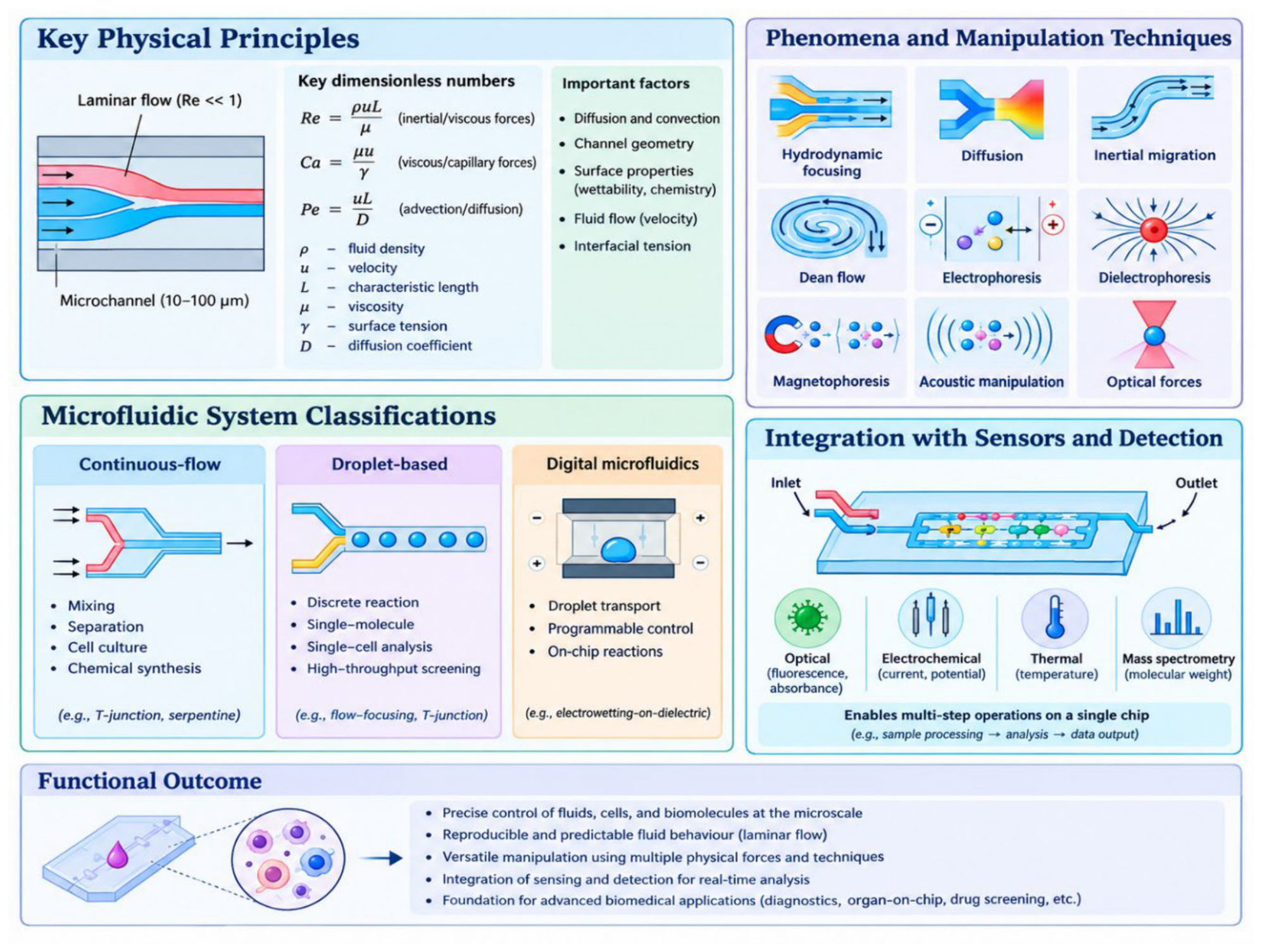
Figure 2.
Major fabrication approaches for microfluidic devices. Representative fabrication workflows include photolithography and etching, soft lithography, injection molding, hot embossing, laser ablation and xurography, three-dimensional printing, micromilling, and paper- and lamination-based fabrication. These methods employ different principles and materials to generate microchannels and integrated microfluidic architectures with varying levels of resolution, geometric complexity, scalability, and manufacturing flexibility. The selection of a fabrication strategy depends on device design, substrate properties, production requirements, and the intended biomedical application.
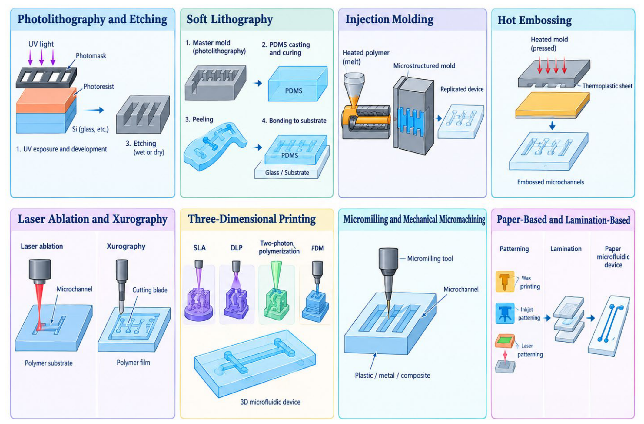
Figure 3.
Microfluidic point-of-care platform for rapid infectious pathogen detection. The schematic illustrates the integrated workflow from clinical sample collection and preparation to cell concentration, nucleic-acid extraction, amplification, and immunological or optical/electrochemical detection. Representative pathogen types and molecular and immunoassay-based detection methods are highlighted. The figure also summarizes the major advantages, clinical relevance, and challenges associated with translating microfluidic POC systems into routine infectious-disease diagnostics.
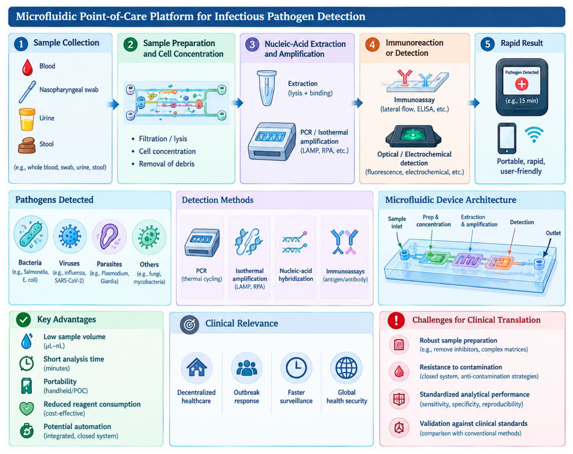
Figure 4.
Microfluidics-enabled liquid biopsy in precision oncology. Microfluidic platforms enrich circulating tumor cells (CTCs), ctDNA, cell-free RNA, exosomes, and tumor-derived proteins from minimally invasive patient samples. Passive and active microfluidic separation strategies enable selective enrichment of rare circulating biomarkers, particularly CTCs. Enriched biomarkers can undergo imaging, enumeration, genotyping, sequencing, PCR, and functional drug-response analysis to characterize tumor heterogeneity and therapeutic resistance. Integration with biosensors and molecular diagnostics supports early cancer detection, prognosis, treatment monitoring, and personalized therapeutic decision-making, while challenges remain in sensitivity, specificity, heterogeneity, and standardization.
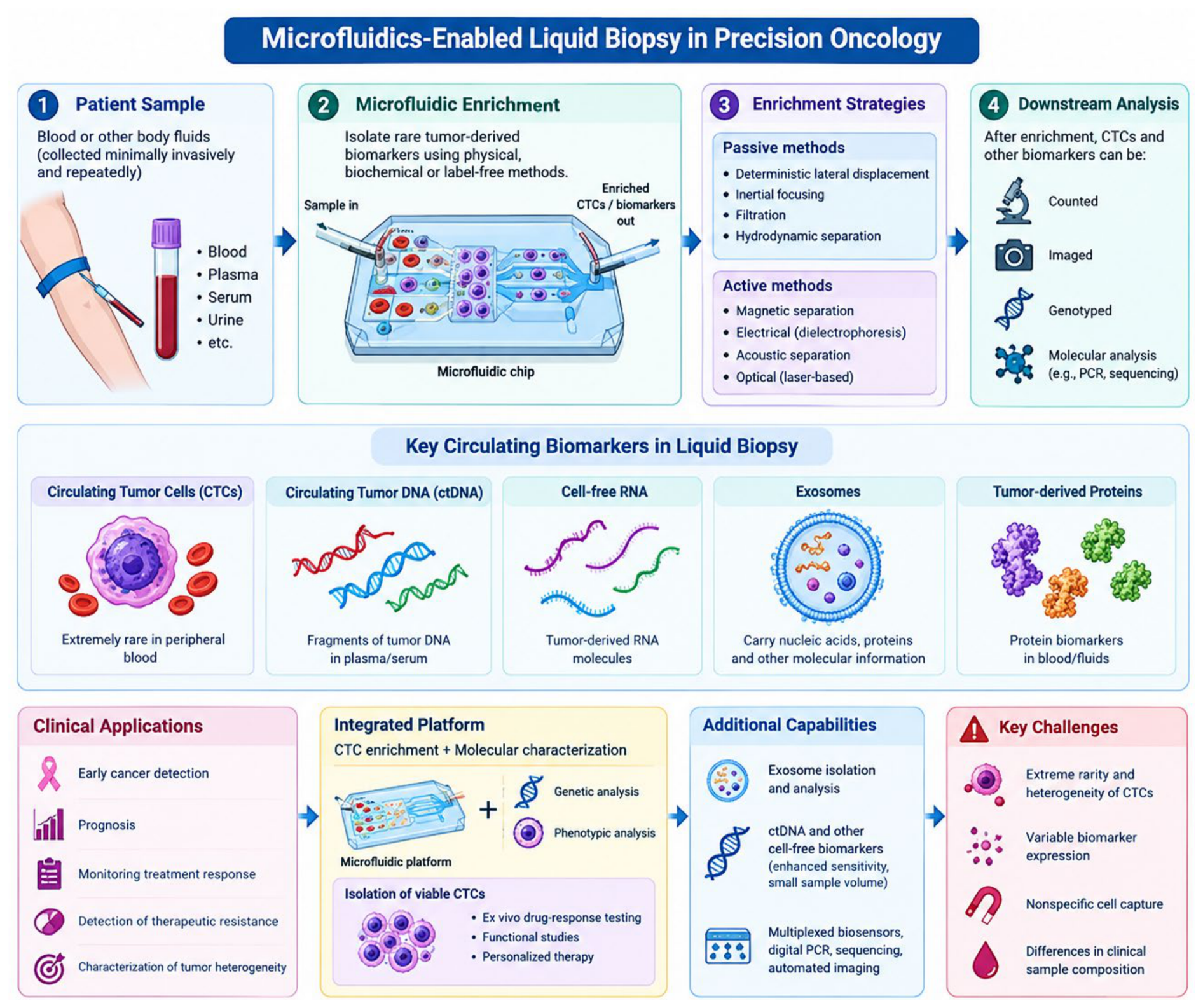
Figure 5.
Microfluidics-enabled cardiovascular disease diagnostics and prognostics. Microfluidic platforms reproduce physiologically relevant blood-flow and cellular environments for investigating thrombosis, hemostasis, atherosclerosis, vascular inflammation, and cardiac dysfunction. (A–B) Thrombosis-on-chip, vessel-on-chip, heart-on-chip, and blood-analysis platforms enable controlled assessment of platelet activity, coagulation, endothelial function, and cardiac responses. (C) Microfluidic biosensors facilitate sensitive and low-volume detection of cardiovascular biomarkers, including cardiac troponins, natriuretic peptides, inflammatory mediators, and coagulation-related proteins. (D–F) Integrated functional and patient-specific analyses support myocardial injury detection, heart-failure assessment, thrombotic-risk stratification, drug-response evaluation, and personalized therapeutic strategies. (G–H) Vascular-on-chip models combined with multiplexed biosensing, automated imaging, and machine-learning analysis provide quantitative assessment of vascular pathology and early disease-associated changes.
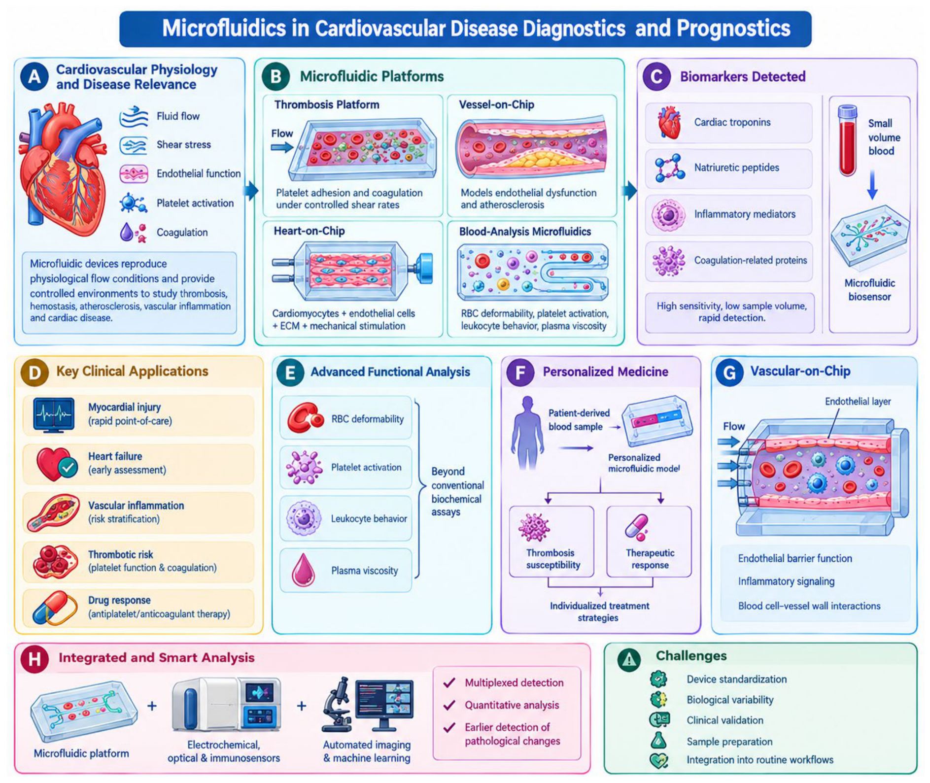
Figure 6.
Tumor- and tumor-microenvironment-on-a-chip platforms for cancer modeling and therapeutic screening. Microfluidic tumor-on-chip systems reconstruct key tumor microenvironment components, including cancer cells, immune cells, endothelial cells, fibroblasts, pericytes, extracellular matrix, and microvascular networks. Controlled microfluidic gradients of oxygen, nutrients, growth factors, and chemotherapeutic agents enable investigation of tumor-cell migration, invasion, angiogenesis, immune infiltration, and metastatic dissemination. Integration of patient-derived cells or organoids, biosensors, real-time imaging, and molecular analysis facilitates personalized drug-response assessment and dynamic monitoring of tumor behaviour. Advanced organoid technology, 3D bioprinting, and multiplexed microfluidic platforms further enhance physiological relevance and support precision oncology applications.
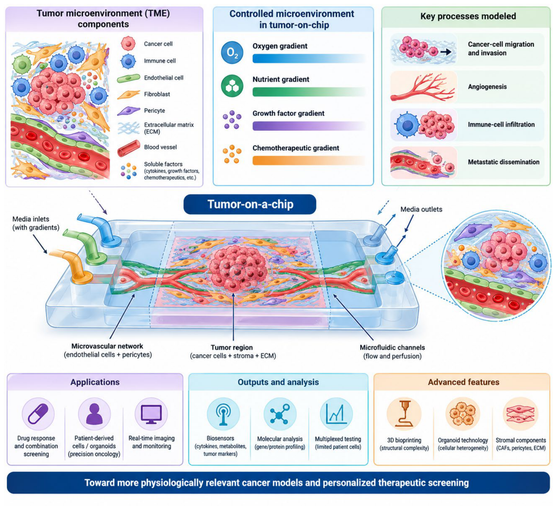
Figure 7.
Organ-on-chip and human-on-chip platforms enabled by microfluidic engineering. Microengineered chips recreate key structural, biochemical, mechanical, and physiological features of human organs using living cells, controlled fluid flow, concentration gradients, and mechanical stimuli. Representative gut-, lung-, liver-, and kidney-on-chip models enable investigation of organ-specific functions, disease mechanisms, drug transport, metabolism, and toxicity. Interconnection of multiple organ modules can model systemic pharmacokinetics, metabolite-mediated effects, and personalized therapeutic responses using patient-derived cells or organoids.
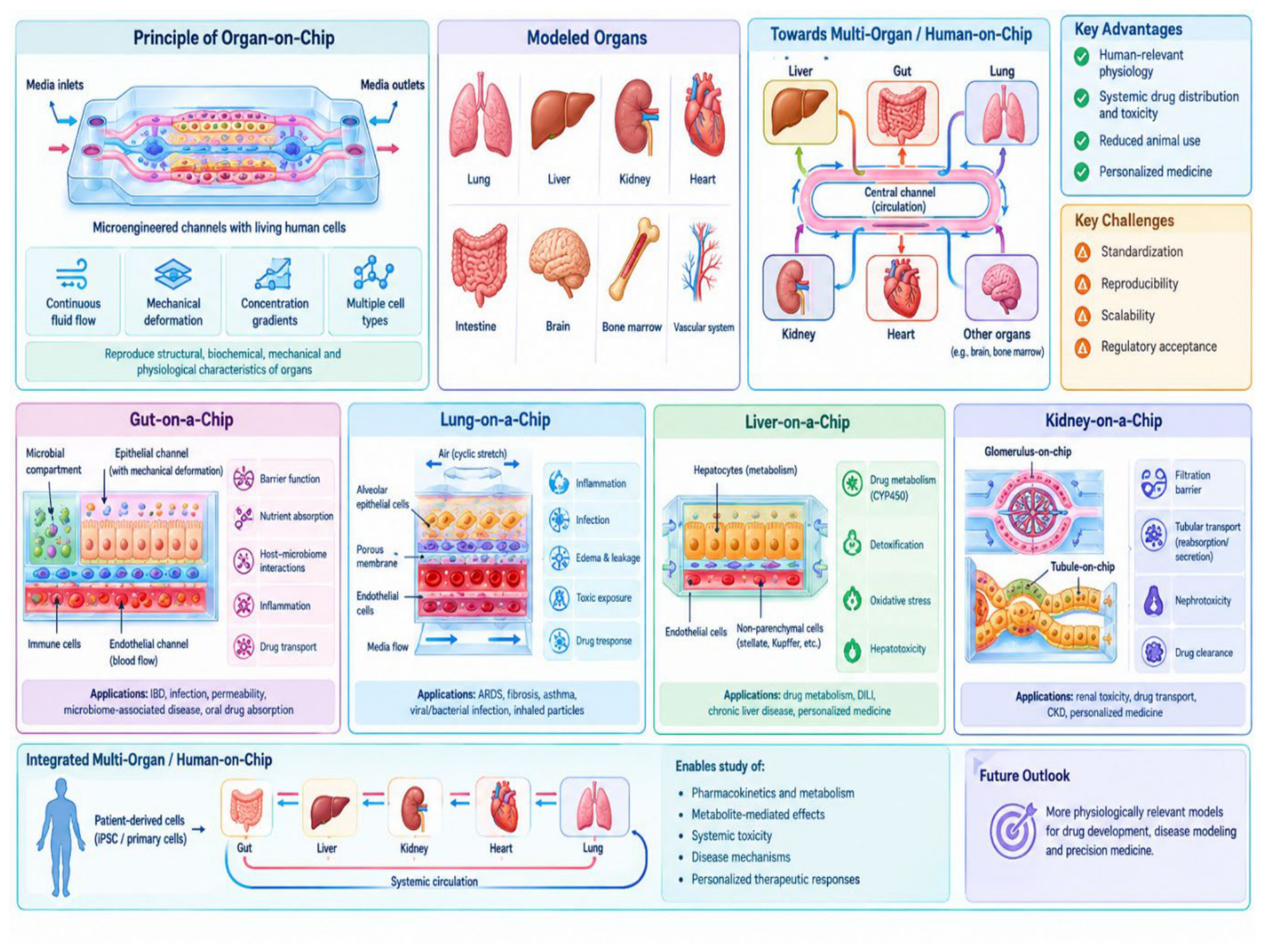
Figure 8.
Microfluidic blood–brain-barrier (BBB)-on-chip and brain-on-chip platforms for neurological disease modeling. BBB-on-chip systems reconstruct key features of the neurovascular unit using endothelial cells, pericytes, astrocytes, neurons, extracellular matrix, and controlled fluid flow.These platforms enable investigation of BBB permeability, drug transport, neuroinflammation, neurotoxicity, neurodegenerative diseases, and brain tumor interactions. Integration of immune cells, brain organoids, patient-derived cells, biosensors, and real-time imaging enhances physiological relevance and supports disease-specific therapeutic screening.
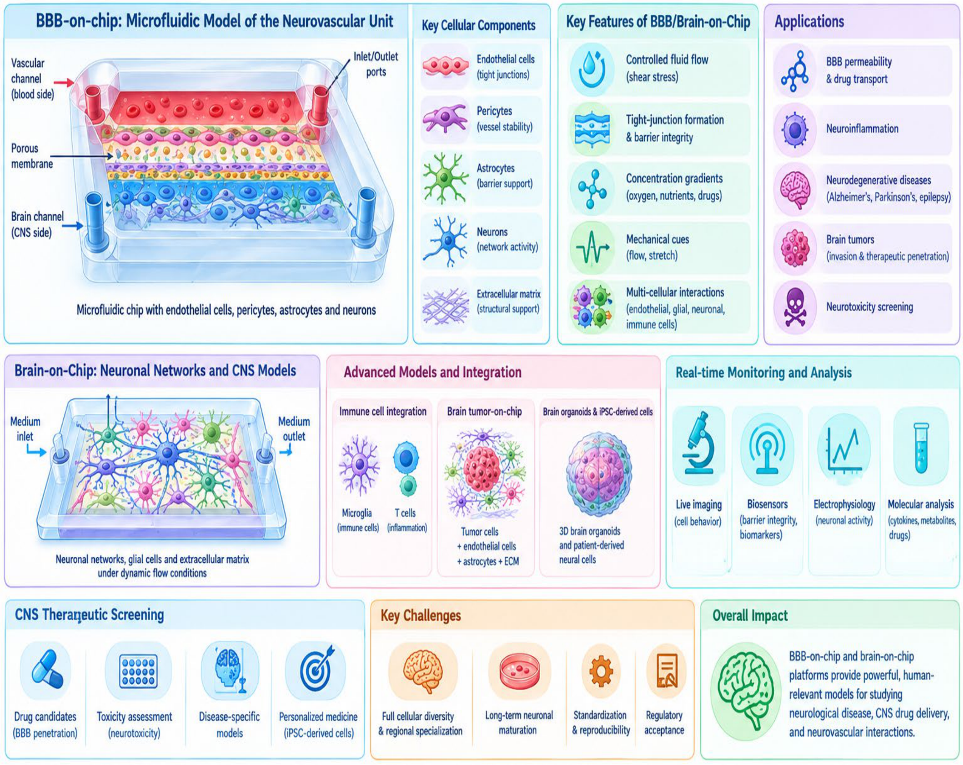
Figure 9.
Microfluidic platforms for pharmaceutical synthesis, drug screening, and drug delivery. Microreactors enable controlled mixing, heat and mass transfer, and continuous synthesis of pharmaceutical compounds and drug-loaded nanoparticles. Microfluidic screening platforms support high-throughput dose–response analysis, toxicity testing, droplet-based assays, and organ-on-chip evaluation. Microfluidic fabrication also enables reproducible production of nanoparticles, liposomes, lipid nanoparticles, and nucleic-acid delivery systems with tunable physicochemical properties.
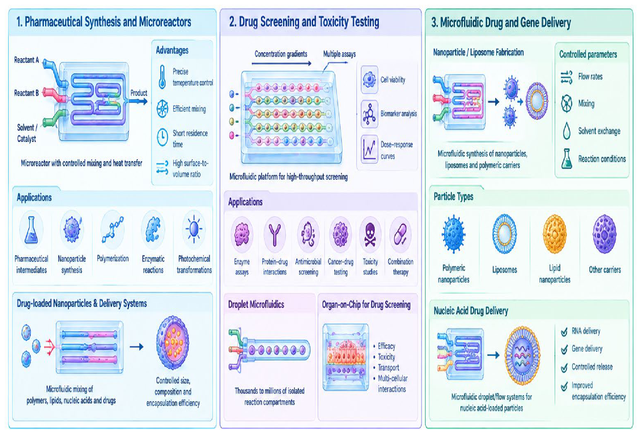
Figure 10.
Microfluidics in single-cell analysis, regenerative medicine, and reproductive technologies. Microfluidic platforms enable precise isolation, manipulation, and molecular characterization of individual cells, while providing controlled cellular microenvironments for stem-cell differentiation, tissue engineering, organoid culture, and bioprinting. In reproductive medicine, microfluidic systems support sperm selection, sperm–oocyte interaction, fertilization, embryo culture, and developmental monitoring. Integration with advanced imaging, sequencing, biomaterials, and automated platforms highlights the potential of microfluidics for precise, personalized, and translational biomedical applications.
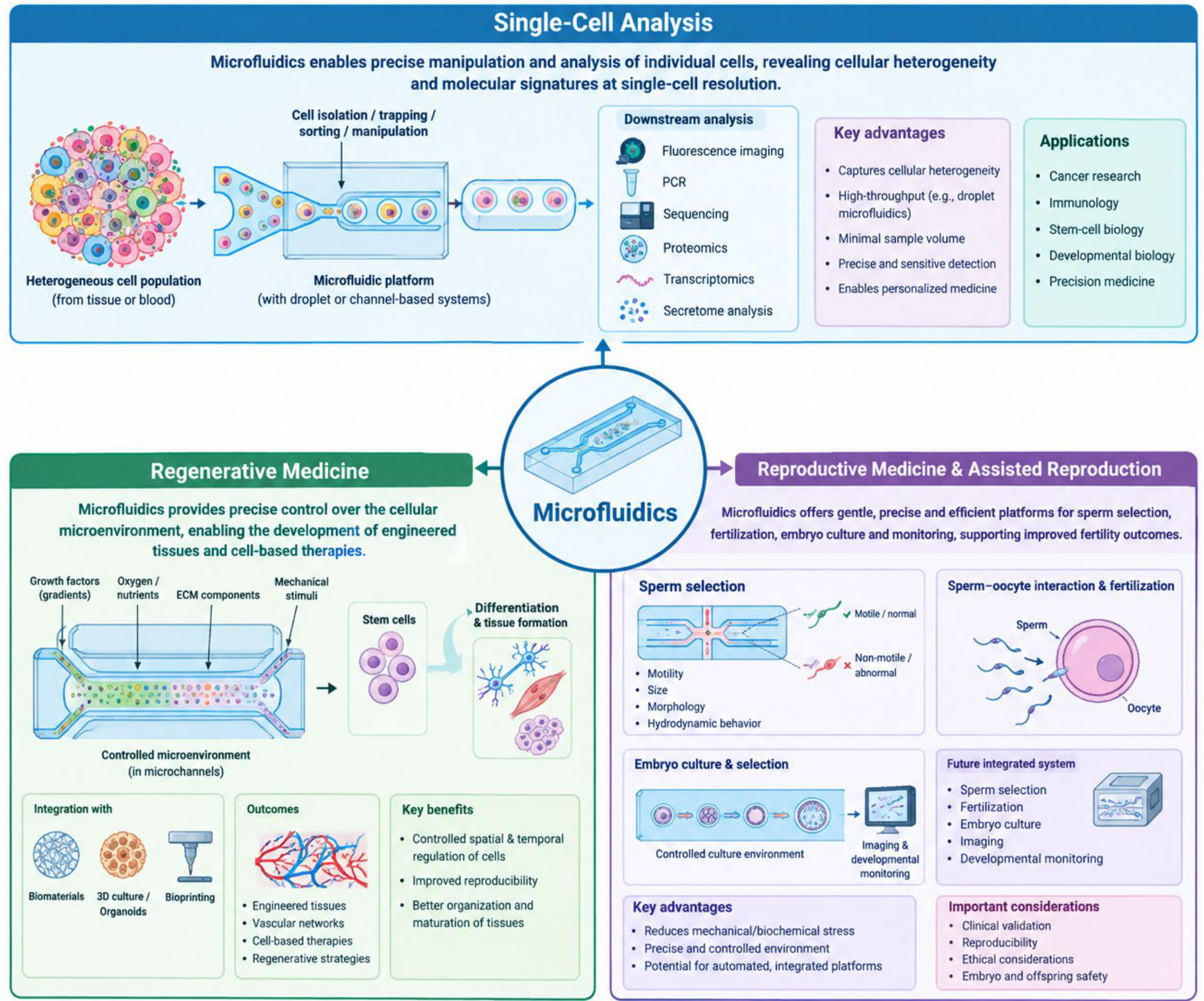
Disclaimer/Publisher’s Note: The statements, opinions and data contained in all publications are solely those of the individual author(s) and contributor(s) and not of MDPI and/or the editor(s). MDPI and/or the editor(s) disclaim responsibility for any injury to people or property resulting from any ideas, methods, instructions or products referred to in the content. |
© 2026 by the authors. Licensee MDPI, Basel, Switzerland. This article is an open access article distributed under the terms and conditions of the Creative Commons Attribution (CC BY) license (http://creativecommons.org/licenses/by/4.0/).
Copyright: This open access article is published under a Creative Commons CC BY 4.0 license, which permit the free download, distribution, and reuse, provided that the author and preprint are cited in any reuse.