

Submitted:
09 October 2026
Posted:
12 October 2026
You are already at the latest version
Abstract
Successful adaptation to severe physiologic stress depends on the coordinated regulation of systemic inflammation and multiple interconnected adaptive biologic systems operating across the entire organism. As introduced in the accompanying Introductory Manuscript, homeostatic correction is conceptualized as a dynamic process that progresses through three partially overlapping phases—the Priming, Modulatory, and Restorative Phases—coordinated by context-dependent glucocorticoid- glucocorticoid receptor alpha (GC–GRα) signaling.Specifically, this review focuses on the integrated regulation of immune, metabolic, mitochondrial, and vascular–endothelial adaptive programs that together establish the biologic foundation for successful homeostatic correction. During the Priming Phase, coordinated danger recognition, innate immune activation, immunometabolic reprogramming, mitochondrial adaptation, and endothelial activation rapidly mobilize host-defense mechanisms while preserving tissue perfusion, bioenergetic homeostasis, and organismal survival. During the Modulatory Phase, these adaptive responses undergo coordinated recalibration that limits excessive inflammatory and metabolic injury while preserving antimicrobial competence, restoring mitochondrial function, stabilizing endothelial integrity, and promoting tissue protection. During the Restorative Phase, integrated immune resolution, metabolic recovery, mitochondrial restoration, and endothelial repair re-establish systemic homeostasis, adaptive resilience, and long-term organ function.Throughout these sequential yet overlapping phases, GC–GRα signaling serves as a central integrative regulator, coordinating immune, metabolic, mitochondrial, and vascular adaptive responses in response to the magnitude, tissue specificity, and temporal evolution of physiologic stress. Rather than acting independently, these foundational biologic systems operate as an interconnected adaptive network that prepares the organism for the complementary neuroendocrine, adaptive immune, lymphatic, microbiome–gut, and regenerative responses reviewed in the second Companion Manuscript. Together with the Introductory Manuscript, these reviews provide a comprehensive systems-level framework for understanding coordinated homeostatic correction during critical illness and other forms of severe physiologic stress.
Keywords:
glucocorticoid receptor alpha (GRα)
; homeostatic correction
; critical illness
; immune regulation
; immunometabolic reprogramming
; mitochondrial bioenergetics
; vascular homeostasis
; endothelial adaptation
; inter-organ communication
; adaptive resilience
Introduction
The accompanying Introductory Manuscript [1] proposes that successful adaptation to severe physiologic stress depends on the coordinated activation of integrated biologic programs that evolve through three dynamic and partially overlapping phases of homeostatic correction—the Priming, Modulatory, and Restorative Phases. Within this systems-level framework, glucocorticoid- glucocorticoid receptor alpha (GC–GRα) signaling serves as a central integrative regulator, coordinating context-dependent adaptive responses across multiple organ systems. To facilitate a comprehensive yet focused discussion of these complex biologic processes, the framework is presented in two complementary companion reviews [2,3] that together encompass the major adaptive systems involved in homeostatic correction.
Among these adaptive programs, the immune, metabolic, and vascular systems form the earliest and most tightly integrated network activated by severe physiologic stress. Rather than operating as independent biological pathways, these systems continuously communicate through inflammatory, neuroendocrine, metabolic, mitochondrial, and vascular signaling networks to coordinate danger recognition, host defense, bioenergetic adaptation, tissue perfusion, and systemic homeostasis. Together, they establish the biologic foundation while complementary neuroendocrine, adaptive immune, lymphatic, microbiome–gut, and regenerative programs interact continuously with these foundational systems throughout homeostatic correction.
This companion review examines the phase-specific regulation of these foundational adaptive systems, emphasizing how GC–GRα signaling coordinates their temporal integration across the Priming, Modulatory, and Restorative Phases of homeostatic correction. By integrating immune regulation, immunometabolic reprogramming, mitochondrial adaptation, and vascular homeostasis within a unified systems-level framework, this review outlines the biologic mechanisms that establish the earliest organism-wide adaptive responses required for successful survival, physiologic stabilization, and progression toward recovery during critical illness and other forms of severe physiologic stress.
Figure 1 (Integrated Multisystem Framework of Phase-Specific Homeostatic Correction During Severe Physiologic Stress) provides an overview of the complete homeostatic correction framework presented across the two companion reviews [2,3]. The present review, The Foundational Immune–Metabolic–Vascular Adaptive Response During Critical Illness: A Phase-Specific GC–GRα Framework [2], examines the integrated immune, metabolic, mitochondrial, and vascular–endothelial adaptive systems that underpin organism-wide adaptation. The companion review, Integrated Neuroendocrine, Immune, Barrier, and Regenerative Adaptive Responses During Severe Physiologic Stress: A Phase-Specific GC–GRα Framework[3] examines the complementary neuroendocrine, adaptive immune, lymphatic, microbiome–gut, epithelial-barrier, and mechanical–structural regenerative systems that interact continuously with and build on these foundational responses throughout the Priming, Modulatory, and Restorative Phases. Together, these two companion reviews [2,3] provide a comprehensive systems-level description of the major biologic domains that coordinate phase-specific homeostatic correction during severe physiologic stress and critical illness.
The Priming, Modulatory, and Restorative Phases should not be interpreted as fixed chronological intervals. Available longitudinal human studies indicate that Priming responses can emerge within minutes to hours of the initiating insult, while Modulatory programs may begin during the first hours to days and Restorative programs may become detectable within several days and continue for weeks or longer. These programs overlap substantially and may evolve asynchronously among biological domains, tissues, and individual patients. Accordingly, chronological time provides an approximate framework for the evolution of critical illness but does not define biological phase; rather, biological phase reflects the patient's predominant, evolving multidomain adaptive state. Successful recovery depends on coordinated progression among these programs, whereas delayed, persistent, or discordant responses may contribute to prolonged organ dysfunction, chronic critical illness, or death. Figure 2 (Temporal Evolution and Divergent Trajectories of Homeostatic Correction During Critical Illness) illustrates the approximate temporal evolution and overlap of these adaptive phases, as well as the divergent trajectories that may lead to recovery or persistent organ dysfunction.
1. Immune Regulation Across the Phases of Homeostatic Correction
1.1. Priming Phase: Immune Sensing, Danger Recognition, and Early Host Defense
The Priming Phase represents the earliest stage of coordinated adaptation during critical illness. Rather than an isolated inflammatory response, it is an organism-wide defense program initiated by infectious and noninfectious danger signals. These signals activate integrated immune, neuroendocrine, metabolic, vascular, mitochondrial, lymphatic, and structural programs that support immediate survival and preserve systemic homeostasis [4,5,6,7]. GC-GRα signaling functions throughout this phase as a central integrative regulator, coordinating tissue-specific, context-dependent adaptive responses while balancing effective host defense with the preservation of metabolic function, vascular integrity, mitochondrial bioenergetics, and tissue homeostasis [7,8].
1.1.1. Immune Sensing
Immune sensing is the initial biological process that enables the host to detect disturbances that threaten homeostasis and initiate coordinated adaptive responses. Rather than relying on a single organ or cell type, immune sensing is mediated by a distributed surveillance network comprising epithelial barriers, vascular endothelial cells, tissue-resident immune cells, peripheral sensory neurons, neuroendocrine pathways, and circulating immune cells. This network continuously monitors the external and internal environments for signs of infection, tissue injury, metabolic stress, ischemia, or other forms of cellular damage. These interconnected surveillance systems communicate through immune, neural, endocrine, vascular, and metabolic signaling pathways to rapidly integrate local danger recognition with organism-wide adaptive responses, thereby linking early threat detection to coordinated homeostatic correction [4,5,6]
1.1.2. Danger Recognition
The Priming Phase begins when distributed cellular surveillance systems, including pattern-recognition receptors (PRRs), epithelial barriers, vascular endothelial cells, tissue-resident immune cells, peripheral sensory neurons, and neuroendocrine stress pathways, recognize infectious and noninfectious danger signals. PRRs—including Toll-like receptors, NOD-like receptors, RIG-I-like receptors, and inflammasome-associated signaling pathways—detect pathogen-associated molecular patterns (PAMPs) and damage-associated molecular patterns (DAMPs), thereby initiating early host-defense signaling.[8,9,10,11]
Importantly, the Priming Phase may be initiated not only by infection but also by sterile danger signals generated during trauma, hemorrhage, ischemia-reperfusion, burns, surgery, pancreatitis, toxic injury, and metabolic stress. These diverse insults release endogenous DAMPs—including HMGB1, ATP, mitochondrial DNA, histones, heat-shock proteins, oxidized lipids, urate crystals, and extracellular matrix fragments—that activate many of the same innate immune pathways triggered by infection, thereby initiating a shared adaptive inflammatory-defense program.[12,13,14,15,16,17]
PRR engagement rapidly activates nuclear factor kappa B (NF-κB), activator protein 1 (AP-1), Janus kinase–signal transducer and activator of transcription (JAK–STAT), mitogen-activated protein kinase (MAPK), phosphoinositide 3-kinase (PI3K), calcium-dependent signaling pathways, and inflammasomes, initiating production of TNF, IL-1β, IL-6, interferons, chemokines, and numerous inflammatory mediators that coordinate leukocyte recruitment, antimicrobial defense, antigen presentation, metabolic adaptation, neuroendocrine activation, vascular responses, and tissue-protective programs. Collectively, these integrated responses establish the biologic foundation for subsequent inflammatory recalibration, tissue protection, and restoration of homeostasis [8,10,18,19]
1.1.3. Acute-Phase Response
Acute systemic inflammation rapidly activates the hepatic acute-phase response (APR), a coordinated adaptive program that integrates inflammatory, metabolic, endocrine, coagulation, and innate host-defense pathways required for survival during severe physiologic stress. The liver serves as a systems integrator, translating inflammatory cytokine signaling into coordinated organism-wide metabolic and host-defense responses.[20,21]
These adaptive hepatic responses support pathogen recognition, complement activation, iron sequestration, coagulation, endothelial protection, antioxidant defense, redistribution of metabolic substrates, and increased free cortisol availability, thereby enhancing GC-GRα-mediated homeostatic correction across multiple organ systems. Although initially adaptive, excessive or persistent APR activation may contribute to hypercoagulability, endothelial dysfunction, hypoalbuminemia, persistent catabolism, metabolic dysregulation, mitochondrial dysfunction, and progression to chronic critical illness [22,23,24]
1.1.4. Neuroendocrine Coordination of Early Host Defense
Activation of the HPA axis provides the principal neuroendocrine coordination of the Priming Phase. Concurrently, GC–GRα signaling participates directly in the early host-defense response. Through reciprocal interactions with NF-κB and AP-1, GRα modifies chromatin accessibility and transcription-factor occupancy, facilitating the coordinated expression of pro-inflammatory and innate host-defense genes.[25,26]. Early GC-GRα signaling integrates with NF-κB- and AP-1-dependent inflammatory pathways to optimize early host defense responses and support neutrophil recruitment, antigen presentation, metabolic substrate mobilization, cardiovascular adaptation, mitochondrial function, and survival-associated transcriptional programs. These observations illustrate that GC-GRα signaling functions not simply as an anti-inflammatory pathway but as an integrative regulator of multiple adaptive biologic programs required for effective host defense [25,26,27,28,29,30,31]
1.1.5. Cellular Priming and Early Immune Programming
Priming is distinct from full inflammatory activation. Rather, it is a coordinated, adaptive preparatory state characterized by transcriptional induction, chromatin remodeling, post-translational modifications, and inflammasome licensing, which together enhance responsiveness to subsequent danger signals. GC-GRα signaling interacts dynamically with NF-κB, AP-1, and STAT pathways, chromatin remodeling complexes, circadian immune programs, and metabolic signaling networks to establish adaptive readiness while preventing progression toward uncontrolled inflammation and tissue injury [32,33,34,35,36,37]
1.1.6. Innate Immune Activation and Transition to Adaptive Immunity
Neutrophils, monocytes, macrophages, and dendritic cells undergo coordinated activation that supports pathogen containment and initiates adaptive immunity. GC-GRα signaling promotes neutrophil mobilization, macrophage reprogramming, mitochondrial bioenergetics, vascular responsiveness, and inflammatory-metabolic integration, whereas dendritic-cell activation and early T-cell priming mark the transition from innate to adaptive immunity. Failure to appropriately regulate or terminate these priming responses contributes to persistent inflammatory amplification, dysregulated host defense, and progression toward multiple-organ dysfunction.[38,39,40,41,42,43,44,45,46]
1.2. Modulatory Phase: Immune Recalibration and Controlled Host Defense
1.2.1. Immune Recalibration and Inflammatory Resolution.
During the Modulatory Phase of critical illness, inflammatory responses undergo progressive recalibration to limit excessive tissue-damaging inflammation while preserving effective antimicrobial defense and supporting the restoration of tissue homeostasis. This phase is characterized by attenuation of excessive cytokine amplification, reduction of neutrophil-dominant inflammatory injury, a transition toward monocyte/macrophage-mediated immune regulation, restoration of endothelial and mitochondrial stability, and initiation of pro-resolving programs that facilitate recovery and prevent immune-mediated organ dysfunction [29,31,47]
1.2.2. Gc-Grα-Mediated Immune Recalibration
GC-GRα signaling plays a central role in orchestrating these adaptive modulatory responses through highly context-dependent genomic and non-genomic mechanisms. Rather than producing indiscriminate anti-inflammatory effects, GRα signaling regulates inflammatory and host-defense programs in a context-, tissue-, and phase-specific manner, shaped by inflammatory burden, pathogen load, metabolic status, and homeostatic requirements.[31,48,49] As the host response transitions from Priming toward Modulation, the same GRα interactions with inflammatory transcription factors that support early host-defense gene expression are redirected toward limiting excessive inflammatory amplification. Chromatin accessibility facilitates GRα–transcription-factor co-occupancy and functional cooperation, including with NF-κB, allowing context-dependent reprogramming of inflammatory gene expression toward controlled inflammation and resolution.[26,50] During this phase, GRα signaling represses excessive NF-κB- and AP-1-mediated cytokine amplification while maintaining coordinated antimicrobial competence, supporting pathogen clearance, preserving innate immune-cell and phagocytic function [31,50,51]
1.2.3. Macrophage Reprogramming and Efferocytosis
A major feature of the Modulatory Phase is the transition from neutrophil-dominant inflammatory responses toward monocyte/macrophage-mediated inflammatory regulation and tissue protection. GRα signaling promotes differentiation into anti-inflammatory and pro-resolving macrophage phenotypes characterized by enhanced phagocytosis, efferocytosis of apoptotic neutrophils, suppression of excessive cytokine production, and support for tissue repair processes.[48,52,53,54] Efferocytosis (the regulated clearance of apoptotic cells by phagocytes) is critical during this phase because clearing apoptotic neutrophils terminates persistent inflammatory activation, prevents secondary tissue injury, and promotes restoration of tissue homeostasis by facilitating the transition from inflammation to resolution and tissue repair.[47,52,54] This transition also promotes inflammatory resolution while responding to persistent or newly emerging infectious threats. Neutrophils often show reduced or tightly regulated GRα signaling during sustained inflammatory activation, limiting excessive anti-inflammatory activity while maintaining effective innate antimicrobial responses. By contrast, monocytes and macrophages maintain or increase GRα responsiveness, favoring anti-inflammatory and pro-resolving programs.[48,49,55,56] A recent spatial hypothesis in non-resolving ARDS further illustrates this functional transition by proposing that macrophage capacity for lipid disposal, efferocytosis, NET clearance, and endothelial–epithelial repair acts as a local “brake” on a self-renewing inflammatory–barrier injury circuit; progressive failure of these macrophage functions may instead favor persistent inflammation, defective barrier restoration, and loss of reversibility.[57]
1.2.4. Endogenous Pro-Resolving Mediators
GC-GRα signaling also induces multiple endogenous pro-resolving mediators that contribute to inflammatory resolution and restoration of immune balance. Among the best characterized are glucocorticoid-induced leucine zipper (GILZ), Annexin A1, and dual-specificity phosphatase-1 (DUSP1/MKP-1), which collectively suppress excessive inflammatory signaling, inhibit leukocyte overactivation, reduce oxidative stress, and promote pro-resolving macrophage progra.[29,31,58] Experimental models further demonstrate that macrophage-specific GILZ overexpression reduces systemic inflammation and improves bacterial clearance, supporting the concept that coordinated inflammatory resolution and preservation of antimicrobial competence are complementary rather than opposing processes during the Modulatory Phase [59]
Rather than simply suppressing macrophage activity, GILZ actively reprograms macrophages away from the highly inflammatory M1 phenotype toward regulatory and resolving phenotypes, characterized by enhanced IL-10 production, improved phagocytosis and efferocytosis, and preservation of antibacterial host-defense functions during severe infection and sepsis.[60,61,62,63] Collectively, these observations support the concept that inflammatory resolution is an active, highly coordinated adaptive process that recalibrates immune responses to preserve host defense while promoting tissue protection, restoring homeostasis, and advancing toward the Restorative Phase.[64]
Annexin A1 further promotes neutrophil apoptosis, limits excessive neutrophil recruitment, enhances monocyte recruitment, and facilitates macrophage-mediated efferocytosis, thereby promoting inflammatory resolution and tissue repair. GILZ–Annexin A1 signaling also limits neutrophil rolling, adhesion, endothelial transmigration, and excessive tissue accumulation while preserving effective host-defense responses. Emerging evidence further suggests that GILZ and Annexin A1 function as a cooperative pro-resolving module, with reciprocal regulatory interactions that coordinate macrophage polarization, efferocytosis, pathogen elimination, and the orderly transition from inflammation to tissue repair during severe infection and sepsis.[60,61,65,66] Collectively, these integrated pathways promote immune recalibration, restoration of tissue homeostasis, and successful progression toward the Restorative Phase.
1.2.5. Cell- and Tissue-Specific Immune Regulation
Importantly, growing evidence shows that GRα-mediated modulation during critical illness is highly cell- and tissue-specific. Neutrophils often show reduced GRα signaling during severe inflammatory stress, thereby limiting excessive immunosuppression, whereas monocytes and macrophages maintain or increase GRα responsiveness to support anti-inflammatory and tissue-protective programs.[49,55] This differential regulation supports the maintenance of effective innate immune function while limiting collateral inflammatory injury in vulnerable tissues, including the lung, vascular endothelium, gastrointestinal tract, and mitochondria-rich organs.[31,48] Increasing evidence further suggests that tissue-specific adaptations preferentially direct stronger GRα activity toward vital organs—including the lung, vascular endothelium, heart, diaphragm, and kidney—while limiting excessive glucocorticoid exposure in tissues more vulnerable to catabolic injury. [31,48] Increasing evidence further suggests that tissue-specific adaptations preferentially direct stronger GRα activity toward vital organs—including the lung, vascular endothelium, heart, diaphragm, and kidney—while limiting excessive glucocorticoid exposure in tissues more vulnerable to catabolic injury [49,67,68]
1.2.6. Preservation of Host Defense and Clinical Translation
GC-GRα signaling also helps maintain effective host-defense function during the Modulatory Phase. Endogenous glucocorticoids preserve coordinated antimicrobial competence, bacterial clearance, natural killer cell regulation, epithelial barrier integrity, and immune defense programs while suppressing excessive inflammatory cytokine production.[69,70,71] Simultaneously, GC-GRα signaling contributes to the restoration of endothelial and epithelial barrier integrity by suppressing NF-κB activation, reducing endothelial adhesion molecule expression, stabilizing tight junctions and the glycocalyx, enhancing endothelial nitric oxide synthase activity, and supporting mitochondrial bioenergetic adaptation and antioxidant defenses.[68,72] These observations strongly support the concept that the Modulatory Phase does not represent immune paralysis or generalized immunosuppression, but rather a coordinated adaptive recalibration program designed to limit immune-mediated tissue injury, preserve pathogen control, and facilitate recovery of systemic homeostasis [29,31,47]
Recent clinical evidence further supports the concept that immune dysregulation during sepsis is heterogeneous and requires individualized therapeutic approaches. In the ImmunoSep randomized clinical trial, biomarker-guided immunotherapy targeting macrophage activation-like syndrome with anakinra or sepsis-induced immunoparalysis with recombinant interferon-γ improved measures of organ dysfunction compared with placebo, but did not significantly reduce 28-day mortality. Specific, phenotype-guided therapeutic strategies that align with the dynamic processes of homeostatic correction in critical illness. Experimental and translational studies increasingly support the concept that GC-GRα-induced pro-resolving mediators can simultaneously attenuate excessive inflammatory injury, support effective microbial clearance, and promote the restoration of tissue homeostasis during severe infection and sepsis.[59,62,73,74]
1.3. Restorative Phase: Immune Resolution, Repair, and Functional Recovery
1.3.1. Immune Resolution and Restoration of Homeostasis
During the Restorative Phase of homeostatic correction, immune and inflammatory responses gradually shift from controlled modulation to coordinated resolution programs that support tissue repair, restore immune balance, and recover organ function. This phase is marked by attenuation of sustained inflammatory activation, macrophage reprogramming toward pro-resolving and reparative phenotypes, restoration of adaptive immune balance, prevention of immune exhaustion, and activation of regenerative pathways that facilitate recovery from severe physiologic stress and critical illness.[58,75,76,77,78] Collectively, these integrated adaptive responses promote the restoration of immune homeostasis while preserving long-term host defense and physiologic resilience. GC-GRα signaling coordinates the shift from inflammatory host-defense responses to pro-resolving and reparative programs that facilitate the restoration of immune homeostasis and functional recovery.
1.3.2. Macrophage Reprogramming and Tissue Repair
A major biologic feature of the Restorative Phase is macrophage reprogramming from inflammatory to reparative, pro-resolving phenotypes, characterized by enhanced efferocytosis, clearance of apoptotic cells and cellular debris, suppression of persistent cytokine amplification, production of IL-10 and transforming growth factor-β (TGF-β), and support for tissue regeneration and extracellular matrix remodeling.[76,79,80,81] Efficient efferocytosis is a critical mechanism for resolving inflammation because clearing apoptotic leukocytes actively suppresses persistent inflammatory signaling, promotes the restoration of immune tolerance, and facilitates macrophage-mediated tissue repair.[77,78]
1.3.3. Endogenous Pro-Resolving Mediators
GC-GRα signaling also induces multiple endogenous pro-resolving mediators, including glucocorticoid-induced leucine zipper (GILZ) and Annexin A1, which promote neutrophil apoptosis, macrophage reprogramming, suppression of excessive leukocyte recruitment, and coordinated inflammatory silencing during recovery from severe systemic inflammation.[58,77] Specialized pro-resolving mediators (SPMs) and resolution-associated lipid mediators further limit persistent leukocyte activation, restore endothelial and epithelial integrity, modulate adaptive immune responses, and promote tissue regeneration during recovery.[82,83,84] Together, GC-GRα signaling, endogenous pro-resolving mediators, and SPMs coordinate inflammatory resolution, tissue repair, and restoration of immune homeostasis, thereby supporting successful homeostatic correction and long-term physiologic recovery.
1.3.4. Recovery of Adaptive Immune Homeostasis
Adaptive immune responses also undergo progressive recalibration during the Restorative Phase. Restoration of balanced T-cell responses, expansion of regulatory T-cell populations, suppression of persistent effector T-cell hyperactivation, and prevention of immune exhaustion contribute to recovery of coordinated host defense while limiting autoimmune and chronic inflammatory injury.[75,83] Beyond regulating inflammatory resolution, GC-GRα signaling directly contributes to restoration of adaptive immune homeostasis by influencing lymphocyte survival, differentiation, cytokine production, and functional recalibration. Through coordinated effects on T- and B-cell responses, GC-GRα signaling facilitates the transition from innate inflammatory-defense programs toward long-term immune regulation, pathogen clearance, tissue repair, and restoration of immune competence.[85] Collectively, these integrated immune-recovery programs re-establish durable immune homeostasis while preserving long-term host defense and physiologic resilience.
1.3.5. Immune–microbiome Crosstalk and Long-Term Host Protection
The innate and adaptive immune systems remain tightly interconnected throughout recovery, with ongoing crosstalk among lymphocytes, antigen-presenting cells, tissue-resident macrophages, and regulatory immune networks, thereby restoring immune balance and sustaining durable host protection. Successful adaptive immune recalibration also supports the development and maintenance of immunologic memory, thereby enhancing long-term pathogen-specific protection while preserving immune homeostasis.
Emerging evidence further suggests that interactions among GC-GRα signaling, the recovering microbiome, and pro-resolving mediators help restore immune tolerance, regulate innate and adaptive immune responses, maintain epithelial barrier integrity, and support long-term immune homeostasis through coordinated effects on dendritic-cell function, antigen presentation, and cytokine-regulatory pathways.[31,80] These integrated interactions illustrate how GC-GRα signaling coordinates immune, epithelial, and microbiome recovery to promote successful homeostatic correction.
1.3.6. Restoration of Systemic Immune Resilience
Successful immune restoration also depends upon recovery of mitochondrial integrity, re-establishment of endothelial and epithelial barrier function, resolution of oxidative stress, and coordinated regenerative remodeling across organ systems. Failure of these restorative mechanisms may contribute to persistent inflammation, maladaptive fibrosis, immune dysfunction, prolonged catabolism, chronic critical illness, and incomplete recovery of organ function, thereby limiting restoration of long-term physiologic resilience and homeostasis.[49,86] Conversely, successful integration of these restorative processes re-establishes organism-wide homeostasis, strengthens adaptive resilience, and prepares the host to respond effectively to future physiologic stress.
Integrative Summary
Collectively, the Priming, Modulatory, and Restorative Phases illustrate that immune regulation during critical illness is a dynamic, integrated continuum rather than a sequence of isolated inflammatory events. The Priming Phase establishes rapid host defense through coordinated danger recognition, innate immune activation, and neuroendocrine integration. The Modulatory Phase recalibrates inflammatory responses to preserve antimicrobial competence while limiting collateral tissue injury through coordinated macrophage reprogramming, endogenous pro-resolving mediators, and cell-specific immune regulation. The Restorative Phase completes this adaptive process by promoting immune resolution, tissue repair, restoration of adaptive immune homeostasis, and long-term physiologic resilience. Throughout all three phases, GC-GRα signaling coordinates immune, metabolic, mitochondrial, vascular, and reparative adaptive programs, thereby facilitating successful homeostatic correction and recovery from critical illness.
2. Immunometabolic Reprogramming and Mitochondrial Adaptation
2.1. Priming Phase: Immunometabolic Reprogramming and Mitochondrial Adaptation
2.1.1. Early Immunometabolic Reprogramming
Early immune activation during the Priming Phase is accompanied by rapid immunometabolic reprogramming, an adaptive process characterized by increased glycolytic flux (accelerated glucose metabolism through glycolysis), heightened ATP demand, mitochondrial activation (enhanced mitochondrial bioenergetic and signaling functions), and redistribution of metabolic substrates that support host-defense functions. Activated immune cells increase glycolysis, redirect pyruvate toward lactate production, and enhance pentose phosphate pathway (PPP) activity to generate ATP, nicotinamide adenine dinucleotide phosphate (NADPH), and biosynthetic substrates that support cytokine production, phagocytosis, respiratory burst activity, proliferation, and cellular survival during severe physiologic stress. Activated immune cells increase glycolysis, redirect pyruvate toward lactate production, and enhance pentose phosphate pathway (PPP) activity to generate ATP, nicotinamide adenine dinucleotide phosphate (NADPH), and biosynthetic substrates that support cytokine production, phagocytosis, respiratory burst activity, proliferation, and cellular survival during severe physiologic stress.[87,88,89,90]
2.1.2. Mitochondrial Metabolic Reprogramming and Immunometabolites
During the Priming Phase, mitochondria undergo functional reprogramming to generate mitochondrial reactive oxygen species (mtROS), integrate inflammatory signaling, and coordinate immunometabolic adaptation. Concurrently, tricarboxylic acid (TCA) cycle intermediates—including citrate, succinate, and itaconate—accumulate as immunometabolites, functioning as signaling molecules and biosynthetic intermediates that regulate inflammatory gene expression, antimicrobial defense, and cellular adaptation.[89,91,92,93,94,95] Collectively, these metabolic adaptations provide the energetic and biosynthetic resources required to sustain innate immune function while coordinating adaptive responses across multiple organ systems during severe physiologic stress.
2.1.3. Gc-Grα Regulation of Mitochondrial Bioenergetics
Simultaneously, GC-GRα signaling helps coordinate metabolic adaptation by regulating mitochondrial function, oxidative phosphorylation, tricarboxylic acid (TCA) cycle activity, mitochondrial biogenesis, redox homeostasis, and stress-responsive transcriptional programs that sustain immune activation and cellular energy balance during critical illness.[31,96,97,98,99]
In addition to its well-established nuclear actions, GRα translocates to mitochondria, where it regulates mitochondrial gene expression and interacts with proteins involved in oxidative phosphorylation, apoptosis, and pyruvate metabolism, thereby directly linking systemic stress signaling to mitochondrial bioenergetic function during severe physiologic stress.[97,100]
Across tissues, GC-GRα signaling also supports metabolic substrate mobilization, tissue-specific mitochondrial function, antioxidant defenses, and preservation of bioenergetic integrity, thereby helping maintain organ performance during critical illness while coordinating metabolic adaptation across multiple organ systems.[49,88,101] Collectively, these actions establish GC-GRα signaling as a central integrator of mitochondrial bioenergetics, cellular metabolism, and organism-wide adaptation during the Priming Phase, thereby providing the energetic and metabolic foundation for effective host defense and subsequent homeostatic correction.
2.1.4. Consequences of Dysregulated Metabolic Adaptation
These adaptive metabolic responses initially support survival and host defense; however, when excessive, persistent, or inadequately regulated, they may contribute to mitochondrial dysfunction, oxidative injury, metabolic failure, immune dysregulation, and ultimately to multi-organ dysfunction during severe critical illness.[88,102,103]
Progressive mitochondrial respiratory impairment, depletion of antioxidant defenses, and bioenergetic failure further compromise immune function, sustain inflammation, and promote multiple-organ dysfunction, ultimately impairing adaptive capacity, tissue repair, and physiologic recovery.[55,104] Thus, successful homeostatic correction during the Priming Phase depends on preserving mitochondrial integrity and on coordinated immunometabolic adaptation, thereby providing the bioenergetic foundation for subsequent Modulatory and Restorative responses.
2.2. Modulatory Phase: Metabolic Recalibration and Bioenergetic Recovery
2.2.1. Metabolic Recalibration and Restoration of Bioenergetic Homeostasis
During the Modulatory Phase, the immunometabolic activation established in the Priming Phase is progressively recalibrated to restore mitochondrial integrity, enhance bioenergetic efficiency, limit oxidative injury, and support organ recovery while meeting the energy demands of host defense and tissue repair. This phase is marked by attenuation of excessive catabolic stress responses, recovery of oxidative phosphorylation (OXPHOS), restoration of mitochondrial quality control mechanisms, reduction of excessive reactive oxygen species (ROS) generation, and optimization of metabolic substrate utilization across vital organs.[87,88,98,102,105]
2.2.2. Gc-Grα Regulation of Mitochondrial Bioenergetics
GC-GRα signaling plays a central role in coordinating metabolic recalibration during the Modulatory Phase through integrated genomic and non-genomic mechanisms that regulate mitochondrial biogenesis, oxidative metabolism, antioxidant defenses, and cellular energy homeostasis.[31,87,88] GRα translocates into mitochondria, where it directly influences mitochondrial gene transcription, oxidative phosphorylation pathways, ATP generation, apoptotic pathways, and mitochondrial stress responses, thereby linking systemic neuroendocrine signaling to bioenergetic recovery and metabolic homeostasis during the Modulatory Phase.[87,98]
2.2.3. Mitochondrial Quality Control and Oxidative Stress
Persistent mitochondrial dysfunction during critical illness may contribute to impaired ATP production, excessive ROS generation, oxidative damage, metabolic inflexibility, and organ dysfunction. GC-GRα signaling helps counteract these maladaptive responses by enhancing mitochondrial biogenesis, promoting mitophagy and mitochondrial quality control, stabilizing electron transport chain function, and restoring redox homeostasis.[68,88,102] Simultaneously, GRα signaling contributes to the induction of antioxidant pathways, including superoxide dismutase, activity, glutathione metabolism, and mitochondrial stress-response programs, thereby reducing oxidative injury while preserving mitochondrial respiratory function during persistent inflammatory stress.[98,102]
2.2.4. Metabolic Flexibility and Substrate Utilization
Metabolic adaptation during the Modulatory Phase also involves coordinated regulation of glucose, lipid, amino acid, and ketone metabolism to ensure adequate substrate availability for energy-dependent organs, including the brain, heart, diaphragm, kidney, immune system, and vascular endothelium. GC-GRα signaling supports metabolic flexibility by coordinating tissue-specific substrate selection and utilization, mitochondrial fatty acid oxidation, gluconeogenesis, insulin sensitivity, and ATP production in response to shifting energy demands during recovery from severe physiologic stress.[68,98,102]
2.2.5. Systemic Recovery and Organ Bioenergetics
Importantly, growing evidence suggests that mitochondrial and metabolic recovery during the Modulatory Phase is closely linked to successful inflammatory resolution, re-establishing tissue homeostasis, and preserving organ function. Restoring mitochondrial integrity supports endothelial barrier recovery, immune cell bioenergetics, epithelial repair, myocardial performance, diaphragmatic function, and neuroendocrine coordination, thereby re-establishing coordinated organ function and systemic homeostasis.[31,87,88] Collectively, these observations support the concept that GC-GRα-mediated metabolic recalibration is an integrated adaptive program that restores efficient energy utilization, preserves mitochondrial resilience, and coordinates recovery across multiple organ systems, thereby establishing the bioenergetic foundation for the Restorative Phase and for successful homeostatic correction.
2.3. Restorative Phase: Restoration of Metabolic Homeostasis and Bioenergetic Recovery
2.3.1. Restoration of Metabolic Homeostasis
During the Restorative Phase of homeostatic correction, the metabolic recalibration established in the Modulatory Phase advances toward restoring metabolic flexibility, recovering mitochondrial integrity, normalizing substrate utilization, and re-establishing efficient cellular bioenergetics, thereby supporting tissue repair and organ recovery. This phase is marked by progressive restoration of oxidative phosphorylation (OXPHOS), improved mitochondrial coupling efficiency, normalization of glucose and lipid metabolism, replenishment of cellular energy stores, and recovery of coordinated ATP production across vital organ systems.[46,106,107] Collectively, these adaptive metabolic responses restore bioenergetic capacity and support tissue regeneration, organ recovery, and long-term physiologic resilience.
2.3.2. Gc-Grα Regulation of Mitochondrial Recovery
GC-GRα signaling remains active throughout the Restorative Phase, serving as a central integrator that coordinates mitochondrial recovery, oxidative metabolism, redox homeostasis, and the restoration of cellular bioenergetics. It supports mitochondrial biogenesis, restoration of electron transport chain activity, substrate mobilization, mitochondrial quality control, and normalization of oxidative phosphorylation, thereby promoting recovery of cellular and organ function after severe systemic inflammation and critical illness.[46,68]
2.3.3. Mitochondrial Quality Control and Redox Homeostasis
Successful recovery during the Restorative Phase depends on the coordinated re-engagement of mitochondrial quality-control programs, including mitophagy, biogenesis, and fusion–fission dynamics, together with the regulation of reactive oxygen species (ROS) homeostasis and the preservation of redox balance. Fusion and fission are complementary processes that continuously remodel the mitochondrial network: fusion allows healthy mitochondria to merge, exchange proteins, lipids, mitochondrial DNA, and metabolites, thereby preserving bioenergetic function, whereas fission separates damaged mitochondrial segments for removal by mitophagy and facilitates mitochondrial replication and redistribution within cells. Balanced, appropriately timed coupling among mitophagy, mitochondrial biogenesis, and fusion–fission dynamics is particularly important for removing damaged mitochondria and restoring functional mitochondrial networks that support tissue repair and organ functional recovery.[107,108,109,110,111]
2.3.4. Recovery of Metabolic Flexibility
Metabolic flexibility improves progressively during recovery as tissues regain the ability to use glucose, fatty acids, ketone bodies, and amino acids in response to shifting energy and repair demands. Survivors of critical illness show restored mitochondrial coupling efficiency, improved fatty acid oxidation, normalized oxidative metabolism, and re-established adaptive substrate switching, all of which help preserve ATP production while limiting oxidative injury and bioenergetic failure.[46,106] Collectively, restoring metabolic flexibility enables tissues to match energy production to shifting functional and reparative demands, thereby supporting organ recovery, physiologic resilience, and successful homeostatic correction.
2.3.5. Systems Integration of Metabolic Recovery
GC-GRα signaling also helps preserve antioxidant defenses and redox homeostasis by coordinating mitochondrial stress responses, antioxidant enzymes, endothelial function, and gut-barrier integrity. Coordinated interactions among GC-GRα signaling, mitochondrial recovery, micronutrient availability, and microbiome-derived metabolic pathways further promote the restoration of systemic metabolic homeostasis and the coordinated recovery of organ structure and function following critical illness.[68] These integrated metabolic adaptations highlight GC-GRα signaling as a central integrator coordinating mitochondrial, metabolic, endothelial, and microbiome recovery, thereby promoting successful homeostatic correction and long-term physiologic resilience.
2.3.6. Consequences of Impaired Metabolic Recovery
Failure to restore mitochondrial integrity, oxidative phosphorylation, metabolic flexibility, and redox balance during the Restorative Phase may contribute to persistent bioenergetic failure, oxidative injury, endothelial dysfunction, prolonged catabolism, impaired tissue repair, chronic critical illness, and incomplete recovery of organ function.[106,107] Collectively, these maladaptive responses impair successful homeostatic correction by limiting coordinated bioenergetic recovery, compromising tissue regeneration, and reducing long-term adaptive resilience. Conversely, successful metabolic recovery restores cellular bioenergetic capacity, supports coordinated tissue regeneration, and re-establishes the metabolic resilience required for sustained organ function and long-term physiologic recovery.
Integrative Summary
Collectively, the three phases demonstrate that immunometabolic adaptation is a dynamic process that provides the energetic foundation for successful homeostatic correction. During the Priming Phase, immunometabolic reprogramming rapidly mobilizes metabolic substrates and mitochondrial bioenergetics to support effective host defense. The Modulatory Phase recalibrates mitochondrial function, oxidative metabolism, redox homeostasis, and metabolic flexibility to restore efficient energy utilization, limit oxidative injury, and preserve organ bioenergetics. The Restorative Phase completes this adaptive trajectory by re-establishing mitochondrial integrity, metabolic resilience, and long-term bioenergetic homeostasis required for tissue regeneration, sustained organ function, and long-term physiologic resilience. Throughout this continuum, GC-GRα signaling serves as the central integrative regulator, coordinating mitochondrial bioenergetics, metabolic homeostasis, and redox balance across multiple organ systems and orchestrating the phase-specific metabolic adaptations required for successful homeostatic correction, organ recovery, and long-term physiologic resilience.
3. Vascular and Endothelial Adaptation
3.1. Priming Phase: Endothelial Activation and Vascular Adaptation
3.1.1. Endothelial Activation and Host Defense
Early in the Priming Phase, endothelial cells transition from a quiescent barrier phenotype to an activated immunovascular interface that participates in inflammatory sensing, leukocyte trafficking, regulation of vascular tone, and coordination of host defense.[112,113,114] Activation of endothelial nuclear factor-κB (NF-κB) and mitogen-activated protein kinase (MAPK) signaling pathways induces expression of adhesion molecules, including intercellular adhesion molecule-1 (ICAM-1), vascular cell adhesion molecule-1 (VCAM-1), and E-selectin, thereby facilitating leukocyte rolling, adhesion, transmigration, and localized inflammatory signaling during critical illness [112,115,116]
3.1.2. Vascular Permeability, Glycocalyx, and Coagulation
Simultaneously, controlled increases in vascular permeability and glycocalyx remodeling facilitate the redistribution of inflammatory mediators, leukocyte trafficking, and the delivery of plasma proteins to injured tissues, thereby supporting early host defense responses. However, when excessive or inadequately regulated, these adaptive responses may also promote interstitial edema, microvascular dysfunction, platelet adhesion, immunothrombosis, and impaired tissue perfusion. Endothelial activation also interacts closely with coagulation pathways through von Willebrand factor release, platelet activation, and microvascular thrombosis, linking inflammatory signaling to microvascular injury, impaired tissue perfusion, and multiorgan dysfunction during severe systemic stress and critical illness [112,114]
3.1.3. Gc-Grα Regulation of Endothelial Function
GC-GRα signaling helps preserve vascular homeostasis during the Priming Phase by inhibiting NF-κB/MAPK-dependent inflammatory signaling, enhancing endothelial nitric oxide synthase (eNOS) activity, stabilizing endothelial junctional proteins, preserving glycocalyx integrity, and regulating vascular permeability and microvascular function.[68,117,118,119] GRα signaling also supports endothelial barrier integrity by regulating occludin, claudins, vascular endothelial cadherin (VE-cadherin), angiopoietin-1 (Angpt-1), and the sphingosine-1-phosphate/sphingosine kinase-1 (S1P/SphK1) pathways, thereby promoting vascular stabilization and endothelial resilience during critical illness.[120,121,122] Collectively, these coordinated actions preserve tissue perfusion, maintain endothelial barrier integrity, limit collateral tissue injury, and support coordinated organ function during severe physiologic stress.
3.1.4. Consequences of Dysregulated Endothelial Responses
Although these vascular responses initially support host defense and tissue adaptation, excessive endothelial activation or impaired GRα-mediated vascular regulation may contribute to persistent capillary leak, endothelial dysfunction, coagulopathy, microvascular collapse, tissue hypoperfusion, and progression to multiorgan dysfunction.[112,123,124] Thus, preserving endothelial integrity, microvascular function, and tissue perfusion is essential to successful homeostatic correction during the Priming Phase.
3.2. Modulatory Phase: Endothelial Stabilization and Vascular Recalibration
3.2.1. Endothelial Stabilization and Barrier Recovery
During the Modulatory Phase of critical illness, vascular responses undergo progressive recalibration to restore endothelial barrier integrity, reduce excessive leukocyte-endothelial interactions, rebalance coagulation pathways, and normalize tissue perfusion, thereby supporting coordinated recovery of organ function and systemic homeostasis.[31,46,124] In contrast to the Priming Phase, characterized by endothelial activation and inflammatory recruitment, the Modulatory Phase features endothelial stabilization programs that limit excessive vascular permeability, attenuate inflammatory endothelial signaling, and promote restoration of microvascular integrity and perfusion [112,124,125]
3.2.2. Gc-Grα Regulation of Vascular Homeostasis
GC-GRα signaling plays a central role in coordinating vascular homeostasis during this phase by integrating genomic and non-genomic mechanisms that suppress excessive NF-κB and MAPK activation, reduce endothelial oxidative stress, stabilize endothelial junctions, and preserve glycocalyx integrity.[31,46,118,119] GRα signaling also upregulates endothelial-protective mediators, including sphingosine kinase-1/sphingosine-1-phosphate (SphK1/S1P), angiopoietin-1 (Angpt-1), glucocorticoid-induced leucine zipper (GILZ), serum/glucocorticoid-regulated kinase-1 (SGK-1), and endothelial nitric oxide synthase (eNOS), thereby promoting endothelial quiescence, vascular tone regulation, endothelial survival, and restoration of microvascular function during recovery from severe systemic inflammation.[31,46,68] Experimental deletion of endothelial GR enhances NF-κB activation, dysregulates nitric oxide signaling, causes vascular collapse, and increases mortality during sepsis, supporting the essential role of endothelial GRα signaling in maintaining vascular homeostasis during critical illness [118]
3.2.3. Glycocalyx Restoration and Microvascular Recovery
Restoration of endothelial barrier integrity during the Modulatory Phase is closely linked to the preservation and recovery of the endothelial glycocalyx and junctional complexes.[124,125,126] Under physiologic conditions, the glycocalyx limits vascular permeability, regulates leukocyte adhesion, preserves anticoagulant surface properties, and supports microvascular rheology and capillary perfusion.[125,127] During severe systemic inflammation, glycocalyx degradation contributes to endothelial barrier dysfunction, capillary leak, platelet-endothelial interactions, immunothrombosis, and impaired tissue perfusion [112,126,128]
GC-GRα signaling promotes glycocalyx preservation and endothelial junctional stability by limiting inflammatory endothelial activation, reducing oxidative stress, and enhancing protective endothelial signaling pathways.
Recovery of glycocalyx structure and function, along with restoration of endothelial junctional stability during the Modulatory Phase, helps normalize vascular permeability, reduce edema, limit excessive leukocyte transmigration, and restore functional capillary density and tissue oxygenation.[112,124,128] Collectively, these coordinated endothelial and microvascular responses preserve microvascular integrity, improve tissue perfusion, facilitate organ recovery, and promote successful homeostatic correction and transition to the Restorative Phase.
3.2.4. Recovery of Leukocyte–endothelial and Coagulation Homeostasis
The Modulatory Phase is also marked by the progressive attenuation of excessive leukocyte-endothelial interactions and the restoration of balanced coagulation homeostasis. GRα signaling suppresses endothelial adhesion molecule expression, reduces neutrophil rolling and transendothelial migration, and limits excessive inflammatory coagulation by inhibiting NF-κB-dependent inflammatory signaling and tissue factor-mediated coagulation.[118,124] Simultaneously, endothelial stabilization pathways involving Angpt-1/Tie2, ROBO4, vascular endothelial cadherin (VE-cadherin), and S1P/SphK1 contribute to endothelial quiescence, normalization of microvascular permeability, reduced microvascular thrombosis, and preservation of tissue perfusion during recovery from critical illness.[120,121,124,129] Collectively, these coordinated endothelial, inflammatory, and coagulation responses restore vascular homeostasis, preserve microvascular perfusion, and support organ recovery during successful homeostatic correction.
3.2.5. Endothelial Bioenergetics and Vascular Function
Emerging evidence further suggests that successful vascular recovery during the Modulatory Phase is closely linked to the restoration of endothelial mitochondrial function, nitric oxide and redox homeostasis, and microvascular responsiveness.[68,125,127] Recovery of endothelial metabolic and mitochondrial integrity supports endothelial barrier integrity, normalizes vasomotor regulation, improves capillary blood flow, and enables the coordinated restoration of organ perfusion, while limiting progression toward persistent endothelial dysfunction and multiorgan injury.[31,112,128] Restoring endothelial bioenergetics provides the metabolic foundation for vascular stabilization, sustained tissue perfusion, and coordinated organ recovery during successful homeostatic correction.
Integrative Summary
Across these three phases, vascular and endothelial adaptation forms a coordinated continuum that extends from early endothelial activation through vascular stabilization to the restoration of endothelial integrity and microvascular homeostasis. During the Priming Phase, endothelial activation promotes inflammatory sensing, vascular permeability, leukocyte trafficking, and coordinated host defense. The Modulatory Phase restores endothelial barrier integrity, glycocalyx structure, coagulation balance, and microvascular function while limiting ongoing vascular injury. The Restorative Phase completes this adaptive process by promoting endothelial repair, normalizing vascular permeability, re-establishing the integrity of the glycocalyx, and optimizing tissue perfusion. Throughout these sequential adaptive responses, GC-GRα signaling serves as the central integrator, coordinating endothelial integrity, vascular homeostasis, microvascular function, and tissue perfusion, thereby orchestrating successful homeostatic correction and long-term physiologic resilience.
Conclusion
Critical illness and other forms of severe physiologic stress are increasingly recognized not merely as disorders of dysregulated systemic inflammation but as states of disrupted systemic adaptation, in which survival depends on the coordinated activation, regulation, and restoration of multiple interconnected biologic systems. This review proposes that immune, metabolic, and vascular responses constitute the foundational adaptive triad that orchestrates the Priming and Modulatory Phases of homeostatic correction. Rather than functioning as independent processes, these systems communicate continuously through bidirectional signaling networks that integrate pathogen recognition, immunometabolic reprogramming, mitochondrial bioenergetics, endothelial adaptation, and tissue-protective mechanisms into a coordinated organism-wide adaptive response.
Within this framework, the Priming Phase establishes adaptive readiness by integrating danger recognition, innate immune activation, metabolic mobilization, mitochondrial reprogramming, and endothelial activation, thereby creating the biologic conditions required for effective host defense. The Modulatory Phase recalibrates these responses by limiting excessive inflammatory and metabolic injury while preserving antimicrobial competence, restoring bioenergetic efficiency, stabilizing endothelial function, and coordinating tissue protection. The Restorative Phase completes the adaptive trajectory by resolving inflammation, restoring mitochondrial and metabolic homeostasis, repairing endothelial function, and restoring physiologic resilience. Although presented as sequential stages for conceptual clarity, these phases represent dynamic, partially overlapping, and continuously interacting adaptive programs that collectively drive homeostatic correction throughout critical illness.
A central concept emerging from this review is that GC-GRα signaling functions not simply as an anti-inflammatory pathway but as a phase-specific, context-dependent, central integrative regulator that coordinates these interconnected adaptive programs according to the nature, magnitude, and temporal evolution of physiologic stress. Through the coordinated regulation of immune, metabolic, mitochondrial, and vascular responses, GC-GRα signaling helps maintain an appropriate balance between effective host defense, tissue protection, and restoration of systemic homeostasis. Failure of this coordinated regulation may contribute to persistent inflammation, bioenergetic failure, endothelial dysfunction, multiple-organ dysfunction, chronic critical illness, and impaired long-term recovery.
The conceptual framework presented here further suggests that the immune, metabolic, and vascular systems form the biologic foundation upon which additional adaptive systems—including neuroendocrine regulation, adaptive immunity, lymphatic function, microbiome-host interactions, and regenerative repair—operate during homeostatic correction. The accompanying Companion manuscripts[3] will examine these complementary systems to provide a more comprehensive, systems-level understanding of the coordinated biologic processes that determine successful adaptation, recovery, and restoration of physiologic resilience during critical illness.
By emphasizing the dynamic integration of these adaptive responses rather than their individual components, this framework offers a broader perspective on the pathobiology of critical illness and other forms of severe physiologic stress while providing a biologic foundation for developing phase-specific, mechanism-based therapeutic strategies aimed at restoring adaptive capacity, accelerating homeostatic correction, and improving clinical outcomes. Ultimately, this framework supports the concept that successful recovery from critical illness depends not simply on suppressing inflammation but on preserving and restoring the coordinated adaptive capacity of interconnected biologic systems, with GC-GRα signaling serving as a central integrative regulator throughout homeostatic correction.
Figures
Legend:
This figure provides an overview of the integrated framework of phase-specific homeostatic correction during severe physiologic stress and critical illness. The framework depicts the dynamic progression from the Priming Phase, characterized by activation of inflammatory-defense, neuroendocrine, metabolic, vascular, mitochondrial, epithelial, lymphatic, microbiome, and structural adaptive responses required for immediate survival, through the Modulatory Phase of controlled inflammatory recalibration, metabolic stabilization, endothelial protection, and immune coordination, and ultimately to the Restorative Phase, where regenerative, reparative, mitochondrial, vascular, microbiome, and tissue-repair programs restore systemic homeostasis and functional recovery. Although this companion review focuses primarily on the integrated immune, metabolic, mitochondrial, and vascular adaptive systems, the figure also illustrates the complementary neuroendocrine, adaptive immune, lymphatic, microbiome–gut, epithelial, and structural regenerative programs that interact continuously with these foundational systems throughout homeostatic correction.
This companion review, The Foundational Immune–Metabolic–Vascular Adaptive Response During Critical Illness,[2] examines the integrated immune, metabolic, mitochondrial, and vascular–endothelial adaptive systems that provide the foundational biologic programs of homeostatic correction across the Priming, Modulatory, and Restorative Phases. A complementary companion review, Integrated Neuroendocrine, Immune, Barrier, and Regenerative Adaptive Responses During Severe Physiologic Stress: A Phase-Specific GC–GRα Framework,[3] examines the neuroendocrine, adaptive immune, lymphatic, microbiome–gut, epithelial-barrier, and mechanical–structural regenerative systems that interact continuously with these foundational systems throughout homeostatic correction. Together, the two companion reviews provide a comprehensive, systems-level framework for understanding coordinated organism-wide adaptation across the Priming, Modulatory, and Restorative Phases
Across all phases, GC-GRα signaling serves as a central integrative regulator, coordinating inter-organ communication networks that maintain immune effectiveness, mitochondrial bioenergetics, vascular integrity, metabolic adaptation, tissue protection, and long-term physiologic resilience. The figure also emphasizes that these biologic phases are dynamic, partially overlapping, and continuously coordinated, underscoring that successful recovery from critical illness depends on temporally coordinated transitions across interconnected adaptive systems rather than on the isolated regulation of individual organs or inflammatory pathways.
Abbreviations: ACTH = adrenocorticotropic hormone; AP-1 = activator protein-1; ATP = adenosine triphosphate; DAMPs = damage-associated molecular patterns; ECM = extracellular matrix; eNOS = endothelial nitric oxide synthase; GC = glucocorticoid; GRα = glucocorticoid receptor alpha; HPA = hypothalamic–pituitary–adrenal; ICAM-1 = intercellular adhesion molecule-1; IL = interleukin; NF-κB = nuclear factor kappa-light-chain-enhancer of activated B cells; NO = nitric oxide; PAMPs = pathogen-associated molecular patterns; ROS = reactive oxygen species; TNF-α = tumor necrosis factor alpha; VCAM-1 = vascular cell adhesion molecule-1.
Legend:
The figure illustrates the approximate temporal evolution, overlap, and divergent trajectories of the Priming, Modulatory, and Restorative Phases of homeostatic correction during severe physiologic stress and critical illness. Priming responses emerge rapidly, within minutes to hours of the initiating insult, and predominate early, whereas Modulatory programs begin within hours to days and overlap with ongoing Priming responses. Restorative programs may emerge within several days, become increasingly prominent during recovery, and continue for weeks to months. The temporal intervals shown are approximate and do not represent fixed phase boundaries. The onset, magnitude, duration, and resolution of each adaptive program vary among biological systems and individual patients; therefore, patients at the same chronological time may occupy different predominant biological states.
Successful homeostatic correction is characterized by coordinated progression from effective host defense through controlled recalibration and stabilization toward repair, regeneration, and recovery. In contrast, persistent or discordant responses—including persistent inflammation and/or immunosuppression, metabolic and mitochondrial dysfunction, endothelial injury, and impaired repair—may contribute to persistent organ dysfunction, chronic critical illness, or death. The direction and rate of these trajectories are influenced by the severity and persistence of the initiating insult, timing and effectiveness of treatment, host adaptive reserve, and the evolving biological response.
Abbreviations: CCI, chronic critical illness; GC, glucocorticoid; GRα, glucocorticoid receptor alpha.
Author Contributions
Conceptualization, G.U.M.; methodology, G.U.M.; investigation and literature review, G.U.M.; writing—original draft preparation, G.U.M.; writing—review and editing, G.U.M. and A.T.; visualization, G.U.M.; critical intellectual review and revision, A.T. All authors have read and agreed to the published version of the manuscript.
Funding
This research received no external funding.
Institutional Review Board Statement
Not applicable.
Informed Consent Statement
Not applicable.
Data Availability Statement
No new data were created or analyzed in this study. Data sharing is not applicable to this article.
Acknowledgments
Artificial intelligence–assisted tools, including Consensus for structured literature discovery and evidence retrieval and ChatGPT (OpenAI) for editorial assistance, language editing, graphical organization, visual refinement, layout optimization, color harmonization, and figure formatting, were used to prepare this manuscript. These tools did not generate the scientific concepts, biologic interpretations, mechanistic model, or conclusions presented in this manuscript. The authors conceived and developed all scientific concepts, biological interpretations, mechanistic framework, manuscript content, and figure content. The authors critically reviewed, verified, and approved all retrieved evidence, manuscript text, figures, and final editorial decisions.
Conflicts of Interest
The authors declare no conflict of interest.
References
- Meduri, G.U.; Torres, A. Homeostatic Correction During Severe Physiologic Stress: A Phase-Specific Model for GC–GRα-Coordinated Organism-Wide Adaptation. Med. Sci. 2026. [Google Scholar] [CrossRef]
- Meduri, G.U.; Torres, Antoni. The Foundational Immune–Metabolic–Vascular Adaptive Response During Critical Illness: A Phase-Specific GC–GRα Framework. Med. Sci. 2026. [Google Scholar] [CrossRef]
- Meduri, G.U.; Torres, A. Integrated Neuroendocrine, Immune, Barrier, and Regenerative Adaptive Responses During Severe Physiologic Stress: A Phase-Specific GC–GRα Framework. Med. Sci. 2026. [Google Scholar] [CrossRef]
- Kroemer, G.; Montégut, L.; Kepp, O.; Zitvogel, L. The danger theory of immunity revisited. Nat. Rev. Immunol. 2024, 24(12), 912–928. [Google Scholar] [CrossRef] [PubMed]
- Chen, R.; Zou, J.; Zhong, X.; Liu, J.; Kang, R.; Tang, D. DAMP signaling networks: from receptors to diverse pathophysiological functions. J. Adv. Res. 2026, 83, 183–218. [Google Scholar] [CrossRef] [PubMed]
- Medzhitov, R. Recognition of microorganisms and activation of the immune response. Nature 2007, 449(7164), 819–26. [Google Scholar] [CrossRef] [PubMed]
- Meduri, G.U. Glucocorticoids and GRα Signaling in Critical Illness: Phase-Specific Homeostatic Corrections Across Systems. Semin Respir. Crit. Care Med. 2026, 47(1), 3–31. [Google Scholar] [CrossRef] [PubMed]
- Meduri, G. Glucocorticoid receptor alpha: origins and functions of the master regulator of homeostatic corrections in health and critical illness. Rev. Explor Endocr. Metab. Dis. 2025, 2, 101426. [Google Scholar]
- Takeuchi, O.; Akira, S. Pattern recognition receptors and inflammation. Cell 2010, 140(6), 805–20. [Google Scholar] [CrossRef] [PubMed]
- Kawai, T.; Akira, S. The role of pattern-recognition receptors in innate immunity: update on Toll-like receptors. Nat. Immunol. 2010, 11(5), 373–84. [Google Scholar] [CrossRef] [PubMed]
- Newton, K.; Dixit, V.M. Signaling in innate immunity and inflammation. Cold Spring Harb. Perspect. Biol. 2012, 4(3). [Google Scholar] [CrossRef] [PubMed]
- Mogensen, T.H. Pathogen recognition and inflammatory signaling in innate immune defenses. Clin. Microbiol. Rev. Table of Contents. 2009, 22(2), 240–73. [Google Scholar] [CrossRef] [PubMed]
- Hirsiger, S.; Simmen, H.P.; Werner, C.M.; Wanner, G.A.; Rittirsch, D. Danger signals activating the immune response after trauma. Mediat. Inflamm. 2012, 2012, 315941. [Google Scholar] [CrossRef] [PubMed]
- Castellheim, A.; Brekke, O.L.; Espevik, T.; Harboe, M.; Mollnes, T.E. Innate immune responses to danger signals in systemic inflammatory response syndrome and sepsis. Scand. J. Immunol. 2009, 69(6), 479–91. [Google Scholar] [CrossRef] [PubMed]
- Huber-Lang, M.; Lambris, J.D.; Ward, P.A. Innate immune responses to trauma. Nat. Immunol. 2018, 19(4), 327–341. [Google Scholar] [CrossRef] [PubMed]
- Huang, Q.; Gao, S.; Yao, Y.; et al. Innate immunity and immunotherapy for hemorrhagic shock. Front Immunol. 2022, 13, 918380. [Google Scholar] [CrossRef] [PubMed]
- Huang, Y.; Jiang, W.; Zhou, R. DAMP sensing and sterile inflammation: intracellular, intercellular and inter-organ pathways. Nat. Rev. Immunol. 2024, 24(10), 703–719. [Google Scholar] [CrossRef] [PubMed]
- Relja, B.; Land, W.G. Damage-associated molecular patterns in trauma. Eur. J. Trauma Emerg. Surg. 2020, 46(4), 751–775. [Google Scholar] [CrossRef] [PubMed]
- Medzhitov, R. Origin and physiological roles of inflammation. Nature 2008, 454(7203), 428–435. [Google Scholar] [CrossRef] [PubMed]
- Kumar, V.; Stewart, J.H., Iv. Pattern-Recognition Receptors and Immunometabolic Reprogramming: What We Know and What to Explore. J. Innate Immun. 2024, 16(1), 295–323. [Google Scholar] [CrossRef] [PubMed]
- Gabay, C.; Kushner, I. Acute-phase proteins and other systemic responses to inflammation. N Engl. J. Med. 1999, 340(6), 448–54. [Google Scholar] [CrossRef] [PubMed]
- Heinrich, P.C.; Castell, J.V.; Andus, T. Interleukin-6 and the acute phase response. Biochem J. 1990, 265(3), 621–36. [Google Scholar] [CrossRef] [PubMed]
- Kotas, M.E.; Medzhitov, R. Homeostasis, inflammation, and disease susceptibility. Cell 2015, 160(5), 816–827. [Google Scholar] [CrossRef] [PubMed]
- Libby, P.; Smith, R.; Rubin, E.J.; Glassberg, M.K.; Farkouh, M.E.; Rosenson, R.S. Inflammation unites diverse acute and chronic diseases. Eur. J. Clin. Invest. 2024, 54(11), e14280. [Google Scholar] [CrossRef] [PubMed]
- Meduri, G.U. Glucocorticoid receptor alpha as a core survival receptor: mechanisms, and implications for health and critical illness. Explor. Endocr. Metab. Dis. 2025, 2, 101451. [Google Scholar] [CrossRef]
- Rao, N.A.; McCalman, M.T.; Moulos, P.; et al. Coactivation of GR and NFKB alters the repertoire of their binding sites and target genes. Genome Res. 2011, 21(9), 1404–1416. [Google Scholar] [CrossRef] [PubMed]
- Biddie, S.C.; John, S.; Sabo, P.J.; et al. Transcription factor AP1 potentiates chromatin accessibility and glucocorticoid receptor binding. Mol. Cell 2011, 43(1), 145–155. [Google Scholar] [CrossRef] [PubMed]
- Galon, J.; Franchimont, D.; Hiroi, N.; et al. Gene profiling reveals unknown enhancing and suppressive actions of glucocorticoids on immune cells. Faseb J. 2002, 16(1), 61–71. [Google Scholar] [CrossRef] [PubMed]
- Lannan, E.A.; Galliher-Beckley, A.J.; Scoltock, A.B.; Cidlowski, J.A. Proinflammatory actions of glucocorticoids: glucocorticoids and TNFα coregulate gene expression in vitro and in vivo. Endocrinology 2012, 153(8), 3701–3712. [Google Scholar] [CrossRef] [PubMed]
- Cain, D.W.; Cidlowski, J.A. Immune regulation by glucocorticoids. Nat. Rev. Immunol. 2017, 17(4), 233–247. [Google Scholar] [CrossRef] [PubMed]
- Shimba, A.; Cui, G.; Tani-Ichi, S.; et al. Glucocorticoids Drive Diurnal Oscillations in T Cell Distribution and Responses by Inducing Interleukin-7 Receptor and CXCR4. Immunity 2018-02-20 2018, 48, 286. [Google Scholar] [CrossRef] [PubMed]
- Mishra, B.; Ivashkiv, L.B. Interferons and epigenetic mechanisms in training, priming and tolerance of monocytes and hematopoietic progenitors. Immunol. Rev. 2024, 323(1), 257–275. [Google Scholar] [CrossRef] [PubMed]
- Song, N.; Liu, Z.S.; Xue, W.; et al. NLRP3 Phosphorylation Is an Essential Priming Event for Inflammasome Activation. Mol. Cell 2017, 68(1), 185–197.e6. [Google Scholar] [CrossRef] [PubMed]
- McKee, C.M.; Coll, R.C. NLRP3 inflammasome priming: A riddle wrapped in a mystery inside an enigma. J. Leukoc. Biol. 2020, 108(3), 937–952. [Google Scholar] [CrossRef] [PubMed]
- Langlais, D.; Couture, C.; Balsalobre, A.; Drouin, J. The Stat3/GR interaction code: predictive value of direct/indirect DNA recruitment for transcription outcome. Mol. Cell 2012, 47(1), 38–49. [Google Scholar] [CrossRef] [PubMed]
- Sacta, M.A.; Chinenov, Y.; Rogatsky, I. Glucocorticoid Signaling: An Update from a Genomic Perspective. Annu Rev. Physiol. 2016, 78, 155–80. [Google Scholar] [CrossRef] [PubMed]
- Meduri, G.U.; Chrousos, G.P. General adaptation in critical illness 1: The glucocorticoid signaling system as master rheostat of homeostatic corrections in concerted action with nuclear factor-κB. In Stress: Immunology and Inflammation; Elsevier, 2024; pp. 231–261. [Google Scholar]
- Miralda, I.; Uriarte, S.M.; McLeish, K.R. Multiple Phenotypic Changes Define Neutrophil Priming. Front Cell Infect. Microbiol. 2017, 7, 217. [Google Scholar] [CrossRef] [PubMed]
- Vogt, K.L.; Summers, C.; Chilvers, E.R.; Condliffe, A.M. Priming and de-priming of neutrophil responses in vitro and in vivo. Eur. J. Clin. Invest. 2018, 48 Suppl 2, e12967. [Google Scholar] [CrossRef] [PubMed]
- Gritsenko, A.; Green, J.P.; Brough, D.; Lopez-Castejon, G. Mechanisms of NLRP3 priming in inflammaging and age related diseases. Cytokine Growth Factor Rev. 2020, 55, 15–25. [Google Scholar] [CrossRef] [PubMed]
- Ronchetti, S.; Ricci, E.; Migliorati, G.; Gentili, M.; Riccardi, C. How Glucocorticoids Affect the Neutrophil Life. Int. J. Mol. Sci. 2018, 19(12). [Google Scholar] [CrossRef] [PubMed]
- Cruz-Topete, D.; He, B.; Xu, X.; Cidlowski, J.A. Krüppel-like factor 13 is a major mediator of glucocorticoid receptor signaling in cardiomyocytes and protects these cells from DNA damage and death. J. Biol. Chem. 2016, 291(37), 19374–19386. [Google Scholar] [CrossRef] [PubMed]
- Jobin, K.; Seetharama, D.; Rüttger, L.; et al. A distinct priming phase regulates CD8 T cell immunity by orchestrating paracrine IL-2 signals. Science 2025, 388(6743), eadq1405. [Google Scholar] [CrossRef] [PubMed]
- Van Den Eeckhout, B.; Tavernier, J.; Gerlo, S. Interleukin-1 as Innate Mediator of T Cell Immunity. Front Immunol. 2020, 11, 621931. [Google Scholar] [CrossRef] [PubMed]
- Newton, R.; Shah, S.; Altonsy, M.O.; Gerber, A.N. Glucocorticoid and cytokine crosstalk: Feedback, feedforward, and co-regulatory interactions determine repression or resistance. J. Biol. Chem. 2017, 292(17), 7163–7172. [Google Scholar] [CrossRef] [PubMed]
- Meduri, G.U.; Chrousos, G.P. General Adaptation in Critical Illness: Glucocorticoid Receptor-alpha Master Regulator of Homeostatic Corrections. Review. Front Endocrinol. 2020-April-22 2020, 11(161), 161. [Google Scholar] [CrossRef] [PubMed]
- Shimba, A.; Ikuta, K. Control of immunity by glucocorticoids in health and disease. Semin Immunopathol. 2020, 42(6), 669–680. [Google Scholar] [CrossRef] [PubMed]
- Quatrini, L.; Ugolini, S. New insights into the cell-and tissue-specificity of glucocorticoid actions. Cell Mol. Immunol. 2020, 1–10. [Google Scholar]
- Téblick, A.; Van Dyck, L.; Van Aerde, N.; et al. Impact of duration of critical illness and level of systemic glucocorticoid availability on tissue-specific glucocorticoid receptor expression and actions: A prospective, observational, cross-sectional human and two translational mouse studies. EBioMedicine 2022, 80. [Google Scholar] [CrossRef] [PubMed]
- Kadiyala, V.; Sasse, S.K.; Altonsy, M.O.; et al. Cistrome-based cooperation between airway epithelial glucocorticoid receptor and NF-κB orchestrates anti-inflammatory effects. J. Biol. Chem. 2016, 291(24), 12673–12687. [Google Scholar] [CrossRef] [PubMed]
- Xavier, A.M.; Anunciato, A.K.; Rosenstock, T.R.; Glezer, I. Gene Expression Control by Glucocorticoid Receptors during Innate Immune Responses. Front Endocrinol. 2016, 7, 31. [Google Scholar] [CrossRef] [PubMed]
- Savill, J.S.; Henson, P.M.; Haslett, C. Phagocytosis of aged human neutrophils by macrophages is mediated by a novel "charge-sensitive" recognition mechanism. J. Clin. Invest. 1989, 84(5), 1518–27. [Google Scholar] [CrossRef] [PubMed]
- Ehrchen, J.; Steinmuller, L.; Barczyk, K.; et al. Glucocorticoids induce differentiation of a specifically activated, anti-inflammatory subtype of human monocytes. Blood 2007, 109(3), 1265–74. [Google Scholar] [CrossRef] [PubMed]
- Liberman, A.C.; Budziñski, M.L.; Sokn, C.; Gobbini, R.P.; Steininger, A.; Arzt, E. Regulatory and Mechanistic Actions of Glucocorticoids on T and Inflammatory Cells. Front Endocrinol. 2018, 9, 235. [Google Scholar] [CrossRef] [PubMed]
- Poupouzas, G.; Lotsios, N.S.; Vrettou, C.S.; et al. Cell-specific expression and signaling of glucocorticoid receptor isoforms over time in critically ill patients with a low inflammatory response. Crit. Care 2025, 29(1), 390. [Google Scholar] [CrossRef] [PubMed]
- Ehrchen, J.M.; Roth, J.; Barczyk-Kahlert, K. More than suppression: glucocorticoid action on monocytes and macrophages. Front Immunol. 2019, 10, 2028. [Google Scholar] [CrossRef] [PubMed]
- Liu, Y.; Shi, P. Source-renewing ectopic ApoB-TRL lipolysis in non-resolving ARDS: a testable spatial hypothesis. Front Immunol. 17, 1859209. [CrossRef] [PubMed]
- Ayroldi, E.; Riccardi, C. Glucocorticoid-induced leucine zipper (GILZ): a new important mediator of glucocorticoid action. Faseb J. 2009, 23doi. [Google Scholar] [CrossRef] [PubMed]
- Ellouze, M.; Vigouroux, L.; Tcherakian, C.; et al. Overexpression of GILZ in macrophages limits systemic inflammation while increasing bacterial clearance in sepsis in mice. Eur. J. Immunol. 2020, 50(4), 589–602. [Google Scholar] [CrossRef] [PubMed]
- Grossi, L.C.; Zaidan, I.; Souza, J.A.M.; et al. GILZ Modulates the Recruitment of Monocytes/Macrophages Endowed with a Resolving Phenotype and Favors Resolution of Escherichia coli Infection. Cells 2023, 12(10). [Google Scholar] [CrossRef] [PubMed]
- Vago, J.P.; Galvão, I.; Negreiros-Lima, G.L.; et al. Glucocorticoid-induced leucine zipper modulates macrophage polarization and apoptotic cell clearance. Pharmacol. Res. 2020, 158, 104842. [Google Scholar] [CrossRef] [PubMed]
- Souza, J.A.M.; Carvalho, A.F.S.; Grossi, L.C.; et al. Glucocorticoid-Induced Leucine Zipper Alleviates Lung Inflammation and Enhances Bacterial Clearance during Pneumococcal Pneumonia. Cells 2022, 11(3). [Google Scholar] [CrossRef] [PubMed]
- Ballegeer, M.; Vandewalle, J.; Eggermont, M.; et al. Overexpression of Gilz Protects Mice Against Lethal Septic Peritonitis. Shock 2019, 52(2), 208–214. [Google Scholar] [CrossRef] [PubMed]
- Brennan, E.; Kantharidis, P.; Cooper, M.E.; Godson, C. Pro-resolving lipid mediators: regulators of inflammation, metabolism and kidney function. Nat. Rev. Nephrol. 2021, 17(11), 725–739. [Google Scholar] [CrossRef] [PubMed]
- Ricci, E.; Ronchetti, S.; Pericolini, E.; et al. Role of the glucocorticoid-induced leucine zipper gene in dexamethasone-induced inhibition of mouse neutrophil migration via control of annexin A1 expression. Faseb J. 2017, 31(7), 3054–3065. [Google Scholar] [CrossRef] [PubMed]
- Metzemaekers, M.; Gouwy, M.; Proost, P. Neutrophil chemoattractant receptors in health and disease: double-edged swords. Cell Mol. Immunol. 2020, 17(5), 433–450. [Google Scholar] [CrossRef] [PubMed]
- Vandewalle, J.; Libert, C. Glucocorticoids in sepsis: to be or not to be. Front Immunol. 2020, 11, 1318. [Google Scholar] [CrossRef] [PubMed]
- Meduri, G.U.; Psarra, Anna-Maria G. The Glucocorticoid System: A Multifaceted Regulator of Mitochondrial Function, Endothelial Homeostasis, and Intestinal Barrier Integrity. Review. Semin. Respir. Crit. Care Med. (Corticosteroids in Critically Ill Patients). 2026, 47, 32–46. [Google Scholar] [CrossRef] [PubMed]
- Meduri, G.U.; Kanangat, S.; Bronze, M.; et al. Effects of methylprednisolone on intracellular bacterial growth. Clin. Diagn. Lab Immunol. 2001, 8(6), 1156–63. [Google Scholar] [CrossRef] [PubMed]
- Khadka, S.; Dziadowicz, S.A.; Xu, X.; et al. Endogenous glucocorticoids are required for normal macrophage activation and gastric Helicobacter pylori immunity. Am. J. Physiol. Gastrointest. Liver Physiol. 2024, 327(4), G531–g544. [Google Scholar] [CrossRef] [PubMed]
- Quatrini, L.; Wieduwild, E.; Escaliere, B.; et al. Endogenous glucocorticoids control host resistance to viral infection through the tissue-specific regulation of PD-1 expression on NK cells. Nat. Immunol. 2018, 19(9), 954–962. [Google Scholar] [CrossRef] [PubMed]
- Heming, N.M.; Sivanandamoorthy, S.M.; Meng, P.M.; Bounab, R.M.; Annane, D.M. Immune effects of corticosteroids in sepsis. Front Immunol. 2018, 9, 1736. [Google Scholar] [CrossRef] [PubMed]
- Vago, J.P.; Tavares, L.P.; Riccardi, C.; Teixeira, M.M.; Sousa, L.P. Exploiting the pro-resolving actions of glucocorticoid-induced proteins Annexin A1 and GILZ in infectious diseases. BioMed Pharmacother. 2021, 133, 111033. [Google Scholar] [CrossRef] [PubMed]
- He, Y.J.; Xu, J.Q.; Sun, M.M.; et al. Glucocorticoid-Induced Leucine Zipper: A Promising Marker for Monitoring and Treating Sepsis. Front Immunol. 2020, 11, 606649. [Google Scholar] [CrossRef] [PubMed]
- Desgeorges, T.; Caratti, G.; Mounier, R.; Tuckermann, J.; Chazaud, B. Glucocorticoids shape macrophage phenotype for tissue repair. Front Immunol. 2019, 10, 1591. [Google Scholar] [CrossRef] [PubMed]
- Schif-Zuck, S.; Gross, N.; Assi, S.; Rostoker, R.; Serhan, C.N.; Ariel, A. Saturated-efferocytosis generates pro-resolving CD11blow macrophages: Modulation by resolvins and glucocorticoids. Eur. J. Immunol. 2011, 41(2), 366–379. [Google Scholar] [CrossRef] [PubMed]
- Ariel, A.; Serhan, C.N. New lives given by cell death: macrophage differentiation following their encounter with apoptotic leukocytes during the resolution of inflammation. Front Immunol. 2012, 3, 4. [Google Scholar]
- McColl, A.; Bournazos, S.; Franz, S.; et al. Glucocorticoids induce protein S-dependent phagocytosis of apoptotic neutrophils by human macrophages. J. Immunol. 2009, 183(3), 2167–75. [Google Scholar] [CrossRef] [PubMed]
- Rodríguez-Morales, P.; Franklin, R.A. Macrophage phenotypes and functions: resolving inflammation and restoring homeostasis. Trends Immunol. 2023, 44(12), 986–998. [Google Scholar] [CrossRef] [PubMed]
- Saas, P.; Vetter, M.; Maraux, M.; Bonnefoy, F.; Perruche, S. Resolution therapy: Harnessing efferocytic macrophages to trigger the resolution of inflammation. Front Immunol. 2022, 13, 1021413. [Google Scholar] [CrossRef] [PubMed]
- Schilperoort, M.; Ngai, D.; Sukka, S.R.; Avrampou, K.; Shi, H.; Tabas, I. The role of efferocytosis-fueled macrophage metabolism in the resolution of inflammation. Immunol. Rev. 2023, 319(1), 65–80. [Google Scholar] [CrossRef] [PubMed]
- Halade, G.V.; Lee, D.H. Inflammation and resolution signaling in cardiac repair and heart failure. EBioMedicine 2022, 79, 103992. [Google Scholar] [CrossRef] [PubMed]
- Soliman, A.M.; Soliman, M.; Shah, S.S.H.; et al. Molecular dynamics of inflammation resolution: therapeutic implications. Front Cell Dev. Biol. 2025, 13, 1600149. [Google Scholar] [CrossRef] [PubMed]
- Li, C.; Wang, Z.; Yang, Y.; et al. Pro-resolving lipid mediators in diseases: exploring the molecular basis and clinical implication. Mol. BioMed 2026, 7(1), 1. [Google Scholar] [CrossRef] [PubMed]
- Zhang, X.; Zink, F.; Hezel, F.; et al. Metabolic substrate utilization in stress-induced immune cells. Intensive Care Med. Exp. 2020, 8 (Suppl 1), 28. [Google Scholar] [CrossRef] [PubMed]
- Yan, L.; Wang, J.; Cai, X.; et al. Macrophage plasticity: signaling pathways, tissue repair, and regeneration. MedComm (2020) 2024, 5(8), e658. [Google Scholar] [CrossRef] [PubMed]
- Psarra, A.-M.G.; Sekeris, C.E. Nuclear receptors and other nuclear transcription factors in mitochondria: regulatory molecules in a new environment. Biochim. Et. Biophys. Acta (BBA) -Mol. Cell Res. 2008, 1783(1), 1–11. [Google Scholar] [CrossRef] [PubMed]
- Kasahara, E.; Inoue, M. Cross-talk between HPA-axis-increased glucocorticoids and mitochondrial stress determines immune responses and clinical manifestations of patients with sepsis. Redox Rep. 2015, 20(1), 1–10. [Google Scholar] [CrossRef] [PubMed]
- Sun, L.; Yang, X.; Yuan, Z.; Wang, H. Metabolic Reprogramming in Immune Response and Tissue Inflammation. Arterioscler. Thromb. Vasc. Biol. 2020, 40(9), 1990–2001. [Google Scholar] [CrossRef] [PubMed]
- Geiß, C.; Salas, E.; Guevara-Coto, J.; Régnier-Vigouroux, A.; Mora-Rodríguez, R.A. Multistability in Macrophage Activation Pathways and Metabolic Implications. Cells 2022, 11(3). [Google Scholar] [CrossRef] [PubMed]
- Kelly, B.; O'Neill, L.A. Metabolic reprogramming in macrophages and dendritic cells in innate immunity. Cell Res. 2015, 25(7), 771–84. [Google Scholar] [CrossRef] [PubMed]
- Mills, E.L.; O'Neill, L.A. Reprogramming mitochondrial metabolism in macrophages as an anti-inflammatory signal. Eur. J. Immunol. 2016, 46(1), 13–21. [Google Scholar] [CrossRef] [PubMed]
- Peace, C.G.; O'Neill, L.A. The role of itaconate in host defense and inflammation. J. Clin. Invest 2022, 132(2). [Google Scholar] [CrossRef] [PubMed]
- Gao, H.; Ding, M.; Liu, Y.; et al. Reprogramming immunity with itaconate: metabolic mechanisms and therapeutic perspectives. Inflamm. Res. 2025, 74(1), 128. [Google Scholar] [CrossRef] [PubMed]
- Qian, X.; Sun, J.; Li, F.; et al. Inflammasomes as the molecular hub of cardiovascular-metabolic-immune comorbidity networks. Front Immunol. 2025, 16, 1690443. [Google Scholar] [CrossRef] [PubMed]
- Du, J.; Wang, Y.; Hunter, R.; et al. Dynamic regulation of mitochondrial function by glucocorticoids. Proc. Natl. Acad. Sci. 2009, 106(9), 3543–3548. [Google Scholar] [CrossRef] [PubMed]
- Moutsatsou, P.; Kokkinopoulou, I. Mitochondrial Glucocorticoid Receptors and Their Actions. Int. J. Mol. Sci. 2021-06-01 2021, 22doi. [Google Scholar] [CrossRef] [PubMed]
- Glombik, K.; Detka, J.; Budziszewska, B. Hormonal Regulation of Oxidative Phosphorylation in the Brain in Health and Disease. Cells 2021, 10(11), 2937. [Google Scholar] [CrossRef] [PubMed]
- Haden, D.W.; Suliman, H.B.; Carraway, M.S.; et al. Mitochondrial biogenesis restores oxidative metabolism during Staphylococcus aureus sepsis. Am. J. Respir. Crit. Care Med. 2007, 176(8), 768–77. [Google Scholar] [CrossRef] [PubMed]
- Meduri, G.U.; Torres, A. Evolutionary Integration and Glucocorticoid Regulation of the Respiratory System: Structure, Function, and Homeostatic Adaptation. Med. Sci. 2026, 14(1). [Google Scholar] [CrossRef] [PubMed]
- Wepler, M.; Preuss, J.M.; Merz, T.; et al. Impact of downstream effects of glucocorticoid receptor dysfunction on organ function in critical illness-associated systemic inflammation. Intensive Care Med. Exp. 2020, 8 (Suppl 1), 37. [Google Scholar] [CrossRef] [PubMed]
- Kraft, B.D.; Chen, L.; Suliman, H.B.; Piantadosi, C.A.; Welty-Wolf, K.E. Peripheral Blood Mononuclear Cells Demonstrate Mitochondrial Damage Clearance During Sepsis. Crit. Cre Med. Online Firstdoi. 2019. [Google Scholar] [CrossRef] [PubMed]
- Kim, H.G.; Rincon, J.C.; Efron, P.A.; Maile, R. DAMP-driven trained immunity: metabolic and epigenetic reprogramming in critical illness and chronic inflammation. Front Immunol. 2025, 16, 1669054. [Google Scholar] [CrossRef] [PubMed]
- McBride, M.A.; Owen, A.M.; Stothers, C.L.; et al. The metabolic basis of immune dysfunction following sepsis and trauma. Front Immunol. 2020, 11, 1043. [Google Scholar] [CrossRef] [PubMed]
- Pålsson-McDermott, E.M.; O'Neill, L.A.J. Targeting immunometabolism as an anti-inflammatory strategy. Cell Res. 2020, 30(4), 300–314. [Google Scholar] [CrossRef] [PubMed]
- McKenna, H.T.; O'Brien, K.A.; Fernandez, B.O.; et al. Divergent trajectories of cellular bioenergetics, intermediary metabolism and systemic redox status in survivors and non-survivors of critical illness. Redox Biol. 2021, 41, 101907. [Google Scholar] [CrossRef] [PubMed]
- Lira Chavez, F.M.; Gartzke, L.P.; van Beuningen, F.E.; et al. Restoring the infected powerhouse: Mitochondrial quality control in sepsis. Redox Biol. 2023, 68, 102968. [Google Scholar] [CrossRef] [PubMed]
- Tang, C.; Cai, J.; Yin, X.M.; Weinberg, J.M.; Venkatachalam, M.A.; Dong, Z. Mitochondrial quality control in kidney injury and repair. Nat. Rev. Nephrol. 2021, 17(5), 299–318. [Google Scholar] [CrossRef] [PubMed]
- Ng, M.Y.W.; Wai, T.; Simonsen, A. Quality control of the mitochondrion. Dev. Cell 2021, 56(7), 881–905. [Google Scholar] [CrossRef] [PubMed]
- Zhu, C.L.; Yao, R.Q.; Li, L.X.; et al. Mechanism of Mitophagy and Its Role in Sepsis Induced Organ Dysfunction: A Review. Front Cell Dev. Biol. 2021, 9, 664896. [Google Scholar] [CrossRef] [PubMed]
- Tian, S.; Zhang, Y.; Liu, C.; et al. Double-edged mitophagy: balancing inflammation and resolution in lung disease. Clin. Sci. (Lond) 2025, 139(19), 1047–72. [Google Scholar] [CrossRef] [PubMed]
- Wu, M.; Yan, Y.; Xie, X.; Bai, J.; Ma, C.; Du, X. Effect of endothelial responses on sepsis-associated organ dysfunction. Chin. Med. J. (Engl) 2024, 137(23), 2782–2792. [Google Scholar] [CrossRef] [PubMed]
- Molema, G.; Zijlstra, J.G.; van Meurs, M.; Kamps, J. Renal microvascular endothelial cell responses in sepsis-induced acute kidney injury. Nat. Rev. Nephrol. 2022, 18(2), 95–112. [Google Scholar] [CrossRef] [PubMed]
- Theofilis, P.; Sagris, M.; Oikonomou, E.; et al. Inflammatory Mechanisms Contributing to Endothelial Dysfunction. Biomedicines 2021, 9(7). [Google Scholar] [CrossRef] [PubMed]
- Cook-Mills, J.M.; Marchese, M.E.; Abdala-Valencia, H. Vascular cell adhesion molecule-1 expression and signaling during disease: regulation by reactive oxygen species and antioxidants. Antioxid. Redox Signal 2011, 15(6), 1607–38. [Google Scholar] [CrossRef] [PubMed]
- Liang, N.W.; Wilson, C.; Davis, B.; et al. Modeling lung endothelial dysfunction in sepsis-associated ARDS using a microphysiological system. Physiol. Rep. 2024, 12(13), e16134. [Google Scholar] [CrossRef] [PubMed]
- Hafezi-Moghadam, A.; Simoncini, T.; Yang, Z.; et al. Acute cardiovascular protective effects of corticosteroids are mediated by non-transcriptional activation of endothelial nitric oxide synthase. Nat. Med. 2002, 8(5), 473–479. [Google Scholar] [CrossRef] [PubMed]
- Goodwin, J.E.; Feng, Y.; Velazquez, H.; Sessa, W.C. Endothelial glucocorticoid receptor is required for protection against sepsis. In Research Support;Extramural.
- Research Support, Non-U.S. Gov't. Proc. Natl. Acad. Sci. USA 2013, 110(1), 306–11. [CrossRef] [PubMed]
- Salvador, E.; Shityakov, S.; Forster, C. Glucocorticoids and endothelial cell barrier function. Cell Tissue Res. 2014, 355(3), 597–605. [Google Scholar] [CrossRef] [PubMed]
- Felinski, E.A.; Cox, A.E.; Phillips, B.E.; Antonetti, D.A. Glucocorticoids induce transactivation of tight junction genes occludin and claudin-5 in retinal endothelial cells via a novel cis-element. Exp. Eye Res. 2008, 86(6), 867–878. [Google Scholar] [CrossRef] [PubMed]
- Chappell, D.; Jacob, M.; Hofmann-Kiefer, K.; et al. Hydrocortisone preserves the vascular barrier by protecting the endothelial glycocalyx. In Vitr. [CrossRef] [PubMed]
- Research Support, Non-U.S. Gov't. Anesthesiology 2007, 107(5), 776–84. [CrossRef] [PubMed]
- Zielinska, K.A.; Van Moortel, L.; Opdenakker, G.; De Bosscher, K.; Van den Steen, P.E. Endothelial Response to Glucocorticoids in Inflammatory Diseases. Front Immunol. 2016, 7, 592. [Google Scholar] [CrossRef] [PubMed]
- Vandewalle, J.; Timmermans, S.; Paakinaho, V.; et al. Combined glucocorticoid resistance and hyperlactatemia contributes to lethal shock in sepsis. Cell Metab. 2021, 33(9), 1763–1776. e5. [Google Scholar] [CrossRef] [PubMed]
- Hellenthal, K.E.M.; Brabenec, L.; Wagner, N.M. Regulation and Dysregulation of Endothelial Permeability during Systemic Inflammation. Cells 2022, 11(12). [Google Scholar] [CrossRef] [PubMed]
- Joffre, J.; Hellman, J. Oxidative Stress and Endothelial Dysfunction in Sepsis and Acute Inflammation. Antioxid. Redox Signal 2021, 35(15), 1291–1307. [Google Scholar] [CrossRef] [PubMed]
- Dolmatova, E.V.; Wang, K.; Mandavilli, R.; Griendling, K.K. The effects of sepsis on endothelium and clinical implications. Cardiovasc Res. 2021, 117(1), 60–73. [Google Scholar] [CrossRef] [PubMed]
- Lupu, F.; Kinasewitz, G.; Dormer, K. The role of endothelial shear stress on haemodynamics, inflammation, coagulation and glycocalyx during sepsis. J. Cell Mol. Med. 2020, 24(21), 12258–12271. [Google Scholar] [CrossRef] [PubMed]
- Wang, R.; Han, Q.; Fan, J.; et al. Sepsis-Induced Endothelial Barrier Dysfunction: Mechanisms, Pathology, and Therapeutic Advances. Research (Wash D C) 2025, 8, 0997. [Google Scholar] [CrossRef] [PubMed]
- Vu, H.H.; Moellmer, S.A.; McCarty, O.J.T.; Puy, C. New mechanisms and therapeutic approaches to regulate vascular permeability in systemic inflammation. Curr. Opin. Hematol. 2025, 32(3), 130–137. [Google Scholar] [CrossRef] [PubMed]
Figure 1.
Integrated Multisystem Framework of Phase-Specific Homeostatic Correction During Severe Physiologic Stress.
Figure 1.
Integrated Multisystem Framework of Phase-Specific Homeostatic Correction During Severe Physiologic Stress.
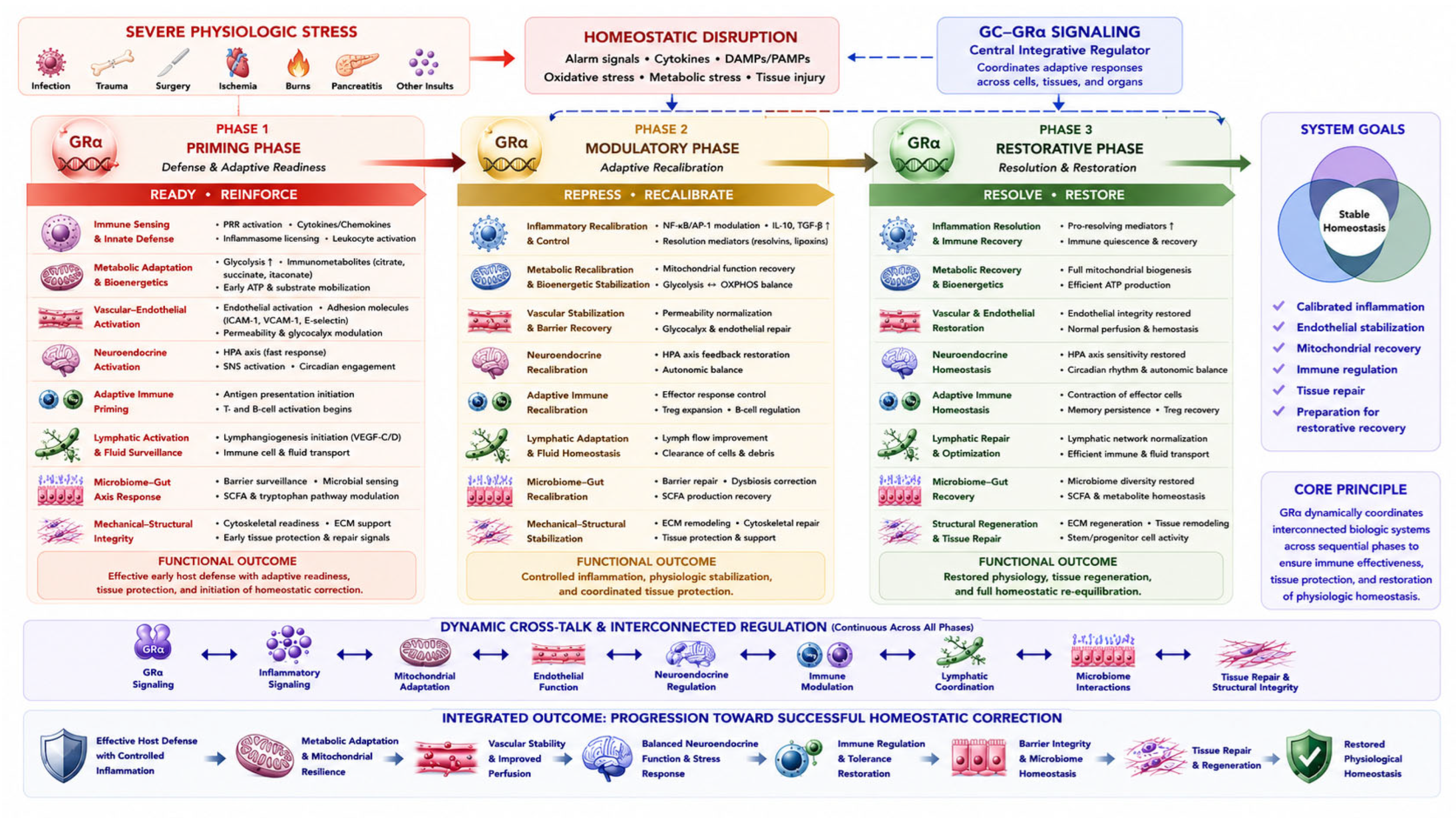
Figure 2.
Temporal Evolution and Divergent Trajectories of Homeostatic Correction During Critical Illness.
Figure 2.
Temporal Evolution and Divergent Trajectories of Homeostatic Correction During Critical Illness.
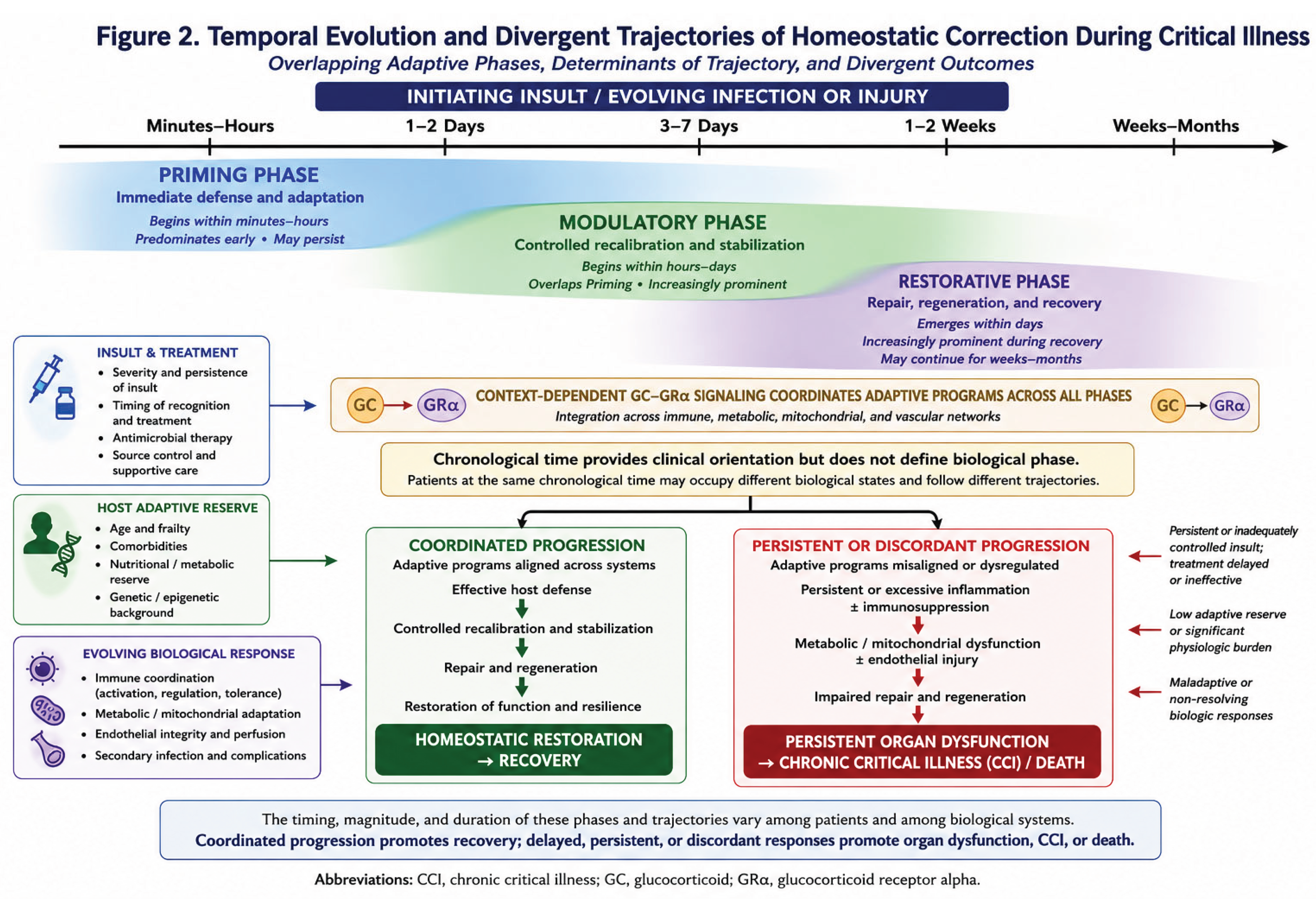
Disclaimer/Publisher’s Note: The statements, opinions and data contained in all publications are solely those of the individual author(s) and contributor(s) and not of MDPI and/or the editor(s). MDPI and/or the editor(s) disclaim responsibility for any injury to people or property resulting from any ideas, methods, instructions or products referred to in the content. |
© 2026 by the authors. Licensee MDPI, Basel, Switzerland. This article is an open access article distributed under the terms and conditions of the Creative Commons Attribution (CC BY) license (http://creativecommons.org/licenses/by/4.0/).
Copyright: This open access article is published under a Creative Commons CC BY 4.0 license, which permit the free download, distribution, and reuse, provided that the author and preprint are cited in any reuse.