
Submitted:
09 October 2026
Posted:
10 October 2026
You are already at the latest version
Abstract
The adult configuration of the aortic arch complex (AoAC) results from transformation of its embryonic precursor. Concepts of the embryology of variants and malformations of the AoAC traditionally focus on transformation processes that are driven by regression and persistence of embryonic vascular segments. However, such concepts cannot explain every variant or malformation since they neglect other morphogenetic processes involved in the transformation of the embryonic AoAC, such as blood vessel “migration”, and septation of the aortic sac (AoS) and distal outflow tract (OFT) of the embryonic heart. This article focuses on the hitherto neglected role of septation of the AoS and embryonic OFT in normal and abnormal patterning of the AoAC. It is shown (1) how phylogenetic “deviations” from the mammalian “standard” of AoS septation lead to the AoAC branching patterns found in non-mammalian vertebrates; and (2) how ontogenetic deviations from the human “standard” can lead to AoAC anomalies, such as “bovine arch”; subclavian artery as first branch of the aortic arch; anomalous origin of supra-aortic branches from the proximal ascending aorta; anomalous origin of one pulmonary artery from the proximal ascending aorta; anomalous origin of one common carotid artery from the proximal pulmonary trunk; and tritruncal hearts.
Keywords:
aortic arch complex
; malformations
; variants
; embryology
; aortic sac
; flow dividers
; septation
; aortico-pulmonary septum
; inter-aortic septum
; mammals
; reptiles
; birds
; frogs
1. Introduction—An Introductory Overview on the Embryology of the Aortic Arch Complex
The great arteries that exit the human heart, the aortic arch with its branches, and the fetal connection between the distal aortic arch and the pulmonary trunk or pulmonary arteries (ductus arteriosus) form an arterial complex characterized by intimate anatomical, ontogenetic, and phylogenetic relationships. This arterial complex is frequently called the aortic arch complex or the aortic arch system. Unfortunately, these terms are used not only for naming of the above-described arterial complex but also for naming of its embryonic precursor whose anatomy deviates in several respects from the anatomy of its mature derivative. In the present article, I will therefore distinguish between the adult aortic arch complex and the embryonic aortic arch complex.
The embryonic aortic arch complex consists roughly of two components. These are, firstly, the arterial segments (intermediate and distal) of the initially undivided outflow tract (OFT) of the embryonic heart and, secondly, a basket-shaped arterial complex that has been named the arterial pharyngeal basket [1]. The arterial segments of the initially undivided OFT of the embryonic heart lie within the embryonic pericardial cavity and provide the roots and intra-pericardial portions of the future great arterial trunks [2,3]. The arterial pharyngeal basket lies downstream to the cardiac OFT. It surrounds the pharynx of the early embryo and is embedded into the pharyngeal mesenchyme, which is of mesodermal and neural crest origin [4,5]. In lung-breathing vertebrates, the arterial pharyngeal basket provides the vascular segments that form the definitive aortic arch(es) and its branches as well as the fetal connection(s) between the pulmonary trunk and aortic arch(es) (=ductus arteriosus).
The arterial pharyngeal basket is a phylogenetically conserved feature among vertebrates [6,7,8,9,10]. Its upstream end is formed by a sac-shaped expansion of the flow path, the so-called aortic sac [11]. The aortic sac (AoS) is a midline structure that lies ventral to the embryonic pharynx. Its function is two-fold, (1) it is the connecting element to the downstream end of the embryonic cardiac OFT; and (2) it is a vascular unit that directs and distributes the blood stream to a set of bilaterally paired pharyngeal arch arteries (PhAAs). These arteries appear in a cranial to caudal sequence during the early phase of cardiovascular development [11,12]. In higher vertebrates, a set of 5 pairs of PhAAs is regularly formed from which the first two pairs normally regress [6,10]. Thus, the late AoS normally gives origin only to the caudal three pairs of PhAAs, which are traditionally named the 3rd (or carotid), 4th (or systemic), and 6th (or pulmonary) arch arteries [7,13]. These arteries take their origins from the left and right lateral walls of the AoS from where they course around the left and right lateral walls of the pharynx. PhAAs connect the AoS with a bilateral pair of dorsal aortae, which run along the dorsal wall of the embryonic pharynx. The left and right dorsal aortae coalesce caudal to the embryonic pharynx to give rise to the initial segment of the unpaired dorsal aorta. This vascular segment harbors the primordia of the future subclavian arteries and, therefore, can be regarded as downstream end of the arterial pharyngeal basket of land-living vertebrates (tetrapods).
The adult aortic arch complex evolves from the transformation of the two above-described components of the embryonic aortic arch complex. This transformation is driven mainly by three morphogenetic processes: (1) the structural division of the initially undivided flow path of the AoS and distal cardiac OFT into systemic and pulmonary compartments due to the formation of mesenchymal septa; (2) the remodeling of the arterial pharyngeal basket by involution of some of its original vascular segments; and (3) the positional shift - “migration” - of some of its vascular elements (e.g., subclavian arteries).
Concepts explaining the morphogenesis of normal as well as aberrant arborisation patterns of the adult aortic arch complex should take into account all three processes. In reality, however, this was rarely done during the past 170 years. Explanations for the morphogenesis of abnormal branching patterns of the adult aortic arch complex traditionally focus on remodeling of the embryonic arterial pharyngeal basket sometimes in combination with the positional shift (migration) of arteries. They mostly neglect the process of septation of the AoS and distal OFT of the embryonic heart. The reason for this may be twofold. (1) Disturbances in septation of the AoS and distal OFT are traditionally made responsible for the morphogenesis of common arterial trunk (formerly called persistent truncus arteriosus), which is a congenital cardiovascular malformation that belongs to the group of so-called conotruncal heart defects [4,5]. Conotruncal heart defects represent a clinically important group of congenital cardiac malformations. Abnormalities in septation of the AoS and distal OFT, thus, might have been perceived mainly as processes contributing to the pathogenesis of congenital heart defects and not to vascular malformations. (2) Since the publication of the seminal works of Rathke [6] and Boas [7] on the aortic arch complex of vertebrates, simplified diagrams of the cardiac OFT and arterial pharyngeal basket are used to conceptualize the transformation of the aortic arch complex during ontogeny as well as phylogeny of vertebrates. Such diagrams are popular since they facilitate the generation of a wide variety of branching patterns simply via graphic remodeling of schemes depicting a hypothetical basic pattern of the aortic arch complex. Unfortunately, such diagrams do not facilitate graphical simulation of the spatially complex process of septation of the AoS and cardiac OFT and, therefore, present the status of these vascular elements usually in the adult condition. Popular examples for such a kind of diagrams are the hypothetical double aortic arch of Edwards [14,15] and Barry’s diagram of the embryonic aortic arch complex of the human embryo [16]. Both diagrams are widely used among those dealing with the diagnosis and treatment of human patients affected by congenital malformations of the adult aortic arch complex.
The above-mentioned kind of diagrams is popular since it can provide simple embryological explanations for a wide variety of variants and malformations of the adult aortic arch complex [15,17,18]. However, every model has its limitations. Simulations of the transformation of the embryonic aortic arch complex that are exclusively driven by persistence and involution of vascular segments of a basic aortic arch complex cannot generate every known variant or malformation of the adult aortic arch complex, especially those in which a branch of the aortic arch or pulmonary trunk takes irregular origin from an arterial segment derived from the AoS or from the distal or intermediate portions of the embryonic OFT. Among such anomalies are cases of (1) so-called bovine aortic arch, which is a frequent variant of the adult aortic arch complex of humans [19,20]; (2) subclavian artery as the first branch of the aortic arch [21,22,23,24,25,26]; (3) anomalous origin of one supra-aortic branch of the aortic arch from the proximal portion of the ascending aorta [27,28]; (4) anomalous origin of one pulmonary artery from the proximal portion of the ascending aorta [29,30,31,32]; (5) anomalous origin of the left common carotid artery from the proximal portion of the pulmonary trunk [33]; and (6) so-called tritruncal hearts [34,35,36,37]. In the past, some of these aberrations from the normal arborisation pattern were explained by abnormal positional shift (migration) or failure of normal positional shift of the origin of the affected arteries [19,20,30,31] or by failure of fusion of arterial segments [35,37]. However, the possibility that such anomalies might result from aberrant septation of the AoS and distal OFT was rarely taken into consideration [29,32,33,34,38].
The purpose of the present article is to make the reader familiar with the hitherto neglected role of septation of the AoS and distal OFT in the generation of normal and abnormal arborisation patterns of the adult aortic arch complex of vertebrates. This review starts with the description of septation of the AoS and distal OFT as seen in humans and other mammals. From the standpoint of human beings, this description defines the standard for “normal” AoS septation. At a second step, septation of the AoS and distal OFT is described as seen in reptiles, birds, and amphibia. These descriptions serve as examples for phylogenetic “deviations” from the human standard and thereby set the scene for a better understanding of the possible role of malseptation of the AoS in the morphogenesis of variants and malformations of the adult aortic arch complex, which is the focus of the third, and final step.
2. Septation of the AoS and Distal OFT as Seen in Humans and Other Mammals
Humans and other mammals have completely septated four-chambered hearts and we usually find two arterial trunks that exit the heart. These are the ascending aorta, which exits the left or systemic ventricle, and the pulmonary trunk, which exits the right or pulmonary ventricle. Both arterial trunks evolve from the structural division of the initially undivided flow path of the AoS and arterial segments of the OFT of the embryonic heart. This morphogenetic process is usually attributed to the upstream growth of an arterial flow divider called the aortico-pulmonary septum (AP-septum) [39,40,41]. This mesenchymal septum arises from merging of a pair of flow dividers located between the orifices of the 4th (systemic) and 6th (pulmonary) PhAA (Figure 1A,B and Figure 2A).
The mesenchymal core of the AP-septum is formed by cells derived from the cardiac neural crest [4,5]. Subsequent to merging of its paired primordia, the AP-septum appears as a horizontal, sickle-shaped crest at the dorsal wall of the AoS from where it growth upstream towards the cardiac OFT (Figure 2B). The initially undivided cavity of the AoS, thereby, becomes divided into one aortic and one pulmonary compartment (Figure 2C,D). Subsequent to division of the AoS, the AP-septum grows further into the distal segment of the OFT where its two spurs merge with the distal margins of paired endocardial ridges that form at the intermediate portion of the OFT. This leads to the structural separation of the systemic and pulmonary flow paths at the level of the future intra-pericardial portions of the great arteries and their roots [2,3]. The AP- septum, finally, looses its septal function due to the development of the mature vessel wall, so that the ascending aorta and pulmonary trunk finally form separate anatomical units that normally do not share a common septum [3].
3. Septation of the AoS and Distal OFT as Seen in Reptiles, Birds and Amphibia
3.1. Reptiles
Among reptiles only crocodiles have completely septated four-chambered hearts, while non-crocodilian reptiles usually have three-chambered hearts that have two atria but only a single, incompletely divided ventricle [42,43]. A striking feature, and remarkable deviation from the mammalian “standard”, is the fact that non-crocodilian reptiles as well as crocodiles usually have three arterial trunks that exit the heart (Figure 3).
These arterial trunks are, (1) the right or systemic aorta, which receives its blood mainly from the cavum arteriosum of the single ventricle (crocodiles: left ventricle); (2) the left or visceral aorta, which receives its blood from the cavum venosum and cavum arteriosum of the single ventricle (crocodiles: right ventricle); and (3) the pulmonary trunk, which receives its blood from the cavum pulmonale of the single ventricle (crocodiles: right ventricle). Thus, in contrast to mammals, which show a bipartite division of their arterial heart segment, reptiles normally show a tripartite division of their arterial heart segment. Furthermore, in contrast to expectations based on Edwards’ hypothetical double aortic arch, the two aortic trunks of reptiles do not show a bilaterally symmetric branching pattern but ramify in an asymmetric pattern. The right aortic trunk gives off both common carotid arteries and the right aortic arch, while the left aortic trunk does not give off any branch and continues directly as the left aortic arch [9].
Studies on reptilian embryos have shown that the above-described arrangement of the adult aortic arch complex of reptiles results exclusively from septation of the AoS and the arterial segments of the OFT of the embryonic heart [39,44,45,46,47,48,49]. In contrast to mammalian embryos, reptilian embryos form not only an AP-septum that divides the AoS into aortic and pulmonary compartments. They furthermore form a so-called aortic or inter-aortic septum that divides the aortic compartment of the aortic sac into left and right-sided compartments [39,44,45,46,47,48,49].
The morphological aspects of the process of septation of the AoS and distal cardiac OFT of reptilian embryos are best described in publications of Alfred Greil [39] and other scholars and colleagues of the Austrian anatomist and embryologist Ferdinand Hochstetter [39,44,45,46,47]. Based on studies on amphibian, reptilian, avian and mammalian embryos, these authors described not only the formation and upstream growth of the AP-septum. They, furthermore, noted that the AP-septum is not the only flow divider that forms within the AoS and contributes to septation of the AoS and distal OFT. They describe a system of AoS flow dividers that, in a basic condition, consists of two median flow dividers and two pairs of lateral flow dividers (Figure 4). The median flow dividers are located at the cranial and caudal margins of the AoS. They form protrusions that are located between the two orifices of the 3rd (carotid) PhAAs and between the two orifices of the 6th (pulmonary) PhAAs. They were named the septum inter-caroticum (cranial pole) and septum inter-pulmonale (caudal pole). The lateral flow dividers are located between the orifices of the 3rd and 4th PhAAs and between the orifices of the 4th and 6th PhAAs. They were named the left and right septum carotico-aorticum and the left and right primordium of the septum aortico-pulmonale.
Starting from the above-described basic condition, the process of septation of the AoS and distal cardiac OFT of reptiles now can be described as follows (Figure 5): Due to bilaterally symmetric growth, the initially paired aortico-pulmonary flow dividers merge and form a common AP-septum that leads to the structural separation of the pulmonary and systemic flow paths in a fashion similar to the fashion observed in mammals. In contrast to mammals, however, substantial growth is found not only at the level of the aortico-pulmonary flow dividers but also at the level of the carotico-aortic flow dividers. Compared to the aortico-pulmonary flow dividers, however, these paired flow dividers show a bilaterally asymmetric growth behavior that mainly affects the left carotico-aortic flow divider. This mesenchymal protrusion grows toward the midline of the AoS where it merges with a midsagittal mesenchymal ridge located at the dorsal wall of the AoS. Thereby a septum is formed that starts to subdivide the aortic compartment of the AoS into right- and left-sided compartments and, therefore, has been named the aortic or inter-aortic septum. This vertically oriented septum grows toward the caudal pole of the AoS where it merges with the horizontally oriented AP-septum. The inter-aortic and AP-septa now form a common septal complex in which the two septal components are arranged perpendicular to each other. This septal complex grows into the distal OFT where it merges with endocardial ridges of the intermediate portion of the OFT and thereby leads to tripartite division of the arterial segments of the heart. Corresponding to mammals, the inter-aortic AP-septal complex, finally, looses its septal function due to the development of the mature vessel wall, so that the three arterial trunks finally form separate anatomical units that normally do not share common septa.
3.2. Birds
For a long time, the chick embryo served as a popular model for embryonic development of the aortic arch complex of higher vertebrates [4,6,50]. Therefore, our current knowledge about this topic is strongly based on descriptive and experimental data from studies on birds. This is especially true for our current knowledge about the role of the cardiac neural crest in AP-septation [4].
Birds belong to the sauropsids, which is a clade of land-living vertebrates that includes modern reptiles, crocodiles, and birds. Among this clade, birds share the feature of a completely septated four-chambered heart with crocodiles. Thus, the cardiovascular system of birds is phylogenetically more closely related to crocodiles than mammals. In contrast to crocodiles, however, birds have only two arterial trunks, - one aortic trunk and one pulmonary trunk - , that exit the heart (Figure 6).
On the first view, this situation seems to correspond to the mammalian rather than reptilian situation and may suggest that AP-septation in birds runs the same way as in mammals. Embryological data, however, have shown that the aortic trunk of birds corresponds to the right aortic trunk of reptiles. Birds normally loose the developing left aorta by regression of the left 4th PhAA [1,7,13,51,52,53]. This is unique among higher vertebrates, which usually retain both 4th PhAAs [10,48,52]. Before regression of the left 4th PhAA, septation of the AoS of birds runs in a mode that shows strong similarities to the reptilian mode described above. The AP-septum forms by growth and merging of initially paired aortico-pulmonary flow dividers and the left carotico-aortic flow divider grows toward the midline of the AoS (Figure 7A,B). In contrast to reptiles, however, the left carotico-aortic septum does not merge with the midline crest to form an inter-aortic septum (Figure 7C). Thus, it is only the AP-septum that grows into the distal outflow tract and reaches the level of the future arterial valves. Corresponding to mammals, the AP-septum of birds, finally, looses its septal function due to the development of the mature vessel wall, so that the aortic and pulmonary trunks finally form separate anatomical units that normally do not share common septa.
3.3. Amphibia
Today, amphibia do not serve as popular models for embryonic development of the aortic arch complex of higher vertebrates. With regard to septation of the AoS and distal cardiac OFT, however, amphibia show some interesting anatomical features that support the above-described concept of a system of multiple AoS flow dividers that have the potential to contribute to septation of the AoS. Among the amphibia, it is the adult aortic arch complex of anurans (frogs, toads) that shows the strongest similarities to the adult aortic arch complex of amniotes (reptiles, birds, mammals). The following description, thus, focuses mainly on the situation found in frogs and toads.
Amphibia have hearts that are composed of two atria, a single undivided ventricle, and a distinct myocardial outflow chamber called the conus. The conus is incompletely divided into systemic and pulmonary flow paths. It is interposed between the undivided ventricle and the arterial segment of the amphibian heart. The latter is called the truncus arteriosus. From its external aspect, the truncus arteriosus of amphibia appears as a single arterial trunk that exits the heart. In anuran amphibia, this arterial trunk is Y-shaped since it bifurcates into a right and a left arm. Each of these truncus arms finally gives off three morphologically distinct arteries that derive from the 3rd, 4th, and 6th PhAAs. They are called the common carotid artery, the aortic arch, and the pulmo-cutaneous artery. The truncus arteriosus and its two arms do not really represent true vascular trunks. They rather represent densely packed bundles of ramifying vascular channels, which are ensheathed by a common outer wall. The arborisation pattern of the truncus channels is most elaborated at the distal part of the non-bifurcated portion of the truncus arteriosus (Figure 8). Here, we find a bundle of three pairs of channels that splits downstream into the right and left-sided bundles of the truncus arms. Each of these bundles includes three vascular channels, which finally feed the common carotid arteries, the aortic arches, and the pulmo-cutaneous arteries. The truncus channels, therefore, are named the carotid, aortic, and pulmo-cutaneous channels.
The channels of the truncus arteriosus are separated from each other by a system of intervascular septa. Thus, we find bilaterally paired aortico-pulmonary and carotico-aortic septa within the truncus arms and the non-bifurcated truncus, as well as unpaired inter-carotid, inter-aortic, and inter-pulmonary septa within the non-bifurcated truncus (Figure 8).
The arrangement of the truncus septa reflects the arborisation pattern of the truncus channels. Thus, the system of truncus septa is most elaborated at the distal part of the non-bifurcated portion of the truncus from where several septa disappear toward the upstream end of the truncus (Figure 9). It is an interesting and largely unknown fact, that the truncus septa of most frogs and toads are arranged in a bilaterally asymmetric fashion. As a result, the truncus channels ramify in a pattern that surprisingly corresponds to the asymmetric arborisation pattern of the great arterial trunks that exits the heart of reptiles. Thus, the left aortic channel of the truncus arteriosus of most frogs leads only to the left aortic arch, while the right aortic channel feeds the right aortic arch and both carotid channels (Figure 9). This fact has been documented for numerous anuran species [51,54,55,56,57,58,59,60,61,62] but, for unknown reasons, did not find its way into historic as well as contemporary textbooks of zoology.
The development of the septa of the amphibian truncus arteriosus was studied by Alfred Greil at the beginning of the 20th century [63]. He reports that the initially undivided lumen of the truncus arteriosus becomes subdivided into its several vascular channels by the upstream growth of flow dividers initially located between the orifices of the PhAAs 3, 4, and 6. This observation not only shows that the process of septation of the AoS seems to run the same way in all extant tetrapods. It furthermore shows that, in contrast to amniotes (reptiles, birds, mammals), the flow dividers of the AoS of amphibia never loose but retain their septal function during the whole lifespan.
4. Possible Role of Malseptation of the AoS in the Morphogenesis of Variants and Malformations of the Adult Aortic Arch Complex
In the preceding sections of this article, we have seen that, among land-living vertebrates, the process of septation of the AoS and distal portion of the embryonic OFT includes more than the popular story of the AP-septum. In the final section, it will be shown how postulated deviations from the normal pattern of AoS and distal OFT septation can explain the morphogenesis of several variants and malformations of the adult aortic arch complex that cannot be explained by transformations of the embryonic aortic arch complex that are exclusively driven by involution or abnormal persistence of some of its segments. The description starts with malformations and variants that are mainly confined to the intra-pericardial subregion of the adult aortic arch complex and ends with variants and malformation confined to the extra-pericardial subregion.
4.1. Tritruncal Hearts
Human cases of the so-called tritruncal heart may represent the most convincing examples for malformations that possibly result from malseptation of the AoS and distal OFT. Human tritruncal hearts are very rare malformations that, on the first view, seem to resemble reptilian hearts, since they also have three arterial trunks that exit the heart. Each of these arterial trunks has its own semilunar valve, and their flow paths are completely separated from each other. In contrast to reptilian hearts, however, human tritruncal hearts have only a single aorta but two pulmonary trunks; one main pulmonary trunk, that continues as the left pulmonary artery and ductus arteriosus, and one so-called intermediate trunk, that continues as the right pulmonary artery. Up to now, only four cases of human tritruncal heart were described in the literature. Two of them had the ventriculo-arterial connection of triple outlet right ventricle [36,37]. In the remaining two cases, the main pulmonary trunk arose from the right ventricle, while the aorta and intermediate trunk arose from the left ventricle [34,35]. Previous explanations for the embryology of tritruncal hearts are based on the idea that the normal pulmonary trunk is formed by fusion of the proximal segments of the 6th pharyngeal arch arteries [64]. The presence of two pulmonary trunks, thus, was explained by failure of the hypothetical fusion of the proximal segments of the 6th pharyngeal arch arteries [35,37]. This pathogenetic hypothesis, however, does not comply with reality since the pulmonary trunk derives from the distal portion of the embryonic cardiac OFT. Only a single group had speculated that human tritruncal hearts might result from a “double septation” of the AoS and distal OFT [34]. On the basis of the above-described knowledge about the flow dividers of the AoS, the postulated process of “double septation” can be ascribed to the upstream growth of an abnormal septal complex formed by merging of the AP-septum with an excessively growing inter-pulmonary septum/flow divider (Figure 10). The observation that the two pulmonary trunks have separate lumens but share a common vessel wall [35,36,37], - which undoubtedly functions as an inter-pulmonary septum -, is suggestive of this idea.
4.2. Anomalous Connections Between Aortic and Pulmonary Segments of the Adult Aortic Arch Complex
According to the traditional morphogenetic diagrams of the aortic arch complex, open vascular connections between one of its aortic and one of its pulmonary segments can be explained only by the abnormal persistence of a ductus arteriosus or by the abnormal persistence of vascular channels traditionally interpreted as 5th PhAAs. However, these pathogenetic scenarios cannot explain every case of abnormal connections between aortic and pulmonary segments of the adult aortic arch complex. In the following, two kinds of such malformation are presented.
4.2.1. Anomalous Origin of One Pulmonary Artery from the Proximal Portion of the Ascending Aorta
The first kind of malformations is represented by cases in which one pulmonary artery takes its origin from the proximal portion of the ascending aorta (Figure 11A). Such an anomaly can affect the left as well as the right pulmonary artery [26]. The initial segment of the affected artery is an intra-pericardial vessel that lies upstream to the derivatives of the so-called 5th and 6th PhAAs [32]. In view of these topographical characteristics, the initial segment of a pulmonary artery that arises from the proximal portion of the ascending aorta can be interpreted neither as persistent ductus arteriosus nor as persistent segment of a so-called 5th pharyngeal arch artery.
Based on incorrect interpretations of embryological data, some authors tried to explain the aberrant origin of an affected pulmonary artery by failure of its normal positional shift during embryonic development [30,31]. Others speculated that such anomalies might have resulted from aberrant septation of the AoS and distal OFT [29,32,38]. On the basis of the above-described knowledge about the flow dividers of the AoS, the postulated process of malseptation can be ascribed to the upstream growth of an abnormal septal complex formed by merging of a hemi-hypotrophic AP-septum with an excessively growing inter-pulmonary septum/flow divider (Figure 11B).
According to this hypothetical scenario, cases of origin of one pulmonary artery from the proximal portion of the ascending aorta might be interpreted as special cases of aorto-pulmonary window in which the pulmonary trunk underwent bipartition due to excessive growth of the inter-pulmonary septum/flow divider.
4.2.2. Anomalous Origin of the Left Common Carotid Artery from the Proximal Portion of the Pulmonary Trunk
This type of abnormal vascular connection between one aortic and one pulmonary segment of the adult aortic arch complex is a rare malformation. It was first observed in neural crest-ablated chicks [65] and later in humans [66]. Up to now only seven human cases were described in the literature [33]. Besides the pulmonary origin of the left common carotid artery, this type of arterial malformation is characterized by the presence of a right aortic arch, aberrant left subclavian artery, and presence of a remnant of the left ductus arteriosus at its normal position (Figure 12A). Because of its topography and the presence of a remnant of the left ductus arteriosus, the initial segment of the abnormally connected left common carotid artery cannot be interpreted as persistent ductus arteriosus or as persistent segment of a so-called 5th pharyngeal arch artery. On the basis of the above-described knowledge about the flow dividers of the AoS, the embryology of this malformation was explained by malseptation of the AoS and distal OFT in combination with regression of the left 4th PhAA and persistence of the distal segment of the right dorsal aorta [65] and this explanation was adopted by several authors who reported on human cases [66,67,68,69]. The postulated process of malseptation is ascribed to the upstream growth of an abnormal septal complex formed by merging of an excessively growing inter-carotid septum with a hemi-hypotrophic AP- septum (Figure 12B).
The observation that the initial segment of the left common carotid artery sometimes shares a common vessel wall (inter-arterial septum) with the ascending aorta [66] is suggestive of the malseptation idea. According to the above-described hypothetical scenario, cases of abnormal origin of one common carotid artery from the proximal portion of the pulmonary trunk might be interpreted as special cases of aorto-pulmonary window in which the developing ascending aorta underwent bipartition due to excessive growth of the inter-carotid septum/flow divider.
4.3. Anomalous Origin of Supra-Aortic Branches of the Aortic Arch from the Proximal (Intrapericardial) Portion of the Ascending Aorta
Human cases in which one supra-aortic branch of the aortic arch takes its origin from the proximal portion of the ascending aorta close to the sinu-tubular junction seems to be exceedingly rare and, therefore, may be of minor clinical relevance. However, these variants of the adult aortic arch complex are of embryological interest since the affected arteries arise from a segment of the adult aortic arch complex that derives from the distal portion of the embryonic OFT. The aberrant origin of the affected arteries, therefore, cannot be explained simply by involution or persistence of segments of the embryonic arterial pharyngeal basket.
The simplest way to explain the embryology of such variants would be usage of the term “migration” of the artery along its stem vessel. This term, however, is a cloudy term since it says nothing about the mechanism behind such migrations. The positional shift of the orifice of an embryonic artery along its stem vessel can result from several morphogenetic processes, such as differential growth of vessel wall segments, partial fusion between the artery and its stem vessel, as well as partial splitting of the stem vessel by ingrowth of an inter-arterial septum [70]. The cranial shift of the origin of the future subclavian arteries from the unpaired aorta to the paired dorsal aortae, for example, is initially driven by downstream splitting of the unpaired dorsal aorta [51].
In the following, the embryology of human cases, in which one supra-aortic branch of the aortic arch takes its origin from the proximal (intrapericardial) portion of the ascending aorta, is explained by arterial “migrations” caused by aberrant growth behavior of the AoS septa/flow dividers.
4.3.1. Anomalous Origin of the Brachiocephalic Artery from the Proximal (Intrapericardial) Portion of the Ascending Aorta
This variant of the origin of the brachiocephalic artery was noted only in a single human case [28]. It was a case of tetralogy of Fallot with left aortic arch. The authors of this case report explained the upstream shift of the orifice of the brachiocephalic artery by a growth defect (hypoplasia) of the right half of the AoS.
The brachiocephalic artery derives from the right horn of the AoS, which is a cranial subdivision of the systemic compartment of the AoS [71]. During normal development, the cranial portion of the systemic compartment of the AoS becomes subdivided into left and right horns by upstream growth of the inter-carotid flow divider [38,39,47,72]. Therefore, a proximal origin of the brachiocephalic artery, which is accompanied by an elongation of this artery, might result from an excessive upstream growth of the inter-carotid septum (Figure 13A). The observation that the initial segment of the affected brachiocephalic artery shares a common vessel wall (inter-arterial septum) with the ascending aorta [28] is suggestive of this idea.
4.3.2. Anomalous Origin of Both Common Carotid Arteries from the Proximal (Intrapericardial) Portion of the Ascending Aorta
This variant of the origin of both common carotid arteries was described only for a single human case [27]. It was a 45-year old man without congenital heart disease. The authors of this case report explained the close proximity of the orifices of the common carotid arteries to the aortic valve by persistence of the early topographical relations between the future aortic valve and the orifices of the future common carotid arteries. They did not provide a description as to how this should have happened. Based on the above-described knowledge about AoS septation, the combined upstream shift of the orifices of both common carotid arteries can be explained by excessive growth of the inter-carotid and carotico-aortic flow dividers (Figure 13B).
4.4. Extrapericardial Variants
4.4.1. Subclavian Artery as First Branch of the Aortic Arch
This variant of the origin of a subclavian artery was found in patients with left as well as right-sided aortic arch [21,22,23,24,25,26]. The essential feature of this anomaly is the absence of the brachiocephalic artery, so that its two normal branches arise separately from the aortic arch. The affected subclavian artery is the first branch of the aortic arch. The right subclavian artery in cases of left aortic arch, and the left subclavian artery in cases of right aortic arch. Cases of this anomaly were regularly associated with so-called cono-truncal heart defects, such as TGA, tetralogy of Fallot, and DORV [21,23,24,25,26].
Several mechanisms were proposed to explain the embryology of this anomaly, which includes persistence of the so-called 5th PhAA combined with regression of the ipsilateral 4th PhAA and a segment of the dorsal aorta [22], or excessive migration of the affected subclavian artery combined with remodeling of the AoS [25,26]. Based on the above-described knowledge about aortic sac septation, it can be postulated that the absence of the brachiocephalic artery, that normally would supply the affected subclavian artery, results from splitting of its embryonic precursor (horn of the AoS) due to excessive growth of the ipsilateral carotico-aortic flow divider (Figure 14).
4.4.2. Trifurcated Brachiocephalic Trunk; the So-Called “Bovine Aortic Arch”
The most common deviation from the normal branching pattern of the adult aortic arch complex of humans is characterized by the presence of two instead of three supra-aortic branches. The first of these branches is a common trunk that gives rise to the right subclavian artery, the right common carotid artery, and the left common carotid artery. The second branch is the left subclavian artery [19, 20, 73). This aortic arch variant is frequently named the bovine aortic arch although it has been noted that this term is a misnomer, since this configuration does not bear resemblance to the aortic arch branching pattern of cattle [19]. The latter is characterized by the presence of a single brachiocephalic trunk that originates from the aortic arch and gives off the bilateral subclavian arteries and the bilateral common carotid arteries.
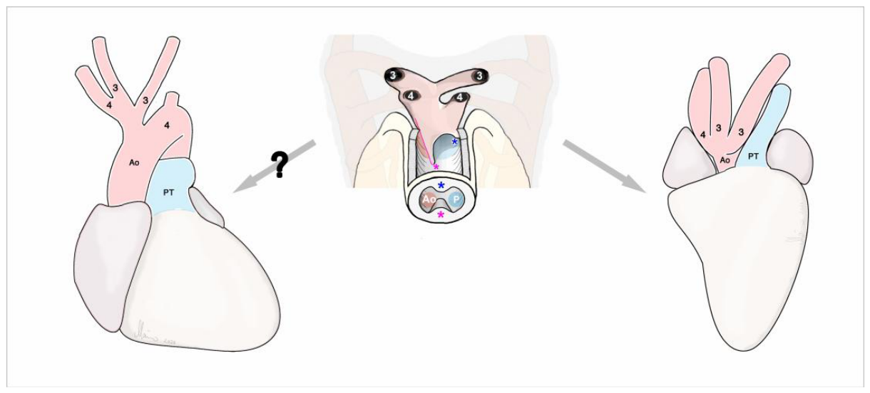
The embryology of the trifurcated brachiocephalic trunk, found in the human “bovine aortic arch”, cannot be explained by remodeling of the arterial pharyngeal basket due to regression or persistence of its segments. Its embryology, therefore, has been explained by other mechanisms, which include upstream migration of the origin of the left common carotid artery due to different growth rates in the various components of the arterial pharyngeal basket [19,20], failure of AoS bifurcation [73], and aberrant septation of the AoS [38]. With regard to the latter explanation, it should be noted, that the trifurcated brachiocephalic trunk of the human “bovine aortic arch” consists of components that derive from the right half of the AoS (initial segments of right subclavian and common carotid arteries) and the cranial portion of the left horn of the AoS (left common carotid artery). These components also form the right aorta of reptiles and the definitive aorta of birds. The trifurcated brachiocephalic trunk of the so-called “bovine aortic arch” of humans obviously bears strong resemblance to the adult aortic arch complex found in sauropsids and, therefore, may be named the sauropsid brachiocephalic trunk rather than bovine brachiocephalic trunk. Its embryology can be explained by a pattern of AoS septation that corresponds to the pattern observed in birds (Figure 15). In contrast to birds, however, the left 4th PhAA does not regress.
Supplementary Materials
There are no supplementary materials.
Author Contributions
This is a single author paper.
Funding
This research received no external funding.
Institutional Review Board Statement
Not applicable.
Informed Consent Statement
Not applicable.
Data Availability Statement
No new data were created or analyzed in this study.
Acknowledgments
The author thanks Mr. Hannes Sydow for photographical assistance.
Conflicts of Interest
The author declares no conflicts of interest.
Abbreviations
The following abbreviations are used in this manuscript:
| AoS | Aortic sac |
| AP-septum | Aortico-pulmonary septum |
| PhAA | Pharyngeal arch artery |
| DORV | Double outlet right ventricle |
References
- Waldo, K.; Kirby, M.L. Development of the great arteries. In Living morphogenesis of the heart. Birkhäuser, Boston, 1998, pp. 187-217.
- Anderson, R.H.; Chaudhry, B.; Mohun, T.J.; Bamforth, S.D.; Hoyland, D.; Phillips, H. M.; Webb, S.; Moorman, A.F.M.; Brown, N.A.; Henderson, D.J. Normal and abnormal development of the intrapericardial arterial trunks in humans and mice. Cardiovascular Research 2012, 95, 108-115. [CrossRef]
- Anderson, R.H.; Lamers, W.H.; Hikspoors, J.P.; Mohun, T.J.; Bamforth, S.D.; Chaudhry, B.; Eley, L.; Kerwin, J.; Crosier, M.; Henderson, D.J. Development of the arterial roots and ventricular outflow tracts. Journal of Anatomy 2024, 244, 497-513. [CrossRef]
- Hutson, M.R.; Kirby, M.L. Model systems for the study of heart development and disease: cardiac neural crest and conotruncal malformations. Seminars in Cell & Developmental Biology 2007, 18, 101-110.
- Keyte, A.; Hutson, M.R. The neural crest in cardiac congenital anomalies. Differentiation 2012, 84, 25-40. [CrossRef]
- Rathke, H. Untersuchungen über die Aortenwurzeln und die von ihnen ausgehenden Arterien der Saurier (Vol. 13). KK Hof-und Staatsdruckerei, Wien, 1857.
- Boas, J.E.V. Text Book of Zoology. Sampson Low, Marston, London, 1896.
- Goodrich, E.S. Studies on the structure & development of vertebrates (Vol. 1). Macmillan and Company, London, 1930.
- Farmer, C.G. On the evolution of arterial vascular patterns of tetrapods. Journal of Morphology 2011, 272, 1325-1341. [CrossRef]
- Graham, A.; Hikspoors, J.P.; Lamers, W.H.; Anderson, R.H.; Bamforth, S.D. Morphogenetic processes in the development and evolution of the arteries of the pharyngeal arches: their relations to congenital cardiovascular malformations. Frontiers in Cell and Developmental Biology 2023, 11, 1259175. [CrossRef]
- Congdon, E.D. Transformation of the aortic-arch system during the development of the human embryo. Contrib Embryol, 1922, 14, 47-110.v.
- Anderson, R.H.; Bamforth, S.D. Morphogenesis of the mammalian aortic arch arteries. Frontiers in Cell and Developmental Biology 2022, 10, 892900. [CrossRef]
- Boas, J.E.V. Über die Arterienbogen der Wirbelthiere. Briefliche Mittheilung an den Herausgeber. Morphol Jahrbuch 1887, 13, 115-118.
- Edwards, J.E. Anomalies of the derivatives of the aortic arch system. Medical Clinics of North America 1948, 32, 925–949. [CrossRef]
- Stewart, J.R.; Kincaid, O.W.; Edwards, J.E. An atlas of vascular rings and related malformations of the aortic arch system. Charles C. Thomas, Springfield, 1964.
- Barry, A. The aortic arch derivatives in the human adult. The Anatomical Record 1951, 111, 221-238. [CrossRef]
- Stojanovska, J.; Cascade, P.N.; Chong, S.; Quint, L.E.; Sundaram, B. Embryology and imaging review of aortic arch anomalies. Journal of Thoracic Imaging 2012, 27, 73-84. [CrossRef]
- Hanneman, K.; Newman, B.; Chan, F. Congenital variants and anomalies of the aortic arch. Radiographics 2017, 37, 32-51. [CrossRef]
- Layton, K.F.; Kallmes, D.F.; Cloft, H.J.; Lindell, E.P.; Cox, V.S. Bovine aortic arch variant in humans: clarification of a common misnomer. American Journal of Neuroradiology 2006, 27, 1541-1542.
- Meyer, A.M.; Turek, J.W.; Froud, J.; Endelman, L.A.; Cavanaugh, N.B.; Torres, J.E.; Ashwath, R. Insights into arch vessel development in the bovine aortic arch. Pediatric Cardiology, 2019, 40, 1445-1449. [CrossRef]
- Boechat, M.I.; Gilsanz, V.; Fellows, K.E. [1978). Subclavian Artery as the First Branch of the Aortic Arch: A Normal Variant. Am J Roentgenol 1978, 131, 721-722. [CrossRef]
- Moes, C.A.F.; Benson, L.N.; Burrows, P.E.; Freedom, R.M.; Williams, W.; Duckworth, J.W. The subclavian artery as the first branch of the aortic arch. Pediatric Cardiology 1991, 12, 39-43. [CrossRef]
- Krishnan, K.S.; Theodore, S.; KVSS, S.K.; Neelakandhan, K.S. Embryologic and surgical considerations in tetralogy of Fallot with right arch and aberrant left subclavian artery from the ascending aorta. The Journal of Thoracic and Cardiovascular Surgery 2005, 130, 215-216. [CrossRef]
- Krishnamoorthy, K.M.; Madan, H.; Tharakan, J.A. Origin of left subclavian artery from ascending aorta: A variant of double aortic arch. The Journal of Thoracic and Cardiovascular Surgery 2006, 131, 245-247. [CrossRef]
- Hernandez, L.E.; Shepard, C.W.; Bamforth, S.D.; Anderson, R.H. The right subclavian artery arising as the first branch of a left-sided aortic arch. World Journal for Pediatric and Congenital Heart Surgery 2014, 5, 456-459. [CrossRef]
- Verma, M.; Pandey, N.N.; Ramakrishnan, S.; Jagia, P. Anomalous origin of one pulmonary artery from aorta: evaluation on computed tomography angiography. Journal of Cardiac Surgery 2022, 37, 483-491. [CrossRef]
- Boerman, R.H.; Padberg, G.W.A.M.; Wondergem, J.; Gittenberger-de Groot, A.C. Anomalous origin of the common carotid arteries as a cause of stroke. Clin Neurol Neurosurg 1991, 94, 317-319. [CrossRef]
- Prabhu, S.; Rao, S.; Siddaiah, S.; Shanmugasundaram, B.; Karl, T.R.; Angelini, A. Abnormally proximal aortic origin of the brachiocephalic artery: surgical implications. Ann Pediatr Cardiol. 2022, 15, 515-517.
- Richardson, J.V.; Doty, D.B.; Rossi N.P.; Ehrenhaft J.L. The spectrum of anomalies of aortopulmonary septation. J Thorac Cardiovasc Surg. 1979, 78, 21-27. [CrossRef]
- Penkoske, P.A.; Castañeda, A.R.; Fyler, D.C.; Van Praagh, R. Origin of pulmonary artery branch from ascending aorta: primary surgical repair in infancy. The Journal of Thoracic and Cardiovascular Surgery 1983, 85, 537-545.
- Kutsche, L.M.; Van Mierop, L.H.S. Anomalous origin of a pulmonary artery from the ascending aorta: associated anomalies and pathogenesis. The American Journal of Cardiology 1988, 61, 850-856. [CrossRef]
- Loomba, R.S.; Aiello, S.; Tretter, J.T.; Gaffar, M.; Reppucci, J.; Brock, M.A.; Spicer, D.; Anderson, R.H. Left pulmonary artery from the ascending aorta: a case report and review of published cases. Journal of Cardiovascular Development and Disease 2020, 8, 1. [CrossRef]
- Männer, J. Congenital isolation of a common carotid artery: a review of cases, phenotypes, and embryology of a rare anomaly of the aortic arch system. Pediatric Cardiology 2025, 1-10. [CrossRef]
- Diaz-Gongora, G.; Quero-Jimenez, M.; Espino-Vela, J.; Arteaga, M.; Bargeron, L. A heart with three arterial trunks (tritruncal heart) Report of a case. Pediatric Cardiology 1982, 3, 293-299.
- Pelletier, G.J.; Sokoloski, M.; Kardon, R. Diagnosis and management of tritruncal heart in an infant. The Journal of Thoracic and Cardiovascular Surgery 2009, 137, 753-755. [CrossRef]
- Tingo, J.E.; Carroll, S.J.; Crystal, M.A. Triple outlet right ventricle: a previously unknown cardiac malformation. Cardiology in the Young 2015 25, 576-579. [CrossRef]
- Traa, B.S.; Alfieris, G.M.; Wittlieb-Weber, C.A. Successful surgical palliation of a triple-outlet right ventricle: a rare congenital cardiac malformation. Cardiology in the Young 2016, 26, 403-405. [CrossRef]
- Seidl, W.; Steding, G. Contribution to the Development of the Heart. Part III: The aortic arch complex. Normal development and morphogenesis of congenital malformations. The Thoracic and Cardiovascular Surgeon 1981, 29, 359-368.
- Greil, A. Beitrage zur vergleichenden Anatomie und Entwicklungsgeschichte des Herzens und des Truncus arteriosus der Wirbelthiere. Morphologisches Jahrbuch, 1903, 31, 123-210.
- Oostra, R.J.; Steding, G.; Lamers, W.H.; Moorman, A.F. Steding’s and Virágh’s scanning electron microscopy atlas of the developing human heart. New York, NY: Springer New York, 2007.
- Yang, Y.P.; Li, H.R.; Cao, X.M.; Qiao, C.J.; Ya, J. Septation of the intrapericardial arterial trunks in the early human embryonic heart. Chinese Medical Journal 2018, 131, 1457-1464. [CrossRef]
- Jensen, B.; Moorman, A.F.; Wang, T. Structure and function of the hearts of lizards and snakes. Biological Reviews 2014, 89, 302-336. [CrossRef]
- Cook, A.C.; Tran, V.H.; Spicer, D.E.; Rob, J.M.; Sridharan, S.; Taylor, A.; Anderson, R.H.; Jensen, B. Sequential segmental analysis of the crocodilian heart. Journal of Anatomy 2017, 231, 484-499. [CrossRef]
- Langer, A. Uber die Entwicklungsgeschichte des Bulbus cordis bei Amphibien und Reptilien. Morphol Jahrbuch 1894, 21, 40-68.
- Hochstetter, F. Beiträge zur Anatomie und Entwickelungsgeschichte des Blutgefässsystemes der Krokodile. In: Voeltzkow, A. (ed): Reise in Ostafrika. Wissenschaftliche Ergebnisse. Anat Entwicklungsgesch 1906, 4: 1–133.
- Spitzer, A. Über die Ursache und den Mechanismus der Zweiteilung des Wirbeltierherzens: II. Teil. Die doppelte Septierung des arteriellen Herzschenkels und deren sekundäre Vereinfachung. Archiv für Entwicklungsmechanik der Organismen 1921, 47, 511-570. [CrossRef]
- Pernkopf, E.; Wirtinger, W. Die Transposition der Herzostien – ein Versuch der Erklärung dieser Erscheinungen. Die Phoronomie der Herzentwicklung als morphogenetische Grundlage der Erklärung. I. Teil. Die Phoronomie der Herzentwicklung. Z Anat Entwickl Gesch. 1933, 100, 564-711.
- Poelmann, R.E.; Gittenberger-de Groot, A.C.; Biermans, M.W.; Dolfing, A.I.; Jagessar, A.; van Hattum, S.; Hoogenboom, A.; Wisse, L.J.; Vicente-Steijn, R.; de Bakker, M.A.G.; Vonk, F.J.; Hirasawa, T.; Kuratani, S.; Richardson, M.K. Outflow tract septation and the aortic arch system in reptiles: lessons for understanding the mammalian heart. Evodevo 2017, 8, 9. [CrossRef]
- Poelmann, R.E.; Gittenberger-de Groot, A.C.; Goerdajal, C.; Grewal, N.; De Bakker, M.A.; Richardson, M.K. Ventricular septation and outflow tract development in crocodilians result in two aortas with bicuspid semilunar valves. Journal of Cardiovascular Development and Disease 2021 8, 132. [CrossRef]
- von Baer, K.E. Ueber die Kiemen und Kiemengefässe bei den Embryonen der Wirbelthiere. Archiv für Anatomie und Physiologie 1827, 12, 556-568.
- Hochstetter, F. Die Entwicklung des Blutgefässsystems. In Handbuch der Vergleichenden and Experimentellen Entwicklungslehre der Wirbeltiere, 3. Band, 2. Teil; Hertwig, O., Ed.; Fischer Jena: 1906; pp. 21–166.
- Bremer, J.L. Experiments on the aortic arches in the chick. The Anatomical Record 1928, 37, 225-254. [CrossRef]
- Shaner, R.F. Comparative development of the bulbus and ventricles of the vertebrate heart with special reference to Spitzer’s theory of heart malformations. The Anatomical Record 1962, 142, 519-529. [CrossRef]
- Boas, J.E.V. Uber den Conus arteriosus und die Arterienbogen der Amphibien. Morphol Jahrbuch 1882, 7, 488-568.
- Zimmermann, W. Über die Carotidendrüse von Rana esculenta. Inaug.-Diss., Med. Facult. Berlin University, Dr. v. Jul. Feiertag, 1887.
- Gaupp, E. Anatomie des Frosches. Zweite Abtheilung, Lehre vom Nerven- und Gefässsystem. 2nd edition; Vieweg & Sohn, Braunschweig, Germany, 1899.
- Pohlman, A.G. On the phylogenesis of the heart. Anatomical Record 1914, 8, 130-131.
- Holmes, S.J. The biology of the frog. 3rd edition, Macmillan; New York, 1916.
- Rau, A.S. Observations on the anatomy of the heart, lungs and related parts of Ceratophrys. Journal of Anatomy 1924, 58[Pt 4), 306-327.
- Peelle, M.L. The truncus arteriosus of American frogs, Rana sphenocephala, palustris, pipiens, clamitans and catesbeiana, as contrasted to European species. Journal of the Faculty of Science, Hokkaido Imperial University: Zoology 1931, 1, 55-93.
- Holmes, E.B. A reconsideration of the phylogeny of the tetrapod heart. Journal of Morphology 1975, 147, 209-228. [CrossRef]
- Nigam, H.C. Structure and function of the truncus arteriosus of the Indian frog Rana tigerina (Daud). Proceedings: Animal Sciences 1983, 92, 55-63. [CrossRef]
- Greil, A. Ueber die Entwicklung des Truncus arteriosus der Anamnier. Anat Anz 1903b 23 (Suppl. Verh Anat Ges 1903), 123-210.
- Skidmore F.D. Development of the right outflow tract and pulmonary arterial supply. Ann R Coll Surg Engl. 1975, 57, 186-197.
- Männer, J.; Seidl, W.; Steding, G. The formal pathogenesis of isolated common carotid or innominate arteries: the concept of malseptation of the aortic sac. Anatomy and Embryology 1997, 196, 435-445. [CrossRef]
- Kaushik, N.; Saba, Z.; Rosenfeld, H.; Patel, H.T.; Martin, K.; Reinhartz, O.; Lamberti, J.J. An isolated left common carotid artery from the main pulmonary artery: possible malseptation of the truncoaortic sac. Pediatr Cardiol 2005, 26, 707–709. [CrossRef]
- Joshi, A.R.; Joshi, S.; Kale, K.; Jain, R.; Bava, J.S. Isolation of left common carotid artery with its origin proximal to patent ductus arteriosus presenting in adult age. Case Reports in Radiology, 2016, 2016, 4149365. [CrossRef]
- Juergensen, M.S.; Quezada, M.E.; Gossett, M.J.G.; Kouretas, M.P.; Reddy, M.V.M.; Hogan, M.W. Breaking the rules: left common carotid artery from the main pulmonary artery in an infant with a vascular ring. Case: Cardiovasc Imaging Case Rep, 2021, 5, 262-266.
- Sachdeva, S.; Gupta, S.K.; Rajashekar, P.; Jagia, P.; Sebastian, L.J.D. Isolated left common carotid artery: multidisciplinary management is the key to success. Cardiology in the Young 2022, 32, 1519-1526. [CrossRef]
- Hochstetter, F. Über den Ursprung der Arteria caudalis beim Orang und beim Kaninchen: Nebst Bemerkungen über sogenannte “Gefässwurzelwanderung”. Anatomische Hefte 1911, 43, 243-334.
- Hikspoors, J.P.J.M.; Kruepunga N.; Mommen, G.M.C.; Mommen, E.; Anderson, R.H.; Lamers, W.H. A pictorial account of the human embryonic heart between 3.5 and 8 weeks of development. Communications Biology 2022, 5, 226. [CrossRef]
- Wang, J.J.; Liu, H.X.; Song, L.; Li, H.R.; Yang, Y.P.; Zhang, T.; Jing, Y. Isl-1 positive pharyngeal mesenchyme subpopulation and its role in the separation and remodeling of the aortic sac in embryonic mouse heart. Developmental Dynamics 2019 248, 771-783. [CrossRef]
- Lale, P.; Toprak, U.; Yagız, G.; Kaya, T.; Uyanık, S.A. Variations in the branching pattern of the aortic arch detected with computerized tomography angiography. Advances in Radiology, 2014, 2014, 969728. [CrossRef]
Figure 1.
These semi-schematic drawings depict a low magnification (A) and higher magnification (B) ventral view on the AoS and arterial pharyngeal basket before the onset of septation. The ventral wall of the AoS is removed to facilitate view on its dorsal wall and on the openings of the PhAAs 3, 4, and 6. Arteries are shown as embedded into the semitransparent pharyngeal mesenchyme. Yellow areas mark the semitransparent dorsal wall of the pericardial cavity. At the preseptation stage, a bilateral pair of aortico-pulmonary flow dividers is found between the openings of the 4th and 6th PhAAs (marked by magenta color). These flow dividers are the forerunners of the AP-septum. Abbreviations: L-DAo = left dorsal aorta; PA = pulmonary arteries; R-DAo = right dorsal aorta. Drawings are based on original image data from [11] (Figure 33) and [40] (Figure 4.10) showing the normal AoS and arterial pharyngeal basket of a human embryo at post-fertilization week 5 (Carnegie stage 14).
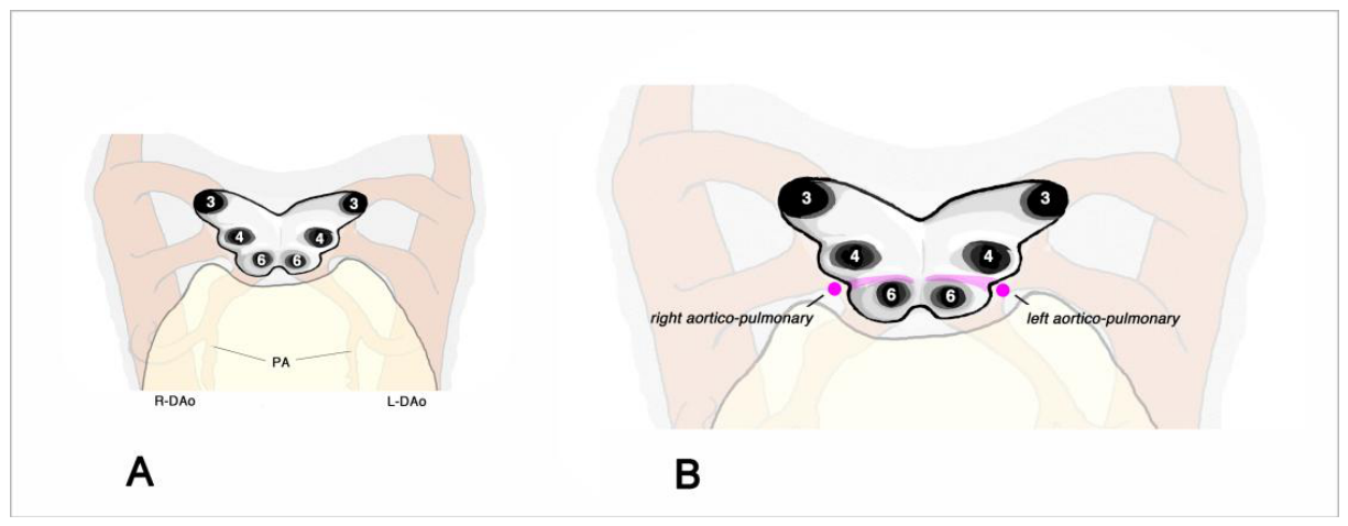
Figure 2.
These semi-schematic drawings depict the process of septation of the AoS and distal OFT as seen in mammals. Ventral views into the opened AoS (A–D) and distal OFT (B,C). (A). Preseptation stage. A bilateral pair of aortico-pulmonary flow dividers is found between the openings of the 4th and 6th PhAAs (marked by magenda color). (B). The paired aortico-pulmonary flow dividers have merged to form the AP-septum. (C). The AP-septum grows into the distal OFT, which undergoes anticlockwise torsion. Its upstream spurs (marked by asterisks) grow toward the mesenchymal ridges of the intermediate portion of the OFT (marked by corresponding asterisks). (D). Subsequent to growth into the distal OFT, the AoS is divided into one aortic (red) and one pulmonary (blue) compartment. The aortic compartment is incompletely subdivided into left and right horns of the AoS. As indicated by red arrows in the inset (picture of a human heart), these horns form the brachiocephalic artery (right horn) and a segment of the definitive aortic arch (left horn). Abbreviations: Ao = flow path of the future aorta; P = flow path of the future pulmonary trunk; other abbreviations as used before. Drawings are based on original image data from [11] (Figure 33) and [40] (Figures 4.10 and 4.15) showing the normal AoS and arterial pharyngeal basket of human embryos at post-fertilization week 5 and 6 (Carnegie stages 14 and 15).
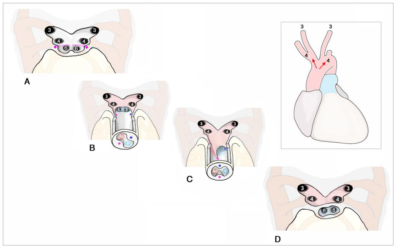
Figure 3.
This drawing depicts the arrangement of the great arterial trunks that exit the heart of a lizard. Numbers mark the derivatives of the 3rd and 4th PhAAs: 3 = left and right common carotid artery; 4 = left and right aortic arch. Abbreviations: LA = left atrium; L-Ao = left aorta; PT = pulmonary trunk; RA = right atrium; R-Ao = right aorta; V = ventricle; other abbreviations as used before.
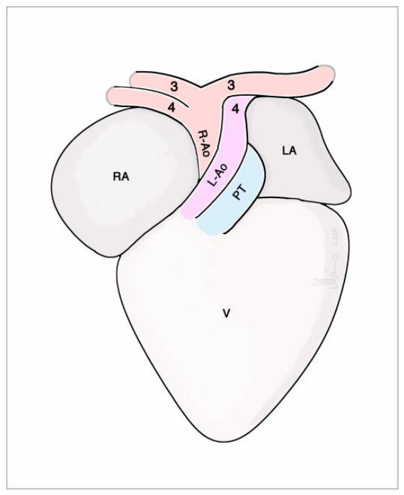
Figure 4.
This semi-schematic drawing depicts the system of flow dividers found in the AoS of amniotes (reptiles, birds. mammals).
Figure 4.
This semi-schematic drawing depicts the system of flow dividers found in the AoS of amniotes (reptiles, birds. mammals).
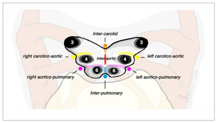
Figure 5.
These semi-schematic drawings depict the process of septation of the AoS and distal OFT as seen in reptiles. Same views as shown in Figure 1. (A). Preseptation. The AoS and PhAAs show a bilaterally symmetric patterning. Flow dividers that will contribute to septation are marked by the same color code as used in Figure 4. (B). The AP-septum has formed by merging of the paired aortico-pulmonary flow dividers. Growth of the left carotico-aortic flow divider has led to a bilaterally asymmetric branching pattern of the PhAAs. (C). The left carotico-aortic flow divider/septum has merged with the midline crest to form the inter-aortic septum. The latter is fused with the AP-septum. This septal complex grows into the distal portion of the OFT. Its upstream spurs (marked by asterisks) grow toward the mesenchymal ridges of the intermediate portion of the OFT (marked by corresponding asterisks). (D). Subsequent to growth into the distal OFT, the AoS is divided into two aortic (red) and one pulmonary (blue) compartment. The inset shows the definite branching pattern of the aortic arch complex as seen in lizards. Abbreviations: A1 = flow path of the future right aorta; A1 = flow path of the future left aorta; other abbreviations as used before.
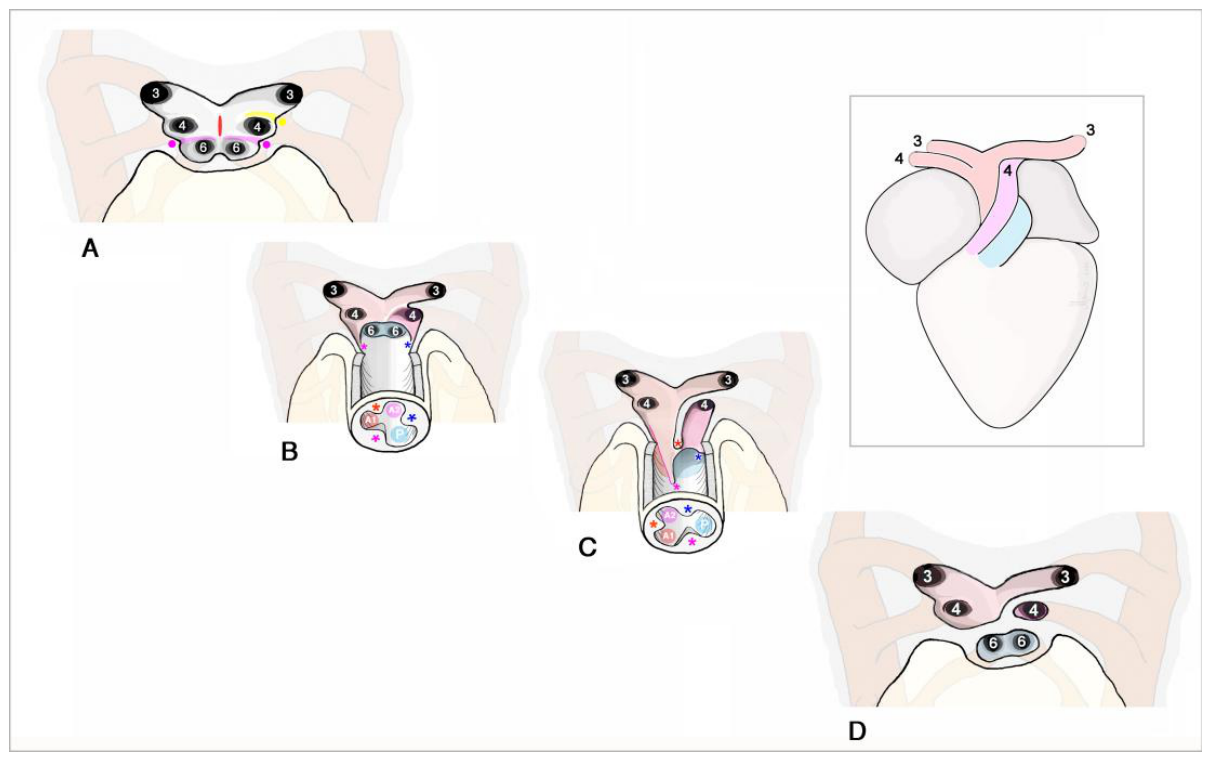
Figure 6.
This drawing depicts the arrangement of the great arteries that exit the heart of a chick. Abbreviations: Ao = aorta; LV = left ventricle; RV = right ventricle; other abbreviations as used before.
Figure 6.
This drawing depicts the arrangement of the great arteries that exit the heart of a chick. Abbreviations: Ao = aorta; LV = left ventricle; RV = right ventricle; other abbreviations as used before.
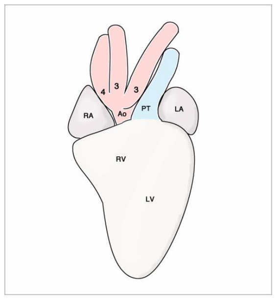
Figure 7.
These scanning electron microscopic images depict the development of the aortic sac septa in birds (chick embryos). Ventral views into the opened AoS and proximal portions of PhAAs 3 and 4. Flow dividers/septa are marked by the same color code as used before. Embryos from the 3rd (A), 4th (B), and 6th (C) day of incubation. (A). At the end of the 3rd incubation day, the AoS and PhAAs show a bilaterally symmetric patterning. (B). During the 4th incubation day, the AP-septum has formed by growth and merging of the paired aortico-pulmonary flow dividers, and growth of the left carotico-aortic septum has led to a bilaterally asymmetric branching pattern of the PhAAs. (C). The left carotico-aortic septum does not merge with the midline crest, so that chicks do not form an inter-aortic septum that grows upstream into the distal portion of the OFT. Scale bars = 40 mm. Abbreviations as used before.
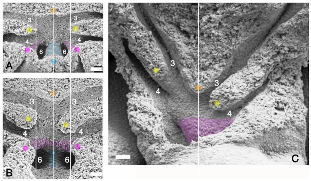
Figure 8.
These semi-schematic drawings depict the arrangement of the arterial channels and inter-arterial septa in the downstream portions of the truncus arteriosus of a frog heart as seen on sections perpendicular to the flow path. Blue lines A, B, C in the inset mark the positions of the sections shown in the main figure. Abbreviations: L-AC = left aortic channel; L-CC = left carotid channel; L-PC = left pulmocutaneous channel; R-AC = right aortic channel; R-CC = right carotid channel; R-PC = right pulmocutaneous channel.
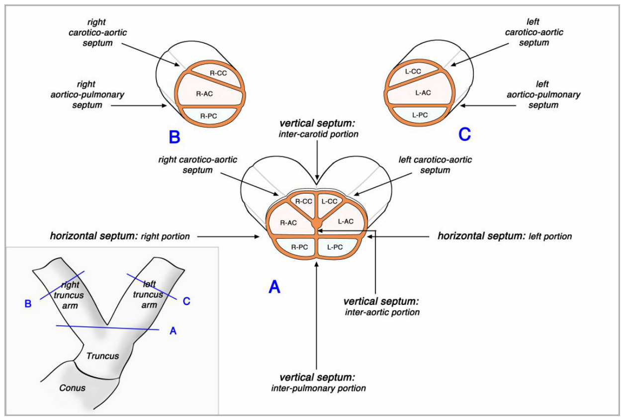
Figure 9.
These semi-schematic drawings depict the arrangement of the arterial channels and inter-arterial septa in the non-bifurcated portion of the truncus arteriosus of a frog heart. Blue lines A-D in the left inset mark the positions of the sections shown in the main figure. The system of truncus septa is most elaborated at the distal part of the non-bifurcated portion of the truncus (section D) from where several septa disappear toward the upstream end of the truncus (C-A). The truncus septa are arranged in a bilaterally asymmetric fashion. As a result, the truncus channels ramify in a pattern that corresponds to the arborisation pattern of the great arterial trunks that exits the heart of reptiles (right inset). Abbreviations as used before.
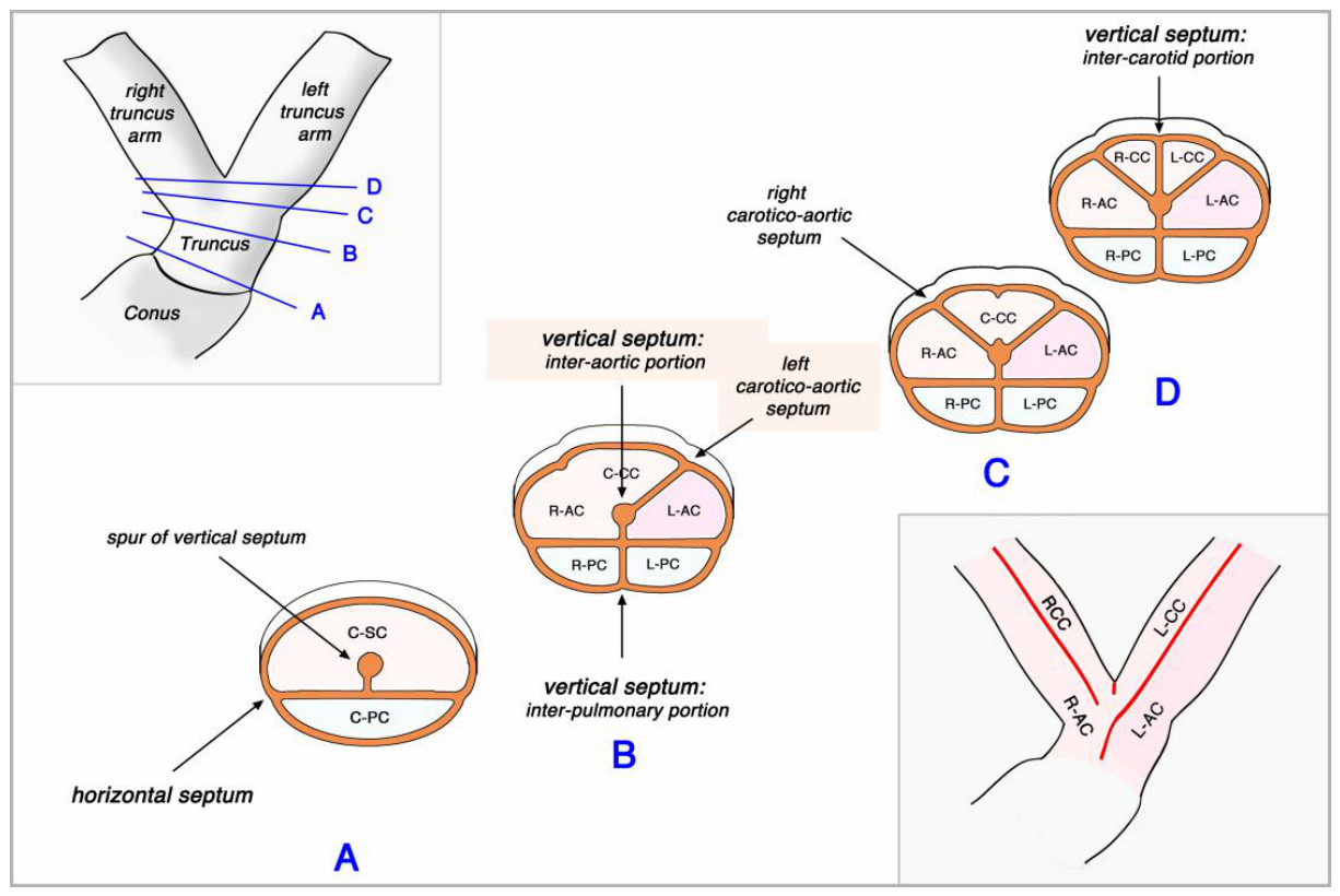
Figure 10.
This drawing depicts the arrangement of the arterial trunks in human tritruncal hearts. There are three arterial trunks that exit the heart: one pulmonary trunk (PT) that continues as the left pulmonary artery; one intermediate trunk (IT) that continues as the right pulmonary artery; and one aortic trunk. The blue wedge illustrates the hypothesis that the two pulmonary trunks were separated from each other by excessive up-stream growth of the inter-pulmonary septum of the AoS. Abbreviations as used before.
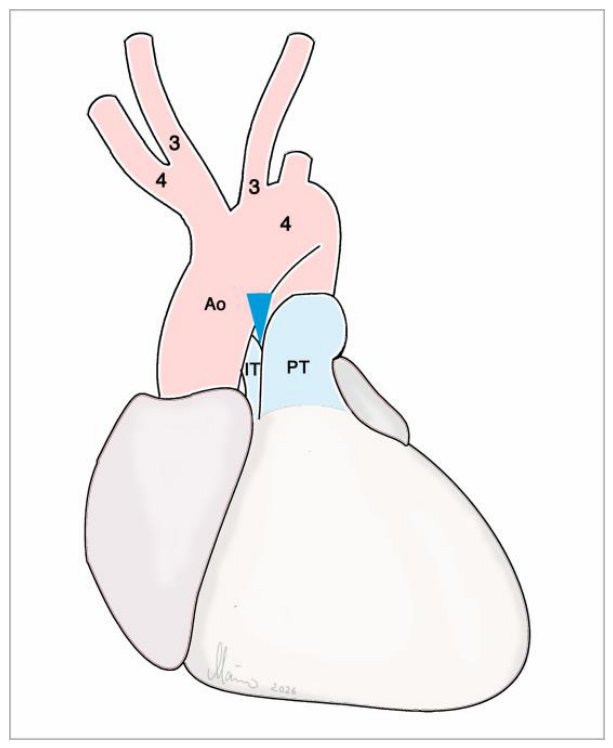
Figure 11.
These drawings depict the arrangement of the great arteries in human cases of anomalous origin of the left pulmonary artery from the proximal portion of the ascending aorta (A), and its postulated embryology due to malseptation of the AoS (B). Asterisks mark the margin of the opening between the aortic and pulmonary compartments possibly resulting from hemi-hypotrophy of the AP-septum. The difference in the spatial orientation of this opening between the embryonic and adult situation can be explained by ingrowth of the hemi-hypothrophic AP-septum into the OFT, which undergoes anticlockwise torsion. Excessive growth of the inter-pulmonary septum may have caused the separation (isolation) of the left pulmonary artery from the pulmonary trunk. Abbreviations: L-P = left pulmonary artery; R-P = right pulmonary artery, other abbreviations as used before.
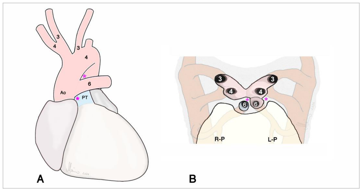
Figure 12.
These drawings depict the arrangement of the great arteries in human cases of anomalous origin of the left common carotid artery from the proximal portion of the pulmonary trunk (A), and its postulated embryology due to malseptation of the AoS (B). Asterisks mark the margin of the opening between the systemic and pulmonary compartments possibly resulting from hemi-hypotrophy of the AP-septum. The difference in the spatial orientation of this opening between the embryonic and adult situation can be explained by ingrowth of the hemi-hypothrophic AP-septum into the OFT, which undergoes anticlockwise torsion. Excessive growth of the inter-carotid septum may have caused the separation (isolation) of the left common carotid artery from the aorta. Abbreviations: L-Duct = left ductus arteriosus; R-Duct = right ductus arteriosus; other abbreviations as used before.
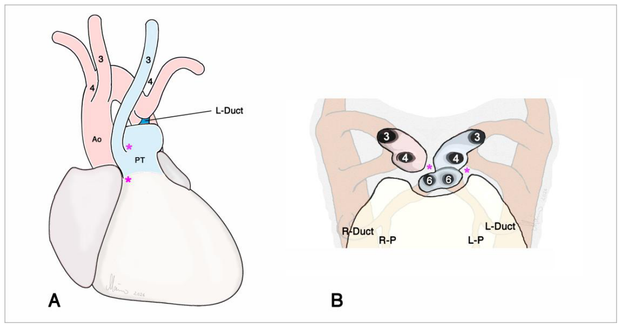
Figure 13.
These drawings depict the arrangement of the great arteries in human cases of anomalous origin of the brachiocephalic artery (A) and both common carotid arteries (B) from the proximal portion of the ascending aorta. The orange wedges illustrate how excessive up-stream growth of the inter-carotid septum may have led to splitting of the derivatives of the left and right halves of the AoS. The yellow sickle in B illustrates how excessive up-stream growth of both carotico-aortic flow dividers may have led to the formation of a common carotico-aortic septum that led to extensive separation of the carotid and aortic flow paths. Abbreviations as used before.
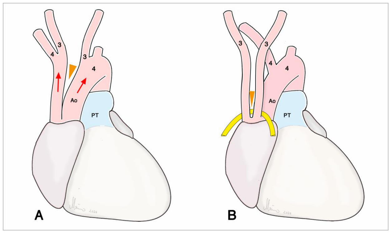
Figure 14.
This drawing depicts the arrangement of the great arteries in a human heart in which the right subclavian artery arises as the first branch of the left aortic arch. The yellow sickle illustrates how excessive up-stream growth of right carotico-aortic flow divider may have led to splitting of the right horn of the AoS, which normally forms the future brachiocephalic artery. Abbreviations as used before.
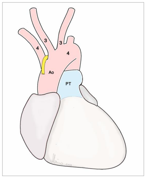
Figure 15.
This scheme illustrates the fact that the trifurcated brachiocephalic artery of humans bears strong resemblance to the right aorta of birds and, therefore, may result from a corresponding growth behavior of the AoS septa. Abbreviations as used before.
Figure 15.
This scheme illustrates the fact that the trifurcated brachiocephalic artery of humans bears strong resemblance to the right aorta of birds and, therefore, may result from a corresponding growth behavior of the AoS septa. Abbreviations as used before.
Disclaimer/Publisher’s Note: The statements, opinions and data contained in all publications are solely those of the individual author(s) and contributor(s) and not of MDPI and/or the editor(s). MDPI and/or the editor(s) disclaim responsibility for any injury to people or property resulting from any ideas, methods, instructions or products referred to in the content. |
© 2026 by the authors. Licensee MDPI, Basel, Switzerland. This article is an open access article distributed under the terms and conditions of the Creative Commons Attribution (CC BY) license (http://creativecommons.org/licenses/by/4.0/).
Copyright: This open access article is published under a Creative Commons CC BY 4.0 license, which permit the free download, distribution, and reuse, provided that the author and preprint are cited in any reuse.