

Submitted:
09 October 2026
Posted:
10 October 2026
You are already at the latest version
Abstract
Environmental contamination in veterinary healthcare facilities may contribute to the persistence and potential dissemination of multidrug-resistant (MDR) bacteria, thereby creating challenges for infection control and clinical management. A structured litera-ture search was conducted in PubMed, Scopus, and Google Scholar for studies published between 2010 and 2025 that investigated the bacterial contamination of small-animal veterinary environments, equipment, and/or veterinary personnel. A total of 96 studies met the inclusion criteria. Swabbing was the predominant sampling method, used ex-clusively in 56 studies (58.3%). Resistance-targeted selective and/or chromogenic culture media were used in 41 studies (42.7%). Among the clinical areas, intensive care units showed the highest proportion of studies reporting MDR bacteria (13/20, 65.0%). Re-garding individual sampling sites, MDR bacteria were reported in 27 of 44 studies sam-pling cages (61.4%). High proportions of MDR detection were also reported for scales (8/11, 72.7%), ultrasound equipment (10/15, 66.7%), sinks (13/21, 61.9%), and computer keyboards (19/32, 59.4%). Staphylococcus was the predominant reported genus (58 studies, 60.4%), while MDR S. pseudintermedius and S. aureus were reported in 27 (28.1%) and 25 (26.0%) studies, respectively. Among resistance determinants, mecA was the most fre-quently reported gene. These findings highlight the widespread detection of MDR bac-teria across veterinary clinical environments and support the need for systematic envi-ronmental surveillance and strengthened infection prevention measures.
Keywords:
evidence map
; veterinary hospitals
; environmental samples
; equipment
; veterinary personnel
; bacterial contamination
; multidrug resistance
1. Introduction
Antimicrobial resistance (AMR) represents a major global health threat [1]. A critical concern is the emergence and spread of antimicrobial-resistant organisms in healthcare-associated infections (HAIs), which constitute a persistent challenge in clinical settings and are associated with treatment failure, prolonged illness, and increased mortality in human medicine [2]. The persistence and dissemination of HAI-associated pathogens are influenced not only by infected patients but also by the healthcare environment, where contaminated surfaces, inanimate fomites, and transiently colonized healthcare personnel may act as interconnected reservoirs and potential transmission routes [3].
In veterinary medicine, multidrug-resistant (MDR) infections are increasingly reported worldwide, involving critical pathogens such as ESKAPE group organisms, methicilin-resistant staphylococci (MRS), and carbapenem-resistant Enterobacterales (CRE) [4,5]. Previous evidence has shown that pathogens associated with healthcare-associated infections and zoonotic transmission can be recovered from clinical cases, environmental surfaces, and equipment in veterinary hospitals [6]. However, available evidence on antimicrobial resistance and environmental contamination in veterinary facilities remains limited and heterogeneous, underscoring the need for improved awareness, early detection, and infection prevention practices among veterinary healthcare personnel [7].
Despite the growing evidence of bacterial contamination in veterinary healthcare environments, important knowledge gaps remain regarding the distribution of MDR bacteria across different clinical areas, environmental surfaces, equipment and veterinary personnel. In routine practice, attention is often focused on the diagnosis and treatment of individual MDR infections, while potential environmental sources of cross-contamination may require greater attention
To address these gaps, this evidence map aims to collect and synthesize the available evidence on bacterial isolation from veterinary healthcare environments, clinical equipment, and veterinary personnel. Specifically, this study aims to characterize the sampling approaches and culture methods used across studies, describe the bacterial taxa and MDR phenotypes identified, examine the distribution of MDR bacteria across functional clinical areas and individual sampling sites, and summarize the reported antimicrobial resistance determinants.
2. Materials and Methods
A structured literature search was conducted to identify studies investigating bacterial contamination and antimicrobial resistance in companion-animal veterinary clinical environments and among veterinary healthcare personnel. The search covered peer-reviewed publications from 2010 to 2025 to capture contemporary trends and relevant evidence. The databases searched were PubMed, Scopus, and Google Scholar, using combinations of terms related to veterinary hospitals and clinics, environmental contamination, veterinary healthcare personnel, bacterial isolation, antimicrobial resistance, and multidrug-resistant (MDR) bacteria.
During the screening process, particular emphasis was placed on studies involving sample collection followed by bacterial culture, isolation, and phenotypic and/or molecular identification from veterinary clinical settings. Eligible sources of bacterial samples included environmental surfaces and areas within veterinary hospitals or clinics, clinical equipment and fomites, and occupational specimens obtained from veterinary personnel, such as nasal or hand swabs and personal items (e.g., shoes, mobile phones, and footwear). Studies were included when bacterial recovery was based on microbiological culture, with subsequent phenotypic and/or molecular identification where reported. Conversely, studies without microbiological sampling or bacterial culture, as well as studies focusing exclusively on animal clinical cases, in vitro disinfection assays without in situ sampling, or narrative reviews, were excluded.
Retrieved records were initially screened at the title and abstract level, followed by detailed full-text assessment according to the eligibility criteria. Extracted information was synthesized into predefined categories: (i) study characteristics, including author(s), publication year, country, and facility type; (ii) sampling characteristics, including target site, sample type, and sampling technique; (iii) microbiological methods, including isolation media and bacterial taxa identified; (iv) antimicrobial resistance characteristics, including antimicrobial susceptibility testing methods, reported resistance, multidrug-resistant strains, and associated sample origins; and (v) resistance determinants, including detected antimicrobial resistance genes (ARGs).
Multidrug-resistant (MDR) status was assigned according to the definitions reported by the individual studies or, where analytical antimicrobial susceptibility profiles were available, evaluated against established criteria [8]. Furthermore, for the purposes of the present evidence synthesis, ESBL-producing organisms, methicillin-resistant staphylococci (MRS), and carbapenem-resistant Enterobacterales (CRE) were included within the MDR-related findings, when reported by the original studies, given their resistance profiles and clinical significance.
Particular attention was given to the origin of the samples, which were categorized according to the clinical area, environmental surface, medical or diagnostic equipment, or veterinary personnel sampled. When reported, the specific sample source associated with MDR bacterial recovery was also recorded. Information was extracted as reported by the original studies, while sampling locations were standardized into functional clinical areas and individual sampling sites to facilitate comparisons across studies. Descriptive frequencies were calculated at the study level; therefore, the percentages represent the proportion of the included studies reporting a given bacterial taxon, resistance phenotype, or sampling location and do not represent the proportion of individual isolates.
3. Results
3.1. Included Studies and Generic Characteristics
The database search across PubMed (180 records), Scopus (1,094 records), and Google Scholar (6,916 records) initially yielded 8,190 records published between 2010 and 2025. Following the removal of duplicate records and preliminary screening of titles and abstracts against the predefined eligibility criteria, 382 records were retained for full-text assessment. Full-text articles were subsequently evaluated for eligibility based on the predefined inclusion criteria, with particular emphasis on studies reporting microbiological sampling and culture-based bacterial isolation from small-animal veterinary clinical environments, equipment, or fomites, and/or veterinary healthcare personnel.
Following the full-text assessment, 96 peer-reviewed studies met the eligibility criteria and were included in the evidence map for data extraction and qualitative evidence synthesis. A study selection flow diagram informed by the PRISMA 2020 framework was used to summarize the identification, screening, eligibility assessment, and inclusion of studies (Figure 1).
The detailed characteristics of each included study, encompassing the year, country, origin of samples, and sampling technique, are summarized in Table 1.
| Year | Country | Sample Origin | Sampling Technique | Reference |
| 2010 | Japan | Personnel, Environment/equipment | Swabs | [10] |
| 2010 | UK | Environment/equipment | Dipslides/contact agar | [11] |
| 2010 | South Korea | Personnel, Environment/equipment | Swabs | [12] |
| 2010 | UK | Personnel | Swabs | [13] |
| 2010 | USA | Personnel | Swabs | [14] |
| 2010 | Canada | Environment/equipment | Electrostatic cloths/gloved hands | [15] |
| 2011 | The Netherlands | Personnel, Environment/equipment | Swabs/gauze pads | [16] |
| 2011 | USA | Environment/equipment | Swabs/electrostatic cloths | [17] |
| 2011 | South Korea | Personnel, Environment/equipment | Swabs | [18] |
| 2012 | Malaysia | Personnel, Environment/equipment | Swabs | [19] |
| 2012 | USA | Environment/equipment | Swabs | [20] |
| 2012 | USA | Environment/equipment | Swabs | [21] |
| 2012 | Canada | Personnel | Electrostatic cloths | [22] |
| 2013 | USA | Environment/equipment | Swabs | [23] |
| 2013 | USA | Environment/equipment | Impaction air sampling | [24] |
| 2013 | USA | Environment/equipment | Impaction air sampling | [25] |
| 2013 | Canada | Personnel | Electrostatic cloths | [26] |
| 2013 | USA | Environment/equipment | Swabs/electrostatic cloths | [27] |
| 2013 | Germany | Personnel, Environment/equipment | Swabs | [28] |
| 2014 | South Korea | Personnel, Environment/equipment | Swabs | [29] |
| 2014 | Finland | Environment/equipment | Swabs | [30] |
| 2014 | USA | Environment/equipment | Sponges | [31] |
| 2014 | Zambia | Environment/equipment | Swabs | [32] |
| 2015 | Colombia | Environment/equipment | Swabs | [33] |
| 2016 | Israel | Environment/equipment | Swabs | [34] |
| 2016 | Malaysia | Environment/equipment | Swabs | [35] |
| 2016 | UK | Environment/equipment | Swabs | [36] |
| 2016 | UK | Environment/equipment | Boot socks/wipe cloths | [37] |
| 2017 | Thailand | Personnel, Environment/equipment | Swabs | [38] |
| 2017 | Costa Rica | Environment/equipment | Swabs/electrostatic cloths | [39] |
| 2017 | Mexico | Personnel, Environment/equipment | Impaction air sampling/swabs | [40] |
| 2018 | UK | Personnel | Contact agar | [41] |
| 2018 | Germany | Personnel, Environment/equipment | Swabs | [42] |
| 2018 | USA | Environment/equipment | Electrostatic cloths | [43] |
| 2018 | Japan | Personnel, Environment/equipment | Swabs | [44] |
| 2018 | Australia | Personnel, Environment/equipment | Swabs | [45] |
| 2018 | South Korea | Personnel, Environment/equipment | Swabs/contact agar | [46] |
| 2019 | USA | Environment/equipment | Swabs | [47] |
| 2020 | China | Personnel, Environment/equipment | Swabs/settle plates | [48] |
| 2020 | USA | Environment/equipment | Swabs | [49] |
| 2020 | France | Environment/equipment | Swabs | [50] |
| 2020 | USA | Environment/equipment | Electrostatic cloths | [51] |
| 2020 | Switzerland | Personnel, Environment/equipment | Swabs | [52] |
| 2020 | Brazil | Environment/equipment | Swabs | [53] |
| 2020 | Thailand | Personnel, Environment/equipment | Swabs | [54] |
| 2021 | Nigeria | Environment/equipment | Swabs | [55] |
| 2021 | Thailand | Personnel, Environment/equipment | Swabs | [56] |
| 2021 | Italy | Environment/equipment | Sponges | [57] |
| 2021 | USA | Environment/equipment | Electrostatic cloths | [58] |
| 2021 | Brazil | Environment/equipment | Swabs/settle plates | [59] |
| 2021 | Egypt | Personnel, Environment/equipment | Swabs | [60] |
| 2021 | Australia | Environment/equipment | Electrostatic cloths | [61] |
| 2021 | Hungary | Personnel, Environment/equipment | Swabs | [62] |
| 2021 | Thailand | Personnel, Environment/equipment | Swabs | [63] |
| 2021 | Slovenia | Environment/equipment | Swabs | [64] |
| 2021 | Switzerland | Personnel, Environment/equipment | Swabs | [65] |
| 2021 | Brazil | Environment/equipment | Swabs | [66] |
| 2021 | Portugal | Environment/equipment | Swabs | [67] |
| 2021 | UK | Personnel | Swabs | [68] |
| 2022 | Chile | Personnel, Environment/equipment | Swabs | [69] |
| 2022 | Brazil | Environment/equipment | Swabs/membrane filtration | [70] |
| 2022 | Thailand | Environment/equipment | Swabs | [71] |
| 2022 | Brazil | Environment/equipment | Swabs/settle plates | [72] |
| 2022 | France | Environment/equipment | Swabs | [73] |
| 2022 | Germany | Environment/equipment | Not specified | [74] |
| 2022 | China | Environment/equipment | Not specified | [75] |
| 2022 | Brazil | Personnel, Environment/equipment | Swabs/settle plates | [76] |
| 2022 | The Netherlands | Environment/equipment | Not specified | [77] |
| 2022 | USA | Environment/equipment | Electrostatic cloths | [78] |
| 2022 | Switzerland | Personnel, Environment/equipment | Swabs | [79] |
| 2022 | Thailand | Environment/equipment | Swabs | [80] |
| 2023 | Brazil | Personnel, Environment/equipment | Swabs | [81] |
| 2023 | Ireland | Personnel, Environment/equipment | Swabs | [82] |
| 2023 | UK | Environment/equipment | Electrostatic cloths | [4] |
| 2024 | China | Personnel, Environment/equipment | Swabs | [83] |
| 2024 | Portugal | Personnel, Environment/equipment | Swabs/contact agar | [84] |
| 2024 | China | Personnel, Environment/equipment | Swabs | [85] |
| 2024 | USA | Environment/equipment | Sponges | [86] |
| 2024 | Portugal | Environment/equipment | Swabs | [87] |
| 2024 | Spain | Environment/equipment | Swabs | [88] |
| 2024 | Spain | Personnel, Environment/equipment | Swabs | [89] |
| 2024 | Spain | Personnel, Environment/equipment | Swabs/sponges | [90] |
| 2024 | South Africa | Personnel | Glove juice technique | [91] |
| 2024 | Brazil | Environment/equipment | Swabs | [92] |
| 2024 | China | Environment/equipment | Impaction air sampling | [93] |
| 2025 | Brazil | Environment/equipment | Swabs | [94] |
| 2025 | Brazil | Environment/equipment | Swabs | [95] |
| 2025 | Portugal | Personnel, Environment/equipment | Swabs/contact agar | [96] |
| 2025 | Brazil | Personnel, Environment/equipment | Swabs | [97] |
| 2025 | USA | Environment/equipment | Sponges | [98] |
| 2025 | UK | Environment/equipment | Swabs/sponges | [99] |
| 2025 | USA | Environment/equipment | Sponges | [100] |
| 2025 | Peru | Environment/equipment | Swabs | [101] |
| 2025 | Italy | Environment/equipment | Swabs | [102] |
| 2025 | Bangladesh | Environment/equipment | Swabs | [103] |
| 2025 | Italy | Personnel, Environment/equipment | Swabs/sponges | [104] |
A total of 96 studies published between 2010 and 2025 were included in the evidence set. Their temporal distribution indicates a progressive increase in relevant research during the later years of the review period. In particular, 51 studies (53.1%) were published between 2021 and 2025. This indicates an increasing research interest in bacterial contamination and antimicrobial resistance within veterinary clinical settings in recent years.
Regarding sample origin, most studies investigated environmental surfaces and/or veterinary clinical equipment. Specifically, 55 studies (57.3%) sampled environmental or equipment-associated sources exclusively, whereas 34 studies (35.4%) included both veterinary personnel and environmental/equipment-associated sources. Only seven studies (7.3%) focused exclusively on veterinary personnel-associated samples.
A wide range of sampling techniques have been reported. Swabbing was by far the most frequently used technique, being reported as the sole sampling approach in 56 studies (58.3%). Other studies combined swabbing with additional sampling approaches, including settle plates (4 studies), electrostatic cloths (3 studies), contact agar (3 studies), sponges (3 studies), impaction air sampling (1 study), and membrane filtration (1 study).
Electrostatic cloths were used as a standalone sampling technique in eight studies (8.3%), whereas sponges were used independently in five studies (5.2%). Active air sampling using impaction-based methods was reported in three studies (3.1%), with one additional study combining impaction air sampling with surface swabbing. Contact-based approaches were also identified, including dipslides and contact agar sampling. Less frequently used approaches included boot socks/wipe cloths, gauze pads, and the glove juice technique, each representing a limited proportion of the included evidence.
3.2. Geographic Distribution of Included Studies
The geographic distribution of the selected studies (n = 96) included 33 countries across six continents, reflecting the global interest in antimicrobial resistance in veterinary clinical settings (Figure 2).
Most studies originated from North America, Europe, and South America. The United States contributed the highest number of publications (n = 17, 17.7%), followed by Brazil (n = 11, 11.5%), the United Kingdom (n = 8, 8.3%), and Thailand (n = 6, 6.3%). Additional contributions were reported from China (n = 5), Portugal (n = 4), and South Korea (n = 4); Canada, Germany, Italy, Spain, and Switzerland each contributed three studies. The remaining 21 countries contributed one or two studies each, indicating a broad but uneven geographical distribution of available evidence.
3.3. Sample Characteristics and Veterinary Facility Settings
The number of samples collected varied substantially across studies. Across articles with available data, the mean number of samples per study was 282.8 (95% CI, 127.6–438.0), with a median of 150 and a range of 4–6,400 samples.
When sample numbers were considered by sample origin, environmental and equipment sampling was more frequently reported than personnel sampling was. Among studies with numerical sample counts, the mean number of environmental samples per study was 160.1 (95% CI, 102.2–218.1), with a median of 95 samples. For personnel-associated sampling, numerical sample counts yielded a mean of 133.2 samples per study (95% CI, 65.5–200.8) and a median of 77.
The included studies were conducted in various veterinary clinical settings. An academic or teaching veterinary facility was identified in 55 of the 96 studies (57.3%). Most studies were conducted within a single veterinary facility (70/96, 72.9%), whereas 23 studies (24.0%) included multiple facilities. These multi-facility studies involved combinations of teaching hospitals, private veterinary hospitals or clinics, referral hospitals, and academic and private facilities.
3.4. Culture Media and Antimicrobial Susceptibility Testing
Substantial heterogeneity was observed in the culture media used for bacterial isolation across the included studies. Selective culture approaches specifically targeting antimicrobial resistance phenotypes or resistance-associated bacterial groups were reported in 41 studies (42.7%). These approaches (Table 2) included selective and/or chromogenic media targeting methicillin-resistant staphylococci, extended-spectrum β-lactamase (ESBL)-producing organisms, carbapenem-resistant organisms etc. In several studies, selective enrichment steps were combined with resistance-specific selective or chromogenic agar, indicating a targeted strategy for the recovery of resistant bacterial populations.
In a group of 39 studies (40.6%), selective and/or differential culture media were used, without an explicit focus on a specific antimicrobial-resistance phenotype. These included media such as MacConkey agar, eosin methylene blue agar, Baird–Parker agar, mannitol salt agar, Slanetz–Bartley agar, and other selective or differential media targeting particular bacterial groups. These approaches were primarily intended to facilitate the recovery and differentiation of specific bacterial taxa rather than directly screen for a defined resistance phenotype.
In contrast, only 11 studies (11.5%) relied solely on general-purpose or non-selective culture media, including blood agar, sheep blood agar, horse blood agar, brain heart infusion agar, Columbia agar, and Plate Count Agar. Finally, in five studies (5.2%), culture media were either not reported or no culture medium was specified.
Overall, the culture strategies ranged from broad bacterial recovery using general-purpose media to highly targeted selective screening for specific antimicrobial resistance phenotypes.
Antimicrobial susceptibility testing (AST) also demonstrated methodological variations. Disk diffusion was the most frequently reported individual approach, being used in 41 studies (42.7%). MIC-based methods were reported in 31 studies (32.3%), including automated or semi-automated systems such as VITEK and MicroScan, Sensititre-based testing, broth microdilution, agar dilution, and other microdilution approaches. A further 11 studies (11.5%) employed combined susceptibility-testing strategies, incorporating disk diffusion together with MIC-based methods or other complementary approaches, such as E-test or resistance-specific screening procedures. Finally, in 13 studies (13.5%), the antimicrobial susceptibility testing method was not specified.
Thus, although disk diffusion is the most common AST approach, MIC-based methods are also widely used, either alone or in combination with other susceptibility-testing techniques.
Overall, the evidence base showed considerable variation in both culture media and AST methodology, reflecting differences in the approaches used to recover, identify, and characterize antimicrobial-resistant bacteria across studies.
3.5. Bacterial Findings and Distribution
A broad range of bacterial taxa were reported in veterinary clinical environments and personnel-associated samples, across the included studies. Detailed data are presented in Table 3. Staphylococcus spp. were the most frequently reported genus, identified in 58 studies (60.4%), followed by Escherichia coli (32, 33.3%), Pseudomonas spp. (20, 20.8%), Klebsiella spp. (18, 18.8%), Acinetobacter spp. (17, 17.7%), Enterococcus spp. (16, 16.7%), and Enterobacter spp. (14, 14.6%). Less frequently reported taxa included Bacillus spp. and Serratia spp. (7 studies each), Citrobacter spp. (6 studies), and Stenotrophomonas, Salmonella, and Micrococcus spp. (5 studies each) (Table 3).
Beyond the predominant taxa, diverse opportunistic and environmental bacteria were recovered across a limited number of studies (n ≤ 3). These included various Gram-negative bacilli, such as Hafnia alvei, Leclercia adecarboxylata, Moraxella spp., Elizabethkingia meningoseptica, Alcaligenes spp., Raoultella spp., and other sporadically reported genera (e.g., Pasteurella, Bordetella, Aeromonas, Achromobacter, and Providencia). Less frequently isolated Gram-positive organisms included Corynebacterium spp., Rothia spp., Streptococcus spp. (including S. canis and S. agalactiae), Paenibacillus spp., Aerococcus spp., and Clostridioides difficile.
As mentioned above, species of the Staphylococcus genus were the most frequently reported. The detailed distribution of staphylococcal species is presented in Table 4.
Among the 58 studies reporting Staphylococcus spp., S. aureus was the most frequently identified species (35 studies, 36.5%), followed by S. pseudintermedius (29 studies, 30.2%) and S. epidermidis (12 studies, 12.5%). Other species were reported less frequently than the above-mentioned species. Overall, the findings demonstrate a marked predominance of staphylococci within the evidence base, with both coagulase-positive and coagulase-negative taxa represented.
3.6. Multidrug-Resistant Bacteria and Antimicrobial Resistance Determinants Identified
The number of MDR-positive samples varied substantially across studies and between environmental and personnel-associated sampling. Among the studies with numerically reported data, the mean number of MDR-positive environmental samples was 23.1 per study (95% CI, 13.3–32.9), based on 64 studies with available numerical counts. For personnel-associated samples, the corresponding mean was 14.0 MDR-positive samples per study (95% CI, 6.1–21.9), based on 31 studies with numerical data. When the MDR-positive environmental and personnel counts were combined within the same study, the mean total number of MDR-positive samples was 28.9 per study (95% CI, 13.9–43.9) among the 26 studies for which numerical counts were available for both sample sources.
Numerous taxa of multidrug-resistant (MDR) bacteria were reported across the included studies. For the study-level synthesis, species nomenclature was standardized and resistance phenotypes were grouped with the corresponding bacterial species when species-level identification was available. S. pseudintermedius was the most frequently reported MDR species, identified in 27 studies (28.1%), followed by S. aureus in 25 studies (26.0%) and E. coli in 16 studies (16.7%). Other frequently reported MDR taxa included S. epidermidis (12 studies, 12.5%), S. haemolyticus (7, 7.3%), E. faecium and K. pneumoniae (5 studies each, 5.2%), S. schleiferi, P. aeruginosa and Acinetobacter baumannii (4 studies each, 4.2%), Stenotrophomonas maltophilia and S. hominis (3 studies each, 3.1%) (Table 5).
Several studies have reported multidrug resistance solely at the genus level (e.g., Enterococcus spp., Acinetobacter spp.) or as broad resistance phenotypes without species determination (e.g., VRE, CRE/CPE, ESBL, or unspecified MRS); consequently, these were excluded from this specific species-level synthesis. This may result in an underestimation of the true occurrence of certain MDR pathogens.
Molecular screening for antimicrobial resistance genes (ARGs) was conducted in 64 of the 96 studies (66.7%). The detailed data regarding the relevant findings are presented in Table 6.
Reflecting the high frequency of methicillin-resistant staphylococci, the mecA gene was the single most prevalent resistance determinant, detected across 37 studies (57.8% of studies with molecular investigation; 38.5% overall). Other Gram-positive resistance determinants included the penicillinase gene blaZ (n = 10), macrolide-lincosamide-streptogramin B determinants (erm genes; n = 6), and vancomycin resistance gene vanA (n = 2).
Among Gram-negative isolates, beta-lactamase genes were widespread. ESBL genes of the blaCTX-M lineage were identified in 14 studies (21.9% of tested studies), followed by blaTEM (n = 11) and blaSHV (n = 7). Plasmid-mediated and chromosomal AmpC determinants (blaCMY, blaDHA, blaEBC, blaCIT, and AmpC) were detected in 10 studies. Acquired carbapenemases or intrinsic/lineage-specific variants of blaOXA gene were identified in 13 studies. Other carbapenemase genes of critical public health concern were identified in multiple facilities, including variants of the metallo-beta-lactamase blaNDM (n = 7), and sporadically blaKPC, blaVIM, and blaIMP (n = 2).
Resistance determinants targeting other key antimicrobials included diverse tet genes conferring tetracycline resistance (n = 12), aminoglycoside-modifying enzyme genes (n = 9), plasmid-mediated quinolone resistance and mutation markers (n = 6), and sulfonamide/trimethoprim resistance determinants (sul, dfr; n = 6). Additionally, four recent investigations utilized sequencing technologies or shotgun metagenomics, demonstrating extensive resistome diversity in veterinary settings.
3.7. Distribution of MDR Bacteria Across Functional Clinical Areas
Hospital areas were grouped into standardized categories (Figure 3) to allow comparison across studies. The analysis focused on areas examined in at least 20 studies. Various terms were merged into primary sectors: hospitalization wards included canine and feline wards, kennels, and recovery rooms; surgery preparation included pre-op rooms and anesthesia induction areas; and examination rooms included consultation rooms and screening offices.
The proportion of studies reporting MDR detection varied across functional clinical areas. MDR bacteria were reported in 13 of 20 studies (65.0%) sampling ICU/critical-care/emergency areas, while none of these studies explicitly reported absence of MDR detection. Surgery preparation rooms (55.0%, 11/20) and hospitalization wards (52.8%, 19/36) also showed high MDR detection rates across studies. These areas may represent settings with frequent patient contact and intensive clinical activity; however, differences in sampling strategies and reporting across studies should also be considered.
In contrast, operating and examination rooms had fewer studies in which MDR was detected (36.4% and 31.4%, respectively) and higher rates of studies with no MDR bacteria reported (24.2% and 22.9%, respectively).
3.8. Distribution of MDR Bacteria Across Environmental Surfaces and Sampling Sites
To allow for comparisons across studies, individual sampling sites were considered separately. The analysis focused on sampling points examined in more than 10 studies. Sampling locations included environmental surfaces, clinical tables and equipment, facility-associated objects, and specimens collected from veterinary healthcare personnel. For each sampling point, studies were categorized according to whether MDR bacteria were detected, whether the MDR outcome was not specified, or whether no MDR bacteria were reported (Figure 4).
The proportion of studies reporting MDR detection varied substantially across sampling sites, with several high-touch surfaces and diagnostic tools exhibiting high frequencies. Among patient-housing facilities, animal cages were the most commonly investigated site, with MDR pathogens detected in 61.4% (27/44) of the studies. High detection rates were also identified on veterinary scales (72.7%, 8/11) and ultrasound equipment/tables (66.7%, 10/15), followed by examination tables (48.6%, 17/35) and surgical tables (47.6%, 10/21). Handheld clinical instruments in direct contact with patients also demonstrated notable MDR detection rates across studies, including clippers (54.5%, 6/11), thermometers (54.5%, 6/11), and stethoscopes (40.0%, 8/20).
Moist clinical environments and handwashing facilities were prominent reservoirs, with MDR bacteria reported in 61.9% (13/21) of studies evaluating sinks and 45.0% (9/20) of studies evaluating faucets/taps. Communal touchpoints and non-clinical fomites also yielded frequent contamination; computer keyboards showed confirmed MDR recovery in 59.4% (19/32) of the studies, followed by door handles (42.9%, 15/35), computer mice (42.9%, 6/14), desk/counter surfaces (38.5%, 10/26), and telephones (38.1%, 8/21). Floors yielded MDR isolates in 54.3% (19/35) of investigations, although an additional 37.1% (13/35) reported facility-level MDR without specifying floor-level counts. Conversely, lower proportions of confirmed MDR detection and higher rates of negative studies were observed for X-ray units (27.3% MDR vs. 63.6% no MDR), light switches (28.6% MDR vs. 57.1% no MDR), and anesthesia machines/rebreathing circuits (40.0% MDR vs. 53.3% no MDR).
Surveillance of veterinary staff revealed frequent reports of MDR isolation in the relevant studies. Nasal carriage among personnel yielded the highest proportion of confirmed MDR detection across all evaluated sites (81.8%, 18/22), primarily driven by methicillin-resistant staphylococci. Similarly, isolation of MDR strains from personnel hands was reported in 52.0% (13/25) of studies, highlighting the dual role of veterinary healthcare workers as both potential reservoirs and mechanical vectors in hospital transmission networks.
4. Discussion
This review synthesizes data from 96 studies published between 2010 and 2025 to map bacterial contamination and resistance patterns across companion-animal veterinary facilities. Despite substantial methodological heterogeneity, particularly in sampling approaches, selective culture protocols, and reporting detail, the synthesized findings provide consistent indications that multidrug-resistant bacteria circulate within veterinary clinical environments. Repeated recovery from both animal-contact surfaces, clinical equipment, and high-touch administrative fomites has been documented, while personnel-associated samples are regularly correlated with MDR isolates. Together, these patterns point to potential transmission pathways linking facilities, equipment, staff, and animal patients, highlighting the need for long-term surveillance protocols and structured risk management in veterinary practice.
Although studies originate from multiple countries and regions, the evidence remains clustered within a few countries and is predominantly derived from veterinary teaching hospitals. While these academic centers provide controlled settings and detailed protocols, they do not necessarily reflect everyday primary care clinics, where variations in infrastructure, patient flow, cleaning routines, and facility management are substantial.
Staphylococci were the most frequently reported bacterial group in both the overall bacterial findings and the MDR findings of the included studies. This predominance is biologically anticipated, given the well-established ability of methicillin-resistant staphylococci (MRS), particularly methicillin-resistant Staphylococcus aureus (MRSA), to persist in healthcare environments and their recognized association with healthcare-associated infections and colonization of healthcare personnel [105]. Furthermore, MRSA isolates recovered from dogs and cats have been reported to be indistinguishable from strains associated with human infections and healthcare settings, highlighting the potential relevance of veterinary environments within the broader epidemiology of methicillin resistance [106]. Other staphylococcal species, particularly S. pseudintermedius and S. schleiferi, are important pathogens in small-animal practice, including dermatological and other clinical infections, while also being recognized as colonizing organisms [107]. Coagulase-negative staphylococci, including S. epidermidis and S. haemolyticus, were also reported among the recovered and MDR bacteria, although less frequently [108].
The frequent reporting of staphylococci was also accompanied by molecular detection of antimicrobial resistance determinants. Among them, mecA was the most frequently identified, consistent with the predominance of methicillin-resistant phenotypes among the MDR staphylococci reported in the included studies. Other resistance determinants were regularly reported, including blaZ and erm genes, reflecting resistance to β-lactam and macrolide-lincosamide-streptogramin antimicrobial classes, respectively.
However, the predominance of staphylococci should be interpreted with caution, considering the sampling and culture methods used in the included studies. A significant proportion of studies specifically targeted staphylococcal species or methicillin-resistant phenotypes using selective or resistance-targeted media [10,11,14,17,22,25,26,27,28,30,32,38,39,42,44,45,48,51,52,68,69,71,78,81,92,95,104]. These targeted approaches may have increased the likelihood of recovering and reporting these species, particularly methicillin-resistant strains, and may have contributed to their apparent predominance.
In addition to staphylococci, several other opportunistic bacteria, including E. coli, Klebsiella, Pseudomonas, Acinetobacter, and Enterococcus species, were detected across facilities, illustrating the broad range of contaminants present. Worryingly, these isolates frequently exhibited high-priority resistance phenotypes, confirmed by the detection of the relevant ARGs, most notably ESBL production [4,36,37,41,65,66,70,73,79,83,85,102], carbapenem resistance [52,66,79,85,98,100,102,103,109], and vancomycin-resistant enterococci (VRE) [46,66]. The clinical relevance of these findings is further highlighted by the increasing recognition of these bacterial groups as causes of difficult-to-treat infections in companion animals, particularly when associated with antimicrobial resistance [110,111,112].
The frequent isolation of MDR pathogens from the ICU and preoperative environments mirrors the findings from human hospitals, where a significant proportion of critically ill patients contract healthcare-associated infections during their ICU stay [113,114]. This issue is particularly critical in veterinary teaching hospitals and referral centers that admit the most severe clinical cases. Together, suppressed patient immunity and heavy antibiotic use make hospital ICUs potential hotspots for the persistence and transmission of resistant bacteria.
Beyond clinical areas, several individual sampling sites showed frequent reporting of MDR bacteria, particularly sites that are repeatedly exposed to direct animal contact, frequent handling by veterinary personnel, or both. Cages, scales, and examination tables represent surfaces with frequent direct contact with animals and may therefore become contaminated through the spread of bacteria from colonized or infected animals. In contrast, frequently touched surfaces such as door handles and computer keyboards are primarily associated with contact by veterinary personnel and may provide opportunities for the transfer of microorganisms between hands and the surrounding environment. Other items, including stethoscopes, thermometers, and selected clinical equipment such as ultrasound devices, may act at the interface between animals and personnel, as they are repeatedly handled by staff while also coming into direct or close contact with animals. The frequent detection of MDR bacteria at these sites highlights the importance of considering both animal-contact and personnel-contact surfaces when assessing potential environmental reservoirs within veterinary healthcare facilities. Similar to human healthcare settings [115], the potential dissemination of MDR bacteria in veterinary facilities is likely to involve interconnected pathways linking animal patients, contaminated environmental surfaces and equipment, and veterinary personnel, although the relative contribution of each pathway remains to be determined.
Sinks and faucets/taps also showed frequent reporting of MDR bacteria in the included studies. This finding is consistent with the recognized challenges that healthcare plumbing systems pose for infection prevention and control (IPC). Wet surfaces within sinks and drains provide a solid–liquid interface that can favor biofilm formation, which has been associated with the persistence and dissemination of healthcare-associated pathogens and, in some settings, with outbreaks [116]. In addition, organic material and waste disposed of into sinks and drains may provide nutrients that support bacterial persistence within the plumbing environment [117]. The connection between clinical plumbing systems and the wider environment may raise broader One Health considerations.
Finally, several studies have reported the isolation of MDR bacteria from samples collected from veterinary personnel. These findings indicate that staff working in clinical environments can harbor resistant strains, raising valid concerns about their potential role as carriers. Beyond the risk of exposure for hospitalized animals, possible public health implications are highlighted, particularly the potential for these bacteria to reach personnel’s household contacts and the broader community. Comparable concerns are frequently discussed in human healthcare, where more extensive data describe potential exposure risks for medical personnel [118,119]. In veterinary medicine, however, the available evidence is much more limited and less conclusive, leaving the actual impact of occupational carriage unclear.
This study has certain limitations that should be considered when interpreting its findings. First, the included studies were predominantly observational and descriptive, limiting the extent to which causal relationships or transmission pathways could be evaluated. Second, substantial methodological heterogeneity was observed across studies, particularly with respect to sampling methods, culture media, antimicrobial susceptibility testing (AST) methods, and the definitions and reporting of multidrug resistance (MDR). In addition, a considerable number of studies used selective or resistance-targeted culture media to investigate specific bacterial taxa and resistance phenotypes. Such targeted approaches may have influenced the spectrum of bacteria recovered and, therefore, may have affected the distribution of bacterial and MDR findings observed in the present evidence map.
Because of this methodological heterogeneity, the analysis was primarily conducted at the study level rather than at the individual sample level. Direct comparison of sample-level frequencies across studies would have been difficult to interpret reliably. Finally, some degree of terminology harmonization and grouping was necessary to allow comparison across studies. Different studies often used different terms for similar clinical areas, equipment, or sampling sites, while in some cases, different parts of the same area or piece of equipment were sampled separately. Although these categories were standardized to facilitate synthesis, this process may have resulted in some loss of site-specific detail and should therefore be considered when interpreting the spatial distribution of MDR detection.
However, this evidence map provides a broad synthesis of the available information on bacterial contamination and antimicrobial resistance in small-animal veterinary healthcare environments, incorporating environmental surfaces, clinical equipment and fomites, and veterinary personnel-associated samples. It also provides an overview of the methodological approaches currently used for bacterial recovery and antimicrobial susceptibility assessment, the main bacterial pathogens reported, and their associated antimicrobial profiles and determinants. Importantly, the mapping of MDR detection across functional clinical areas and individual sampling sites provides an overview of locations that are more frequently associated with the reporting of MDR phenotypes across the available literature. Collectively, these findings provide a useful evidence base for understanding how bacterial contamination and antimicrobial resistance have been investigated in small-animal veterinary healthcare settings and may support the development of more targeted environmental surveillance strategies, infection prevention practices, and standardized approaches for future investigations.
Future research should focus on developing standardized environmental sampling protocols that are practical and compatible with the routine workflows of various veterinary specialties. Greater consistency in the selection of sampling sites, sampling frequency, microbiological methods, antimicrobial susceptibility testing, and reporting would improve comparability between studies and provide a more reliable basis for monitoring MDR bacteria in veterinary healthcare settings. Concurrently, the application of molecular approaches, including whole-genome sequencing, where appropriate, could provide further characterization of resistance determinants and help determine the relatedness of isolates recovered from animals, environmental sites, equipment, and veterinary personnel. These approaches may contribute to a better understanding of potential transmission pathways in veterinary facilities. Longitudinal studies would further help determine the persistence and recurrence of MDR bacteria in specific clinical areas and sampling sites over time.
Future research could move beyond the description of bacterial recovery and resistance profiles and assess their clinical and epidemiological significance. This could include evaluating the frequency and severity of infections associated with the recovered pathogens, the clinical relevance of their resistance determinants, and the potential risk associated with particular sampling sites and exposed populations. Integrating this data could ultimately support more targeted risk assessments and help identify priorities for environmental surveillance and infection prevention in companion animal veterinary facilities.
5. Conclusions
Overall, the available evidence indicates that MDR bacteria are consistently reported across a wide range of small animal veterinary clinical environments. Certain functional areas, high-contact surfaces, frequently used clinical equipment, and veterinary personnel appear more often associated with the detection of MDR organisms, while specific bacterial taxa—particularly staphylococci and certain Gram-negative pathogens—are repeatedly identified across studies. These patterns suggest that MDR contamination is not randomly distributed but may be linked to specific ecological niches within veterinary healthcare settings. However, the current evidence base does not allow reliable estimation of the true distribution of MDR contamination, nor does it permit clear definition of transmission pathways among the environment, animals, and veterinary personnel. This limitation is largely driven by methodological heterogeneity, differences in sampling strategies, and the frequent use of targeted culture approaches. Therefore, there is a clear need for standardized surveillance frameworks, harmonized microbiological methodologies, and integrated One Health-oriented epidemiological studies. Such approaches will clarify MDR occurrence and transmission dynamics and support the development of targeted prevention and control strategies in veterinary clinical practice.
Author Contributions
Conceptualization, M.L., I.C. and G.V.; methodology, M.L., I.C. and G.V.; formal analysis, M.L., I.C. and G.V.; investigation, M.L. and I.C.; writing—original draft preparation, M.L. and I.C.; writing—review and editing, G.V.; supervision, G.V.; All authors have read and agreed to the published version of the manuscript.
Funding
This research received no external funding.
Institutional Review Board Statement
Not applicable.
Informed Consent Statement
Not applicable.
Data Availability Statement
The original contributions presented in this study are included in the article/supplementary material. Further inquiries can be directed to the corresponding author(s).
Acknowledgments
During the preparation of this manuscript, the authors used ChatGPT (GPT-5.6 Luna; OpenAI) for the purposes of language refinement and proofreading assistance. The authors have reviewed and edited the output and take full responsibility for the content of this publication.
Conflicts of Interest
The authors declare no conflicts of interest.
References
- Bertagnolio, S.; Dobreva, Z.; Centner, C.M.; Olaru, I.D.; Donà, D.; Burzo, S.; Huttner, B.D.; Chaillon, A.; Gebreselassie, N.; Wi, T.; et al. WHO Global Research Priorities for Antimicrobial Resistance in Human Health. Lancet Microbe 2024, 5, 100902. [Google Scholar] [CrossRef] [PubMed]
- Raoofi, S.; Pashazadeh Kan, F.; Rafiei, S.; Hosseinipalangi, Z.; Noorani Mejareh, Z.; Khani, S.; Abdollahi, B.; Seyghalani Talab, F.; Sanaei, M.; Zarabi, F.; et al. Global Prevalence of Nosocomial Infection: A Systematic Review and Meta-Analysis. PLoS ONE 2023, 18, e0274248. [Google Scholar] [CrossRef] [PubMed]
- Chia, P.Y.; Sengupta, S.; Kukreja, A.; S.L. Ponnampalavanar, S.; Ng, O.T.; Marimuthu, K. The Role of Hospital Environment in Transmissions of Multidrug-Resistant Gram-Negative Organisms. Antimicrob. Resist Infect. Control 2020, 9, 29. [Google Scholar] [CrossRef] [PubMed]
- Zendri, F.; Isgren, C.M.; Devaney, J.; Schmidt, V.; Rankin, R.; Timofte, D. Resistome-Based Surveillance Identifies ESKAPE Pathogens as the Predominant Gram-Negative Organisms Circulating in Veterinary Hospitals. Front. Microbiol. 2023, 14, 1252216. [Google Scholar] [CrossRef] [PubMed]
- Rincón-Real, A.A.; Suárez-Alfonso, M.C. Carbapenem Resistance in Critically Important Human Pathogens Isolated from Companion Animals: A Systematic Literature Review. PHRP 2022, 13, 407–423. [Google Scholar] [CrossRef] [PubMed]
- Sebola, D.C.; Oguttu, J.W.; Kock, M.M.; Qekwana, D.N. Hospital-Acquired and Zoonotic Bacteria from a Veterinary Hospital and Their Associated Antimicrobial-Susceptibility Profiles: A Systematic Review. Front. Vet. Sci. 2023, 9, 1087052. [Google Scholar] [CrossRef] [PubMed]
- Stull, J.W.; Weese, J.S. Hospital-Associated Infections in Small Animal Practice. Vet. Clin. N. Am. Small Anim. Pract. 2015, 45, 217–233. [Google Scholar] [CrossRef] [PubMed]
- Magiorakos, A.-P.; Srinivasan, A.; Carey, R.B.; Carmeli, Y.; Falagas, M.E.; Giske, C.G.; Harbarth, S.; Hindler, J.F.; Kahlmeter, G.; Olsson-Liljequist, B.; et al. Multidrug-Resistant, Extensively Drug-Resistant and Pandrug-Resistant Bacteria: An International Expert Proposal for Interim Standard Definitions for Acquired Resistance. Clin. Microbiol. Infect. 2012, 18, 268–281. [Google Scholar] [CrossRef] [PubMed]
- Page, M.J.; McKenzie, J.E.; Bossuyt, P.M.; Boutron, I.; Hoffmann, T.C.; Mulrow, C.D.; Shamseer, L.; Tetzlaff, J.M.; Akl, E.A.; Brennan, S.E.; et al. The PRISMA 2020 Statement: An Updated Guideline for Reporting Systematic Reviews. BMJ 2021, n71. [Google Scholar] [CrossRef] [PubMed]
- Ishihara, K.; Shimokubo, N.; Sakagami, A.; Ueno, H.; Muramatsu, Y.; Kadosawa, T.; Yanagisawa, C.; Hanaki, H.; Nakajima, C.; Suzuki, Y.; et al. Occurrence and Molecular Characteristics of Methicillin-Resistant Staphylococcus Aureus and Methicillin-Resistant Staphylococcus Pseudintermedius in an Academic Veterinary Hospital. Appl. Environ. Microbiol. 2010, 76, 5165–5174. [Google Scholar] [CrossRef] [PubMed]
- Aksoy, E.; Boag, A.; Brodbelt, D.; Grierson, J. Evaluation of Surface Contamination with Staphylococci in a Veterinary Hospital Using a Quantitative Microbiological Method. J. Small Anim. Pract. 2010, 51, 574–580. [Google Scholar] [CrossRef] [PubMed]
- Youn, J.-H.; Hwang, S.Y.; Kim, S.H.; Koo, H.C.; Shin, S.; Park, Y.K.; Lim, S.-K.; Park, Y.H. mecA Gene Transferrability and Antibiogram of Zoonotic Staphylococcus Intermedius from Animals, Staff and the Environment in Animal Hospitals in Korea. J. Microbiol. Biotechnol. 2010, 20, 420–427. [Google Scholar] [CrossRef]
- Loeffler, A.; Pfeiffer, D.U.; Lloyd, D.H.; Smith, H.; Soares-Magalhaes, R.; Lindsay, J.A. Meticillin-Resistant Staphylococcus Aureus Carriage in UK Veterinary Staff and Owners of Infected Pets: New Risk Groups. J. Hosp. Infect. 2010, 74, 282–288. [Google Scholar] [CrossRef] [PubMed]
- Morris, D.O.; Boston, R.C.; O’Shea, K.; Rankin, S.C. The Prevalence of Carriage of Meticillin-resistant Staphylococci by Veterinary Dermatology Practice Staff and Their Respective Pets. Vet. Dermatol. 2010, 21, 400–407. [Google Scholar] [CrossRef] [PubMed]
- Murphy, C.P.; Reid-Smith, R.J.; Boerlin, P.; Weese, J.S.; Prescott, J.F.; Janecko, N.; Hassard, L.; McEwen, S.A. Escherichia coli and selected veterinary and zoonotic pathogens isolated from environmental sites in companion animal veterinary hospitals in southern Ontario. Can. Vet. J. 2010, 51(9), 963. [Google Scholar] [PubMed]
- Van Duijkeren, E.; Kamphuis, M.; Van Der Mije, I.C.; Laarhoven, L.M.; Duim, B.; Wagenaar, J.A.; Houwers, D.J. Transmission of Methicillin-Resistant Staphylococcus Pseudintermedius between Infected Dogs and Cats and Contact Pets, Humans and the Environment in Households and Veterinary Clinics. Vet. Microbiol. 2011, 150, 338–343. [Google Scholar] [CrossRef] [PubMed]
- Hoet, A.E.; Johnson, A.; Nava-Hoet, R.C.; Bateman, S.; Hillier, A.; Dyce, J.; Gebreyes, W.A.; Wittum, T.E. Environmental Methicillin-Resistant Staphylococcus Aureus in a Veterinary Teaching Hospital During a Nonoutbreak Period. Vector-Borne Zoonotic Dis. 2011, 11, 609–615. [Google Scholar] [CrossRef] [PubMed]
- Youn, J.-H.; Yoon, J.W.; Koo, H.C.; Lim, S.-K.; Park, Y.H. Prevalence and Antimicrogram of Staphylococcus Intermedius Group Isolates from Veterinary Staff, Companion Animals, and the Environment in Veterinary Hospitals in Korea. J. VET Diagn. Invest 2011, 23, 268–274. [Google Scholar] [CrossRef] [PubMed]
- Aklilu, E.; Zakaria, Z.; Hassan, L.; Hui Cheng, C. Molecular Relatedness of Methicillin-Resistant S. Aureus Isolates from Staff, Environment and Pets at University Veterinary Hospital in Malaysia. PLoS ONE 2012, 7, e43329. [Google Scholar] [CrossRef] [PubMed]
- Bender, J.B.; Schiffman, E.; Hiber, L.; Gerads, L.; Olsen, K. Recovery of Staphylococci from Computer Keyboards in a Veterinary Medical Centre and the Effect of Routine Cleaning. Vet. Rec. 2012, 170, 414–414. [Google Scholar] [CrossRef] [PubMed]
- KuKanich, K.S.; Ghosh, A.; Skarbek, J.V.; Lothamer; Ludek Zurek, K.M. Surveillance of Bacterial Contamination in Small Animal Veterinary Hospitals with Special Focus on Antimicrobial Resistance and Virulence Traits of Enterococci. javma 2012, 240, 437–445. [Google Scholar] [CrossRef] [PubMed]
- Julian, T.; Singh, A.; Rousseau, J.; Weese, J.S. Methicillin-Resistant Staphylococcal Contamination of Cellular Phones of Personnel in a Veterinary Teaching Hospital. BMC Res. Notes 2012, 5, 193. [Google Scholar] [CrossRef] [PubMed]
- Fujita, H.; Hansen, B.; Hanel, R. Bacterial Contamination of Stethoscope Chest Pieces and the Effect of Daily Cleaning. J. Vet. Intern. Med. 2013, 27, 354–358. [Google Scholar] [CrossRef] [PubMed]
- Harper, T.A.M.; Bridgewater, S.; Brown, L.; Pow-Brown, P.; Stewart-Johnson, A.; Adesiyun, A.A. Bioaerosol Sampling for Airborne Bacteria in a Small Animal Veterinary Teaching Hospital. Infect. Ecol. Epidemiol. 2013, 3, 20376. [Google Scholar] [CrossRef] [PubMed]
- Lutz, E.A.; Hoet, A.E.; Pennell, M.; Stevenson, K.; Buckley, T.J. Nonoutbreak-Related Airborne Staphylococcus Spp in a Veterinary Hospital. Am. J. Infect. Control 2013, 41, 648–651. [Google Scholar] [CrossRef] [PubMed]
- Singh, A.; Walker, M.; Rousseau, J.; Monteith, G.J.; Weese, J.S. Methicillin-Resistant Staphylococcal Contamination of Clothing Worn by Personnel in a Veterinary Teaching Hospital. Vet. Surg. 2013, 42, 643–648. [Google Scholar] [CrossRef] [PubMed]
- Van Balen, J.; Kelley, C.; Nava-Hoet, R.C.; Bateman, S.; Hillier, A.; Dyce, J.; Wittum, T.E.; Hoet, A.E. Presence, Distribution, and Molecular Epidemiology of Methicillin-Resistant Staphylococcus Aureus in a Small Animal Teaching Hospital: A Year-Long Active Surveillance Targeting Dogs and Their Environment. Vector-Borne Zoonotic Dis. 2013, 13, 299–311. [Google Scholar] [CrossRef] [PubMed]
- Weiß, S.; Kadlec, K.; Feßler, A.T.; Schwarz, S. Identification and Characterization of Methicillin-Resistant Staphylococcus Aureus, Staphylococcus Epidermidis, Staphylococcus Haemolyticus and Staphylococcus Pettenkoferi from a Small Animal Clinic. Vet. Microbiol. 2013, 167, 680–685. [Google Scholar] [CrossRef] [PubMed]
- Chung, Y.S.; Kwon, K.H.; Shin, S.; Kim, J.H.; Park, Y.H.; Yoon, J.W. Characterization of Veterinary Hospital-Associated Isolates of Enterococcus Species in Korea. J. Microbiol. Biotechnol. 2014, 24, 386–393. [Google Scholar] [CrossRef] [PubMed]
- Grönthal, T.; Moodley, A.; Nykäsenoja, S.; Junnila, J.; Guardabassi, L.; Thomson, K.; Rantala, M. Large Outbreak Caused by Methicillin Resistant Staphylococcus Pseudintermedius ST71 in a Finnish Veterinary Teaching Hospital – From Outbreak Control to Outbreak Prevention. PLoS ONE 2014, 9, e110084. [Google Scholar] [CrossRef] [PubMed]
- Suthar, N.; Roy, S.; Call, D.R.; Besser, T.E.; Davis, M.A. An Individual-Based Model of Transmission of Resistant Bacteria in a Veterinary Teaching Hospital. PLoS ONE 2014, 9, e98589. [Google Scholar] [CrossRef] [PubMed]
- Youn, J.-H.; Park, Y.H.; Hang’ombe, B.; Sugimoto, C. Prevalence and Characterization of Staphylococcus Aureus and Staphylococcus Pseudintermedius Isolated from Companion Animals and Environment in the Veterinary Teaching Hospital in Zambia, Africa. Comp. Immunol. Microbiol. Infect. Dis. 2014, 37, 123–130. [Google Scholar] [CrossRef] [PubMed]
- Bernal-Rosas, Y.; Osorio-Muñoz, K.; Torres-García, O. Pseudomonas Aeruginosa: An Emerging Nosocomial Trouble in Veterinary. Rev. MVZ Córdoba 2015, 20, 4937–4946. [Google Scholar] [CrossRef]
- Kuzi, S.; Blum, S.E.; Kahane, N.; Adler, A.; Hussein, O.; Segev, G.; Aroch, I. Multi-drug-resistant Acinetobacter calcoaceticus-Acinetobacter Baumannii Complex Infection Outbreak in Dogs and Cats in a Veterinary Hospital. J. Small Anim. Pract. 2016, 57, 617–625. [Google Scholar] [CrossRef] [PubMed]
- Ng, S.Y.; Saleha, A.A.; Bejo, S.K.; Dhaliwal, G.K. Occurrence of multidrug-resistant Acinetobacter baumannii and Escherichia coli in veterinary healthcare facilities in Klang Valley, Malaysia. J. Vet. Malays. 2016, 28(2), 12–16. [Google Scholar]
- Timofte, D.; Maciuca, I.E.; Williams, N.J.; Wattret, A.; Schmidt, V. Veterinary Hospital Dissemination of CTX-M-15 Extended-Spectrum Beta-Lactamase–Producing Escherichia Coli ST410 in the United Kingdom. Microb. Drug Resist. 2016, 22, 609–615. [Google Scholar] [CrossRef] [PubMed]
- Tuerena, I.; Williams, N.J.; Nuttall, T.; Pinchbeck, G. Antimicrobial-resistant Escherichia Coli in Hospitalised Companion Animals and Their Hospital Environment. J. Small Anim. Pract. 2016, 57, 339–347. [Google Scholar] [CrossRef] [PubMed]
- Fungwithaya, P.; Brikshavana, P.; Chanchaithong, P.; Prapasarakul, N. Distribution of Methicillin-Resistant Coagulase-Positive Staphylococci (MRCoPS) in a Surgical Unit and Cystotomy Operation Sites in a Veterinary Teaching Hospital. J. Vet. Med. Sci. 2017, 79, 359–365. [Google Scholar] [CrossRef] [PubMed]
- Rojas, I.; Barquero-Calvo, E.; Van Balen, J.C.; Rojas, N.; Muñoz-Vargas, L.; Hoet, A.E. High Prevalence of Multidrug-Resistant Community-Acquired Methicillin-Resistant Staphylococcus Aureus at the Largest Veterinary Teaching Hospital in Costa Rica. Vector-Borne Zoonotic Dis. 2017, 17, 645–653. [Google Scholar] [CrossRef] [PubMed]
- Velázquez-Guadarrama, N.; Olivares-Cervantes, A.L.; Salinas, E.; Martínez, L.; Escorcia, M.; Oropeza, R.; Rosas, I. Presence of Environmental Coagulase-Positive Staphylococci, Their Clonal Relationship, Resistance Factors and Ability to Form Biofilm. Rev. Argent. De Microbiol. 2017, 49, 15–23. [Google Scholar] [CrossRef] [PubMed]
- Espadale, E.; Pinchbeck, G.; Williams, N.J.; Timofte, D.; McIntyre, K.M.; Schmidt, V.M. Are the Hands of Veterinary Staff a Reservoir for Antimicrobial-Resistant Bacteria? A Randomized Study to Evaluate Two Hand Hygiene Rubs in a Veterinary Hospital. Microb. Drug Resist. 2018, 24, 1607–1616. [Google Scholar] [CrossRef] [PubMed]
- Feßler, A.T.; Schuenemann, R.; Kadlec, K.; Hensel, V.; Brombach, J.; Murugaiyan, J.; Oechtering, G.; Burgener, I.A.; Schwarz, S. Methicillin-Resistant Staphylococcus Aureus (MRSA) and Methicillin-Resistant Staphylococcus Pseudintermedius (MRSP) among Employees and in the Environment of a Small Animal Hospital. Vet. Microbiol. 2018, 221, 153–158. [Google Scholar] [CrossRef] [PubMed]
- Mathys, D.A.; Mollenkopf, D.F.; Van Balen, J.C.; Wittum, T.E. β-Lactam and Fluoroquinolone-Resistant Enterobacteriaceae Recovered from the Environment of Human and Veterinary Tertiary Care Hospitals. Vector-Borne Zoonotic Dis. 2018, 18, 620–623. [Google Scholar] [CrossRef] [PubMed]
- Sato, T.; Usui, M.; Maetani, S.; Tamura, Y. Prevalence of Methicillin-Resistant Staphylococcus Aureus among Veterinary Staff in Small Animal Hospitals in Sapporo, Japan, between 2008 and 2016: A Follow up Study. J. Infect. Chemother. 2018, 24, 588–591. [Google Scholar] [CrossRef] [PubMed]
- Worthing, K.A.; Brown, J.; Gerber, L.; Trott, D.J.; Abraham, S.; Norris, J.M. Methicillin-Resistant Staphylococci amongst Veterinary Personnel, Personnel-Owned Pets, Patients and the Hospital Environment of Two Small Animal Veterinary Hospitals. Vet. Microbiol. 2018, 223, 79–85. [Google Scholar] [CrossRef] [PubMed]
- Oh, Y.-I.; Baek, J.Y.; Kim, S.H.; Kang, B.-J.; Youn, H.-Y. Antimicrobial Susceptibility and Distribution of Multidrug-Resistant Organisms Isolated from Environmental Surfaces and Hands of Healthcare Workers in a Small Animal Hospital. Jpn. J. Vet. Res. 2018, 66(3), 193–202. [Google Scholar] [CrossRef]
- Shoen, H.R.C.; Rose, S.J.; Ramsey, S.A.; De Morais, H.; Bermudez, L.E. Analysis of Staphylococcus Infections in a Veterinary Teaching Hospital from 2012 to 2015. Comp. Immunol. Microbiol. Infect. Dis. 2019, 66, 101332. [Google Scholar] [CrossRef] [PubMed]
- Chen, L.; Tang, Z.-Y.; Cui, S.-Y.; Ma, Z.-B.; Deng, H.; Kong, W.-L.; Yang, L.-W.; Lin, C.; Xiong, W.-G.; Zeng, Z.-L. Biofilm Production Ability, Virulence and Antimicrobial Resistance Genes in Staphylococcus Aureus from Various Veterinary Hospitals. Pathogens 2020, 9, 264. [Google Scholar] [CrossRef] [PubMed]
- Gentile, D.; Allbaugh, R.A.; Adiguzel, M.C.; Kenne, D.E.; Sahin, O.; Sebbag, L. Bacterial Cross-Contamination in a Veterinary Ophthalmology Setting. Front. Vet. Sci. 2020, 7, 571503. [Google Scholar] [CrossRef] [PubMed]
- Keck, N.; Dunie-merigot, A.; Dazas, M.; Hirchaud, E.; Laurence, S.; Gervais, B.; Madec, J.-Y.; Haenni, M. Long-Lasting Nosocomial Persistence of Chlorhexidine-Resistant Serratia Marcescens in a Veterinary Hospital. Vet. Microbiol. 2020, 245, 108686. [Google Scholar] [CrossRef] [PubMed]
- Perkins, A.V.; Sellon, D.C.; Gay, J.M.; Lofgren, E.T.; Moore, D.A.; Jones, L.P.; Davis, M.A. Prevalence of Methicillin-Resistant Staphylococcus Pseudintermedius on Hand-Contact and Animal-Contact Surfaces in Companion Animal Community Hospitals. Can. Vet. J. 2020, 61(6), 613. [Google Scholar] [PubMed] [PubMed Central]
- Schmidt, J.S.; Kuster, S.P.; Nigg, A.; Dazio, V.; Brilhante, M.; Rohrbach, H.; Bernasconi, O.J.; Büdel, T.; Campos-Madueno, E.I.; Gobeli Brawand, S.; et al. Poor Infection Prevention and Control Standards Are Associated with Environmental Contamination with Carbapenemase-Producing Enterobacterales and Other Multidrug-Resistant Bacteria in Swiss Companion Animal Clinics. Antimicrob. Resist Infect. Control 2020, 9, 93. [Google Scholar] [CrossRef] [PubMed]
- Stella, A.E.; Lima, T.F.; Moreira, C.N.; De Paula, E.M.N. Characterization of Staphylococcus Aureus Strains Isolated from Veterinary Hospital. Int. J. Microbiol. 2020, 2020, 1–5. [Google Scholar] [CrossRef] [PubMed]
- Nakbubpa, K.; Phattanakitjumroon, K.; Chukiatsiri, T.; Rommaneeyachitto, K.; Jivarangsan, V.; et al. Prevalence of Klebsiella Pneumoniae in Surgical Practice and Laboratory Dog Husbandry Room Environments at Rajamangala University of Technology Tawan-Ok Veterinary Hospital. VIS 2020, 19, 269–276. [Google Scholar] [CrossRef]
- Akwuobu, C.A.; Ngbede, E.O.; Mamfe, L.M.; Ezenduka, E.V.; Chah, K.F. Veterinary Clinic Surfaces as Reservoirs of Multi-Drug- and Biocide-Resistant Gram-Negative Bacteria. Access Microbiol. 2021, 3. [Google Scholar] [CrossRef] [PubMed]
- Churak, A.; Poolkhet, C.; Tamura, Y.; Sato, T.; Fukuda, A.; Thongratsakul, S. Evaluation of Nosocomial Infections through Contact Patterns in a Small Animal Hospital Using Social Network Analysis and Genotyping Techniques. Sci. Rep. 2021, 11, 1647. [Google Scholar] [CrossRef] [PubMed]
- Cocca, G.; Piva, S.; Magno, S.D.; Scarpellini, R.; Giacometti, F.; Serraino, A.; Giunti, M. Prevalence and Patterns of Antimicrobial Resistance among Escherichia Coli and Staphylococcus Spp. in a Veterinary University Hospital. Vet. Sci. 2021, 8, 308. [Google Scholar] [CrossRef] [PubMed]
- Feyes, E.E.; Diaz-Campos, D.; Mollenkopf, D.F.; Horne, R.L.; Soltys, R.C.; Ballash, G.A.; Shelby, J.A.; Reed, E.E.; Stevenson, K.B.; Daniels, J.B.; et al. Implementation of an Antimicrobial Stewardship Program in a Veterinary Medical Teaching Institution. javma 2021, 258, 170–178. [Google Scholar] [CrossRef] [PubMed]
- Giacon, M.M.; Siqueira, F.M.; Da Motta, A.D.S. Microbial Contamination and Antimicrobial Resistance Profiles Indicate Potential Risks of Infection at the Veterinary Medical Teaching Hospital - UFRGS, Porto Alegre, Brazil. Acta Sci. Vet. 2021, 49. [Google Scholar] [CrossRef]
- Hassan, O.; Ali, M.; El-agrab, H. Isolation of Staphylococcus Spp. and Pseudomonas Spp from Small Animals’ Clinics: A Potential Hazard for Nosocomial Infections. Vet. Med. J. 2021, 0, 26–32. [Google Scholar] [CrossRef]
- Horsman, S.; Rynhoud, H.; Zhou, X.; Soares Magalhães, R.J.; Gibson, J.S.; Meler, E. Environmental Recovery of Nosocomial Bacteria in a Companion Animal Shelter Before and After Infection Control Procedures. Front. Vet. Sci. 2021, 7, 608901. [Google Scholar] [CrossRef] [PubMed]
- Kerek, Á.; Sterczer, Á.; Somogyi, Z.; Kovács, D.; Jerzsele, Á. Investigation of the Environmental Presence of Multidrug-Resistant Bacteria at Small Animal Hospitals in Hungary. Acta Vet. Hung. 2021, 68, 387–392. [Google Scholar] [CrossRef] [PubMed]
- Nakbubpa, K.; Janchawna, R.; Thumchop, W.; Panboonthong, A.; Pornsukarom, S. Prevalence, Species, and Antimicrobial Resistance of Acinetobacter in Surgical Practice and Laboratory Dog Husbandry Room Environments. Korean J. Vet. Res. 2021, 61, e26. [Google Scholar] [CrossRef]
- Papić, B.; Golob, M.; Zdovc, I.; Kušar, D.; Avberšek, J. Genomic Insights into the Emergence and Spread of Methicillin-Resistant Staphylococcus Pseudintermedius in Veterinary Clinics. Vet. Microbiol. 2021, 258, 109119. [Google Scholar] [CrossRef] [PubMed]
- Schmitt, K.; Kuster, S.P.; Zurfluh, K.; Jud, R.S.; Sykes, J.E.; Stephan, R.; Willi, B. Transmission Chains of Extended-Spectrum Beta-Lactamase-Producing Enterobacteriaceae at the Companion Animal Veterinary Clinic–Household Interface. Antibiotics 2021, 10, 171. [Google Scholar] [CrossRef] [PubMed]
- Sfaciotte, R.A.P.; Parussolo, L.; Melo, F.D.; Bordignon, G.; Israel, N.D.; Salbego, F.Z.; Wosiacki, S.R.; Ferraz, S.M. Detection of the Main Multiresistant Microorganisms in the Environment of a Teaching Veterinary Hospital in Brazil. Pesq. Vet. Bras. 2021, 41, e06706. [Google Scholar] [CrossRef]
- Verdial, C.; Carneiro, C.; Machado, I.; Tavares, L.; Almeida, V.; Oliveira, M.; Gil, S. Controlling Bacteriological Contamination of Environmental Surfaces at the Biological Isolation and Containment Unit of a Veterinary Teaching Hospital. Ir. Vet. J. 2021, 74, 18. [Google Scholar] [CrossRef] [PubMed]
- Vinall, G.; Cogan, T.; Jeffery, A.; Tivers, M. Staphylococcal Bacterial Contamination of Portable Electronic Devices in a Large Veterinary Hospital. J. Small Anim. Pract. 2021, 62, 253–256. [Google Scholar] [CrossRef] [PubMed]
- Abusleme, F.; Galarce, N.; Quezada-Aguiluz, M.; Iragüen, D.; González-Rocha, G. Characterization and Antimicrobial Susceptibility of Coagulase-Positive Staphylococcus Isolated in a Veterinary Teaching Hospital in Chile. Rev. Argent. De Microbiol. 2022, 54, 192–202. [Google Scholar] [CrossRef] [PubMed]
- Oliveira, A.F.D.; Arrais, B.R.; Bannwart, P.F.; Pinto, J.F.N.; Stella, A.E. Detection of Beta-Lactamase-Producing Enterobacteriaceae in a Veterinary Hospital Environment. Braz. J. Vet. Res. Anim. Sci. 2022, 59, e191724. [Google Scholar] [CrossRef]
- Fungwithaya, P.; Sontigun, N.; Boonhoh, W.; Boonchuay, K.; Wongtawan, T. Antimicrobial Resistance in Staphylococcus Pseudintermedius on the Environmental Surfaces of a Recently Constructed Veterinary Hospital in Southern Thailand. Vet. World 2022, 1087–1096. [Google Scholar] [CrossRef] [PubMed]
- Giotto, C.C.; Koprovski, N.V.F.C.; Souza, T.T.D.; Barros, R.A.M.D.; Starikoff, K.R. Microbiological Contamination in a University Veterinary Hospital. Cienc. Rural 2022, 52, e20210475. [Google Scholar] [CrossRef]
- Haenni, M.; Boulouis, H.J.; Lagrée, A.C.; Drapeau, A.; Va, F.; Billet, M.; Châtre, P.; Madec, J.Y. Enterobacterales High-Risk Clones and Plasmids Spreading Bla ESBL/AmpC and Bla OXA-48 Genes within and between Hospitalized Dogs and Their Environment. J. Antimicrob. Chemother. 2022, 77, 2754–2762. [Google Scholar] [CrossRef] [PubMed]
- Jacobmeyer, L.; Semmler, T.; Stamm, I.; Ewers, C. Genomic Analysis of Acinetobacter Baumannii Isolates Carrying OXA-23 and OXA-58 Genes from Animals Reveals ST1 and ST25 as Major Clonal Lineages. Antibiotics 2022, 11, 1045. [Google Scholar] [CrossRef] [PubMed]
- Lin, H.; Chen, W.; Zhou, R.; Yang, J.; Wu, Y.; Zheng, J.; Fei, S.; Wu, G.; Sun, Z.; Li, J.; et al. Characteristics of the Plasmid-Mediated Colistin-Resistance Gene Mcr-1 in Escherichia Coli Isolated from a Veterinary Hospital in Shanghai. Front. Microbiol. 2022, 13, 1002827. [Google Scholar] [CrossRef] [PubMed]
- Menezes, M. P.; Borzi, M. M.; Ruaro, M. A.; Cardozo, M. V.; Rabelo, R. C.; Verbisck, N. V.; Moraes, P. C. Multidrug-Resistant Bacteria Isolated from Surgical Site of Dogs, Surgeon’s Hands and Operating Room in a Veterinary Teaching Hospital in Brazil. Top. Companion Anim. Med. 2022, 49, 100638. [Google Scholar] [CrossRef] [PubMed]
- Naing, S.Y.; Hordijk, J.; Duim, B.; Broens, E.M.; Van Der Graaf-van Bloois, L.; Rossen, J.W.; Robben, J.H.; Leendertse, M.; Wagenaar, J.A.; Zomer, A.L. Genomic Investigation of Two Acinetobacter Baumannii Outbreaks in a Veterinary Intensive Care Unit in The Netherlands. Pathogens 2022, 11, 123. [Google Scholar] [CrossRef] [PubMed]
- Perkins, A.V.; Sellon, D.C.; Gay, J.M.; Lofgren, E.T.; Jones, L.P.; Davis, M.A. Longitudinal Observation of Meticillin-resistant Staphylococcus Pseudintermedius Pulsotypes in Six Veterinary Hospitals in the North-western United States. Vet. Rec. Open 2022, 9, e241. [Google Scholar] [CrossRef] [PubMed]
- Schmitt, K.; Biggel, M.; Stephan, R.; Willi, B. Massive Spread of OXA-48 Carbapenemase-Producing Enterobacteriaceae in the Environment of a Swiss Companion Animal Clinic. Antibiotics 2022, 11, 213. [Google Scholar] [CrossRef] [PubMed]
- Soonthornsit, J.; Pimwaraluck, K.; Kongmuang, N.; Pratya, P.; Phumthanakorn, N. Molecular Epidemiology of Antimicrobial-Resistant Pseudomonas Aeruginosa in a Veterinary Teaching Hospital Environment. Vet. Res. Commun. 2023, 47, 73–86. [Google Scholar] [CrossRef] [PubMed]
- Leite, D.P.D.S.B.M.; Barbosa, I.C.; Da Silva, R.A.; Fernandes, P.R.; Abad, A.C.A.; Da Silva, J.G.; Mota, R.A.; Porto, T.S. Occurrence of Antimicrobial-Resistant Staphylococcus Aureus in a Brazilian Veterinary Hospital Environment. Braz. J. Microbiol. 2023, 54, 2393–2401. [Google Scholar] [CrossRef] [PubMed]
- Singaravelu, A.; Leggett, B.; Leonard, F.C. Improving Infection Control in a Veterinary Hospital: A Detailed Study on Patterns of Faecal Contamination to Inform Changes in Practice. Ir. Vet. J. 2023, 76, 4. [Google Scholar] [CrossRef] [PubMed]
- An, L.; Wu, Y.; Zhang, B.; Xu, Q.; Liao, L.; Wu, S.; Xu, X.; He, Q.; Pei, X.; Chen, J. Transmission Chains and Molecular Characterizations of Extended-Spectrum β-Lactamase Producing Enterobacteriaceae at a Veterinary Hospital in Chengdu, China. Infect. Genet. Evol. 2024, 124, 105658. [Google Scholar] [CrossRef] [PubMed]
- Moreira Da Silva, J.; Menezes, J.; Fernandes, L.; Marques, C.; Costa, S.S.; Timofte, D.; Amaral, A.; Pomba, C. Dynamics of blaOXA-23 Gene Transmission in Acinetobacter Spp. from Contaminated Veterinary Environmental Surfaces: An Emerging One Health Threat? J. Hosp. Infect. 2024, 146, 116–124. [Google Scholar] [CrossRef] [PubMed]
- Dai, H.; Shao, D.; Song, Y.; An, Q.; Zhang, Z.; Zhang, H.; Chen, S.; Wu, C.; Shen, J.; Lyu, Y.; et al. Transmission of Ceftazidime-Avibactam-Resistant Escherichia Coli among Pets, Veterinarians and Animal Hospital Environment. Biosaf. Health 2024, 6, 191–198. [Google Scholar] [CrossRef] [PubMed]
- DeStefano, I.; Fellman, C.L.; Bergeron, A.C.; Golato, L.M.; Doron, S.; Cumming, M.; Brown, C.M.; Leaf, J.; Dietrich, J.; Wagner, R.; et al. Effective Mitigation of an Outbreak of New Delhi Metallo-β-Lactamase–Producing Escherichia Coli Infections in a Small Animal Veterinary Teaching Hospital. javma 2025, 263, 1–10. [Google Scholar] [CrossRef] [PubMed]
- Geraldes, C.; Araújo, C.; Pinheiro, A.C.; Afonso, M.; Carapeto, S.; Verdial, C.; Cunha, E.; Abreu, R.; Tavares, L.; Chambel, L.; et al. Pathogenicity Potential of Enterococci Isolated from a Veterinary Biological Isolation and Containment Unit. Front. Vet. Sci. 2024, 11, 1458069. [Google Scholar] [CrossRef] [PubMed]
- Pérez Jiménez, J.A.; Penelo Hidalgo, S.; Baquero Artigao, M.; Ortiz-Díez, G.; Ayllón Santiago, T. Prevalence, Distribution and Antimicrobial Susceptibility of Enterobacteriaceae and Non-Fermenting Gram-Negative Bacilli Isolated From Environmental Samples in a Veterinary Clinical Hospital in Madrid, Spain. Environ. Microbiol. Rep. 2024, 16, e70055. [Google Scholar] [CrossRef] [PubMed]
- Riacuteos, A.; Penelo, S.; Barquero, M.; Aylloacute; n, T.; OrtizDíez, G. Prevalence and Antimicrobial Resistance of Staphylococcus Spp. Isolated from the Environment and Veterinary Personnel in a Spanish Veterinary Teaching Hospital. Vet. Res. Forum 2024. [Google Scholar] [CrossRef] [PubMed]
- Scarpellini, R.; Vélez De Mendizábal, L.L.; Quevedo-Caraballo, S.; Blanco, J.L.; García, M.E.; Pérez-Sancho, M.; Portero Fuentes, M.; Penelo, S.; Esposito, E.; Mondo, E.; et al. Active Surveillance of Antimicrobial Resistance in Companion Animals: A Pilot Study in a Spanish Veterinary Teaching Hospital. Comp. Immunol. Microbiol. Infect. Dis. 2024, 108, 102169. [Google Scholar] [CrossRef] [PubMed]
- Sebola, D.C.; Oguttu, J.W.; Malahlela, M.N.; Kock, M.M.; Qekwana, D.N. Occurrence and Characterization of ESKAPE Organisms on the Hands of Veterinary Students before Patient Contact at a Veterinary Academic Hospital, South Africa. BMC Vet. Res. 2024, 20, 475. [Google Scholar] [CrossRef] [PubMed]
- De Souto Sobrinho, J.D.; De Valença Silva, A.K.; De Medeiros, K.B.; Silva, M.L.C.R.; De Medeiros, A.B.M.; De Sousa, D.L.C.; De Azevedo, S.S.; De Sousa Américo Batista Santos, C. Antimicrobial Resistance, Enterotoxin and Biofilm Production Genes in Staphylococcus Spp. Isolated from Facilities and Fomites in Veterinary Hospital in the Caatinga Biome. Braz. J. Microbiol. 2024, 55, 2885–2892. [Google Scholar] [CrossRef] [PubMed]
- Zhu, D.M.; Yan, Y.S.; Wang, H.; Zhong, Y.; Inam; Gao, Y.H.; Li, G.M.; Mu, G.D.; Dong, H.F.; Li, Y.; et al. Transmission of Human-Pet Antibiotic Resistance via Aerosols in Pet Hospitals of Changchun. One Health 2024, 18, 100765. [Google Scholar] [CrossRef] [PubMed]
- Bridi, V.; Do Prado, D.P.G.; Ferreira, S.R.R.; Pedretti, C.P.; Filho, E.G.P.; Santos, W.G.D.; Rezende, H.H.A. Antimicrobial Resistance and Biofilm Formation in Bacterial Species Isolated from a Veterinary Hospital. Pathogens 2025, 14, 845. [Google Scholar] [CrossRef] [PubMed]
- Da Silva, L.X.; Dos Santos, G.S.; Da Silva, B.C.T.; Sakauchi, V.T.S.; De Zoppa, A.L.V.; Gaino, R.; Sellera, F.P.; Heinemann, M.B.; Gaeta, N.C. Environmental Spread of mecA-Carrying Staphylococcaceae in a Brazilian Veterinary Teaching Hospital: A Phenotypic and Molecular Epidemiological Investigation. Vet. Res. Commun. 2026, 50, 41. [Google Scholar] [CrossRef] [PubMed]
- Moreira Da Silva, J.; Menezes, J.; Fernandes, L.; Marques, C.; Costa, S.S.; Timofte, D.; Amaral, A.J.; Pomba, C. Evaluation of Multidrug-Resistant Bacteria and Their Molecular Mechanisms Found in Small Animal Veterinary Practices in Portugal. Front. Cell. Infect. Microbiol. 2025, 15, 1582411. [Google Scholar] [CrossRef] [PubMed]
- Possa De Menezes, M.; Vedovelli Cardozo, M.; Pereira, N.; Bugov, M.; Verbisck, N.V.; Castro, V.; Figueiredo De Castro Nassar, A.; Castro Moraes, P. Genotypic Profile of Staphylococcus Spp., Enterococcus Spp., and E. Coli Colonizing Dogs, Surgeons, and Environment during the Intraoperative Period: A Cross-Sectional Study in a Veterinary Teaching Hospital in Brazil. BMC Vet. Res. 2025, 21, 147. [Google Scholar] [CrossRef] [PubMed]
- DeStefano, I.M.; Fellman, C.L.; Vagnone, P.M.S.; Cumming, M.A.; Dale, J.L.; Ruhland, A.; Dietrich, J.; Brown, C.M.; Fortes, E.; Epie, N.N.; Beaudoin, A.; Janiszewski, K.; Leaf, J.; Doron, S.; Doucette, M.; Cole, S.D. Human Cases of Carbapenemase-Producing Escherichia Coli Linked to Spread Between Animals and the Environment in a Veterinary Facility—Massachusetts, USA. Clin. Infect. Dis. 2023, 82(6), e1138–e1144. [Google Scholar] [CrossRef] [PubMed]
- Elton, L.; Mateos, A.D.; Frosini, S. M.; Jepson, R.; Rofael, S.; McHugh, T.D.; Wey, E.Q. A Metagenomic Approach to One Health Surveillance of Antimicrobial Resistance in a UK Veterinary Centre. Microb. Genom. 2025, 11(9), 001471. [Google Scholar] [CrossRef] [PubMed]
- Holzman, R.M.; Hsieh, E.; Dietrich, J.; Cline, K.; Dale, J.L.; Wagner, R.; Vagnone, P.S.; Kollmann, L.; Cole, S.D. Veterinary Hospital Environments Contain Carbapenemase-Producing Enterobacterales with Clonal Spread between Facilities (Philadelphia, PA, 2022–2023). ajvr 2025, 86, ajvr.25.01.0024. [Google Scholar] [CrossRef] [PubMed]
- De Lima Núñez, T.A.; Ramírez-Reyez, R.P. Microbiological Characterization of Two Surfaces of Veterinarians’ Stethoscopes in Trujillo, Peru. Arch. Vet. Sci. 2025, 30. [Google Scholar] [CrossRef]
- Mezzasalma, N.; Spadini, C.; Rega, M.; Montanaro, S.L.; Iannarelli, M.; Cataldo, E.; Fidanzio, F.; Corsini, A.; Quintavalla, C.; Scaltriti, E.; et al. Genomic Tracing and Resolution of Environmental Klebsiella Pneumoniae ST307 CTX-M-15 Linked to a Nosocomial Urinary Tract Infection in a Cat: A Veterinary Teaching Hospital Case Study. BMC Vet. Res. 2025, 21, 618. [Google Scholar] [CrossRef] [PubMed]
- Rahman, Md.H.; Siddique, A.B.; Zihadi, Md.A.H.; Soheb Ahmed, S.M.; Sumon, Md.S.H.; Ahmed, S. Prevalence and Molecular Characterization of Multi-Drug and Extreme Drug Resistant Escherichia Coli in Companion Animals in Bangladesh. Sci. Rep. 2025, 15, 16419. [Google Scholar] [CrossRef] [PubMed]
- Scarpellini, R.; Cordovana, M.; Ambretti, S.; Esposito, E.; Mondo, E.; Giunti, M.; Piva, S. Epidemiological Multilevel Surveillance of Methicillin-Resistant Staphylococci in a Veterinary Teaching Hospital and First Characterization of Staphylococcus Pseudintermedius Isolates Using IR Biotyper®. Vet. J. 2025, 314, 106469. [Google Scholar] [CrossRef]
- Silva-Santana, G. Staphylococcus Aureus: Dynamics of Pathogenicity and Antimicrobial-Resistance in Hospital and Community Environments - Comprehensive Overview. Res. Microbiol. 2025, 176, 104267. [Google Scholar] [CrossRef] [PubMed]
- Leonard, F.C.; Markey, B.K. Meticillin-Resistant Staphylococcus Aureus in Animals: A Review. Vet. J. 2008, 175, 27–36. [Google Scholar] [CrossRef] [PubMed]
- Morris, D.O.; Loeffler, A.; Davis, M.F.; Guardabassi, L.; Weese, J.S. Recommendations for Approaches to Meticillin-resistant Staphylococcal Infections of Small Animals: Diagnosis, Therapeutic Considerations and Preventative Measures.: Clinical Consensus Guidelines of the World Association for Veterinary Dermatology. Vet. Dermatol. 2017, 28, 304. [Google Scholar] [CrossRef] [PubMed]
- Ruzauskas, M.; Siugzdiniene, R.; Klimiene, I.; Virgailis, M.; Mockeliunas, R.; Vaskeviciute, L.; Zienius, D. Prevalence of Methicillin-Resistant Staphylococcus Haemolyticus in Companion Animals: A Cross-Sectional Study. Ann. Clin. Microbiol. Antimicrob. 2014, 13, 56. [Google Scholar] [CrossRef] [PubMed]
- DeStefano, I.; Fellman, C.L.; Bergeron, A.C.; Golato, L.M.; Doron, S.; Cumming, M.; Brown, C.M.; Leaf, J.; Dietrich, J.; Wagner, R.; et al. Effective Mitigation of an Outbreak of New Delhi Metallo-β-Lactamase–Producing Escherichia Coli Infections in a Small Animal Veterinary Teaching Hospital. javma 2025, 263, 1–10. [Google Scholar] [CrossRef] [PubMed]
- Köck, R.; Daniels-Haardt, I.; Becker, K.; Mellmann, A.; Friedrich, A.W.; Mevius, D.; Schwarz, S.; Jurke, A. Carbapenem-Resistant Enterobacteriaceae in Wildlife, Food-Producing, and Companion Animals: A Systematic Review. Clin. Microbiol. Infect. 2018, 24, 1241–1250. [Google Scholar] [CrossRef] [PubMed]
- Silva, J.M.D.; Menezes, J.; Marques, C.; Pomba, C.F. Companion Animals—An Overlooked and Misdiagnosed Reservoir of Carbapenem Resistance. Antibiotics 2022, 11, 533. [Google Scholar] [CrossRef] [PubMed]
- Wada, Y.; Irekeola, A.A.; E.A.R., E.N.S.; Yusof, W.; Lih Huey, L.; Ladan Muhammad, S.; Harun, A.; Yean, C.Y.; Zaidah, A.R. Prevalence of Vancomycin-Resistant Enterococcus (VRE) in Companion Animals: The First Meta-Analysis and Systematic Review. Antibiotics 2021, 10, 138. [Google Scholar] [CrossRef] [PubMed]
- Ababneh, Q.; Abulaila, S.; Jaradat, Z. Isolation of Extensively Drug Resistant Acinetobacter Baumannii from Environmental Surfaces inside Intensive Care Units. Am. J. Infect. Control 2022, 50, 159–165. [Google Scholar] [CrossRef] [PubMed]
- Strich, J.R.; Palmore, T.N. Preventing Transmission of Multidrug-Resistant Pathogens in the Intensive Care Unit. Infect. Dis. Clin. N. Am. 2017, 31, 535–550. [Google Scholar] [CrossRef] [PubMed]
- Blanco, N.; O’Hara, L.M.; Harris, A.D. Transmission Pathways of Multidrug-Resistant Organisms in the Hospital Setting: A Scoping Review. Infect. Control Hosp. Epidemiol. 2019, 40, 447–456. [Google Scholar] [CrossRef] [PubMed]
- Chia, P.Y.; Sengupta, S.; Kukreja, A.; S.L. Ponnampalavanar, S.; Ng, O.T.; Marimuthu, K. The Role of Hospital Environment in Transmissions of Multidrug-Resistant Gram-Negative Organisms. Antimicrob. Resist Infect. Control 2020, 9, 29. [Google Scholar] [CrossRef] [PubMed]
- Grabowski, M.; Lobo, J.M.; Gunnell, B.; Enfield, K.; Carpenter, R.; Barnes, L.; Mathers, A.J. Characterizations of Handwashing Sink Activities in a Single Hospital Medical Intensive Care Unit. J. Hosp. Infect. 2018, 100, e115–e122. [Google Scholar] [CrossRef] [PubMed]
- Dulon, M.; Peters, C.; Schablon, A.; Nienhaus, A. MRSA Carriage among Healthcare Workers in Non-Outbreak Settings in Europe and the United States: A Systematic Review. BMC Infect. Dis. 2014, 14, 363. [Google Scholar] [CrossRef] [PubMed]
- Peters, C.; Dulon, M.; Nienhaus, A.; Schablon, A. Occupational Infection Risk with Multidrug-Resistant Organisms in Health Personnel—A Systematic Review. IJERPH 2019, 16, 1983. [Google Scholar] [CrossRef] [PubMed]
Figure 1.
PRISMA 2020 flow diagram [9] illustrating the selection process for studies investigating bacterial contamination and antimicrobial resistance in small animal veterinary clinical environments and personnel.
Figure 1.
PRISMA 2020 flow diagram [9] illustrating the selection process for studies investigating bacterial contamination and antimicrobial resistance in small animal veterinary clinical environments and personnel.
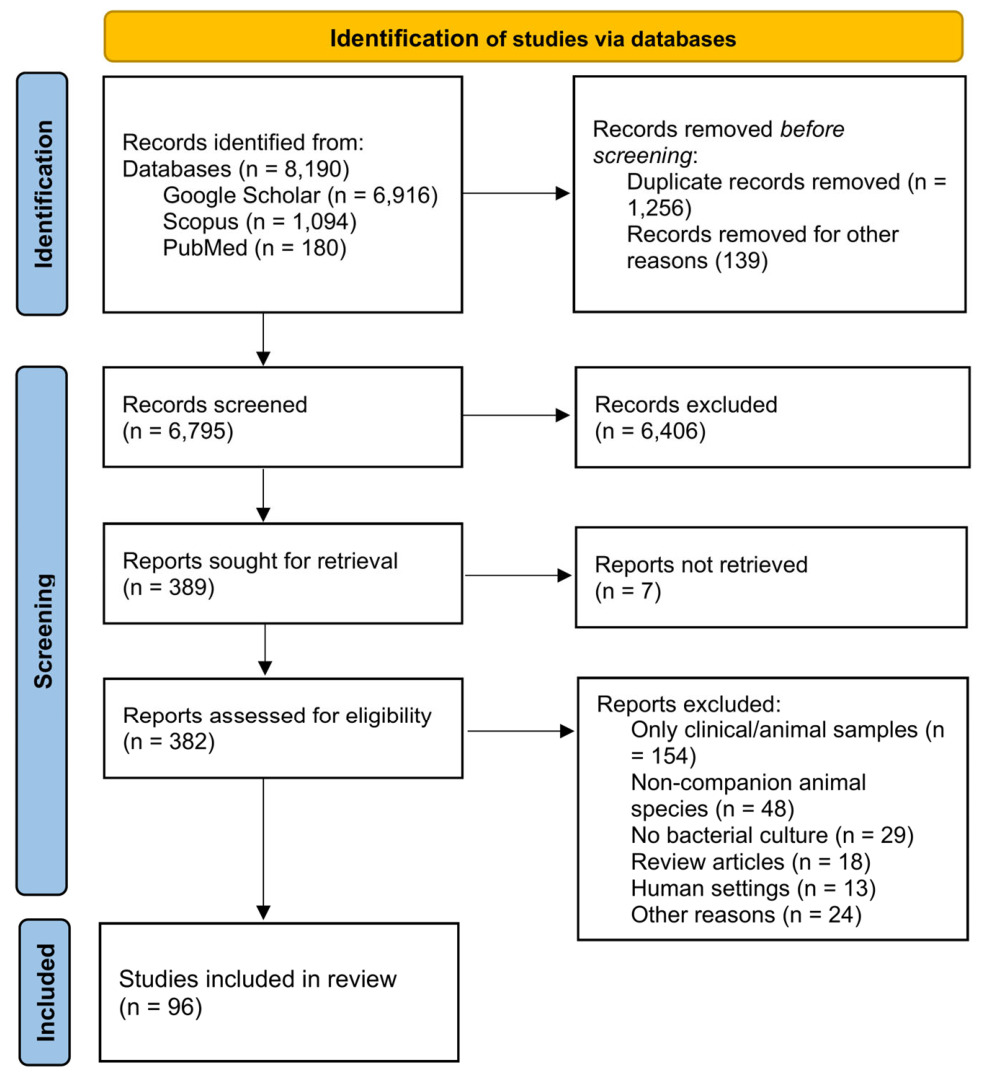
Figure 2.
Geographic distribution of the included studies. Color intensity corresponds to the number of published studies per country (Map generated using MapChart, https://www.mapchart.net/, accessed on 05 September 2026).
Figure 2.
Geographic distribution of the included studies. Color intensity corresponds to the number of published studies per country (Map generated using MapChart, https://www.mapchart.net/, accessed on 05 September 2026).
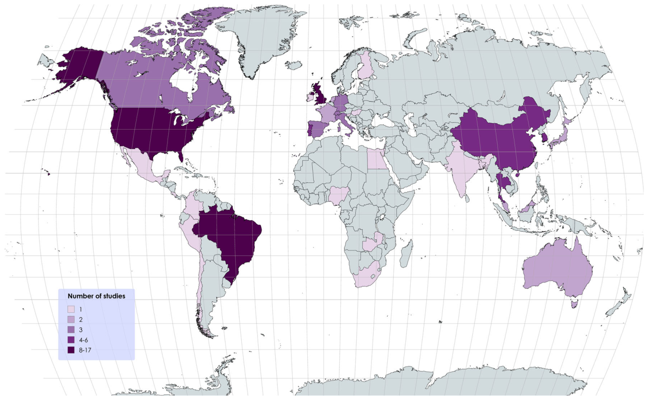
Figure 3.
Environmental multidrug-resistant (MDR) bacterial detection and reporting patterns across primary functional clinical areas in small animal veterinary facilities. The percentages represent the study-level proportions. Unspecified MDR location represents studies that confirmed the isolation of multidrug-resistant bacteria from a general veterinary facility environment but did not provide disaggregated, room-specific isolation rates.
Figure 3.
Environmental multidrug-resistant (MDR) bacterial detection and reporting patterns across primary functional clinical areas in small animal veterinary facilities. The percentages represent the study-level proportions. Unspecified MDR location represents studies that confirmed the isolation of multidrug-resistant bacteria from a general veterinary facility environment but did not provide disaggregated, room-specific isolation rates.
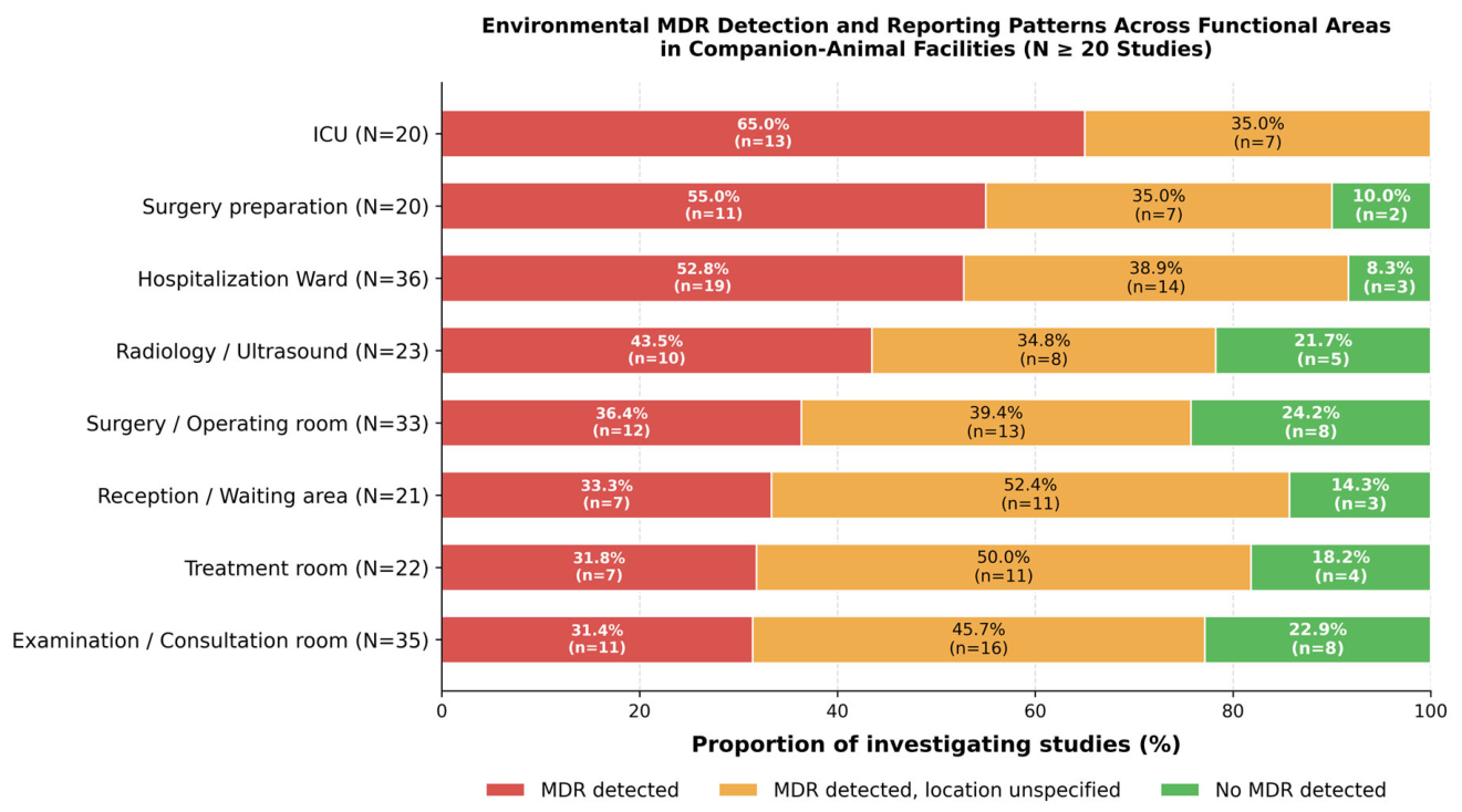
Figure 4.
Multidrug-resistant (MDR) bacterial detection and reporting across samples from environmental surfaces, clinical equipment, and personnel in small animal veterinary facilities. The percentages represent study-level proportions. Unspecified MDR location represents studies that confirmed the isolation of multidrug-resistant bacteria from a general veterinary facility environment but did not provide, site-specific isolation rates.
Figure 4.
Multidrug-resistant (MDR) bacterial detection and reporting across samples from environmental surfaces, clinical equipment, and personnel in small animal veterinary facilities. The percentages represent study-level proportions. Unspecified MDR location represents studies that confirmed the isolation of multidrug-resistant bacteria from a general veterinary facility environment but did not provide, site-specific isolation rates.
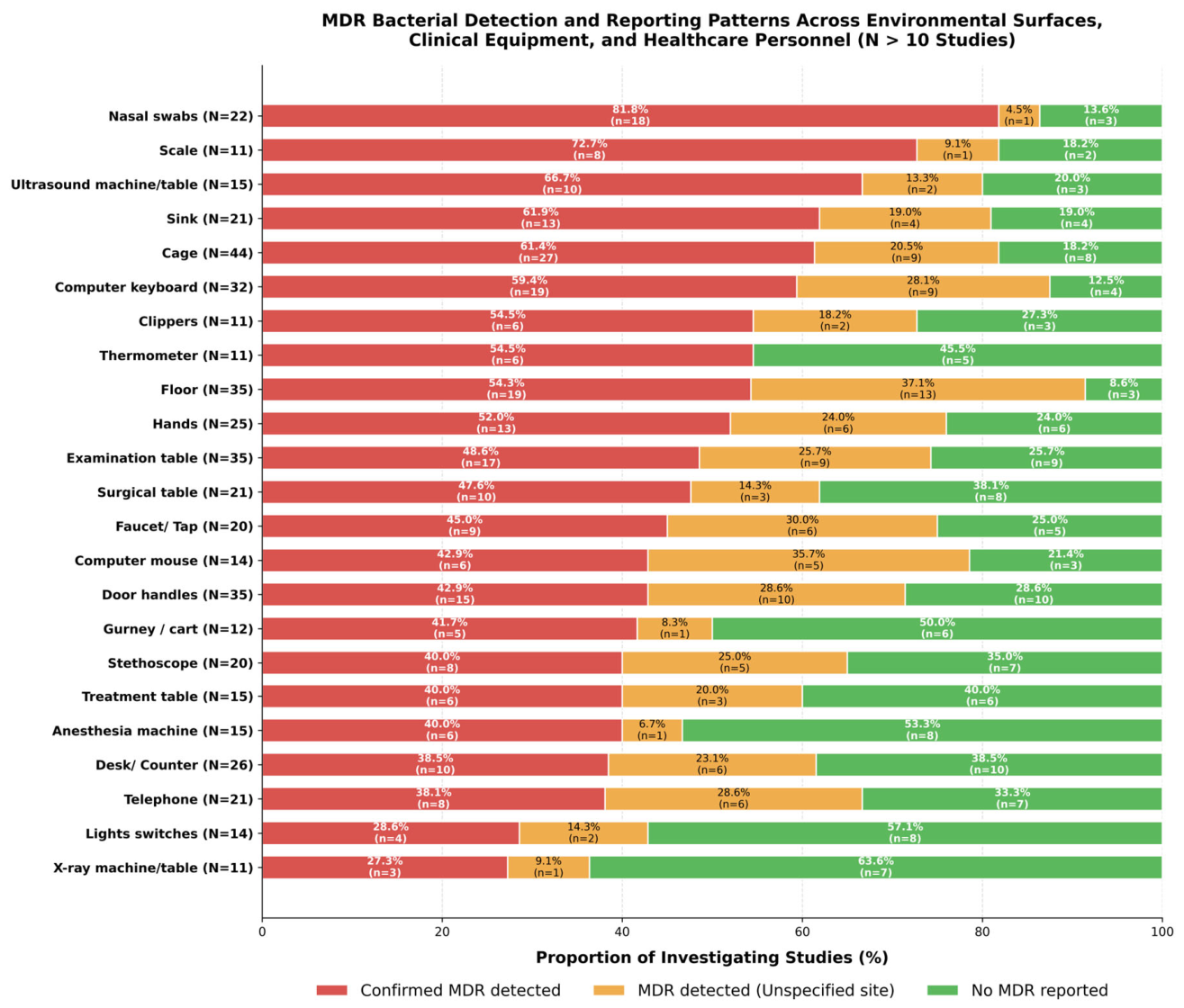

Table 2.
Distribution of targeted antimicrobial resistance categories across the included studies.
| Targeted Resistance Category | Studies, n | % of Studies |
| MRSA1/MRS / Oxacillin resistance | 27 | 28.1% |
| ESBL/ Extended-spectrum cephalosporin resistance | 15 | 15.6% |
| Carbapenem resistance | 10 | 10.4% |
| Other targeted resistance phenotypes (e.g., VRE2, colistin-resistance, and other specific targets) | 7 | 7.3% |
1 Methicillin-Resistant Staphylococcus aureus. 2 Vancomycin-resistant enterococci.
Table 3.
Bacterial taxa reported across the included studies (taxa reported in ≥ 5 studies included).
Table 3.
Bacterial taxa reported across the included studies (taxa reported in ≥ 5 studies included).
| Bacterial taxa | Studies, n | % of total studies |
| Staphylococcus spp.1 | 58 | 60.4% |
| Escherichia coli | 32 | 33.3% |
| Pseudomonas spp.2 | 20 | 20.8% |
| Klebsiella spp.3 | 18 | 18.8% |
| Enterococcus spp.4 | 16 | 16.7% |
| Acinetobacter spp.5 | 17 | 17.7% |
| Enterobacter spp.6 | 14 | 14.6% |
| Bacillus spp. | 7 | 7.3% |
| Serratia spp.7 | 7 | 7.3% |
| Citrobacter spp.8 | 6 | 6.2% |
| Stenotrophomonas maltophilia (incl. spp.) | 5 | 5.2% |
| Salmonella spp. | 5 | 5.2% |
| Micrococcus spp. (incl. M. luteus) | 5 | 5.2% |
1 Detailed species distribution is presented in Table 4. 2 Includes P. aeruginosa, P. fluorescens, and Pseudomonas spp. 3 Includes K. pneumoniae, K. oxytoca, and Klebsiella spp. 4 Includes E. faecium, E. faecalis, E. hirae, E. gallinarum, E. avium and Enterococcus spp. 5 Includes A. baumannii, A. lwoffii, A. calcoaceticus, A. nosocomialis, A. haemolyticus, A. junii, Acinetobacter spp. 6 Includes En. cloacae, En. (Pantoea) agglomerans, En. aerogenes, En. amnigenus, En, mori, En. Cobei, Enterobacter spp. 7 Includes S. marcescens, S. rubidaea, S. plymuthica and Serratia spp. 8 Includes C. freundii, C. braakii and Citrobacter spp.
Table 4.
Distribution of Staphylococcus species reported across the included studies (n = 96).
| Staphylococcal species | Studies, n | % of total studies |
| Staphylococcus aureus | 35 | 36.5% |
| Staphylococcus pseudintermedius | 29 | 30.2% |
| Staphylococcus epidermidis | 12 | 12.5% |
| Staphylococcus schleiferi | 6 | 6.2% |
| Staphylococcus hominis | 6 | 6.2% |
| Staphylococcus haemolyticus | 5 | 5.2% |
| Staphylococcus warneri | 4 | 4.2% |
| Staphylococcus simulans | 4 | 4.2% |
| Staphylococcus cohnii | 4 | 4.2% |
| Staphylococcus intermedius | 3 | 3.1% |
| Staphylococcus capitis | 3 | 3.1% |
| Other coagulase-negative staphylococciᵃ | 5 | 5.2% |
| Mammaliicoccus (Staphylococcus) sciuri | 2 | 2.1% |
| Unspecified Staphylococcus spp. | 21 | 21.9% |
| Staphylococcus spp. (overall genus) | 58 | 60.4% |
ᵃ Includes S. felis (n = 2 studies), S. succinus (n = 2), S. xylosus (n = 2), S. vitulinus (n = 2), S. equorum (n = 2), S. pettenkoferi (n = 1), S. urealyticus (n = 1), S. saccharolyticus (n = 1), and S. saprophyticus (n = 1).
Table 5.
Distribution of MDR species reported in the included studies (n = 96).
| MDR bacteria1 | Studies, n | % of studies |
| Staphylococcus pseudintermedius (including MRSP) | 27 | 28.1 |
| Staphylococcus aureus (including MRSA) | 25 | 26.0 |
| Escherichia coli | 16 | 16.7 |
| Staphylococcus epidermidis (including MRS. epidermidis) | 12 | 12.5 |
| Staphylococcus haemolyticus | 7 | 7.3 |
| Enterococcus faecium | 5 | 5.2 |
| Klebsiella pneumoniae | 5 | 5.2 |
| Staphylococcus schleiferi | 4 | 4.2 |
| Pseudomonas aeruginosa | 4 | 4.2 |
| Acinetobacter baumannii | 4 | 4.2 |
| Staphylococcus hominis | 3 | 3.1 |
| Stenotrophomonas maltophilia | 3 | 3.1 |
| Enterococcus faecalis | 2 | 2.1 |
1 Data are restricted to definitively identified bacterial species with documented multidrug-resistant (MDR) profiles reported in ≥ 2 studies. Categories were not mutually exclusive; individual studies could report multiple MDR species.
Table 6.
Distribution of detected antimicrobial resistance genes (ARGs) across veterinary environmental and personnel sampling studies.
Table 6.
Distribution of detected antimicrobial resistance genes (ARGs) across veterinary environmental and personnel sampling studies.
| Antimicrobial Class / Mechanism | Representative ARGs* | Studies, n | % of Tested Studies (n=64) | % of Total Studies (N=96) |
| Methicillin resistance | mecA | 37 | 57.8% | 38.5% |
| ESBLs | blaCTX-M variants | 14 | 21.9% | 14.6% |
| Carbapenemases/ Oxacillinases | blaOXA variants | 13 | 20.3% | 13.5% |
| Tetracycline resistance | tet genes | 12 | 18.8% | 12.5% |
| Broad-spectrum β-lactamases | blaTEM variants | 11 | 17.2% | 11.5% |
| AmpC β-lactamases | blaCMY, blaDHA, blaEBC, blaCIT, AmpC | 10 | 15.6% | 10.4% |
| Penicillinases | blaZ | 10 | 15.6% | 10.4% |
| Aminoglycoside resistance | aac, aph, ant, aad, strA/B | 9 | 14.1% | 9.4% |
| Broad-spectrum β-lactamases | blaSHV variants | 7 | 10.9% | 7.3% |
| Metallo-β-lactamases (MBLs) | blaNDM variants | 7 | 10.9% | 7.3% |
| MLSB1 (Macrolides/Lincosamides) | erm genes | 6 | 9.4% | 6.2% |
| Quinolone resistance | qnr, oqxAB, norA/C, gyrA | 6 | 9.4% | 6.2% |
| Sulfonamides / Trimethoprim | sul and dfr variants | 6 | 9.4% | 6.2% |
| WGS / Metagenomics | Multi-class resistomes | 4 | 6.2% | 4.2% |
| Carbapenemases (Other) | blaKPC, blaVIM, blaIMP | 2 | 3.1% | 2.1% |
| Glycopeptides | vanA | 2 | 3.1% | 2.1% |
1 MLSB: Macrolide-Lincosamide-Streptogramin B. * Categories were not mutually exclusive; individual studies could report multiple resistance determinants.
Disclaimer/Publisher’s Note: The statements, opinions and data contained in all publications are solely those of the individual author(s) and contributor(s) and not of MDPI and/or the editor(s). MDPI and/or the editor(s) disclaim responsibility for any injury to people or property resulting from any ideas, methods, instructions or products referred to in the content. |
© 2026 by the authors. Licensee MDPI, Basel, Switzerland. This article is an open access article distributed under the terms and conditions of the Creative Commons Attribution (CC BY) license (http://creativecommons.org/licenses/by/4.0/).
Copyright: This open access article is published under a Creative Commons CC BY 4.0 license, which permit the free download, distribution, and reuse, provided that the author and preprint are cited in any reuse.