

Submitted:
08 October 2026
Posted:
10 October 2026
You are already at the latest version
Abstract
The traditional nanocarrier-mediated nucleic acid delivery relies on random Brownian diffusion, suffering from slow transport speed and insufficient single-cell targeting ability. In this study, we proposed a targeted siRNA delivery strategy that uses optical tweezers to deliver the siRNA-PS probes to target cells. The nanoscale inter-facial stability and trapping performance of the probes are evaluated. Delivery is achieved in less than 10 s, two orders of magnitude faster than passive-diffusion-based delivery. Using Nicotiana protoplasts as a proof-of-concept cellular model, single siRNA-PS probe target delivery tests achieved a gene-silencing efficiency of up to 92%. This work demonstrates an optical tweezers-driven targeted delivery strategy and provides experimental insights into nanophotonic manipulation of microscale colloidal assemblies.
Keywords:
plant protoplast
; gene silencing
; siRNA-PS probes
; optical tweezers
; targeted delivery
1. Introduction
Reducing agriculture’s reliance on chemical pesticides is a pressing imperative for environmental health [1,2]. RNA interference (RNAi) represents an eco-friendly strategy to address this challenge, enabling the development of pest- and pathogen-resistant crops through sequence-specific gene silencing [3,4]. However, broad practical deployment of RNAi is still hindered by a major bottleneck: the low efficiency of delivering small interfering RNA (siRNA) [5]. Conventional physical delivery methods, such as gene gun bombardment, typically achieve only 0.5 - 2% transient delivery efficiency in Nicotiana cells and frequently cause cell mortality exceeding 30% [6]. While nanocarrier systems [7] have improved delivery efficiency to 30 - 70% relying on Brownian-motion-driven diffusion [8,9], their insufficient target specificity and unresolved long-term biosafety concerns limit practical application [10]. These limitations motivate the exploration of efficient and bio-compatible delivery platforms to unlock the capability of RNAi-related technologies.
Optical tweezers, which use a tightly focused laser beam to create a three-dimensional optical trap, allow noncontact, noninvasive and high spatiotemporal-resolution manipulation of micro- and nanoparticles [11,12]. Such capabilities have fostered broad applications in precision measurement [13,14,15], nanotechnology [16,17,18], and biological science [19,20]. In biomedicine, optical tweezers have been adopted for targeted delivery of diverse biomolecular cargoes, such as siRNA, plasmid DNA, proteins, and isolated mitochondria [21,22]. Furthermore, “nano-tweezers” enable noninvasive extraction of single-molecule biomolecules from living cells [23]. Despite the promising prospects of optical tweezers techniques in those fields, using optical tweezers to deliver nucleic acids into plant cells has yet to be experimentally demonstrated. The precise and non-invasive nature of optical tweezers presents a promising alternative to overcome the difficulty of achieving targeted delivery in plant protoplast transfection.
Colloidal microcarriers loaded with biological molecules have been widely used in targeted delivery [24]. However, most existing works mainly focus on delivery performance, while ignoring the nanoscale interfacial stability between the loaded molecules and the microcarriers in optical tweezers [25]. Intense local optical field gradients and photothermal effects in optical tweezers may perturb the nanometer-thin adsorption interface and trigger premature dissociation of adsorbed biomolecules [26,27]. Without evaluation of such interfacial stability, it remains difficult to judge whether the cargo can stay firmly bound to microcarriers through-out the optical manipulation process [28,29,30].
In this work, amino-modified polystyrene (PS) microspheres are synthesized and assembled with siRNA to construct cargo-carrying probes. We investigate the nanoscale interfacial stability of such assemblies using optical tweezers, and establish an optical tweezers-assisted strategy for delivering siRNA into single cells. As shown in Figure 1, the trapped siRNA-PS probes can achieve rapid, controllable and noninvasive gene delivery to Nicotiana protoplasts. This approach avoids physical damage and eliminates the randomness inherent in passive nanocarrier uptake. The reduction in green fluorescent protein fluorescence intensity and mRNA expression levels confirm effective gene-silencing performance. Our work establishes a precise and efficient optical delivery platform, providing experimental support for nanophotonic manipulation of micro-scale colloidal assemblies for single-cell cargo delivery.
2. Methods
Synthesis and characterization of siRNA-PS probes. Nucleic acid probes are fabricated via electrostatic binding between amino-modified blue-fluorescent PS microspheres and CY3-labeled siRNA (Figure 2a). Zeta potential measurements and confocal laser scanning microscopy (CLSM) are employed to evaluate binding efficiency and probe fluorescence properties. Overlapping fluorescence signals in CLSM images (Figure 2b) confirmed successful siRNA adsorption. Zeta potential analysis (Figure 2c) revealed surface charge inversion. The potential shifted from +6.17 mV to -20.7 mV after siRNA loading, further verifying siRNA-PS probe formation. Dynamic light scattering (DLS) is used to assess whether siRNA loading triggers particle aggregation or alters monodispersity. As shown in Figure 2d, the hydrodynamic diameter only marginally increased from 1073.77 nm to 1082.53 nm upon siRNA modification, demonstrating negligible aggregation.
To find the optimal loading ratio of siRNA to PS microspheres, we conduct gel electrophoresis [31], designing the mass ratio of siRNA to PS ranging from 1:1 to 1:50. When the ratio of siRNA to PS is inappropriate, bands appear in the gel. As shown in Figure 2e, the siRNA bands disappear in the range of 1:15 to 1:30 mass ratios, indicating that the siRNA reaches a saturated loading state on the PS micro-spheres. To maximize the amount of siRNA loaded, we select the 1:15 ratio for subsequent experiments. The amount of siRNA in our probe is 40000-fold higher than that of the nanocarrier at a single microsphere level [32], as shown in Figure 2f.
3. Results and Discussion
Stability of siRNA-PS Probes. We characterize the siRNA-PS probes using transmission electron microscopy (TEM) and energy-dispersive X-ray spectroscopy (EDX). TEM images in Figure 2a reveal no morphological damage to PS micro-spheres after siRNA loading. Since PS microspheres are phosphorus-free, the presence of phosphorus clearly indicates siRNA loading. EDX analysis demonstrates uniform phosphorus distribution across the microsphere surface, confirming homogeneous siRNA loading, as shown in Figure 3a and Table 1.
We analyzed the effects of short-term storage, temperature fluctuations, and enzymatic degradation on the stability of siRNA-PS probes. As shown in Figure 3b, after the probes are stored at room temperature (25 ℃) for 12 hours, no significant siRNA release is observed, indi-cating their short-term stability. Then, the probes demonstrate excellent thermal stability, with considerable siRNA release occurring only at temperatures exceeding 35 ℃, as shown in Figure 3c. This suggests that the probes remain stable under typical plant physiological conditions and during standard laboratory handling. Furthermore, an effective delivery system must protect nucleic acids from enzymatic degradation in the environment [33]. As illustrated in Figure 3d, free siRNA is rapidly degraded by nucleases, whereas siRNA encapsulated within the PS microspheres remains predominantly intact under the same conditions. This confirms the PS microspheres’ protective function and highlights their importance in maintaining siRNA stability for successful delivery into plant cell.
Optical Tweezers-Assisted Delivery. We use optical tweezers to trap and deliver siRNA-PS probes, as shown in Figure 4a. The system is constructed around an inverted microscope. Key components include a high numerical aperture oil-immersion objective (100×, NA = 1.25) , which tightly focuses the trapping laser to form stable optical traps, and a near-infrared semiconductor laser (980 nm, 100 mW) providing a linearly polarized, fundamental-mode Gaussian beam [34,35]. Functionalized PS microspheres (radius = 0.5 μm) serve as siRNA delivery probes. The tightly focused laser beam traps and manipulates the microspheres, with their displacement controlled by a piezoelectric platform [36,37]. A 10× objective lens and a charge-coupled device (CCD) camera are used for real-time observation, while a quadrant photodetector (QPD) detects the forward-scattered signal from the probe [38,39]. We measured the confined Brownian motion and calculated the transverse optical trap stiffness using the power spectrum method, as shown in Figure 4b,c. The results show that the trapping stiffness in the X and Y directions is 3.2 and 3.0 pN/μm/mW, respectively.
To evaluate whether the local photothermal effect at the center of the optical trap causes siRNA to dissociate from the surface of the PS microspheres, we first trap a siRNA-PS probe and observe it for 10 minutes. At this time, the temperature at the center of the light trap is 25.3 ℃. Subsequently, we place the trapping chamber loaded with the probe under CLSM for observation. The results in Figure 4d show that the red fluorescent siRNA remains bound to the surface of the PS micro-sphere and does not appear in the surrounding medium. This indicates that the local photothermal effect at the center of the optical trap does not affect the stability of the probe on the surface of the microspheres.
As shown in Figure 5a, we deliver siRNA-PS to the plant protoplasts using optical tweezers within 12 s. Conventional nanocarriers rely on random Brownian motion for cell contact and internalization, requiring approximately 1 h for siRNA delivery [40,41]. Our approach reduces delivery time by two orders of magnitude, thereby improving delivery efficiency. As shown in Figure 5b and Figure 5c, We design three paths (linear, L-shaped, and π-shaped) for delivering siRNA–PS probes to the plant protoplasts [42]. The delivery times are 10 s, further demonstrating the targeting and high efficiency of our optical tweezers-assisted delivery method.
Gene silence. In this experiment, the gene silencing effect is evaluated using CLSM and quantitative reverse-transcription PCR (RT-qPCR). Green fluorescent protein (GFP) is used as a fluorescent reporter. GFP can produce visible green fluorescence inside cells, and weaker fluorescence signals mean stronger inhibition of target gene expression [43]. CLSM qualitatively assessed gene silencing by monitoring fluorescence changes of GFP protein, whereas RT-qPCR provides quantitative analysis of intracellular GFP messenger-RNA (mRNA) expression levels. mRNA carries gene information inside cells, and lower mRNA content corresponds to better gene silencing performance.
All experimental data are statistically analyzed and expressed as means. Stu-dent’s t-test for two-group comparisons and one-way analysis of variance (ANOVA) for multiple-group comparisons are adopted to judge whether the numerical difference between different cell groups comes from experimental treatment or random test error. The P value is a probability index calculated by statistical methods to measure the reliability of group differences. Data analysis and graphing are per-formed using GraphPad Prism 9.0. Results are considered statistically significant at a P value < 0.05, which means the observed difference is not caused by accidental measurement fluctuations.
Nicotiana protoplasts carrying the GFP gene are adopted for this test. Six treatment groups are designed to evaluate the silencing performance of optical tweezers-assisted delivery (Figure 6a). Untreated protoplasts serve as the blank control (CK group). The PS group containing only PS microspheres verifies that the carrier itself cannot trigger gene silencing. The Ns-S group with PS microspheres and non-specific siRNA tests the specificity of RNA interference. The free siRNA group confirms the inherent silencing capacity of siRNA molecules. The siR-NA-PS-R group represents conventional passive delivery relying on Brownian diffu-sion of siRNA-loaded microspheres. The siRNA-PS-OT group corresponds to siR-NA-PS probes delivered with optical tweezers.
Figure 6b and Figure 6c respectively display fluorescence images and corresponding fluorescence intensities for all experimental groups. Strong GFP fluorescence intensity is observed in the CK, PS, and Ns-PS groups, which proves that no obvious silencing of the GFP gene occurs within these groups. GFP fluorescence in-tensity drops in the free siRNA group and declines further in the siRNA-PS-R group. The siRNA-PS-OT group exhibits the weakest fluorescence signal. These results in-dicate that only specific siRNAs can achieve gene silencing effectively, and the optical tweezers-assisted delivery method produces the optimal silencing effect.
Gene silencing efficiency is quantified by calculating the relative reduction of GFP mRNA expression in each group compared with the CK group. Figure 6d presents the relative expression level of GFP mRNA. The calculated silencing efficiencies reach 58% for the free siRNA group, 75% for the siRNA-PS-R group and 92% for the siRNA-PS-OT group. Therefore, the delivery strategy based on optical tweezers achieves the the best gene silencing performance.
4. Conclusions
In this study, we proposed an optical tweezers-assisted strategy for targeted siRNA delivery into plant protoplasts to achieve fast siRNA transport and high-efficiency gene silencing at the single-cell scale. This approach reduces delivery time by two orders of magnitude, dropping from around 1 h to under 10 s, and improves gene-silencing efficiency from 75% up to 92%. Traditional nanocarrier delivery relies on random Brownian motion diffusion for cell uptake and lacks real-time monitoring and single-probe manipulation capabilities, whereas our strategy provides precise, controllable cargo delivery.
Sonication and microfluidic-based delivery methods possess limited targeting performance, they rely on batch processing and tend to disrupt cell membranes, threatening cell viability and inducing off-target delivery. Optical tweezers, however, support minimally invasive, high-precise single-probe delivery, which is particularly suitable for fragile plant protoplasts [44,45].
Our optical tweezers-assisted siRNA delivery realizes controllable, site-specific gene regulation. Unlike conventional nanocarrier delivery, which relies on random Brownian motion and lacks single-cell control, this method enables precise manipulation of individual plant cells with minimal invasiveness. The proposed method not only achieves efficient gene silencing but also provides a versatile platform for delivering other biomolecules into plant cells, offering new opportunities for plant functional genomics, crop improvement, and sustainable agriculture.
Data availability
The authors declare that all data supporting the findings of this study are available within the paper.
Declaration of competing interest
The authors declare no competing interests.
Author Contributions
Yilun Liu: Conceptualization, Investigation, Formal analysis, Visualization, Writ-ing-original draft; Pengpeng Zhou: Visualization, Writing-editing; Xinlin Chen: Methodology, Writing-review; Miao Peng: Conceptualization, Software, Methodology, Funding acquisition, Writing review and editing; Guangzong Xiao: Resources, Writing-review; Hui Luo: Funding acquisition, Supervision; Yong Li: Conceptual-ization, Project administration, Writing-review, Supervision.
Acknowledgments
This work is supported by the National Natural Science Foundation of China (62405379); Science Fund for Distinguished Young Scholars of Hunan Province (2024JJ2055); Scientific Research Project of Hunan Provincial Department of Edu-cation (24B0236).:
References
- Stokstad, E. A new leaf. Science 2018, 362, 144–147. [CrossRef]
- Wang, D.; Saleh, N.B.; Byro, A.; Zepp, R.; Sahle-Demessie, E.; Luxton, T.P.; Ho, K.T.; Burgess, R.M.; Flury, M.; White, J.C.; Su, C. Nano-enabled pesticides for sustainable agriculture and global food security. Nat. Nanotechnol. 2022, 17, 347–360. [CrossRef]
- Squire, H.J.; Tomatz, S.; Wang, J.W.T.; González-Grandío, E.; Landry, M.P. Best Practices and Pitfalls in Developing Nanomaterial Delivery Tools for Plants. ACS Nano. 2025, 19, 7–12. [CrossRef]
- Iwakawa, H.; Tomari, Y. Life of RISC: Formation, action, and degradation of RNA-induced silencing complex. Mol. Cell 2022, 82, 30–43. [CrossRef]
- Zhang, H.; Zhang, H.; Demirer, G.S.; González-Grandío, E.; Fan, C.; Landry, M.P. Engineering DNA nanostructures for siRNA delivery in plants. Nat. Protoc. 2020, 15, 3064–3087. [CrossRef]
- Dong, O.X.; Ronald, P.C. Targeted DNA insertion in plants. Proc. Natl. Acad. Sci. U.S.A. 2021, 118. [CrossRef]
- Horejs, C. Nanoparticle transport in plants. Nat. Rev. Mater. 2022, 7. [CrossRef]
- Wang, P.; Zhao, F.J.; Kopittke, P.M. Engineering Crops without Genome Integration Using Nanotechnology. Trends Plant Sci. 2019, 24, 574–577. [CrossRef]
- Cai, Y.; Liu, Z.; Wang, H.; Meng, H.; Cao, Y. Mesoporous Silica Nanoparticles Mediate SiRNA Delivery for Long-Term Multi-Gene Silencing in Intact Plants. Adv. Sci. 2024, 11. [CrossRef]
- Zhang, H.; Goh, N.S.; Wang, J.W.; Pinals, R.L.; González-Grandío, E.; Demirer, G.S.; Butrus, S.; Fakra, S.C.; Flores, A.D.R.; Zhai, R.; Zhao, B.; Park, S.J.; Landry, M.P. Nanoparticle cellular internalization is not required for RNA delivery to mature plant leaves. Nat. Nanotechnol. 2022, 17, 197–205. [CrossRef]
- Zhang, X.; Zhao, K.; He, Z.; Yan, J.; Li, Y.; Wu, T.; Zhang, Y. Creating color patterns using optical manipulation and scattering of silicon nanoparticles. Nano Mater. Sci. 2025, 7, 533–538. [CrossRef]
- Monteiro, F.; Afek, G.; Carney, D.; Krnjaic, G.; Wang, J.; Moore, D.C. Search for Composite Dark Matter with Optically Levitated Sensors. Phys. Rev. Lett. 2020, 125. [CrossRef]
- Kuang, T.; Huang, R.; Xiong, W.; Zuo, Y.; Han, X.; Nori, F.; Qiu, C.W.; Luo, H.; Jing, H.; Xiao, G. Nonlinear multi-frequency phonon lasers with active levitated optomechanics. Nat. Phys. 2023,19 ,414-419. [CrossRef]
- Xiao, G.; Kuang, T.; He, Y.; Chen, X.; Xiong, W.; Han, X.; Tan, Z.; Luo, H.; Jing, H. Giant enhancement of nonlinear harmonics of an optical-tweezer phonon laser. eLight. 2024, 4, 90–98. [CrossRef]
- Dong, Y.; Chen, X.D.; Guo, G.C.; Sun, F.W. Robust scalable architecture for a hybrid spin-mechanical quantum entanglement system. Phys. Rev. B. 2019, 100. [CrossRef]
- Bustamante, C.J.; Chemla, Y.R.; Liu, S.; Wang, M.D. Optical tweezers in single-molecule biophysics. Nat. Rev. Methods Primers. 2021, 1. [CrossRef]
- Shan, X.; Wang, F.; Wang, D.; Wen, S.; Chen, C.; Di, X.; Nie, P.; Liao, J.; Liu, Y.; Ding, L.; Reece, P.J.; Jin, D. Optical tweezers beyond refractive index mismatch using highly doped upconversion nanoparticles. Nat. Nanotechnol. 2021, 16, 531–537. [CrossRef]
- Lin, R.J.; Su, V.-C.; Wang, S.; Chen, M.K.; Chung, T.L.; Chen, Y.H.; Kuo, H.Y.; Chen, J.W.; Chen, J.; Huang, Y.-T.; Wang, J.H.; Chu, C.H.; Wu, P.C.; Li, T.; Wang, Z.; Zhu, S.; Tsai, D.P. Achromatic metalens array for full-colour light-field imaging. Nat. Nanotechnol. 2019, 14, 227–231. [CrossRef]
- Pan, A.; Zuo, C.; Yao, B. High-resolution and large field-of-view Fourier ptychographic microscopy and its applications in biomedicine. Rep. Prog. Phys. 2020, 83. [CrossRef]
- Zhao, T.; Hao, H.; Wang, Z.; Liang, Y.; Feng, K.; He, M.; Yun, X.; Bianco, P.R.; Sun, Y.; Yao, B.; Lei, M. Multi-color structured illumination microscopy for live cell imaging based on the enhanced image recombination transform algorithm. Biomed. Opt. Express. 2021, 12, 3474–3484. [CrossRef]
- Català-Castro, F.; Schäffer, E.; Krieg, M. Exploring cell and tissue mechanics with optical tweezers. J. Cell Sci. 2022, 135. [CrossRef]
- Stevenson, D.J.; Gunn-Moore, F.J.; Campbell, P.; Dholakia, K. Single cell optical transfection. J. R. Soc. Interface 2010, 7, 863–871. [CrossRef]
- Nadappuram, B.P.; Cadinu, P.; Barik, A.; Ainscough, A.J.; Devine, M.J.; Kang, M.; Gonzalez-Garcia, J.; Kittler, J.T.; Willison, K.R.; Vilar, R.; Actis, P.; Wojciak-Stothard, B.; Oh, S.H.; Ivanov, A.P.; Edel, J.B. Nanoscale tweezers for single-cell biopsies. Nat. Nanotechnol. 2019, 14, 80–88. [CrossRef]
- Libermann, T.A.; Zerbini, L.F. Targeting Transcription Factors for Cancer Gene Therapy. Curr. Gene Ther. 2006, 6, 17–33. [CrossRef]
- Padmos, J.D.; Boudreau, R.T.M.; Weaver, D.F.; Zhang, P. Impact of Protecting Ligands on Surface Structure and Antibacterial Activity of Silver Nanoparticles. Langmuir 2015, 31, 3745–3752. [CrossRef]
- Gao, Z.; Sun, M.; Sui, X.; Li, N.; Xia, F.; Huang, Y.; Jiang, L. Artificial intelligence approaches in nanopore sensing for single-molecule analysis: From machine learning to deep learning. Nano Mater. Sci. 2026. [CrossRef]
- Brès, C.S.; Della Torre, A.; Grassani, D.; Brasch, V.; Grillet, C.; Monat, C.Supercontinuum in integrated photonics: generation, applications, challenges, and perspectives. Nanophotonics 2023, 12, 1199–1244. [CrossRef]
- Elder, R.M.; Forster, A.L.; Krishnamurthy, A.; Dennis, J.M.; Akiba, H.; Yamamuro, O.; Ito, K.; Evans, K.M.; Soles, C.; Sirk, T.W. Relative effects of polymer composition and sample preparation on glass dynamics. Soft Matter 2022, 18, 6511–6516. [CrossRef]
- Tuo, J.; Lu, L.; Jiang, X.; Duan, Y.; Liu, J.; Zhi, S.; Li, X.; Si, C. Biomass-based materials for intelligent responsive pesticide delivery system: Focusing on performance optimization, mechanism exploration, and multi-functional design. Nano Mater. Sci. 2026. [CrossRef]
- Demirer, G.S.; Zhang, H.; Goh, N.S.; Pinals, R.L.; Chang, R.; Landry, M.P. Carbon nanocarriers deliver siRNA to intact plant cells for efficient gene knockdown. Sci. Adv. 2020, 6. [CrossRef]
- Gao, Y.; Liu, X.L.; Li, X.R. Research progress on siRNA delivery with nonviral carriers. Int. J. Nanomed. 2011, 6, 1017–1025. [CrossRef]
- Wu, H.; Li, Z.; Xiao, Y.; Liu, H.; Li, Y.; Pan, Y.; Zhang, H.; Liu, Q.; Jiang, G. Plant Trait Regulation Enabled by Nanoplastic Nucleic Acid Carriers.Environ. Sci. Technol. 2025, 59, 20179–20189. [CrossRef]
- Pei, M.; Jin, S.; Chen, X.; Kuang, T.; Xiong, W.; Han, X.; Xiao, G.; Luo, H. Direct measurement of the van der Waals force between a pair of microspheres based on photonic force microscopy. Opt. Eng. 2021, 60. [CrossRef]
- He, Y.; Kuang, T.; Han, X.; Feng, Z.; Chen, X.; Xiong, W.; Jin, S.; Tan, Z.; Zhang, Q.; Luo, H.; Jing, H.; Xiao, G. Coherent acoustic frequency comb via Floquet engineering of optical tweezer phonon lasers. Sci. Adv. 2025, 11. [CrossRef]
- Liu, J.X.; Jiao, Y.F.; Li, Y.; Xu, X.W.; He, Q.Y.; Jing, H. Phase-controlled asymmetric optomechanical entanglement against optical backscattering. Sci. China Phys. Mech. Astron. 2023, 66. [CrossRef]
- Lu, T.X.; Wang, Y.; Xia, K.; Xiao, X.; Kuang, L.M.; Jing, H. Quantum squeezing induced nonreciprocal phonon laser. Sci. China Phys. Mech. Astron. 2024, 67. [CrossRef]
- Zou, X.; Zheng, Q.; Wu, D.; Lei, H. Controllable Cellular Micromotors Based on Optical Tweezers. Adv. Funct. Mater. 2020, 30. [CrossRef]
- Yang, J.Y.; Li, N.; Pan, Y.; Yang, J.; Chen, Z.M.; Cai, H.; Wang, Y.; Han, C.; Chen, X.F.; Liu, C.; Hu, H. From Photon Momentum Transfer to Accelerometer Based on Optical Levitated Microsphere at Dynamic Input from 0.1 μg to 1 g. ACS Photonics 2025, 12, 194–201. [CrossRef]
- Sun, X.D.; Ma, J.Y.; Feng, L.J.; Duan, J.L.; Yuan, X.Z. Precise tracking of nanoparticles in plant roots.Nat. Protoc. 2025, 20, 248–271. [CrossRef]
- Hu, P.; An, J.; Faulkner, M.M.; Wu, H.; Li, Z.; Tian, X.; Giraldo, J.P. Nanoparticle Charge and Size Control Foliar Delivery Efficiency to Plant Cells and Organelles. ACS Nano. 2020, 14, 7970–7986. [CrossRef]
- Pan, T.; Shi, Y.; Zhao, N.; Xiong, J.; Xiao, Y.; Xin, H.; Li, B. Bio-Micromotor Tweezers for Noninvasive Bio-Cargo Delivery and Precise Therapy. Adv. Funct. Mater. 2022, 32. [CrossRef]
- Jones, C.M.; List, N.H.; Martínez, T.J. Steric and Electronic Origins of Fluorescence in GFP and GFP-like Proteins. J. Am. Chem. Soc. 2022, 144, 12732–12746. [CrossRef]
- Demirer, G.S. Detecting and quantifying nanoparticle-mediated biomolecule delivery in plants. Nat. Rev. Methods Primers 2023, 3. [CrossRef]
- Collins, D.J.; Devendran, C.; Ma, Z.; Ng, J.W.; Neild, A.; Ai, Y. Acoustic tweezers via sub–time-of-flight regime surface acoustic waves. Sci. Adv. 2016, 2. [CrossRef]
- Zhang, Y.; Min, C.; Dou, X.; Wang, X.; Urbach, H.P.; Somekh, M.G.; Yuan, X. Plasmonic tweezers: for nanoscale optical trapping and beyond. Light Sci. Appl. 2021, 10. [CrossRef]
Figure 1.
Schematic of optical tweezers-assisted siRNA-targeted transfection in single cells.
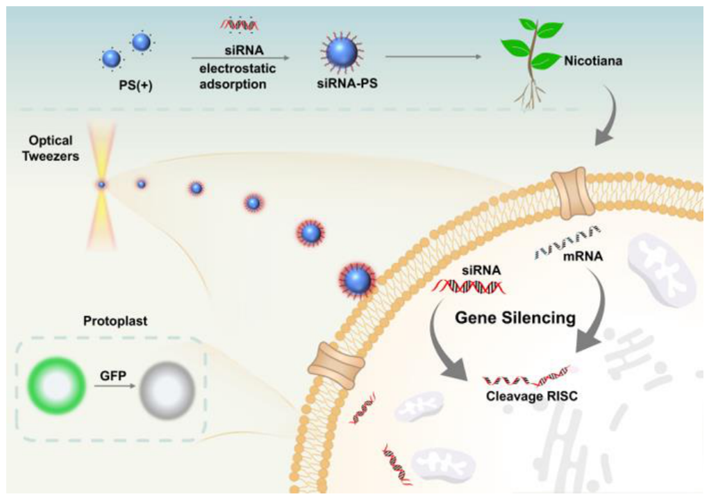
Figure 2.
Preparation and characterization of siRNA-PS Probes. (a) Schematic illustration of electrostatic adsorption of siRNA-PS probes. (b) Fluorescence images of siRNA-PS probes. (c) Zeta potential measurements showing the interaction between siRNA and PS microspheres. (d) Hydrodynamic diameter changes of PS microspheres before and after siRNA loading. (e) Optimization of the ratio of siRNA to PS microspheres by gel electrophoresis to determine the best binding ratio. (f) The amount of siRNA on the surface of a single micron-sized carrier and nanocarrier.
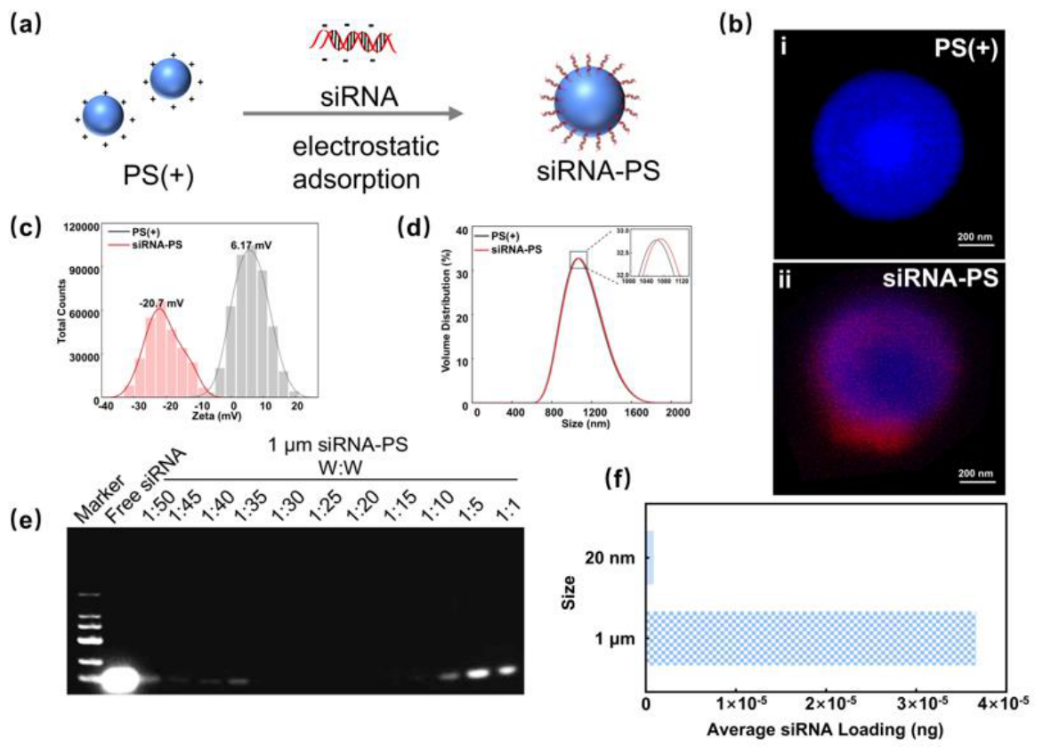
Figure 3.
(a) Transmission electron microscopy (TEM) images of siRNA-PS probe. (b) Stability of the siRNA-PS probes under exposure durations. (c) Thermal stability of probes at different temperatures (d) Anti-degradation properties of the probes under enzymatic treatment.
Figure 3.
(a) Transmission electron microscopy (TEM) images of siRNA-PS probe. (b) Stability of the siRNA-PS probes under exposure durations. (c) Thermal stability of probes at different temperatures (d) Anti-degradation properties of the probes under enzymatic treatment.
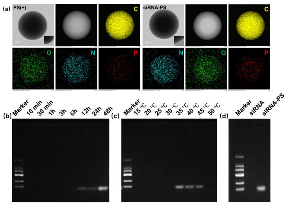
Figure 4.
(a) Schematic diagram of the optical tweezers system. (b) Power spectral density (PSD) of microspheres trapped in the X-axis direction. (c) PSD of microspheres trapped in the Y-axis direction. (d) fluorescence microscopy images of siRNA-loaded probes after optical tweezers trapping. Red dashed circles indicate siRNA-PS probes.
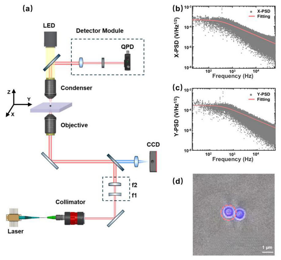
Figure 5.
Optical tweezers-assisted targeted delivery of siRNA-PS probe. (a) Schematic illustration comparing delivery time between conventional delivery and optical tweezers-assisted delivery. (b) Schematic diagrams and real-time images of microspheres for optical tweezers-assisted delivery in different movement paths. White dashed circles indicate plant protoplast cells; black dashed circles indicate siRNA-PS probes. (c) Scanning electron microscopy (SEM) images of protoplasts in contact with microspheres.
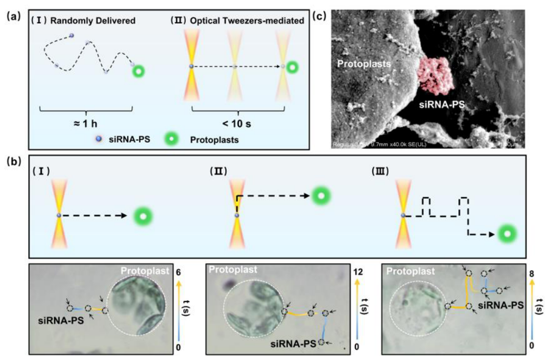
Figure 6.
GFP gene silencing effect. (a) Schematic diagram of each group design. (b) CLSM images of GFP in protoplasts across experimental groups, with (c) corresponding fluorescence gray-scale values. (d) RT-qPCR determination of GFP mRNA expression levels in different experimental groups. Asterisks (****) indicate the statistical significance (p < 0.0001).
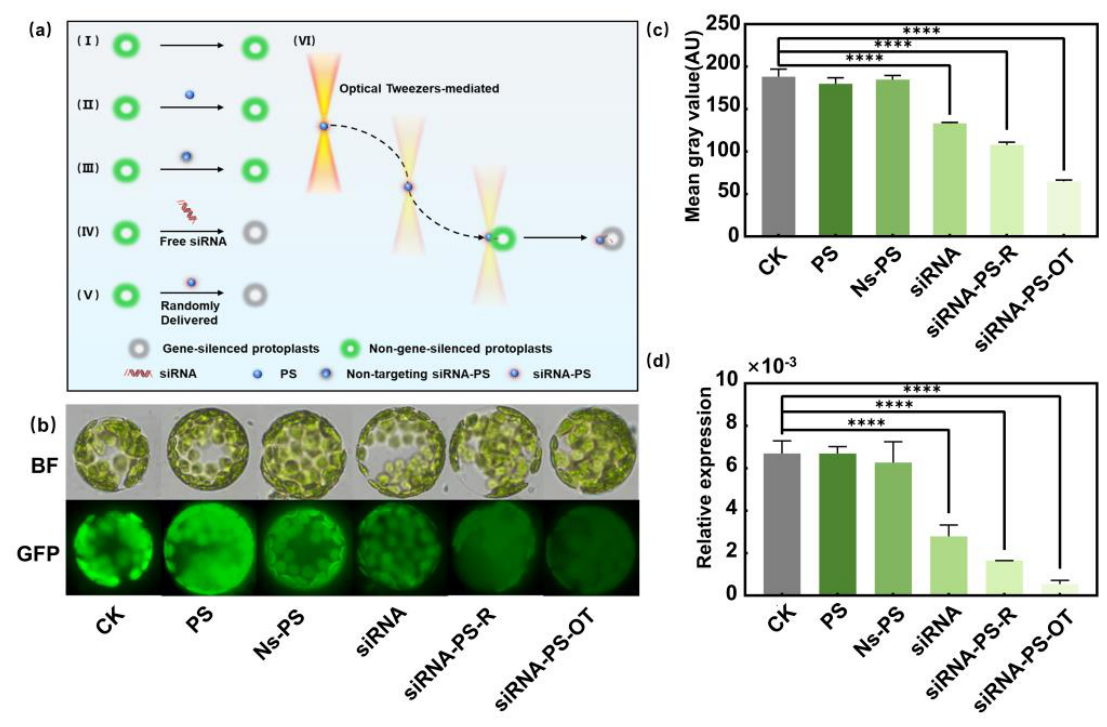

Table 1.
Relative elemental composition of siRNA-PS probes from EDX measurements.
| Group | Mass (%) | |||
| C | N | O | P | |
| PS (+) | 99.10 | 0.29 | 0.61 | 0.00 |
| siRNA-PS | 98.52 | 0.53 | 0.91 | 0.04 |
Disclaimer/Publisher’s Note: The statements, opinions and data contained in all publications are solely those of the individual author(s) and contributor(s) and not of MDPI and/or the editor(s). MDPI and/or the editor(s) disclaim responsibility for any injury to people or property resulting from any ideas, methods, instructions or products referred to in the content. |
© 2026 by the authors. Licensee MDPI, Basel, Switzerland. This article is an open access article distributed under the terms and conditions of the Creative Commons Attribution (CC BY) license (http://creativecommons.org/licenses/by/4.0/).
Copyright: This open access article is published under a Creative Commons CC BY 4.0 license, which permit the free download, distribution, and reuse, provided that the author and preprint are cited in any reuse.