

Submitted:
07 October 2026
Posted:
09 October 2026
You are already at the latest version
Abstract
Resistance to tyrosine kinase inhibitor therapy in patients with chronic myeloid leukemia remains a clinical challenge of which the underlying mechanisms are not always fully understood. The leukemic bone marrow microenvironment is increasingly recognized as an important factor protecting leukemic cells from therapeutic pressure. However, studying this leukemic bone marrow is complicated by limited accessibility of the bone marrow in vivo and the limited physiological relevance of conventional in vitro disease models. Microfluidic bone-marrow-on-a-chip models provide a platform for reconstructing the cellular, biochemical, and biophysical components of the bone marrow niche under controlled conditions. These models may more accurately recapitulate the in vivo microenvironment while avoiding some of the limitations inherent in animal models. Their ability to mimic the bone marrow niche provides novel opportunities to investigate the complex interactions between leukemic cells and their microenvironment. Moreover, the incorpora-tion of patient-derived cells highlights the potential of personalized chronic myeloid leu-kemia-on-a-chip models to study interpatient heterogeneity in treatment response. This review explores the potential of chronic myeloid leukemia-on-a-chip models as platforms for basic research and clinical translation.
Keywords:
chronic myeloid leukemia
; organ-on-a-chip
; bone marrow microenvironment
1. Introduction
The bone marrow is the primary site of hematopoiesis throughout adult life. Studying the bone marrow in health and disease is challenging because of its limited accessibility and the complexity of the microenvironment. The bone marrow niche comprises a dynamic network of interactions between hematopoietic and nonhematopoietic stromal cells, extracellular matrix components, soluble factors, and biophysical cues that collectively regulate hematopoiesis. Chronic myeloid leukemia (CML) should therefore be studied in the context of this niche, as the bone marrow microenvironment plays a critical role in disease development and is actively remodeled during leukemic progression [1,2].
Although most CML patients achieve favorable and durable responses to tyrosine kinase inhibitor (TKI) therapy, a subset of patients has an inadequate response or develops treatment resistance, with a risk of progression to blast phase [3]. TKI resistance may be mediated by mutations in the ABL1 kinase domain of BCR::ABL1 that impair drug binding [4,5]. However, such mutations are not detected in all patients with treatment resistance, indicating that additional BCR::ABL1-independent mechanisms are involved [4]. Interactions between leukemic stem cells (LSCs) and the bone marrow microenvironment may promote LSC quiescence, survival, and persistence during TKI therapy, thereby contributing to treatment resistance [6,7]. Interpatient variation in both leukemic cell-intrinsic characteristics and the bone marrow niche may therefore influence treatment response.
Elucidating these interactions requires experimental platforms that enable LSCs to be studied within a physiologically relevant environment. Conventional two-dimensional (2D) in vitro systems are simple and convenient but fail to recapitulate the key architectures and characteristics of the in vivo bone marrow niche [8,9]. Microfluidic ‘organ-on-a-chip’ technology has advanced rapidly and enables selected components of tissue environments to be investigated under controlled conditions [10]. In this context, bone-marrow-on-a-chip models represent a promising approach for studying the interactions between CML cells and their surrounding niche. Incorporating patient-derived cells into these models could facilitate the investigation of interpatient heterogeneity in treatment response and, ultimately, support the development of patient-specific approaches to therapeutic evaluation.
This review critically evaluates the current literature on interpatient variability in TKI response in CML treatment and on bone marrow-on-a-chip technology. We assess the potential of patient-derived CML-on-a-chip models as platforms for studying niche-mediated treatment resistance and discuss the opportunities and challenges associated with their future application in personalized medicine.
2. Interindividual Variability in TKI Response
Resistance against TKI treatment can broadly be divided into BCR::ABL1-dependent and BCR::ABL1-independent mechanisms. BCR::ABL1-dependent resistance is primarily mediated by mutations in the ABL1 kinase domain that reduce TKI binding, or by increased BCR::ABL1 expression or amplification, which may restore downstream oncogenic signaling despite TKI treatment [11,12,13,14,15]. In this context, the so-called ‘gatekeeper’ T315I mutation in the ATP-binding pocket is one of the most extensively studied and clinically relevant resistance mutations. It confers resistance to imatinib, nilotinib, dasatinib and bosutinib treatment, whereas ponatinib and asciminib retain activity [16,17,18,19]. Other BCR::ABL1 mutations have specific TKI sensitivity profiles making selection of subsequent therapy dependent on the specific mutation detected [11].
In the absence of resistance mutations, inadequate responses may result from a variety of factors. These include suboptimal medication adherence, pharmacokinetic variability, drug influx and efflux alterations, activation of alternative signaling pathways, epigenetic changes, and the protective effect of the bone marrow microenvironment on the leukemic cells [20,21,22,23,24,25,26]. Importantly, poor adherence and pharmacological variability do not constitute true biological resistance, although both may lead to inadequate TKI exposure and an apparently resistant phenotype.
Most TKIs are metabolized by cytochrome P450 enzymes, particularly CYP3A4 and CYP3A5 [27]. Variations in the activity of these enzymes may therefore lead to differences in systemic TKI exposure. Higher activity of CYP3A4 and CYP3A5 was reported in CML patients achieving improved molecular remission rates compared to poor responders, suggesting that active metabolites might contribute to this seemingly counterintuitive association [28]. Altered TKI metabolism due to cytochrome P450 polymorphisms was also related to improved TKI response [29]. Genetic variation and enzyme activity of cytochrome P450 enzymes have been investigated as a potential determinant of TKI pharmacokinetics and treatment response, although its predictive value for clinical response remains inconsistent [11,30,31,32,33,34].
Besides hepatic metabolism, interindividual differences in drug transporting protein expression might contribute to differences in TKI response. The expression of the organic cation transporter 1 has been suggested as a predictor of long-term imatinib response in CML patients in several studies [35,36]. However, other experimental and clinical studies have shown conflicting results [37,38,39,40,41,42]. Efflux transporters such as P-glycoprotein and breast cancer resistance protein are involved in TKI efflux, but, likewise, evidence on their application as predictive biomarkers is lacking [43,44].
Epigenetic mechanisms may provide another source of interpatient variability. Differences in DNA methylation and histone modifications might influence CML progression and response to drug treatment [45,46,47]. MicroRNAs may also modify TKI sensitivity by regulating expression of tumor suppressor genes and oncogenes involved in cell survival. Differences in microRNA expression between responding and non-responding patients have therefore been proposed as potential biomarkers to predict clinical TKI resistance, although further clinical validation is required [48,49].
Thus, although several cellular mechanisms of TKI resistance are well-characterized, resistance cannot always be explained by ABL1 kinase domain mutations or pharmacogenetic factors. Increasing evidence indicates that CML cells may be protected by interactions with nonmalignant cells and extracellular components of the leukemic niche [50]. Interindividual differences in both the leukemic cell population and its microenvironment may consequently influence the depth and durability of response to TKI therapy.
3. The Bone Marrow Niche in CML Pathophysiology
The bone marrow consists of a dynamic microenvironment composed of, amongst others, hematopoietic, mesenchymal, endothelial, adipocytic, osteoblastic, and immune cells that collectively regulate hematopoiesis, immune function, and skeletal homeostasis. Historically, spatially and functionally heterogeneous microenvironments within the bone marrow have been described as distinct endosteal and vascular niches [51]. However, current evidence suggests that these environments are not discrete, but rather interconnected compartments that collectively provide survival and maintenance signals to hematopoietic stem and progenitor cells (HSPCs) [52]. Studying the exact cellular composition of ex vivo-derived bone marrow samples is hampered by the incomplete enzymatic digestion prior to flow cytometric analysis [53].
Hematopoietic stem cells (HSCs) form the most primitive compartment within the hematopoietic system [54,55]. CD34+ HSPCs are used for most clinical gene therapy applications, making CD34+ a widely accepted HSPC marker [56]. The majority of HSPCs lie proximal to sinusoidal surfaces, indicating the importance of the vascular network within the bone marrow [57]. Besides its function in nutrient delivery, waste removal, cellular trafficking, and angiocrine-mediated regulation of hematopoiesis, the vascular system fulfills an important structural role. Moreover, endothelial cells of the vascular system are involved in HSPC maintenance through, amongst others, stem cell factor (SCF) signaling [7,58,59,60,61,62,63,64,65,66]. Mesenchymal stromal cells (MSCs) are multipotent progenitor cells capable of differentiation towards adipocytes, chondrocytes, reticular stromal cells, and osteoblasts. The latter cell type has been studied extensively in the context of HSC maintenance [52]. Collectively, these cells provide structural support and regulatory factors important for hematopoiesis such as interleukin-6, RANKL, Jagged1, angiopoietin-1, interleukin-7, SCF, and CXCL12 [67,68,69,70,71]. Neural cells have been proposed to regulate hematopoiesis through both direct effects on HSPCs and indirect effects mediated by their influence on the stromal components [72]. For example, non-myelinating Schwann cells regulate TGFβ activation in HSCs, inducing phosphorylation of SMAD2/3 proteins that regulates transcriptional programs associated with HSC quiescence and maintenance [73].
The CML-associated microenvironment, known as the LSC niche, is known to provide a protective sanctuary that gives these cells a clonal advantage to outcompete HSCs and contributes to drug resistance [7,74,75,76]. The BCR::ABL1 translocation is the most striking distinction between HSCs and CML LSCs, resulting in constitutive activation of the ABL1 kinase and key downstream signaling pathways including JAK/STAT, Ras/MAPK and PI-3K that increase proliferation of malignant cells [77]. LSCs are characterized by their ability to initiate and serially propagate disease upon transplantation [78]. Besides BCR::ABL1, increased ROS and the dependence on upregulated oxidative metabolism differ between HSCs and LSCs [79,80].
LSCs and HSCs differ in their interactions with the bone marrow niche, including both how they influence and respond to their surrounding microenvironment. LSCs are more dependent on CD44 and selectins to facilitate extracellular anchoring, homing, and engraftment in the bone marrow compared to HSCs [81,82]. Moreover, inhibiting E-selectin-mediated adhesion of CML-initiating cells to the bone marrow endothelium enhanced the efficacy of imatinib treatment and prolonged survival in mice with CML compared with imatinib alone [83]. In addition, in vitro co-culture experiments showed that contact with endothelial cells favors the maintenance of primitive CML cells in culture [84].
CML progenitors were also shown to exhibit reduced adhesion to stroma, in part caused by upregulation of dipeptidyl peptidase-4 (DPP4 or CD26), which results in disruption of the CXCR4-CXCL12 axis by cleaving CXCL12 on stromal cells [85,86,87,88,89,90]. Moreover, MSCs isolated from CML patients exhibit an abnormal gene expression pattern, possibly established during the leukemogenic process, that persists in patients in deep molecular response [91]. CML-MSC-derived cells also play a role in the suppression of autologous T lymphocytes and the overexpression of immunomodulatory factors like TGFß, IL6, and IL10, contributing to CML immune escape [92].
Collectively, these findings highlight the complex interplay between the bone marrow microenvironment and HSPCs or LSCs, emphasizing that oncogenic transformation cannot be fully understood in isolation from its surrounding niche. A comprehensive understanding of the cellular composition of the bone marrow microenvironment and the intricate interactions between its constituent cell populations is therefore essential to elucidate how the niche influences the behavior of both normal and transformed stem cells. Consequently, disease models aiming to faithfully recapitulate the bone marrow microenvironment should incorporate relevant cell types, including LSCs, vascular cells, stromal cells, and neural cells. Moreover, the model should account for cell-matrix interactions, rather than focusing solely on the leukemic cell population.
4. Experimental Models of the Bone Marrow Niche
Various in vitro models have been proposed to study the interaction between HSCs or LSCs and the bone marrow niche. 2D co-cultures of bone marrow-derived cell types have shed light on processes like angiogenesis and osteogenesis in the bone marrow [93]. However, these 2D cultures lack components that might influence leukemic stem cell behavior such as the spatial organization, immune complexity, vascular involvement, matrix composition, and metabolic gradients, which has driven the development of more complex 3D culture systems. Examples of these include organoids, spheroids, and scaffold-based systems, which more closely reproduce the architecture and cell-cell/cell-matrix interactions within a human bone marrow-like milieu [9,94,95,96,97,98,99,100,101]. Nevertheless, these static 3D cultures cannot recapitulate tissue-tissue interfaces, vascular perfusion, interstitial flow, circulating immune cells, or organ-specific mechanical cues that are critical for accurate mimicry of the absorption, distribution, metabolism, and excretion of pharmaceutical compounds and ultimately pharmacokinetics and pharmacodynamics [102]. Moreover, oxygen and nutrient supply may also be a critical aspect.
3D models with a perfused bioreactor setup have therefore been introduced, combining 3D cellular organization with controlled perfusion. Studies suggest that this bioreactor-based 3D setup provides an advantageous system for maintaining immature progenitor cells compared to a 2D system, as an increased proportion of CD34+ and CD34+/CD38- cells was maintained in this system [103]. Meanwhile, these models have been shown to accurately predict patient responses [104].
Other groups have used xenotransplantation of humanized bone marrow microenvironments into immunocompromised mice. The subsequent engraftment of normal and leukemic human hematopoietic cells served as a tool to recapitulate and model human hematopoietic development and malignancies in vivo [105]. The use of this model facilitates the study of hematopoiesis and malignancies in a physiological context, although the mouse-derived vascular and bone marrow sinusoidal structures limit the translatability to the human context. Similar limitations apply to transgenic mouse models used in CML research; they may provide valuable information on disease initiation, progression, and niche remodeling, but differences between murine and human hematopoiesis, immunology, stromal biology, and pharmacology limit translation to humans [75,106,107]. Regulatory authorities have removed the emphasis on animal testing and allowed certain qualified non-animal methods in preclinical safety assessment, making ‘new approach methodologies’ (NAMs) increasingly attractive [108,109]. With the growing understanding of the role of the bone marrow niche in leukemia pathogenesis, this development has created a framework for bone-marrow-on-a-chip and leukemia-on-a-chip models to emerge as promising tools for studying disease biology.
5. Microfluidic Bone Marrow and Leukemia Models
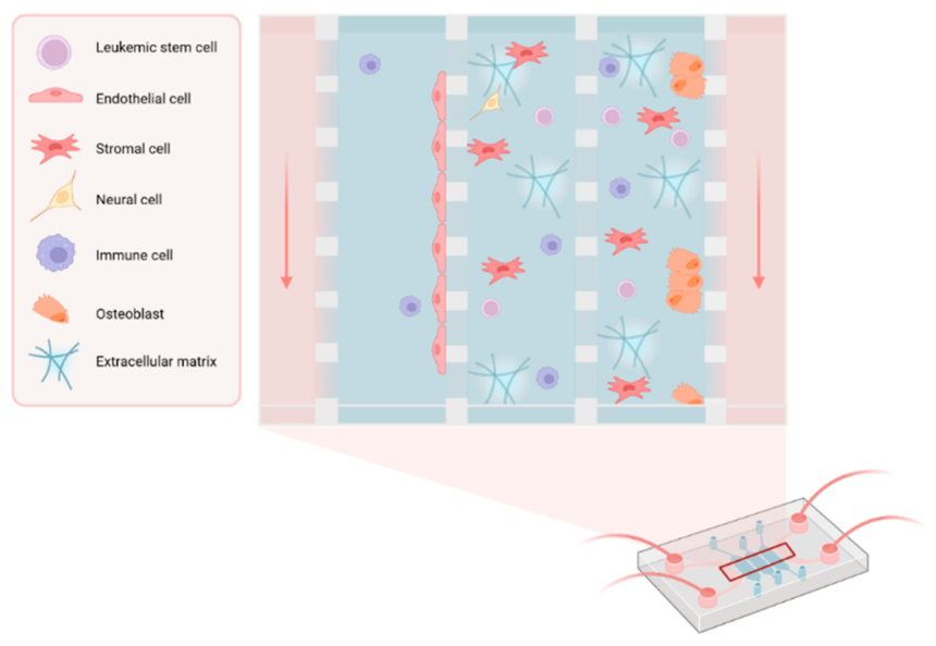
Bone marrow-on-a-chip and leukemia-on-a-chip models are in vitro micro-engineered microfluidic systems designed to reproduce key structural, cellular, biochemical, and biophysical features of the bone marrow microenvironment. By combining human cells, three-dimensional extracellular matrices, spatially organized compartments, and controlled fluid flows, these platforms enable the study of hematopoiesis, disease processes, and therapeutic responses under defined and physiologically relevant conditions (Figure 1).
To produce the device itself, a replication technique is applied, for which a master mold with inverted micro- or nanostructures is fabricated [110]. The resulting products are often made from polydimethylsiloxane (PDMS), silicone, polycarbonate (PC), polymethylmethacrylate (PMMA), polystyrene (PS), polyimide (PI), and polyvinyl chloride (PVC) [111], of which PDMS is the material that is most often used. The choice of material for the fabrication of microfluidic devices depends on multiple factors, including optical transparency for microscopic examination, biological and chemical compatibility, mechanical properties, ease of fabrication, and costs.
To mimic the HSPC population in microfluidic models, CD34+ cells, e.g., isolated from bone marrow, mobilized peripheral blood, or umbilical cord, are integrated [112,113,114,115,116,117]. Human umbilical vein endothelial cells or mouse endothelial C166 cells [118] are mostly used as a practical substitute for specialized bone marrow sinusoidal endothelial cells [112,113,115,116]. To represent the mesenchymal compartment, commercial cell lines can be integrated [112,113]. However, isolation of MSCs from ex vivo femoral heads obtained after hip replacement surgery has also been described [117]. Sometimes MSC-derived osteoblasts are directly integrated into the model [119].
Cells can be integrated in the microfluidic model using a hydrogel that can be designed to closely mimic the native ECM, and possesses additional desirable properties such as optical clarity, which allows for the real-time microscopic observation of cell behavior within the structures [120]. Another option to integrate cells in the microfluidic model is using a scaffold, such as hydroxyapatite, polystyrene, or decellularized bone, which is intended to mimic the in vivo trabecular bone, especially regarding its structural and mechanical properties. A key advantage of a scaffold-based approach is the close imitation of the hematopoietic niche [65]. A disadvantage is the lack of optical transparency, which may interfere with high-resolution imaging.
Traditionally, microfluidic modeling has been a labor-intensive process characterized by limited throughput, substantial experimental complexity, and challenges in model validation and standardization. However, with the emergence of artificial intelligence (AI) and automation technologies, the field is increasingly moving towards more scalable and reproducible platforms. Recent advances, including AI-assisted image analysis integrated into high-throughput bone-on-a-chip systems, highlight the potential of automated workflows to improve data acquisition, analysis, and the application of microfluidic models for drug discovery and preclinical testing [121]. However, automation and artificial intelligence do not in themselves establish biological validity. Meaningful translation will require standardized performance criteria, reproducibility across laboratories, comparison with conventional models, and validation against relevant human biological or clinical outcomes.
6. Applications of Bone Marrow-on-a-Chip Models in CML Research
Various bone-marrow-on-a-chip models have been developed, although in vitro models specifically designed to reproduce the leukemic bone marrow niche remain scarce (Table 1). Existing microfluidic models differ substantially in cellular composition, matrix materials, perfusion systems, experimental setup and experimental readouts [112,113,115,116,119]. The following section provides an overview of these published models and highlights the key differences between them.
In the model by Georgescu et al. [112], induction of vasculogenic self-assembly resulted in a 3D vascularized, perfusable tissue construct resembling the hematopoietic vascular niche of the human bone marrow that was capable of multilineage hematopoiesis. Chou et al. [115] developed a model that recapitulated aspects of bone marrow injury, including myeloerythroid toxicity after clinically relevant exposures to chemotherapeutic drugs and ionizing radiation. Moreover, regimen-specific bone marrow toxicities of the clinically used anti-cancer drug 5-fluorouracil could be observed on chip, an effect that was not reproduced in static cultures.
Aleman et al. [119] used the microfluidic bone marrow-on-a-chip model to study homing of normal and malignant hematopoietic stem cells, to elucidate niche interactions. They observed that cells of an acute myeloid leukemia (AML) cell line (MOLM13) preferentially localized to the osteoblastic compartment, whereas normal CD34+ HSPCs showed a more balanced distribution across the different niches.
Ma et al. developed an in vitro organotypic ‘leukemia-on-a-chip’ [122] to study the interactions between B-cell acute lymphoblastic leukemia (B-ALL) cells and the bone marrow niches during pharmacological treatment. This model combines control over various biological parameters, real-time visualization of physiological and pathophysiological dynamics, with compatibility with high-throughput assays [122].
The 3D microfluidic model presented by Houshmand et al. [123] combined AML cells with MSCs to evaluate the cytotoxic effects of azacitidine and cytarabine. Compared with conventional 2D cultures, leukemic cells cultured in a 3D system exhibited improved phenotypic retention and a higher proliferation rate. Additionally, cytotoxicity assays illustrated drug resistance in 3D culture conditions, demonstrating the importance of studying these leukemic cells in their niche [123]. A similar approach to study TKI treatment and LSC-niche interactions for BCR::ABL1+ cells would be highly beneficial.
Collectively, these studies demonstrate that, despite substantial variability in model design, microfluidic platforms provide a versatile approach to investigate the bone marrow niche and its role in leukemia development and progression. By enabling the controlled recapitulation of cellular, biochemical, and biophysical interactions within the bone marrow microenvironment, these models offer opportunities to address biological questions that are difficult to study using conventional two-dimensional cultures.

Table 1.
Papers presenting biomimetic leukemia-on-a-chip models.
| Disease* | Year | Integrated cells | Main message | Ref. |
|---|---|---|---|---|
| ALL | 2015 | ALL cells (SUP-B15), primary human bone marrow stromal cells, human osteoblasts. | Leukemic cells in the engineered 3D microfluidic ALL model exhibit decreased chemotherapeutic drug sensitivity compared to those in 2D models. | [124] |
| Lymphoma, promyelocytic leukemia & CML | 2016 | Human bone marrow stromal cells (HS5), endothelial cells (HUVECs), leukemic cell lines (U937, HL60, K562). | Leukemia-induced angiogenesis in the bone marrow can be studied using a microfluidic model. | [125] |
| AML | 2017 | AML cells (TF-1), primary bone marrow mesenchymal stem cells. | Leukemic cells in the developed 3D microfluidic device retain their phenotype and exhibit a significantly higher proliferation rate compared to cells under 2D conditions. | |
| AML | 2018 | Endothelial cells (HUVEC), stromal fibroblasts (HS-5, HS27a), marrow mesenchymal stem cells, healthy and AML patient-derived CD34+ cells. | Microfluidic models can be employed to study differences in hematopoietic cell trafficking dependent on cellular niche composition. | [126] |
| Lymphoma & AML | 2019 | Sinusoidal endothelial cells, arterial endothelial cells, mesenchymal stromal cells, osteoblasts, human bone marrow-derived CD34+ cells, leukemic cell lines (U937, MOLM-13). | A human bone marrow-on-a-chip model was developed that enables identification and quantification of preferential interactions of circulating normal and malignant HSPCs with the niche. | [119] |
| B-ALL | 2020 | Endothelial cells (HUVECs), human mesenchymal stem cells (hMSCs), human osteoblasts (hFOB), human cord blood derived CD34+ cells, B-ALL cells (various cell lines and patient-derived primary cells). | Niche-derived factors maintain B-ALL cell survival and quiescence in the developed multi-niche microfluidic leukemia-on-a-chip model. The model can be used to test niche-co-targeting regimens in a preclinical setting. | [122] |
| B-ALL | 2025 | Endothelial cells (HUVEC), human mesenchymal stem cells (hMSCs), bone marrow stromal cells (NHLF, hMSC-derived osteoblasts), bone marrow immune cells (patient-derived mononuclear cells), 4-1BBζ-CAR T cells, leukemic cell lines (Reh, K562). | The engineered organotypic and immunocompetent microfluidic model recapitulates microarchitectural and pathophysiological characteristics. The model can be used to recreate clinically observed heterogeneous responses under CAR T cell-therapy and to map factors that drive therapeutic success or failure. | [127] |
| B-ALL | 2026 | Stromal cells (HS-5), endothelial cells (EA.hy926), osteoblasts (Saos-2), B-ALL cells (Reh). | The multi-niche microfluidic bone marrow model presented in the current study captures relevant aspects of niche-associated signaling and provides a platform for investigating leukemia-microenvironment interactions. | [128] |
*ALL, acute lymphoblastic leukemia; AML, acute myeloid leukemia; CML, chronic myeloid leukemia; B-ALL, B-cell acute lymphoblastic leukemia.
7. Potential Applications of Microfluidic Technologies in CML
Lab-on-a-chip technologies have emerged as promising tools for leukemia diagnostics because they require only small sample volumes, enable high-throughput parallel processing and multiplexed readouts, and can be produced and operated at relatively low cost [129]. Bone marrow specimens from patients with leukemia, including CML, have previously been analyzed using biochip-based fluorescence in situ hybridization [130]. Similar approaches have been developed for antibody- and DNA-based analyses in AML and CLL [131,132]. More recently, a microfluidic chip combining amplification of BCR::ABL1 transcripts with gold-nanoprobe colorimetric-based detection was described as a rapid and potentially low-cost alternative to conventional molecular assays [133]. Although these approaches do not reconstruct the in vivo bone marrow microenvironment and should therefore not be presented as bone marrow-on-a-chip models, they do illustrate the value of microfluidic models for processing small clinical samples and generate rapid measurements.
Microfluidic models can be used to monitor drug response. Faley et al. [134] presented a microfluidic model that served to trap individual live cells, so that intracellular signaling mechanisms underlying CML and TKI treatment could be studied on a single-cell level. Jeon et al. [135] subsequently introduced a device that supports the characterization of cell viability across various TKI types and concentrations, yielding comprehensive metrics to assess drug efficacy in both CML cell lines and patient-derived CML marrow cells. A similar approach was taken by Rocci et al. [136], where a microfluidic platform was used to profile drug response in acute myeloid leukemia. The model showed good predictive power by comparing ex vivo drug response with clinical outcomes. This model is thought to hold potential to guide personalized treatment in hematologic malignancies, and is seen as a bedside analysis platform requiring only a small sample of bone marrow aspirate and simple pre-processing, with results provided within 24 hours [136]. In addition to applications aimed at rapid assessment of patient-specific treatment responses, microfluidic bone marrow models have also been developed to investigate the efficacy of emerging immunotherapies. For example, Ma et al. [127] developed an organotypic bone marrow microfluidic model to monitor the efficacy of CAR T-cell immunotherapy.
Microfluidic models have also been applied to monitor drug response to prodrug treatment in pediatric leukemia. As these prodrugs require metabolic activation to have their effect, and these metabolites can be short-lived and unstable, a microfluidic device was developed that enables co-culturing of patient-derived leukemia cells, MSCs and human liver microtissues. This approach allows for the multiplex ex vivo testing of patient-derived leukemia samples, offering great potential for the selection and optimization of treatments for leukemia patients [137].
8. Potential Contribution to Personalized CML Treatment
The currently recommended TKI doses were largely established in early phase I studies, following a ‘one-dose-fits-all’ strategy. However, accumulating clinical and real-world evidence suggests that these dosing strategies may not be optimal for long-term therapy, and thus carefully monitored dose reduction can reduce treatment-related toxicity and treatment burden while maintaining disease control [138]. In addition, treatment-free remission (TFR), the maintenance of a deep molecular response after TKI therapy discontinuation, has recently emerged as a new goal of CML treatment. Therefore, personalization of TKI therapy through dose optimization is receiving increasing attention. Currently, dose reductions are implemented based on clinical monitoring of treatment response and adverse effects. Whereas the current approach works for the majority of patients, very rare instances of disease progression to blast phase or development of kinase domain mutations has been reported upon TKI dose reduction [139]. A patient-specific microfluidic model could provide a complementary in vitro platform to assess the effects of TKI dose reductions on treatment efficacy, potentially supporting personalized dose optimization. Despite this, future application of a CML-on-a-chip in the selection of patients for TKI dose reduction or TKI withdrawal would require prospective demonstration of its predictive power beyond currently established clinical and molecular factors.
Beyond mechanistic studies, diagnostic purposes, and drug response monitoring, CML-on-a-chip models may facilitate investigation of interpatient variability in TKI response. By incorporating primary CML cells together with autologous stromal and vascular components, these platforms can recapitulate individualized leukemic niches. Such models may help identify niche-mediated mechanisms of resistance and support optimization of patient-specific treatment strategies.
9. Limitations and Open Challenges
Despite the significant advances in microfluidic platforms for modeling complex biological environments, several limitations remain. The complexity and limited accessibility of the in vivo bone marrow hamper its characterization, thereby complicating the development of in vitro models. Moreover, factors such as the variability in fabrication methods, the lack of standardization, and technical limitations remain challenging for initiatives aiming to recapitulate organ function using microfluidic models. To integrate CML-on-a-chip models in drug development pipelines and patient care, with the ultimate goal of using them as a personalized tool to predict patient drug responses, pharmaceutical companies and regulatory agencies will require rigorous demonstration of equivalent or superior performance compared to current models. This will require the definition of specific validation methods and performance criteria, in addition to successful completion of statistically well-powered validation studies that demonstrate that a specific organ chip model can generate human-relevant results in a reproducible and statistically robust manner, independent of where the work is carried out [102]. This may require both advances in (AI-driven) automation and increased commercial involvement to reach this point [140]. Importantly, a clear commercial opportunity has emerged, as it was predicted that incorporation of predictive organ-on-a-chip systems could substantially improve drug discovery and development, allowing manufacturers to bring safer, more effective medicines to market in less time and at lower costs [141].
10. Conclusions
Interactions between CML LSCs and the bone marrow microenvironment may contribute to leukemia cell persistence and variability in TKI response. Bone marrow-on-a-chip technology has emerged as a promising platform for more accurate recapitulation of the bone marrow microenvironment in CML. This platform may therefore hold potential in CML research, diagnostics, drug response monitoring, and personalized medicine. A major advantage of a bone marrow-on-a-chip model is its potential to capture interindividual variation in leukemic niche composition and function. This may enable identification of patient-specific mechanisms of TKI resistance and provide a foundation for personalized therapeutic decision-making in CML. Future studies should focus on prospective validation, in which patient-specific bone marrow-on-a-chip responses to TKIs are compared with subcellular distribution, molecular responses, treatment failure rates, and long-term clinical outcomes. Demonstration of predictive value will be essential before these systems can be incorporated into personalized treatment strategies.
Author Contributions
Writing—original draft preparation, N.E.V.; writing—review and editing, J.B.K., J.J.W.M.J. and F.G.M.R. All authors have read and agreed to the published version of the manuscript.
Funding
This research received no external funding.
Institutional Review Board Statement
Not applicable.
Informed Consent Statement
Not applicable.
Data Availability Statement
No new data were created or analyzed in this study. Data sharing is not applicable to this article.
Conflicts of Interest
The authors declare no conflicts of interest.
References
- Gao, X.; et al. Chronic myelogenous leukemia cells remodel the bone marrow niche via exosome-mediated transfer of miR-320. Theranostics 2019, 9(19), 5642–5656. [Google Scholar] [CrossRef] [PubMed]
- Dolinska, M.; et al. Characterization of the bone marrow niche in patients with chronic myeloid leukemia identifies CXCL14 as a new therapeutic option. Blood 2023, 142(1), 73–89. [Google Scholar] [CrossRef] [PubMed]
- Apperley, J.F.; et al. 2025 European LeukemiaNet recommendations for the management of chronic myeloid leukemia. Leukemia 2025, 39(8), 1797–1813. [Google Scholar] [CrossRef] [PubMed]
- Seman, Z.A.; et al. Mutation analysis of BCR-ABL1 kinase domain in chronic myeloid leukemia patients with tyrosine kinase inhibitors resistance: a Malaysian cohort study. BMC Res. Notes 2024, 17(1), 111. [Google Scholar] [CrossRef] [PubMed]
- Soverini, S. Resistance mutations in CML and how we approach them. Hematol. Am. Soc. Hematol. Educ. Program 2023, 2023(1), 469–475. [Google Scholar] [CrossRef] [PubMed]
- Patterson, S.D.; Copland, M. The Bone Marrow Immune Microenvironment in CML: Treatment Responses, Treatment-Free Remission, and Therapeutic Vulnerabilities. Curr. Hematol. Malig. Rep. 2023, 18(2), 19–32. [Google Scholar] [CrossRef] [PubMed]
- Shah, M.; Bhatia, R. Preservation of Quiescent Chronic Myelogenous Leukemia Stem Cells by the Bone Marrow Microenvironment. Adv. Exp. Med. Biol. 2018, 1100, 97–110. [Google Scholar] [CrossRef] [PubMed]
- Scala, P.; Serio, B.; Giudice, V. 3D In Vitro Models of the Bone Marrow Niche. ACS Biomater. Sci. Eng. 2026, 12(1), 110–127. [Google Scholar] [CrossRef] [PubMed]
- de Janon, A.; Mantalaris, A.; Panoskaltsis, N. Three-Dimensional Human Bone Marrow Organoids for the Study and Application of Normal and Abnormal Hematoimmunopoiesis. J. Immunol. 2023, 210(7), 895–904. [Google Scholar] [CrossRef] [PubMed]
- Tian, C.; et al. Recent advances in microfluidic technologies for organ-on-a-chip. TrAC Trends Anal. Chem. 2019, 117, 146–156. [Google Scholar] [CrossRef]
- Kaehler, M.; Cascorbi, I. Pharmacogenomics of Impaired Tyrosine Kinase Inhibitor Response: Lessons Learned From Chronic Myelogenous Leukemia. Front Pharmacol. 2021, 12, 696960. [Google Scholar] [CrossRef] [PubMed]
- Gorre, M.E.; et al. Clinical resistance to STI-571 cancer therapy caused by BCR-ABL gene mutation or amplification. Science 2001, 293(5531), 876–80. [Google Scholar] [CrossRef] [PubMed]
- Jabbour, E.; et al. Chronic myeloid leukemia: mechanisms of resistance and treatment. Hematol. Oncol. Clin. North Am. 2011, 25((5) v.), 981–95. [Google Scholar] [CrossRef] [PubMed]
- Baccarani, M.; et al. European LeukemiaNet recommendations for the management of chronic myeloid leukemia: 2013. Blood 2013, 122(6), 872–84. [Google Scholar] [CrossRef] [PubMed]
- Rosti, G.; et al. Tyrosine kinase inhibitors in chronic myeloid leukaemia: which, when, for whom? Nat. Rev. Clin. Oncol. 2017, 14(3), 141–154. [Google Scholar] [CrossRef] [PubMed]
- Mauro, M.J.; et al. Asciminib monotherapy in patients with CML-CP without BCR::ABL1 T315I mutations treated with at least two prior TKIs: 4-year phase 1 safety and efficacy results. Leukemia 2023, 37(5), 1048–1059. [Google Scholar] [CrossRef] [PubMed]
- Pierro, F.; et al. Chronic Myeloid Leukemia and the T315I BCR::ABL1 Mutation. Int. J. Mol. Sci. 2025, 26(23). [Google Scholar] [CrossRef] [PubMed]
- Haddad, F.G.; et al. Characteristics and outcomes of patients with chronic myeloid leukemia and T315I mutation treated in the pre- and post-ponatinib era. Am. J. Hematol. 2023, 98(10), 1619–1626. [Google Scholar] [CrossRef] [PubMed]
- Hughes, T.P.; et al. Asciminib in Chronic Myeloid Leukemia after ABL Kinase Inhibitor Failure. N Engl. J. Med. 2019, 381(24), 2315–2326. [Google Scholar] [CrossRef] [PubMed]
- Alves, R.; et al. Resistance to Tyrosine Kinase Inhibitors in Chronic Myeloid Leukemia-From Molecular Mechanisms to Clinical Relevance. Cancers (Basel) 2021, (19), 13. [Google Scholar]
- Marin, D.; et al. Adherence is the critical factor for achieving molecular responses in patients with chronic myeloid leukemia who achieve complete cytogenetic responses on imatinib. J. Clin. Oncol. 2010, 28(14), 2381–8. [Google Scholar] [CrossRef] [PubMed]
- Machova Polakova, K.; Koblihova, J.; Stopka, T. Role of Epigenetics in Chronic Myeloid Leukemia. Curr. Hematol. Malig. Rep. 2013, 8(1), 28–36. [Google Scholar] [CrossRef] [PubMed]
- Picard, S.; et al. Trough imatinib plasma levels are associated with both cytogenetic and molecular responses to standard-dose imatinib in chronic myeloid leukemia. Blood 2007, 109(8), 3496–9. [Google Scholar] [CrossRef] [PubMed]
- Illmer, T.; et al. P-glycoprotein-mediated drug efflux is a resistance mechanism of chronic myelogenous leukemia cells to treatment with imatinib mesylate. Leukemia 2004, 18(3), 401–8. [Google Scholar] [CrossRef] [PubMed]
- Bewry, N.N.; et al. Stat3 contributes to resistance toward BCR-ABL inhibitors in a bone marrow microenvironment model of drug resistance. Mol. Cancer Ther. 2008, 7(10), 3169–75. [Google Scholar] [CrossRef] [PubMed]
- Elias, M.H.; et al. HOXA4 gene promoter hypermethylation as an epigenetic mechanism mediating resistance to imatinib mesylate in chronic myeloid leukemia patients. BioMed Res. Int. 2013, 2013, 129715. [Google Scholar] [CrossRef] [PubMed]
- Peng, B.; Lloyd, P.; Schran, H. Clinical pharmacokinetics of imatinib. Clin. Pharmacokinet. 2005, 44(9), 879–94. [Google Scholar] [CrossRef] [PubMed]
- Gréen, H.; et al. CYP3A activity influences imatinib response in patients with chronic myeloid leukemia: a pilot study on in vivo CYP3A activity. Eur. J. Clin. Pharmacol. 2010, 66(4), 383–6. [Google Scholar] [CrossRef] [PubMed]
- Maddin, N.; et al. Impact of CYP3A4*18 and CYP3A5*3 Polymorphisms on Imatinib Mesylate Response Among Chronic Myeloid Leukemia Patients in Malaysia. Oncol. Ther. 2016, 4(2), 303–314. [Google Scholar] [CrossRef] [PubMed]
- Cargnin, S.; et al. Impact of SLC22A1 and CYP3A5 genotypes on imatinib response in chronic myeloid leukemia: A systematic review and meta-analysis. Pharmacol. Res. 2018, 131, 244–254. [Google Scholar] [CrossRef] [PubMed]
- Liu, T.C.; et al. Polymorphism analysis of CYP3A5 in myeloid leukemia. Oncol. Rep. 2002, 9(2), 327–9. [Google Scholar] [CrossRef]
- Kim, D.H.; et al. Clinical relevance of a pharmacogenetic approach using multiple candidate genes to predict response and resistance to imatinib therapy in chronic myeloid leukemia. Clin. Cancer Res. 2009, 15(14), 4750–8. [Google Scholar] [CrossRef] [PubMed]
- Bedewy, A.M.; El-Maghraby, S.M. Do SLCO1B3 (T334G) and CYP3A5*3 polymorphisms affect response in Egyptian chronic myeloid leukemia patients receiving imatinib therapy? Hematology 2013, 18(4), 211–6. [Google Scholar] [CrossRef] [PubMed]
- Harivenkatesh, N.; et al. Influence of MDR1 and CYP3A5 genetic polymorphisms on trough levels and therapeutic response of imatinib in newly diagnosed patients with chronic myeloid leukemia. Pharmacol. Res. 2017, 120, 138–145. [Google Scholar] [CrossRef] [PubMed]
- White, D.L.; et al. OCT-1-mediated influx is a key determinant of the intracellular uptake of imatinib but not nilotinib (AMN107): reduced OCT-1 activity is the cause of low in vitro sensitivity to imatinib. Blood 2006, 108(2), 697–704. [Google Scholar] [CrossRef] [PubMed]
- Engler, J.R.; et al. Chronic myeloid leukemia CD34+ cells have reduced uptake of imatinib due to low OCT-1 activity. Leukemia 2010, 24(4), 765–70. [Google Scholar] [CrossRef] [PubMed]
- Watkins, D.B.; Hughes, T.P.; White, D.L. OCT1 and imatinib transport in CML: is it clinically relevant? Leukemia 2015, 29(10), 1960–9. [Google Scholar] [CrossRef] [PubMed]
- Davies, A.; et al. Nilotinib concentration in cell lines and primary CD34(+) chronic myeloid leukemia cells is not mediated by active uptake or efflux by major drug transporters. Leukemia 2009, 23(11), 1999–2006. [Google Scholar] [CrossRef] [PubMed]
- Nies, A.T.; et al. Cellular uptake of imatinib into leukemic cells is independent of human organic cation transporter 1 (OCT1). Clin. Cancer Res. 2014, 20(4), 985–94. [Google Scholar] [CrossRef] [PubMed]
- Jaurès Blanc, M.; et al. Imatinib Uptake into Cells is Not Mediated by Organic Cation Transporters OCT1, OCT2, or OCT3, But is Influenced by Extracellular pH. Drug Metab. Lett. 2019, 13(2), 102–110. [Google Scholar] [CrossRef] [PubMed]
- Hu, S.; et al. Interaction of Imatinib with Human Organic Ion Carriers. Clin. Cancer Res. 2008, 14(10), 3141–3148. [Google Scholar] [CrossRef] [PubMed]
- Nies, A.T.; et al. Cellular Uptake of Imatinib into Leukemic Cells Is Independent of Human Organic Cation Transporter 1 (OCT1). Clin. Cancer Res. 2014, 20(4), 985–994. [Google Scholar] [CrossRef] [PubMed]
- Bruckmueller, H.; Cascorbi, I. ABCB1, ABCG2, ABCC1, ABCC2, and ABCC3 drug transporter polymorphisms and their impact on drug bioavailability: what is our current understanding? Expert Opin. Drug Metab. Toxicol. 2021, 17(4), 369–396. [Google Scholar] [CrossRef] [PubMed]
- Verhagen, N.E.; et al. Characterizing P-glycoprotein and Breast Cancer Resistance Protein interactions of asciminib among other tyrosine kinase inhibitors used in chronic myeloid leukemia. Cancer Chemother. Pharmacol. 2026, 96(1). [Google Scholar] [CrossRef] [PubMed]
- Jelinek, J.; et al. Aberrant DNA methylation is associated with disease progression, resistance to imatinib and shortened survival in chronic myelogenous leukemia. PLoS ONE 2011, 6(7), e22110. [Google Scholar] [CrossRef] [PubMed]
- Machova Polakova, K.; Koblihova, J.; Stopka, T. Role of epigenetics in chronic myeloid leukemia. Curr. Hematol. Malig. Rep. 2013, 8(1), 28–36. [Google Scholar] [CrossRef] [PubMed]
- Heller, G.; et al. Next-generation sequencing identifies major DNA methylation changes during progression of Ph+ chronic myeloid leukemia. Leukemia 2016, 30(9), 1861–8. [Google Scholar] [CrossRef] [PubMed]
- Klümper, T.; et al. Expression differences of miR-142-5p between treatment-naïve chronic myeloid leukemia patients responding and non-responding to imatinib therapy suggest a link to oncogenic ABL2, SRI, cKIT and MCL1 signaling pathways critical for development of therapy resistance. Exp. Hematol. Oncol. 2020, 26. [Google Scholar] [CrossRef] [PubMed]
- San José-Enériz, E.; et al. MicroRNA expression profiling in Imatinib-resistant Chronic Myeloid Leukemia patients without clinically significant ABL1-mutations. Mol. Cancer 2009, 8, 69. [Google Scholar] [CrossRef] [PubMed]
- Zhang, W.; et al. Single cell sequencing reveals cell populations that predict primary resistance to imatinib in chronic myeloid leukemia. Aging 2020, 12(24), 25337–25355. [Google Scholar] [CrossRef] [PubMed]
- Morrison, S.J.; Scadden, D.T. The bone marrow niche for haematopoietic stem cells. Nature 2014, 505(7483), 327–334. [Google Scholar] [CrossRef] [PubMed]
- Kiel, M.J.; Morrison, S.J. Uncertainty in the niches that maintain haematopoietic stem cells. Nat. Rev. Immunol. 2008, 8(4), 290–301. [Google Scholar] [CrossRef] [PubMed]
- Nombela-Arrieta, C.; Manz, M.G. Quantification and three-dimensional microanatomical organization of the bone marrow. Blood Adv. 2017, 1(6), 407–416. [Google Scholar] [CrossRef] [PubMed]
- Schmachtel, T.; Bonig, H.; Rieger, M.A. FACS-Based Assessment of Human Hematopoietic Stem and Progenitor Cells. Int. J. Mol. Sci. 2025, 26(17), 8381. [Google Scholar] [CrossRef] [PubMed]
- Kiel, M.J.; et al. SLAM family receptors distinguish hematopoietic stem and progenitor cells and reveal endothelial niches for stem cells. Cell 2005, 121(7), 1109–21. [Google Scholar] [CrossRef] [PubMed]
- Bhoopalan, S.V.; et al. Editing human hematopoietic stem cells: advances and challenges. Cytotherapy 2023, 25(3), 261–269. [Google Scholar] [CrossRef] [PubMed]
- Bandyopadhyay, S.; et al. Mapping the cellular biogeography of human bone marrow niches using single-cell transcriptomics and proteomic imaging. Cell 2024, 187(12), 3120–3140.e29. [Google Scholar] [CrossRef] [PubMed]
- Yao, L.; et al. Bone marrow dysfunction in mice lacking the cytokine receptor gp130 in endothelial cells. Blood 2005, 106(13), 4093–101. [Google Scholar] [CrossRef] [PubMed]
- Li, W.; et al. Hematopoietic stem cell repopulating ability can be maintained in vitro by some primary endothelial cells. Exp. Hematol. 2004, 32(12), 1226–37. [Google Scholar] [CrossRef] [PubMed]
- Butler, J.M.; et al. Endothelial cells are essential for the self-renewal and repopulation of Notch-dependent hematopoietic stem cells. Cell Stem Cell 2010, 6(3), 251–64. [Google Scholar] [CrossRef] [PubMed]
- Doan, P.L.; Chute, J.P. The vascular niche: home for normal and malignant hematopoietic stem cells. Leukemia 2012, 26(1), 54–62. [Google Scholar] [CrossRef] [PubMed]
- Furuhashi, K.; et al. Bone marrow niches orchestrate stem-cell hierarchy and immune tolerance. Nature 2025, 638(8049), 206–215. [Google Scholar] [CrossRef] [PubMed]
- Barker, J.E. Early transplantation to a normal microenvironment prevents the development of Steel hematopoietic stem cell defects. Exp. Hematol. 1997, 25(6), 542–7. [Google Scholar] [PubMed]
- Wolf, N.S. Dissecting the hematopoietic microenvironment. III. Evidence for a positive short range stimulus for cellular proliferation. Cell Tissue Kinet. 1978, 11(4), 335–45. [Google Scholar] [PubMed]
- Morrison, S.J.; Scadden, D.T. The bone marrow niche for haematopoietic stem cells. Nature 2014, 505(7483), 327–34. [Google Scholar] [CrossRef] [PubMed]
- Ding, L.; et al. Endothelial and perivascular cells maintain haematopoietic stem cells. Nature 2012, 481(7382), 457–462. [Google Scholar] [CrossRef] [PubMed]
- Visnjic, D.; et al. Hematopoiesis is severely altered in mice with an induced osteoblast deficiency. Blood 2004, 103(9), 3258–64. [Google Scholar] [CrossRef] [PubMed]
- Zhu, J.; et al. Osteoblasts support B-lymphocyte commitment and differentiation from hematopoietic stem cells. Blood 2007, 109(9), 3706–12. [Google Scholar] [CrossRef] [PubMed]
- Calvi, L.M.; et al. Osteoblastic cells regulate the haematopoietic stem cell niche. Nature 2003, 425(6960), 841–846. [Google Scholar] [CrossRef] [PubMed]
- Raaijmakers, M.H.G.P.; et al. Bone progenitor dysfunction induces myelodysplasia and secondary leukaemia. Nature 2010, 464(7290), 852–857. [Google Scholar] [CrossRef] [PubMed]
- Kwon, M.; et al. Hematopoietic Stem Cells and Their Niche in Bone Marrow. Int. J. Mol. Sci. 2024, 25, 6837. [Google Scholar] [CrossRef] [PubMed]
- Canaani, J.; Kollet, O.; Lapidot, T. Neural regulation of bone, marrow, and the microenvironment. Front Biosci (Schol Ed) 2011, 3(3), 1021–31. [Google Scholar] [CrossRef] [PubMed]
- Yamazaki, S.; et al. Nonmyelinating Schwann cells maintain hematopoietic stem cell hibernation in the bone marrow niche. Cell 2011, 147(5), 1146–58. [Google Scholar] [CrossRef] [PubMed]
- Colmone, A.; et al. Leukemic cells create bone marrow niches that disrupt the behavior of normal hematopoietic progenitor cells. Science 2008, 322(5909), 1861–5. [Google Scholar] [CrossRef] [PubMed]
- Zhang, B.; et al. Altered microenvironmental regulation of leukemic and normal stem cells in chronic myelogenous leukemia. Cancer Cell 2012, 21(4), 577–92. [Google Scholar] [CrossRef] [PubMed]
- Schepers, K.; et al. Myeloproliferative neoplasia remodels the endosteal bone marrow niche into a self-reinforcing leukemic niche. Cell Stem Cell 2013, 13(3), 285–99. [Google Scholar] [CrossRef] [PubMed]
- Sawyers, C.L. Chronic myeloid leukemia. N Engl. J. Med. 1999, 340(17), 1330–40. [Google Scholar] [CrossRef] [PubMed]
- Schepers, K.; Campbell, T.B.; Passegué, E. Normal and leukemic stem cell niches: insights and therapeutic opportunities. Cell Stem Cell 2015, 16(3), 254–67. [Google Scholar] [CrossRef] [PubMed]
- Nieborowska-Skorska, M.; et al. Rac2-MRC-cIII-generated ROS cause genomic instability in chronic myeloid leukemia stem cells and primitive progenitors. Blood 2012, 119(18), 4253–63. [Google Scholar] [CrossRef] [PubMed]
- Kuntz, E.M.; et al. Targeting mitochondrial oxidative phosphorylation eradicates therapy-resistant chronic myeloid leukemia stem cells. Nat. Med. 2017, 23(10), 1234–1240. [Google Scholar] [CrossRef] [PubMed]
- Krause, D.S.; et al. Requirement for CD44 in homing and engraftment of BCR-ABL-expressing leukemic stem cells. Nat. Med. 2006, 12(10), 1175–80. [Google Scholar] [CrossRef] [PubMed]
- Gordon, M.Y.; et al. Altered adhesive interactions with marrow stroma of haematopoietic progenitor cells in chronic myeloid leukaemia. Nature 1987, 328(6128), 342–344. [Google Scholar] [CrossRef] [PubMed]
- Godavarthy, P.S.; et al. The vascular bone marrow niche influences outcome in chronic myeloid leukemia via the E-selectin - SCL/TAL1 - CD44 axis. Haematologica 2020, 105(1), 136–147. [Google Scholar] [CrossRef] [PubMed]
- Torres-Barrera, P.; et al. Cell Contact with Endothelial Cells Favors the In Vitro Maintenance of Human Chronic Myeloid Leukemia Stem and Progenitor Cells. Int. J. Mol. Sci. 2022, 23(18), 10326. [Google Scholar] [CrossRef] [PubMed]
- Gordon, M.Y.; et al. Altered adhesive interactions with marrow stroma of haematopoietic progenitor cells in chronic myeloid leukaemia. Nature 1987, 328(6128), 342–4. [Google Scholar] [CrossRef] [PubMed]
- Sugiyama, T.; et al. Maintenance of the hematopoietic stem cell pool by CXCL12-CXCR4 chemokine signaling in bone marrow stromal cell niches. Immunity 2006, 25(6), 977–88. [Google Scholar] [CrossRef] [PubMed]
- Tzeng, Y.S.; et al. Loss of Cxcl12/Sdf-1 in adult mice decreases the quiescent state of hematopoietic stem/progenitor cells and alters the pattern of hematopoietic regeneration after myelosuppression. Blood 2011, 117(2), 429–39. [Google Scholar] [CrossRef] [PubMed]
- Ding, L.; Morrison, S.J. Haematopoietic stem cells and early lymphoid progenitors occupy distinct bone marrow niches. Nature 2013, 495(7440), 231–5. [Google Scholar] [CrossRef] [PubMed]
- Matteucci, E.; Giampietro, O. Dipeptidyl peptidase-4 (CD26): knowing the function before inhibiting the enzyme. Curr. Med. Chem. 2009, 16(23), 2943–51. [Google Scholar] [CrossRef] [PubMed]
- Herrmann, H.; et al. Dipeptidylpeptidase IV (CD26) defines leukemic stem cells (LSC) in chronic myeloid leukemia. Blood 2014, 123(25), 3951–62. [Google Scholar] [CrossRef] [PubMed]
- Aggoune, D.; et al. Bone marrow mesenchymal stromal cell (MSC) gene profiling in chronic myeloid leukemia (CML) patients at diagnosis and in deep molecular response induced by tyrosine kinase inhibitors (TKIs). Leuk. Res. 2017, 60, 94–102. [Google Scholar] [CrossRef] [PubMed]
- Giallongo, C.; et al. Mesenchymal Stem Cells (MSC) Regulate Activation of Granulocyte-Like Myeloid Derived Suppressor Cells (G-MDSC) in Chronic Myeloid Leukemia Patients. PLoS ONE 2016, 11(7), e0158392. [Google Scholar] [CrossRef] [PubMed]
- Gurel Pekozer, G.; Kose, G. Torun; Hasirci, V. Influence of co-culture on osteogenesis and angiogenesis of bone marrow mesenchymal stem cells and aortic endothelial cells. Microvasc. Res. 2016, 108, 1–9. [Google Scholar] [CrossRef] [PubMed]
- Khan, A.O.; et al. Human Bone Marrow Organoids for Disease Modeling, Discovery, and Validation of Therapeutic Targets in Hematologic Malignancies. Cancer Discov. 2023, 13(2), 364–385. [Google Scholar] [CrossRef] [PubMed]
- Olijnik, A.A.; et al. Generating human bone marrow organoids for disease modeling and drug discovery. Nat. Protoc. 2024, 19(7), 2117–2146. [Google Scholar] [CrossRef] [PubMed]
- Ackermann, M.; et al. Standardized generation of human iPSC-derived hematopoietic organoids and macrophages utilizing a benchtop bioreactor platform under fully defined conditions. Stem Cell Res. Ther. 2024, 15(1), 171. [Google Scholar] [CrossRef] [PubMed]
- Chen, K.; et al. Establishment of human hematopoietic organoids for evaluation of hematopoietic injury and regeneration effect. Stem Cell Res. Ther. 2024, 15(1), 133. [Google Scholar] [CrossRef] [PubMed]
- Motazedian, A.; et al. Multipotent RAG1+ progenitors emerge directly from haemogenic endothelium in human pluripotent stem cell-derived haematopoietic organoids. Nat. Cell Biol. 2020, 22(1), 60–73. [Google Scholar] [CrossRef] [PubMed]
- Faria, C.; et al. Patient-derived lymphoma spheroids integrating immune tumor microenvironment as preclinical follicular lymphoma models for personalized medicine. J. Immunother. Cancer 2023, 11(10). [Google Scholar] [CrossRef] [PubMed]
- Kastenschmidt, J.M.; et al. A human lymphoma organoid model for evaluating and targeting the follicular lymphoma tumor immune microenvironment. Cell Stem Cell 2024, 31(3), 410–420.e4. [Google Scholar] [CrossRef] [PubMed]
- Zhang, Q.; et al. Calcium-Infiltrated Biphasic Hydroxyapatite Scaffolds for Human Hematopoietic Stem Cell Culture. Tissue Eng. Part A 2018, 24(21-22), 1563–1573. [Google Scholar] [CrossRef] [PubMed]
- Ingber, D.E. Human organs-on-chips for disease modelling, drug development and personalized medicine. Nat. Rev. Genet 2022, 23(8), 467–491. [Google Scholar] [CrossRef] [PubMed]
- Hou, L.; et al. Long-term culture of leukemic bone marrow primary cells in biomimetic osteoblast niche. Int. J. Hematol. 2009, 90(3), 281–291. [Google Scholar] [CrossRef] [PubMed]
- Barbaglio, F.; et al. Three-dimensional co-culture model of chronic lymphocytic leukemia bone marrow microenvironment predicts patient-specific response to mobilizing agents. Haematologica 2021, 106(9), 2334–2344. [Google Scholar] [CrossRef] [PubMed]
- Reinisch, A.; et al. Generation and use of a humanized bone-marrow-ossicle niche for hematopoietic xenotransplantation into mice. Nat. Protoc. 2017, 12(10), 2169–2188. [Google Scholar] [CrossRef] [PubMed]
- Koschmieder, S.; et al. Inducible chronic phase of myeloid leukemia with expansion of hematopoietic stem cells in a transgenic model of BCR-ABL leukemogenesis. Blood 2005, 105(1), 324–34. [Google Scholar] [CrossRef] [PubMed]
- Welner, R.S.; et al. Treatment of chronic myelogenous leukemia by blocking cytokine alterations found in normal stem and progenitor cells. Cancer Cell 2015, 27(5), 671–81. [Google Scholar] [CrossRef] [PubMed]
- Consolidated Appropriations Act, 2023; Public Law 117–328, Sec. 3209 (FDA Modernization Act 2.0). 29 December 2022. Available online: https://www.govinfo.gov/content/pkg/PLAW-117publ328/html/PLAW-117publ328.htm.
- Beilmann, M.; et al. Application of new approach methodologies for nonclinical safety assessment of drug candidates. Nat. Rev. Drug Discov. 2025, 24(9), 705–725. [Google Scholar] [CrossRef] [PubMed]
- Xu, J.; Harasek, M.; Gföhler, M. From Soft Lithography to 3D Printing: Current Status and Future of Microfluidic Device Fabrication. Polymers 2025, 17(4). [Google Scholar] [CrossRef] [PubMed]
- Santos Rosalem, G.; et al. Microfluidics and organ-on-a-chip technologies: A systematic review of the methods used to mimic bone marrow. PLoS ONE 2020, 15(12), e0243840. [Google Scholar] [CrossRef] [PubMed]
- Georgescu, A.; et al. Self-organization of the hematopoietic vascular niche and emergent innate immunity on a chip. Cell Stem Cell 2024, 31(12), 1847–1864.e6. [Google Scholar] [CrossRef] [PubMed]
- Nelson, M.R.; et al. A multi-niche microvascularized human bone marrow (hBM) on-a-chip elucidates key roles of the endosteal niche in hBM physiology. Biomaterials 2021, 270, 120683. [Google Scholar] [CrossRef] [PubMed]
- Koenig, L.; et al. A microfluidic bone marrow chip for the safety profiling of biologics in pre-clinical drug development. Commun. Biol. 2025, 8(1), 754. [Google Scholar] [CrossRef] [PubMed]
- Chou, D.B.; et al. On-chip recapitulation of clinical bone marrow toxicities and patient-specific pathophysiology. Nat. BioMed Eng. 2020, 4(4), 394–406. [Google Scholar] [CrossRef] [PubMed]
- Glaser, D.E.; et al. Organ-on-a-chip model of vascularized human bone marrow niches. Biomaterials 2022, 280, 121245. [Google Scholar] [CrossRef] [PubMed]
- Sieber, S.; et al. Bone marrow-on-a-chip: Long-term culture of human haematopoietic stem cells in a three-dimensional microfluidic environment. J. Tissue Eng. Regen. Med. 2018, 12(2), 479–489. [Google Scholar] [CrossRef] [PubMed]
- Sharipol, A.; et al. Bone Marrow Microenvironment-On-Chip for Culture of Functional Hematopoietic Stem Cells. Frontiers in Bioengineering and Biotechnology 2022, 10. [Google Scholar] [CrossRef] [PubMed]
- Aleman, J.; et al. Deconstructed Microfluidic Bone Marrow On-A-Chip to Study Normal and Malignant Hemopoietic Cell-Niche Interactions. Small 2019, 15(43), e1902971. [Google Scholar] [CrossRef] [PubMed]
- Campbell, S.B.; et al. Beyond Polydimethylsiloxane: Alternative Materials for Fabrication of Organ-on-a-Chip Devices and Microphysiological Systems. ACS Biomater. Sci. Eng. 2021, 7(7), 2880–2899. [Google Scholar] [CrossRef] [PubMed]
- Paek, K.; et al. A high-throughput biomimetic bone-on-a-chip platform with artificial intelligence-assisted image analysis for osteoporosis drug testing. Bioeng. Transl. Med. 2023. 8, 1, e10313. [Google Scholar] [CrossRef] [PubMed]
- Ma, C.; et al. Leukemia-on-a-chip: Dissecting the chemoresistance mechanisms in B cell acute lymphoblastic leukemia bone marrow niche. Sci. Adv. 2020, 6(44). [Google Scholar] [CrossRef] [PubMed]
- Houshmand, M.; et al. Mimicking the Acute Myeloid Leukemia Niche for Molecular Study and Drug Screening. Tissue Eng. Part C Methods 2017, 23(2), 72–85. [Google Scholar] [CrossRef] [PubMed]
- Bruce, A.; et al. Three-Dimensional Microfluidic Tri-Culture Model of the Bone Marrow Microenvironment for Study of Acute Lymphoblastic Leukemia. PLoS ONE 2015, 10(10), e0140506. [Google Scholar] [CrossRef] [PubMed]
- Zheng, Y.; et al. Angiogenesis in Liquid Tumors: An In Vitro Assay for Leukemic-Cell-Induced Bone Marrow Angiogenesis. Adv. Healthc. Mater. 2016, 5(9), 1014–1024. [Google Scholar] [CrossRef] [PubMed]
- Kotha, S.S.; et al. Engineering a multicellular vascular niche to model hematopoietic cell trafficking. Stem Cell Res. Ther. 2018, 9(1), 77. [Google Scholar] [CrossRef] [PubMed]
- Ma, C.; et al. Bioengineered immunocompetent preclinical trial-on-chip tool enables screening of CAR T cell therapy for leukaemia. Nat. Biomed. Eng. 2025. 9, 12, 2098–2114. [Google Scholar] [CrossRef] [PubMed]
- Rosalem, G.S.; et al. A microfluidic bone marrow model combining CFD and organ-on-a-chip technologies to study leukemia niche dynamics. Front Bioeng. Biotechnol. 2026, 1766296. [Google Scholar] [CrossRef] [PubMed]
- Park, S.; et al. High-throughput on-chip leukemia diagnosis. Int. J. Lab Hematol. 2013, 35(5), 480–90. [Google Scholar] [CrossRef] [PubMed]
- Lee, D.S.; et al. Application of high throughput cell array technology to FISH: Investigation of the role of deletion of p16 gene in leukemias. J. Biotechnol. 2007, 127(3), 355–360. [Google Scholar] [CrossRef] [PubMed]
- Huang, P.Y.; et al. Surface profiles for subclassification of chronic lymphocytic leukemia. Leuk. Lymphoma 2012, 53(6), 1046–56. [Google Scholar] [CrossRef] [PubMed]
- Bullinger, L.; Valk, P.J. Gene expression profiling in acute myeloid leukemia. J. Clin. Oncol. 2005, 23(26), 6296–305. [Google Scholar] [CrossRef] [PubMed]
- Alves, P.U.; et al. Multifunctional microfluidic chip for optical nanoprobe based RNA detection – application to Chronic Myeloid Leukemia. Sci. Rep. 2018, 8(1), 381. [Google Scholar] [CrossRef] [PubMed]
- Faley, S.; et al. Intracellular protein trafficking kinetics in chronic myeloid leukemia stem cells using a microfluidic platform. Integr. Biol. (Camb) 2012, 4(4), 368–73. [Google Scholar] [CrossRef] [PubMed]
- Jeon, H.; et al. Microfluidic single-cell drug screening: toward personalized precision therapy in chronic myeloid leukemia. Lab Chip 2025, 25(21), 5638–5652. [Google Scholar] [CrossRef] [PubMed]
- Rocchi, L.; et al. Ex-Vivo Drug Response Profiling for Precision Medicine Approaches in Acute Myeloid Leukemia with the Open Microwell Microfluidic Platform. Blood 2016, 128(22), 1675–1675. [Google Scholar] [CrossRef]
- Gökçe, F.; et al. Microphysiological Drug-Testing Platform for Identifying Responses to Prodrug Treatment in Primary Leukemia. Adv. Healthc. Mater. 2023, 12(6), e2202506. [Google Scholar] [CrossRef] [PubMed]
- Dalgetty, M.; Cortes, J. Considerations on the dose and schedule of tyrosine kinase inhibitors for chronic myeloid leukemia: does dose matter? Blood 2025, 145(23), 2701–2708. [Google Scholar] [CrossRef] [PubMed]
- Claudiani, S.; et al. TKI dose reduction can effectively maintain major molecular remission in patients with chronic myeloid leukaemia. Br. J. Haematol. 2021, 193(2), 346–355. [Google Scholar] [CrossRef] [PubMed]
- Zhang, B. M. Radisic, Organ-on-a-chip devices advance to market. Lab Chip 2017, 17(14), 2395–2420. [Google Scholar] [PubMed]
- Ewart, L.; et al. Performance assessment and economic analysis of a human Liver-Chip for predictive toxicology. Commun. Med. 2022, 2(1), 154. [Google Scholar] [CrossRef] [PubMed]
Figure 1.
Schematic representation of a conceptual microfluidic leukemia-on-a-chip model. The model incorporates relevant cellular and microenvironmental components of the leukemic bone marrow niche within a controlled microfluidic environment. Created in BioRender. Verhagen, N. (2026) https://BioRender.com/ifzb75s.
Figure 1.
Schematic representation of a conceptual microfluidic leukemia-on-a-chip model. The model incorporates relevant cellular and microenvironmental components of the leukemic bone marrow niche within a controlled microfluidic environment. Created in BioRender. Verhagen, N. (2026) https://BioRender.com/ifzb75s.
Disclaimer/Publisher’s Note: The statements, opinions and data contained in all publications are solely those of the individual author(s) and contributor(s) and not of MDPI and/or the editor(s). MDPI and/or the editor(s) disclaim responsibility for any injury to people or property resulting from any ideas, methods, instructions or products referred to in the content. |
© 2026 by the authors. Licensee MDPI, Basel, Switzerland. This article is an open access article distributed under the terms and conditions of the Creative Commons Attribution (CC BY) license (http://creativecommons.org/licenses/by/4.0/).
Copyright: This open access article is published under a Creative Commons CC BY 4.0 license, which permit the free download, distribution, and reuse, provided that the author and preprint are cited in any reuse.