

Submitted:
07 October 2026
Posted:
09 October 2026
You are already at the latest version
Abstract
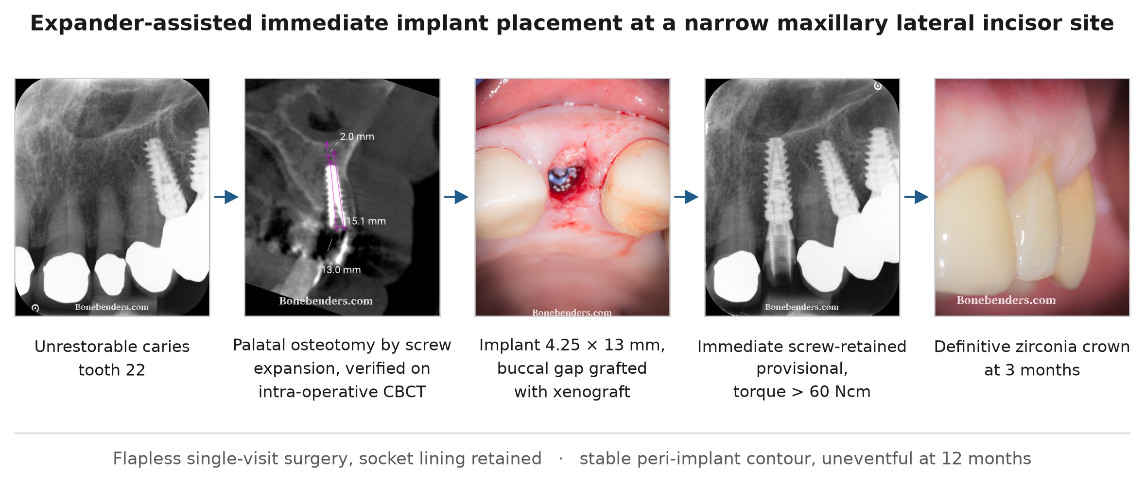
Background. Replacement of a maxillary lateral incisor is among the most demanding indications in implant dentistry. The site is typically narrow bucco-palatally, the buccal plate is frequently thin and largely bundle-bone dependent, and the aesthetic tolerance for ridge collapse is minimal. Immediate (Type 1) placement shortens treatment and preserves the soft-tissue architecture, but it does not prevent the remodelling of the buccal wall that follows extraction, and the osteotomy itself risks fenestration or loss of primary stability. Case presentation. A 60-year-old male smoker (about 20 cigarettes/day) with a history of stage IV periodontitis presented with an unrestorable, almost circumferential carious lesion of the maxillary left lateral incisor, already restored with a metal-ceramic crown (FDI 22). After atraumatic flapless extraction, the osteotomy was initiated on the palatal wall of the socket with a lance bur and a 2.2 mm pilot drill, and completed with a sequence of screw bone expanders instead of rotary drills. Expander position and axis were verified intraoperatively with a periapical radiograph and a CBCT scan before the osteotomy was finalized. An implant (“Newton”, Kalodon, 4.25 × 13 mm) was placed in a palatally shifted position with apical bone expansion of the nasal-floor cortex (part of the “supramaxillary arch buttress”, a nomenclature we have proposed), achieving an insertion torque exceeding 60 Ncm. The residual buccal gap was filled with a deproteinized bovine bone mineral xenograft to compensate for the expected resorption of the bundle bone, and a screw-retained provisional restoration was delivered the same day. Healing was uneventful. At three months the peri-implant mucosa was stable and well contoured, with no visible collapse of the buccal contour; the site was digitally scanned and a definitive screw-retained zirconia crown was delivered. Radiographically, the marginal bone level appeared stable at all controls. The patient was then followed for 12 months after crown delivery, with an uneventful course. Conclusions. In a narrow lateral incisor socket, combining “apical expansion” with immediate implant placement, buccal gap grafting and immediate provisionalization allowed a single-surgery, three-month rehabilitation with preserved mucosal architecture. Intraoperative three-dimensional verification of the expander before finalizing the osteotomy was decisive in confirming the implant axis in a site with minimal (bundle) buccal bone. The result was obtained in a heavy smoker with a history of severe periodontitis and should be confirmed with quantitative, longer-term data. Engaging the “supramaxillary arch buttress” of the splanchnocranium ensures the necessary stability.
Keywords:
immediate implant placement
; ridge expansion
; bone expander
; xenograft
; bundle bone
; immediate provisionalization
; maxillary lateral incisor
; aesthetic zone
1. Introduction
The dimensional changes that follow tooth extraction are well characterized. In the classic dog model, the buccal wall of the socket — composed to a large extent of bundle bone, a tooth-dependent structure that loses its function with the periodontal ligament — undergoes marked vertical and horizontal resorption during the first weeks of healing.1 Placing an implant into the fresh socket does not arrest this process: the buccal and lingual walls remodel essentially as they would in an ungrafted socket, and the implant surface may become exposed where the wall was thin.2,3
In the anterior maxilla these changes translate directly into aesthetic risk. Systematic reviews of immediate (Type 1, according to the 2004 ITI Consensus Conference) placement report acceptable survival but a non-negligible frequency of mid-facial recession, which correlates with a thin buccal phenotype, a buccally positioned implant shoulder and a flapped approach.4–6 A buccal bone thickness of at least 2 mm has been proposed as the target for long-term stability of the facial contour,7 a value rarely available at the maxillary lateral incisor, where the ridge is frequently narrow or knife-edged.
Two adjunctive strategies have been developed to mitigate this. The first is grafting of the horizontal defect (gap) between the implant and the buccal plate, usually with a slowly resorbing xenograft; the second is immediate provisionalization, which supports the soft tissue during the critical remodelling phase. The combination — the "dual-zone" concept — was described by Chu and co-workers,8 and retrospective cohorts from the same group showed that grafting and/or provisional restoration reduce facial–palatal ridge dimensional change and better preserve peri-implant soft-tissue height and thickness.9,10 Contemporary meta-analyses confirm a modest but consistent benefit of socket grafting11 and of immediate provisionalization12 on aesthetic outcomes, and ITI consensus statements identify immediate restoration/loading as a valid protocol when primary stability and case selection permit.13
A separate problem is how to prepare the osteotomy in a narrow ridge. Rotary instrumentation in a thin crest removes bone and may perforate the buccal plate. Summers introduced osteotomes as an alternative: by compacting rather than excising bone, the ridge-expansion osteotomy widens a narrow crest and increases bone density around the implant.14,15 Applying this principle inside a fresh extraction socket — where the palatal wall must guide the implant away from the buccal plate — is technically demanding, because the expander tends to follow the path of least resistance along the socket axis. A second problem is where to find primary stability. In a single post-extraction site the socket walls rarely provide it, and anchorage has to be sought beyond the socket, in dense cortical bone that largely coincides with the resistance pillars (buttresses) of the facial skeleton. For the maxillary incisors, the cortical floor of the nasal fossa and the canine pillar are the structures most commonly available for this purpose.
This report describes a case in which these elements were combined in a single surgical session at a maxillary lateral incisor site, with intraoperative CBCT verification of the expander before the osteotomy was finalized. The report follows the CARE guidelines.
2. Case Presentation
2.1. Patient Information and Chief Complaint
A 60-year-old male patient (ASA II) attended with acute pain due to an unrestorable carious lesion of the vital maxillary left lateral incisor (tooth 22). He was a smoker of about 20 cigarettes per day and reported no bisphosphonate or other antiresorptive therapy and no relevant allergies. His periodontal diagnosis was periodontitis stage IV, grade C. At the time of treatment the full-mouth periodontal status was: maximum probing depth 4 mm, bleeding on probing 20%, furcation involvement up to degree I and tooth mobility up to degree 1. The patient had previously been rehabilitated with implant-supported restorations in the left posterior maxilla, and posterior mandible.
2.2. Clinical and Radiographic Findings
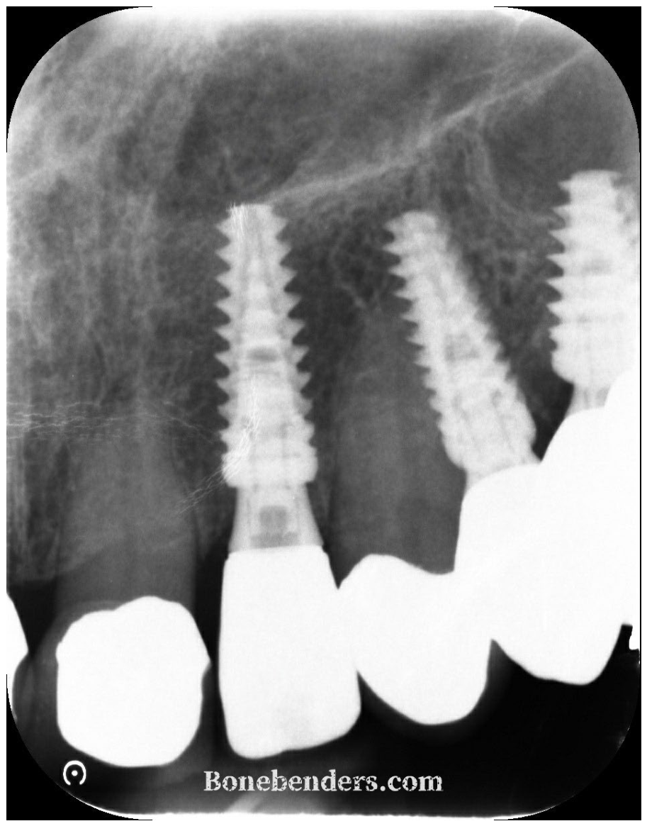
Clinically, upon probing, the maxillary left lateral incisor presented an extensive carious lesion extending subgingivally. The gingival phenotype was thick. The preoperative periapical radiograph (Figure 1) showed a radiolucent defect of the coronal and cervical portion of the tooth; intraoperative inspection confirmed that the lesion was almost circumferential and extended apically beyond the level at which a ferrule could be obtained, so that the tooth was deemed unrestorable. Two osseointegrated implants supporting fixed restorations were present distally in the same quadrant. Peri-apical bone appeared intact and there was no radiographic sign of apical pathology.
2.3. Diagnosis and Treatment Plan
Diagnosis: unrestorable subgingival caries of a vital tooth 22 (FDI numbering system) with an intact socket.
Given the intact socket walls and the absence of infection, immediate implant placement with immediate provisionalization was planned, with the explicit intention of (i) placing the implant palatally and apically to the socket, engaging the nasal-floor cortex for primary stability, (ii) preparing the osteotomy predominantly by ridge expansion rather than drilling in order to preserve and compact the thin buccal plate, and (iii) grafting the residual buccal gap with a xenograft. Written informed consent was obtained, including consent to publish anonymized clinical images.
2.4. Surgical Procedure
Under local anaesthesia (Articaine 40 mg/mL + Epinephrine 1:100,000, Pierrelgroup, Capua (CE)Italy), tooth 22 was extracted flaplessly and atraumatically with a sagittal apico-coronal section using a carbide bur on a high-speed handpiece, periotomes and thin-tip root elevators, taking care not to luxate the buccal plate. The socket was purposely not debrided, in order to preserve part of the periodontal ligament (PDL) cells and the reactive tissue lining the socket, which has been proposed to act as an in-situ regenerative reservoir.16 It was inspected with a periodontal probe, and all four bony walls, including the buccal plate, were found to be intact.
The osteotomy was initiated by marking and penetrating the palatal wall of the socket with a lance bur at low speed (100 rpm) without irrigation. The direction was deliberately changed with respect to the original axis of the tooth, which in this site is too buccal: a buccal position of the implant in the original socket would risk fracturing the thin bundle bone and, in the absence of a strong apical bone structure, would not provide an insertion torque sufficient for immediate loading.
A 2.2 mm pilot drill (100 rpm, without irrigation) was then used to slightly enlarge the access, after which a sequence of screw bone expanders with the same shape as the final implant (Bonbender expander system, Kalodon Implants, Italy) was introduced along the planned palatal–apical axis. The expanders were inserted with the implant contra-angle at 15 rpm and high torque, or with a manual wrench. The final osteotomy was clearly distinct from the original socket (Figure 2).
A periapical radiograph was taken with the 3.8 × 13 mm expander in position (Figure 3) and, because primary stability was essential and the margin for error at the buccal plate minimal, a cone-beam CT scan was acquired intraoperatively with the expander in situ before the osteotomy was finalized (Figure 4). The sagittal reconstruction confirmed the expander axis within the residual bone on the palatal side of the socket and the close proximity of its apical portion to the nasal floor. Three measurements were taken on this scan: the length of the expander in situ (13.0 mm), the depth that a 15 mm implant would have reached along the same axis (15.1 mm), and the residual bone height between the apex of the expander and the nasal floor (2.0 mm) — that is, the additional depth still available beyond the expander. A 15 mm implant would have used that additional depth, but its coronal portion would then have remained too superficial. It was therefore judged safer to exploit the full available depth with a 13 mm implant, seated deeper than the expander so as to engage the apical cortical bone.
A “Newton” (Kalodon) 4.25 × 13 mm implant was then inserted along the prepared axis with the shoulder positioned 5 mm apical to the mid-buccal gingival margin and shifted palatally to maintain a buccal gap. Primary stability was obtained by engaging the cortical plate of the nasal floor apically, within the bony framework we have proposed to describe as the “supramaxillary arch”17; insertion torque exceeded 60 Ncm and ISQ was not measured. The immediate postoperative periapical radiograph documented the implant position and the apical cortical engagement of the nasal floor (Figure 5).
The residual horizontal gap between the implant surface and the buccal plate was filled with a deproteinized bovine bone mineral xenograft (S1 Sticky Bone, MEDPARK DENTAL, Seoul, Republic of Korea; a commercially agglutinated bovine-derived particulate xenograft, supplied in a “sticky” formulation without the addition of autologous fibrin) delivered without a membrane and condensed to the level of the crestal bone, with the explicit purpose of compensating for the resorption of the bundle bone (Figure 6). No flap was raised and no sutures were required.
2.5. Immediate Provisionalization
A screw-retained provisional crown was fabricated chairside on an anatomical titanium abutment and delivered immediately after surgery. The emergence profile was shaped to support the gingival margin and the papillae while remaining free of centric and eccentric contacts. A periapical radiograph confirmed complete seating of the provisional and the marginal bone level at delivery (Figure 7).
Postoperative instructions comprised the prescription of ibuprofen twice daily if necessary, prohibition of rinsing of any kind for 5 days, a soft diet and avoidance of any function on the provisional. The patient was also instructed to start using the provisional gradually after the first week, with the goal to return to normal function after a month.
2.6. Follow-Up and Outcomes
Healing was uneventful. At 30 days the control radiograph showed an unchanged marginal bone level, with no peri-implant radiolucency (Figure 8).
At three months the provisional was removed for the definitive impression. The peri-implant mucosa was healthy, with a well-defined and stable emergence profile reproducing the shape of the provisional (Figure 9). Osseointegration was confirmed clinically by the absence of mobility and of pain on percussion and on removal/reseating of the provisional. A digital scan was taken at the same appointment.
A definitive screw-retained zirconia crown was fabricated and delivered. On the day of delivery the soft-tissue architecture was examined with the provisional removed (Figure 10) and after seating the definitive crown (Figure 11): the mid-facial mucosal margin was at the level of the contralateral/adjacent reference, the buccal contour was convex without visible collapse, and the papillae filled the embrasures. The corresponding periapical radiograph showed a stable marginal bone level and a correctly seated crown (Figure 12). The patient was subsequently followed for 12 months after delivery of the definitive crown; the course was uneventful, with no biological or technical complications.
2.7. Timeline2.8. Patient Perspective
| Time point | Event |
| Day 0 — surgery | Presentation with acute pain; tooth 22 diagnosed unrestorable. Flapless atraumatic extraction without socket curettage; palatal osteotomy with lance bur, 2.2 mm pilot drill and screw expanders; intraoperative periapical radiograph and CBCT verification of the expander; placement of a 4.25 × 13 mm implant with insertion torque exceeding 60 Ncm and apical engagement of the nasal-floor cortex; buccal gap grafted with bovine xenograft; screw-retained provisional delivered free of occlusal contacts. |
| Days 0–5 | Ibuprofen as required; no rinsing of any kind; soft diet; no function on the provisional. |
| Week 1 to month 1 | Progressive return to normal function on the provisional. |
| 30 days | Periapical control: marginal bone level unchanged, no peri-implant radiolucency. |
| 3 months | Provisional removed; peri-implant mucosa healthy with a stable emergence profile; digital scan; definitive screw-retained zirconia crown delivered; periapical control. |
| 3 to 15 months | Twelve months of clinical follow-up after crown delivery; uneventful, with no biological or technical complications. |
I seeked an immediate check with my dentist about an acute pain on the left side of my mouth. The pain did not respond to ibuprofen and other drugs I had at home. The doctor told me the pain originated from a deep cavity underneath an old crown of one of my front upper teeth. I asked him to take care of it. And so he did.
They acted rapidly at the office and replaced my tooth with an immediate implant. I took an antibiotic and ibuprofen on the same day. The next day all pain was gone and I went on with my normal life.
3. Discussion
This case combined three interventions that address different components of the same problem, and it is worth separating what each can and cannot be expected to do.
Apical bone expansion in a fresh socket and engagement of the supramaxillary arch buttress. The expander was used instead of rotary drilling because the residual bone palatal to the socket was narrow and because compaction, rather than excision, preserves bone volume and improves the density of the implant bed.14,15 The trade-off is control: within a fresh socket the expander is guided as much by the existing socket walls as by the operator, so the risk is that the final osteotomy reproduces the socket axis — which in the lateral incisor is typically too buccal — rather than the prosthetically correct palatal axis. This is precisely why the position of the expander was checked radiographically and then three-dimensionally before the osteotomy was finalized (Figs. 3 and 4). Intraoperative CBCT is not a routine step and carries an additional radiation dose that must be justified for each patient; here it was considered justified by the combination of a narrow alveolar crest, a flapless approach that precluded direct visual inspection of the buccal plate, and apical engagement of the nasal-floor cortex (part of the supramaxillary arch buttress) to obtain high stability. Static or dynamic guided surgery would be a reasonable alternative in comparable sites, although a guide is of limited help with a screw-expander protocol.
Grafting of the buccal gap. The experimental evidence is unambiguous that implant placement does not prevent remodelling of the buccal wall,2,3 and that the thinner the wall, the more of it is lost. Filling the gap with a slowly resorbing xenograft does not stop bundle-bone resorption at the biological level, but it maintains the volume of the compartment while it occurs. The retrospective cohorts of Tarnow and Chu showed less facial–palatal dimensional change and better soft-tissue height and thickness when grafting, provisionalization, or both, were used in flapless post-extraction sites.9,10 A meta-analysis of immediate placement with or without socket grafting likewise favours grafting for aesthetic outcomes, albeit with modest effect sizes and heterogeneous protocols.11 In the present case the graft remained radiopaque at the buccal aspect at every control, and the buccal contour at crown delivery showed no visible collapse — but it must be stated plainly that a two-dimensional periapical radiograph cannot image the buccal plate, and that only a comparative CBCT or a superimposed intraoral scan can quantify contour change. This is a limitation of the present documentation.
Immediate provisionalization. The provisional served to shape and support the peri-implant mucosa from day one; the soft-tissue profile recorded at the three-month scan (Figure 9) is a direct imprint of its emergence profile. Immediate provisionalization in immediate placement has been associated with less mid-facial recession in meta-analysis,12 and current ITI consensus supports immediate restoration where primary stability is sufficient and occlusal loading is controlled.13 The prerequisite is primary stability, obtained here by apical engagement of the nasal-floor cortex rather than by the socket walls — a strategy widely used in the anterior maxilla but one that places the implant apex against a thin cortical plate and therefore requires careful control of the apical axis, reinforcing the rationale for the intraoperative imaging.
Case selection. Buser and co-workers have argued that the advantages of immediate placement apply to a restricted subset of patients: intact socket walls, a thick gingival phenotype, a thick buccal plate, no acute infection and a low smile line.6 Evans and Chen showed that recession after immediate placement is greater at sites with a thin phenotype and a buccally placed implant shoulder.5 The present site met the criteria of an intact socket and an absence of acute infection; the narrow ridge, however, is a risk factor rather than a favourable feature, and the result reported here should not be read as evidence that immediate placement is appropriate in all narrow lateral incisor sites. The patient also presented two recognized risk factors that are not captured by these site-level criteria: heavy smoking (about 20 cigarettes per day) and a history of stage IV periodontitis. Both are associated with a higher risk of biological complications around implants, and the favourable short-term result in this patient does not reduce the need for strict supportive peri-implant care.
Retention of the socket lining. The socket was deliberately not curetted, in order to preserve part of the periodontal ligament and of the reactive tissue lining its walls.16 This departs from the conventional instruction to debride a socket thoroughly before immediate placement, and it is reported here as a deliberate deviation rather than an omission. Two separate lines of evidence make it defensible. First, the biological value of retained ligament tissue is the explicit premise of partial extraction therapy: in the socket-shield technique a buccal root fragment with its periodontal ligament is deliberately left in place, and the original proof-of-principle report described preservation of the buccal tissues at the retained fragment.18 Second, the concern that residual soft tissue compromises osseointegration is not borne out by the clinical literature on compromised sockets: a systematic review of immediate placement into infected sites found survival comparable to that reported for non-infected sites, while noting the limited quality of the available evidence,19 and prospective human series of implants placed into periapically and periodontally infected fresh sockets have reported comparable survival and marginal bone behaviour over follow-up of up to four years.20,21 Neither line of evidence tests the specific choice made here — retaining the lining of a non-infected socket in a heavy smoker with treated stage IV periodontitis — and the uneventful course in this single patient cannot be taken as validation of it.
Limitations. This is a single case with a short observation period (three months to definitive crown delivery, followed by 12 months of uneventful clinical follow-up). Clinical outcomes are described qualitatively up to crown delivery: no quantitative aesthetic index, no quantitative radiographic bone-level measurement and no volumetric comparison of the buccal contour are reported. The 12-month course after crown delivery was clinically uneventful but was not documented with standardized photographs or radiographs, so it supports the absence of complications rather than a measured stability of the soft-tissue and bone levels. Longer follow-up with standardized photographs, superimposed intraoral scans and, if clinically indicated, a comparative CBCT would be required to document the behaviour of the grafted buccal compartment over time.
4. Conclusion
In a narrow maxillary lateral incisor site with an unrestorable tooth and intact socket walls, immediate implant placement performed with an expander-based osteotomy, palatal–apical implant positioning with nasal-floor cortical engagement, buccal gap grafting with a xenograft and same-day provisionalization produced a stable, well-contoured peri-implant soft-tissue profile at the time of definitive crown delivery three months later. Intraoperative verification of the expander axis — periapical and then three-dimensional — was the step that made a flapless approach in a knife-edge ridge controllable. Longer-term and quantitative follow-up is needed before the approach can be recommended beyond carefully selected sites.
Declarations
Funding.: This research received no external funding.
Author: contributions. E.B. performed the treatment, collected the documentation and wrote the manuscript.
Ethics: and consent. Ethical approval was not required for this single-patient case report, which describes treatment delivered as part of routine clinical care. Treatment was performed in accordance with the Declaration of Helsinki. The patient provided written informed consent to the treatment and to the publication of anonymized clinical photographs and radiographs.
Data: availability. The clinical and radiographic data supporting this report are available from the corresponding author on reasonable request, within the limits of patient confidentiality.
Conflicts: of Interest. The author contributed to the development of the bone expanders used in this case and receives no royalties from them. The author declares no other conflicts of interest.
References
- Araújo, M.G.; Lindhe, J. Dimensional ridge alterations following tooth extraction. An experimental study in the dog. J. Clin. Periodontol. 2005, 32(2), 212–218. [Google Scholar] [CrossRef] [PubMed]
- Araújo, M.G.; Sukekava, F.; Wennström, J.L.; Lindhe, J. Ridge alterations following implant placement in fresh extraction sockets: an experimental study in the dog. J. Clin. Periodontol. 2005, 32(6), 645–652. [Google Scholar] [CrossRef] [PubMed]
- Araújo, M.G.; Wennström, J.L.; Lindhe, J. Modeling of the buccal and lingual bone walls of fresh extraction sites following implant installation. Clin. Oral Implant. Res. 2006, 17(6), 606–614. [Google Scholar] [CrossRef] [PubMed]
- Chen, S.T.; Buser, D. Esthetic outcomes following immediate and early implant placement in the anterior maxilla — a systematic review. Int. J. Oral Maxillofac. Implant. 2014, 29 (Suppl), 186–215. [Google Scholar] [CrossRef] [PubMed]
- Evans, C.D.; Chen, S.T. Esthetic outcomes of immediate implant placements. Clin. Oral Implant. Res. 2008, 19(1), 73–80. [Google Scholar] [CrossRef] [PubMed]
- Buser, D.; Chappuis, V.; Belser, U.C.; Chen, S. Implant placement post extraction in esthetic single tooth sites: when immediate, when early, when late? Periodontol. 2000 2017, 73(1), 84–102. [Google Scholar] [CrossRef] [PubMed]
- Grunder, U.; Gracis, S.; Capelli, M. Influence of the 3-D bone-to-implant relationship on esthetics. Int. J. Periodontics Restor. Dent. 2005, 25(2), 113–119. [Google Scholar] [PubMed]
- Chu, S.J.; Salama, M.A.; Salama, H.; Garber, D.A.; Saito, H.; Sarnachiaro, G.O.; Tarnow, D.P. The dual-zone therapeutic concept of managing immediate implant placement and provisional restoration in anterior extraction sockets. Compend Contin. Educ. Dent. 2012, 33(7), 524–532, 534. [Google Scholar] [PubMed]
- Tarnow, D.P.; Chu, S.J.; Salama, M.A.; Stappert, C.F.J.; Salama, H.; Garber, D.A.; Sarnachiaro, G.O.; Sarnachiaro, E.; Gotta, S.L.; Saito, H. Flapless postextraction socket implant placement in the esthetic zone: part 1. The effect of bone grafting and/or provisional restoration on facial–palatal ridge dimensional change — a retrospective cohort study. Int. J. Periodontics Restor. Dent. 2014, 34(3), 323–331. [Google Scholar] [CrossRef] [PubMed]
- Chu, S.J.; Salama, M.A.; Garber, D.A.; Salama, H.; Sarnachiaro, G.O.; Sarnachiaro, E.; Gotta, S.L.; Reynolds, M.A.; Saito, H.; Tarnow, D.P. Flapless postextraction socket implant placement, part 2: the effects of bone grafting and provisional restoration on peri-implant soft tissue height and thickness — a retrospective study. Int. J. Periodontics Restor. Dent. 2015, 35(6), 803–809. [Google Scholar] [CrossRef] [PubMed]
- Seyssens, L.; Eeckhout, C.; Cosyn, J. Immediate implant placement with or without socket grafting: a systematic review and meta-analysis. Clin. Implant Dent. Relat. Res. 2022, 24(3), 339–351. [Google Scholar] [CrossRef] [PubMed]
- Pitman, J.; Seyssens, L.; Christiaens, V.; Cosyn, J. Immediate implant placement with or without immediate provisionalization: a systematic review and meta-analysis. J. Clin. Periodontol. 2022, 49(10), 1012–1023. [Google Scholar] [CrossRef] [PubMed]
- Morton, D.; Wismeijer, D.; Chen, S.; Hamilton, A.; Wittneben, J.; Casentini, P.; et al. Group 5 ITI Consensus Report: implant placement and loading protocols. Clin. Oral Implant. Res. 2023, 34 (Suppl 26), 349–356. [Google Scholar] [CrossRef] [PubMed]
- Summers, R.B. A new concept in maxillary implant surgery: the osteotome technique. Compendium 1994, 15(2), 152–162. [Google Scholar] [PubMed]
- Summers, R.B. The osteotome technique: part 2 — the ridge expansion osteotomy (REO) procedure. Compendium 1994, 15(4), 422–436. [Google Scholar] [PubMed]
- Bruschi, G.B.; De Nale, M.; Bruschi, E.; Viganò, P.; Tuscano, D. Granulation tissue as an in-situ regenerative reservoir in periodontal, implant, and bone-regeneration surgery: a demineralized-bone-matrix hypothesis [preprint]. Zenodo 2026. [Google Scholar] [CrossRef]
- Bruschi, E. The supramaxillary arch: the hidden buttress of the splanchnocranium: technical note. Zenodo 2026. [Google Scholar] [CrossRef]
- Hürzeler, M.B.; Zuhr, O.; Schupbach, P.; Rebele, S.F.; Emmanouilidis, N.; Fickl, S. The socket-shield technique: a proof-of-principle report. J. Clin. Periodontol. 2010, 37(9), 855–862. [Google Scholar] [CrossRef] [PubMed]
- Waasdorp, J.A.; Evian, C.I.; Mandracchia, M. Immediate placement of implants into infected sites: a systematic review of the literature. J. Periodontol. 2010, 81(6), 801–808. [Google Scholar] [CrossRef] [PubMed]
- Crespi, R.; Capparè, P.; Gherlone, E. Fresh-socket implants in periapical infected sites in humans. J. Periodontol. 2010, 81(3), 378–383. [Google Scholar] [CrossRef] [PubMed]
- Crespi, R.; Capparè, P.; Gherlone, E. Immediate loading of dental implants placed in periodontally infected and non-infected sites: a 4-year follow-up clinical study. J. Periodontol. 2010, 81(8), 1140–1146. [Google Scholar] [CrossRef] [PubMed]
Figure 1.
Preoperative periapical radiograph. Unrestorable carious lesion of tooth 22, intraoperatively found to be almost circumferential. Two osseointegrated implants are present distally in the same quadrant.
Figure 1.
Preoperative periapical radiograph. Unrestorable carious lesion of tooth 22, intraoperatively found to be almost circumferential. Two osseointegrated implants are present distally in the same quadrant.
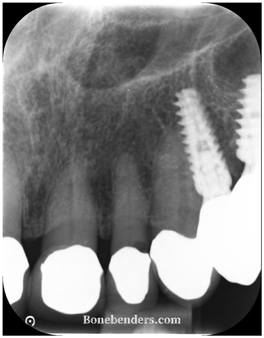
Figure 2.
Occlusal clinical view immediately before implant insertion. The final osteotomy created, essentially, with screw expanders, is on the palatal side and distinct from the original socket on the buccal side.
Figure 2.
Occlusal clinical view immediately before implant insertion. The final osteotomy created, essentially, with screw expanders, is on the palatal side and distinct from the original socket on the buccal side.
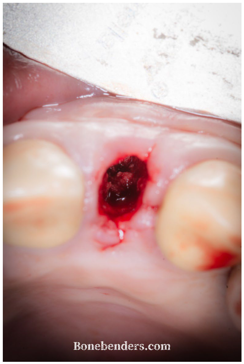
Figure 3.
Periapical radiograph taken with the 3.8 × 13 mm bone expander in position during preparation of the osteotomy, used to verify the axis before proceeding.
Figure 3.
Periapical radiograph taken with the 3.8 × 13 mm bone expander in position during preparation of the osteotomy, used to verify the axis before proceeding.
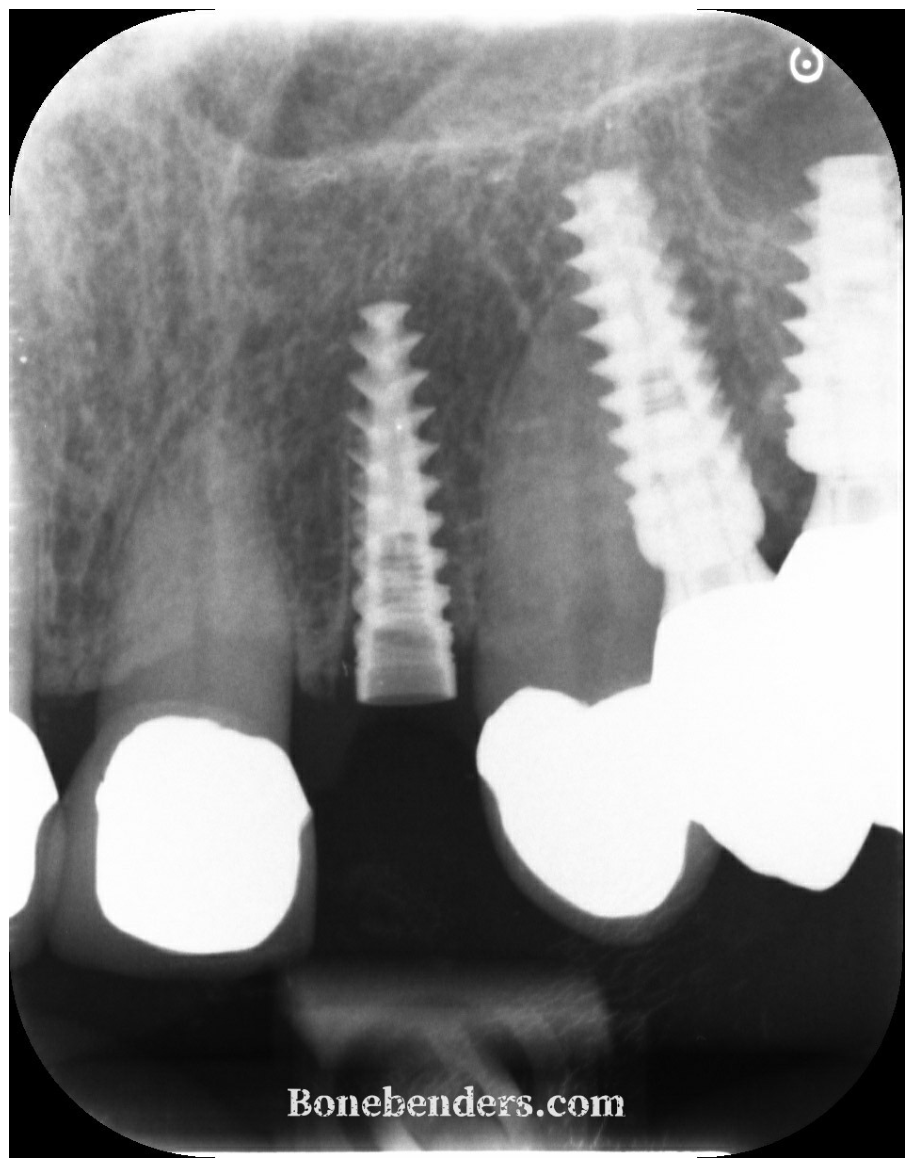
Figure 4.
Intraoperative CBCT, sagittal reconstruction, with the 13 mm expander in situ before the osteotomy was finalized. 13.0 mm: length of the expander; 15.1 mm: the depth that a 15 mm implant would have reached along the same axis; 2.0 mm: the residual bone height between the apex of the expander and the nasal floor, i.e. the additional depth still available beyond the expander.
Figure 4.
Intraoperative CBCT, sagittal reconstruction, with the 13 mm expander in situ before the osteotomy was finalized. 13.0 mm: length of the expander; 15.1 mm: the depth that a 15 mm implant would have reached along the same axis; 2.0 mm: the residual bone height between the apex of the expander and the nasal floor, i.e. the additional depth still available beyond the expander.
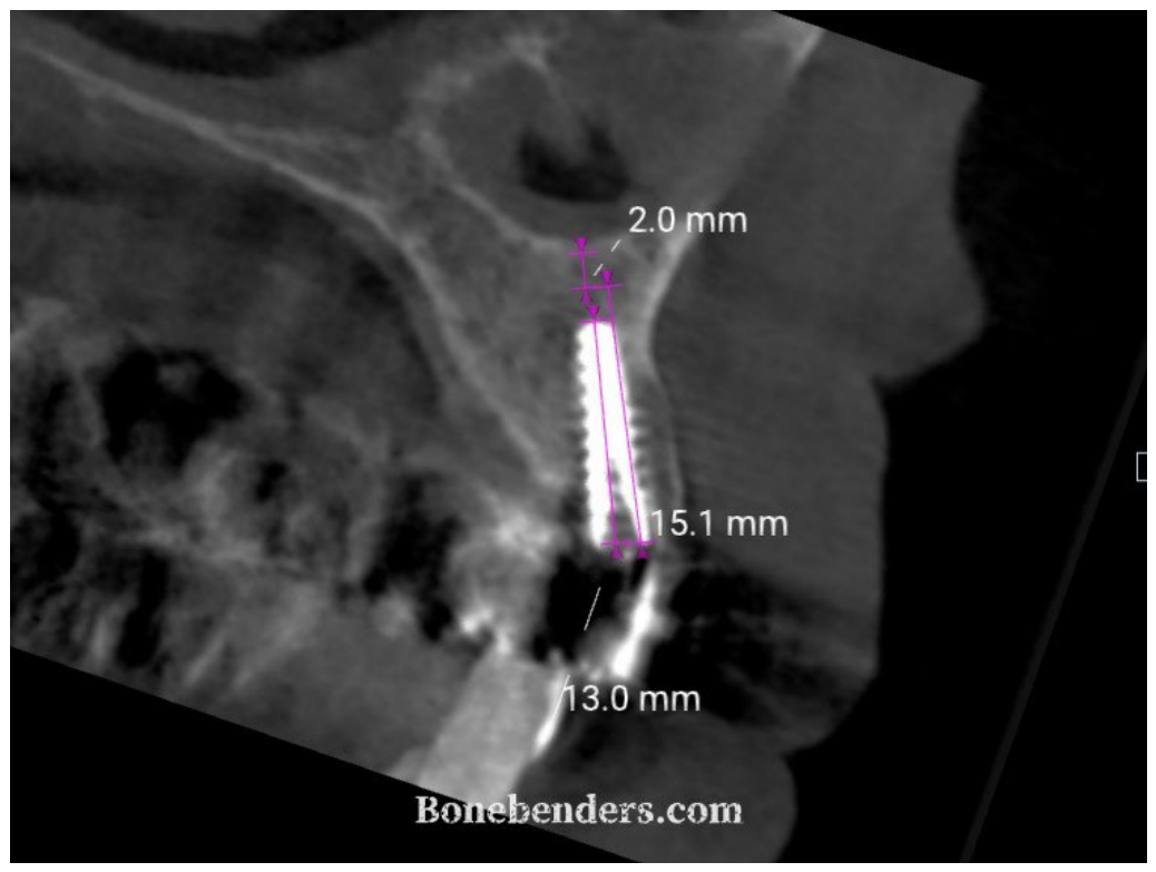
Figure 5.
Periapical radiograph immediately after implant insertion, showing the palatal–apical position and engagement of the nasal-floor cortex.
Figure 5.
Periapical radiograph immediately after implant insertion, showing the palatal–apical position and engagement of the nasal-floor cortex.
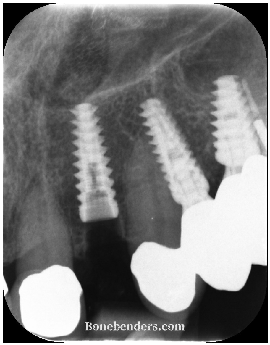
Figure 6.
Occlusal clinical view immediately after implant placement at site 22. The residual buccal gap has been filled with a deproteinized bovine sticky bone (S1 Bone) mineral xenograft to compensate for the expected resorption of the bundle bone.
Figure 6.
Occlusal clinical view immediately after implant placement at site 22. The residual buccal gap has been filled with a deproteinized bovine sticky bone (S1 Bone) mineral xenograft to compensate for the expected resorption of the bundle bone.
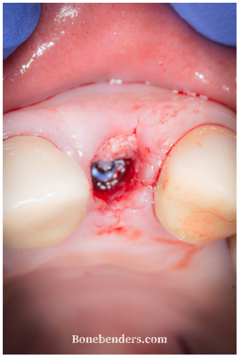
Figure 7.
Periapical radiograph after delivery of the screw-retained immediate provisional restoration.
Figure 7.
Periapical radiograph after delivery of the screw-retained immediate provisional restoration.
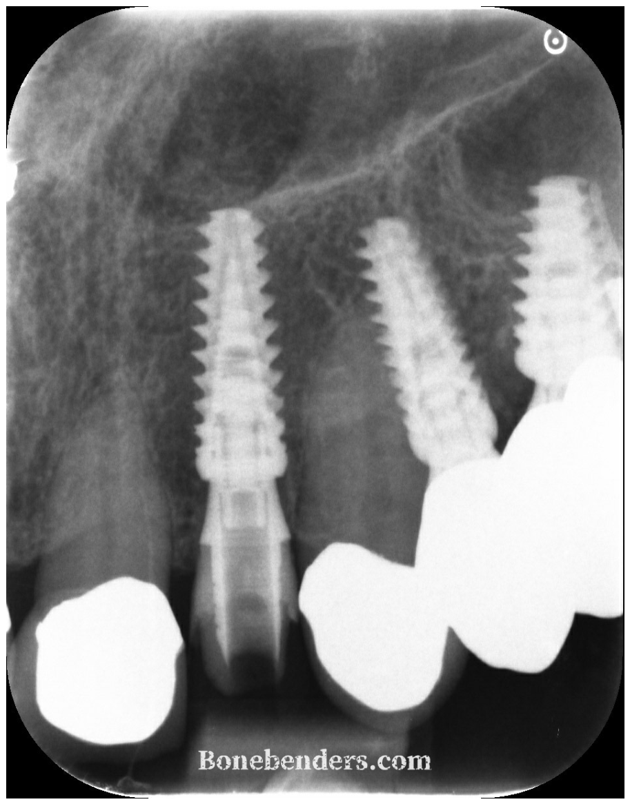
Figure 8.
Control radiograph at 30 days: implant and provisional in situ, marginal bone level unchanged, buccal graft still radiopaque.
Figure 8.
Control radiograph at 30 days: implant and provisional in situ, marginal bone level unchanged, buccal graft still radiopaque.
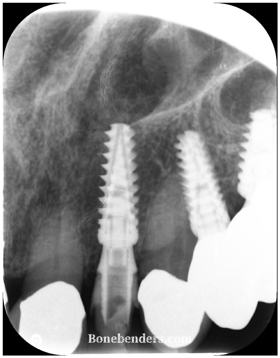
Figure 9.
Soft-tissue appearance at three months, at the appointment for the digital scan, with the provisional removed. The emergence profile created by the provisional is maintained.
Figure 9.
Soft-tissue appearance at three months, at the appointment for the digital scan, with the provisional removed. The emergence profile created by the provisional is maintained.
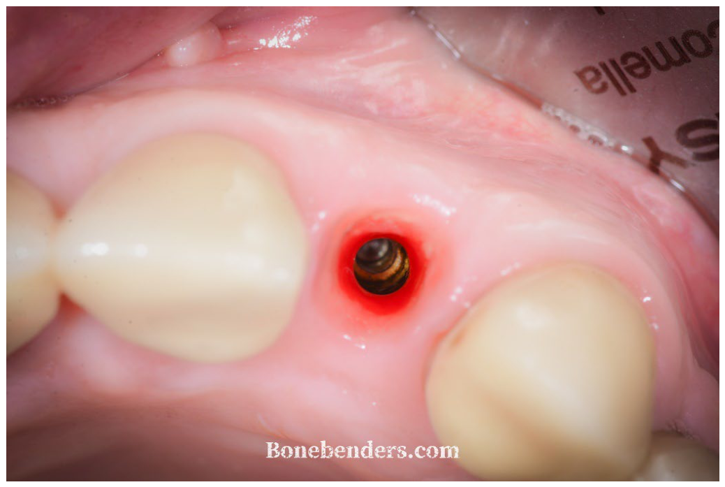
Figure 10.
Lateral clinical view on the day of delivery of the definitive zirconia crown, with the restoration removed.
Figure 10.
Lateral clinical view on the day of delivery of the definitive zirconia crown, with the restoration removed.
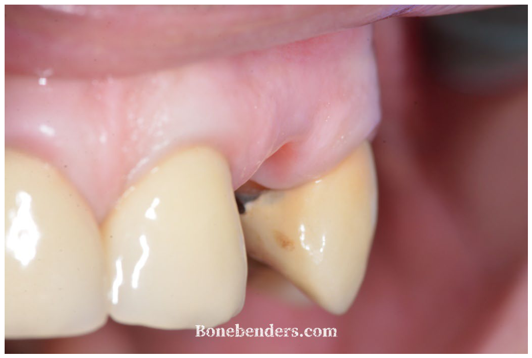
Figure 11.
Lateral clinical view on the day of delivery, with the definitive zirconia crown seated.
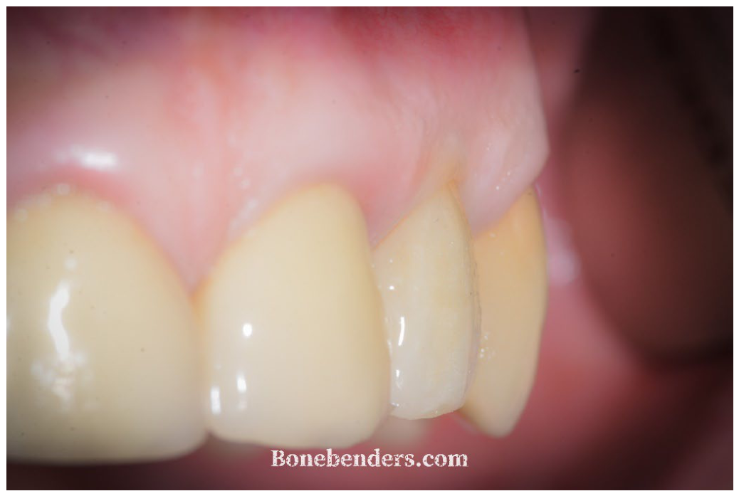
Figure 12.
Periapical radiograph at three months, after delivery of the definitive crown.
Disclaimer/Publisher’s Note: The statements, opinions and data contained in all publications are solely those of the individual author(s) and contributor(s) and not of MDPI and/or the editor(s). MDPI and/or the editor(s) disclaim responsibility for any injury to people or property resulting from any ideas, methods, instructions or products referred to in the content. |
© 2026 by the author. Licensee MDPI, Basel, Switzerland. This article is an open access article distributed under the terms and conditions of the Creative Commons Attribution (CC BY) license.
Copyright: This open access article is published under a Creative Commons CC BY 4.0 license, which permit the free download, distribution, and reuse, provided that the author and preprint are cited in any reuse.