

Submitted:
07 October 2026
Posted:
09 October 2026
You are already at the latest version
Abstract
Purpose:
Maxillofacial metastases from prostate cancer are accounts for 9–11% of metastatic jaw lesions. Optimal imaging for suspected maxillofacial metastases remains undefined. Conventional imaging often lacks sensitivity for early disease, while PET/CT may be limited due to physiologic tracer uptake in adjacent salivary glands. With increasing use of PET/CT in advanced prostate cancer, its diagnostic performance in the mandibular region requires further evaluation.
Methods:
We conducted a single-institution paired retrospective study using the prospectively maintained Mayo Clinic Advanced Prostate Cancer Registry. Patients with clinically confirmed mandibular metastases who underwent molecular PET/CT ([⁶⁸Ga]Ga-PSMA-11 or [¹¹C]choline) and conventional imaging (CT or MRI) within 1 month were included. All studies were blindly reviewed by a board-certified radiologist specialized in nuclear medicine. Detection rates, anatomic distribution, and quantitative imaging parameters were assessed. paired detection rates were compared using a one-sided exact McNemar test, with a two-sided exact test performed as a sensitivity analysis.
Results:
Eighteen patients with clinically confirmed mandibular metastases underwent 36 paired imaging evaluations (18 molecular PET/CT and 18 conventional studies). Median age was 70.5 years (IQR, 60–77), and median Gleason score was 8 (IQR, 7–9). At detection of mandibular metastasis, 56% of our cohort had hormone-sensitive and 44% had castration-resistant disease. Molecular PET/CT detected mandibular involvement in 18/18 patients (100%; 95% CI, 81–100%) versus 13/18 (72.2%; 95% CI, 47–90%) with conventional imaging. All five discordant cases were detected by molecular PET/CT but not conventional imaging, with no discordant cases favoring conventional imaging (one-sided exact McNemar p = 0.031; two-sided p = 0.063). Mandibular involvement was predominantly unilateral (94.4%), most commonly involving the body (50%), followed by the ramus (28%) and condyle (17%). Median SUVmax was 6.3 (IQR, 4.4–16.9) for PSMA PET/CT and 4.3 (IQR, 3.8–8.5) for choline PET/CT; median lesion volume was 5.0 mL (IQR, 1.1–11.3).
Conclusions:
Molecular PET/CT identified all clinically confirmed mandibular prostate cancer metastases in this paired cohort, including five cases not detected by conventional imaging. Despite physiologic tracer uptake in adjacent salivary structures, mandibular metastatic disease remained detectable on molecular imaging. These findings suggest that molecular PET/CT may provide complementary value when mandibular metastasis is suspected, particularly when conventional imaging is negative or equivocal.
Keywords:
neoplasm metastasis
; mandible
; positron-emission tomography
; jaw neoplasms
; conventional imaging
1. Introduction
Prostate cancer represents a significant global health burden, with bone being the most common site of distant metastasis at presentation, observed in approximately 82% of patients with de novo metastatic disease [1,2]. Osseous metastases to the axial skeleton are predominant, with involvement of the oral and maxillofacial region is uncommon. Metastatic tumors to the oral cavity account for only 1-3% of all malignant oral lesions, with prostate cancer representing approximately 9-11% of these cases [3,4]. The mandible is more frequently affected than the maxilla or oral soft tissues, with metastases often presenting with non-specific symptoms including pain, swelling, paresthesia, or numb chin syndrome [5,6,7]. Importantly, oral metastases represent the first indication of metastatic spread or an undiscovered primary malignancy in approximately 23-25%; underscoring the importance of accurate detection and characterization [3,8].
Conventional imaging of suspected osseous metastases has traditionally relied on technetium-99m-methylene diphosphonate (99mTc-MDP) bone scintigraphy in combination with anatomic [5,6,7]imaging computed tomography (CT) or magnetic resonance imaging (MRI). However, these modalities demonstrate significant limitations, with pooled sensitivity of bone scan ranging from 79-88% and specificity of 75-82% for bone metastasis detection [9,10,11]. The modality's reliance on osteoblastic activity rather than direct tumor detection results in high rates of equivocal findings (15-25% in large trials) and false-negative examinations, particularly problematic in patients with low prostate-specific antigen (PSA) levels [9,12]. Additionally, plain radiography, while useful for symptomatic regions, cannot detect bone lesions until nearly 50% of mineral content is lost or gained, further limiting its utility in early disease detection [13].
The advent of molecular imaging with prostate-specific membrane antigen (PSMA) and choline positron emission tomography/computed tomography (PET/CT) has revolutionized the diagnostic landscape for prostate cancer staging and restaging. Despite these advances, the interpretation of PSMA and choline PET/CT in the mandibular region presents unique challenges due to intense physiologic tracer uptake in salivary glands and other head and neck structures. PSMA radiotracers demonstrate consistent high uptake in the salivary glands [17,18,19]. Additional pitfalls include PSMA uptake in sympathetic ganglia, benign bone lesions (fractures, osteophytes, fibrous-dysplasia-mccune-albright-syndrome), and non-prostatic malignancies, all of which can complicate interpretation in the head and neck region [20,21,22]. The proximity of these sites of physiologic or nonspecific uptake to the mandible may complicate differentiation of metastatic lesions from benign or physiologic findings.
Accurate detection of mandibular metastases has clinical implications beyond disease staging, as these lesions often occur in the setting of widespread metastatic disease and have been associated with a median survival of only 6–8 months following diagnosis [8,23]. Mandibular involvement may also substantially affect quality of life through pain, pathologic fracture, and functional impairment. Current National Comprehensive Cancer Network (NCCN) guidelines incorporate PSMA PET/CT into the evaluation of prostate cancer, including staging of high-risk disease and assessment of biochemical recurrence. Despite the increasing clinical adoption of molecular PET imaging, published experience specifically evaluating its use for mandibular prostate cancer metastases remains limited. This evidence gap is particularly relevant given the potential interpretive challenges created by physiologic tracer uptake in adjacent salivary structures.
Therefore, we evaluated advanced molecular imaging in patients with mandibular metastases from prostate cancer and compared lesion detection with conventional imaging, with particular emphasis on the potential impact of physiologic salivary gland uptake on lesion identification.
2. Method
2.1. Study Design and Patient Population
We conducted a single-institution paired retrospective study from 2008-2024 using a maintained advanced prostate cancer registry. Patients with advanced prostate cancer and clinically confirmed mandibular metastases who underwent a paired radiographic evaluation with PSMA PET/CT or choline PET/CT and conventional imaging (CT or MRI) within 1 month of each other were included.
Mandibular metastases were confirmed through comprehensive clinical review incorporating radiographic findings on multiple imaging modalities, PSA levels, presence of other metastatic sites, radiographic progression on serial imaging, and/or response to systemic therapy. The study protocol was approved by the Institutional Review Board, and all patient data were de-identified in accordance with institutional and regulatory requirements before analysis.
2.2. Imaging Acquisition
Conventional imaging included CT and/or MRI of the head and neck, while molecular imaging included [⁶⁸Ga]Ga-PSMA-11 PET/CT or [¹¹C]choline PET/CT scans. All imaging studies were independently and blindly reviewed by a radiologist with expertise in nuclear medicine. Readers were blinded to clinical information except for prostate cancer diagnosis and were blinded to results of other imaging modalities. For each modality, presence or absence of mandibular metastases was recorded on per patient. Anatomic location, lesion characteristics, and laterality of involvement were documented. For PET/CT Quantitative PET/CT analysis included SUVmax, metabolic lesion volume. For conventional imaging, lesion size and morphologic features were measured.
2.3. Statistical Analysis
Descriptive statistics were performed using JMP version 18 to summarize patient demographics, clinical characteristics, and lesion distribution. Continuous variables, including age and SUVmax values, were reported as median and interquartile range (IQR), while categorical variables were expressed as counts and percentages. The detection rates of mandibular metastases by PSMA PET/CT, choline PET/CT, CT, and MRI were compared. Differences in quantitative PET/CT parameters between PSMA and choline tracers were assessed. A p-value < 0.05 was considered statistically significant.
Because all included patients had clinically confirmed mandibular metastases, the analysis compared the relative detection performance of the imaging modalities rather than overall diagnostic accuracy. Accordingly, specificity and false-positive rates could not be assessed. Because of the rarity of mandibular metastatic disease, PSMA and choline PET/CT examinations were grouped as molecular PET imaging for the primary analysis. The study was not powered for tracer-specific comparisons. Detection rates of advanced PET/CT and conventional imaging were compared using an exact one-tailed McNemar test for paired data. A one-tailed test was prespecified because extensive evidence indicates that PSMA- and choline-based PET/CT are more sensitive than conventional imaging for detecting bone metastases, making superior performance of conventional imaging biologically unlikely. A two-sided exact McNemar test was additionally performed as a sensitivity analysis.
3. Results
3.1. Patient Characteristics
The median age at diagnosis was 70.5 years (interquartile range [IQR], 60–77), and the median [IQR] primary Gleason score was 8 (7–9). Nearly half of the patients presented with metastatic disease at initial diagnosis and received upfront chemohormonal therapy, whereas the remaining patients developed metastatic progression following initial local treatment. At the time of detection of mandibular metastases, 56% of patients had hormone-sensitive disease, while 44% had castration-resistant prostate cancer [See Table 1]
3.2. Imaging Analysis and Detection Rates
Eighteen patients with clinically confirmed mandibular metastases were included. Each patient underwent paired molecular PET imaging and conventional imaging, yielding 36 imaging studies for analysis, including 18 PET/CT scan (PSMA PET/CT ([⁶⁸Ga]Ga-PSMA-11) or choline PET/CT ([¹¹C]choline)) and 18 conventional imaging studies (CT or MRI). Advanced PET/CT imaging detected mandibular metastases in 100% (95% CI, 81–100%) of cases, compared with 73% (95% CI, 47–90%) by conventional imaging (one-tailed McNemar exact test, p = 0.031). Among 5 discordant cases, all were detected by PET/CT but missed by conventional imaging; no cases were detected by conventional imaging alone [see Table 2]. Among the conventional imaging modalities (CT or MRI) failed to identify mandibular metastatic involvement in 27% of cases that were subsequently detected on advanced PET/CT imaging. In cases where both modalities identified the lesion, advanced PET/CT imaging provided additional information regarding metabolic activity and tracer avidity that was not available on conventional imaging.
3.3. Anatomic Distribution of Mandibular Metastases
The majority of lesions were unilateral (94.4%), with only 6% demonstrating bilateral involvement. The mandibular body was the most commonly involved anatomic site, accounting for 50% of all mandibular metastases. The ramus and the condyle were involved in 28%, 17% of cases, respectively, while the multifocal involvement was present in 5.6% of cases. No lesions were identified in the maxilla or other facial bones in this cohort. [see Table 3] [Figure 1]
3.4. Quantitative PET/CT Imaging Parameters
Quantitative analysis of tracer uptake revealed differences between PSMA PET/CT and choline PET/CT imaging. The median (IQR) SUVmax of mandibular lesions on PSMA PET/CT was 6.3 (4.4–16.9), demonstrating higher tracer avidity compared to choline PET/CT, which showed a median (IQR) SUVmax of 4.3 (3.8–8.5). The median
(IQR) lesion volume was 5.0 mL (1.1–11.3) [see Table 4].
4. Discussion
In this paired retrospective study, molecular PET/CT demonstrated a higher detection rate for clinically confirmed mandibular prostate cancer metastases than conventional imaging. Molecular PET/CT identified mandibular involvement in all 18 patients (100%), compared with 13 of 18 patients (72.2%) using conventional CT or MRI. Importantly, all five discordant cases were detected by molecular PET/CT but not by conventional imaging, with no discordant cases favoring conventional imaging. These findings are particularly relevant given the rarity of mandibular prostate cancer metastases and the potential interpretive challenges posed by physiologic tracer uptake in adjacent salivary and other head and neck structures.
Metastatic tumors involving the oral and maxillofacial region are rare, accounting for approximately 1% of all malignant oral neoplasms [24] Among metastatic lesions involving the jaws, prostate carcinoma accounts for approximately 9–11% of cases, making it one of the more frequent primary malignancies metastasizing to the jaw in male patients [3,4]. Within the maxillofacial skeleton, the mandible is the most commonly affected site, followed by the maxilla, with a particular predilection for the posterior mandibular region [8]. Despite these recognized patterns, evidence comparing molecular PET/CT with conventional imaging specifically for mandibular prostate cancer metastases remains limited and has largely consisted of case reports and small case series. Our paired cohort therefore provides a more structured assessment of the relative detection of mandibular metastatic involvement using molecular and conventional imaging.
These results are consistent with the broader evidence supporting improved detection of prostate cancer metastases using molecular imaging. A meta-analysis comparing 68Ga-PSMA-11 PET/CT with 99mTc-MDP bone scintigraphy reported pooled sensitivity and specificity of 98% and 97% for PSMA PET/CT versus 83% and 68% for bone scintigraphy [14]. Similarly, the proPSMA trial demonstrated that PSMA PET/CT offers 27% greater accuracy than conventional imaging for detecting metastatic disease in high-risk patients [25]. Additional systematic reviews have confirmed that PSMA PET/CT significantly outperforms CT, MRI, and bone scintigraphy for detecting bone metastases, with accuracy values of 0.95 versus 0.85, 0.81and 0.80 [26,27]. PSMA PET/CT may also identify osseous metastatic disease in patients with negative bone scintigraphy, whereas bone scintigraphy appears to provide limited additional diagnostic value following negative PSMA PET/CT findings [14,15]. Choline PET/CT has similarly demonstrated high diagnostic performance for detecting osseous metastases, with reported pooled sensitivity of 89–91% and specificity of 98–99% [12,16]. Although these studies establish the broader diagnostic advantages of molecular imaging for osseous metastatic disease, the present study extends these observations to the mandible, an anatomically distinct and potentially challenging region.
The mandibular region presents unique challenges for molecular PET interpretation because of physiologic tracer uptake in adjacent head and neck structures, particularly the salivary glands. The parotid and submandibular glands demonstrate consistently high tracer uptake, with reported mean SUVmax values of 12.3 and 11.7, respectively [17]. Furthermore, intense radioligand accumulation in salivary tissue does not appear to correlate with high PSMA expression on immunohistochemistry, suggesting that salivary uptake may occur through mechanisms that are not entirely PSMA-mediated [28]. Other potential interpretive pitfalls include tracer accumulation in sympathetic ganglia, inflammatory processes, and benign osseous abnormalities. Despite these potential sources of interference, molecular PET/CT identified all clinically confirmed mandibular metastases in our cohort. The unidirectional discordance observed in the five patients with negative conventional imaging is consistent with the ability of molecular imaging to identify tracer-avid metastatic deposits that may lack conspicuous structural abnormalities on CT or MRI. Importantly, however, our findings should not be interpreted as demonstrating earlier detection, as the paired imaging examinations were obtained within the same clinical interval.
From a clinical perspective, accurate recognition of mandibular metastatic disease may influence disease staging, symptom attribution, and treatment planning. In patients presenting with mandibular pain, swelling, or numb chin syndrome, advanced molecular imaging may facilitate identification of metastatic disease when conventional imaging findings are inconclusive. Given that all discordant cases in the present cohort favored molecular imaging, PET/CT may be a useful adjunct when mandibular metastatic disease is suspected, and conventional imaging findings are negative or equivocal.
To our knowledge, this represents one of the first studies comparing PSMA or choline PET/CT with conventional imaging for the detection of mandibular prostate cancer metastases. The paired design, in which each patient underwent both molecular and conventional imaging within 1 month, allowed direct within-patient comparison and represents an important strength given the rarity of this metastatic presentation. In addition, multidisciplinary evaluation incorporating expertise from nuclear medicine, radiology, oral and maxillofacial surgery, and oncology enabled characterization of anatomic distribution, lesion features, and quantitative PET/CT parameters. Together, these findings provide preliminary evidence supporting the complementary clinical utility of molecular PET/CT in the evaluation of suspected mandibular metastatic involvement.
Our study has several limitations. First, its retrospective design and single-institution setting may limit generalizability of our findings, although the limited cohort reflects the rarity of mandibular metastases from prostate cancer. Second, molecular imaging included both PSMA and choline PET/CT, while conventional imaging included CT and MRI, introducing heterogeneity within the imaging groups. The limited sample size precluded formal tracer-specific comparisons, and PSMA- and choline-specific quantitative findings should therefore be considered descriptive. Third, because all included patients had clinically confirmed mandibular metastatic disease, the study evaluates relative detection rates rather than overall diagnostic accuracy; specificity, false-positive rates, and predictive values could not be assessed. Histopathologic confirmation was not available for all lesions, and the diagnosis of mandibular metastasis was instead established through integrated clinical, laboratory, longitudinal, and imaging assessment. This composite reference standard may introduce potential incorporation bias because imaging findings contributed to the clinical determination of metastatic involvement. Finally, the relatively small number of discordant pairs limits statistical precision despite the consistent direction of discordance. Larger prospective and multicenter studies incorporating standardized imaging protocols and, when feasible, histopathologic confirmation are warranted to validate these findings and better define the diagnostic performance of molecular PET/CT for mandibular prostate cancer metastases.
5. Conclusions
Advanced molecular imaging identified mandibular metastatic lesions not visualized by conventional imaging and remained effective despite the potential interpretive challenge of physiologic salivary gland uptake. All discordant findings favored molecular imaging, with no lesion detected exclusively by conventional imaging. These findings support incorporation of molecular PET imaging into the evaluation of suspected mandibular metastatic lesions in patients with advanced prostate cancer.
Author Contributions
M.E.M., W.J.F., A.T.K., E.A., E.D.K., J.A., N.D., and A.E.A. contributed to study conception, design, scientific ideation, and manuscript review. M.E.M. and A.E.A. performed data analysis, data interpretation, figure preparation, and writing of the Results section. M.E.M., N.D., E.A., M.M., and R.H. contributed to data collection. M.E.M. wrote the first draft of the manuscript with the help of W.J.F. and A.T.K., who provided study supervision, contributed to data interpretation, and reviewed the manuscript.
Funding
The author(s) did not receive any financial support for the research, authorship, or publication of this work.
Institutional Review Board Statement
This retrospective study was conducted in accordance with the ethical standards of the institutional research committee and with the 1964 Helsinki Declaration and its later amendments. The study protocol was approved by the Institutional Review Board of the Mayo Clinic, Rochester, MN (IRB No. 25-013579).
Informed Consent Statement
Individual patient consent was not required due to retrospective nature of the study.
Data Availability Statement
The original contributions presented in this study are included in the article/supplementary material. Further inquiries can be directed to the corresponding author(s).
Conflicts of Interest
The author(s) report no conflicts of interest related to the research, authorship, or publication of this article.
References
- Patrikidou, A.; Brureau, L.; Casenave, J.; Albiges, L.; DiPalma, M.; Patard, J.-J.; et al. Locoregional symptoms in patients with de novo metastatic prostate cancer: Morbidity, management, and disease outcome. Urol. Oncol. 2015, 33(5), 202.e9–e17. [Google Scholar] [CrossRef]
- Raychaudhuri, R.; Lin, D.W.; Montgomery, R.B. Prostate Cancer. JAMA 2025, 333(16), 1433–46. [Google Scholar] [CrossRef]
- Hirshberg, A.; Shnaiderman-Shapiro, A.; Kaplan, I.; Berger, R. Metastatic tumours to the oral cavity – Pathogenesis and analysis of 673 cases. Oral Oncol. 2008, 44(8), 743–52. [Google Scholar] [CrossRef]
- Oliver-Puigdomènech, C.; González-Navarro, B.; Polis-Yanes, C.; Estrugo-Devesa, A.; Jané-Salas, E.; López-López, J. Incidence rate of metastases in the oral cavity: a review of all metastatic lesions in the oral cavity. Med. Oral Patol. Oral Cir. Bucal 2021, 26(5), e619–e25. [Google Scholar] [CrossRef]
- Lombardo, E.M.; Ponce, K.; Tregnago, L.; Pagnoncelli, R.M.; Heitz, C.; Brücker, M.R.; et al. Metastasis of Prostate Adenocarcinoma in the Mandible: Rare Occurrence in Maxillary Bones. J. Craniofac Surg. 2020, 31(1), e75–e8. [Google Scholar] [CrossRef]
- Ayranci, F.; Omezli, M.M.; Torul, D.; Ay, M. Metastatic Prostate Adenocarcinoma of the Mandible Diagnosed With Oral Manifestations. J. Craniofac Surg. 2020, 31(3), e220–e2. [Google Scholar] [CrossRef]
- Soares, E.C.S.; Costa, F.W.G.; Rocha-Filho, F.D.; Ferreira, FVdA; Alves, A.P.N.N. Metastatic Prostate Adenocarcinoma Associated With Numb Chin Syndrome. J. Craniofac Surg. 2011, 22(6), 2366–8. [Google Scholar] [CrossRef]
- Kirschnick, L.B.; Schuch, L.F.; Cademartori, M.G.; Vasconcelos, A.C.U. Metastasis to the oral and maxillofacial region: A systematic review. Oral Dis. 2020, 28(1), 23–32. [Google Scholar] [CrossRef]
- Lecouvet, F.E.; Oprea-Lager, D.E.; Liu, Y.; Ost, P.; Bidaut, L.; Collette, L.; et al. Use of modern imaging methods to facilitate trials of metastasis-directed therapy for oligometastatic disease in prostate cancer: a consensus recommendation from the EORTC Imaging Group. Lancet Oncol. 2018, 19(10), e534–e45. [Google Scholar] [CrossRef]
- Shen, G.; Deng, H.; Hu, S.; Jia, Z. Comparison of choline-PET/CT, MRI, SPECT, and bone scintigraphy in the diagnosis of bone metastases in patients with prostate cancer: a meta-analysis. Skelet. Radiol. 2014, 43(11), 1. [Google Scholar] [CrossRef]
- Shen, C.-T.; Qiu, Z.-L.; Han, T.-T.; Luo, Q.-Y. Performance of 18F-Fluoride PET or PET/CT for the Detection of Bone Metastases. Clin. Nucl. Med. 2015, 40(2), 103–10. [Google Scholar] [CrossRef]
- Trabulsi, E.J.; Rumble, R.B.; Jadvar, H.; Hope, T.; Pomper, M.; Turkbey, B.; et al. Optimum Imaging Strategies for Advanced Prostate Cancer: ASCO Guideline. J. Clin. Oncol. 2020, 38(17), 1963–96. [Google Scholar] [CrossRef]
- Kushwaha, N.S.; Abbas, M.B. Radiographs of Metastatic Bone Tumours. In A Guide to Musculoskeletal Radiology; Kushwaha, N.S., Abbas, M.B., Eds.; Springer Nature Singapore: Singapore, 2023; pp. 91–3. [Google Scholar]
- Zhao, G.; Ji, B. Head-To-Head Comparison of 68 Ga-PSMA-11 PET/CT and 99m Tc-MDP Bone Scintigraphy for the Detection of Bone Metastases in Patients With Prostate Cancer: A Meta-Analysis. AJR Am. J. Roentgenol. 2022, 219(3), 386–95. [Google Scholar] [CrossRef]
- Wang, Y.; Qiu, Y.; Yan, X. Prostate-specific membrane antigen PET versus [99mTc]Tc-MDP bone scan for diagnosing bone metastasis in prostate cancer: a head-to-head comparative meta-analysis. Front Med. 2024, 11. [Google Scholar] [CrossRef]
- Guo, Y.; Wang, L.; Hu, J.; Feng, D.; Xu, L. Diagnostic performance of choline PET/CT for the detection of bone metastasis in prostate cancer: A systematic review and meta-analysis. PLoS ONE 2018, 13(9), e0203400. [Google Scholar] [CrossRef]
- KleinNulent, T.J.W.; Valstar, M.H.; deKeizer, B.; Willems, S.M.; Smit, L.A.; Al-Mamgani, A.; et al. Physiologic distribution of PSMA-ligand in salivary glands and seromucous glands of the head and neck on PET/CT. Oral Surg. Oral Med. Oral Pathol. Oral Radiol. 2018, 125(5), 478–86. [Google Scholar] [CrossRef]
- Kirchner, J.; Schaarschmidt, B.M.; Sawicki, L.M.; Heusch, P.; Hautzel, H.; Ermert, J.; et al. Evaluation of Practical Interpretation Hurdles in 68Ga-PSMA PET/CT in 55 Patients. Clin. Nucl. Med. 2017, 42(7), e322–e7. [Google Scholar] [CrossRef]
- Demirci, E.; Sahin, O.E.; Ocak, M.; Akovali, B.; Nematyazar, J.; Kabasakal, L. Normal distribution pattern and physiological variants of 68Ga-PSMA-11 PET/CT imaging. Nucl. Med. Commun. 2016, 37(11), 1169–79. [Google Scholar] [CrossRef]
- Fendler, W.P.; Eiber, M.; Beheshti, M.; Bomanji, J.; Calais, J.; Ceci, F.; et al. PSMA PET/CT: joint EANM procedure guideline/SNMMI procedure standard for prostate cancer imaging 2.0. Eur. J. Nucl. Med. Mol. Imaging 2023, 50(5), 1466–86. [Google Scholar] [CrossRef]
- Sheikhbahaei, S.; Afshar-Oromieh, A.; Eiber, M.; Solnes, L.B.; Javadi, M.S.; Ross, A.E.; et al. Pearls and pitfalls in clinical interpretation of prostate-specific membrane antigen (PSMA)-targeted PET imaging. Eur. J. Nucl. Med. Mol. Imaging 2017, 44(12), 2117–36. [Google Scholar] [CrossRef]
- Rischpler, C.; Beck, T.I.; Okamoto, S.; Schlitter, A.M.; Knorr, K.; Schwaiger, M.; et al. 68 Ga-PSMA-HBED-CC Uptake in Cervical, Celiac, and Sacral Ganglia as an Important Pitfall in Prostate Cancer PET Imaging. J. Nucl. Med. 2018, 59(9), 1406–11. [Google Scholar] [CrossRef]
- Pruckmayer, M.; Glaser, C.; Marosi, C.; Leitha, T. Mandibular pain as the leading clinical symptom for metastatic disease: Nine cases and review of the literature. Ann. Oncol. 1998, 9(5), 559–64. [Google Scholar] [CrossRef]
- Piattelli, A.; Fioroni, M.; Rubini, C. Gingival Metastasis From a Prostate Adenocarcinoma: Report of a Case. J. Periodontol. 1999, 70(4), 441–4. [Google Scholar] [CrossRef]
- Hofman, M.S.; Lawrentschuk, N.; Francis, R.J.; Tang, C.; Vela, I.; Thomas, P.; et al. Prostate-specific membrane antigen PET-CT in patients with high-risk prostate cancer before curative-intent surgery or radiotherapy (proPSMA): a prospective, randomised, multicentre study. Lancet 2020, 395(10231), 1208–16. [Google Scholar] [CrossRef]
- Chow, K.M.; So, W.Z.; Lee, H.J.; Lee, A.; Yap, D.W.T.; Takwoingi, Y.; et al. Head-to-head Comparison of the Diagnostic Accuracy of Prostate-specific Membrane Antigen Positron Emission Tomography and Conventional Imaging Modalities for Initial Staging of Intermediate- to High-risk Prostate Cancer: A Systematic Review and Meta-analysis. Eur. Urol. 2023, 84(1), 36–48. [Google Scholar] [CrossRef]
- Zhang, H.; Xie, C.; Huang, C.; Jiang, Z.; Tang, Q. Comparison of Prostate-Specific Membrane Antigen Positron Emission Tomography and Conventional Imaging Modalities in the Detection of Biochemical Recurrence of Prostate Cancer and Assessment of the Role of Artificial Intelligence: A Systematic Review and Meta-analysis. Acad. Radiol. 2025, 32(12), 7260–80. [Google Scholar] [CrossRef]
- Rupp, N.J.; Umbricht, C.A.; Pizzuto, D.A.; Lenggenhager, D.; Töpfer, A.; Müller, J.; et al. First Clinicopathologic Evidence of a Non–PSMA-Related Uptake Mechanism for 68 Ga-PSMA-11 in Salivary Glands. J. Nucl. Med. 2019, 60(9), 1270–6. [Google Scholar] [CrossRef]
Figure 1.
Distribution of metastatic mandibular Lesion.
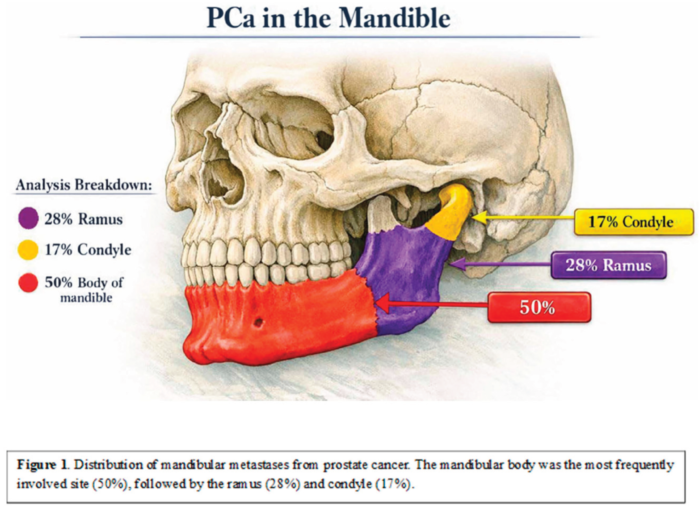

Table 1.
Patient demographics and characteristics.
| Patient variables | Value |
| Median (IQR) age at diagnosis | 70.5 years (60–77) |
| Median (IQR) primary Gleason score | 8 (7–9) |
| Metastatic disease at initial diagnosis | 9 (50%) |
| Upfront treatment in metastatic cases | Chemohormonal therapy |
| Hormone-sensitive disease at mandibular metastasis detection | 10 (56%) |
Table 2.
Imaging Studies and Detection Rates.
| Imaging Modality | Number of Studies | Detection of Mandibular Metastases | P-value |
| PSMA PET/CT or choline PET/CT | 18 | 18 (100%) | 0.031 |
| Conventional CT or MRI | 18 | 13 (73%) | 0.031 |
| Total imaging studies reviewed | 36 | — | — |
Table 3.
Distribution of metastatic mandibular Lesions.
| Location | N (%) |
| Unilateral lesions | 17 (94.4%) |
| Body of Mandible | 9 (50%) |
| Ramus | 5 (28%) |
| Condyle | 3 (17%) |
| Multifocal | 1 (5.6%) |
Table 4.
PET/CT Imaging Characteristics.
| Parameter | PSMA PET/CT | Choline PET/CT |
| Median (IQR) SUVmax | 6.3 (4.4–16.9) | 4.3 (3.8–8.5) |
| Median (IQR) lesion size (all lesions) | 5.0 ml (1.1–11.3) | 5.0 ml (1.1–11.3) |
Disclaimer/Publisher’s Note: The statements, opinions and data contained in all publications are solely those of the individual author(s) and contributor(s) and not of MDPI and/or the editor(s). MDPI and/or the editor(s) disclaim responsibility for any injury to people or property resulting from any ideas, methods, instructions or products referred to in the content. |
© 2026 by the authors. Licensee MDPI, Basel, Switzerland. This article is an open access article distributed under the terms and conditions of the Creative Commons Attribution (CC BY) license (http://creativecommons.org/licenses/by/4.0/).
Copyright: This open access article is published under a Creative Commons CC BY 4.0 license, which permit the free download, distribution, and reuse, provided that the author and preprint are cited in any reuse.