

Submitted:
06 October 2026
Posted:
09 October 2026
You are already at the latest version
Abstract
The process by which fascial membranes emerge during fetal development remains incompletely understood. Recent developmental anatomical studies suggest that recognizable fascial lamination appears as a late fetal event coinciding with extracellular matrix maturation, skeletal stiffening, and the emergence of a body-wide tension network. Within this developmental framework, Poisson-compression-driven syneresis may transform hydrated loose mesenchyme into dense fascial membranes through mechanical phase separation.We propose that aging represents the reverse rotation of this developmental process. In development, increasing tension fields generate orthogonal compression, fluid expulsion, collagen densification, and membrane formation. In aging, progressive decline of tension fields reduces Poisson compression, allowing Donnan osmotic swelling pressure to predominate. This shift promotes rehydration, re-expansion, loss of structural density, ligamentous creep, and eventual membrane failure.This framework provides a unified biomechanical explanation for several age-related disorders characterized by failure of tension-dependent supporting membranes, including pelvic organ prolapse, abdominal wall and inguinal hernia, and age-related spinal kyphosis. Rather than interpreting these conditions as isolated local diseases, we suggest they represent different anatomical manifestations of a common biomechanical cascade:
Plaintext
Tension field decay
↓
Loss of Poisson compression
↓
Loss of syneresis
↓
Re-expansion
↓
Creep
↓
Membrane failure
We further propose a Two-Hit Framework in which systemic fascial decompression establishes vulnerability, while local trauma or shear loading determines the anatomical site of failure. This model connects developmental morphogenesis and age-related structural collapse through a single physical principle and offers a conceptual foundation for future investigation of connective tissue aging.Keywords:
aging
; fascial membranes
; phase separation
; poisson compression
; syneresis
; connective tissue aging
; pelvic organ prolapse
Chapter 1. Introduction
1.1. Development and Aging as Opposite Directions of Morphological Change
Developmental biology and aging research have traditionally been studied as separate disciplines. Development investigates how biological structures emerge, whereas aging investigates how those structures deteriorate. Despite this apparent opposition, remarkably little attention has been directed toward the possibility that both phenomena may represent different phases of the same physical process.
In connective tissues, this question is particularly relevant. During fetal development, highly hydrated mesenchymal tissues undergo progressive structural organization to form dense tension-bearing membranes, ligaments, fascial sheets, and supporting connective tissue networks. Conversely, aging is characterized by gradual loss of structural integrity, increasing tissue laxity, progressive deformation, and ultimately failure of these same anatomical systems.
Conventional theories generally explain developmental morphogenesis through tissue differentiation and aging through degeneration. However, such explanations provide limited insight into why many connective tissue structures appear to undergo a characteristic sequence of swelling, loss of stiffness, elongation, and mechanical failure during aging. An alternative possibility is that developmental morphogenesis and age-related degeneration represent opposite directions of a common biomechanical trajectory.
1.2. The Developmental Phase Separation Framework
Recent developmental studies suggest that fascial membranes emerge relatively late during fetal development despite completion of organ positional rearrangements much earlier. In the retroperitoneum, fetal anatomical studies demonstrated that recognizable fascial lamination becomes evident primarily during the second trimester, particularly around the twentieth gestational week.
This developmental transition coincides with several biomechanically important events: maturation of the extracellular matrix, increasing collagen deposition, skeletal stiffening, and the establishment of more effective long-range force transmission across the fetal trunk. Within the framework of Poisson Compression Theory (PCT), these events may collectively permit the emergence of a systemic tension field capable of generating orthogonal compression within hydrated mesenchymal tissues. Such compression promotes fluid expulsion, collagen densification, and membrane formation through a process analogous to mechanical phase separation.
1.3. An Unresolved Paradox of Connective Tissue Aging
Many age-related disorders demonstrate biomechanical features that are difficult to reconcile with classical wear-and-tear models. Pelvic organ prolapse, abdominal wall hernias, spinal deformities, and numerous connective tissue disorders often display progressive tissue laxity, loss of geometric stability, ligamentous elongation, and increased hydration prior to structural failure.
Importantly, these tissues frequently exhibit swelling or thickening before collapse rather than simple loss of substance. Such observations raise an important question: Why do aging connective tissues frequently fail through deformation and expansion rather than simple material loss?
1.4. Central Hypothesis
We propose that aging may represent the reverse rotation of developmental phase separation.
During development:
Plaintext
Hydrated mesenchyme → Tension field formation → Poisson compression → Syneresis → Collagen densification → Membrane formation
During aging:
Plaintext
Tension field decay → Loss of Poisson compression → Loss of syneresis → Rehydration → Re-expansion → Creep → Membrane failure
Within this framework, developmental morphogenesis and connective tissue aging are interpreted as mechanically symmetrical processes operating in opposite directions.
1.5. Scope of the Present Study
The purpose of this article is to focus specifically on diseases in which the primary pathology appears to involve loss of competence of tension-dependent supporting membranes. Three representative conditions are examined: pelvic organ prolapse (POP), abdominal wall and inguinal hernia, and age-related spinal kyphotic deformity.
Chapter 2. Developmental Phase Separation and the Biomechanical Origin of Fascial Membranes
2.1. Developmental Morphogenesis as Mechanical Condensation
The formation of fascial membranes represents one of the least understood problems in connective tissue biology. Early fetal connective tissues are characterized by a highly hydrated extracellular environment rich in hyaluronan and loosely organized mesenchyme (Toole, 2001). Progressive extracellular matrix maturation and collagen organization subsequently accompany the emergence of identifiable fascial structures (Cho et al., 2009; Kinugasa et al., 2008; Matsubara et al., 2009). The mature fascial network displays fundamentally different material properties, characterized by dense, anisotropic collagen organization and high tensile competence (Stecco et al., 2017). The developmental transition from hydrated mesenchyme to mechanically competent membrane therefore requires a substantial reorganization of tissue architecture. We propose that this transition may be interpreted as a form of biomechanical phase separation driven by increasing systemic tension.
2.2. Emergence of Tension Fields
The appearance of recognizable fascial lamination exhibits a striking temporal pattern, emerging primarily during the second trimester. This coincides with accelerated skeletal ossification and increasing trunk rigidity (Nowlan, 2015; Senevirathne et al., 2025). Differential growth among skeletal, muscular, and connective tissues generates mechanical incompatibilities. As growth proceeds, locally generated stresses become increasingly coupled through continuous connective tissue networks, resulting in the emergence of a body-wide tension field capable of transmitting force over long anatomical distances (Ingber, 2003; Verbruggen et al., 2018).
2.3. Poisson Compression
The critical physical consequence of increasing tension fields is the generation of transverse compressive forces through the Poisson effect. Collagen networks demonstrate strong transverse coupling during extension (Vader et al., 2009). Ban et al. (2019) demonstrated that collagen networks may exhibit pronounced anomalous Poisson behavior, producing substantial transverse densification during tensile loading.
A potentially important anatomical observation supporting this framework in vivo was reported by Kim et al. (2015). In their fetal study of Denonvilliers' fascia, the authors documented the emergence of distinct fascial structures during the late fetal period, accompanied by a characteristic lateral displacement of the neurovascular bundle (NVB). They further reported a prominent linear organization of the surrounding mesenchymal tissue.
Conventionally, these findings have been interpreted primarily as descriptive anatomical observations. However, within the framework of Poisson Compression Theory (PCT), they can be viewed as anatomical signatures of mechanical tissue reorganization induced by orthogonal compression. Specifically, the lateral displacement of the NVB is consistent with a mechanical exclusion effect, whereby the developing fascial layer spatially reorganizes adjacent structures during progressive tissue compression.
Furthermore, the linear alignment observed by Kim et al. demonstrates a striking conceptual correspondence with the strain-induced collagen alignment reported by Vader et al. (2009) and the anomalous Poisson effect observed by Ban et al. (2019). In these experimental systems, tensile loading generates orthogonal matrix compression and directional collagen reorganization. Therefore, the fetal observations reported by Kim et al. provide a potentially critical in vivo analogue, connecting experimentally observed Poisson-related phenomena with actual fascial morphogenesis in the developing human body. As systemic tension fields increase during fetal growth, hydrated mesenchymal tissues experience this progressive orthogonal compression.
2.4. Syneresis
Compression alone is insufficient to explain the formation of dense fascial structures. Hydrated connective tissues behave as poroelastic systems consisting of a solid collagen-proteoglycan network and an interstitial fluid phase (Mow et al., 1980). Under sustained compression, fluid is progressively expelled from the matrix. This process is known as syneresis (Chaudhuri et al., 2020; Maroudas, 1976). As fluid leaves the network, tissue density increases and collagen fibers become more closely packed.
2.5. Donnan Swelling Pressure
A complete physical description requires consideration of swelling forces as well as compressive forces. Many connective tissues contain substantial concentrations of proteoglycans possessing fixed negative charges. These fixed charges generate Donnan osmotic swelling pressure, creating a continuous tendency for water influx (Maroudas, 1976; Urban & McMullin, 1988). Consequently, connective tissues exist within a dynamic equilibrium between swelling pressure and mechanical confinement (Antoniou et al., 1996). During fetal development, tension-field-derived compression exceeds Donnan swelling pressure, driving fluid expulsion and tissue condensation.
2.6. Phase Separation
The resulting structure resembles a mechanically induced phase separation process (Chaudhuri et al., 2020; Toole, 2001). Condensed regions become fascial membranes and fibrous boundaries, whereas preserved hydrated regions become gliding planes and interfascial spaces. Development therefore transforms a mechanically homogeneous system into a heterogeneous structural network.
Chapter 3. Aging as the Reverse Rotation of Developmental Phase Separation
3.1. Connective Tissue Aging
Most contemporary theories of connective tissue aging emphasize cumulative damage, cellular senescence, or declining repair capacity (Kirkwood, 2005; Lakatta & Levy, 2003). While relevant, these mechanisms do not fully explain why structural failure frequently occurs after a prolonged phase of tissue laxity, swelling, and deformation rather than simple tissue loss.
3.2. Tension Field Decay
The maintenance of mature fascial architecture requires the continuous presence of a systemic tension field. Unlike embryonic development, we hypothesize that aging may progressively reduce the capacity of connective tissue systems to maintain long-range mechanical integration. While direct measurement of whole-body tension remains challenging, aging is frequently associated with diminished postural activity, reduced efficiency of force transmission, and declining mechanobiological responsiveness (Huijing, 2009; Ingber, 2003; Langevin et al., 2013). As the tension field weakens, Poisson-derived orthogonal compression would correspondingly decline, leading to a progressive reduction in the mechanical constraint that previously stabilized condensed connective tissue structures.
3.3. Persistence of Swelling Forces
A critical feature of this model is that the decline of compressive forces does not imply a corresponding disappearance of swelling forces. Proteoglycans continue to generate fixed-charge-associated osmotic effects throughout life (Maroudas, 1976; Urban & McMullin, 1988). With aging, tension field decay progressively lowers the magnitude of mechanical restraint, causing the equilibrium to shift toward swelling dominance.
3.4. Re-Expansion
Rather than remaining in a condensed state, aging connective tissues become increasingly susceptible to passive water accumulation. Early disc degeneration is frequently accompanied by increased hydration and swelling rather than immediate loss of volume (Urban & McMullin, 1988; Antoniou et al., 1996; Caligaris et al., 2009). While these conditions likely have multifactorial etiologies, these observations are highly consistent with a decompression-based interpretation. Re-expansion is therefore proposed as the earliest macroscopic manifestation of developmental phase separation operating in reverse.
3.5. Creep
Increases in hydration increase matrix compliance, while loss of collagen packing reduces structural stiffness. As a result, normal physiological forces generate progressively larger deformations that accumulate over time (Solomonow et al., 2003; Woo et al., 1981). Creep is therefore interpreted as the mechanical bridge connecting re-expansion to clinical structural failure.
Chapter 4. The Two-Hit Framework
4.1. Why Does Failure Occur in Different Anatomical Locations?
If aging primarily reflects a systemic biomechanical process, one might expect all connective tissues to fail simultaneously. Clinical reality, however, demonstrates considerable heterogeneity. To explain this, we propose a Two-Hit Framework that determines the ultimate site and timing of structural failure. This framework consists of two essential components—a systemic biomechanical regression (Systemic Hit) and a localized mechanical stressor (Local Hit). Clinical failure occurs precisely when and where these two hits overlap.
4.2. The Systemic Hit: Global Fascial Decompression
One component is the systemic decay of the tension field, loss of Poisson compression, and subsequent passive rehydration driven by Donnan swelling pressure (fascial decompression). This process is not region-specific; rather, it globally diminishes the stiffness and mechanical reserve of connective tissues throughout the aging body.
4.3. The Local Hit: Mechanical Triggers
The other component consists of regional mechanical events acting upon specific anatomical sites. These triggers include massive overstretching during childbirth, surgical incisions, postural asymmetries, or repetitive shear loading associated with obesity.
4.4. Chronological Independence of the Two Hits
Crucially, the chronological order in which these two hits occur is not fixed and varies by etiology. In some conditions, such as incisional hernias in the elderly, the Systemic Hit (age-related vulnerability) precedes the Local Hit (surgical trauma), leading to immediate structural failure. Conversely, in conditions like pelvic organ prolapse (POP), the Local Hit (childbirth) can precede the Systemic Hit by decades. In such cases, the robust tension field of youth biomechanically compensates for the local damage, preserving it as a 'silent defect'. It is only decades later, when the Systemic Hit (age-related decompression) occurs and dismantles this compensatory mechanism, that the disease clinically manifests. Thus, regardless of their chronological sequence, macroscopic structural failure ensues when both hits finally intersect.
4.5. Differentiation from Classical Epidemiological Models
The clinical observation that obstetric trauma combined with aging leads to pelvic floor failure has long been recognized in traditional literature. However, conventional models describe age-related changes merely as vague biological deterioration, such as "hormonal decline," "muscle atrophy," or "wear-and-tear." These classical models fail to explain the mechanical paradox of why aging connective tissues frequently undergo swelling and creep (elongation) prior to failure, rather than simple attritional tissue loss. A key conceptual feature of the present Two-Hit Framework is its reinterpretation of the "Systemic Hit".
We redefine age-related vulnerability not as ambiguous biological decay, but as a precise thermodynamic and biomechanical state shift: the reverse rotation of developmental phase separation, driven by the loss of Poisson mechanical confinement and the unmasking of Donnan osmotic swelling pressure. This reinterpretation may provide a biomechanical framework that complements conventional epidemiological models.
4.6. Pelvic Organ Prolapse as a Two-Hit Disorder
While traditional explanations emphasize childbirth-related levator injury and fascial tears (DeLancey, 1992; Dietz, 2005; Dietz, 2008), prolapse frequently becomes clinically apparent decades after childbirth. Within the present framework, the pathogenesis of age-related pelvic organ prolapse (POP) is explained as a time-delayed two-hit model. The first hit (local structural damage) is childbirth, which introduces massive concentrated shear forces and overstretching to the pelvic floor. However, during youth, the systemic tension field is highly robust, and the fascial membrane maintains high density and rigidity through Poisson compression. Consequently, this local damage is biomechanically compensated for and does not immediately manifest as clinical POP, remaining as a 'silent defect' (Urbankova et al., 2019). The second hit (systemic biomechanical decompensation) is the age-related decay of the tension field. As aging progresses, Poisson compression is lost (fascial decompression), and Donnan swelling pressure drives the passive re-expansion of the membrane into a spongy, low-density state. This systemic mechanical regression acts as the trigger, causing the long-contained first hit to rapidly expand and culminate in macroscopic structural failure. This model rationally explains the epidemiological observation that delayed age at first delivery significantly increases future POP risk (Wang et al., 2021), as older membranes have already begun to lose their tension-dependent structural reserve.
4.7. Hernia as a Two-Hit Disorder
Similarly, abdominal wall hernias reflect localized failure occurring within a globally weakened fascial environment (LeBlanc et al., 2018; Simons et al., 2009). The abdominal wall functions as a tension-bearing containment membrane. Loss of membrane condensation permits gradual re-expansion and creep of the abdominal support system (First Hit). Surgical intervention, congenital weak points, or localized stress concentration (such as obesity-related intra-abdominal pressure) then acts as a mechanical second hit, introducing a localized discontinuity into a membrane whose capacity for force redistribution is already impaired.
4.8. Spinal Deformity as a Two-Hit Disorder
Age-related kyphosis can be interpreted as a failure of the axial tension system (Adams & Roughley, 2006; Katzman et al., 2010). Re-expansion of discal tissues and ligamentous creep gradually reduce structural coherence. Regional forces function as secondary triggers superimposed upon a generalized decline in connective tissue competence.
4.9. A Tension-Shear Landscape of Failure and the U-Shaped Risk Distribution
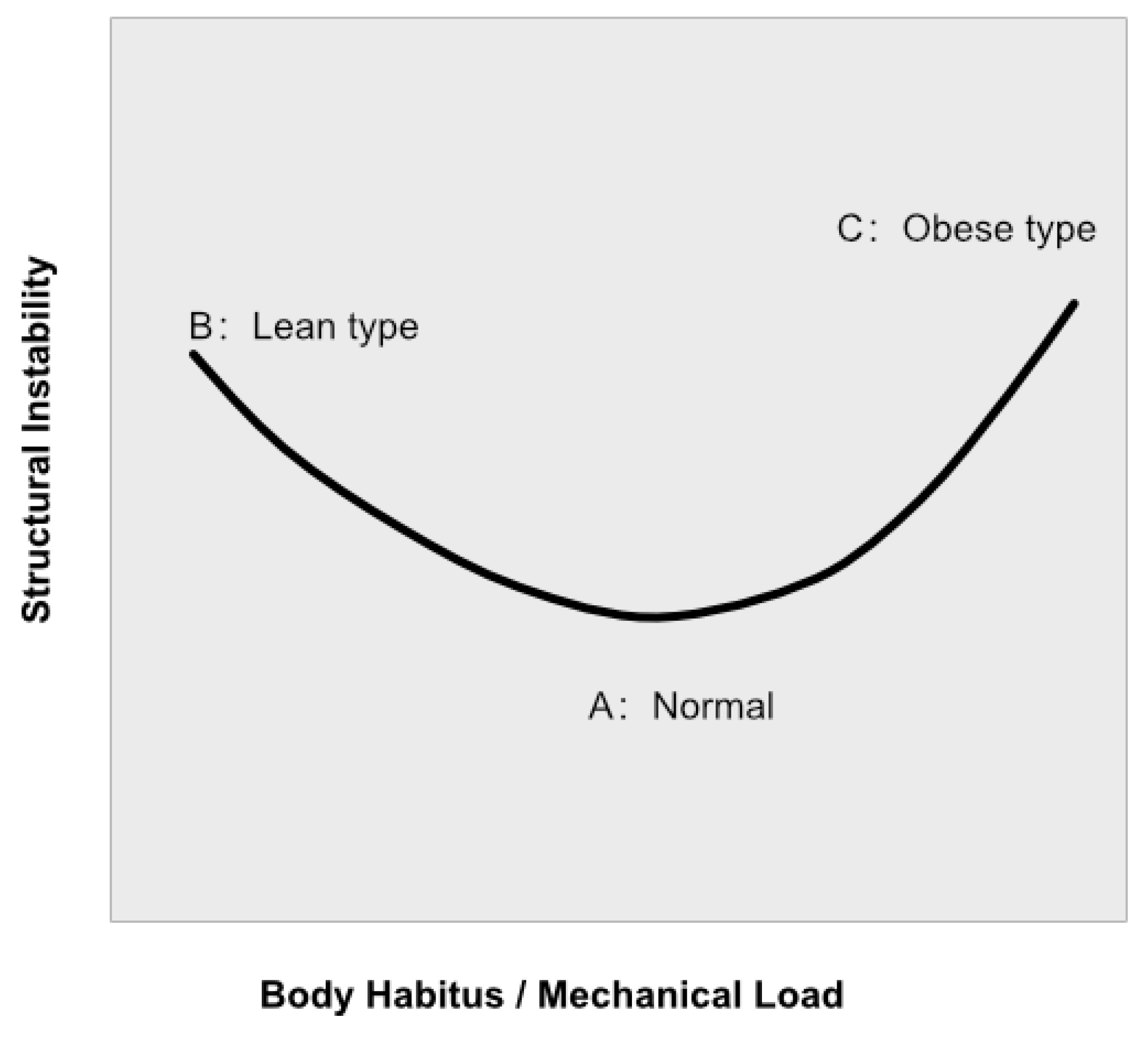
The present framework conceptualizes connective tissue disorders within a two-dimensional mechanical landscape mapping 'Maintenance of Tension' against 'Shear Exposure'. This conceptualization logically explains the U-shaped epidemiological risk distribution frequently observed in conditions like POP and hernias, where both extremes of body habitus—frailty and obesity—exhibit high rates of structural failure (see Figure 2). At one end of the spectrum (the left arm of the U), frail or sarcopenic individuals represent a collapse-dominant phenotype. Here, the systemic tension field is severely depleted (low tension), leading to complete fascial decompression and marked re-expansion. Even in the absence of massive external loading, failure occurs primarily through progressive tissue creep and central sagging, manifesting as pure relaxation and collapse. At the opposite end (the right arm of the U), obese individuals or those subjected to chronic mechanical loading represent a shear-dominant phenotype. While their tension field may be partially preserved due to maintained muscle mass, the massive local shear forces (e.g., from elevated intra-abdominal pressure) may exceed the local mechanical tolerance of the membrane, leading to lateral detachments or acute structural tears. Between these two extremes lies the optimal mechanical state, where adequate tension maintenance balances physiological shear forces, minimizing the overall risk of failure.
Figure 1.
The H-shaped Suspended Membrane Model in Aging Mechanics. The vertical pillars represent lateral anatomical supports, while the horizontal line represents the tension-dependent fascial membrane. The thickness and color of the horizontal line indicate the relative degree of membrane re-expansion and loss of structural density. (A) Youth / Intact Tension Field: The membrane is thin, dense (black), and mechanically competent, maintained by strong Poisson compression. (B) Lean Phenotype (Central Collapse): Severe tension decay may lead to marked re-expansion and loss of structural density (thick, gray). The weakened membrane becomes increasingly susceptible to central sagging through progressive creep. (C) Obese Phenotype (Lateral Shear Failure): The tension field is partially preserved, resulting in less re-expansion than in the lean phenotype (intermediate thickness, gray). However, when subjected to massive lateral shear loading, the local shear forces may exceed the mechanical tolerance of the attachment site, potentially resulting in structural detachment at the lateral junction.
Figure 1.
The H-shaped Suspended Membrane Model in Aging Mechanics. The vertical pillars represent lateral anatomical supports, while the horizontal line represents the tension-dependent fascial membrane. The thickness and color of the horizontal line indicate the relative degree of membrane re-expansion and loss of structural density. (A) Youth / Intact Tension Field: The membrane is thin, dense (black), and mechanically competent, maintained by strong Poisson compression. (B) Lean Phenotype (Central Collapse): Severe tension decay may lead to marked re-expansion and loss of structural density (thick, gray). The weakened membrane becomes increasingly susceptible to central sagging through progressive creep. (C) Obese Phenotype (Lateral Shear Failure): The tension field is partially preserved, resulting in less re-expansion than in the lean phenotype (intermediate thickness, gray). However, when subjected to massive lateral shear loading, the local shear forces may exceed the mechanical tolerance of the attachment site, potentially resulting in structural detachment at the lateral junction.
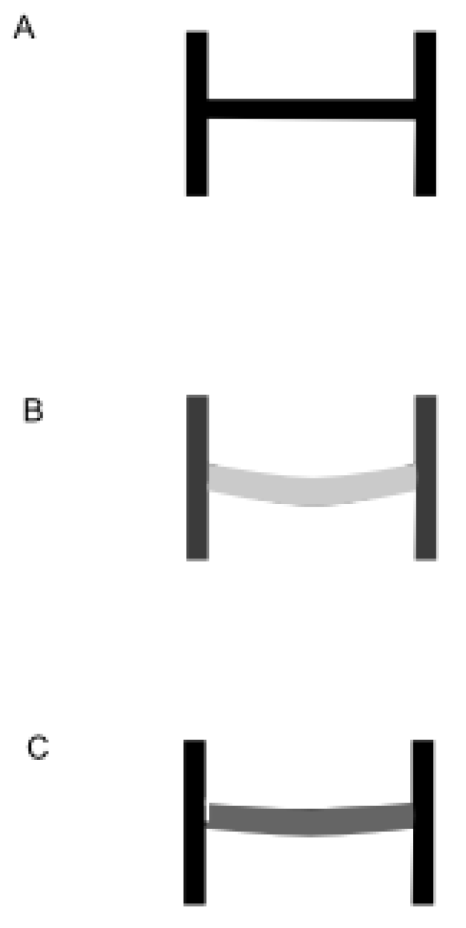
Figure 2.
Conceptual U-shaped relationship between body habitus and structural instability. The x-axis represents body habitus and the corresponding mechanical loading environment, ranging from frailty/sarcopenia (left) to obesity/chronic overload (right). The y-axis represents the hypothesized degree of structural instability and associated risk of fascial support failure (e.g., pelvic organ prolapse or hernia). The left side of the curve illustrates a collapse-dominant phenotype, in which deterioration of the systemic tension field may promote fascial re-expansion and increased central sagging. The right side illustrates a shear-dominant phenotype, in which chronic mechanical loading and elevated local shear forces may increase the likelihood of focal attachment failure. The trough of the curve represents a biomechanically balanced state in which tension maintenance and shear loading are optimally matched.
Figure 2.
Conceptual U-shaped relationship between body habitus and structural instability. The x-axis represents body habitus and the corresponding mechanical loading environment, ranging from frailty/sarcopenia (left) to obesity/chronic overload (right). The y-axis represents the hypothesized degree of structural instability and associated risk of fascial support failure (e.g., pelvic organ prolapse or hernia). The left side of the curve illustrates a collapse-dominant phenotype, in which deterioration of the systemic tension field may promote fascial re-expansion and increased central sagging. The right side illustrates a shear-dominant phenotype, in which chronic mechanical loading and elevated local shear forces may increase the likelihood of focal attachment failure. The trough of the curve represents a biomechanically balanced state in which tension maintenance and shear loading are optimally matched.
Chapter 5. Clinical Manifestations of Fascial Membrane Failure
5.1. Overview: Anatomical Expressions of a Common Mechanical Cascade
The preceding chapters proposed that aging may represent the reverse rotation of developmental phase separation. Within this framework, connective tissue aging is interpreted as a progressive shift from mechanically condensed fascial membranes toward increasingly hydrated, compliant, and creep-prone structures. This model predicts that anatomically distinct disorders may emerge as different manifestations of the same underlying mechanical process. Rather than representing unrelated diseases, pelvic organ prolapse, abdominal wall hernia, and age-related spinal deformity may be viewed as regional expressions of failure occurring within tension-dependent supporting membranes.
5.2. Pelvic Organ Prolapse: Failure of the Pelvic Support Membrane System
Pelvic organ prolapse (POP) has traditionally been attributed to factors including childbirth trauma, levator ani injury, connective tissue weakness, and hormonal changes (DeLancey, 1992; Dietz, 2008; Hendrix et al., 2002; Wu et al., 2014). These mechanisms undoubtedly contribute to disease development. However, several observations remain difficult to reconcile with purely local explanations. For example, substantial prolapse may occur decades after childbirth, and prolapse risk increases progressively with aging even in the absence of major obstetric injury (Dietz, 2005).
Within the present framework, the pelvic floor is viewed as an integrated, tension-dependent support membrane system. The precise pathogenesis of POP is driven not merely by muscle weakness or simple wear-and-tear, but by the intersection of childbirth (the Local Hit) and age-related fascial decompression (the Systemic Hit). As detailed in Chapter 4, the chronological relationship between these two hits—whether resulting in functional repair, a decades-long time lag, or earlier structural failure—depends heavily on the biomechanical phase state of the membrane at the time of local loading. By reconceptualizing the pelvic floor as a tension-dependent membrane and applying the Two-Hit Framework, the complex epidemiology of POP (including its occurrence in nulliparous women, elevated risks associated with advanced maternal age, and prolonged onset latency) can be consistently explained through a single biomechanical mechanism.
5.3. Abdominal and Inguinal Hernias: Failure of the Fascial Containment System
Conventional theories of hernia formation emphasize elevated intra-abdominal pressure, congenital weakness, obesity, surgical disruption, and mechanical strain (Burcharth et al., 2013; Burger et al., 2004; Ruhl & Everhart, 2007). While each factor contributes, none fully explains why anatomically similar stresses cause hernias only in a subset of individuals (LeBlanc et al., 2018; Simons et al., 2009). The abdominal wall functions as a tension-bearing containment membrane. The loss of membrane condensation due to tension field decay (the Systemic Hit) permits the gradual re-expansion and creep of the abdominal support system. Subsequently, surgical interventions or local stress concentrations act as the Local Hit, introducing regional discontinuities into a membrane whose force-distributing capacity is already compromised. This mechanical failure of the abdominal wall also perfectly follows the U-shaped mechanical landscape described in Chapter 4. As previously reported, low BMI is an independent risk factor for inguinal hernias (Ruhl & Everhart, 2007). In the collapse-dominant phenotype (frailty/sarcopenia), severe tension decay drives widespread creep and decompression of the entire abdominal wall, manifesting as poorly defined, lax eventrations such as massive rectus diastasis or broad direct inguinal hernias (the left arm of the U). Conversely, in the shear-dominant phenotype (obesity/elevated intra-abdominal pressure), while the physical presence of adipose tissue may be paradoxically protective against inguinal hernias, the extreme internal pressure acts as a massive local shear force. This sharply tears through anatomical weak points like surgical scars or the umbilical ring, resulting in well-defined, "blowout-type" shear failures such as incisional or umbilical hernias (the right arm of the U).
5.4. Age-Related Kyphosis: Failure of the Axial Support System and Reinterpretation of Fractures
Age-related hyperkyphosis has traditionally been interpreted as a consequence of vertebral degeneration, muscular weakness, or osteoporotic vertebral compression fractures (Adams & Roughley, 2006; Belavý et al., 2012; Katzman et al., 2010). However, epidemiological data reveal that only approximately 36-37% of older adults with hyperkyphosis have evident vertebral fractures, meaning the majority (over 60%) progress to kyphosis without bone fracture (Katzman et al., 2010). This highlights that bone failure or muscle loss alone cannot fully explain the primary mechanism of kyphosis. Within the present framework, the root cause of age-related kyphosis may involve failure of the axial tension system (including annular containment structures and longitudinal ligaments). The decay of the systemic tension field (the Systemic Hit) shifts this continuous tension-distributing support system into a hydrated, highly compliant state, driving postural creep (ligamentous kyphosis) even in the absence of fractures. Crucially, the preceding decompression and creep of this soft tissue system eliminate the spine's shock-absorbing and force-distributing capacity. Consequently, the spinal load-bearing axis shifts anteriorly, causing even minor everyday mechanical loads, such as sneezing or light lifting (the Local Hit), to bypass soft-tissue dissipation and concentrate directly as abnormal compressive and shear stresses on the anterior bone (vertebral bodies). Thus, clinically observed vertebral compression fractures—particularly the prevalent "occult fractures" occurring without overt trauma— may in some cases be interpreted as a secondary mechanical consequence of loss of the tension-dependent axial support system. Furthermore, this soft-tissue-driven mechanism perfectly aligns with the U-shaped risk landscape described in Chapter 4. In the collapse-dominant phenotype (frailty/sarcopenia), severe tension decay leads to pure ligamentous creep (postural kyphosis), and the loss of shock absorption combined with bone fragility frequently precipitates "occult" compression fractures under minor everyday loads (the left arm of the U). Conversely, in the shear-dominant phenotype (obesity/chronic overload), massive axial compression and shear forces concentrate on a spine that has lost its soft-tissue cushioning, precipitating shear-driven structural failures such as annular tears and degenerative spondylolisthesis (the right arm of the U).
5.5. Shared Mechanical Features Across Disorders
Although pelvic prolapse, hernia, and spinal deformity occur in different anatomical regions, they demonstrate strikingly similar mechanical patterns. All three involve tension-dependent support structures, progressive loss of geometric stability, increased compliance, time-dependent creep, and susceptibility to local loading conditions.
5.6. Toward a Unified Concept of Fascial Membrane Failure
The proposed unifying sequence suggests that the primary pathology is not prolapse, hernia, or spinal deformity itself. The primary pathology is age-related failure to maintain the mechanically condensed state originally established during developmental phase separation.
Chapter 6. Discussion
6.1. Reframing Connective Tissue Aging: The Symmetry of Development and Degeneration
The central proposition of the present work is that connective tissue aging and developmental morphogenesis can be interpreted as opposite trajectories within a shared biomechanical continuum. Connective tissue architecture can therefore be understood not merely as a two-stage process (growth and decay), but as progressing through three distinct biomechanical states:
Plaintext
State I: Developmental Condensation
↓
State II: Mechanical Maintenance
↓
State III: Reverse Phase Separation (Aging)
- State I (Developmental Condensation): Hydrated mesenchyme exists as a mechanically compliant continuum. As developmental tension fields emerge, Poisson compression drives syneresis and collagen densification, producing mechanically competent fascial membranes.
- State II (Mechanical Maintenance): Once formed, fascial membranes are not static anatomical objects. Rather, biological form is a dynamically maintained state. Continuous force transmission, postural activity, and mechanotransduction balance intrinsic swelling pressures with mechanical constraints to preserve architecture.
- State III (Reverse Phase Separation): As tension maintenance progressively declines during aging, this balance shifts. Loss of Poisson compression allows persistent osmotic swelling forces to dominate, leading to rehydration, re-expansion, and creep.
6.2. Beyond Wear-and-Tear: Mechanical Aging vs. Molecular Degeneration
Current aging theories emphasize molecular mechanisms such as oxidative damage, cellular senescence, and matrix turnover. The present model does not replace these biological theories but provides a systems-level biomechanical framework within which they can be integrated. Impaired mechanotransduction, altered collagen turnover, and cellular senescence (Ingber, 2003; Langevin et al., 2013) can be viewed as upstream biological events that ultimately trigger the downstream physical cascade of fascial decompression.
Crucially, this model moves beyond classical engineering "wear-and-tear" analogies. Conventional wear models cannot fully account for a consistent observation across connective tissues: failure is frequently preceded by swelling, laxity, and geometric distortion rather than simple material loss (Urban & McMullin, 1988; Antoniou et al., 1996; Caligaris et al., 2009). By recognizing re-expansion as the transitional stage between structural competence and overt collapse, this framework explains why aging tissues frequently become thicker and more hydrated, yet mechanically incompetent, prior to failure.
6.3. The Two-Hit Framework and the Tension-Shear Landscape
To explain why a systemic aging process produces diverse, region-specific anatomical failures, we propose a Two-Hit Framework mapping onto a Tension-Shear mechanical landscape. Generalized tension field decay leads to widespread fascial decompression (the Systemic Hit), while regional shear or trauma introduces localized mechanical stress (the Local Hit). As previously discussed, these two hits are chronologically independent, but their intersection dictates structural failure. This framework helps resolve epidemiological puzzles, such as the U-shaped risk distribution in connective tissue disorders, differentiating between collapse-dominant phenotypes (low-tension) and shear-dominant phenotypes (high-shear).
6.4. Clinical, Surgical, and Preventive Implications
If fascial architecture is maintained by force transmission throughout adulthood (State II), habitual movement takes on renewed biological significance. Population studies consistently suggest that habitual physical activity is associated with reduced risk of several age-related connective tissue disorders (Shiri & Falah-Hassani, 2017; Townsend et al., 2008). Walking, posture regulation (Hodges & Richardson, 1996), and whole-body stretching behaviors may act as continuous tension-field restoration mechanisms. Behaviors that repeatedly engage distributed fascial loading may actively preserve the mechanical integration established during development.
6.5. Limitations and Future Directions
The proposed framework remains conceptual and requires experimental validation. Objective methods to quantify whole-body tension transmission and tissue re-expansion in vivo must be developed. Additionally, while the model effectively describes pelvic organ prolapse, hernias, and kyphosis, its applicability to other age-related conditions requires dedicated investigation.
Finally, a critical conceptual clarification must be made regarding the central hypothesis. The present framework should not be interpreted as implying that aging literally recreates developmental mesenchyme. Rather, it proposes that aging may involve the progressive loss of the mechanically condensed state established during development, resulting in tissue behaviors that become increasingly consistent with a less condensed connective tissue architecture.
Chapter 7. Conclusion
We propose that connective tissue aging can be interpreted as the reverse rotation of developmental phase separation. During fetal development, the emergence of whole-body tension fields induces Poisson compression and syneresis, transforming hydrated mesenchyme into dense, structurally competent fascial membranes. During aging, the progressive decay of this systemic tension network relieves mechanical constraint, permitting intrinsic osmotic forces to drive rehydration, re-expansion, and ligamentous creep.
Within this unified biomechanical cascade, diverse age-related disorders—including pelvic organ prolapse, abdominal wall hernias, and spinal kyphotic deformity—are not independent diseases, but rather distinct anatomical expressions of a common failure within tension-dependent supporting membranes. The Two-Hit Framework bridges systemic aging and localized disease by demonstrating how global fascial decompression establishes vulnerability, while regional mechanical loading determines the site of structural collapse.
The present theory integrates developmental anatomical observations (Cho et al., 2009; Kinugasa et al., 2008; Matsubara et al., 2009) with contemporary connective tissue biomechanics (Ban et al., 2019; Vader et al., 2009) and classical swelling-pressure theory (Maroudas, 1976; Urban & McMullin, 1988). Extending the tradition initiated by Thompson's proposition that biological form reflects the action of physical forces (Thompson, 1917), the present framework suggests that development, maintenance, and aging may represent successive states of a single mechanically organized continuum (Ingber, 2003; Nowlan, 2015). By uniting morphogenesis, tissue maintenance, and structural aging into a single physical principle, this framework offers a foundational new perspective for the study of connective tissue pathology.
Use of Artificial Intelligence in the Manuscript Preparation
During the preparation of this manuscript, the author used generative artificial intelligence tools (Gemini and Microsoft Copilot) for language editing, stylistic refinement, and drafting assistance. The central hypothesis, theoretical framework, biomechanical interpretations, literature selection, and conceptual development were conceived and developed by the author. After using these tools, the author carefully reviewed, revised, and verified all content and takes full responsibility for the concepts, interpretations, and final content of the manuscript.
References
- Adams, M. A.; Roughley, P. J. What is intervertebral disc degeneration, and what causes it? Spine 2006, 31(20), 2151–2161. [Google Scholar] [CrossRef]
- Antoniou, J.; et al. The human lumbar intervertebral disc: evidence for changes in the biosynthesis and denaturation of the extracellular matrix with growth, maturation, ageing, and degeneration. J. Clin. Investig. 1996, 98(4), 996–1003. [Google Scholar] [CrossRef]
- Ban, E.; et al. Strongly coupled biomechanics and fluid flow in poroelastic collagen networks. Proc. Natl. Acad. Sci. 2019, 116(14), 6790–6799. [Google Scholar]
- Belavý, D. L.; et al. Muscle atrophy and changes in spinal morphology: is the cervical spine different? J. Appl. Physiol. 2012, 113(1), 86–94. [Google Scholar]
- Burcharth, J.; et al. The epidemiology and risk factors for inguinal hernia. Surgery 2013, 153(2), 241–247. [Google Scholar]
- Burger, J. W.; et al. Long-term follow-up of a randomized controlled trial of suture versus mesh repair of incisional hernia. Ann. Surg. 2004, 240(4), 578. [Google Scholar] [CrossRef]
- Caligaris, M.; et al. Differential mechanisms of extracellular matrix regulation in early disc degeneration. J. Biomech. 2009, 42(10), 1461–1466. [Google Scholar]
- Chaudhuri, O.; et al. Extracellular matrix viscoelasticity and its impact on cellular behaviour. Nature 2020, 584(7822), 535–546. [Google Scholar] [CrossRef]
- Cho, B. H.; et al. The fascial structures of the retroperitoneum: a topographical and histological study. J. Anat. 2009, 215(4), 438–450. [Google Scholar]
- DeLancey, J. O. Anatomic aspects of vaginal eversion after hysterectomy. Am. J. Obstet. Gynecol. 1992, 166(6), 1717–1728. [Google Scholar] [CrossRef]
- Dietz, H. P. The prevention of pelvic floor trauma. Curr. Opin. Obstet. Gynecol. 2005, 17(5), 452–458. [Google Scholar] [CrossRef]
- Dietz, H. P. Prolapse worsens with age, doesn't it? Aust. N. Z. J. Obstet. Gynaecol. 2008, 48(6), 587–591. [Google Scholar] [CrossRef]
- Hendrix, S. L.; et al. Pelvic organ prolapse in the Women's Health Initiative: gravity and gravidity. Am. J. Obstet. Gynecol. 2002, 186(6), 1160–1166. [Google Scholar] [CrossRef]
- Hodges, P. W.; Richardson, C. A. Inefficient muscular stabilization of the lumbar spine associated with low back pain. Spine 1996, 21(22), 2640–2650. [Google Scholar] [CrossRef]
- Huijing, P. A. Epimuscular myofascial force transmission: a historical review and implications for new research. J. Biomech. 2009, 42(1), 9–21. [Google Scholar] [CrossRef]
- Ingber, D. E. Tensegrity I. Cell structure and hierarchical systems biology. J. Cell Sci. 2003, 116(7), 1157–1173. [Google Scholar] [CrossRef]
- Katzman, W. B.; et al. Age-related hyperkyphosis: its causes, consequences, and management. J. Orthop. Sports Phys. Ther. 2010, 40(6), 352–360. [Google Scholar] [CrossRef]
- Kim, J. H.; Kinugasa, Y.; Hwang, S. E.; Murakami, G.; Cho, B. H. Denonvilliers' fascia revisited. Surg. Radiol. Anat. 2015, 37(2), 187–197. [Google Scholar] [CrossRef]
- Kinugasa, Y.; et al. The surgical anatomy of the fascial structures of the pelvis. Clin. Anat. 2008, 21(6), 548–556. [Google Scholar]
- Kirkwood, T. B. Understanding the odd science of aging. Cell 2005, 120(4), 437–447. [Google Scholar] [CrossRef]
- Lakatta, E. G.; Levy, D. Arterial and ventricular aging: physiology in health and frailty. Circulation 2003, 107(1), 139–146. [Google Scholar]
- Langevin, H. M.; et al. Connective tissue fibroblast response to acupuncture: dose-dependent effect of bidirectional needle rotation. J. Altern. Complement. Med. 2013, 19(11), 861–867. [Google Scholar]
- LeBlanc, K. A.; et al. Management of Abdominal Hernias; Springer, 2018. [Google Scholar]
- Maroudas, A. Swelling pressure versus collagen tension in normal and degenerate articular cartilage. Nature 1976, 260(5554), 808–809. [Google Scholar] [CrossRef]
- Matsubara, A.; et al. The fascial architecture of the retroperitoneum. J. Anat. 2009, 214(1), 130–141. [Google Scholar]
- Mow, V. C.; et al. Biphasic creep and stress relaxation of articular cartilage in compression? Theory and experiments. J. Biomech. Eng. 1980, 102(1), 73–84. [Google Scholar] [CrossRef]
- Nowlan, N. C. Biomechanics of foetal movement. Eur. Cells Mater. 2015, 29, 1–21. [Google Scholar] [CrossRef]
- Ruhl, C. E.; Everhart, J. E. Risk factors for inguinal hernia among adults in the US population. Am. J. Epidemiol. 2007, 165(10), 1154–1161. [Google Scholar] [CrossRef]
- Senevirathne, G.; Fernandopulle, S.C.; Richard, D.; Baumgart, S.L.; Christensen, A.L.; Fabbri, M.; et al. The evolution of hominin bipedalism in two steps. Nature 2025, 645, 952–963. [Google Scholar] [CrossRef]
- Shiri, R.; Falah-Hassani, K. Does leisure time physical activity protect against low back pain? Systematic review and meta-analysis of 36 prospective cohort studies. Br. J. Sports Med. 2017, 51(19), 1410–1418. [Google Scholar] [CrossRef]
- Simons, M. P.; et al. European Hernia Society guidelines on the treatment of inguinal hernia in adult patients. Hernia 2009, 13(4), 343–403. [Google Scholar] [CrossRef]
- Solomonow, M.; et al. Biomechanics and electromyography of a cumulative lumbar disorder: response to static flexion. Clin. Biomech. 2003, 18(10), 890–898. [Google Scholar] [CrossRef]
- Stecco, C.; et al. Functional Atlas of the Human Fascial System; Churchill Livingstone, 2017. [Google Scholar]
- Thompson, D. W. On Growth and Form; Cambridge University Press, 1917. [Google Scholar]
- Toole, B. P. Hyaluronan in morphogenesis. Semin. Cell Dev. Biol. 2001, 12(2), 79–87. [Google Scholar] [CrossRef]
- Townsend, M.K.; Danforth, K.N.; Rosner, B.; Curhan, G.C.; Resnick, N.M.; Grodstein, F. Physical activity and incident urinary incontinence in middle-aged women. J. Urol. 2008, 179(3), 1012–1017. [Google Scholar] [CrossRef]
- Urban, J. P.; McMullin, J. F. Swelling pressure of the lumbar intervertebral discs: influence of age, spinal level, composition, and degeneration. Spine 1988, 13(2), 179–187. [Google Scholar]
- Urbankova, I.; Grohregin, K.; Hanacek, J.; et al. The effect of the first vaginal birth on pelvic floor anatomy and dysfunction. Int. Urogynecology J. 2019, 30(10), 1689–1696. [Google Scholar] [CrossRef]
- Vader, D.; et al. Strain-induced alignment in collagen gels. PLoS ONE 2009, 4(6), e5902. [Google Scholar] [CrossRef]
- Verbruggen, S. W.; et al. Mechanobiology of the human skeleton. Curr. Osteoporos. Rep. 2018, 16(4), 433–441. [Google Scholar]
- Wang, R.; Tulikangas, P. K.; Tunitsky-Bitton, E. Relationship Between Maternal Age at First Delivery and Subsequent Pelvic Organ Prolapse. Female Pelvic Med. Reconstr. Surg. 2021, 27(2), e423–e426. [Google Scholar] [CrossRef]
- Woo, S. L.; et al. The biomechanical and biochemical properties of swine tendons—long term effects of exercise on the digital extensors. Biorheology 1981, 18(1), 101–108. [Google Scholar]
- Wu, J. M.; et al. Lifetime risk of stress urinary incontinence or pelvic organ prolapse surgery. Obstet. Gynecol. 2014, 123(6), 1201–1206. [Google Scholar] [CrossRef]
Disclaimer/Publisher’s Note: The statements, opinions and data contained in all publications are solely those of the individual author(s) and contributor(s) and not of MDPI and/or the editor(s). MDPI and/or the editor(s) disclaim responsibility for any injury to people or property resulting from any ideas, methods, instructions or products referred to in the content. |
© 2026 by the authors. Licensee MDPI, Basel, Switzerland. This article is an open access article distributed under the terms and conditions of the Creative Commons Attribution (CC BY) license (http://creativecommons.org/licenses/by/4.0/).
Copyright: This open access article is published under a Creative Commons CC BY 4.0 license, which permit the free download, distribution, and reuse, provided that the author and preprint are cited in any reuse.