
Submitted:
29 September 2026
Posted:
08 October 2026
You are already at the latest version
Abstract
Inborn errors of immunity (IEI), which includes primary immunodeficiencies, are a heterogenous group of disorders that affect the immune system leading to infections, autoimmunity, atopy, inflammation, or malignancy. The most recent update by Inter-national Union of Immunological Societies (IUIS) reports more than 500 gene defects in 10 categories. The initial approach to diagnosis should be dictated by the clinical history and physical examination. Traditional laboratory studies and genetic testing represent the backbone of diagnostic testing. The treatment options depend on the specific disorder and include support measures, immunoglobulin replacement therapy, antibiotic prophylaxis, immunosuppression, biological therapy, hematopoietic stem cell transplantation, and gene therapy. The early clinical suspicion and expeditious testing contribute to im-proved outcomes. This review provides a general approach to patients with suspected IEIs, highlighting aspects of infections and non-infectious manifestations, clinical history, physical examination, and immune laboratories, functional and genetic evaluation.
Keywords:
inborn errors of immunity
; primary immunodeficiencies diseases
; warning signs
; recurrent infections
; immune dysregulation
; inflammation
; autoimmunity
; malignancy
; allergies
1. Introduction
The Inborn Errors of Immunity (IEI) are a heterogenous group of conditions that affect morphology, number of cells, and/or function of cells, thereby altering immune function. The frequency ranges from a high of 1:500 to ultra-rare conditions [1,2].
Classically, IEIs were recognized by susceptibility to severe and/or recurrent infections and were called primary immunodeficiencies. However, in 2019, the International Union of Immunological Societies (IUIS) changed the terminology to IEI, as it allows for the recognition of not only infectious but also non-infectious manifestations. [3,4] The most recent update by IUIS reports more than 500 gene defects in 9 categories with an additional category of acquired phenocopies [5].
Infection still comprises the most common presenting manifestation with nearly 70% of patients displaying significant infections leading to diagnosis, although family history, autoimmunity, inflammation, and syndromic features represent significant subsets of presentations [6,7].
Different presentations require different approaches to testing. The heterogeneity and complexity of clinical manifestations are ongoing challenges to recognition. In this article, diagnostic tests are discussed based on the type of presentation.
- 1)
- Approaches to identifying patients with IEI
- 1.0
- Overview
The diagnostic journey for patients with IEI continues to be over 10 years[8]. This finding has not changed over the last two decades, and therefore there is a clear gap between the onset of patient symptoms and recognition of the IEI. Until there is improved recognition, there will continue to be patients who suffer as a result of their ongoing end organ effects. This review is focused on improving recognition of IEI and empowering physicians to initiate the screening tests. Different strategies have been designed over time to promote recognition of IEI. In 1994, the Jeffrey Modell Foundation designed the ‘10 warning signs for immunodeficiency’ for suspected primary immunodeficiency in pediatric population, and subsequently, the alarm signs were created for adults. [9] However, most of the warning signs are focused on infectious manifestations and up to one-third of patients do not present with any of these features [7]. Formal analysis revealed that the ‘10 warning signs for immunodeficiency’ have low specificity and relatively low sensitivity but they have high impact in awareness campaigns [10,11]. Additional approaches have been developed. Adding autoimmunity, severe allergies, and hematological/oncological lymphoproliferation disorders improves the suspicion, detection, and diagnosis of IEIs [12]. In this era of electronic medical records, there have been multiple efforts to harness that computational power to risk stratify patients for IEI [2,13,14,15,16]. A lab flag has also been developed to identify people with low globulins on routine metabolic testing [17]. All these efforts have the potential to markedly improve the time-to-diagnosis, which continues to span over ten years [8]. Yet lacking implementation, we are left with clinical acumen to identify patients who should be tested, and this review will describe common scenarios.
1.1. Infections
Infectious manifestations constitute the most common presentation for people ultimately identified as having IEI [7]. The infectious manifestations can be simplified into two categories. The most traditional category is comprised of patients who have had too many infections, which may also include infections of higher severity than expected. Simple algorithms can identify patients with a high number of infections [14] The second category is comprised of patients who have unusual infections where a single infection constitutes a red flag. These unusual infections are often due to opportunistic organisms but can also be a prolonged or severe infection with a more typical organism [18] (Figure 1).
1.2. Autoimmunity/Inflammation
Autoimmunity is common in the general population and is not in and of itself a significant signal for an IEI. Psoriasis, rheumatoid arthritis, and inflammatory bowel disease all affect approximately one percent of the population [19]. Thus, the presence of autoimmunity or inflammation does not necessarily require a workup. A common approach to ascertaining patients who might have an IEI includes the age of onset and the complexity of the autoimmunity or inflammation. As an example, systemic lupus erythematosus occurs in approximately 1:2000 adult women. In the US and Europe, the likelihood of finding an IEI is approximately 1% in adults with a majority of adult patients having C2 deficiency. Among young children with systemic lupus erythematosus, the likelihood of finding an IE is much higher, in the range of 30% [20].
Similarly, it is very infrequent to find an IEI in adults presenting with inflammatory bowel disease. However, among infants and young children, the rate of IEI is approximately 10 to 20% in the United States (US) and even higher in other regions [21,22].
The second element related to ascertainment among patients presenting with autoimmunity or inflammation is the complexity. Children with multiple autoimmune conditions or recalcitrant autoimmunity should have an evaluation for IEI (Figure 2).
1.3. Atopy
Atopy is even more common in the general population than autoimmunity and thus in isolation has a relatively low positive predictive value for IEI. Nevertheless, there are scenarios involving early onset severe atopy or atopy plus significant infections that warrants an evaluation [23].
1.4. Malignancy
Today, many children with malignancy will have genetic testing performed on the tumor to identify the optimal therapy. It is not unusual to identify germline variants at the time of analysis for somatic variants. The main reason to highlight malignancy in this review article is to underscore the importance of malignancy in the family history [24,25]. It is infrequent that patients present with malignancy who have IEI. It is more commonly seen later in disease course.
2. The role of the History
The complete medical history is always important in pediatrics. A focused history should include age-of-onset of symptoms, perinatal history (intrauterine growth restriction, birth weight and length), severe reactions to live vaccines, time of umbilical cord separation, and newborn screening or prenatal genetic testing. DNA repair defects, which often have an element of immune compromise, are often associated with growth failure – even beginning in utero. Failure to control the attenuated live viruses in vaccines is always a red flag and prolonged attachment of the umbilical cord, particularly with infection, can be a sign of a neutrophil disorder. The family history is also central to every evaluation for IEI. Key points include consanguinity, endogamy, recurrent miscarriage, infant early deaths, known IEIs in the family, autoimmunity, or malignancy.
3. Role of the Physical Examination
A detailed and thorough physical examination with attention to anthropometric measurements, facial features, dysmorphism, dental alterations, absent tonsils, lymphadenopathy, hepatosplenomegaly, silver hair, telangiectasias, neurological features, and signs of atopy are central. Today, failure to thrive is unusual in IEI, but is still seen [26,27]. In the absence of immune suppressive medication, absent tonsils suggest impaired B cell development. On the other hand, significant tonsillar hypertrophy, splenomegaly, and significant adenopathy can arise in some of the immune dysregulation conditions where there is unchecked proliferation of lymphocytes.
4.0. Diagnostic Approach to IEI with Infections
4.1. Overview
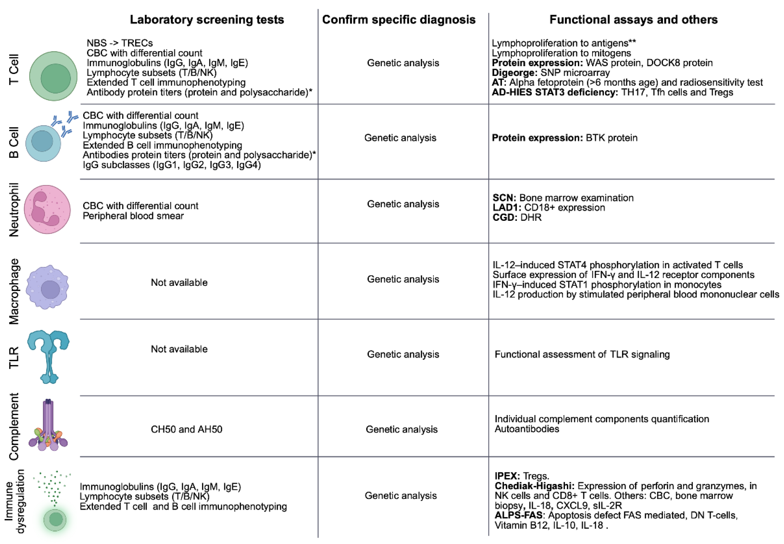
In the era where inborn errors of immunity were called primary immunodeficiencies, the most common manifestation was recurrent sinopulmonary diseases. This continues to be the most common manifestation and is the phenotype most closely aligned with antibody and complement disorders. Screening tests for these are widely available through total IgG, IgA, IgM levels and a CH50. In contrast, infants and children with a history dominated by viral opportunistic infections or Pneumocystis are most likely to have a T cell deficiency. Lymphocyte subset analysis with quantification of T cells (CD3+, CD3+CD4+, CD3+CD8+), B cells (CD19+, CD20+), and NK cells (CD16+CD56+) is the most common starting point for this clinical picture [28] (Figure 3).
Deep fungal infections for which there is no explanation and unusual bacterial infections should trigger testing for chronic granulomatous disease. The most widely available test is the Dihydrorhodamine 1,2,3 response (DHR) test, but testing for chronic granulomatous disease can sometimes be labeled as a neutrophil oxidative burst test. This test is generally straightforward to interpret; however, neutrophils have a short half-life in phlebotomy tubes and sample handling can sometimes give artifactual results [29].
Complement testing is often paired with an antibody evaluation. Many of the infections that arise in complement deficient patients can also be seen in people with antibody deficiencies. Stronger predictors of complement deficiency include sepsis and Neisserial infections. Invasive Pneumococcal infections also constitute a relatively strong predictor for both complement and antibody deficiencies.
According to the clinical suspicion and diagnostic resources available at each center, additional assessments may be included and when to consider genetic testing is an evolving topic.
Finally, it is important to highlight a comprehensive evaluation to rule out secondary immunodeficiencies such as Human Immunodeficiency Virus (HIV) infection, anatomical abnormalities (including asplenia), prematurity, cystic fibrosis, malnutrition, immunosuppressive or other immunomodulatory therapies, nephrotic syndrome, protein-losing enteropathy, lymphangiectasias and other acquired conditions that may impair immune function [30].
4.2. Recognition of Patterns of IEI Infectious Manifestations
4.2.1. T Cell Disorders
Phenotype. Patients with T cell defects commonly present with early recurrent and severe infections caused by bacterial, viral and fungal microorganisms. Additional clinical manifestations may include severe adverse reactions to live vaccines, chronic diarrhea, autoimmunity (see immune dysregulation section), and failure to thrive. Although many T cell defects present during infancy, milder or hypomorphic disorders may remain undiagnosed until adolescence or adulthood. Severe Combined Immunodeficiency (SCID) constitutes a medical emergency because affected infants are at high risk for life-threatening infections and require prompt diagnosis and definitive treatment [31]. Today, in the US, there is newborn screening for SCID and therefore most infants are diagnosed prior to infectious manifestations.
Initial evaluation. The initial laboratory evaluation includes a complete blood count (CBC) with differential. When a significant or persistent lymphopenia is present, a T cell disorder should be suspected. Lymphocyte subset analysis by flow cytometry identifies the specific lymphocyte lines (T, B, and NK cells) and can be further expanded to characterize naive and memory T cells and recent thymic emigrant [6] enumeration [31]. In SCID, T cell production is profoundly impaired, with markedly reduced or absent T cells; depending on the underlying genetic defect, B cell and NK cells can also be affected. Total lymphocytes and T lymphocytes are higher in the first months of life and decrease with age. As a rule, lymphopenia in infants is determined when the absolute lymphocyte count is <2500 cells/mm3 and in adults when it is <1000 cells/mm3 [28]. Other IEIs, particularly combined immunodeficiencies (CIDs), may present with less profound T cell lymphopenia and variable abnormalities in T cell differentiation and function. Concordantly, their infection pattern is less profound.
Advanced studies. T cell proliferation assays evaluate the capacity of T cells to proliferate in response to mitogenic or antigenic stimulation and is usually markedly reduced or absent in patients with significant T cell dysfunction. Antigen-specific proliferation can be assessed following stimulation with antigens such as tetanus toxoid or Candida antigens. However, these assays may be difficult to interpret during the first months of life because young infants have had limited exposure to foreign antigens [28]. In contrast, mitogen-induced proliferation assesses the intrinsic proliferative capacity of T cells and can be evaluated phytohemagglutinin (PHA), concanavalin A (ConA), and pokeweed mitogen (PWM) to test T cell capacity to proliferate [31]. These assays can require a large blood volume and are highly influenced by the clinical state of the patient. Therefore, it is reasonable to move from identification of persistent lymphopenia to genetic testing without testing for proliferation.
Newborn screening. The newborn screening for SCID has been implemented throughout the United States and Puerto Rico since 2018 [32]. The screening program relies on measurement of T cell receptor excision circles (TRECs) in dried blood spots. TRECs are nonreplicating DNA circles generated as a byproduct of T cell receptor gene rearrangement during T cell development in the thymus and serve as a marker of newly generated T cells. Low or absent TREC levels can identify infants with SCID and other conditions associated with profound T cell lymphopenia or impaired thymic output, including congenital athymia and selected CIDs [33].
An abnormal TREC screening result should be followed promptly by a comprehensive immunologic evaluation, including a CBC with differential and flow-cytometric analysis of peripheral blood lymphocyte subsets with particular attention to naïve T cell populations. Additional functional and genetic studies should then be guided by the initial findings and clinical presentation to confirm the diagnosis and determine the therapeutic approach [31].
Example case. A two-year-old boy has been seen more than 20 times for viral infections with some bacterial superinfections requiring antibiotics. In his life, he has had greater than 10 courses of antibiotics - most often for otitis media. When he received his one-year vaccines, he had a three week rash and fever that eventually self-resolved. He had a single CBC on record at the time of an emergency room visit that demonstrated an absolute lymphocyte count of 500cells/mm3. Upon further evaluation, he was found to have a T cell count that is less than 10% of the age-appropriate range. Genetic testing was performed that revealed two variants in the gene RAG1. This gene is associated with both SCID and milder forms of T cell deficiency. He was referred for bone marrow transplant, which was successfully performed, and he has gone on to lead a normal life.
4.2.2. Antibody Disorders
Phenotype: Predominantly antibody deficiencies is the most common category of IEIs. Their clinical spectrum is broad, ranging from asymptomatic individuals to patients with recurrent or severe infections depending in part of the specific diagnosis. Sinopulmonary infections caused by bacteria and gastrointestinal infections are common.
Initial evaluation: The laboratory evaluation should include quantitative serum immunoglobulins (IgG, IgA, IgM, IgE), and evaluation of antibody responses to vaccines.
Low serum IgG is generally defined as a value more than 2 standard deviations below the age-adjusted reference mean on at least two measurements obtained more than 3 weeks apart [28,34]. When the initial IgG concentration is markedly reduced (<100–300 mg/dL, depending on age), a single measurement may be sufficient to expedite further diagnostic evaluation and management [28,34].
Advanced studies: Measurement of IgG subclasses has a more limited diagnostic utility. IgG subclass concentrations vary substantially during infancy and early childhood, complicating interpretation, and reduced subclass levels may be completely benign. [35] However, for some teenagers and adults, IgG subclass measurements may provide insight into their immune function. B cell enumeration and B cell subset evaluations can also be useful. Markedly reduced or absent B cells (<2% of total lymphocytes) strongly suggests agammaglobulinemia. In addition, deficiency of circulating isotype-switched memory B cells (CD27+IgM-IgD-cells) with impaired antibody response and reduced levels of IgG and low IgA and/or low IgM can help to identify patients with common variable immunodeficiency (CVID) [36].
Example case: An eight-year-old girl who has been previously active in sports, was admitted for a severe pneumococcal pneumonia six months ago and has continued to have fatigue and recurrent infections that have kept her out of school. A review of her prescriptions revealed multiple trips to Urgent Care for which antibiotics were prescribed. Immunoglobulin testing revealed an IgG <200 mg/dL and undetectable IgA and IgM. Her vaccine responses were non-protective for all that were tested. She therefore fulfilled the criteria for CVID. This condition is unusual in that it can arise at any age. She was started on immunoglobulin replacement and has had few subsequent infections.
4.2.3. Complement Disorders
Phenotype: Complement factor deficiencies may result in a broad spectrum of infections and immune dysregulation Encapsulated microorganisms such as Streptococcus pneumoniae, Haemophilus influenzae, and Neisseria meningitidis comprise the most common infecting organisms. Deficiencies involving the terminal complement pathway (e.g., C7, C8, C9) and the alternative pathway (e.g., regulators Factor H, Factor I, and properdin) are associated with a marked susceptibility to meningitis caused by Neisseria meningitidis infections. Autoimmunity (lupus and renal disease) is common for some specific defects [37].
Initial evaluation: A CH50 and an AH50 are excellent screening tests for complement deficiencies associated with infection (but not for disorders associated with hemolytic uremic syndrome).
Advanced studies: In the setting of an abnormal CH50 or AH50 (or both) and a compatible clinical phenotype, the next step would typically involve genetic testing. It is also possible to measure serum levels of each of the individual protein components using immunoassays, such as enzyme-linked immunosorbent assays (ELISA), or nephelometric techniques to confirm and further characterize a suspected complement deficiency. [35] Appropriate attention should be given to blood sample collection and handling, particularly when individual complement component levels are being measured because the complement proteins are relatively unstable.
Example case: A 15-year-old boy was admitted yesterday for meningitis and Neisseria meningitidis was recovered from his lumbar puncture. His mother noted that his older brother also had meningitis as a young adult. A single case of Neisseria has a likelihood of only about 5% identification of a complement deficiency, however, when there is a positive family history, the likelihood rises to 30%. Thus, this young man was tested with a CH50 which returned the next day as zero. Genetic testing identified a pathogenic homozygous variant in C6, and when his brother was tested, he was also found to have the same C6 deficiency. Management of terminal complement component deficiencies involves optimizing vaccination and discussion of antibiotic management.
4.2.4. Neutrophil Disorders
Phenotype: The clinical manifestations of neutropenia and neutrophil dysfunction include recurrent or severe bacterial and fungal infections, typically involving the skin and internal organs. Common sites of infection include lung, mastoid, skin, and gingiva.
Initial evaluation: A CBC with differential and peripheral blood smear will allow detection of neutrophil quantitative defects and some morphologic changes associated with neutrophil dysfunction.
Advanced studies: In cyclic neutropenia, neutrophil counts typically fluctuate at approximately 21-day intervals, although cycles may range from 14 to 36 days. Serial CBCs once or twice weekly for up to 6 weeks can help establish the diagnosis [38]. In contrast, severe congenital neutropenia is characterized by persistent, profound neutropenia.
When the clinical picture suggests a neutrophil disorder and the neutrophil counts are normal, one should focus on the DHR. The DHR is a specific test for chronic granulomatous disease (CGD) which is characterized by impaired production of reactive oxygen species by phagocytes, resulting in defective intracellular microbial killing. Alternative testing is widely available and is sometimes ordered as a neutrophil oxidative burst test [29].
Example case 1: A seven-year-old girl has a past medical history significant for two cutaneous abscesses that required incision and drainage and very prolonged courses of antibiotics for delayed healing. She was subsequently admitted for a pneumonia, and her bronchoalveolar lavage was positive for aspergillus growth. This clinical constellation was very suggestive of CGD and she had appropriate testing with a DHR test, which was consistent with autosomal recessive CGD. Today, some mild cases of CGD are treated conservatively but most people opt for bone marrow transplant. After bone marrow transplant, she had a full recovery.
Example case 2: A 15-month-old baby had a past medical history significant for delayed umbilical cord separation (>4 weeks) with subsequent omphalitis. She also had other severe, recurrent infections requiring admission for antibiotics with marked leukocytosis and neutrophilia. Her most recent admission was for presumed mastoiditis, and she had drainage through an overlying ulcer. Upon surgical resection of the mastoid, the surgeon noted limited pus. Concern for leukocyte adhesion deficiency was raised and the CD18+ expression on the surface of neutrophils and monocytes by flow cytometry was very low. She subsequently had genetic testing which revealed two pathogenic variants in ITGB2. She then underwent a bone marrow transplant which was curative for her.
4.2.5. Macrophage Disorders
Phenotype: Mendelian susceptibility to mycobacterial disease (MSMD) is characterized by abnormal killing of mycobacteria resulting from defects in the IL-12/IFN-γ axis. Patients typically present with severe or recurrent infections caused by Mycobacterium species, including tuberculosis and nontuberculous mycobacteria, and may also develop infections caused by Salmonella spp. or herpesviruses.
Evaluation: Genetic testing is the cornerstone of testing in this set of conditions. There are limited options for functional testing.
Advanced studies: Functional assays include assessment of IL-12–induced STAT4 phosphorylation in activated T cells, IFN-γ–induced STAT1 phosphorylation in monocytes, IL-12 production by stimulated peripheral blood mononuclear cells, and surface expression of IFN-γ and IL-12 receptor components [35].
Example case: A 5-year-old boy had an insidious ache in his lower leg. Over the last two months, it increased to the point where weight bearing was difficult. He had some fevers and weight loss. On examination, there was mild swelling over the lower tibia with significant point tenderness. Hepatosplenomegaly was found. An X-ray showed osteomyelitis and given its extensive nature, he was referred for surgical debridement. The culture grew an M. avium complex organism. Additional imaging revealed smaller lesions in two vertebrae. Genetic testing was performed and two pathogenic variants were identified in the IL12RB1 gene. He was managed with prolonged antimicrobials and recovered. Long term, he was placed on suppressive azithromycin.
4.2.6. Toll-like Receptor Disorders (TLRs)
Phenotype: Some IEIs affect the toll-like receptor pathway. These include inherited mutations in MYD88, and IRAK4, which are both associated with severe bacterial infections in infancy. Mutations affecting TLR3, UNC93B1 are associated with viral encephalitis [39].
Evaluation: Functional assessment of TLR signaling is available in the US and tests for many, but not all the disorders. Thus, genetic testing is often performed as an initial approach.
Example case: An 8-month-old girl was admitted for periorbital cellulitis. She previously had S. pneumoniae sepsis with a complicated course. Her brother died four years ago from S. pneumoniae sepsis and meningitis. Genetic testing was performed and two pathogenic variants in MYD88 were identified. She recovered from her cellulitis and was started on immunoglobulin and was given a protocol for fever. The natural history of this disorder is gradual improvement in the severity of the infections.
5.0. Diagnostic Approach to IEI with Immune Dysregulation
5.1. Overview
Immune dysregulation is characterized by pronounced autoimmunity, autoinflammation, atopy, lymphoproliferation, and/or malignancy. Its evaluation can be complex because, in addition to immune system dysfunction, multiple organs and systems may be affected in diverse ways and the evolution over time complicates assessment [40]. One diagnostic approach focuses first on the immune system with classical testing, specialized studies, functional assays followed by genetic analysis. However, the routine laboratory tests are often limited in their ability to identify the specific diagnosis. For that reason, the other diagnostic approach comprises early genetic testing in patients with clinical manifestations of immune dysregulation, autoinflammatory disorders, atypical atopy, and bone marrow failure as a first step.
5.2. Immune Dysregulation in T Cell Disorders
Phenotype: Autoimmunity is extremely common in T cell disorders with autoimmune cytopenias being particularly common [41]. In addition to classic autoimmunity, inflammatory diseases are also common in this category of disorders.
A particular set of disorders is termed primary immune dysregulation disorders because the immune dysregulation is prominent. Hemophagocytic lymphohistiocytosis (HLH) is an intrinsic T cell disorder and certain disorders leading to inflammatory bowel disease, are examples. Elevated IgE levels, eosinophilia, and platelet abnormalities may suggest primary atopic conditions, including the actinopathy T cell disorders. Markedly elevated IgE levels (>2,000 IU/mL), particularly when associated with early-onset or refractory severe allergic manifestations and infections can be a sign of a primary atopic condition [42].
Evaluations: This category may be suspected through the identification of lymphopenia on a CBC and T cell lymphopenia identified on flow cytometry but not all immune dysregulation conditions have that feature. Assessment of cytotoxic function may also be informative, particularly in patients with suspected NK-cell disorders or HLH. Evaluation of the expression of key cytotoxic proteins, including perforin and granzymes, in NK cells and CD8+ T cells is available in specialty labs. However, the specific diagnosis nearly always requires genetic testing [43]. The diversity of potential etiologies and the lack of overall screening options, supports an early genetic testing approach.
Example case 1: A ten-year-old girl with recalcitrant Evan’s syndrome was being considered for B cell depleting therapy. She had no history of infections but the question of doing an immune evaluation prior to B cell depletion was discussed. In children, 40% of patients with Evan’s syndrome in one study were found to have an IEI. Her IgG was normal but genetic testing revealed a pathogenic variant in CTLA4. CTLA4 deficiency has a high rate of lymphoma in adulthood and a significant risk of later development of hypogammaglobulinemia. She was started on abatacept with resolution of her autoimmune cytopenias. She required ongoing monitoring programs for her lifetime risks of hypogammaglobulinemia and lymphoma.
Example case 2: A two-year-old with severe recalcitrant eczema and extensive molluscum in the last three months was admitted for anaphylaxis to walnuts with ongoing wheezing. Her IgE was >10,000 IU/mL and her eosinophil count was 2500 cells/mm3. This is one of the most difficult settings to risk stratify for genetic testing. However, extremely high IgE and eosinophils are useful red flags and the extensive molluscum was certainly a red flag. This child was found to have DOCK8 deficiency, one of the actinopathies, and was referred for bone marrow transplant.
5.3. Immune Dysregulation in Antibody Disorders
Phenotype: The most common treatable IEI is CVID, which occurs in about 1:10,000 adults [44]. Among the conditions in this review, it is unusual in its broad age of onset which ranges from 2 years of age though 80+ years of age. Although classically, sinopulmonary infections are the most common manifestation, about half of the patients have immune dysregulation and the autoimmunity/immune dysregulation can precede the identification of hypogammaglobulinemia. Autoimmune cytopenias are the most common feature of immune dysregulation but gastrointestinal disease (small bowel enteropathy and inflammatory bowel disease) is also common [36].
Initial evaluation: Immunoglobulin levels and vaccine titers are a reasonable if imperfect screen. They are required to diagnose CVID, however, some cases present with autoimmunity or inflammation prior to overt hypogammaglobulinemia.
Advanced studies: If the suspicion of immune dysregulation is high and immunoglobulin levels are normal, genetic testing is the approach most likely to yield a diagnosis.
Example case: A 17-year-old was in Europe as part of a summer abroad program. While there, he developed profuse watery, diarrhea. He was given antibiotics for presumed bacterial gastroenteritis, which were ineffective. His past history was significant for idiopathic thrombocytopenia at five years of age from which he fully recovered. Upon return, his parents were shocked at how much weight he lost and sent him for an evaluation. Stool cultures and parasite exam were negative. He was referred for endoscopy which showed villous atrophy and increased intraepithelial lymphocytes. Based on this, he was tested for celiac disease and he was found to be IgA deficient. Gluten restriction was not effective and further testing revealed that he also had a low IgG level and poor titers to vaccines. Thus, he fulfilled the diagnostic criteria for CVID. He was started on immunoglobulin replacement and sirolimus with full recovery. His genetic testing was not revealing. Only about 25% of people with CVID have had a genetic etiology identified.
5.4. Immune Dysregulation in Complement Disorders
Phenotype: there are only three immune dysregulation phenotypes amongst the complement deficiencies. 1) The most common phenotype is systemic lupus, or cutaneous lupus, which is found in people with the early classical pathway complement deficiencies. These conditions are treated in the standard way although infection risk is high for these patients and precautions are typically taken. 2) The second phenotype is atypical hemolytic uremic syndrome. This is due to a variety of altered proteins that ultimately lead to overactivity of the alternative pathway of the complement cascade. These disorders are typically not fully penetrant and are most often episodically manifested. Management includes the use of complement inhibitors. 3) A subset of patients with Factor I deficiency have had neuroinflammatory disease or vasculitis which has responded to complement inhibitors.
Initial evaluation: CH50 is not routinely performed for all patients with lupus, however, in the setting of a positive family history or a significant infection history, it is reasonable to perform. It should be performed for early onset lupus. For atypical hemolytic uremic syndrome, the testing relies on genetics.
Advanced studies: There can be autoantibodies directed to complement components that mimic the inherited forms of atypical hemolytic uremic syndrome.
Example case: A one-year-old had a photosensitive rash throughout the summer. After an upper respiratory infection, she developed autoimmune hemolytic anemia and significant arthritis. An antinuclear antibody (ANA) was positive, and she had protein in her urine. Genetic testing was sent which revealed two pathogenic variants in C1QA. This led to C1q deficiency, which is known to be associated with early onset and quite severe lupus. Management of these cases is not entirely clear; however, bone marrow transplant has been successful and the family considered that approach but has pursued a conservative approach.
5.5. Immune Dysregulation in Neutrophil Disorders.
Phenotype: Immune dysregulation is not common across all neutrophil disorders, but CGD is associated strongly with inflammatory bowel disease and is also enriched for autoimmune cytopenias [45].
Initial evaluation: Among all cases of childhood-onset inflammatory bowel disease, CGD is found in approximately 1%. However, the ramification of this finding is extremely high. Thus, it is appropriate to test for CGD in early onset inflammatory bowel disease (IBD) (usually defined as onset <6 years of age).
Example case: A boy with known CGD immigrated to the US recently. The parents revealed that he has had a five-pound weight loss in the last two months. His erythrocyte sedimentation rate (ESR) was 65mm/hr. He said he saw blood in his stools but denied diarrhea. Endoscopy revealed inflammatory bowel disease and he was referred for bone marrow transplant which is typically curative for both the bowel disease and infection risk.
5.6. Macrophage and Toll-like Receptor Defects
These conditions are infrequently associated with autoimmunity or immune dysregulation.
6.0. Genetic Diagnosis (Diagnostic Laboratory Testing, Molecular Testing)
6.1. Overview
Genetic testing has become an integral component of the comprehensive evaluation of patients with suspected IEIs [46]. First, there is more accessibility to testing because the costs have decreased over time. Genetic testing can establish the specific etiology of an IEI, enabling individualized management, precision medicine, and genetic counseling for patients and their families. In addition, it is particularly useful for children where initial immunological studies can be normal and in the immune dysregulation conditions. The availability of genetic testing varies according to region, clinical center, and resources [47,48]. Some centers incorporate genetic testing early in the diagnostic evaluation and others reserve genetic testing for patients in whom conventional immunologic evaluation is inconclusive.
Current genetic testing options include Sanger sequencing, targeted IEI gene panels, whole-exome sequencing (WES), and whole-genome sequencing (WGS). Emerging approaches include optical genome mapping, RNA sequencing, and proteomic analyses [49]. Today, most commercial laboratories in the US are doing next generation sequencing of some type but targeted analysis of a set of genes rather than Sanger sequencing.
6.2. Sequencing Technologies
6.2.1. Sanger Sequencing
Sanger Sequencing remains an important, targeted method for variant confirmation and is particularly useful in patients with suspected IEI who have a similarly affected family member with a known genetic defect. It may also be appropriate for genes or genomic regions that are not reliably assessed by high-throughput sequencing approaches, including pseudogenes. In such cases, Sanger sequencing can facilitate discrimination between the functional gene and its pseudogene - which are nonfunctional genetic segments that evolutionarily arose from duplication of existing genes [28].
6.2.2. Targeted Gene Panels
Targeted Gene Panels is a method to detect mutations in known IEI-related genes [51]. They are particularly useful when the clinical phenotype or family history strongly suggests a monogenic disorder and the selected panel provides adequate coverage of the relevant genes. However, the diagnostic yield of targeted panels may be limited when the phenotype is atypical [52].
6.2.3. Whole-Exome Sequencing (WES)
Whole-Exome Sequencing (WES) evaluates protein-coding regions of the genome, which comprise approximately 1%–2% of the human genome, focusing on the exonic regions and nearby splice sites. [42] WES is particularly useful in patients with IEIs who have additional phenotypic features suggestive of a multisystem disorder, such as cardiac or neurologic abnormalities, or when a new monogenic disorder is suspected in the setting of parental consanguinity [28,53]. The chemistry behind WES is more technically complicated than whole genome sequencing and for that reason, many commercial testing companies have switched to the chemistry used for whole genome sequencing.
6.2.4. Whole-Genome Sequencing (WGS)
Whole-Genome Sequencing (WGS) provides broader genomic coverage than WES. Although WGS offers greater potential for identifying variants outside the exome, its interpretation requires specialized bioinformatic and clinical expertise and may be associated with greater costs [54]. WGS may be particularly valuable in patients with suspected IEIs and multisystem phenotypes, a strong suspicion of a noncoding or structural variant, or a phenotype suggestive of a genetic defect not adequately captured by clinical exome sequencing. It may also facilitate the identification of novel disease-associated genes [55].
Conclusion
The IEIs have heterogeneous and complex clinical manifestations. Based on clinical suspicion, a diagnostic pathway can be designed to guide the selection of initial, advanced, and specific tests. However, genetic testing is increasingly being used as a first-line diagnostic approach, as it can facilitate early diagnosis and enable timely implementation of appropriate management strategies, ultimately improving patient outcomes.
Author Contributions
Estefanía Vásquez Echeverri, and Kathleen E. Sullivan: Conceptualization, writing-original draft preparation, review and editing critical manuscript revision. All authors have read and agreed to the published version of the manuscript.
Funding
This research received no external funding.
Acknowledgments
We acknowledge our teachers and the patients who have guided us.
Conflicts of Interest
The authors declare no conflicts of interest.
Abbreviations
The following abbreviations are used in this manuscript:
| IEI | Inborn errors of immunity |
| IUIS | International Union of Immunological Societies |
| US | United States |
| NK | Natural Killer |
| DHR | Dihydrorhodamine 1,2,3 response |
| HIV | Human Immunodeficiency Virus |
| SCID | Severe combined immunodeficiency |
| CBC | Complete blood count |
| CID | combined immunodeficiencies |
| PHA | Phytohemagglutinin |
| ConA | Concanavalin A |
| PWM | Pokeweed Mitogen |
| TRECs | T cell receptor excision circles |
| DNA | Deoxyribonucleic acid |
| RAG1 | Recombination-activating gene 1 |
| CVID | Common Variable Immunodeficiency |
| ELISA | Enzyme-linked immunosorbent assays |
| CGD | Chronic granulomatous disease |
| ITGB2 | Integrin subunit beta 2 |
| MSMD | Mendelian susceptibility to mycobacterial disease |
| TLR | Toll-like receptor |
| MYD88 | Myeloid differentiation primary response 88 |
| IRAK4 | Interleukin-1 receptor associated kinase 4 |
| TLR3 | Toll-like receptor 3 |
| UNC93B1 | Protein unc-93 homolog B1 |
| HLH | Hemophagocytic Lymphohistiocytosis |
| CTLA4 | Cytotoxic T-lymphocyte associated protein 4 |
| DOCK8 | Dedicator of cytokinesis 8 |
| URI | Upper Respiratory Infection |
| ANA | Antinuclear antibodies |
| C1QA | Complement C1q A chain |
| IBD | Inflammatory bowel disease |
| ESR | Erythrocyte sedimentation rate |
| WES | Whole-exome sequencing |
| WGS | Whole-genome sequencing |
| RNA | Ribonucleic acid |
References
- Akalu, Y.T.; Bogunovic, D. Inborn errors of immunity: an expanding universe of disease and genetic architecture. Nat. Rev. Genet 2024, 25(3), 184–195. [Google Scholar] [CrossRef]
- Rider, N.L.; et al. Validating inborn error of immunity prevalence and risk with nationally representative electronic health record data. J. Allergy Clin. Immunol. 2024, 153(6), 1704–1710. [Google Scholar] [CrossRef]
- Bousfiha, A.; et al. Human Inborn Errors of Immunity: 2019 Update of the IUIS Phenotypical Classification. J. Clin. Immunol. 2020, 40(1), 66–81. [Google Scholar] [CrossRef]
- Tangye, S.G.; et al. Human Inborn Errors of Immunity: 2019 Update on the Classification from the International Union of Immunological Societies Expert Committee. J. Clin. Immunol. 2020, 40(1), 24–64. [Google Scholar] [CrossRef]
- Poli, M.C.; et al. Human inborn errors of immunity: 2024 update on the classification from the International Union of Immunological Societies Expert Committee. J. Hum. Immun. 2025. 1, 1, e20250003. [Google Scholar] [CrossRef]
- Cortesi, M.; et al. Unmasking inborn errors of immunity: identifying the red flags of immune dysregulation. Front Immunol. 2024, 15, 1497921. [Google Scholar] [CrossRef]
- Thalhammer, J.; et al. Initial presenting manifestations in 16,486 patients with inborn errors of immunity include infections and noninfectious manifestations. J. Allergy Clin. Immunol. 2021, 148(5), 1332–1341 e5. [Google Scholar] [CrossRef]
- Nikzad, S.; et al. Quantifying the Diagnostic Odyssey Burden Among Persons with Inborn Errors of Immunity. J. Clin. Immunol. 2025, 45(1), 61. [Google Scholar] [CrossRef]
- Quinn, J.; et al. Growth in diagnosis and treatment of primary immunodeficiency within the global Jeffrey Modell Centers Network. Allergy Asthma Clin. Immunol. 2022, 18(1), 19. [Google Scholar] [CrossRef]
- Aden, A.; et al. Clinical Warning Signs in Detecting Inborn Errors of Immunity in Children: A Diagnostic Accuracy Systematic Review. J. Allergy Clin. Immunol. Pract. 2026, 14(4), 920–936 e1. [Google Scholar] [CrossRef]
- Ghosh, A.; et al. Competency of Jeffrey Modell Foundation warning signs and routine laboratory tests in suspecting primary immunodeficiencies: A cross-sectional multi-centric prospective study from eastern India. Int. J. Rheum. Dis. 2024, 27(11), e15405. [Google Scholar] [CrossRef]
- Dabrowska, A.; et al. Extended List of Warning Signs in Qualification to Diagnosis and Treatment of Inborn Errors of Immunity in Children and Young Adults. J. Clin. Med. 2023, 12(10). [Google Scholar] [CrossRef]
- FitzPatrick, A.M.; et al. Piloting an automated query and scoring system to facilitate APDS patient identification from health systems. Front Immunol. 2024, 15, 1508780. [Google Scholar] [CrossRef]
- Alsaati, N.; et al. A predictive model for identification of pediatric individuals with common variable immunodeficiency through electronic medical records. J. Allergy Clin. Immunol. 2025, 156(1), 186–194. [Google Scholar] [CrossRef]
- Johnson, R.; et al. Electronic health record signatures identify undiagnosed patients with common variable immunodeficiency disease. Sci. Transl. Med. 2024, 16(745), eade4510. [Google Scholar] [CrossRef]
- Messelink, M.A.; et al. Clinical Validation of a Primary Antibody Deficiency Screening Algorithm for Primary Care. J. Clin. Immunol. 2023, 43(8), 2022–2032. [Google Scholar] [CrossRef]
- Jolles, S.; et al. Implementing calculated globulin screening for immunodeficiencies: The benefits and challenges. J. Hum. Immun. 2026. 2, 5, e20260028. [Google Scholar] [CrossRef]
- Costagliola, G.; Peroni, D.G.; Consolini, R. Beyond Infections: New Warning Signs for Inborn Errors of Immunity in Children. Front Pediatr. 2022, 10, 855445. [Google Scholar] [CrossRef]
- Abend, A.H.; et al. Estimation of prevalence of autoimmune diseases in the United States using electronic health record data. J. Clin. Invest 2024, 135(4). [Google Scholar] [CrossRef]
- Nelson, M.; et al. Genetic determinants of childhood onset systemic lupus erythematosus. Lupus Sci. Med. 2026, 13(1). [Google Scholar] [CrossRef]
- Kelsen, J.R.; et al. North American Society for Pediatric Gastroenterology, Hepatology, and Nutrition Position Paper on the Evaluation and Management for Patients With Very Early-onset Inflammatory Bowel Disease. J. Pediatr. Gastroenterol. Nutr. 2020, 70(3), 389–403. [Google Scholar] [CrossRef]
- Demirtas Guner, D.; et al. Genetic Variants in Early-Onset Inflammatory Bowel Disease: Monogenic Causes and Clinical Implications; Children (Basel), 2025; 5, p. 12. [Google Scholar]
- Sams, L.; et al. Atopic manifestations of inborn errors of immunity. Curr. Opin. Allergy Clin. Immunol. 2023, 23(6), 478–490. [Google Scholar] [CrossRef]
- Sanchez-Ramon, S.; et al. Navigating primary and secondary immunodeficiency intersections: how to find IEI hidden within SID. Allergy Asthma Clin. Immunol. 2026, 22(1). [Google Scholar] [CrossRef]
- Chandra, S.; et al. Primary Immunodeficiencies and Hematologic Malignancies: A Diagnostic Approach. Front Immunol. 2022, 13, 852937. [Google Scholar] [CrossRef]
- Nima Rezaeia, b.; Esther de Vriesc, d; Eleonora Gambinerie, f; Meytsg, Isabelle; Haddadh, Elie. Common presentations and diagnostic approaches. Stiehm’s Immune Deficiencies: inborn errors for immunity, 2nd ed.; S.E. Sullivan, K.E., Ed.; Elsevier: London, 2020. [Google Scholar]
- Tran, P.; et al. Eosinophilic gastrointestinal disorders in patients with inborn errors of immunity: Data from the USIDNET registry. Front Immunol. 2022, 13, 987895. [Google Scholar] [CrossRef]
- Chinen, J.; Horner, C.C.; Orange, J.S. Practice parameter for inborn errors of immunity: What is new in the 2025 update. J. Allergy Clin. Immunol. 2026, 158(2), 368–373. [Google Scholar] [CrossRef]
- Kim, H.Y.; et al. Rapid determination of chimerism status using dihydrorhodamine assay in a patient with X-linked chronic granulomatous disease following hematopoietic stem cell transplantation. Ann. Lab Med. 2013, 33(4), 288–92. [Google Scholar] [CrossRef]
- Tuano, K.S.; Seth, N.; Chinen, J. Secondary immunodeficiencies: An overview. Ann. Allergy Asthma Immunol. 2021, 127(6), 617–626. [Google Scholar] [CrossRef]
- Dvorak, C.C.; et al. The diagnosis of severe combined immunodeficiency (SCID): The Primary Immune Deficiency Treatment Consortium (PIDTC) 2022 Definitions. J. Allergy Clin. Immunol. 2023, 151(2), 539–546. [Google Scholar] [CrossRef]
- Puck, J.M.; et al. Expert commentary: practical issues in newborn screening for severe combined immune deficiency (SCID). J. Clin. Immunol. 2012, 32(1), 36–8. [Google Scholar] [CrossRef]
- Puck, J.M. Laboratory technology for population-based screening for severe combined immunodeficiency in neonates: the winner is T-cell receptor excision circles. J. Allergy Clin. Immunol. 2012, 129(3), 607–16. [Google Scholar] [CrossRef]
- Lee, T.K.; Gereige, J.D.; Maglione, P.J. State-of-the-art diagnostic evaluation of common variable immunodeficiency. Ann. Allergy Asthma Immunol. 2021, 127(1), 19–27. [Google Scholar] [CrossRef]
- Klangkalya, N.; Fleisher, T.A.; Rosenzweig, S.D. Diagnostic tests for primary immunodeficiency disorders: Classic and genetic testing. Allergy Asthma Proc. 2024, 45(5), 355–363. [Google Scholar] [CrossRef]
- Cunningham-Rundles, C. Current concepts: Common variable immunodeficiency. Ann. Allergy Asthma Immunol. 2026, 136(4), 376–386. [Google Scholar] [CrossRef]
- Brodszki, N.; et al. European Society for Immunodeficiencies (ESID) and European Reference Network on Rare Primary Immunodeficiency, Autoinflammatory and Autoimmune Diseases (ERN RITA) Complement Guideline: Deficiencies, Diagnosis, and Management. J. Clin. Immunol. 2020, 40(4), 576–591. [Google Scholar] [CrossRef]
- Skokowa, J.; et al. Severe congenital neutropenias. Nat. Rev. Dis. Primers 2017, 3, 17032. [Google Scholar] [CrossRef]
- Maglione, P.J.; N. Simchoni, C. Cunningham-Rundles, Toll-like receptor signaling in primary immune deficiencies. Ann. N Y Acad. Sci. 2015, 1356(1), 1–21. [Google Scholar] [CrossRef]
- Roberti, G.; et al. Inborn Errors of Immunity in Pediatric Hematology and Oncology: Diagnostic Principles for Clinical Practice. J. Clin. Med. 2025, 14(17). [Google Scholar] [CrossRef]
- Fischer, A.; et al. Autoimmune and inflammatory manifestations occur frequently in patients with primary immunodeficiencies. J. Allergy Clin. Immunol. 2017, 140(5), 1388–1393 e8. [Google Scholar] [CrossRef]
- Roganovic, J. G. Bellesi, Inborn Errors of Immunity: New Insights. Acta Med. Acad. 2024, 53(3), 293–302. [Google Scholar] [CrossRef]
- Henter, J.I.; et al. Diagnostic guidelines for familial hemophagocytic lymphohistiocytosis revisited. Blood 2024, 144(22), 2308–2318. [Google Scholar] [CrossRef]
- Jeevarathnum, A.C.; et al. Common variable immunodeficiency disorders: What generalists should know. Afr. J. Thorac. Crit. Care Med. 2021, 27(3). [Google Scholar] [CrossRef]
- Ouahed, J.; et al. Very Early Onset Inflammatory Bowel Disease: A Clinical Approach With a Focus on the Role of Genetics and Underlying Immune Deficiencies. Inflamm. Bowel Dis. 2020, 26(6), 820–842. [Google Scholar] [CrossRef]
- Chinn, I.K.; et al. Diagnostic interpretation of genetic studies in patients with primary immunodeficiency diseases: A working group report of the Primary Immunodeficiency Diseases Committee of the American Academy of Allergy, Asthma & Immunology. J. Allergy Clin. Immunol. 2020, 145(1), 46–69. [Google Scholar] [CrossRef]
- Chinn, I.K.; Orange, J.S. A 2020 update on the use of genetic testing for patients with primary immunodeficiency. Expert Rev. Clin. Immunol. 2020, 16(9), 897–909. [Google Scholar] [CrossRef]
- Platt, C.D.; et al. Efficacy and economics of targeted panel versus whole-exome sequencing in 878 patients with suspected primary immunodeficiency. J. Allergy Clin. Immunol. 2021, 147(2), 723–726. [Google Scholar] [CrossRef]
- von Hardenberg, S.; et al. Current genetic diagnostics in inborn errors of immunity. Front Pediatr. 2024, 12, 1279112. [Google Scholar] [CrossRef]
- Abolhassani, H.; L. Hammarstrom, C. Cunningham-Rundles, Current genetic landscape in common variable immune deficiency. Blood 2020, 135(9), 656–667. [Google Scholar] [CrossRef]
- Chinen, J.; et al. Practical approach to genetic testing for primary immunodeficiencies. Ann. Allergy Asthma Immunol. 2019, 123(5), 433–439. [Google Scholar] [CrossRef]
- Cifaldi, C.; et al. Targeted NGS Platforms for Genetic Screening and Gene Discovery in Primary Immunodeficiencies. Front Immunol. 2019, 10, 316. [Google Scholar] [CrossRef]
- Meyts, I.; et al. Exome and genome sequencing for inborn errors of immunity. J. Allergy Clin. Immunol. 2016, 138(4), 957–969. [Google Scholar] [CrossRef]
- Grumach, A.S.; Goudouris, E.S. Inborn Errors of Immunity: how to diagnose them? J. Pediatr. (Rio J) 2021, 97 Suppl 1(Suppl 1), S84–S90. [Google Scholar] [CrossRef]
- Turro, E.; et al. Whole-genome sequencing of patients with rare diseases in a national health system. Nature 2020, 583(7814), 96–102. [Google Scholar] [CrossRef]
Figure 1.
Infections as Red Flags in Inborn Errors of Immunity.
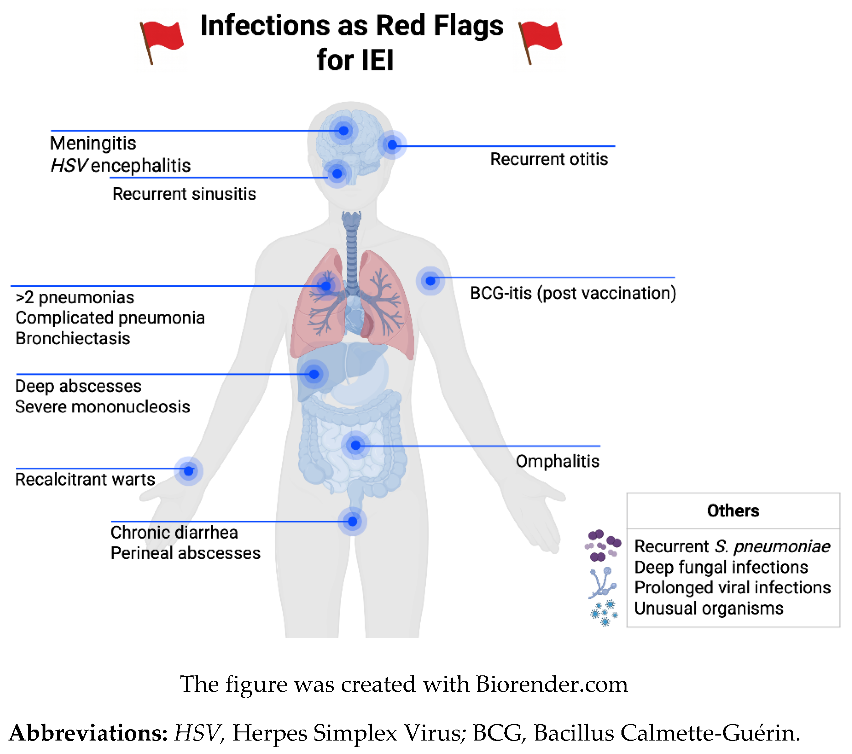
Figure 2.
Non-Infectious Red Flags for Inborn Errors on Immunity.
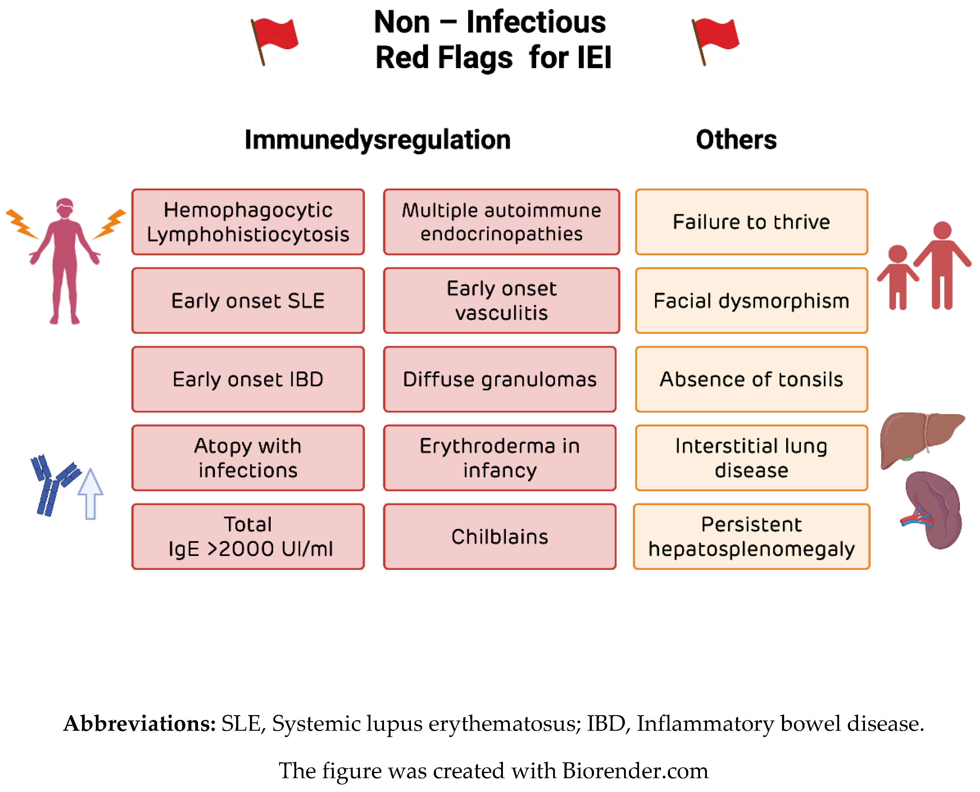
Figure 3.
Immunological approach for Inborn Errors of Immunity diagnosis. *Antibody polysaccharide titers responses it is recommends after 2 years of age. ** Lymphoproliferation to antigens responses it is recommend after 1 year of age. Abbreviations: NBS, Newborn screening; TRECs, T cell receptor excision circles; CBC, Complete blood count; SNP, Single-nucleotide polymorphism; AT, Ataxia Telangiectasia; AD-HIES, Autosomal dominant hyper-IgE Syndrome; STAT3, Signal Transducer and Activator of Transcription 3; BTK, Bruton's tyrosine kinase; STAT4, Signal Transducer and Activator of Transcription 4; IFN, Interferon; STAT1, Signal Transducer and Activator of Transcription 1; SCN, Severe congenital neutropenia; LAD1, Leukocyte Adhesion Deficiency Type 1; CGD, Chronic granulomatous disease; TLR, Toll-like receptor; IPEX, Immune dysregulation, polyendocrinopathy, enteropathy, X-linked; CXCL9, C-X-C motif chemokine ligand 9; sIL-2R, sIL-2R, or soluble CD25; IL, Interleukin; DN, Double-Negative. The figure was created with Biorender.com.
Figure 3.
Immunological approach for Inborn Errors of Immunity diagnosis. *Antibody polysaccharide titers responses it is recommends after 2 years of age. ** Lymphoproliferation to antigens responses it is recommend after 1 year of age. Abbreviations: NBS, Newborn screening; TRECs, T cell receptor excision circles; CBC, Complete blood count; SNP, Single-nucleotide polymorphism; AT, Ataxia Telangiectasia; AD-HIES, Autosomal dominant hyper-IgE Syndrome; STAT3, Signal Transducer and Activator of Transcription 3; BTK, Bruton's tyrosine kinase; STAT4, Signal Transducer and Activator of Transcription 4; IFN, Interferon; STAT1, Signal Transducer and Activator of Transcription 1; SCN, Severe congenital neutropenia; LAD1, Leukocyte Adhesion Deficiency Type 1; CGD, Chronic granulomatous disease; TLR, Toll-like receptor; IPEX, Immune dysregulation, polyendocrinopathy, enteropathy, X-linked; CXCL9, C-X-C motif chemokine ligand 9; sIL-2R, sIL-2R, or soluble CD25; IL, Interleukin; DN, Double-Negative. The figure was created with Biorender.com.
Disclaimer/Publisher’s Note: The statements, opinions and data contained in all publications are solely those of the individual author(s) and contributor(s) and not of MDPI and/or the editor(s). MDPI and/or the editor(s) disclaim responsibility for any injury to people or property resulting from any ideas, methods, instructions or products referred to in the content. |
© 2026 by the authors. Licensee MDPI, Basel, Switzerland. This article is an open access article distributed under the terms and conditions of the Creative Commons Attribution (CC BY) license (http://creativecommons.org/licenses/by/4.0/).
Copyright: This open access article is published under a Creative Commons CC BY 4.0 license, which permit the free download, distribution, and reuse, provided that the author and preprint are cited in any reuse.