

Submitted:
04 October 2026
Posted:
08 October 2026
You are already at the latest version
Abstract
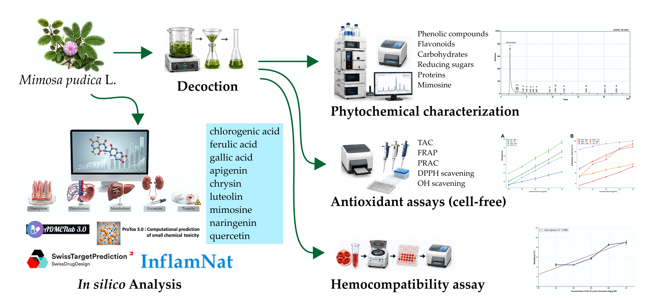
Mimosa pudica L., a pantropical medicinal herb rich in polyphenols, but also containing the non-protein amino acid mimosine with known cytotoxic properties, is a case for xenobiotic pharmacology, yet its safety profile remains inadequately characterized. In this study, we quantified the major metabolite classes of an aqueous decoction of M. pudica aerial parts, evaluated its in vitro hemocompatibility in human erythrocytes, and assessed its antioxidant activity using five cell-free assays. We also employed computational tools (ADMETlab 3.0, ProTox 3.0, SwissTargetPrediction, and InflamNat) to predict ADMET properties, toxicity alerts, and potential protein targets. The decoction showed high phenolic (316.41 ± 19.58 mg GAE/g) and flavonoid (122.99 ± 12.78 mg QE/g) contents, with mimosine levels of 2.11 ± 0.13 mg/g. Antioxidant activity was concentration-dependent, with the lowest IC₅₀ in the DPPH• assay (0.23 ± 0.02 mg/mL). Hemolysis remained below 9% at all tested concentrations (0.2–1.0 mg/mL), with an HC₅₀ > 1.0 mg/mL, consistent with biocompatibility criteria for plant extracts. In silico predictions indicated that most of flavonoids and phenolic acids complied with Lipinski's rule and showed a favorable oral bioavailability, but also, significant CYP450 inhibition and alerts for genotoxicity and nephrotoxicity. Predicted molecular targets included redox-modulatory proteins such as AKR1B1, XDH, NOX4, and GSK3B. Collectively, our results demonstrate that M. pudica is a complex mixture of xenobiotics with antioxidant effects, and suggest benefits and risks associated to their individual constituents. Further in vitro and in vivo studies are needed to validate these predictions and to determine whether M. pudica can be developed into safe and effective nutraceutical or pharmaceutical preparations.
Keywords:
Mimosa pudica
; oxidative stress
; xenobiotic pharmacology
; antioxidant activity
; ADMET prediction
; erythrocyte hemolysis
; mimosine
; target prediction
1. Introduction
Oxidative stress, a key driver of xenobiotic-induced toxicity, arises when the production of reactive oxygen and nitrogen species (ROS/RNS) surpasses the endogenous antioxidant defenses [1]. Environmental pollutants, drugs, and industrial chemicals often trigger this imbalance by generating hydrogen peroxide, superoxide anions, and hydroxyl radicals, highly reactive molecules that damage lipids, proteins, and DNA [2]. To counteract this threat, mammalian cells have evolved the Nrf2/ARE pathway, which coordinates the expression of cytoprotective genes encoding phase II detoxifying enzymes and endogenous antioxidants such as superoxide dismutase (SOD), catalase (CAT), and glutathione peroxidase (GPx) [3]. However, the metabolic machinery involved in xenobiotics’ biotransformation, the cytochrome P450 (CYP450) superfamily, can paradoxically generate ROS as by-products, thus amplifying the oxidative burden [4]. This dual role of CYP450 highlights the subtle balance between detoxification and toxicity. Plant-derived polyphenols and flavonoids have emerged as promising modulators of both the Nrf2/ARE pathway and CYP450 activity, offering a potential strategy to mitigate oxidative damage [5]. Yet, their therapeutic significance must be evaluated against their own xenobiotic nature and potential toxicities.
While plant metabolites are often regarded as safe due to their natural origin, these compounds constitute a complex array of xenobiotics, synthesized through pathways that are evolutionarily distinct from mammalian metabolism [6]. This perspective has implications for the study of plant-based preparations, leading to examine not only their beneficial effects but also their potential toxicological risks, and their interactions with other phytochemicals, and/or conventional drugs [7]. Therefore, ethnomedicinal practices which mostly depend on multi-plant formulations, demand a rigorous xenobiotic framework to assess safety and efficacy, rather than the misleading, assumption of inherent harmlessness.
Mimosa pudica L. (Fabaceae), commonly known as the sensitive plant, is a pantropical herb with a well-documented history in traditional medicine across Asia, Africa, and Latin America. Ethnopharmacological studies report its use for pain, inflammation, wounds, and neurological disorders [8]. Modern phytochemical analyses show a complex metabolomic profile that includes alkaloids, tannins, saponins, and a rich diversity of phenolic compounds [8]. These findings encouraged pharmacological investigations, in which both aqueous and ethanolic extracts of M. pudica attenuated oxidative and nitrosative stress in experimental models of epilepsy and diabetes, restoring antioxidant enzyme activities and glutathione pools [9,10]. Leaf and stem extracts also scavenge DPPH• radicals, inhibit lipid peroxidation, and enhance catalase activity, pointing to strong antioxidant properties that deserve mechanistic analysis at the molecular level [11]. Despite these findings, the therapeutic potential of M. pudica cannot be fully assessed without examining its chemical constituents as individual xenobiotic entities, each with the potential to interact with mammalian detoxification pathways, transport systems, and cellular targets in ways that may be either beneficial or deleterious.
The antioxidant properties of M. pudica are largely attributed to its phenolic and flavonoid constituents. Aqueous extracts have yielded a significant number of aglycones including apigenin, luteolin, quercetin, naringenin, and chrysin, together with phenolic acids such as gallic, chlorogenic, and ferulic acids [9]. These compounds act through multiple redox mechanisms: they scavenge free radicals via hydrogen atom and electron transfer, chelate transition metals, and activate the Nrf2/ARE-dependent antioxidant response [12]. Among flavonoids, apigenin, luteolin, and quercetin are recognized Nrf2 activators, whereas naringenin and chrysin modulate CYP450 activity [13]. Phenolic acids, in turn, contribute to metal chelation and inhibit the pro-inflammatory NF-κB pathway [13]. From a xenobiotic perspective, while these compounds may offer therapeutic benefits, their simultaneous presence in the extract raises the possibility of complex interactions with drug-metabolizing enzymes and transport systems, as well as synergistic or antagonistic effects among themselves.
A concerning feature of M. pudica is its content of L-mimosine, a non-protein amino acid with reported cytotoxic and antiproliferative activities. Historically recognized as a reversible inhibitor of DNA replication and cell cycle progression, mimosine has more recently been linked to apoptosis induction via mitochondrial permeability transition, cytochrome c release, and caspase activation, all processes associated with intracellular hydrogen peroxide generation and glutathione depletion [14]. In plant-plant allelopathic interactions, mimosine also triggers oxidative stress in target species, increasing CAT and SOD activities together with malondialdehyde accumulation [15]. The presence of a rich pool of antioxidant polyphenols in M. pudica coexisting with a potentially pro-oxidant amino acid highlights the need for a comprehensive toxicological and pharmacokinetic evaluation before plant derived preparations can be recommended as therapeutic or nutraceutical agents.
Despite extensive ethnopharmacological reports, there are still unexplored critical aspects in our understanding of M. pudica’s pharmacology and toxicology. Most antioxidant studies were carried out on crude extracts, which prevents a clear attribution of activity to specific metabolites and the potential synergistic or antagonistic interactions among them. The cellular safety profile of M. pudica is not enough characterized, particularly its hemocompatibility and effects on human erythrocyte membrane integrity, a crucial early endpoint in xenobiotic risk assessment that has not been systematically evaluated [16]. Moreover, the absorption, distribution, metabolism, excretion, and toxicity (ADMET) properties of its key metabolites, essential for predicting oral bioavailability, blood-brain barrier permeability, CYP450-mediated drug interactions, and organ-specific toxicity, have been scarcely addressed, especially through computational approaches. The specific protein targets involved in redox regulation and xenobiotic metabolism (e.g., Nrf2, Keap1, CYP450 isoforms, peroxiredoxins) remain largely unknown, limiting the mechanistic interpretation of its antioxidant effects. These gaps underline the requirement for an integrated approach that combines experimental phytochemistry, toxicology, and computational pharmacology to advance M. pudica from traditional/complementary remedy to a rationally characterized source of phytomedicines.
The present study was designed to comprehensively characterize the chemical composition, antioxidant capacity, and safety profile of M. pudica decoction. We aimed to: quantify the major groups of phenolic metabolites, including flavonoids and phenolic acids; evaluate its in vitro antioxidant activity through a multiparametric battery of cell-free assays (DPPH• scavenging, reducing power, phosphomolybdenum total antioxidant capacity , permanganate-reducing antioxidant capacity, and hydroxyl radical scavenging); assess the hemotoxicity and membrane-protective effects in human erythrocytes; and predict the ADMET profile and identify protein targets involved in oxidative stress responses and xenobiotic metabolism using in silico tools (ADMETlab 3.0, ProTox 3.0, SwissTargetPrediction, and InflamNat).
We hypothesize that the decoction’s antioxidant activity correlates with its phenolic content and that in silico predictions will identify molecular targets including redox-modulatory proteins and potential safety concerns not evident from traditional use. By integrating experimental phytochemistry and toxicology with computational pharmacology, this work seeks to provide rational bases for the future development of M. pudica-derived antioxidant products with known benefits and risks.
2. Materials and Methods
2.1. Reagents and Standards
All reagents were of analytical grade unless otherwise stated. Folin–Ciocalteu reagent, hydrogen peroxide (H₂O₂; 30% v/v), salicylic acid, 3,5-dinitrosalicylic acid (DNS), and 2,2-diphenyl-1-picrylhydrazyl (DPPH•) were purchased from Sigma-Aldrich (St. Louis, MO, USA). Sodium carbonate (Na₂CO₃), sodium hydroxide (NaOH), aluminium chloride (AlCl₃), sodium nitrite (NaNO₂), phenol, sulfuric acid (H₂SO₄), trichloroacetic acid (TCA), ferric chloride (FeCl₃), sodium potassium tartrate tetrahydrate (NaKC₄H₄O₆·4H₂O), copper(II) sulfate (CuSO₄), sodium dihydrogen phosphate (NaH₂PO₄), orthophosphoric acid (H₃PO₄), ammonium heptamolybdate tetrahydrate ((NH₄)₆Mo₇O₂₄·4H₂O), potassium ferricyanide (K₃[Fe(CN)₆]), potassium permanganate (KMnO₄), ferrous sulfate heptahydrate (FeSO₄·7H₂O), and HPLC grade methanol were supplied by Merck KGaA (Darmstadt, Germany).
The following analytical standards were obtained from Sigma-Aldrich (St. Louis, MO, USA) and used as indicated: gallic acid (total phenolics), quercetin (total flavonoids), D-(+)-glucose (total carbohydrates and reducing sugars), bovine serum albumin fraction V (BSA; total proteins), L-mimosine (HPLC-UV quantification), and L-(+)-ascorbic acid (positive control and reference standard in the TAC, FRAP, PRAC, DPPH•, and •OH assays).
2.2. Plant Material Collection and Taxonomic Classification
Aerial parts (stems and leaves) of M. pudica were collected from a natural population of more than 20 individuals in El Caney, Santiago de Cuba, Cuba (20.0569° N, −75.7719° W). To minimize variability due to circadian rhythms, collections were performed early in the morning (08:00–10:00 h) from vegetative-stage specimens during March 2024. A voucher specimen was taxonomically identified by specialists at the Centro Oriental de Ecosistemas y Biodiversidad (BIOECO, Santiago de Cuba) and deposited at the BSC Herbarium “Jorge Sierra Calzado” under accession number 218237.
Plant material was air-dried in the shade at room temperature to constant weight over 14 days in the shade. Dried aerial parts were then pulverized in a MRC KM-700 knife mill (Berlin, Germany) and stored in airtight containers until use.
2.3. Preparation of the Decoction and Quantification of Primary and Secondary Metabolites
An aqueous decoction of M. pudica (dMp) was prepared by boiling dried plant material in distilled water. Briefly, 10 g of shadow-dried leaves and stems were combined with 100 mL of water, boiled for 5 minutes, filtered, and stored at 4 °C until required. The extract was evaporated to dryness in a rotary evaporator at 45 °C (Kika Werke RV 05, Germany), and the yield was expressed as dry extract (DE). Quantitative analysis of the decoction was performed by spectrophotometric techniques adapted to 96-well microplates (USA Scientific CytoOne, cat. CC7682-7596) using a SpectraMax ABS PLUS reader (Molecular Devices, San Jose, CA, USA). All assays were performed in triplicate (of three biological replicates), and results were expressed as milligram equivalents of the respective standard per gram of dry extract (mg Eq/g DE).
2.3.1. Total Phenolic Compounds
The Folin–Ciocalteu method was employed [17]. To each well, 20 µL of sample or standard (gallic acid, 20-500 µg/mL), 100 µL of Folin–Ciocalteu reagent, and 100 µL of Na₂CO₃ (200 g/L) were added. The microplate was incubated at 40 °C for 30 min, and absorbance was measured at 765 nm against a blank prepared with distilled water. The calibration curve was: y = 0.007x − 0.028 (r² = 0.9987). Results were expressed as mg gallic acid equivalents per g DE (mg GAE/g DE).
2.3.2. Total Flavonoids
Quantification was performed by the aluminium chloride (AlCl₃) method [17]. Distilled water (100 µL), sample or standard (quercetin, 20-500 µg/mL; 50 µL), and NaNO₂ (5%; 20 µL) were mixed. After 5 min, AlCl₃ (10%; 20 µL) and NaOH (1 mol/L; 100 µL) were sequentially added. Absorbance was read at 415 nm against the corresponding blank. The calibration equation was: y = 0.0004x + 0.0665 (r² = 0.9985). Results were expressed as mg quercetin equivalents per g DE (mg QE/g DE).
2.3.3. Total Carbohydrates
The phenol–sulfuric acid method was used [18]. Sample or standard (glucose, 16-250 µg/mL; 80 µL) and phenol (5%; 40 µL) were added to each well. Concentrated H₂SO₄ (200 µL) was immediately added, and the mixture was homogenized for 10 min. The plate was incubated at 30 °C for 20 min, and absorbance was measured at 490 nm. The calibration curve was: y = 0.004x + 0.0014 (r² = 0.9960). Results were expressed as mg glucose equivalents per g DE (mg GE/g DE).
2.3.4. Reducing Sugars
Reducing sugars content was determined by the 3,5-dinitrosalicylic acid (DNS) method [19]. Sample or standard (glucose, 0.5-2.0 g/mL; 25 µL) was mixed with DNS reagent (25 µL). The microplate was wrapped in aluminum foil and incubated at 92 °C for 5 min. The reaction was stopped by cooling on ice for 5 min, after which 250 µL of distilled water was added. Absorbance was read at 540 nm. The calibration equation was: y = 0.4174x − 0.0071 (r² = 0.9942). Results were expressed as mg glucose equivalents per g DE (mg GE/g DE).
2.3.5. Proteins
Quantification was performed by the Lowry’s method [20]. Sample or standard (BSA, 10–200 µg/mL; 100 µL) and carbonate reagent (200 µL) were added. The mixture was allowed to stand for 10 min at room temperature, after which 20 µL of Folin–Ciocalteu reagent (50%) was added and incubated for 30 min in the dark. Absorbance was measured at 760 nm. The calibration curve was: y = 0.001x − 0.0005 (r² = 0.9920). Results were expressed as mg BSA equivalents per g DE (mg BSAE/g DE).
2.3.6. Mimosine Quantification by High-Performance Liquid Chromatography (HPLC-UV)
The decoction was clarified by precipitation with activated charcoal (0.03 g per 10 mL of decoction), filtered through syringe filter and then, mimosine was extracted with HCl 0.1 mol/L (1:1, v/v). Mimosine was quantified by reversed-phase HPLC following the validated method of Mohamed-Musthaba et al., according to International Council for Harmonisation (ICH) guidelines [21].
Chromatographic analysis was performed at room temperature on a Knauer Smartline HPLC system (Berlin, Germany) coupled to a programmable variable-wavelength UV–Vis detector, controlled by ClarityChrom software v7.2.0.73 (Knauer, Berlin, Germany). Separation was carried out on a reversed-phase C18 column (Phenomenex; 250 × 4.6 mm, 5 µm particle size).
The mobile phase consisted of water–orthophosphoric acid (99.8:0.2, v/v), adjusted to pH 3.0 with orthophosphoric acid, filtered through a 0.22 µm membrane (Millipore), and sonicated before use. Elution was performed in isocratic mode at a flow rate of 1.0 mL/min, with a run time of 12 min. System pressure was maintained at 100-140 bar (1450-2030 psi) with the column temperature controlled at 25-30 °C.
Decoction samples (20 µL) were manually injected. Eluted mimosine was monitored spectrophotometrically at 284 nm. A mimosine calibration curve (5–500 µg/mL) was employed. Results were expressed as mg mimosine per g DE (mg/g DE).
2.4. Evaluation of In Vitro Antioxidant Activity
The antioxidant activity of the dMp and ascorbic acid (AA) as reference was evaluated by five independent spectrophotometric assays, performed in triplicate at increasing extract concentrations (0.2, 0.4, 0.6, 0.8, and 1.0 mg/mL) adapted to microplates using a SpectraMax ABS PLUS reader (Molecular Devices, San Jose, CA, USA).
2.4.1. Total Antioxidant Capacity (TAC). Phosphomolybdenum Method
The phosphomolybdenum complex method described by Elsherif et al. was used [22]. In 1.5 mL vials, 40 µL of sample (or distilled water for the blank) was mixed with 400 µL of reagent solution (0.6 M H₂SO₄, 28 mM sodium phosphate, and 4 mM ammonium heptamolybdate). The mixture was incubated at 95 °C for 90 min. Subsequently, 300 µL was transferred to a 96-well microplate, and absorbance was measured at 695 nm. The ascorbic acid calibration curve (2–10 mM) was: y = 0.1672x + 0.334 (r² = 0.9984). Results were expressed as mg ascorbic acid equivalents per g DE (mg AAE/g DE).
2.4.2. Ferric Reducing Antioxidant Power (FRAP)
Reducing power was evaluated by the potassium ferricyanide method [23]. In 1.5 mL vials, 200 µL of sample (or distilled water for the blank) was mixed with 200 µL of potassium ferricyanide (1%). The mixture was incubated at 50 °C for 20 min, after which 200 µL of TCA (10%) was added and centrifuged at 2000 rpm for 10 min. From the supernatant, 125 µL was taken, mixed with 125 µL of distilled water and 25 µL of FeCl₃ (0.1%) in a microplate, and absorbance was read at 700 nm. The ascorbic acid calibration curve (2–10 mM) was: y = 0.147x − 0.091 (r² = 0.9983). Results were expressed as mg AAE/g DE.
2.4.3. Permanganate Reducing Antioxidant Capacity (PRAC)
PRAC was determined following the method of Mohamed et al. [24]. Volumes of 1 mL of samples or control were placed in test tubes, to which 1 mL of freshly prepared potassium permanganate solution (0.35 mM KMnO₄) and 500 µL of H₂SO₄ (2 M) were added. Subsequently, 300 µL was transferred to a 96-well microplate, and the reduction in absorbance was measured at 530 nm for samples and for ascorbic acid as standard. Reducing capacity was calculated as: PRAC (%) = [(AC − AS)/AC] x 100, where AC is the absorbance of the control and AS is the absorbance of the sample.
2.4.4. DPPH• Radical Scavenging Assay
For the evaluation of 2,2-diphenyl-1-picrylhydrazyl (DPPH•) radical scavenging, volumes of 100 µL of samples or control were placed in a microplate, and 100 µL of DPPH solution (0.09 mM in methanol) was added. The mixture was incubated for 30 min protected from light, and absorbance was measured at 517 nm [23]. The DPPH• radical scavenging activity was calculated as a percentage of DPPH discoloration using the following equation: % DPPH• Scavenging = [(ADPPH − AS)/ADPPH] x 100, where AS is the absorbance of the sample and ADPPH is the absorbance of the DPPH solution.
The half-maximal inhibitory concentration (IC₅₀) was determined by linear regression from the dose-response curve of percentage inhibition versus extract concentration.
2.4.5. Hydroxyl Radical (•OH) Scavenging Assay
•OH scavenging was determined by the salicylic acid degradation method [24]. Volumes of 50 µL of samples and standard (ascorbic acid, 10 mg/mL) were added to a microplate, followed by 50 µL of FeSO₄·7H₂O (9.0 mM) and 50 µL of H₂O₂ (9.0 mM). The mixture was incubated for 10 min at room temperature, and the reaction was initiated by adding 50 µL of salicylic acid (9.0 mM in methanol), incubating for 60 min at 37 °C. Absorbance was measured at 510 nm. The percentage of hydroxyl radical inhibition was calculated as: •OH Inhibition (%) = [1 − (AS − ASELF − AB)/AC] x 100, where AS is the absorbance of the sample, ASELF is the absorbance of the solution containing all reagents except H₂O₂; AB is the absorbance of the blank without sample or salicylic acid; and AC is the absorbance of the control containing all reagents except the sample. The IC₅₀ was determined by linear regression from the dose-response curve.
2.5. In Vitro Cytotoxicity Assay in Human Erythrocytes
Human erythrocytes were obtained from fresh venous blood collected in sodium citrate (1:10) as anticoagulant. After centrifugation at 500–1000 × g for 5–10 min, plasma and the leukocyte layer were discarded. The erythrocyte pellet was washed three times with phosphate-buffered saline (PBS; pH 7.4) under the same conditions to remove plasma residues, and then reconstituted (1:40 v/v) to a final cell concentration of 8 x 10⁹ cells/mL, as determined using a Neubauer counting chamber [25].
The cytotoxicity of the decoction (dMp) was evaluated by the in vitro hemolysis assay according to Invittox Protocol No. 37, described by Berenguer et al. [25]. Erythrocyte suspensions (8 x 10⁹ cells/mL) were incubated with serial concentrations of the decoction (0.2–1.0 mg/mL) in PBS for 60 min at 37 °C with gentle agitation. PBS and distilled water were used as negative (0% hemolysis) and positive (100% hemolysis) controls, respectively. After centrifugation (1000 × g, 5–10 min), released hemoglobin was quantified spectrophotometrically at 540 nm using a SpectraMax ABS PLUS reader (Molecular Devices, San Jose, CA, USA). The percentage of hemolysis was calculated as: Hemolysis (%) = [(AS − ANC) / (APC − ANC)] × 100, where AS is the absorbance of the sample, ANC is the absorbance of the negative control and APC is the absorbance of the positive control.
The half-maximal hemolytic concentration (HC₅₀) was determined by linear regression from the dose–response curve of hemolysis percentage versus decoction concentration, as an indicator of membrane cytotoxicity.
2.6. In Silico Analysis
Nine metabolites (phenolics/flavonoids) of M. pudica were selected for in silico analysis based on two criteria: (i) they are highly conserved across species from the main geographical areas where the plant has been studied; (ii) they have been identified in aqueous extracts and decoctions. The selected compounds were chlorogenic acid, ferulic acid, gallic acid, apigenin, chrysin, luteolin, mimosine, naringenin, and quercetin. Their structures in SMILES format were retrieved from PubChem (https://pubchem.ncbi.nlm.nih.gov/) and used as input for the computational platforms described below.
2.6.1. ADMET Property Prediction
The pharmacokinetic and toxicological profiles of the selected compounds were predicted using ADMETlab 3.0 (https://ADMETlab3.scbdd.com). The following endpoints were assessed: physicochemical properties (molecular weight, logP, TPSA, solubility, pKₐ); medicinal chemistry (QED, Lipinski and Pfizer rules, synthetic accessibility); absorption (Caco-2 and MDCK permeability, PAMPA, human intestinal absorption [HIA], oral bioavailability [F20%, F30%, F50%]); distribution (plasma protein binding [PPB], volume of distribution [VD], blood–brain barrier [BBB] penetration); metabolism (interaction with CYP1A2, CYP2C9, CYP2C19, CYP2D6, CYP3A4, CYP2B6, and CYP2C8, and stability in human liver microsomes [HLM]); and acute and chronic toxicity (hERG, DILI, Ames mutagenicity, rat acute oral toxicity, carcinogenicity, ocular irritation, genotoxicity, and cytotoxicity) [26].
2.6.2. Systemic Toxicity Prediction
Systemic toxicity was predicted using ProTox 3.0 (https://tox.charite.de), a web tool developed by the Institute of Medical Biology at Charité University (Berlin, Germany). This platform uses machine-learning models built from high-quality experimental data to predict diverse toxicity profiles. ProTox 3.0 was used to evaluate organ-level toxicity (hepatotoxicity, neurotoxicity, nephrotoxicity, respiratory toxicity, cardiotoxicity), toxicity endpoints (carcinogenicity, immunotoxicity, mutagenicity, cytotoxicity), blood–brain barrier interaction, ecotoxicity, and effects on Tox21 signalling pathways (nuclear receptors, stress response, molecular initiating events) and cytochrome P450 metabolism [27].
2.6.3. Molecular Target Prediction
Potential protein targets of the evaluated compounds were identified using SwissTargetPrediction, developed by the Swiss Institute of Bioinformatics (SIB) and the Molecular Modeling Group of the University of Lausanne [28] (http://www.swisstargetprediction.ch). Target predictions were based on predictive probability and the number of active ligands reported in the ChEMBL database. Probability values were classified as high (0.700–1.0), moderate (0.300–0.700), and low (0–0.300); only predictions with probability > 0.400 (i.e., high or moderate) were considered relevant for this study.
2.6.4. Anti-Inflammatory Activity Prediction
The anti-inflammatory properties of the selected compounds were evaluated through the InflamNat (accessible at: http://www.inflamnat.com/). InflamNat predicted the inhibitory activity of nitric oxide (NO) production by macrophages (IC₅₀ < 50 µM) without cytotoxicity [29].
2.7. GenAI Disclosure
During the preparation of this manuscript, the authors used generative artificial intelligence (Kimi K3, Moonshot AI, China) to improve English language fluency and grammatical clarity. All the content was critically reviewed, edited, and validated by the authors, who assume full responsibility for the accuracy, interpretation, and scientific integrity of the final manuscript. No AI tools were used for data generation, data analysis, or the creation of figures or tables.
2.8. Statistical Analysis
All spectrophotometric assays were conducted in independent triplicates (n = 3), and results were expressed as mean ± standard deviation (SD).
Dose–response relationships for antioxidant activity (PRAC, DPPH•, and •OH) and hemolytic activity were analyzed by linear regression to determine the half-maximal inhibitory concentration (IC₅₀) and the half-maximal hemolytic concentration (HC₅₀), respectively. For the antioxidant assays, percentage inhibition was plotted against the decimal logarithm of extract concentration (log[C]), and the IC₅₀ was calculated by interpolation from the fitted linear equation when the 50% inhibition threshold fell within the experimental range. For the hemolysis assay, percentage hemolysis was regressed against extract concentration; when the 50% threshold lay outside the tested range, the HC₅₀ was reported as exceeding the maximum concentration tested. The assumptions of linear regression (normality, homoscedasticity) were met. Statistical significance of regression was considered at p < 0.05 and GraphPad Prism version 9.5.1 (GraphPad Software Inc., La Jolla, CA, USA) was used in all analysis.
For the in silico predictions, each web server (ADMETlab 3.0, ProTox 3.0, SwissTargetPrediction, and InflamNat) employs its own statistical algorithms and confidence metrics, as described in their respective original publications.
3. Results
3.1. Quantification of Metabolites in the Decoction
Phytochemical analysis confirmed that the decoction (dMp) was rich in phenolic metabolites (Table 1). Total phenolic content was 316.41 ± 19.58 mg GAE/g DE, while total flavonoid content was 122.99 ± 12.78 mg QE/g DE. With respect to flavonoids’ quantification, the aluminum chloride method is specific for flavonoids with ortho-dihydroxy structures; therefore, molecules lacking this structure (e.g., chrysin, which is present in M. pudica) would be underestimated. Mimosine, a characteristic metabolite of the species, was detected at 2.11 ± 0.13 mg/g DE (Figure 1). In contrast, carbohydrates, reducing sugars, and proteins showed lower values, consistent with an extract enriched in secondary metabolites rather than primary ones.
3.2. In Vitro Cell-Free Antioxidant Activity
Figure 2 shows the antioxidant activity of the decoction (dMp) and ascorbic acid (AA) as reference across the different assays.
In the TAC assay, dMp showed a concentration-dependent increase in absorbance, from 0.125 ± 0.010 to 0.421 ± 0.009 (range 0.2-1.0 mg/mL), whereas AA reached values from 0.106 ± 0.006 to 0.770 ± 0.003 over the same interval. AA exhibited higher values from 0.4 mg/mL onward, with a maximum difference of 0.349 absorbance units at 1.0 mg/mL (Figure 2A).
In the FRAP assay, both groups exhibited similar behavior, but with higher absorbance values: dMp ranged from 0.199 ± 0.026 to 0.904 ± 0.033, whereas AA reached 1.125 ± 0.036 at the maximum concentration. The difference between dMp and AA was smaller in FRAP (0.221 units at 1.0 mg/mL) than in TAC, suggesting that dMp possesses a relatively more efficient ferric reducing capacity than its total antioxidant capacity.
The two radical scavenging assays (DPPH•, and •OH) and PRAC also demonstrated concentration-dependent activity (Figure 2B). AA exhibited the highest antioxidant potential in all methods, reaching near-complete inhibitions in DPPH• (99.96 ± 0.71% at 1.0 mg/mL) and values above 90% in PRAC and •OH. The plant decoction, in turn, showed moderate but consistent activity, with maximum values of 88.76 ± 0.90% (DPPH•), 56.29 ± 1.62% (•OH), and 51.98 ± 0.85% (PRAC) at 1.0 mg/mL
The decoction exhibited the lowest IC₅₀ in the DPPH• assay (0.23 ± 0.02 mg/mL), indicating that this was its most potent antioxidant mechanism, followed by •OH (0.77 ± 0.06 mg/mL) and PRAC (0.964 ± 0.03 mg/mL). AA showed a significant higher radical scavenging potential, with IC₅₀ values of 0.37 ± 0.02 mg/mL (PRAC) and 0.21 ± 0.01 mg/mL (•OH). In the DPPH• assay, AA exceeded 50% inhibition even at the lowest concentration tested (0.2 mg/mL).
3.3. Cytotoxicity in Human Erythrocytes
The results of the vitro hemolysis assay are shown in Figure 3. The decoction induced a concentration-dependent effect on the human erythrocyte membrane. Concentrations of 0.2 and 0.4 mg/mL produced hemolysis percentages (4.43% and 4.39%, respectively) that did not differ significantly from spontaneous hemolysis (~5%). However, from 0.6 mg/mL onward, a significant increase was observed, reaching 5.75%, 8.53%, and 8.99% at 0.6, 0.8, and 1.0 mg/mL, respectively.
Linear regression analysis revealed a positive and statistically significant dependence between hemolysis percentage and decoction concentration (R² = 0.898; p = 0.014). Since none of the evaluated concentrations reached 50% hemolysis, the conventional HC₅₀ parameter was determined to be >1.0 mg/mL within the experimental range.
3.4. In Silico ADMET Analysis
3.4.1. Physicochemical and Drug-Like Properties
The estimated properties for the nine evaluated metabolites are summarized in Table 2. Molecular weights ranged from 170.1 (gallic acid) to 354.1 (chlorogenic acid), all within the optimal range for oral drugs (<500 Da). LogP values at pH 7.4 showed wide variability: mimosine presented a negative value (-3.17), indicative of high hydrophilicity, whereas chrysin showed the greatest lipophilic character (logP 3.60). Most flavonoids and phenolic acids complied with Lipinski’s rule (≤1 violation), except chlorogenic acid, which exceeded the hydrogen bond donor limit (nHD = 6; limit = 5) and presented a QED (0.234) below the optimal threshold (>0.67). Naringenin and ferulic acid showed the best QED profiles (0.742 and 0.715, respectively).
3.4.2. Absorption and Bioavailability
Predicted human intestinal absorption (HIA) was favorable for all bioactive compounds, with probabilities of limited absorption (HIA ≥ 30%) near 0.0 for gallic acid, apigenin, chrysin, luteolin, naringenin, and mimosine. Chlorogenic and ferulic acid and quercetin showed intermediate values (0.106-0.249), suggesting moderate absorption. Caco-2 permeability was medium-to high (optimal: higher than −5.15 log units) for ferulic acid, apigenin, chrysin, and naringenin, whereas the remaining compounds fell within the low permeability range. Predicted oral bioavailability (F20%) indicated a high probability of low bioavailability for chlorogenic acid, ferulic acid, and luteolin (0.925-0.992); moderate for gallic acid, apigenin and quercetin (0.351-0.565); and high for chrysin (0.155), naringenin (0.002); and, notably, mimosine (probability 0.0 of F < 20%)
3.4.3. Distribution and Metabolism
Plasma protein binding (PPB) was very high (>90%) for apigenin, chrysin, luteolin, naringenin, and quercetin, which could limit their pharmacologically active free concentration. In contrast, mimosine and gallic acid showed low PPB (13.4% and 37.6%, respectively). None of the compounds showed significant blood–brain barrier (BBB) permeability (BBB+), all being classified as BBB−.
Regarding metabolism, significant interaction with the cytochrome P450 system was predicted (Table 3). The flavonoids (apigenin, chrysin, luteolin, naringenin, quercetin) were characterized by strong inhibition of CYP1A2, CYP2D6, CYP3A4, and CYP2C8 (probabilities >0.9 in most cases). Mimosine, conversely, showed scarce interaction with CYPs, with slight probabilities of being a substrate for CYP2C9 and CYP2D6.
3.5. In Silico Toxicity Profile
3.5.1. Acute and Chronic Toxicity (ADMETlab and ProTox)
The predicted toxicological profile revealed important alerts that varied according to chemical structure (Table 4). Most compounds showed low risk of hERG potassium channel blockade (probabilities <0.11), with some flavonoids presenting intermediate values at 10 µM. Drug-induced liver injury (DILI) risk was high for apigenin (0.743), chrysin (0.872), luteolin (0.796), and quercetin (0.783), whereas mimosine showed the lowest value (0.110).
Ames’ mutagenicity showed moderate-to-high probabilities for most compounds (0.586–0.716), being particularly notable in mimosine (0.716) and luteolin (0.650). Predicted genotoxicity was highest for apigenin (0.987), chrysin (0.966), luteolin (0.986), naringenin (0.978), quercetin (0.975), and mimosine (0.984). All phenolic acids showed low probability (0.054-0.251) of oral acute toxicity in mammals, whereas flavonoids were predicted to have moderate toxicity (0.480-0.530). Mimosine showed a moderate probability (0.380) in the rat oral acute toxicity prediction.
3.5.2. Organ Toxicity and Signaling Pathways (ProTox)
ProTox 3.0 predicted active nephrotoxicity for all evaluated metabolites (probabilities 0.56–0.69). Hepatotoxicity was predicted as inactive in most cases, except for ferulic acid, which showed a borderline probability (0.51 inactive vs. active).
In the Tox21 pathway screening, significant activation of the estrogen receptor (ERα and ERβ) and androgen receptor (AR) was observed for apigenin, chrysin, luteolin, and quercetin. Furthermore, activation of the mitochondrial membrane potential (MMP) and p53 pathways was predicted for several flavonoids, suggesting potential apoptosis-inducing or mitochondrial oxidative stress potential. The aryl hydrocarbon receptor (AhR) pathway was also activated for apigenin, chrysin, luteolin, and quercetin.
Predictions were obtained using ADMETlab 3.0 (for hERG, DILI, Ames, genotoxicity, and rat oral acute toxicity) and ProTox 3.0 (for nephrotoxicity and hepatotoxicity). Values represent predicted probabilities; for ProTox predictions, “active” indicates a positive toxicity alert and “inactive” indicates no alert. hERG: human ether-à-go-go-related gene; DILI: drug-induced liver injury.
3.6. Prediction of Anti-Inflammatory Activity and Molecular Targets of Redox Modulation and Inflammation
Analysis by InflamNat revealed that the metabolites with the highest probability of inhibiting nitric oxide (NO) production in macrophages (IC₅₀ < 50 µM) were chlorogenic acid (0.687), ferulic acid (0.662), apigenin (0.621), quercetin (0.611), and luteolin (0.568). ADMETlab showed high predictive values of SR/ARE for all flavonoids (0.905-0.996) and gallic acid (0.942). A series of shared molecular targets among the metabolites were identified with SwissTargetPrediction, many of them related to oxidative, inflammatory, and metabolic processes. Those with the most prominent (probability > 0.300) are presented in Table 5.
Additionally, specific therapeutic targets such as telomerase reverse transcriptase (TERT), poly(ADP-ribose) polymerase 1 (PARP1), and various cyclin-dependent kinases (CDK1, CDK2, CDK6) were identified, which supports the antiproliferative and antioxidant potential of these metabolites.
4. Discussion
The maintenance of balance between free radical production and antioxidant defenses is an essential condition for normal organism functioning, and when this equilibrium is disrupted, damaged cell structures (cell membranes, proteins, etc.) accumulate [30,31]. In our study, the aqueous decoction of M. pudica aerial parts proved to be a rich source of phenolic compounds and flavonoids, while also containing the non-protein amino acid mimosine at 2.11 mg/g dry extract. This dual composition is central to our interpretation of the pharmacological activity of the decoction, which reflects the combined contributions of individual constituents, each with distinct chemical properties and biological activities. Whereas phenolic compounds are likely responsible for the observed antioxidant effects, the presence of the DNA replication inhibitor mimosine raises safety concerns that demand a careful risk-benefit evaluation [32].
The phenolic and flavonoid content we measured in Cuban M. pudica (316.41 mg GAE/g and 122.99 mg QE/g) were higher than those reported for specimens from Nepal (3.7-418.6 mg GAE/g and 4.37-14.13 mg QE/g) [33,34], India (15.6 mg GAE/g and 1.97 mg QE/g) [35], Vietnam (154.23 mg GAE/g and 38.58 mg GAE/g) [36], and Cameroon (72.90 mg QE/g and 128.93 mg QE/g) [37]. We interpret this difference as a likely consequence of environmental stress factors, particularly drought and high UV radiation, which are well known to stimulate phenolic compound accumulation in plants [38]. This variability in phenolic and flavonoid content may explain, at least in part, the discrepancies in biological activity and safety reported for extracts and fractions of this species across different studies, including its antioxidant effects. This aspect has not received enough attention in the scientific literature; and results reported for a specific geographical area should not be strictly extrapolated to another. Therefore, bioprospecting efforts must be preceded by a rigorous characterization of local plant material.
The decoction exhibited concentration-dependent antioxidant activity across all five assays, with varying efficacy. The lowest IC₅₀ was recorded in the DPPH• assay (0.23 mg/mL), suggesting a strong hydrogen-donating capacity against lipophilic radicals, a mechanism that could translate into protection of biological membranes [1]. This value was higher (i.e., less potent) than that reported for a hydroethanolic extract of the whole plant (IC₅₀ 0.10 mg/mL) [39] and for isolated flavonoids from ethyl acetate-soluble fractions (IC₅₀ 0.056 mg/mL) [40]. Interestingly, the IC₅₀ of the decoction matches that reported for hydroethanolic extracts of L-mimosine alone (IC₅₀ 0.23 mg/mL) [39], which raises a hypothesis about mimosine contribute to the overall antioxidant activity of the decoction, and it possible interaction with the polyphenol pool. Future studies are needed for fractionation and synergy analysis in the characterization of M. pudica decoctions.
The hydroxyl radical scavenging activity we observed (IC₅₀ 0.77 mg/mL) is particularly relevant in an in vivo context, because •OH is a highly reactive ROS that directly damages DNA, lipids, and proteins [1]. The capacity of the decoction to attenuate •OH generation via the Fenton reaction points to a protective role against iron-driven oxidative damage, a mechanism frequently implicated in xenobiotic-induced cellular toxicity [41]. Conversely, the permanganate-reducing antioxidant capacity (PRAC; IC₅₀ 0.96 mg/mL) and the phosphomolybdenum method (TAC) reflect the total electron-donating capacity of the extract, encompassing both slow- and fast-acting reductants. Similarly, the ferric reducing antioxidant power (FRAP) confirmed the presence of reductants capable of recycling oxidized transition metals, thereby preventing metal-catalyzed radical propagation. These assays suggest that M. pudica decoction acts through multiple, complementary redox mechanisms, rather than a single dominant pathway, a potential advantage for counteracting the complex oxidative challenges exerted by xenobiotic exposure.
Although ascorbic acid (AA) showed a higher effect than the decoction in absolute terms, particularly in the DPPH• and •OH assays, dMp displayed a broader mechanistic spectrum. AA acts primarily through direct radical scavenging and metal chelation; in contrast, the polyphenolic matrix of M. pudica may simultaneously modulate redox signalling pathways, inhibit pro-oxidant enzymes, and upregulate endogenous antioxidant defences. This pattern suggests that M. pudica polyphenols could activate Nrf2-dependent antioxidant responses in addition to their direct scavenging activity, a dual mechanism reported for other plant polyphenols [13,42]. We acknowledge, however, that this remains as a hypothesis; our data demonstrate antioxidant activity, but they do not directly prove Nrf2 activation or enzyme inhibition. Distinguishing direct scavenging from signaling-mediated effects will require cell-based assays and, ultimately, in vivo validation.
Hemolysis assays in human erythrocytes provided insight into the extract’s potential membrane toxicity. Erythrocytes, lacking nuclei and organelles but rich in unsaturated lipids and hemoglobin, are a sensitive model for detecting membrane damage and oxidative lysis [43]. At concentrations of 0.2 and 0.4 mg/mL, the decoction induced hemolysis levels (~4.4%) indistinguishable from spontaneous hemolysis, indicating adequate hemocompatibility at low exposure levels. Importantly, these concentrations encompass the DPPH• IC₅₀ (0.23 mg/mL), which means that the extract can exert significant radical-scavenging activity without compromising erythrocyte membrane integrity. The HC₅₀ exceeding 1.0 mg/mL is consistent with biocompatibility criteria for plant extracts [43]. For comparison, M. pudica gum and its nanoparticle formulations showed hemolysis below 15% at 10% concentration [44], whereas mimosine alone induced erythrocyte cytotoxicity at 0.1–0.5 mM in animal models [45].
Over the past decade, flavonoids and other phenolic compounds have become some of the most intensively studied plant metabolites for preventing cellular membrane disruption, largely due to their antioxidant potential. Based on our results, we propose the hypothesis that the phytochemical constituents of the decoction may permeate the erythrocyte membrane and exert protective effects on both sides of the lipid bilayer. The relatively planar structures of the flavones apigenin, luteolin, and chrysin, as well as the flavonol quercetin, would be expected to facilitate their transport across the erythrocyte membrane [25]. This membrane-partitioning behavior could explain, at least in part, why the decoction remains hemocompatible even at concentrations where mimosine alone would be cytotoxic.
In silico tools have become indispensable for assessing the ADMET properties of natural compounds, offering a practical alternative to animal testing and enabling early identification of pharmacokinetic and toxicological liabilities [46]. Several web-based platforms are particularly well suited to the field of natural products, including ADMETlab 3.0, ProTox 3.0, SwissTargetPrediction, and InflamNat. We selected these tools because they cover complementary aspects of xenobiotic behavior: ADMETlab 3.0 provides a broad panel of physicochemical, pharmacokinetic, and toxicity predictions; ProTox 3.0 focuses on organ-level toxicity and signaling pathways; SwissTargetPrediction identifies potential protein targets based on chemical similarity; and InflamNat predicts anti-inflammatory activity. Together, they allow us to build a multidimensional profile of M. pudica metabolites, difficult to obtain through in vitro assays alone. At the same time, we acknowledge the limitations of these predictions; they are hypothesis-generating rather than confirmatory, and their accuracy depends on the quality and coverage of the underlying training data.
The in silico ADMET predictions revealed a divergence between in vitro potency and in vivo bioavailability; most bioactive compounds showed favorable systemic bioavailability, but some polyphenols had significant limitations. Most flavonoids and phenolic acids exhibited a high probability of excellent human intestinal absorption (HIA > 30%), an essential prerequisite for oral bioavailability. However, Caco-2 permeability, a replacement system for human intestinal epithelium, was high only for ferulic acid, apigenin, chrysin, and naringenin. Gallic acid, apigenin, quercetin, chrysin, naringenin, and mimosine displayed moderate-to-high oral bioavailability (F ≥ 20%). In contrast, chlorogenic acid, ferulic acid, and luteolin were predicted to have low bioavailability (F < 20%), a finding consistent with the known presystemic metabolism of dietary polyphenols, including phase II conjugation and efflux by transporters such as P-glycoprotein [47]. This discrepancy has practical implications, because of compounds that appear highly active in vitro may not reach effective concentrations in vivo, which limits their therapeutic potential when administered orally. We therefore suggest that future studies should explore alternative delivery routes (e.g., topical, sublingual, or nanotechnology-based formulations) for those metabolites with poor oral bioavailability.
The predicted plasma protein binding (PPB) profiles revealed a clear dichotomy among the metabolites. Flavonoid aglycones (apigenin, chrysin, luteolin, naringenin, and quercetin) showed very high PPB (> 90%), which may limit their tissue distribution but could also extend their circulatory retention. In contrast, mimosine and gallic acid displayed low PPB (13.4% and 37.6%, respectively), suggesting that, if absorbed, they would circulate largely as free ligands with greater access to renal filtration and potential tubular secretion. Regarding blood–brain barrier (BBB) permeability, none of the metabolites were predicted to cross the BBB, a finding that is reassuring from a neurotoxicological perspective for the phenolic fraction, but that also confines their antioxidant action to peripheral compartments. However, ProTox 3.0 predicted neurotoxicity for mimosine, despite its lack of BBB permeability, raising safety concerns for the traditional use of M. pudica decoctions for neurological disorders [48]. It is possible that mimosine exerts its neurotoxic effects indirectly, for example, through systemic oxidative stress or mitochondrial dysfunction [49], rather than through direct CNS penetration. Alternatively, the prediction may reflect limitations in the training data for this unusual non-protein amino acid.
The flavonoid constituents were predicted to inhibit multiple CYP450 isoforms (CYP1A2, CYP2D6, CYP3A4, CYP2C8) with probabilities exceeding 0.9. From a xenobiotic pharmacology perspective, this finding has two opposing implications. On one hand, CYP inhibition could attenuate the activation of procarcinogen, offering a potential chemopreventive benefit. On the other hand, it raises the potential for clinically significant drug-drug interactions, particularly with CYP3A4 substrates [50,51]. This dual behavior positions M. pudica not only as an antioxidant candidate, but as an active modulator of the xenobiotic-metabolizing machinery, capable of altering the systemic exposure of co-administered drugs. Mimosine, by contrast, showed negligible CYP interactions, suggesting that its toxicity is independent of metabolic bioactivation, an important distinction that reinforces the need to consider each constituent individually rather than treating the extract as a single entity. Future clinical development of M. pudica preparations must include systematic drug–herb interaction studies, particularly for patients under polypharmacy.
Despite their antioxidant activity, flavonoids (apigenin, chrysin, luteolin, quercetin) were predicted to carry a high risk of drug-induced liver injury (DILI), with probabilities ranging from 0.74 to 0.87. This alert is mechanistically plausible with known CYP-mediated bioactivation to reactive quinones or o-quinone intermediates, exerted by flavonoids, which deplete glutathione and covalently modify hepatic proteins [52]. Such predictive alerts, although do not prove hepatotoxicity, emphasize a potential risk that deserves experimental investigation.
Oral acute toxicity was predicted for all compounds with variable probabilities. Flavonoids and mimosine showed moderate probabilities of being toxic at doses above 500 mg/kg, whereas phenolic acids exhibited low predicted toxicity. Particularly, none of the compounds were predicted to have high rat oral acute toxicity, a result that aligns with in vivo evaluations, where the plant showed no mortality even at doses of 5000 mg/kg [9,53,54]. However, a recent 28-day oral toxicity study in NMRI mice (1000 mg/kg) reported 100% survival but significant weight loss, along with mild histopathological alterations in multiple organs [55]. This discrepancy may be the result of the underestimation by acute toxicity studies of risks associated with prolonged exposure, particularly at doses that are sublethal but still capable of causing cumulative damage. The observed weight loss and histological changes, though mild, suggest that M. pudica may not be as innocuous as consider by its traditional use. These findings reinforce the need for subchronic and chronic toxicity studies. Until such data are available, we recommend that M. pudica preparations be used with caution, particularly in chronic or high-dose regimens.
All metabolites were predicted to be genotoxic with probabilities exceeding 0.96, and most showed moderate-to-high Ames’ mutagenicity alerts. These predictions, however, require careful interpretation. For flavonoids, the genotoxicity alerts may reflect structural features, such as catechol groups, that are typically detoxified in vivo through conjugation or antioxidant defenses, meaning that the in silico prediction may overestimate the actual risk [56]. In contrast, mimosine’s alert is consistent with its known DNA replication inhibition and mitochondrial pro-oxidant activity [56]. Reports on the genotoxic potential of M. pudica plant extracts demonstrated through the Ames’ test that mucilages of this plant and its silver and zinc oxide nanoparticles lack mutagenic activity in the TA98 and TA100 strains of Salmonella typhimurium, supporting their safety for pharmaceutical use [44]. On the other hand, mimosine, studied in Leucaena species, which are closely related to M. pudica, induced persistent DNA double-strand breaks in cell lines deficient in non-homologous end joining (NHEJ), homologous recombination (HR), and nucleotide excision repair (NER), as evidenced by neutral comet assays and γH2AX focus formation [14]. For M. pudica, the available evidence suggests that the flavonoid fraction is likely safe at typical exposure levels, whereas mimosine deserves caution, especially in formulations intended for chronic use or for individuals with impaired DNA repair mechanisms.
ProTox predicted nephrotoxicity for all evaluated metabolites (active in ProTox), with probabilities ranging from 0.56 to 0.69. Several mechanisms could account for this prediction, including renal clearance, active tubular secretion, or accumulation in the proximal tubule, where high concentrations might trigger oxidative stress and mitochondrial dysfunction [57]. This prediction should be interpreted with caution, as all nine metabolites were classified as nephrotoxic, it raises the possibility of a false-positive signal, particularly for compounds with low plasma protein binding (e.g., mimosine and gallic acid), which are more likely to be efficiently cleared by the kidney without accumulating to toxic concentrations. Moreover, traditional use of M. pudica has not been associated with reports of nephrotoxicity, suggesting that the predicted risk may not translate into clinical reality at typical exposure levels. In contrast, the hepatotoxicity prediction was negative for all metabolites (inactive in ProTox), which is consistent with the plant’s reported hepatoprotective properties through its antioxidant activity [58]. Future studies should prioritize experimental validation of the nephrotoxicity prediction, ideally using renal cell models or repeated-dose in vivo designs before definitive conclusions were established.
The antioxidant response element (ARE) signaling pathway is a valuable predictor of a compound’s ability to ameliorate oxidative stress. ADMETlab 3.0 uses the CellSensor ARE-bla HepG2 cell line (Invitrogen) to predict SR-ARE activation through the Nrf2/antioxidant response pathway. The high SR-ARE activation values predicted for all flavonoids and gallic acid suggest that these compounds may be capable of activating the endogenous antioxidant defense mediated by Nrf2 [59]. This finding is mechanistically consistent with our in vitro results: the decoction’s broad antioxidant activity across multiple assays (DPPH•, •OH, FRAP, TAC, PRAC) could reflect, at least in part, an Nrf2-mediated component rather than purely direct radical scavenging. Nrf2 activation can protect normal cells against oxidative damage; however, constitutive Nrf2 activation has been implicated in cancer chemoresistance and tumor progression. Therefore, the potential therapeutic benefit of Nrf2-activating compounds from M. pudica must be evaluated in a context-dependent manner.
Using SwissTargetPrediction, we identified several protein targets related to redox homeostasis and inflammation. Among them, aldose reductase (AKR1B1/AKR1B10) emerged as a high-probability target for chlorogenic acid, apigenin, luteolin, and quercetin. AKR1B1 is a key enzyme in the polyol pathway, and its inhibition has been proposed to reduce oxidative stress in diabetes [60]. In this context, the polyphenols of M. pudica could attenuate a major source of endogenous ROS production by blocking AKR1B1-mediated glucose flux through the polyol pathway [61]. This mechanism is particularly interesting because it connects the antioxidant activity of the plant to a specific metabolic context, diabetes, where oxidative stress is a central pathological driver. Whether the predicted AKR1B1 inhibition translates into a net therapeutic benefit will require experimental validation in diabetic models, ideally with simultaneous assessment of both antioxidant and metabolic endpoints.
In a similar way, apigenin, luteolin, and quercetin were predicted to target xanthine dehydrogenase/oxidase (XDH/XO), suggesting a capacity to suppress superoxide generation during purine catabolism, a mechanism of particular relevance in ischemia-reperfusion injury and hyperuricemia [62]. Additionally, GSK3B inhibition by flavonoids may stabilize Nrf2, potentially contributing to the antioxidant activity we observed in vitro [63]. Taken together with the AKR1B1 findings, these predictions suggest that polyphenols of M. pudica appear to target multiple sites of the redox network, not just as direct radical scavengers, but as modulators of enzymatic sources of ROS and of endogenous antioxidant signaling. This multi-target profile is pharmacologically attractive, as it could produce synergistic effects that are difficult to achieve with single-target drugs.
The predicted inhibition of NADPH oxidase 4 (NOX4) by apigenin, luteolin, and quercetin reinforces a potential role in reducing vascular and fibrogenic ROS signaling. These flavonoids were predicted to interact with key enzymes of the eicosanoid cascade: apigenin with cyclooxygenase-2 (PTGS2), and apigenin, luteolin, and quercetin with lipoxygenases (ALOX5, ALOX12, ALOX15). This links the antioxidant activity of M. pudica to the attenuation of eicosanoid-mediated inflammation, a mechanistic bridge that is particularly relevant for a plant traditionally used to treat inflammatory conditions [64]. In line with this, InflamNat predicted anti-inflammatory potential for all evaluated metabolites, with a significant probability of inhibiting NO production by macrophages without cytotoxicity. Experimental evidence supports these predictions. An aqueous extract of M. pudica reduced carrageenan-induced paw oedema in Swiss albino mice, and the authors attributed this effect to the inhibition of prostaglandin-like substances and phospholipase release [65]. Furthermore, compounds such as mimosine and gallic acid in M. pudica aqueous extract were shown to suppress pro-inflammatory mediators, including interleukin-1β (IL-1β), tumor necrosis factor-alpha (TNF-α), and nitric oxide (NO), in lipopolysaccharide (LPS)-stimulated models [66].
Additional predicted targets include matrix metalloproteinases (MMPs), estrogen receptors, telomerase reverse transcriptase (TERT), poly(ADP-ribose) polymerase 1 (PARP1), and cyclin-dependent kinases (CDKs), pointing to possible antiproliferative effects [67]. Especially, mimosine did not retrieve any high-probability targets in SwissTargetPrediction; it probably reflects the underrepresentation of this non-protein amino acid in the ChEMBL ligand space, which limits the predictive power of chemical similarity-based approaches for structurally unusual xenobiotics. This as an important methodological limitation of our study due to the used tool itself. For mimosine, experimental target identification (e.g., via chemoproteomics) would be more informative than further computational predictions.
Taken together, our experimental and computational data reveal a complex risk-benefit profile for M. pudica. On the benefit side, the decoction exhibits multi-parameter antioxidant activity, and its hemotoxicity behavior at the effective concentrations for radical scavenging (0.2–0.4 mg/mL) is consistent with biocompatibility criteria for plant extracts. Moreover, moderate-to-high predicted bioavailability suggests that systemic antioxidant effects after oral administration may be achieved. On the risk side, the retrieved toxicity alerts, hepatotoxicity, genotoxicity, nephrotoxicity, and CYP-mediated drug interactions, confirm that M. pudica cannot be considered safe only because it is natural or traditionally used. The predicted CYP inhibition potential calls for systematic drug–herb interaction studies, while the mimosine content (2.11 mg/g) deserves deeper toxicological research and suggests that its removal during processing should be evaluated. Although phenolic compounds were not predicted to cross the blood–brain barrier, the potential neurotoxicity of mimosine remains a concern for neurological uses, especially given the popularity of M. pudica for such conditions in traditional medicine. On the positive side, the predicted molecular targets agree with the observed antioxidant activity and support the traditional use of M. pudica for inflammatory illness, providing a mechanistic rationale for its ethnopharmacological applications. We conclude that M. pudica is a complex mixture of xenobiotics whose therapeutic potential and risks must be evaluated, and this evaluation should guide any future development of nutraceutical or pharmaceutical preparations from this plant.
5. Conclusions
In this study, we characterized the chemical composition, antioxidant capacity, and safety profile of an aqueous decoction of M. pudica aerial parts, integrating experimental assays with computational predictions. The decoction exhibited concentration-dependent antioxidant activity and acceptable hemocompatibility at 0.2–0.4 mg/mL, although in silico predictions revealed potential pharmacokinetic and toxicological limitations. Specifically, alerts for hepatotoxicity, genotoxicity, nephrotoxicity, and CYP-mediated drug interactions should be taken into consideration before any clinical application. Our results suggest that M. pudica may be most suitable for topical or gastrointestinal applications, where local concentrations can be achieved without systemic exposure. The predicted molecular targets, including AKR1B1, XDH, NOX4, and GSK3B, require experimental validation, and the mimosine content of the decoction (2.11 mg/g) deserves ongoing toxicological monitoring. More broadly, this work illustrates the value of integrating in vitro experimental assays with computational pharmacology and toxicology to characterize the biological activity of locally used medicinal plants. We conclude that M. pudica is a complex mixture of xenobiotics with antioxidant effects, and differential benefits and risks associated to its individual constituents. Further in vitro and in vivo studies are needed to corroborate the hypotheses emerging from this investigation and to determine whether M. pudica can be developed into safe and effective nutraceutical or pharmaceutical preparations. Until such data are available, we recommend that its use be guided by a rigorous risk-benefit assessment rather than by tradition alone.
Author Contributions
Conceptualization, O.F.L., C.A.B.R. and H.J.M.Q.; methodology, O.F.L., C.A.B.R. and H.J.M.Q.; software, D.G.L. and C.A.B.R.; validation, O.F.L., D.G.L. and C.A.B.R.; formal analysis, O.F.L., C.A.B.R. and H.J.M.Q.; investigation, O.F.L., C.A.B.R., D.G.L. and A.O.R.F.; resources, O.F.L., C.A.B.R. and H.J.M.Q.; data curation, O.F.L., D.G.L. and A.O.R.F.; writing—original draft preparation, O.F.L. and C.A.B.R.; writing—review and editing, O.F.L., C.A.B.R. and H.J.M.Q.; visualization, O.F.L., D.G.L. and A.O.R.F.; supervision, H.J.M.Q. and C.A.B.R.; project administration, O.F.L.; funding acquisition, H.J.M.Q., C.A.B.R. and O.F.L. All authors have read and agreed to the published version of the manuscript.
Funding
Monetary sponsorship was received from the Cuban Ministry of Public Health (MINSAP) via the sectoral project entitled “Pharmacological potential of Cuban plant species for therapeutic use” (Project code PS1086SC001).
Institutional Review Board Statement
Human blood was obtained from healthy volunteer donors, after obtaining informed consent. The study was conducted in accordance with the Declaration of Helsinki and approved by the Institutional Ethics Committee of TOXIMED (protocol TOXIMED/CEI-078/5-2025).
Informed Consent Statement
Informed consent was obtained from volunteer donors.
Data Availability Statement
All data needed to evaluate the conclusion in the paper are present in the manuscript. Additional data related to this study are available on request to the corresponding author upon reasonable request.
Acknowledgments
The authors extend their gratitude to the Belgian Development Cooperation through VLIR-UOS (Flemish Interuniversity Council-University Cooperation for Development) in the context of the Institutional University Cooperation Program with Universidad de Oriente, especially by means of the VLIR-TEAM project entitled “Climate-adapted Production of Plants with Neuroactive Compounds in the South-Eastern Region of Cuba,” (CU2022TEA497A104) in collaboration with Hasselt University, Belgium. During the preparation of this manuscript, the authors used Kimi (Kimi K3, Moonshot AI, China) for language polishing and initial structuring of some sections. The authors have critically reviewed, edited and validated all AI-generated content and take full responsibility for the final version of the manuscript.
Conflicts of Interest
The authors declare no conflicts of interest.
Abbreviations
The following abbreviations are used in this manuscript:
| ADMET | Absorption, distribution, metabolism, excretion, and toxicity |
| ROS | Reactive oxygen species |
| RNS | Reactive nitrogen species |
| Nrf2 | Nuclear factor erythroid 2-related factor 2 |
| ARE | Antioxidant response element |
| SOD | Superoxide dismutase |
| CAT | Catalase |
| GPx | Glutathione peroxidase |
| dMp | Mimosa pudica decoction |
| AA | Ascorbic acid |
| TPSA | Topological Polar Surface Area |
| QED | Quantitative Estimate of Drug-likeness |
| MDCK | Madin-Darby canine kidney cells |
| PAMPA | Parallel artificial membrane permeability assay |
| HIA | Human Intestinal Absorption |
| BBB | Blood–brain barrier |
| PPB | Plasma protein binding |
| DILI | Drug-induced liver injury |
| TAC | Total antioxidant capacity |
| FRAP | Ferric Reducing Antioxidant Power |
| PRAC | Permanganate Reducing Antioxidant Capacity |
| AKR1B | Aldose reductase |
| CA | Carbonic anhydrase |
| XDH | Xanthine dehydrogenase |
| MAO | Monoamine oxidase |
| NOX | NADPH oxidase |
| GSK3B | Glycogen synthase kinase-3 beta |
| SYK | Spleen Tyrosine kinase |
| ALOX | Arachidonate lipoxygenase |
| PTGS | Prostaglandin-endoperoxide synthase (Cyclooxygenase) |
| ESR | Estrogen receptor |
| MMP | Matrix metalloproteinase |
| TERT | Telomerase reverse transcriptase |
| PARP1 | Poly(ADP-ribose) polymerase 1 |
| CDK | Cyclin-dependent kinase |
References
- Vicidomini, C.; Palumbo, R.; Moccia, M.; Roviello, G.N. Oxidative processes and xenobiotic metabolism in plants: Mechanisms of defense and potential therapeutic implications. J. Xenobiot. 2024, 14, 1541–1569. [Google Scholar] [CrossRef] [PubMed]
- Manuguerra, S.; Carli, F.; Scoditti, E.; Santulli, A.; Gastaldelli, A.; Messina, C.M. Effects of mixtures of emerging pollutants and drugs on modulation of biomarkers related to toxicity, oxidative stress, and cancer. Metabolites 2024, 14(10), 559. [Google Scholar] [CrossRef] [PubMed]
- Avendaño-Briseño, K.A.; Escutia-Martínez, J.; Hernández-Cruz, E.Y.; Pedraza-Chaverri, J. Antioxidant effect of curcumin and its impact on mitochondria: Evidence from biological models. J. Xenobiot. 2025, 15, 139. [Google Scholar] [CrossRef] [PubMed]
- Fuertes-Agudo, M.; Luque-Tévar, M.; Cucarella, C.; Martín-Sanz, P.; Casado, M. Advances in understanding the role of NRF2 in liver pathophysiology and its relationship with hepatic-specific cyclooxygenase-2 expression. Antioxidants 2023, 12(8), 1491. [Google Scholar] [CrossRef] [PubMed]
- Belka, M.; Gostyńska-Stawna, A.; Stawny, M.; Krajka-Kuźniak, V. Activation of Nrf2 and FXR via natural compounds in liver inflammatory disease. Int. J. Mol. Sci. 2024, 25(20), 11213. [Google Scholar] [CrossRef] [PubMed]
- Singh, M.; Mishra, R.C.; Shah, I.; Wadhwa, V.; Mor, V. Xenobiotics: Sources, Pathways, Degradation, and Risk Associated with Major Emphasis on Pharmaceutical Compounds. In Xenobiotics in Urban Ecosystems; Singh, R., Singh, P., Tripathi, S., Chandra, K.K., Bhadouria, R., Eds.; Springer Cham: Switzerland AG, 2023; pp. 87–106. [Google Scholar] [CrossRef]
- Heinrich, M.; Jalil, B.; Abdel-Tawab, M.; Echeverria, J.; Kulić, Ž.; McGaw, L.J.; Pezzuto, J.M.; Potterat, O.; Wang, J.B. Best practice in the chemical characterization of extracts used in pharmacological and toxicological research—The ConPhyMP-Guidelines. Front. Pharmacol. 2022, 13, 953205. [Google Scholar] [CrossRef] [PubMed]
- Harshita, S.; Spandana, A.R.; Shilpa, R.; Abhishek, K.S. Phytochemistry, pharmacological activities, and ethnomedical significance of Mimosa pudica L. J. Pharma Insights Res. 2025, 3(5), 252–259. [Google Scholar] [CrossRef]
- Mambou, H.M.A.Y.; Pale, S.; Bopda, O.S.M.; Jugha, V.T.; Musa, N.S.O.; Ojongnkpot, T.A.; Wanyu, B.Y.; Bila, R.B.; Herqash, R.N.; Shahat, A.A.; Taiwe, G.S. Mimosa pudica L. aqueous extract protects mice against pilocarpine-picrotoxin kindling-induced temporal lobe epilepsy, oxidative stress, and alteration in GABAergic/cholinergic pathways and BDNF expression. Front. Pharmacol. 2025, 15, 1301002. [Google Scholar] [CrossRef] [PubMed]
- Sapkota, B.K.; Khadayat, K.; Sharma, K.; Raut, B.K.; Aryal, D.; Thapa, B.B.; Parajuli, N. Phytochemical analysis and antioxidant and antidiabetic activities of extracts from Bergenia ciliata, Mimosa pudica, and Phyllanthus emblica. Adv. Pharmacol. Pharm. Sci. 2022, 2022(1), 4929824. [Google Scholar] [CrossRef] [PubMed]
- Palanichamy, C.; Nayak Ammunje, D.; Pavadai, P.; Ram Kumar Pandian, S.; Theivendren, P.; Kabilan, S.J.; Babkiewicz, E.; Maszczyk, P.; Kunjiappan, S. Mimosa pudica Linn. extract improves aphrodisiac performance in diabetes-induced male Wister rats. J. Biomol. Struct. Dyn. 2025, 43(4), 1621–1640. [Google Scholar] [CrossRef] [PubMed]
- Stranieri, C.; Guzzo, F.; Gambini, S.; Cominacini, L.; Fratta Pasini, A.M. Intracellular polyphenol wine metabolites oppose oxidative stress and upregulate Nrf2/ARE pathway. Antioxidants 2022, 11, 2055. [Google Scholar] [CrossRef] [PubMed]
- Sarkar, C.; Chaudhary, P.; Jamaddar, S.; Janmeda, P.; Mondal, M.; Mubarak, M.S.; Islam, M.T. Redox activity of flavonoids: Impact on human health, therapeutics, and chemical safety. Chem. Res. Toxicol. 2022, 35(2), 140–162. [Google Scholar] [CrossRef] [PubMed]
- Pradeep, S.P.; Kalaria, S.; Glazer, P.M.; Bahal, R. Investigating the role of mimosine-induced genotoxic stress through DNA repair profiling. Mol. Ther. Nucleic Acids 2026, 37(2). [Google Scholar] [CrossRef] [PubMed]
- Kato-Noguchi, H.; Kato, M. Evolution of the defense compounds against biotic stressors in the invasive plant species Leucaena leucocephala. Molecules 2025, 30(11), 2453. [Google Scholar] [CrossRef] [PubMed]
- Pagano, M.; Faggio, C. The use of erythrocyte fragility to assess xenobiotic cytotoxicity. Cell Biochem. Funct. 2015, 33(6), 351–355. [Google Scholar] [CrossRef] [PubMed]
- Sari, K.R.P.; Ikawati, Z.; Danarti, R.; Hertiani, T. Micro-titer plate assay for measurement of total phenolic and total flavonoid contents in medicinal plant extracts. Arab. J. Chem. 2023, 16(9), 105003. [Google Scholar] [CrossRef]
- Bagale, R.; Acharya, S.; Gupta, A.; Chaudhary, P.; Chaudhary, G.P.; Pandey, J. Antibacterial and antioxidant activities of Prinsepia utilis royle leaf and seed extracts. J. Trop. Med. 2022, 2022(1), 3898939. [Google Scholar] [CrossRef] [PubMed]
- Montañez, L.J.B. Cuantificación de azúcares reductores del sustrato en residuos de piña con el método del ácido 3,5-dinitrosalicílico. Rev. Investig. 2020, 13(1), 57–66. [Google Scholar] [CrossRef]
- Vershinina, Y.S.; Mitin, I.V.; Garmay, A.V.; Sugakov, G.K.; Veselova, I.A. Simple and robust approach for determination of total protein content in plant samples. Foods 2025, 14(3), 358. [Google Scholar] [CrossRef] [PubMed]
- Musthaba, S.; Athar, M.T.; Kamal, Y.T.; Ahmad, S.; Ali, J.; Baboota, S. Quantitative estimation of mimosine in anti-psoriatic ayurvedic formulation containing whole plant extract of Mimosa pudica Linn. by validated isocratic RP-HPLC method. Acta Chromatogr. 2011, 23(3), 531–538. [Google Scholar] [CrossRef]
- Elsherif, K.M.; Ewlad-Ahmed, A.M.M.; Alhlbad, E.A.A.; Iqneebir, A.M. Phytochemical screening, antioxidant capacity measurement, and mineral content determination of Thymus vulgaris L. extracts. Walisongo J. Chem. 2023, 6(2), 168–180. [Google Scholar] [CrossRef]
- Mouna, D.I.B.; Benbott, A. FTIR analysis and study of some physicochemical parameters and antioxidant activity of Opuntia ficus indica seed oil from Tebessa region, Algeria. Not. Sci. Biol. 2023, 15(1), 11345. [Google Scholar] [CrossRef]
- Mohamed, H.R.; El-Wakil, E.A.; Abdel-Hameed, E.S.S.; El-Hashash, M.M.; Shemis, M. Evaluation of total phenolics, flavonoids, and antioxidant and cytotoxic potential of Ailanthus altissima (Mill.) swingle leaves. J. Rep. Pharm. Sci. 2021, 10(1), 130–136. [Google Scholar] [CrossRef] [PubMed]
- Berenguer, C.A.; Fong, O.; Escalona, J.C.; de la Vega, J.; Arro, D.J.; Guisado, F.; Llauradó, G.; Morris, H.J. Cytoprotective activity of extracts from Adelia ricinella L. aerial parts. J. Pharm. Pharmacogn. Res. 2021, 9(6), 892–904. [Google Scholar] [CrossRef]
- Fu, L.; Shi, S.; Yi, J.; Wang, N.; He, Y.; Wu, Z.; Peng, J.; Deng, Y.; Wang, W.; Wu, C.; Lyu, A.; Zeng, X.; Zhao, W.; Hou, T.; Cao, D. ADMETlab 3.0: An updated comprehensive online ADMET prediction platform enhanced with broader coverage, improved performance, API functionality and decision support. Nucleic Acids Res. 2024, 52(W1), W422–W431. [Google Scholar] [CrossRef] [PubMed]
- Banerjee, P.; Kemmler, E.; Dunkel, M.; Preissner, R. ProTox 3.0: A webserver for the prediction of toxicity of chemicals. Nucleic Acids Res. 2024, 52(W1), W513–W520. [Google Scholar] [CrossRef] [PubMed]
- Daina, A.; Michielin, O.; Zoete, V. SwissTargetPrediction: Updated data and new features for efficient prediction of protein targets of small molecules. Nucleic Acids Res. 2019, 47(W1), W357–W364. [Google Scholar] [CrossRef] [PubMed]
- Zhang, R.; Ren, S.; Dai, Q.; Shen, T.; Li, X.; Li, J.; Xiao, W. InflamNat: Web-based database and predictor of anti-inflammatory natural products. J. Cheminform. 2022, 14(1), 30. [Google Scholar] [CrossRef] [PubMed]
- Kotzaeroglou, A.; Tsamesidis, I. The role of equilibrium between free radicals and antioxidants in depression and bipolar disorder. Medicines 2022, 9, 57. [Google Scholar] [CrossRef] [PubMed]
- Mittova, V.; Pirtskhalava, M.; Tsetskhladze, Z.R.; Makalatia, K.; Loladze, A.; Bebiashvili, I.; Barblishvili, T.; Gogoladze, A.; Roviello, G.N. Antioxidant potential and antibacterial activities of Caucasian endemic plants Sempervivum transcaucasicum and Paeonia daurica subsp. mlokosewitschii extracts and molecular in silico mechanism insights. J. Xenobiot. 2025, 15, 109. [Google Scholar] [CrossRef] [PubMed]
- O’Reilly, A.; Zhao, W.; Wickström, S.; Arnér, E.S.; Kiessling, R. Reactive oxygen species: Janus-faced molecules in the era of modern cancer therapy. J. Immunother. Cancer 2024, 12(12), e009409. [Google Scholar] [CrossRef] [PubMed]
- Joshi, B.; Bhandari, N.; Shrestha, S.; Gyawali, R.; Thapa, P. Comparative study of polyphenol, flavonoid, and antioxidant activity of various medicinal plants collected from different altitudes. Asian J. Pharm. Clin. Res. 2021, 9(14), 87–93. [Google Scholar] [CrossRef]
- Mandal, A.K.; Pandey, A.; Sah, R.K.; Baral, A.; Sah, P. In vitro antioxidant and antimicrobial potency of Mimosa pudica of Nepalese Terai Region: Insight into L-mimosine as an antibacterial agent. Evid.-Based Complement. Altern. Med. 2022, 2022, 6790314. [Google Scholar] [CrossRef] [PubMed]
- Patro, G.; Bhattamisra, S.K.; Mohanty, B.K.; Sahoo, H.B. In vitro and in vivo antioxidant evaluation and estimation of total phenolic, flavonoidal content of Mimosa pudica L. Pharmacogn. Res. 2016, 8(1), 22. [Google Scholar] [CrossRef] [PubMed]
- Ngoc, Q.N.; Van, T.N.; Minh, T.N.; Cong, H.N.; Tri, N.P.; Tan, P.D.; Yen, N.T.T. Mimosa pudica (sensitive plant): Phenolic content, flavonoid content, and antioxidant activity. AIP Conf. Proc. 2024, 3014(1). [Google Scholar] [CrossRef]
- Hayatou, M.U.; Tembe, E.A.; Herve, B.; Borgia, N.N.; Fokunang, C.N. Qualitative and quantitative phytochemical characterization of leaf extracts of Mimosa pudica (Mimosaceae). J. Complement. Altern. Med. Res. 2023, 23(2), 1–13. [Google Scholar] [CrossRef]
- Ahlawat, Y.K.; Singh, M.; Manorama, K.; Lakra, N.; Zaid, A.; Zulfiqar, F. Plant phenolics: Neglected secondary metabolites in plant stress tolerance. Braz. J. Bot. 2024, 47(3), 703–721. [Google Scholar] [CrossRef]
- Parmar, F.; Kushawaha, N.; Highland, H.; George, L.-B. In vitro antioxidant and anticancer activity of Mimosa pudica linn extract and L-mimosine on lymphoma Daudi cells. Int. J. Pharm. Pharm. Sci. 2015, 7, 100–104. [Google Scholar]
- Jose, J.; Sudheesh, S.; Dhanya, A.; Kumar, T.S.; Sony, J.; Variyar, E.J. In vitro studies of immunomodulatory and free radical scavenging activities of flavonoid isolated from Mimosa pudica. Int. J. Pharm. Sci. Res. 2014, 5, 4254–4261. [Google Scholar] [CrossRef]
- Zhao, Z. Hydroxyl radical generations form the physiologically relevant Fenton-like reactions. Free Radic. Biol. Med. 2023, 208, 510–515. [Google Scholar] [CrossRef] [PubMed]
- Kim, M.; Jee, S.C.; Sung, J.S. Hepatoprotective effects of flavonoids against benzo[a]pyrene-induced oxidative liver damage along its metabolic pathways. Antioxidants 2024, 13(2), 180. [Google Scholar] [CrossRef] [PubMed]
- Romo-Sáenz, C.I.; Rodríguez-Garza, N.E.; Delgado-Miranda, A.L.; Clark-Perez, D.L.; Castro-Valenzuela, B.E.; Quiñones-Flores, C.M.; Castillo-González, A.R.; Garcia, A.; Tamez-Guerra, P.; Gomez-Flores, R. Classical effective techniques to evaluate biological compounds and materials toxicity using red blood cells as biosensors. Chemosensors 2026, 14(3), 55. [Google Scholar] [CrossRef]
- Mumtaz, A.; Munir, H.; Zubair, M.T.; Arif, M.H. Mimosa pudica gum based nanoparticles development, characterization, and evaluation for their mutagenicity, cytotoxicity and antimicrobial activity. Mater. Res. Express 2019, 6(10), 105308. [Google Scholar] [CrossRef]
- Brahmachari, A.K. Evaluation of mimosine toxicity in goat and buffalo erythrocyte system in vitro. Explor. Anim. Med. Res. 2012, 2(1), 66–70. [Google Scholar]
- Ancuceanu, R.; Lascu, B.E.; Drăgănescu, D.; Dinu, M. In Silico ADME Methods Used in the Evaluation of Natural Products. Pharmaceutics 2025, 17, 1002. [Google Scholar] [CrossRef] [PubMed]
- Fernández-Moreno, P.; del Carmen Villegas-Aguilar, M.; Huertas-Camarasa, F.; Fernández-Ochoa, Á. Metabolization pathways and bioavailability of polyphenols. In Bioactive Polyphenols for Health and Pathology Treatment; Academic Press: Cambridge, MA, USA, 2025; pp. 43–80. [Google Scholar] [CrossRef]
- Fong-Lores, O.; Pütz-Claro, S.M.; de la Vega-Acosta, J.E.; Berenguer-Rivas, C.A. Ethnopharmacological study of medicinal plants with activity on the Central Nervous System in the Eastern Region of Cuba. Orange J. 2024, 6(11), 43–55. [Google Scholar] [CrossRef]
- Imtiyaz, K.; Shafi, M.; Mirza, B.; Rizvi, M.M.A. Mimosine: A multifaceted non-protein amino acid with potent anticancer potential across diverse malignancies. Pharmacol. Res.-Nat. Prod. 2025, 100347. [Google Scholar] [CrossRef]
- Mokhosoev, I.M.; Astakhov, D.V.; Terentiev, A.A.; Moldogazieva, N.T. Human cytochrome P450 cancer-related metabolic activities and gene polymorphisms: A review. Cells 2024, 13(23), 1958. [Google Scholar] [CrossRef] [PubMed]
- Choi, M.K.; Song, I.S. Pharmacokinetics and drug interactions. Pharmaceutics 2026, 18(1), 67. [Google Scholar] [CrossRef] [PubMed]
- Vrankova, S.; Cebova, M.; Klimentova, J.; Pechanova, O. Redox paradox of polyphenols: Bioavailability, hormesis, and safety concerns. Phytother. Res. 2026, 40(5), 2437–2455. [Google Scholar] [CrossRef] [PubMed]
- Sajid, I.; Karmaker, B.K.; Rashid, Z.; Islam, M.M.; Haque, M.,E. CNS Depressant and Antinociceptive Activities of the Aerial Parts of Mimosa pudica. Eur. J. Appl. Sci. 2013, 5(4), 127–133. [Google Scholar] [CrossRef]
- Nzeakor, T.A.; Udobi, M.I.; Eke, I.G.; Ezeh, I.O.; Idika, K.I.; Eze, U.U.; Dan-Jumbo, S.O.; Nwosu, C.O. Evidence-based investigations into the ethnoveterinary use of Mimosa pudica L. (Fabaceae) as an anthelmintic. Trop. J. Pharm. Res. 2020, 19(12), 2623–2630. [Google Scholar] [CrossRef]
- Fong-Lores, O.; Berenguer-Rivas, C.A.; de la Vega-Acosta, J.E.; Mora-Tassé, Y.; Morris-Quevedo, H.J. Repeated dose oral toxicity of Mimosa pudica L. (Fabaceae) aerial parts in mice. J. Pharm. Pharmacogn. Res. 2025, 13(2), 475–483. [Google Scholar] [CrossRef] [PubMed]
- Çavuşoğlu, D.; Çavuşoğlu, K. Effect of exogenous application of L-mimosine on physiological, cytogenetic, biochemical and anatomical characteristics of Allium cepa L. S. Afr. J. Bot. 2024, 175, 744–754. [Google Scholar] [CrossRef]
- Fontecha-Barriuso, M.; Lopez-Diaz, A.M.; Guerrero-Mauvecin, J.; Miguel, V.; Ramos, A.M.; Sanchez-Niño, M.D.; Ruiz-Ortega, M.; Ortiz, A.; Sanz, A.B. Tubular Mitochondrial Dysfunction, Oxidative Stress, and Progression of Chronic Kidney Disease. Antioxidants 2022, 11, 1356. [Google Scholar] [CrossRef] [PubMed]
- Hayatou, M.U.; Tembe, E.A.; Herve, B.; Borgia, N.N.; Fokunang, C.N. Evaluation of the hepatoprotective activity of leaf extracts of Mimosa pudica on Wistar rats. Asian J. Res. Rep. Endocrinol. 2023, 6(10), 115–123. [Google Scholar]
- Colomba, M.; Benedetti, S.; Fraternale, D.; Guidarelli, A.; Coppari, S.; Freschi, V.; Crinelli, R.; Kass, G.E.N.; Gorassini, A.; Verado, G.; et al. Nrf2-Mediated pathway activated by Prunus spinosa L. (Rosaceae) fruit extract: Bioinformatics analyses and Experimental validation. Nutrients 2023, 15, 2132. [Google Scholar] [CrossRef] [PubMed]
- Wu, T.T.; Chen, Y.Y.; Ho, C.Y.; Yeh, T.C.; Sun, G.C.; Tseng, C.J.; Cheng, P.W. 3H-1,2-dithiole-3-thione protects lens epithelial cells against fructose-induced epithelial-mesenchymal transition via activation of AMPK to eliminate AKR1B1-induced oxidative stress in diabetes mellitus. Antioxidants 2021, 10(7), 1086. [Google Scholar] [CrossRef] [PubMed]
- Tanawattanasuntorn, T.; Rattanaburee, T.; Thongpanchang, T.; Graidist, P. Trans-(±)-kusunokinin binding to AKR1B1 inhibits oxidative stress and proteins involved in migration in aggressive breast cancer. Antioxidants 2022, 11(12), 2347. [Google Scholar] [CrossRef] [PubMed]
- Nelson, K.L.; Voruganti, V.S. Implication of xanthine oxidoreductase in oxidative stress-related chronic diseases. Front. Endocrinol. 2025, 16, 1662037. [Google Scholar] [CrossRef] [PubMed]
- Soni, D.; Kumar, P. GSK-3β-mediated regulation of Nrf2/HO-1 signaling as a new therapeutic approach in the treatment of movement disorders. Pharmacol. Rep. 2022, 74(4), 557–569. [Google Scholar] [CrossRef] [PubMed]
- Ussia, S.; Ritorto, G.; Macrì, R.; Serra, M.; Tavernese, A.; Altomare, C.; Dardano, D.M.; Idone, C.; Palma, E.; Muscoli, C.; et al. The pivotal role of plant derivatives and eicosanoid signaling modulation in counteracting cardiomiopathy. Int. J. Mol. Sci. 2026, 27, 4849. [Google Scholar] [CrossRef] [PubMed]
- Basumatary, H.; Bordoloi, P.; Deka, D. Evaluation of anti-inflammatory activity of aqueous extract of Mimosa pudica on swiss albino mouse. Int. J. Pharm. Pharm. Sci. 2024, 16(8), 12–6. [Google Scholar] [CrossRef]
- Munasi, H.A.; Fotio, A.L.; Tagne, M.A.F.; Noubissi, P.A.; Nguepi, M.S.D.; Emégam, N.K.; Kamgang, R. Mimosa pudica leaf aqueous extract attenuates experimental ulcerative colitis in rats via suppression of MPO and IL-1 signaling pathways and improvement of the oxidative status. Phytomed. Plus. 2024, 4(2), 100559. [Google Scholar] [CrossRef]
- Cacabelos, R. Epigenomic networking in drug development: From pathogenic mechanisms to pharmacogenomics. Drug Dev. Res. 2014, 75(6), 348–365. [Google Scholar] [CrossRef] [PubMed]
Figure 1.
RP-HPLC chromatogram confirming the presence of mimosine in the decoction of M. pudica aerial parts, previously extracted with HCl 0.1 mol/L. Detection was performed at 284 nm; the peak eluting at the retention time matching the mimosine standard is indicated.
Figure 1.
RP-HPLC chromatogram confirming the presence of mimosine in the decoction of M. pudica aerial parts, previously extracted with HCl 0.1 mol/L. Detection was performed at 284 nm; the peak eluting at the retention time matching the mimosine standard is indicated.
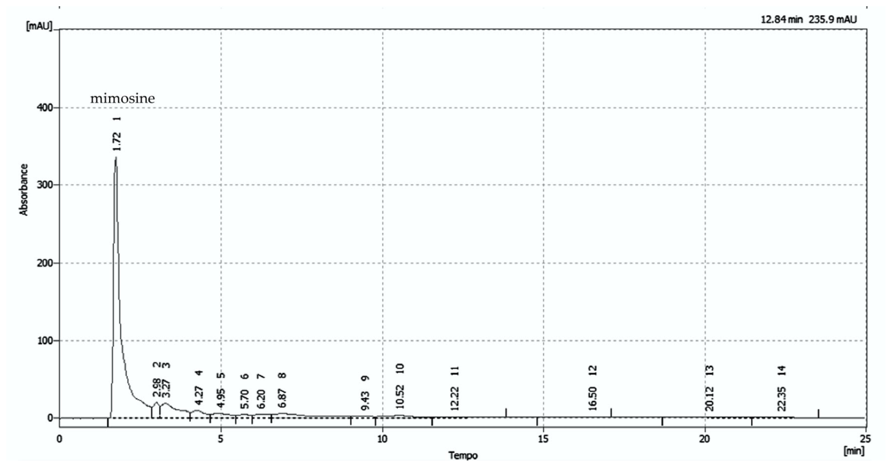
Figure 2.
Concentration-dependent antioxidant activity of the M. pudica decoction (dMp) and ascorbic acid (AA). (A) Absorbance-based assays: total antioxidant capacity (TAC) and ferric reducing antioxidant power (FRAP). (B) Inhibition- or activity-based assays: DPPH• scavenging, hydroxyl radical (•OH) scavenging, and permanganate-reducing antioxidant capacity (PRAC). dMp and AA are represented by continuous or dashed lines, respectively. Points represent the mean; error bars indicate standard deviation (n = 3).
Figure 2.
Concentration-dependent antioxidant activity of the M. pudica decoction (dMp) and ascorbic acid (AA). (A) Absorbance-based assays: total antioxidant capacity (TAC) and ferric reducing antioxidant power (FRAP). (B) Inhibition- or activity-based assays: DPPH• scavenging, hydroxyl radical (•OH) scavenging, and permanganate-reducing antioxidant capacity (PRAC). dMp and AA are represented by continuous or dashed lines, respectively. Points represent the mean; error bars indicate standard deviation (n = 3).
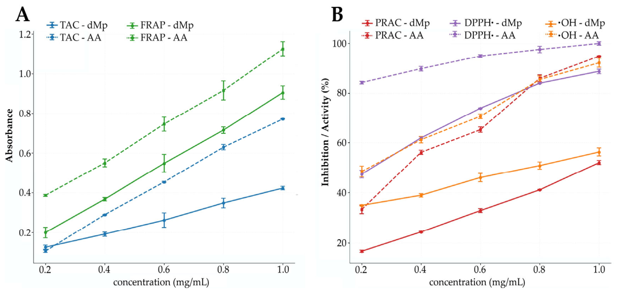
Figure 3.
Hemolytic activity of the M. pudica decoction on human erythrocytes. Erythrocytes were incubated with increasing concentrations of the decoction (0.2–1.0 mg/mL) for 60 min at 37 °C. Points represent the mean and bars the standard deviation (n = 3). The dashed line represents the linear regression plot.
Figure 3.
Hemolytic activity of the M. pudica decoction on human erythrocytes. Erythrocytes were incubated with increasing concentrations of the decoction (0.2–1.0 mg/mL) for 60 min at 37 °C. Points represent the mean and bars the standard deviation (n = 3). The dashed line represents the linear regression plot.
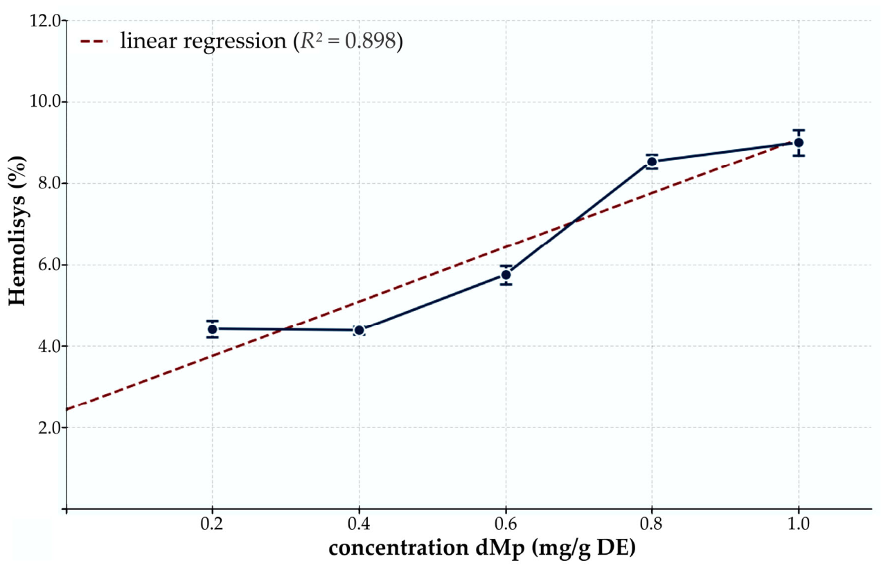

Table 1.
Content of Primary and Secondary Metabolites in the Decoction of M. pudica Aerial Parts.
| Metabolites | Mean ± SD |
| Total phenolic compounds (mg GAE/g DE) | 316.41 ± 19.58 |
| Total flavonoids (mg QE/g DE) | 122.99 ± 12.78 |
| Total carbohydrates (mg GE/g DE) | 34.44 ± 0.65 |
| Reducing sugars (mg GE/g DE) | 31.09 ± 1.10 |
| Proteins (mg BSAE/g DE) | 4.02 ± 0.04 |
| Mimosine (mg/g DE) | 2.11 ± 0.13 |
Values are expressed as mean ± standard deviation (n = 3) per gram of dry extract (DE). GAE: gallic acid equivalents; QE: quercetin equivalents; GE: glucose equivalents; BSAE: bovine serum albumin equivalents.
Table 2.
Predicted Physicochemical and Pharmacokinetic Properties of M. pudica Metabolites.
| Bioactive compound | MW | logP | TPSA | QED | Caco-2 P | HIA | F20% | PPB | BBB |
| Chlorogenic acid | 354.1 | 1.04 | 164.75 | 0.234 | -6.426 | 0.106 | 0.992 | 64.8 | 0.000 |
| Ferulic acid | 194.1 | 1.65 | 66.76 | 0.715 | -4.989 | 0.249 | 0.957 | 68.6 | 0.001 |
| Gallic acid | 170.1 | 0.69 | 97.99 | 0.460 | -5.604 | 0.017 | 0.351 | 37.6 | 0.002 |
| Apigenin | 270.1 | 2.98 | 90.90 | 0.632 | -5.129 | 0.002 | 0.565 | 96.5 | 0.013 |
| Chrysin | 254.1 | 3.60 | 70.67 | 0.700 | -4.980 | 0.002 | 0.155 | 97.8 | 0.277 |
| Luteolin | 286.1 | 2.25 | 111.13 | 0.511 | -5.192 | 0.015 | 0.925 | 97.6 | 0.012 |
| Mimosine | 198.1 | -3.17 | 105.55 | 0.575 | -6.052 | 0.000 | 0.000 | 13.4 | 0.000 |
| Naringenin | 272.1 | 2.60 | 86.99 | 0.742 | -4.987 | 0.000 | 0.002 | 95.0 | 0.000 |
| Quercetin | 302.0 | 1.45 | 131.36 | 0.434 | -6.177 | 0.134 | 0.504 | 98.7 | 0.000 |
MW: Molecular Weight; logP: The logarithm of the n-octanol/water distribution coefficients at pH=7.4; TPSA: Topological Polar Surface Area; QED: Quantitative Estimate of Drug-likeness; HIA: Human Intestinal Absorption; F20%: Bioavailability < 20%; PPB (%): Plasma Protein Binding (%); BBB: Blood-Brain Barrier Penetration. HIA, F20%, and BBB values represent predicted probabilities (0–1 scale).
Table 3.
Predicted Interactions of M. pudica Metabolites with Cytochrome P450 Isoforms Using ADMETlab 3.0.
Table 3.
Predicted Interactions of M. pudica Metabolites with Cytochrome P450 Isoforms Using ADMETlab 3.0.
|
Bioactive compound |
CYP1A2 | CYP2C19 | CYP2C9 | CYP2D6 | CYP3A4 | CYP2B6 | CYP2C8 | ||||||
| I | S | I | S | I | S | I | S | I | S | I | S | I | |
| Chlorogenic acid | ● | ● | ● | ● | ● | ● | ● | ● | ● | ● | ● | ● | ● |
| Ferulic acid | ● | ● | ● | ● | ● | ● | ● | ● | ● | ● | ● | ● | ● |
| Gallic acid | ● | ● | ● | ● | ● | ● | ● | ● | ● | ● | ● | ● | ● |
| Apigenin | ● | ● | ● | ● | ● | ● | ● | ● | ● | ● | ● | ● | ● |
| Chrysin | ● | ● | ● | ● | ● | ● | ● | ● | ● | ● | ● | ● | ● |
| Luteolin | ● | ● | ● | ● | ● | ● | ● | ● | ● | ● | ● | ● | ● |
| Mimosine | ● | ● | ● | ● | ● | ● | ● | ● | ● | ● | ● | ● | ● |
| Naringenin | ● | ● | ● | ● | ● | ● | ● | ● | ● | ● | ● | ● | ● |
| Quercetin | ● | ● | ● | ● | ● | ● | ● | ● | ● | ● | ● | ● | ● |
I: Inhibitor; S: Substrate; Colors indicate probability ranges: ● high (0.7-1.0); ● moderate (0.3-0.7) and ● low (0.0-0.3).
Table 4.
Predicted In Silico Toxicity Profile of M. pudica Metabolites.
|
Bioactive compound |
hERG (ADMET) |
DILI (ADMET) |
Ames (ADMET) |
Genotoxicity (ADMET) |
Rat Oral Acute Tox. (ADMET) |
Nephrotoxicity (ProTox) | Hepatotoxicity (ProTox) |
| Chlorogenic acid | 0.025 | 0.291 | 0.386 | 0.243 | 0.054 | Active (0.56) | Inactive (0.72) |
| Ferulic acid | 0.036 | 0.636 | 0.252 | 0.120 | 0.081 | Active (0.62) | Inactive (0.51) |
| Gallic acid | 0.026 | 0.704 | 0.482 | 0.760 | 0.251 | Active (0.69) | Inactive (0.61) |
| Apigenin | 0.100 | 0.743 | 0.618 | 0.987 | 0.520 | Active (0.60) | Inactive (0.68) |
| Chrysin | 0.105 | 0.872 | 0.628 | 0.966 | 0.539 | Active (0.60) | Inactive (0.68) |
| Luteolin | 0.069 | 0.796 | 0.650 | 0.986 | 0.510 | Active (0.62) | Inactive (0.69) |
| Mimosine | 0.057 | 0.110 | 0.716 | 0.984 | 0.380 | Active (0.60) | Inactive (0.79) |
| Naringenin | 0.107 | 0.220 | 0.703 | 0.978 | 0.500 | Active (0.61) | Inactive (0.67) |
| Quercetin | 0.053 | 0.783 | 0.586 | 0.975 | 0.480 | Active (0.62) | Inactive (0.69) |
Table 5.
Predicted molecular targets of M. pudica metabolites with implications for redox modulation and inflammation.
Table 5.
Predicted molecular targets of M. pudica metabolites with implications for redox modulation and inflammation.
| Bioactive compound | InflamNat | SR/ARE (ADMETlab) |
Molecular Targets (SwissTargetPrediction) |
| Chlorogenic acid | 0.687 | 0.206 | AKR1B1/AKR1B10 (1.0) |
| Ferulic acid | 0.662 | 0.285 | CA2/CA5B (0.934) |
| Gallic acid | 0.396 | 0.942 | CA2/CA5B (0.999) |
| Apigenin | 0.621 | 0.996 | AKR1B1 (1.0); XDH (1.0); MAOA (1.0); NOX4 (1.0); GSK3B (1.0); SYK (1.0); PTGS2 (1.0); ESR1/ESR2 (1.0); ALOX5 (0.600) |
| Chrysin | 0.514 | 0.968 | AKR1B1 (1.0); XDH (1.0); ALOX12 (0.528); ALOX15 (0.528); ESR1/ESR2 (0.418) |
| Luteolin | 0.568 | 0.974 | AKR1B1 (1.0); XDH (1.0); MAOA (1.0); NOX4 (1.0); GSK3B (1.0); SYK (1.0); ALOX5 (1.0); MMP2/MMP9 (1.0); ALOX12 (0.436); ALOX15 (0.435) |
| Mimosine | 0.535 | 0.006 | |
| Naringenin | 0.372 | 0.905 | ESR1/ESR2 (1.0) |
| Quercetin | 0.611 | 0.937 | AKR1B1 (1.0); XDH (1.0); MAOA (1.0); NOX4 (1.0); GSK3B (1.0); ALOX5 (1.0); ALOX12 (1.0); ALOX15 (1.0); MMP9 (1.0); SYK (0.680) |
SR/ARE: Stress Response/Antioxidant Response Element; AKR1B: Aldose Reductase; CA: Carbonic anhydrase; XDH: Xanthine dehydrogenase; MAO: Monoamine oxidase; NOX: NADPH oxidase; GSK3B: Glycogen synthase kinase-3 beta; SYK: Spleen Tyrosine kinase; ALOX: Arachidonate lipoxygenase; PTGS: Cyclooxygenase; ESR: Estrogen Receptor; MMP: Matrix Metalloproteinase.
Disclaimer/Publisher’s Note: The statements, opinions and data contained in all publications are solely those of the individual author(s) and contributor(s) and not of MDPI and/or the editor(s). MDPI and/or the editor(s) disclaim responsibility for any injury to people or property resulting from any ideas, methods, instructions or products referred to in the content. |
© 2026 by the authors. Licensee MDPI, Basel, Switzerland. This article is an open access article distributed under the terms and conditions of the Creative Commons Attribution (CC BY) license (http://creativecommons.org/licenses/by/4.0/).
Copyright: This open access article is published under a Creative Commons CC BY 4.0 license, which permit the free download, distribution, and reuse, provided that the author and preprint are cited in any reuse.