

Submitted:
04 October 2026
Posted:
07 October 2026
You are already at the latest version
Abstract
Sex chromosomes originate from a homologous pair of chromosomes becoming restricted to one sex (referred to as Y in males in the X/Y system; W in females in the ZW system) via inhibition of crossing over followed by gene loss. In contrast, most other forms of chromosomal dosage change caused by meiotic or mitotic errors, or experimental aneuploidy, are strongly deleterious or lethal. The viability of individuals with the heteromorphic sex chromosomes in various species comes from the acquisition of some type of dosage compensation that counteracts any issues with “genomic imbalance”. Here we review lessons learned about gene expression modulation from experimentally produced aneuploidy and from evolutionary genomics. These lessons from genome balance principles are applied to how different examples of sex chromosome dosage compensation (Drosophila, mosquito, mammals, nematodes) have evolved to counteract any detrimental global effects on gene expression. New perspectives and questions are raised to revise ideas about sex chromosomal evolution and to direct re-analyses of previous hypotheses.
Keywords:
dosage compensation
; sex chromosomes
; genomic balance
; Drosophila
; mammals
; mosquito
; nematodes
Introduction
When an individual eukaryotic gene is varied in copy number, its mRNA (and more often than not its protein) usually changes proportionally (e.g. Grell 1962), as illustrated in Figure 1. In contrast, varying entire chromosomes or large chromosomal segments often yields dosage compensation, so that the total output remains close to that of a normal karyotype. This includes heteromorphic sex chromosomes, which typically show some level of dosage compensation. Why this dichotomy of dosage effects or the lack thereof exists and what are the evolutionary forces driving it have been the subject of investigations for nearly a hundred years. Recently, several review articles on dosage compensation have been published (Kaiser and Bachtrog 2010; Chen et al. 2020; Furman et al. 2020; Saunders and Muyle 2024; Cecalev et al. 2024; Xie et al. 2025; Kalita and Valsecchi 2025; Zhu et al. 2025; Charlesworth and Charlesworth 2025; Toups and Vicoso 2025). They can be consulted for specific details related to the many species covered. Our emphasis will be on the lessons learned from studies on gene expression during genomic imbalance, the presumed reason for sex chromosome dosage compensation evolution, and how they apply to this scientific problem. The term “dosage compensation” has been used to refer to the evolutionary phenomenon favoring maintaining ancestral levels of products of dosage sensitive genes in the wake of genomic copy number changes and new regulatory networks that evolved to secure that outcome. In other instances, it has been used to refer to the experimental observation that the total output of gene products from aneuploidy is more or less equal to a balanced diploid control. We will not introduce new terms and indeed it is obvious to which situation one is referring. Indeed, it is likely that what is found experimentally and what has evolved over millions of years is related because they both reflect gene regulatory mechanisms.
Almost 50 years ago, it was noted (Birchler 1979) that small segmental trisomies in Drosophila, used to map structural genes by gene dosage effects, almost always had inverse effects on one or more of the enzyme activities assayed (O’Brien and Gethman 1973; Rawls and Lucchesi 1974; Hodgetts 1975; Hall and Kankel 1976; Pipkin et al. 1977; Oliver et al. 1978; Moore and Sullivan 1978; Detwiler and MacIntyre, 1978). In other words, multiple segmental trisomies, not including the structural gene itself but being present in the genome, would reduce the measured activity more or less to 2/3 of the diploid control. A parallel study in maize revealed a similar pattern (Birchler 1979). The same publication pointed out that still 50 years prior, Blakeslee reported that every trisomy in the plant Datura would quantitatively alter pigment level but not in a ploidy series in which the whole genome was varied (Blakeslee 1921). These effects were usually ignored perhaps because multiple regions of the genome produced the same effect and were relegated to the ‘detrimental effects of aneuploidy”. The fact that many regions of the genome can produce the same type of dosage effect in trans is worth emphasizing again to illustrate the nature of genome imbalance and what sex chromosome evolution must address.
The maize study involved a 1-4 dosage series of the long arm of chromosome one and the whole series showed an inverse effect (illustrated in Figure 2) indicating that it was unlikely to be a secondary effect of aneuploidy because there was no correlation with plant vigor (Birchler, 1979). Subsequently, it was found that multiple regions of the genome could produce an inverse effect on any one protein (Birchler and Newton, 1981) and on any one mRNA (Guo and Birchler 1994). It should be noted that directly proportional trans-acting effects were also observed in these studies. These “trans-acting” effects occurred when varying the dosage of segments of the genome that were different from the region containing the monitored gene product.
The inverse effect can account for dosage compensation of gene products in a dosage series (Birchler 1979) (illustrated in Figure 3). This was shown by division of a chromosome arm into small regions around a studied structural gene, which produces a gene dosage effect, while other regions of the same arm would inversely modulate the same gene product (Birchler 1981). The two opposing effects cancel each other when varied together in a larger aneuploidy. The same principle was demonstrated in Drosophila in a large trisomy showing compensation with the added finding that the inverse effect worked via the promoter region by using a promoter-reporter construct (Birchler et al. 1990). Other authors reported dosage compensation for many genes in large autosomal trisomies of Drosophila (Devlin et al. 1982; 1984), which were accompanied by inverse effects on many unlinked genes (Devlin et al. 1988).
To learn whether single genes could produce these dosage effects, mutageneses were conducted. The rationale was that heterozygous loss of function mutations would essentially be a single gene “monosomy”. These could be examined for modulation of a leaky allele of the white eye color gene in Drosophila (white-apricot) as a reporter gene. The first example found also exhibited an inverse effect on the normal white allele at the RNA level as well as on related genes (Rabinow et al. 1991). It required a normal promoter region of white to be effective. A summary of a large collection of such modifiers found that for those with a known molecular function they represented transcription factors, signal transduction components, and chromatin proteins (Birchler et al. 2001). More recently, some of these were subjected to genomic RNA-seq and found to modulate hundreds of targets with more inverse than direct effects found (Zhang et al. 2021; Liu et al. 2025).
How are dosage effects achieved by either a direct or inverse relationship to the chromosome copy number in aneuploids? A solution was offered by Reiner Veitia (Veitia 2002) based on modeling of the assembly of macromolecular complexes (Bray and Lay 1997). Varying the copy number of a single subunit, while the others remain constant, perturbs assembly of the entire complex. Depending on the abundance, position, and stoichiometry of each subunit, this perturbation can increase or decrease the complex’s activity. Thus, whether a particular regulatory molecule can act to positively or negatively affect a target gene will depend on these parameters relative to its interactors. Such a scenario can potentially explain why multiple regions of the genome can produce similar effects on a single gene product because each region could modulate different subunits that comprise the whole regulatory machinery. Birchler and Newton (Birchler and Newton 1981) rationalized the multiple effects on any one gene product by suggesting that the stoichiometry of the regulatory machinery was responsible considering that there are much fewer effects in a ploidy series in which all subunits would be varied more or less in concert.
Insights from Evolutionary Genomics
As genomic sequences began to emerge it became obvious that many species of eukaryotes had a whole genome duplication (WGD) in their evolutionary past as revealed by related syntenic duplicated genes present in the sequence (e.g. Papp et al. 2003; Blanc and Wolfe 2004; Maere et al. 2005; Thomas et al. 2006; Hakes et al. 2007; Freeling 2009). The functional categories of these duplicates, however, were not random. Instead, genes involved in macromolecular complexes are preferentially retained such as transcription factors, signaling components, and ribosomal subunits among others. This result suggested that a stoichiometric dosage effect would be selected against if only some components were deleted. Further studies found that segmental duplications showed an underrepresentation of the same classes of genes that are retained after WGD. This also indicates selection against stoichiometric changes in the copy number of components of macromolecular complexes (Papp et al. 2003; Freeling 2009; Tasdighian et al. 2017; Zhu et al. 2026). These effects have been found to occur for genes involved in protein-protein interactions (Defoort et al. 2019; Shi et al. 2020). Also, microRNA genes follow a similar pattern (Abrouk et al. 2012; Desvignes et al. 2021), potentially because of their quantitative modulation of transcription factors. Collectively, the results are consistent with the aneuploidy effects on the organism resulting from the dosage sensitivity of the varied transcription factors and signaling components (Birchler et al. 2001; Birchler et al. 2005; Veitia et al. 2008; Birchler and Veitia 2012; Veitia et al. 2013; Birchler and Veitia 2021). Considering the widespread finding across phyla of a constraint against imbalance of these classes of genes in evolutionary time, this aspect must be considered in discussing sex chromosome evolution. The important lesson from evolutionary genomics is that both deletion and duplication of individual components of macromolecular complexes have reduced fitness (Papp et al. 2003; Makanae et al. 2013; Robinson et al. 2021).
Genome Wide Studes of Gene Expression in Aneuploids
With the emergence of RNA-seq technologies, an example of gene expression across the genome was performed involving trisomy for the left arm of chromosome 2 (2L) in Drosophila (Sun et al. 2013). This arm comprises about 20% of the genome. Each expressed gene was used to produce a trisomy/normal ratio, and the ratios were plotted in a distribution. The distributions were separated into the genes on the varied chromosome arm versus the remainder of the genome. Across the genome a large fraction of genes was reduced in expression with a peak at the inverse level (0.67) compared to the diploid control. For genes on 2L, a large fraction was near the same level as the control thus showing autosomal dosage compensation. A lesser subset of genes showed a peak surrounding the 1.50 ratio illustrating a gene dosage effect. The compensation of genes on 2L can be brought about by the inverse effect operating on them but their increased dosage results in a total expression near the diploid level.
Complete Genome Analysis in a Multicellular Eukaryote
Hou and colleagues (Hou et al. 2018) examined gene expression in all five trisomies of Arabidopsis and compared them to a diploid, triploid, and tetraploid ploidy series. The ratio distributions revealed the most common trans-acting effect was inverse for chromosomes 1-4 and dosage compensation for a subset of the genes on the varied chromosomes. Trisomy for chromosome 5 had the least trans effects and the least amount of dosage compensation of cis genes. Interestingly, trisomy disrupted the transcription factor-target gene relationships predicted from regulatory networks for a slight majority of cases examined, indicating that stoichiometric changes will alter this connection. This study also analyzed published data from yeast disomies (Torres et al. 2007) and murine trisomies (Williams et al. 2008) via ratio distributions and found a prevalence of inverse effects in trans in both cases.
Further studies in maize involved a wider range of dosage changes including 1-4 doses in an otherwise diploid background and disomies in an otherwise haploid background with comparisons to a 1-4 whole genome ploidy series (Shi et al. 2021; Yang et al. 2021). These results revealed that with an increase in chromosomal dosage the most common modulations were an inverse effect across the genome, but direct trans-acting effects were also realized. With segmental monosomy, the inverse versus direct trans-acting effects were more equally represented. There were also cases in which reductions in trans occurred in both monosomy and trisomy for the same chromosomal segment. In general, the magnitude of effects was greater with greater dosage difference in the various aneuploids and with a greater dosage differential between the varied segment relative to the background ploidy. Another generalization was that a greater number of genes showing an inverse effect in trans in various aneuploids was associated with a greater number of genes in cis approaching dosage compensation. Lastly, the transcriptome size was estimated in haploid disomies versus normal. Several disomies inversely affected the whole transcriptome size. This dataset was subsequently analyzed for modulation of transposable elements (Yang et al. 2023), microRNAs (Shi et al. 2022), and long noncoding RNAs (Yue et al. 2025) with all categories being modulated more so inversely than directly. For any one aneuploid, the distributions of each type of RNA could be quite distinct from the other types. As a general rule, the number of trans effects is greater with a greater cytological length of an aneuploid and greater in magnitude with a greater stoichiometric difference with the background ploidy.
Yang and colleagues (Yang et al. 2025) examined numerous combination aneuploidies including trisomy-trisomy, monosomy-monosomy, and trisomy-monosomy. As a general rule trisomy-trisomy and monosomy-monosomy resulted in a greater number of genes showing an inverse or direct effect with few increases in the magnitude of the effects. With the increase in the number of genes being inversely affected in trans, more cis genes approached dosage compensation. To the extent that heteromorphic sex chromosome evolution occurs in a stepwise manner (Lahn and Page 1999; Lemaitre et al. 2009), modulation of gene expression across the genome would be predicted to become more prevalent with each step unless counteracted. Trisomy-monosomy resulted in a variety of effects including re-balancing of the genome or multiplicative effects with a greater magnitude. Transcriptome size was variously affected in the combinations. These principles might eventually provide insight into how effects on autosomes during sex chromosome evolution become counteracted.
Zhang and colleagues (Zhang et al. 2024) analyzed published data from various sources for the impact in human cells for trisomies 13, 18, and 21. Although differences were present for different cell types and conditions, overall, the major effect in ratio distributions was an inverse effect on a subset of genes particularly for trisomies 13 and 18. These were accompanied by dosage compensation for a subset of genes residing on the trisomic chromosome. This study also compared the patterns of mRNAs to long noncoding RNAs and found modulations of both but not necessarily with overlapping distributions. The fraction of the RNA pool contributed by the trisomic chromosome cannot account for the much larger pool that is inversely regulated (Zhang et al. 2024). Indeed, single genes can produce the inverse effect (Rabinow et al. 1991; Birchler et al. 2001) and these single regulatory genes can modulate hundreds of genes (Zhang et al. 2021). Also examined was the relationship of dosage sensitive transcription factors and their predicted targets. When a transcription factor showed a dosage effect in a trisomy, the targets could be directly or inversely modulated with the latter predominating. This observation suggests that whether a transcription factor has a positive or negative effect on its targets can be affected by its stoichiometry with other regulatory components.
The need for dosage compensation for monosomic sex chromosomes is illustrated by the fact that no whole chromosome monosomy survives to adulthood in diploid multicellular eukaryotes of which we are aware (e.g. Fitz-Earle and Holm 1979; Schinzel 2001). Only in paleopolyploid maize or more recent polyploids (tobacco, wheat) where the effective imbalance is less than ½ are they viable (yet still exhibit abnormalities). The intermediate evolutionary stage of maize positioned it to provide insight into gene expression changes in monosomies.
Sex Chromosome Evolution Must Accommodate Gene Expression Modulation from Genomic Imbalance.
In discussions of sex chromosome evolution, the focus is often on the sex chromosome itself in isolation. However, when sex chromosomes evolve, and many genes become hemizygous, the genome must accommodate the resulting trans-acting effects of dosage-sensitive regulators. The inverse effect can bring about dosage compensation of the varied genes on the evolving sex chromosome, but autosomal modulations must be addressed. Alternatively, if the dosage sensitivity of regulators could be mitigated, trans effects would be reduced. In vertebrates, the XY system (mammals) and ZW system (birds, snakes) retain many dosage sensitive developmental regulators on both sex chromosomes while other genes are deleted from the degenerating one (Bellott et al. 2014; Bellott et al. 2017; Bellott and Page 2021; Naqvi et al. 2018). This situation is reminiscent of the retention of dosage sensitive genes following WGD as described above. Indeed, dosage compensation of many classes of genes is probably not necessary for full viability and fertility. Those genes that are dosage sensitive as members of multicomponent complexes are probably the most critical. The retention of dosage sensitive regulatory genes on both sex chromosomes in vertebrates illustrates the critical nature of these gene classes in sex chromosome evolution. They must be accommodated if they are not retained, for example in Drosophila and C. elegans. Some dosage sensitive genes might be selected to lack compensation if they foster advantageous sexual dimorphisms or if they in fact bring about dosage compensation of sex-linked targets by inversely affecting them.
Some discussions of dosage compensation have considered whether equalizing expression between the sex chromosomes and the autosomes or between the sexes is the most critical aspect. Certainly, the sex chromosomal to autosomal relationship is needed to foster cell and organismal viability. The “between the sexes” relationship must ensure compatibility, but gene expression might diverge if sexual dimorphism is conditioned by the sex chromosome. Indeed, sex chromosomes condition differences between the sexes!
How do sex chromosomes seem to “know” their dosage to adjust compensation accordingly? If a dosage sensitive gene on the sex chromosome interacts with other components on the autosomes and their stoichiometry affects their collective function, the appropriate relative effect on target genes will be realized. This requires that the sex chromosomal regulatory genes themselves escape dosage compensation and the interacting autosomal genes are not modulated. There is a tendency to view dosage compensation as a whole chromosome phenomenon influenced no doubt by the dramatic cytological images of X inactivation in mammals and the Male Specific Lethal complex coating the X in Drosophila. In reality, there are genes that escape these processes making for a gene-by-gene compensation to some degree (see specific examples described below). In some species, dosage compensation might be even more piecemeal, but any detrimental dosage sensitivity would need accommodation. Some authors reflect on piecemeal dosage compensation or an apparent lack of compensation as showing an imbalanced gene expression, which is technically correct. However, evolutionary genomics reveals, as noted above, that the most critical genes from a dosage perspective are those involved in multicomponent complexes including regulatory genes. Few studies, with exception (e.g. Makino et al. 2010; Xiong et al. 2010; Pessia et al. 2012; Lin et al. 2012), specifically focused on these classes of genes in considering sex chromosome evolution.
As the sex restricted chromosome continues to deteriorate and the hemizygous regions expand on the homolog, genome wide effects might be mitigated by increased expression of the remaining copy via altered transcription, translation, mRNA stability, its interaction with miRNAs that modulate translation, or protein stability (Wang et al. 2020; Lister et al. 2024). Gene duplication is another potential path either on the sex chromosome or transposition to an autosome. It is also possible that downregulation of an autosomal interactor could occur. Another possibility is epigenetic effects that increase histone marks that mediate increased expression. These events would prevent many trans-acting effects across the genome that would otherwise occur with such hemizygosity.
Otherwise, hemizygosity of transcription factors that inversely affect many targets could bring about dosage compensation in the haplo-region but would also tend to affect genes outside the limits. Whether this scenario alleviates the negative fitness consequences versus creates a more serious one would likely depend on the individual case. Genes on large trisomic regions in Drosophila (Devlin et al. 1982; Sun et al. 2013a; Sun et al. 2013b) show expression values quite close to the diploid level for a large fraction of genes encoded thereupon, but there are also modulations of gene expression on other chromosomes.
Genes that are not particularly dosage sensitive could become dosage compensated as (Stephen Jay) Gouldian spandrels because they are included as targets in gene regulatory networks with dosage sensitive genes that are more critical and driving selection. Indeed, it is not clear how many sex chromosome genes “need” to be compensated. In experimental systems, for example in Drosophila, there is no need whatsoever for genes to be compensated in large aneuploidies, but the process is nevertheless quite robust (Devlin et al. 1982; Sun et al. 2013a; Sun et al. 2013b; Zhang et al. 2021). This illustrates that in these cases it is a natural process of how gene expression is regulated and would likely spill over for sex chromosome evolution. In some discussions of dosage compensation, it is proposed that the hemizygous chromosome has no upregulation relative to its progenitor and in the opposite sex the two sex chromosomes are downregulated to match. On the surface this would seem an unusual proposal from a genomic balance perspective because it would suggest evolution toward a hard lethal situation. For this to be the case, the autosomes would need to be downregulated to form a viable genomic stoichiometry. In our view, it is more critical to have a balance within a sex for normal cellular functions than between sexes.
In the following sections we discuss four examples of sex chromosome dosage compensation that illustrate various processes involved and for which recent findings provide new insights. Emphasis will be placed on how direct and inverse dosage effects must either be counteracted or utilized during sex chromosome evolution and why effects on the autosomes play a role.
Drosophila Dosage Compensation
Herman Muller was the first to recognize dosage compensation as a phenomenon (Muller 1932). Using the leaky apricot allele of the white eye color gene on the X chromosome in Drosophila, he observed equal expression between males and females. By creating different doses just surrounding the gene, a dosage effect was realized but with each dose in a male being about double the same dose in a female (Figure 4). Later he varied other small regions of the X and found direct and inverse effects on white-apricot (Muller 1950). This latter finding has fallen into obscurity but matches the findings of Blakeslee (Blakeslee 1921) before him and many studies subsequently (described above) that the dosage of many regions of the genome can quantitatively modulate the expression of a single target gene. Dosage effects on white-apricot are not restricted to the X and occur across the genome (Sabl and Birchler 1993). These effects are similar to the segmental aneuploidy results mentioned in the Introduction.
Muller (Muller 1950) suggested that the change in expression from male to female would continue to decline into triple X metafemales in an exponential relationship as the reason for the low viability of this genotype. Curt Stern (Stern, 1960), however, found that metafemales have a similar expression of apricot as normal females (see Figure 4). Subsequent studies found this to be the case for the majority of X linked genes in metafemales (Sun et al. 2013; Birchler et al. 1989; Birchler 1992; Sun and Birchler 2009) with most autosomal genes being inversely affected—a likely explanation for their reduced viability.
The MSL Model of Dosage Compensation
A model to explain dosage compensation in Drosophila involves the products of the male specific lethal (MSL) loci (Lucchesi and Kuroda 2015). These mutations are specifically lethal as homozygotes in males. The lethality occurs at the transition from the larval to the pupal stage. The members of the complex include the maleless (mle) mutation (Belote and Lucchesi 1980), male specific lethal 1, 2 and 3 (msl1, msl2, msl3), and the males absent on the first (mof) (Hilfiker et al. 1997). The maleless mutation was examined for its effect on transcription by studying the incorporation of tritiated uridine into nascent RNA on the X chromosome versus a portion of the autosomes (Belote and Lucchesi 1980). The data were analyzed by comparing the X chromosome relative to the autosomal values, producing a ratio that was reduced. This led to the interpretation that the products of the male specific lethal genes were responsible for X chromosomal dosage compensation.
Subsequently, the protein encoded by the mle gene was identified and localized on the X chromosome of males (Kuroda et al. 1991). The other members of the complex followed the same pattern. One component, males absent on the first (MOF), encodes a histone acetyl transferase that is responsible for acetylation of lysine 16 on histone H4 (H4Lys16Ac) (Hilfiker et al. 1997), which is enriched on the male X chromosome (Bone et al. 1994). These observations led to the hypothesis that the MSL complex sequesters the histone modifier MOF to cause the upregulation of the single X chromosome. In addition to the protein components, there are also two loci (roX1, roX2) that produce noncoding RNAs for which at least one must be present in the complex (Meller et al. 1997). The JIL1 histone kinase is also enriched on the male X chromosome although mutations in this gene are lethal in both sexes (Jin et al. 1999; Jin et al. 2000; Wang et al. 2001). The CLAMP protein links the MSL complex to the X chromosome (Soruco et al. 2013). There is an extensive literature on how the MSL complex recognizes the X chromosome (e.g. Lucchesi and Kuroda 2015; Makki and Meller 2024; Biswas et al. 2024), but we do not cover it because it does not involve issues of gene expression per se.
Dosage Compensation Without the MSL Complex
While the MSL hypothesis seems straightforward, there are other findings that it cannot explain (Birchler and Veitia 2021; Birchler 2016). First, metafemales with three X chromosomes show dosage compensation. In these animals, X-linked genes must be downregulated so that each copy is expressed at roughly two-thirds of the level observed in normal XX females. Because there is no MSL complex in metafemales (Sun and Birchler, 2009), this compensation cannot be mediated by the MSL complex. Moreover, there is evidence that compensation in males and metafemales is related (Birchler 1992) in that male compensating alleles of the white eye color locus also compensate in metafemales, and the male non-compensating alleles do not compensate in metafemales. Indeed, genomic balance results described above indicate a relationship of an inverse effect between trisomy and monosomy of a particular chromosomal region such that it is continuous through a dosage series. While the three X chromosomes show compensation in metafemales (Sun et al. 2013; Zhang et al. 2021; Stern 1960; Birchler et al. 1989; Birchler 1992; Sun and Birchler 2009), diploid autosomal genes show reduced expression (Birchler et al. 1989; Sun and Birchler 2009; Sun et al. 2013; Zhang et al. 2021). Further, it has long been known that hypomorphic alleles of autosomal eye color genes of diverse functions such as purple, pink-peach, glass, light as well as mini-white transgenes are expressed more highly in males than in females (e.g. Smith and Lucchesi 1969). This is likely a weak inverse effect on the autosomes that is diminished by the sequestration of chromatin modifiers to the X as elaborated below. Of note is the fact that these mutants with increased expression in males have decreased expression in metafemales (Sun et al. 2013; Birchler et al. 1989).
While the above-mentioned studies involved randomly selected example genes, global RNA-Seq experiments on larvae compared males to females and females to metafemales (Sun et al. 2013). The majority of genes on the X chromosome are dosage compensated in metafemales with a small number that exhibit an expression ratio of 1.5 (a dosage effect) (Sun et al. 2013; Zhang et al. 2021), which include transcription factors that have a different stoichiometry with their interactors than they do in normal females. For autosomal genes, the majority was reduced to near 67% (the inverse of the 3/2 X dosage) compared to normal females with a subset being unaffected. In males, the X showed most genes approaching an expression ratio of 1.0 relative to females, the value of dosage compensation, but there was a subset at the 0.5 ratio that lack compensation, which include transcription factors that have a different stoichiometry with their interactors than they do in normal females.
Analysis and Reanalysis of Gene Expression in Msl Mutants
When the expression of example genes was examined in the mle mutant, with emphasis on absolute measurements rather than X to autosomal normalization, the genes on the X chromosome in the mutant males generally did not lose dosage compensation, but autosomal genes were generally increased in expression (Hiebert and Birchler 1994). Subsequently, using transgenes for the white eye color and the yellow body genes on the X and the autosomes in a background of respective null alleles with disruption of the MSL complex, both transgenes retained dosage compensation when present on the X but were increased in expression within about a two-fold range on the autosomes (Bhadra et al. 1999). Further, an RNA fluorescence in situ (FISH) assay was developed for embryos and various X and autosomal genes were examined for their expression levels (Pal-Bhadra et al. 2005). Coincident with the studies in larvae, the X linked genes did not show a loss of dosage compensation and expression of the autosomal genes studied was increased. This FISH assay provided an absolute measurement of gene expression that is not confounded by X to autosomal normalization.
These results might seem contradictory to those from the original autoradiographic results on the mle mutant (Belote and Lucchesi 1980). As noted above, the transcriptional data were treated as a ratio of the X to the autosomes. This approach showed an X/A ratio in the mle/mle females of 0.95 and in mle/mle males of 0.76. In the context of the times, comparing the X expression to the autosomes seemed reasonable. A theoretical loss of dosage compensation would be 0.50, which was not observed. What is obvious from the data is that the absolute values of the X in mle/mle males was slightly greater than normal males and the values for the autosomal region in mle/mle males were even greater. In addition to assaying nascent transcription, these authors also assayed DNA replication. If one normalizes the transcription data to the replication data, then the X is increased 1.48-fold in the mle/mle mutant males, and the autosomes are increased 2.38-fold. If one normalizes the mle/mle male values to those of females, the X has a 1.31-fold increase, and the autosomal segment had a 1.86-fold increase. The same type of absolute results of no loss of X compensation but an increased autosomal expression for msl3 is present in the data of Okuno and colleagues (Okuno et al. 1984). In other words, the absolute values, the normalization to replication values, and the normalization to female values all indicate no loss of X chromosomal dosage compensation but an increase of the autosomes in the mle mutant male. Considering the universal nature of the inverse effect across phyla, such a result can now make sense. Indeed, with the current knowledge that transcription factors are usually dosage sensitive, a result of a reduction of X expression without an effect throughout the genome is highly unlikely. Thus, the foundational study leading to the idea that the msl genes condition male X chromosome dosage compensation was apparently misinterpreted.
Several independent lines of evidence are consistent with the reanalysis interpretation. First, the X-linked dosage sensitive transcription factor Runt was examined in mle, msl1, and msl2 mutant backgrounds (Gergen 1987). The phenotype of runt was unchanged in all three backgrounds, although if the dosage of runt alone was changed, there were obvious phenotypic effects on embryo morphology. The change in phenotype is an absolute measurement. This is an example of dosage compensation that does not require the MSL complex. Second, in the study by Pal Bhadra and colleagues (Pal-Bhadra et al. 2005), the genotype mof; mle/mle was embryonic lethal whereas mle and mof individually are lethal much later in development at the larval/pupal transition. In the mof; mle/mle genotype the X linked genes that ordinarily are dosage compensated all fail to compensate. The value is basically at 50% of the normal male. This RNA-FISH assay is an absolute measurement and not subject to normalization issues. Thus, when it is clear that there is a loss of dosage compensation, it is an embryonic lethal condition. In the same population of embryos, the mle/mle genotype showed no loss of dosage compensation for X linked genes in males. The autosomal genes in the mle/mle genotype were increased in expression. All the genotypes were in the same population mix and thus validate that mle and mof alone do not lose dosage compensation. The embryonic lethality resulting from a loss of dosage compensation is in parallel to the lethality of autosomal whole arm monosomy that is comparable in chromosome length to the X (Fitz-Earle and Holm 1979). These results indicate that genotypes that survive to the larval/pupal transition are unlikely to exhibit a complete loss of dosage compensation. Indeed, heterozygous deficiencies of lengths of only a few percent of the genome are cell inviable (Ripoll 1980) suggesting that the X as a whole retains some level of expression adjustment in the absence of the MSL complex. Third, Cheng and colleagues (Cheng et al. 2016) found that Spiroplasma infection would cause male lethality at the larval/pupal transition similarly to msl mutants. This is accompanied by dissociation of the MSL complex from the male X and redistribution of MOF and H4Lys16Ac to be uniform in the nucleus as previously found (Bhadra et al. 1999). Changes in gene expression were predominantly upregulation of the X and more so of the autosomes. These results agree with the reanalyzed autoradiographic data of Belote and Lucchesi described above and those of Pal-Bhadra et al. (2005). Fourth, as elaborated below, with ectopic expression of MSL2 in females, the acetylation goes from being uniform across the genome to being reduced on the autosomes and increased on the X chromosomes. This lowered acetylation on the autosomes is accompanied by reduced expression of some autosomal genes (Bhadra et al. 2000). Therefore, upon release of MOF and a return to a uniform acetylation level in msl mutants, autosomal gene expression would be expected to increase. These independent lines of evidence indicate that the X is not reduced to 50% of normal, as a general rule, in the msl mutants. This result can occur from a regulatory imbalance between the X chromosome and the autosomes. Whether it involves the same stoichiometric different present in normal males or newly established would be an interesting question to pursue.
Normalization is also an issue in various subsequent gene expression studies on msl mutants that presumably show a reduced X expression in males as previously described (Lucchesi and Kuroda 2015). These studies, in some cases intentionally, normalized X-linked expression to autosomal expression, implicitly assuming that the autosomal output is stable. In other cases, there could be unwitting normalization of the X to the autosomes by the nature of global gene expression data analysis. These studies fall victim to the same problems exhibited by the Belote and Lucchesi foundational study re-analyzed above. If there is a major shift in expression for part of the genome in the same direction and then RNA samples are subjected to analysis in equal proportions in experimental and control samples, it is difficult to discern the absolute expression pattern. For example, the first global analysis of disruption of the MSL complex examined expression in roX- mutants that dissociate the MSL complex from the male X (Deng and Meller 2006). The major peak of the X chromosome in a log2 distribution is shifted down only slightly with a lesser shoulder still above the 50% level. For autosomal genes the distribution is shifted toward upregulation. This raises the question of why some studies find no loss of compensation with elevated autosomal expression, while other appear to show a loss of compensation of the X. Given the lesser than 50% reduction for the X, do these studies have the same caveats as the Belote and Lucchesi (1980) study? Further, determination of Polymerase II occupancy in msl2 mutant cells (Conrad et al. 2012) is subject to the same issues as to whether there are X specific or genome-wide differences. As highlighted previously (Loven et al. 2012), when a large fraction of the transcriptome changes in the same direction, standard normalization procedures can obscure those changes. Loven and colleagues also note that standard spike-in procedures have caveats depending on whether cells, total RNA, or poly A RNA is spiked. In these studies, there could be a sequencing bias resulting from an elevated expression of ~80% of the genome that causes an apparent but not real reduction of the ~20% of the genes from the X chromosome. In contrast, all the studies that attempt absolute measures of the X and autosomes suggest the X is not substantially changed, but there is an inverse effect on autosomal expression (Hiebert and Birchler 1994; Bhadra et al. 1999; Pal-Bhadra et al. 2005; Bhadra et al. 2000; Pal-Bhadra et al. 2006). Given that transcription factors and signaling components are typically dosage-sensitive (for reviews see Birchler et al. 2001; Birchler and Veitia 2012; Birchler and Veitia 2021), it is extremely unlikely that a reduction to half of X-linked gene expression would occur without concomitant global effects on the rest of the genome, making the interpretation that dosage compensation is lost without global effects untenable.
Indeed, it was found that in the maleless mutant males, the histone acetylase MOF became uniformly redistributed across the genome and this was accompanied by an increase of H4Lys16Ac on the autosomes (Bhadra et al. 1999), results that have been confirmed from Spiroplasma infection (Chen et al. 2016) and further by global ChIP-seq studies (Mendjian et al. 2006; Valsecchi et al. 2021). If an increase in acetylation causes an increase in gene expression in the absence of the MSL complex (Sun et al. 2013), it seems likely that the autosomes would be increased in expression when the MSL complex is disrupted with a consequent increase of H4Lys16Ac on the autosomes.
Ectopic Expression or Targeting of the MSL Complex: Evidence for a Constraining Activity on Histone Acetylation
On the hypothesis that the MSL complex can cause a two-fold increase in gene expression when present on the male X, then one might predict that ectopic targeting of the MSL complex to the X chromosome or a transgene in females would produce a two-fold increase in expression given that this would be similar to the situation in normal males. In yeast experiments, attaching the GAL4 DNA binding domain to the MOF protein will target the fusion protein to a reporter that was preceded by an Upstream Activating Sequence (UAS) recognized by the GAL4 domain (Akhtar and Becker 2000). This targeting caused the expression of the reporter to be increased about 10-fold. While this experiment clearly demonstrates that targeting of MOF will increase the expression of the target, it occurs in isolation of other members of the MSL complex, which constrains this effect as described below.
When the MSL complex was engineered to be expressed in females, it accumulates on the X chromosomes, but there is no observed increase in expression of any X linked gene sampled in multiple experiments (Bhadra et al. 1999; Sun and Birchler 2009; Sun et al. 2013; Zhang et al. 2021). Under normal circumstances, the protein product of the Sex lethal (Sxl) gene is expressed in females (Cline and Meyer 1996). This protein is a splicing factor and affects its own expression such that no protein is produced in males (Bell et al. 1991). In females, the SXL protein binds to the messenger RNA for MSL2 and prevents its translation (Kelley et al. 1997). The MSL2 protein is the one required to organize the members of the MSL complex for localization to the X chromosome. By removing the SXL binding site, the MSL2 mRNA can be translated in females. The resulting protein will organize the complex on the Xs causing an increase in H4Lys16 acetylation. However, gene expression assays in females show no X linked genes increased in expression but many autosomal genes are reduced (Bhadra et al. 1999; Pal-Bhadra et al. 2005; Sun and Birchler 2009; Zhang et al. 2021). In a Sxl- genotype per se the MSL complex associates with the X’s, but again there was no evidence for an increase in expression of any X-linked gene assayed and some autosomal genes are reduced (Bhadra et al. 2000; Pal-Bhadra et al. 2006).
In experiments targeting MOF alone to a reporter in females, which would be missing other components of the MSL complex, there was increasing expression with ascending copies of the MOF targeting construct (Sun et al. 2013), thereby showing a concentration effect. In contrast, there was no such effect in males in which all the components of the MSL complex become colocalized with MOF. In parallel, the targeting of the MSL2 protein directly to reporters gave no elevated expression in either sex despite an increase in histone acetylation. With a different reporter construct, targeting of MOF had a greater effect in females than in males, again suggesting a constraining activity (Prestel et al. 2010). However, targeting these same reporters with MSL2 showed no effect (Sun et al. 2013). In an RNA-seq experiment of ectopic expression of MSL2, there was no evidence of X chromosomal increased expression and even a subset of genes encoded on the X that was reduced in expression (Sun et al. 2013). Collectively, the experiments indicate that increased MOF alone on a gene increases the H4 acetylation and expression but not the expression when the full MSL complex is present.
Placement of a transgene carrying the roX genes onto an autosome caused enrichment and spreading of the MSL complex (Park et al. 2002). A mini-white reporter encompassed by the spreading was derepressed from a near null phenotype to near full red pigment and has been interpreted as showing the MSL complex can confer dosage compensation. However, this effect is significantly greater than a two-fold effect and thus likely represents an interesting de-repression phenomenon because it is not a two-fold effect typical of dosage compensation. Moreover, in retrospect, it is unknown how the constraining activity would operate in these circumstances.
When first developed, the ectopic expression of the MSL complex in females was assumed to affect dosage compensation by increasing the expression of the X chromosomes because it was associated with some degree of sex specific lethality (Kelley et al. 1995), but no tests of gene expression were performed. Sex specific lethality COULD indicate an effect on dosage compensation of X-linked genes but if global modulations occur across the genome, viability could be affected by autosomal increases or decreases without a major change for the sex chromosome as occurs in cases of autosomal dosage compensation. The lethality that occurs in the msl mutants is not necessarily because the X chromosome expression drops to half. Changes in autosomal expression could contribute. As noted above, in multiple experiments of gene expression with ectopic MSL expression in females, the X chromosomes are not increased in expression, but some autosomal genes have reduced expression (Zhang et al. 2021; Sun and Birchler 2009; Bhadra et al. 1999; Pal-Bhadra et al. 2005; Sun et al. 2013).
In another instance, changing the amino acid sequence of Histone 4 such that it could not be acetylated resulted in male specific lethality (Copur et al. 2018), but no studies of gene expression were conducted. The lethality stage of these substitution lines was similar to the msl mutants, namely at the larval/pupal transition. These authors (Copur et al. 2018) and others (Samata et al. 2020) did, however, find that by depleting MOF both maternally and zygotically, there was an embryonic lethality. This lethal stage is expected if there is a bona fide loss of dosage compensation similarly to the lethal stage of whole arm monosomies (Fitz-Earle and Holm 1979). Considering the data described above that genotypes that survive to the larval/pupal transition do not have X expression reduced to half, the consequences of these substitutions are not clear.
In other experiments involving amino acid substitutions in Histone 4 that could not be acetylated (Armstrong et al. 2018), gene expression was assayed in wing imaginal disc cells. Differential gene expression analysis found more decreases than increases with 72% of the reductions being from the X and 96% of the increases from the autosomes. The peak of X reductions was approximately at 75% of normal and the distribution of all genes was shifted to an increased expression. In other studies, live imaging of transcription burst amplitude for selected genes previously known that do or do not show dosage compensation suggest that genes that do not show compensation are reduced to near the 50% level in males compared to females (Beadle et al. 2023) indicating the level that would be reached if there were complete loss of compensation.
The Amstrong et al results were compared to RNAi knockdowns of MSL2 and MOF in S2 tissue culture cells in which the MSL complex is organized on the X’s (Zhang et al. 2010). The results were found to be similar in magnitude. The latter authors concluded that these effects were weak and that compensation involved “both fixed and dose dependent components”, referring to the involvement of the MSL complex as well as dosage modulation. The caveat should be noted that S2 cells are essentially tetraploid and highly aneuploid involving almost a quarter of the genome (Zhang et al. 2010), a larger fraction of the genome than the X itself. This aneuploidy would change gene expression via gene dosage effects, genome wide modulations, and potentially the whole transcriptome size compared to diploid cells. Studies with S2 cells have typically not corrected for the multiple gene copy number changes in the genome. Moreover, S2 cells have likely acquired mutations that condition gene expression changes that foster rapid proliferation in culture. How these features of S2 cells affect the constraining activity is unknown. The nature and extent of these genomic imbalance effects on gene expression experiments in S2 cells is also not known and difficult to establish in the absence of a balanced diploid control.
Using stage 15 embryos (near the time of zygotic genome activation) with maternally depleted MOF, Oellers and colleagues (Oellers et al. 2026) set out to test whether dosage compensation involved transcriptional initiation versus elongation, which had been proposed previously (Larschan et al. 2011), with the results favoring the former. Interestingly, they reported that global gene expression was reduced in both male and female stage 15 embryos with the male X being reduced more than the female X’s and the autosomes of both sexes. It is as if genomic expression is compromised generally leading to a gene dosage effect for the expression that remains possible and is consistent with a loss of dosage compensation being an embryonic lethal condition. How the apparent transcriptome size reduction is brought about is unknown and unaddressed. It would be interesting to know how maternally depleted MOF impacts dosage compensation in the metafemale triple X genotype and even for autosomal dosage compensation.
In the same study (Oellers et al. 2026), knockdowns (KD) of msl1 in S2 cells led to significant reduction (not necessarily to 50%) of 56% of the expressed X linked genes. Knockdown of msl2 reduced 60% of expressed X linked genes and KD of mof reduced 39% of the X linked genes. For autosomal genes, msl1 KD increased 7% and reduced 3%; msl2 KD increased 13% and reduced 8%; mof KD increased 13% and reduced 8%. Thus, a slight majority of X linked genes are reduced to some degree while autosomal changes are both down and up regulation with the latter predominating. The parameters of RNA polymerase II led to the same conclusion as reached with maternally depleted MOF embryos, namely that transcriptional initiation was affected. Genes on the X with the greatest loss of H4Lys16ac in the KDs had the most down regulation, but a correlation was not found between the rate of transcription and the amount of acetylation. How these results relate to the multiple lines of evidence for a constraining activity of the MSL complex, described below, is unknown.
Nature of the Constraining Activity
How does the constraining function attributed to the MSL complex operate? The male X chromosome is bloated in mutants for the chromatin remodeling components ISWI (Deuring et al. 2000; Brehm et al. 2000), nurf (Badenhorst et al. 2005), JIL-1 kinase (Wang et al. 2001), Topo2 (Hohl et al. 2012), over-expression of DNA supercoiling factor (Furuhashi et al. 2006) and for the heterochromatin genes, HP1 and Su(var)3-7 (Delattre et al. 2004; Spierer et al. 2005). Heterochromatin Protein 1 (HP1), which can have a transcriptional repressive effect, is enriched on the male X chromosome (Delattre et al. 2004; Spierer et al. 2005; deWit et al. 2005) and might be involved in the constraining activity. The effect of the ISWI mutation on the bloated X phenotype was found to be correlated with the level of H4Lys16Ac (Corona et al. 2002), suggesting that the normal genotype constrains the effect. Indeed, RNA FISH expression studies of the ISWI mutant in embryos showed elevated male specific X chromosome expression (Pal-Bhadra et al. 2005), suggesting a release of the constraint.
The most studied candidate involved with a constraining activity is the nuclear pore complex component, Megator (Mtor) (Aleman et al. 2021). It is enriched on the male X chromosome (Vaquerizas et al. 2010) and interacts with MOF (Mendjan et al. 2006; Aleman et al. 2021). Knockdowns of Mtor result in a large increase of X linked gene expression specifically in males without any change of H4Lys16Ac levels. The expression of selected X-linked genes was validated by single molecule RNA FISH (smRNA FISH), which provides an absolute measurement. In the Mtor knockdown, the magnitude of the upregulation correlates with the amount of MOF and H4Lys16Ac signal on the respective genes, consistent with a constraining effect on expression in normal males. There were also many modulations of autosomal genes with more being downregulated than upregulated. Using tissue culture cells, transient transcriptome sequencing applied to Mtor knockdowns indicated the constraining activity operates on the transcriptional level (Aleman et al. 2021). Interestingly, a combined Mtor RNAi and hypomorphic roX- genotype resulted in viable flies, which are unlikely to occur if dosage compensation were not operating, providing still further evidence that the MSL complex is not needed for dosage compensation in general.
What could be the evolutionary progression for the acquisition of the constraining activity? We speculate that the constraining activity might have evolved initially by the enrichment of MOF to specific genes on the male X chromosome to increase their expression as its homologue degenerated during the evolution of the X and Y chromosomes. Another candidate is MSL2, which associates with dosage sensitive autosomal genes and modifies their expression (Valsecchi et al. 2018) and could perform the same function on an emerging X chromosome. As noted in the Introduction, there is evidence that virtually any very small segment of the genome can contain dosage sensitive regulators that cause effects across the genome. This illustrates that a stepwise nature of sex chromosome evolution is needed. With the further accumulation of MOF and H4Lys16 modifications on the male X, a constraining activity evolved to prevent overexpression of dosage critical genes and eventually others in the same regulatory network.
Acetylation Has a Concentration Effect in the Absence of the Constraining Activity
There are several lines of evidence that H4Lys16Ac has a concentration effect on gene expression in the absence of the MSL constraining activity. First, the male specific bloated X chromosomal phenotype is modulated by the amount of MOF when ISWI is mutant (Corona et al. 2002). Second, as the dosage of the GAL4MOF transgene is increased in females, the expression of the targeted reporter is greater (Sun et al. 2013). Third, when MSL2 is ectopically expressed in females and the level of autosomal acetylation is lowered, many autosomal genes have a reduced expression (Zhang et al. 2021; Sun and Birchler 2009; Bhadra et al. 1999; Pal-Bhadra et al. 2005). Fourth, in the Mtor knockdowns, the magnitude of male X-linked genes with increased expression correlates with the level of acetylation (Aleman et al. 2021).
Is it the case that H4Lys16Ac must be present for the inverse effect to occur but the MSL complex places a constraint on its concentration effect? This would be consistent with the low uniform level across the whole genome in msl- mutants that have X compensation and inversely affected autosomes. The ectopic expression of MSL would have no effect on the X’s in females because there is already H4Lys16Ac present and the complex would constrain any increase. Metafemales would have a low level across the whole genome, which would allow compensation of the X and autosomal reductions. What it does not explain is how in male Mtor mutants in which the MSL complex is still X sequestered, the autosomes are modulated and more so inversely. This idea also does not explain the finding that the ectopic expression of MSL2 in metafemales does not eliminate the inverse effect on the autosomes (Zhang et al. 2021; Sun and Birchler 2009) but in fact further decreases expression there. Also, the autosomal dosage compensation seen with trisomy 2L and the inverse effects across the genome are not eliminated when the MSL complex sequesters MOF to the X’s in females (Zhang et al. 2021). These issues will require further study.
Collectively, these data indicate that X dosage compensation does not require the MSL complex generally nor is it conferred by the MSL complex. The available evidence indicates that one action of the MSL complex is to constrain the high level of histone acetylation on the male X chromosome (Bhadra et al. 1999; Pal-Bhadra et al. 2005) to allow the inverse effect to provide the correct level of expression for dosage compensation. How the MSL complex affects the action of JIL1 kinase has not been studied because this gene is homozygous lethal in both sexes, complicating such analyses. Nor have any studies examined how shifting the distribution of the MSL complex intersects with the enrichment of HP1. An inverse effect of an imbalanced genome can account for dosage compensation in all the various genotypes [males (1X:2A), metafemales (3X:2A), intersexes (2X:3A), metamales (1X:3A)] in which it occurs (Birchler 1996) and for the fact that all genotypes have different levels of modulation. A blanket two-fold effect would not produce different levels of modulation.
In other words, the stoichiometry of the sex chromosome encoded regulators to those in the remainder of the genome is a critical parameter in driving dosage compensation evolution, which is consistent with genomic balance principles established over the past one hundred years. The imbalance could arise from non-compensated genes on the X (Sun et al. 2013; Pal et al. 2025), but there is also evidence that MSL2 associates with dosage sensitive regulatory genes on the autosomes and modulates their expression (Valsecchi et al. 2018). Whether the imbalance is determined by non-compensated genes on the X, upregulation of interactors on the autosomes, a combination of the two, or other scenarios is to be determined. This follows from classical studies of gene balance (Blakeslee and Belling 1924; Satina et al. 1937) that the effect of an extra chromosome depends on the relative dosage to the background ploidy. In other words, an extra chromosome added to the karyotype of haploids, diploids, or tetraploids has progressively less severe effects. This has been found also on the molecular level in that the magnitude of modulation of mRNAs, miRNAs, and lncRNAs in aneuploidy depends on the same relationship (Guo and Birchler 1994; Shi et al. 2021; Yang et al. 2021; Yang et al. 2013; Shi et al. 2022; Yue et al. 2025). Specifically, with regard to Drosophila X chromosome dosage compensation, it occurs with different doses of the X in diploids and triploids and accommodates the dosage of the X to the dose of the background ploidy.
Specifically, the expression per gene is similar in diploid females compared to triploid females (Lucchesi and Rawls 1973a; Rabinow et al. 1991), which is consistent with other ploidy comparisons across species. Flies that have two X chromosomes and three sets of autosomes are triploid intersexes. The X chromosomes of triploid intersexes show dosage compensation by having an upregulation to the triploid female level via a ~1.5-fold increase per gene (Lucchesi and Rawls 1973b; Maroni and Plaut 1973). In triploid metamales, there is only one X chromosome present in an otherwise triploid background. It exhibits dosage compensation approaching the expression level of the triploid female (Lucchesi et al. 1977). In this case the upregulation per gene is ~ 3-fold. On the hypothesis that the MSL complex alone conditions a two-fold increased expression, there would be overcompensation in triploid intersexes and under-compensation in triploid metamales, neither of which occurs.
Inverse Effect Hypothesis for Dosage Compensation
In summary, the inverse effect hypothesis for dosage compensation (Birchler and Veitia 2021; Birchler 2016)(Figure 5) posits that dosage compensation in the one dose male X chromosome and the triple X metafemales together with the generalized two-thirds reduction of the autosomes in the latter (Sun et al. 2013) is brought about by the empirically documented inverse dosage effect as found in aneuploidies across eukaryotes (Birchler 1979; Birchler and Newton 1981; Hou et al. 2018; Birchler and Yang 2025). The inverse effect results from a stoichiometric imbalance of dosage sensitive components of the transcriptional machinery between the varied chromosome and the remainder of the genome. The inverse effect can bring about the trend of dosage compensation of target genes on the varied chromosome by canceling what would otherwise be a gene dosage effect (Birchler 1981; Birchler et al. 1990; Devlin et al. 1982; Devlin et al. 1984; Sun et al. 2013; Hou et al. 2018; Shi et al. 2021; Yang et al. 2021). The inverse effect can be modulated by chromatin modifiers such that what would otherwise be an inverse effect on the autosomes in males is diminished by the sequestration and enrichment of MOF (Hilfiker et al. 1997), JIL1 Kinase (Jin et al. 1999), HP1 (deWit et al. 2005), and Mtor (Mendjan et al. 2006; Aleman et al. 2021) on the male X. When the MSL complex is dissociated from the X, the chromatin modifiers are released across the genome allowing the inverse effect of the X to act not only on the X but also on the autosomes because the modifications become uniform in the genome (Bhadra et al. 1999; Pal-Bhadra et al. 2005; Cheng et al. 2016). It is speculated that the current state of the male X chromosome might have evolved initially by the targeting of MOF or MSL2 to dosage sensitive genes on the male X chromosome to increase their expression as its homologue degenerated during the evolution of the X and Y chromosomes. Genomic balance studies indicate that dosage sensitive transcription factors in monosomies can modulate targets either directly or inversely, so it is possible that selection operated to favor an inverse effect. MSL2 associates with dosage sensitive autosomal genes in both Drosophila and murine cells (Valsecchi et al. 2018; Sun et al. 2023). Thus, it is likely to have been pre-existing before the emergence of the Drosophila sex chromosomes and thus performed a similar function on the progenitor X with expansion of this role as the hemizygous state evolved. With the further accumulation of MOF, HP1, and JIL1 kinase modifications on the male X and expansion of hemizygous regions, a constraining activity evolved to prevent overexpression of dosage critical genes and others in the same regulatory network because the action of the modifiers is otherwise concentration dependent (Bhadra et al. 1999; Pal-Bhadra et al. 2005; Sun et al. 2013; Prestel et al. 2010; Aleman et al. 2021). The constraining activity and the MSL complex itself are maintained because disruption of them will cause global changes in gene expression that are detrimental. The constraint will allow the appropriate two-fold inverse effect to achieve proper dosage compensation of the male X.
Dosage Compensation in Mosquito
The X chromosome of mosquito is derived from the same progenitor chromosome as in Drosophila melanogaster, but its evolution occurred independently (Valsecchi et al. 2021). The X chromosome shows upregulation and is a distinct process from fruit flies (Valsecchi et al. 2021). An autosomal gene, Sex chromosome activation (SOA)/007 (Kalita et al. 2023; Krzywinska et al. 2023) will increase the expression of a subset of X chromosomal genes when overexpressed and decrease them when knocked out. The gene product was found to associate with the male X chromosome preferentially and reside at gene promoters. Differential gene expression studies indicate this effect occurs on a subset of X linked genes but also revealed an inverse effect on a subset of autosomal genes (Kalita et al. 2025). In other words, when the subset of X-linked genes is reduced in SOA-, a subset of autosomal genes is increased and when a subset of X linked genes is increased with SOA overexpression, a subset of autosomal genes is reduced in expression (Kalita et al. 2023). The authors attributed the autosomal effects to perturbed expression of X-linked transcriptional regulators (Kalita et al. 2023). This case is a nice example of a dosage compensation process that evolved to modulate dosage critical X-linked regulatory genes that otherwise have global effects across the genome. Only a subset of X linked genes are associated with SOA and those without the association appear to show some level of at least partial compensation. Knockdown of SOA affected genes in proportion to the degree of their association and even those, as a general rule, showed some residual compensation without SOA (Kalita et al. 2025). Because the association of SOA is only with a subset of X linked genes, the differential gene expression RNA-seq is likely to be an accurate reflection of the gene expression trends. The association of SOA with selected X linked genes in males appears to ensure a more robust compensation, which prevents genomic global dysregulation.
Eutherian Mammals
There is an extensive literature on dosage compensation in mammals. The reader is referred to the reviews cited in the Introduction for an overview. A classic view holds that the X is upregulated in both sexes, and that X inactivation in females simply restores balance between the X and the autosomes (Ohno 1967). Different studies have concluded either that there is upregulation or not (see Chen et al. 2020). Recent single-cell studies in mice indicate that the two X chromosomes are not hyper-expressed before inactivation; rather, the regulation of the active X appears to occur in concert with silencing of its homologue (Lentini et al. 2022). This raises the question of what is the driving force for X inactivation. In a human study, noting that about a quarter of the genes on the inactive X are expressed, genotypes with differing doses of the inactive X (Xi) were examined for gene expression. It was found that modulation of the active X (Xa) occurred, both positively and negatively (San Roman et al. 2023). This illustrates that the gene-by-gene effects are complicated and that there is an apparent interaction between the Xa and Xi. As X inactivation proceeds along the chromosome, these interactions would come into play to produce the expression output. The results of Lin and colleagues (Lin et al. 2012) showing that genes encoding some members of molecular complexes were those most often compensated might intersect with these data.
In studies of sex chromosome aneuploidies in human, there are modulations of genes on the active X but also on the autosomes (Raznahan et al. 2018; Zhang et al. 2020; Liu et al. 2023). These dosage effects are both direct and inverse modulations with the latter being more prevalent. These dosage effects can differ among different cell types. In a follow-up to the study of the interaction of the Xi and Xa chromosomes (San Roman et al., 2023), the dosage of Xi and the Y was found to modulate thousands of autosomal genes (San Roman et al. 2024), many in a cell type specific manner. A strong plurality of responding autosomal genes was affected positively by the dosage of the ZFX/ZFY transcription factor that is retained between the sex chromosomes. The dosage sensitivity of this transcription factor on global gene expression is a likely explanation why it is maintained on both sex chromosomes.
Taken together, regulation of the active X does not appear to result from an inherent property of each individual gene because in those circumstances in which both Xs are active, there is no apparent upregulation. There are several factors involved with eutherian mammalian sex chromosome evolution: Y gene loss, X inactivation, the conservation of dosage sensitive developmental regulators between the heteromorphic sex chromosomes, the interaction of the escapers of X inactivation with the active X, and the dosage effects of the sex chromosomes on the autosomes. As the inactivation proceeds the effective dosage of different regions of the X comes into play. These interconnected features provide an intricate interaction for the necessary genomic balance of cellular functions in eutherian mammals.
Nematodes
In nematodes, males have one X chromosome (XO) and hermaphrodites have two X’s. Whether the male has upregulation was a matter of discussion for some years with different conclusions (Meyer 2022a; b), but it has generally been considered that there is downregulation of the Xs in hermaphrodites. A recent study of gene expression indicates that there is indeed upregulation of the single X in males (Li et al. 2025). A condensin complex is present throughout the genome in hermaphrodites but more concentrated on the X’s (Jans et al. 2009). Some of the more conspicuous associations on the autosomes include the histone genes, ribosomal subunits, tRNA genes, and miRNAs (Jans et al. 2009), which evolutionary genomics studies indicate are dosage sensitive (except tRNA genes). When the condensin complex is mutationally disrupted, the gene expression changes that occur are not necessarily correlated with the genes with which the complex associates (Jans et al. 2009). The modulations of gene expression in the mutants are mixed but more often are increased expression of the X’s and inverse modulation of autosomal genes. In determinations of polymerase II occupancy in the mutants, it increases overall on the X’s and decreases on the autosomes (Pferdehirt et al. 2011). While many discussions of sex chromosome dosage compensation cite the nematode example as a sex specific chromosome wide mechanism, in reality it is a trend that has genome wide consequences for dosage sensitive genes. It is also another example showing that dosage sensitive genes are a major target involved with dosage compensation mechanisms.
Assumptions to Avoid in Considering Dosage Compensation or Its Disruption
For genes involved in molecular complexes there is evidence for quantitative constraint over evolutionary time depending on the level of connectivity in protein-protein interaction networks (Defoort et al. 2019; Shi et al. 2020). Corrections to potential alterations of this stoichiometric balance are likely a driving force for sex chromosome dosage compensation. Nevertheless, evidence from genome fractionation following whole genome duplication indicates that this balance constraint can adjust over evolutionary time (Birchler et al. 2005; Birchler and Veitia 2012). Such adjustments of balance likely also factor into cases of sex chromosome dosage compensation evolution. Thus, the common assumption that autosomal expression is unchanged between sexes, or when dosage-compensation mechanisms are perturbed, is potentially incorrect. Wherever possible, absolute per-cell measurements of both sex-chromosomal and autosomal expression should be obtained. One can imagine cases where the autosomes might not be affected, but a determination one way or another is important to establish. In fact, it is LIKELY that the autosomes will be affected as the fly, mosquito, mammalian, and nematode examples all illustrate. Thus, normalization of expression of the sex chromosome to that of the autosomes should be avoided.
Gene expression and chromatin assays would benefit from experimental protocols that can determine absolute measures of gene expression per cell. Single nuclei RNA-seq with Unique Molecular Identifiers is a good candidate (Kim et al. 2023). For chromatin studies of histone modifications and RNA polymerase II occupancy, the same caveats apply to them as to RNA-seq studies, so chromatin spike-in protocols could be used (Swaffer et al. 2023). Single-molecule Fluorescence In Situ Hybridization (smFISH) is an absolute measurement that can validate the expression level of selected mRNAs (Haimovich et al. 2017; Weber et al. 2026).
Concluding Remarks
As this narrative illustrates, sex chromosome dosage compensation remains far from fully understood. In each of the selected examples described above, there are indications that the mechanism is involved with modifying the action of dosage sensitive genes, which melds with genomic imbalance principles. These principles indicate that even very small copy number changes can modulate gene expression across the genome and in similar ways by each of the many different dosage changes. These dosage effects are generally mediated by transcription factors, signal transduction components, and chromatin proteins and other cellular components that are members of multi-subunit complexes, for example the ribosome. As larger genomic regions are changed in dosage, the effects on the genome are more prevalent. The dosage of a gene or chromosomal segment relative to the ploidy level of the rest of the genome will generally dictate the magnitude of the modulation effects.
Recent work has overturned several longstanding assumptions and exposed new questions about how genome balance is maintained as heteromorphic sex chromosomes evolve. The examples illustrate the diversity of ways that the trans-acting dosage effects are either counteracted or capitalized upon to maintain a workable balance. Emerging single-cell and quantitative chromatin technologies are well suited to address these issues. The late Scott Hawley was fond of saying that “evolution proceeds like a 16-year-old boy fixing a broken-down car with no money”, and so it seems with sex chromosome evolution.
Acknowledgements
Work on genomic balance was supported by a grant from the National Science Foundation (IOS-1545780). The authors thank Keerthana Saravanakumar and Jianlin Cheng for analyzing published data. The authors are grateful for the comments on an early draft by Patrick Ferree, Winston Bellott, Jianzhi Zhang, and Maya Capelson.
Conflicts of Interest
The authors declare no conflicts of interest.
References
- Abrouk, M.; Zhang, R.; Murat, F.; Li, A.; Point, C.; Mao, L.; Salse, J. Grass microRNA gene paleohistory unveils new insights into gene dosage balance in subgenome partitioning after whole genome duplication. Plant Cell 2012, 24, 1776–1792. [Google Scholar] [CrossRef]
- Akhtar, A.; Becker, P.B. Activation of transcription through histone H4 acetylation by MOF, an acetyltransferase essential for dosage compensation in Drosophila. Mol. Cell 2000, 5, 367–375. [Google Scholar] [CrossRef]
- Aleman, J.R.; Kuhn, T.M.; Pasual-Garcia, P.; Gospocic, J.; Lan, Y.; Bonasio, R.; Little, S.C.; Capelson, M. Correct dosage of the X chromosome transcription is controlled by a nuclear pore component. Cell Rep. 2021, 35, 109236. [Google Scholar] [CrossRef]
- Armstrong, R.L.; Penke, T.J.R.; Strahl, B.D.; Matera, A.G.; McKay, D.J.; MacAlpine, D.M.; Duronio, R.J. Chromatin conformation and transcriptional activity are permissive regulators of DNA replication initiation in Drosophila. Genome Res. 2018, 28, 1688–1700. [Google Scholar] [CrossRef]
- Badenhorst, P.; Xiao, H.; Cherbas, L.; Kwon, S.Y.; Voas, M.; Rebay, I.; Cherbas, P.; Wu, C. The Drosophila nucleosome remodeling factor NURF is required for ecdysteroid signaling and metamorphosis. Genes Dev. 2005, 19, 2540–2545. [Google Scholar] [CrossRef]
- Beadle, L.F.; Zhou, H.; Rattray, M.; Ashe, H.L. Modulation of transcription burst amplitude underpins dosage compensation in the Drosophila embryo. Cell Rep. 2023, 42, 112382. [Google Scholar] [CrossRef]
- Bell, L.R.; Horabin, J.L.; Schedl, P.; Cline, T. Positive autoregulatin of Sex-lethal by alternative splicing maintains the female determined state in Drosophila. Cell 1991, 665, 229–239. [Google Scholar] [CrossRef]
- Bellott, D.W.; Hughes, J.F.; Skaletsky, H.; Brown, L.G.; Pyntikova, T.; Cho, T.-J.; Koutseva, N.; Zaghlul, S.; Graves, T.; Rock, S.; Kremitzki, C.; Fulton, R.S.; Dugan, S.; Ding, Y.; Morton, D.; Khan, Z.; Lewi, L.; Buhar, C.; Wang, Q.; Watt, J.; Holder, M.; Lee, S.; Nazareth, L.; Alfoldi, J.; Rozen, S.; Muzny, D.M.; Warren, W.C.; Gibbs, R.A.; Wilson, R.K.; Page, D.C. Mammalian Y chromosomes retain widely expressed dosage-sensitive regulators. Nature 2014, 508, 494–499. [Google Scholar] [CrossRef]
- Bellott, D.W.; Page, D.C. Dosage-sensitive functions in embryonic development drove the survival of genes and sex-specific chromosomes in snakes, birds, and mammals. Genome Res. 2021, 31, 198–210. [Google Scholar] [CrossRef]
- Bellott, D.W.; Skaletsky, H.; Cho, T.-J.; Brown, L.; Locke, D.; Chen, N.; Galkina, S.; Pyntikova, T.; Koutseva, N.; Graves, T.; Kremitzki, C.; Warren, W.C.; Clark, A.G.; Gaginskaya, E.; Wilson, R.K.; Page, D.C. Avian W and mammalian Y chromosomes convergently retained dosage-sensitive regulators. Nat. Genet 2017, 49, 387–394. [Google Scholar] [CrossRef]
- Belote, J.M.; Lucchesi, J.C. Control of X chromosome transcription by the maleless gene in Drosophila. Nature 1980, 285, 573–575. [Google Scholar] [CrossRef]
- Bhadra, U.; Pal-Bhadra, M.; Birchler, J.A. Role of the male specific lethal (msl) genes in modifying the effects of sex chromosomal dosage in Drosophila. Genetics 1999, 152, 249–268. [Google Scholar] [CrossRef]
- Bhadra, U.; Pal-Bhadra, M.; Birchler, J.A. Histone acetylation and gene expression analysis of sex lethal mutants in Drosophila. Genetics 2000, 155, 753–763. [Google Scholar] [CrossRef]
- Birchler, J.A. A study of enzyme activities in a dosage series of the long arm of chromosome one in maize. Genetics 1979, 92, 1211–1229. [Google Scholar] [CrossRef]
- Birchler, J.A. The genetic basis of dosage compensation of alcohol dehydrogenase-1 in maize. Genetics 1981, 97, 625–637. [Google Scholar] [CrossRef]
- Birchler, J.A. Expression of cis-regulatory mutations of the white locus in metafemales of Drosophila melanogaster. Genet Res. 1992, 59, 11–18. [Google Scholar]
- Birchler, J.A. X chromosome dosage compensation in Drosophila. Science 1996, 272, 1190. [Google Scholar] [CrossRef]
- Birchler, J.A. Parallel universes for models of X chromosome dosage compensation in Drosophila: A review. Cytogenet. Genome Res. 2016, 148, 52–67. [Google Scholar] [CrossRef]
- Birchler, J.A.; Bhadra, U.; Bhadra, M.P.; Auger, D.L. Dosage-dependent gene regulation in multicellular eukaryotes: implications for dosage compensation, aneuploid syndromes, and quantitative traits. Dev. Biol. 2001, 234, 275–288. [Google Scholar] [CrossRef]
- Birchler, J.A.; Hiebert, J.C.; Krietzman, M. Gene expression in adult metafemales of Drosophila melanogaster. Genetics 1989, 122, 869–879. [Google Scholar] [CrossRef]
- Birchler, J.A.; Hiebert, J.C.; Paigen, K. Analysis of autosomal dosage compensation involving the alcohol dehydrogenase locus in Drosophila melanogaster. Genetics 1990, 124, 677–686. [Google Scholar] [CrossRef]
- Birchler, J.A.; Newton, K.J. Modulation of protein levels in chromosomal dosage series of maize: the biochemical basis of aneuploid syndromes. Genetics 1981, 99, 247–266. [Google Scholar] [CrossRef]
- Birchler, J.A.; Riddle, N.C.; Auger, D.L.; Veitia, R.A. Dosage balance in gene regulation: biological implications. Trends Genet. 2005, 21, 219–226. [Google Scholar] [CrossRef]
- Birchler, J.A.; Veitia, R.A. Gene balance hypothesis: connecting issues of dosage sensitivity across biological disciplines. Proc. Natl. Acad. Sci. U.S.A. 2012, 109, 14746–14753. [Google Scholar] [CrossRef]
- Birchler, J.A.; Veitia, R.A. One hundred years of gene balance: how stoichiometric issues affect gene expression, genome evolution, and quantitative traits. Cytogenet. Genome Res. 2021, 161, 529–550. [Google Scholar] [CrossRef]
- Birchler, J.A.; Yang, H. Genomic balance effects on gene expression and the organism. Curr. Opin. Plant Biol. 2025, 87, 102761. [Google Scholar] [CrossRef]
- Biswas, S.; Gurdziel; Meller, V.H. siRNA that participates in Drosophila dosage compensation is produced by many 1.688X and 359 bp repeats. Genetics 2024, 227, iyae074. [Google Scholar] [CrossRef]
- Blakeslee, A.F. Types of mutations and their possible significance in evolution. Am. Nat. 1921, 55, 254–267. [Google Scholar] [CrossRef]
- Blakeslee, A.F.; Belling, J. Chromosomal mutations in the Jimson weed, Datura stramonium. J. Hered. 1924, 15, 195–206. [Google Scholar] [CrossRef]
- Blanc, G.; Wolfe, K.H. Functional divergence of duplicated genes formed by polyploidy during Arabidopsis evolution. Plant Cell 2004, 16, 1679–1691. [Google Scholar] [CrossRef]
- Bone, J.R.; Lavender, J.; Richman, R.; Palmer, M.J.; Turner, B.M.; Kuroda, M.I. Acetylated histone H4 on the male X chromosome is associated with dosage compensation in Drosophila. Genes Dev. 1994, 8, 96–104. [Google Scholar] [CrossRef]
- Bray, D.; Lay, S. Computer-based analysis of the binding steps in protein complex formation. Proc. Natl. Acad. Sci. U.S.A. 1997, 94, 13493–13498. [Google Scholar] [CrossRef]
- Brehm, A.; Langst, G.; Kehle, J.; Clapier, C.R.; Imhof, A.; Eberharter, A.; Muller, J.; Becker, P.B. dMi-2 and ISWI chromatin remodeling factors have distinct nucleosome binding and mobilization properties. EMBO J. 2000, 19, 4332–4341. [Google Scholar] [CrossRef]
- Cecalev, D.; Vicoso, B.; Galupa, R. Compensation of gene dosage on the mammalian X. Development 2024, 151, dev202891. [Google Scholar] [CrossRef]
- Charlesworth, B.; Charlesworth, D. H. J. Muller and the relationship between sex chromosome degeneration and the evolution of dosage compensation. Genome Biol. Evol. 2025, 17, evaf195. [Google Scholar] [CrossRef]
- Chen, J.; Wang, M.; He, X.; Yang, J.-R.; Chen, X. The evolution of sex chromosome dosage compensation in animals. J. Genet. Genom. 2020, 47, 681–693. [Google Scholar] [CrossRef]
- Chen, X.; Zhang, J. The X to autosome expression ratio in haploid and diploid human embryonic stem cells. Mol. Biol. Evol. 2016, 33, 3104–3107. [Google Scholar] [CrossRef]
- Cheng, B.; Kuppanda, N.; Aldrich, J.C.; Akbari, O.S.; Ferree, P.M. Male-Killing Spiroplasma alters behavior of the dosage compensation complex during Drosophila melanogaster embryogenesis. Curr. Biol. 2016, 26, 1339–1345. [Google Scholar] [CrossRef]
- Cline, T.W.; Meyer, B.J. Vive la difference: males and females in flies vs worms. Annu. Rev. Genet. 1996, 30, 637–702. [Google Scholar] [CrossRef]
- Conrad, T.; Cavalli, F.M.G.; Vaquerizas, J.M.; Luscombe, N.M.; Akhtar, A. Drosophila dosage compensation involves enhanced Pol II recruitment to male X-linked promoters. Science 2012, 337, 742–746. [Google Scholar] [CrossRef]
- Copur, O.; Gorchakov, A.; Finkl, K.; Kuroda, M.I.; Muller, J. Sex-specific phenotypes of histone H4 point mutants establish dosage compensation as the critical function of H4K16 acetylation in Drosophila. Proc. Natl. Acad. Sci. USA 2018, 115, 13336–13341. [Google Scholar] [CrossRef]
- Corona, D.F.V.; Clapier, C.R.; Becker, P.B.; Tamkun, J.W. Modulation of ISWI function by site-specific histone acetylation. EMBO Rep. 2002, 31, 242–247. [Google Scholar] [CrossRef]
- Defoort, J.; Van de Peer, Y.; Carretero-Paulet, L. The evolution of gene duplicates in angiosperms and the impact of protein-protein interactions and the mechanism of duplication. Genome Biol. Evol. 2019, 11, 2292–2305. [Google Scholar] [CrossRef]
- Delattre, M.; Spierer, A.; Jaquet, Y.; Spierer, P. Increased expression of Drosophila Su(var)3-7 triggers Su(var)3-9-dependent heterochromatin formation. J. Cell Sci. 2004, 117, 6239–6247. [Google Scholar] [CrossRef]
- Desvignes, T.; Sydes, J.; Montfort, J.; Bobe, J.; Postlethwait, J.H. Evolution after whole genome duplication: teleost microRNAs. Mol. Biol. Evol. 2021, 38, 3308–3331. [Google Scholar] [CrossRef]
- Deng, X.; Meller, V.H. roX RNAs are required for increased expression of X-linked genes in Drosophila melanogaster males. Genetics 2006, 174, 1859–1866. [Google Scholar] [CrossRef]
- Detwiler, C.; MacIntyre, R. A genetic and developmental analysis of an acid deoxyribonuclease in Drosophila melanogaster. Biochem Genet. 1978, 16, 113–1134. [Google Scholar] [CrossRef]
- Deuring, R.; Fanti, L.; Armstrong, J.A.; Sarte, M.; Papoulos, O.; Prestel, M.; Daubresse, G.; Verardo, M.; Moseley, S.L.; Berloco, M.; Tsukiyama, T.; Wu, C.; Pimpinelli, S.; Tamkun, J.W. The ISWI chromatin-remodeling protein is required for gene expression and the maintenance of higher order chromatin structure in vivo. Mol. Cell 2000, 5, 355–365. [Google Scholar] [CrossRef]
- Devlin, R.H.; Grigliatti, T.A.; Holm, D.G. Dosage compensation is transcriptionally regulated in autosomal trisomics of. Drosoph. Chromosom. 1984, 91, 65–73. [Google Scholar] [CrossRef]
- Devlin, R.H.; Holm, D.G.; Grigliatti, T.A. Autosomal dosage compensation in Drosophila melanogaster strains trisomic for the left arm of chromosome 2. Proc. Natl. Acad. Sci. USA 1982, 79, 1200–1204. [Google Scholar] [CrossRef]
- Devlin, R.H.; Holm, D.G.; Grigliatti, T.A. The influence of whole-arm trisomy on gene expression in Drosophila. Genetics 1988, 118, 87–101. [Google Scholar] [CrossRef]
- deWit, E.; Greil, F.; van Steenstel, B. Genome-wide HP1 binding in Drosophila: developmental plasticity and genomic targeting signals. Genome Res. 2005, 15, 1264–1273. [Google Scholar] [CrossRef]
- Fitz-Earle, M.; Holm, D.G. Exploring the potential of compound: free-arm combinations of chromosome two in Drosophila melanogaster for insect control and the survival of pupae of whole-arm trisomies. Genetics 1979, 89, 499–510. [Google Scholar] [CrossRef]
- Freeling, M. Bias in plant gene content following different sorts of duplication: tandem, whole-genome, segmental, or by transposition. Annu Rev. Plant Biol. 2009, 60, 433–453. [Google Scholar] [CrossRef]
- Furman, B.L.S.; Metzger, D.C.H.; Darolti, I.; Wright, A.E.; Sandkam, B.A.; Almeida, P.; Shu, J.J.; Mank, J.E. Sex chromosome evolution: So many exceptions to the rules. Genome Biol. Evol. 2020, 12, 750–763. [Google Scholar] [CrossRef]
- Furuhashi, H.; Nakajima, M.; Hirose, S. DNA supercoiling factor contributes to dosage compensation in Drosophila. Development 2006, 133, 4475–4483. [Google Scholar] [CrossRef]
- Gergen, J.P. Dosage compensation in Drosophila: Evidence that daughterless and Sex-lethal control X chromosome activity at the blastoderm stage of embryogenesis. Genetics 1987, 117, 477–485. [Google Scholar] [CrossRef]
- Grell, E.H. The dose effect of ma-l+ and ry+ on xanthine dehydrogenase activity in Drosophila melanogaster. Z. Für Vererbungslehre 1962, 93, 371–377. [Google Scholar] [CrossRef]
- Guo, M.; Birchler, J.A. Trans-acting dosage effects on the expression of model gene systems in maize aneuploids. Science 1994, 266, 1999–2002. [Google Scholar] [CrossRef]
- Haimovich, G.; Ecker, C.M.; Dunagin, M.C.; Eggan, E.; Raj, A.; Gerst, J.E.; Singer, R.H. Intercellular mRNA trafficking via membrane nanotube-like extensions in mammalian cells. Proc. Natl. Acad. Sci. USA 2017, 114, E9873–E9882. [Google Scholar] [CrossRef]
- Hakes, L.; Pinney, J.W.; Lovell, S.C.; Oliver, S.G.; Robertson, D.L. All duplicates are not equal: the difference between small-scale and genome duplications. Genome Biol. 2007, 8, R209. [Google Scholar] [CrossRef]
- Hall, J.; Kankel, D.R. Genetics of acetylcholinesterase in D. melanogaster. Genetics 1976, 83, 517–533. [Google Scholar] [CrossRef]
- Hiebert, J.C.; Birchler, J.A. Effects of the maleless mutation on X and autosomal gene expression in Drosophila melanogaster. Genetics 1994, 136, 913–926. [Google Scholar] [CrossRef]
- Hilfiker, A.; Hilfiker-Kleiner, D.; Pannuti, A.; Lucchesi, J.C. mof, a putative acetyl transferase gene related to the Tip60 and MOZ human genes and to the SAS of yeast, is required for dosage compensation in Drosophila. EMBO J. 1997, 16, 2054–2060. [Google Scholar] [CrossRef]
- Hodgetts, R.B. Response of DOPA decarboxylase activity variations in gene dosage in Drosophila: A possible location of the structural gene. Genetics 1975, 79, 45–54. [Google Scholar] [CrossRef]
- Hohl, A.M.; Thompson, M.; Soshnev, A.A.; Wu, J.; Morris, J.; Hsieh, T.S.; Wu, C.T.; Geyer, P.K. Restoration of topoisomerase 2 function by complementation of defective monomers in Drosophila. Genetics 2012, 192, 843–856. [Google Scholar] [CrossRef]
- Hou, J.; Shi, X.; Chen, C.; Islam, M.S.; Johnson, A.F.; Kanno, T.; Huettel, B.; Yen, M.-R.; Hsu, F.-M.; Ji, T.; Chen, P.-Y.; Matzke, M.; Matzke, A.J.M.; Cheng, J.; Birchler, J.A. Global impacts of chromosomal imbalance on gene expression in Arabidopsis and other taxa. Proc. Natl. Acad. Sci. U. S. A. 2018, 115, E11321-11330. [Google Scholar] [CrossRef]
- Jans, J.; Gladden, J.M.; Ralston, E.J.; Pickle, C.S.; Michel, A.H.; Pferdehirt, R.R.; Eisen, M.B.; Meyer, B.J. A condensin-like dosage compensation complex acts at a distance to control expression throughout the genome. Genes Dev. 2009, 23, 602–618. [Google Scholar] [CrossRef]
- Jin, Y.; Wang, Y.; Johansen, J.; Johansen, K.M. JIL-1, a chromosomal kinase implicated in regulation of chromatin structure, associates with the male specific lethal (MSL) dosage compensation complex. J. Cell Biol. 2000, 149, 1005–1010. [Google Scholar] [CrossRef]
- Jin, Y.; Wang, Y.; Walker, D.L.; Dong, H.; Conley, C.; Johansen, J.; Johansen, K.M. JIL-1: a novel chromosomal tandem kinase implicated in transcriptional regulation in Drosophila. Mol. Cell 1999, 4, 129–135. [Google Scholar]
- Kaiser, V.B.; Bachtrog, D. Evolution of sex chromosomes in insects. Annu. Rev. Genet. 2010, 44, 91–112. [Google Scholar] [CrossRef]
- Kalita, A.I.; Marois, E.; Kozielska, M.; Weissing, F.J.; Jaouen, E.; Mockel, M.M.; Ruhle, F.; Butter, F.; Basilicata, M.F.; Valsecchi, C.I.K. The sex-specific factor SOA controls dosage compensation in Anopheles mosquitoes. Nature 2023, 623, 175–182. [Google Scholar] [CrossRef]
- Kalita, A.I.; Marois, E.; Ruhle, F.; Valsecchi, C.I.K. Sex-specific transcriptome dynamics of Anopheles gambiae during embryonic development. Genes Dev. 2025, 39, 1106–1126. [Google Scholar] [CrossRef]
- Kalita, A.I.; Valsecchi, I.K. Dosage compensation in non-model insects—progress and perspectives. Trends Genet. 2025, 41, 76–98. [Google Scholar] [CrossRef]
- Kelley, R.L.; Solovyeva, I.; Lyman, L.M.; Richman, R.; Solovyev, V.; Kuroda, M.I. Expression of msl-2 causes assembly of dosage compensation regulators on the X chromosomes and female lethality in Drosophila. Cell 1995, 81, 867–877. [Google Scholar] [CrossRef]
- Kelley, R.L.; Wang, J.; Bell, L.; Kuroda, M.I. Sex lethal controls dosage compensation in Drosophila by a non-splicing mechanism. Nature 1997, 387, 195–199. [Google Scholar] [CrossRef]
- Kim, Y.K.; Cho, B.; Cook, B.P.; Trcka, D.; Wrana, J.L.; Ramalho-Santos, M. Absolute scaling of single-cell transcriptomes identifies pervasive hypertranscription in adult stem and progenitor cells. Cell Rep. 2023, 42, 111978. [Google Scholar] [CrossRef]
- Krzywinska, E.; Ribeca, P.; Ferretti, L.; Hammond, A.; Krzywinski, J. A novel factor modulating X chromosome dosage compensation in Anopheles. Curr. Biol. 2023, 33, 4697–4703. [Google Scholar] [CrossRef]
- Kuroda, M.I.; Kernan, M.J.; Kreber, R.; Ganetsky, B.; Baker, B.S. The maleless protein associates with the X chromosome to regulate dosage compensation in Drosophila. Cell 1991, 66, 6300–6304. [Google Scholar] [CrossRef]
- Lahn, B.T.; Page, D.C. Four evolutionary strata on the human X chromosome. Science 1999, 286, 964–967. [Google Scholar] [CrossRef]
- Larschan, E.; Bishop, E.P.; Kharschenko, P.V.; Core, L.J.; Lis, J.T.; Park, P.J.; Kuroda, M.I. X chromosome dosage compensation via enhanced transcriptional elongation in Drosophila. Nature 2011, 471, 115–118. [Google Scholar] [CrossRef]
- Lemaitre, C.; Braga, M.D.V.; Gautier, C.; Sagot, M.-F.; Tannier, E.; Marais, G.A.B. Footprints of inversions at present and past pseudoautosomal boundaries in human sex chromosomes. Genome Biol. Evol. 2009, 1, 56–66. [Google Scholar] [CrossRef]
- Lentini, A.; Cheng, H.; Noble, J.C.; Papanicolaou, N.; Coucoravas, C.; Andrews, N.; Deng, Q.; Enge, M.; Reinius, B. Elastic dosage compensation by X-chromosome upregulation. Nat. Commun. 2022, 13, 1854. [Google Scholar] [CrossRef]
- Li, J.; Song, S.; Zhang, J. Repeated evolution of transcript dosage compensation of independently formed nematode neo-X chromosomes. Genome Biol. Evol. 2025, 17, evaf061. [Google Scholar] [CrossRef]
- Lin, F.; Xing, K.; Zhang, J.; He, X. Expression reduction in mammalian X chromosome evolution refutes Ohno’s hypothesis of dosage compensation. Proc. Natl. Acad. Sci. USA 2012, 109, 11752–11757. [Google Scholar] [CrossRef]
- Lister, N.C.; Milton, A.M.; Patel, H.R.; Waters, S.A.; Hanrahan, B.J.; McIntyre, K.L.; Livernois, A.M.; Horspool, W.B.; Wee, L.K.; Ringer, A.R.; Mundlos, S.; Robson, M.I.; Shearwin-Whyatt, L.; Grutzner, F.; Marshall Graves, A.; Ruiz-Herrera, A.; Waters, P.D. Incomplete transcriptional dosage compensation of chicken and platypus sex chromosomes is balanced by post-transcriptional compensation. Proc. Natl. Acad. Sci. USA 2024, 121, e2322360121. [Google Scholar] [CrossRef]
- Liu, S.; Akula, N.; Reardon, P.K.; Russ, J.; Torres, E.; Clasen, L.S.; Blumenthal, J.; Lalonde, F.; McMahon, F.J.; Szele, F.; Disteche, C.M.; Cader, M.Z.; Raznahan, A. Aneuploidy effects on human gene expression across three cell types. Proc. Natl. Acad. Sci. USA 2023, 120, e2218478120. [Google Scholar]
- Liu, W.; Deng, L.; Wang, M.; Liu, X.; Ouyang, X.; Wang, Y.; Miao, N.; Luo, X.; Wu, X.; Lu, X.; Xv, X.; Zhang, T.; Li, Y.; Ji, J.; qiao, Z.; Wang, S.; Guan, L.; Li, D.; Dang, Y.; Liu, C.; Lin, C.; Goh, W.S.S.; Zhou, W.; Luo, Z.; Gao, P.; Li, P.; Yu, Y. PCF11/Spt5 condensates stall RNA polymerase II to facilitate termination and piRNA-guided heterochromatin formation. Mol. Cell 2025, 85, 929–947. [Google Scholar] [CrossRef]
- Lucchesi, J.C.; Kuroda, M. Dosage compensation in Drosophila. Cold Spring Harb. Perspect. Biol. 2015, 7, a019398. [Google Scholar] [CrossRef]
- Lucchesi, J.C.; Rawls, J.M., Jr. Regulation of gene function: a comparison of enzyme activity levels in relation to gene dosage in diploids and triploids of Drosophila melanogaster. Biochem Genet 1973a, 9, 41–51. [Google Scholar] [CrossRef]
- Lucchesi, J.C.; Rawls, J.M., Jr. 1973b Regulation of gene function: a comparison of X-linked enzyme activity levels in normal and intersexual triploids of Drosophila melanogaster. Genetics 73, 459–464. [CrossRef]
- Lucchesi, J.C.; Belote, J.M.; Maroni, G. X-linked gene activity in metamales (XY;3A) of Drosophila. Chromosoma 1977, 65, 1–7. [Google Scholar] [CrossRef]
- Loven, J.; Orlando, D.A.; Sigova, A.A.; Lin, C.Y.; Rahl, P.B.; Burge, C.B.; Levens, D.L.; Lee, T.I.; Young, R.A. Revisiting global gene expression analysis. Cell 2012, 151, 476–482. [Google Scholar] [CrossRef]
- Maere, S.; Bodt, S.D.; Raes, J.; Casneuf, T.; Montagu, M.V.; Kuiper, M.; de Peer, Y.V. Modeling gene and genome duplications in eukaryotes. Proc. Natl. Acad. Sci. U.S.A. 2005, 102, 5454–5459. [Google Scholar] [CrossRef]
- Makanae, K.; Kintaka, R.; Makino, T.; Kitano, H.; Moriya, H. Identification of dosage-sensitive genes in Saccharomyces cerevisiae using the genetic tug-of-war method. Genome Res. 2013, 23, 300–311. [Google Scholar] [CrossRef]
- Makino, T.; McLysaght, A. Ohnologs in the human genome are dosage balanced and frequently associated with disease. Proc. Natl. Acad. Sci. U.S.A. 2010, 107, 9270–9274. [Google Scholar] [CrossRef]
- Makki, R.; Meller, V.H. Identification of X chromatin is modulated by complementary pathways in Drosophila melanogaster. G3 Genes/Genomes/Genetics 2024, 14, jkae057. [Google Scholar] [CrossRef]
- Maroni, G.; Plaut, W. Dosage compensation in Drosophila melanogaster triploids. I. Autoradiography study. Chromosoma 1973, 40, 361–377. [Google Scholar]
- Meller, V.H.; Wu, K.H.; Roman, G.; Kuroda, M.I.; Davis, R.L. roX1 RNA paints the X chromosome of male Drosophila and is regulated by the dosage compensation system. Cell 1997, 88, 445–457. [Google Scholar] [CrossRef]
- Mendjan, S.; Taipale, M.; Kind, J.; Holz, H.; Gebhardt, P.; Schelder, M.; Vermeulen, M.; Buscaino, A.; Duncan, K.; Mueller, J.; Wilm, M.; Stunnenberg, H.G.; Saumweber, H.; Akhtar, A. Nuclear pore components are involved in transcriptional regulation of dosage compensation in Drosophila. Mol. Cell 2006, 21, 811–823. [Google Scholar] [CrossRef]
- Meyer, B.J. The X chromosome in C. elegans sex determination and dosage compensation. Curr. Opin. Genet. Dev. 2022a, 74, 101912. [Google Scholar] [CrossRef]
- Meyer, B.J. Mechanisms of sex determination and X chromosome dosage compensation. Genetics 2022b, 220, iyab197. [Google Scholar] [CrossRef]
- Moore, G.P.; Sullivan, D.T. Biochemical and genetic characterization of kynurenine formamidase from Drosophila melanogaster. Biochem. Genet. 1978, 16, 619–634. [Google Scholar] [CrossRef]
- Muller, H.J. Further studies on the nature and causes of gene mutations. Proc. 6th Int. Congr. Genet. 1932, 1, 213–355. [Google Scholar]
- Muller, H.J. Evidence for the precision of genetic adaptation. In The Harvey Lectures, Series XLIII; Chas. C. Thomas: Springfield, IL, 1950; pp. Pp. 165–229. [Google Scholar]
- Naqvi, S.; Bellott, D.W.; Lin, K.S.; Page, D.C. Conserved microRNA targeting reveals preexisting gene dosage sensitivities that shaped amniote sex chromosome evolution. Genome Res. 2018, 28, 474–483. [Google Scholar] [CrossRef]
- O’Brien, S.J.; Gethmann, R.C. Segmental aneuploidy as a probe for structural genes in Drosophila: mitochondrial membrane enzymes. Genetics 1973, 75, 155–167. [Google Scholar] [CrossRef]
- Oellers, R.; Alexiadis, A.; Sun, Y.; Navandar, M.; Kulkarni, T.; Holz, H.; Georgiev, P.; Rabbani, L.; Shvedunova, M.; Erdogdu, N.U.; Cramer, P.; Lidschreiber, M.; Manke, T.; Akhtar, A. Transcription initiation drives dosage compensation establishment and maintenance in male Drosophila. Sci. Adv. 2026, 12, eaec3865. [Google Scholar] [CrossRef]
- Okuno, T.; Satou, T.; Oishi, K. Studies on the sex-specific lethals of Drosophila melanogaster. VII. Sex-specific lethals that do not affect dosage compensation. Jpn. J. Genet 1984, 59, 237–247. [Google Scholar] [CrossRef]
- Ohno, S. Sex chromosomes and sex-linked genes; Springer-Verlag; p. 196.
- Oliver, M.J.; Huber, R.E.; Williamson, J.W. Genetic and biochemical aspects of trehalase from Drosophila melanogaster. Biochem Genet. 1978, 16, 927–940. [Google Scholar] [CrossRef]
- Pal, S.; Oliver, B.; Przytycka, T.M. Cell-type specific variation in X-chromosome dosage compensation in Drosophila. Micropublication Biol. 2025. [Google Scholar] [CrossRef]
- Pal-Bhadra, M.; Bhadra, U.; Birchler, J.A. Gene expression analysis of the function of the male-specific lethal complex in Drosophila. Genetics 2005, 169, 2061–2074. [Google Scholar] [CrossRef]
- Pal-Bhadra, M.; Bhadra, U.; Birchler, J.A. Misregulation of Sex-lethal and disruption of male-specific lethal complex localization in Drosophila species hybrids. Genetics 2006, 174, 1151–1159. [Google Scholar] [CrossRef]
- Papp, B.; Pál, C.; Hurst, L.D. Dosage sensitivity and the evolution of gene families in yeast. Nature 2003, 424, 194–197. [Google Scholar] [CrossRef]
- Park, Y.; Kelley, R.I.; Oh, H.; Kuroda, M.I.; Meller, V.H. Extent of chromatin spreading determined by roX RNA recruitment of MSL proteins. Science 2002, 298, 1620–1623. [Google Scholar] [CrossRef]
- Pessia, E.; Makino, T.; Bailly-Bechet, M.; McLysaght, A.; Marais, G.A.B. Mammalian X chromosome inactivation evolved as a dosage-compensation mechanism for dosage-sensitive genes on the X chromosome. Proc. Natl. Acad. Sci. USA 2012, 109, 5346–5351. [Google Scholar] [CrossRef]
- Pferdehirt, R.R.; Kruesi, W.S.; Meyer, J. An MLL/COMPASS subunit functions in the C. elegans dosage compensation complex to target X chromosomes for transcriptional regulation of gene expression. Genes Dev. 2011, 25, 499–515. [Google Scholar] [CrossRef]
- Pipkin, S.B.; Chakrabartty, P.K.; Bremner, T.A. Location and regulation of Drosophila fumarase. J. Hered. 1977, 68, 245–252. [Google Scholar] [CrossRef]
- Prestel, M.; Feller, C.; Straub, T.; Mitlohner, H.; Becker, P.B. The activation potential of MOF is constrained for dosage compensation. Mol. Cell 2010, 38, 815–826. [Google Scholar] [CrossRef]
- Rabinow, L.; Nguyen-Huynh, A.T.; Birchler, J.A. A trans-acting regulatory gene that inversely affects the expression of the white, brown and scarlet loci in Drosophila. Genetics 1991, 129, 463–480. [Google Scholar] [CrossRef]
- Rawls, J.M.; Lucchesi, J.C. Regulation of enzyme activities in Drosophila. I. The detection of regulatory loci by gene dosage responses. Genet Res. 1974, 24, 59–72. [Google Scholar] [CrossRef]
- Raznahan, A.; Parikshak, N.N.; Chandran, V.; Blumenthal, J.D.; Clasen, L.S.; Alexander, A.F.; Zinn, A.R.; Wangsa, D.; Wise, J.; Murphy, D.G.M.; Bolton, P.F.; Ried, T.; Ross, J.; Giedd, J.N.; Geschwind, D.H. Sex-chromosome dosage effects on gene expression in humans. Proc. Natl. Acad. Sci. U.S.A. 2018, 115, 7398–7403. [Google Scholar] [CrossRef]
- Ripoll, P. Effect of terminal aneuploidy on epidermal cell viability in Drosophila melanogaster. Genetics 1980, 94, 135–152. [Google Scholar] [CrossRef]
- Robinson, D.; Place, M.; Hose, J.; Jochem, A.; Gasch, A.P. Natural variation in the consequences of gene overexpression and its implications for evolutionary trajectories. eLife 2021, 10, e70564. [Google Scholar] [CrossRef]
- Sabl, J.F.; Birchler, J.A. Dosage dependent modifiers of white alleles in Drosophila melanogaster. Genet Res. 1993, 62, 15–22. [Google Scholar] [CrossRef]
- Samata, M.; Alexiadis, A.; Richard, G.; Georgiev, P.; Nuebler, J.; Kulkami, T.; Renschler, G.; Basilicata, M.F.; Zenk, F.L.; Shvedunova, M.; Semplicio, G.; Mimy, L.; Iovino, N.; Akhtar, A. Intergenerationally maintained histone H4 lysine 16 acetylation is instructive for future gene activation. Cell 2020, 182, 127–144. [Google Scholar] [CrossRef]
- San Roman, A.; Godfrey, A.K.; Skaletsky, H.; Bellott, D.W.; Groff, A.F.; Harris, H.L.; Blanton, L.V.; Hughes, J.F.; Brown, L.; Phou, S.; Buscetta, A.; Kruszka, P.; Banks, N.; Dutra, A.; Pak, E.; Lasutschinkow, P.C.; Keen, C.; Davis, S. M.; Tartaglia, N.R.; Samango-Sprouse, C.; Muenke, M.; Page, D.C. The human inactive X chromosome modulates expression of the active X chromosome. Cell Genom. 2023, 3, 100259. [Google Scholar] [CrossRef]
- San Roman, A.; Skaletsky, H.; Godfrey, A.K.; Bokil, N.V.; Teitz, L.; Singh, I.; Blanton, L.V.; Bellott, D.W.; Pyntikova, T.; Lange, J.; Koutseva, N.; Hughes, J.F.; Brown, L.; Phou, S.; Buscetta, A.; Kruszka, P.; Banks, N.; Dutra, A.; Pak, E.; Lasutschinkow, P.C.; Keen, C.; Davis, S.M.; Lin, A.E.; Tartaglia, N. R.; Samango-Sprouse, C.; Muenke, M.; Page, D.C. The human Y and inactive X chromosomes similarly modulate autosomal gene expression. Cell Genom. 2024, 4, 100462. [Google Scholar] [CrossRef]
- Satina, S.; Blakeslee, A.F.; Avery, A.G. Balanced and unbalanced haploids in Datura. J. Hered. 1937, 28, 193–202. [Google Scholar] [CrossRef]
- Saunders, P.A.; Muyle, A. Sex chromosome evolution: Hallmarks and question marks. Mol. Biol. Evol. 2024, 41, msae218. [Google Scholar] [CrossRef]
- Schinzel, A. Catalogue of unbalanced chromosome aberrations in Man, Second and expanded edition; Walter de Gruyter: Berlin, New York, 2001. [Google Scholar]
- Shi, T.; Rahmani, R.S.; Guggar, P.F.; Wang, M.; Li, H.; Zhang, Y.; Li, Z.; Wang, Q.; Van de Peer, Y.; Marchal, K.; Chen, J. Distinct expression and methylation patterns for genes with different fates following a single whole-genome duplication in flowering plants. Mol. Biol. Evol. 2020, 37, 2394–2413. [Google Scholar] [CrossRef]
- Shi, X.; Yang, H.; Chen, C.; Hou, J.; Hanson, K.M.; Albert, P.S.; Ji, T.; Cheng, J.; Birchler, J.A. Genomic imbalance determines positive and negative modulation of gene expression in diploid maize. Plant Cell 2021, 33, 917–939. [Google Scholar] [CrossRef]
- Shi, X.; Yang, H.; Chen, C.; Hou, J.; Ji, T.; Cheng, J.; Birchler, J.A. Dosage-sensitive miRNAs trigger modulation of gene expression during genomic imbalance in maize. Nat. Commun. 2022, 13, 3014. [Google Scholar] [CrossRef]
- Smith, P.D.; Lucchesi, J.C. The role of sexuality in dosage compensation in Drosophila. Genetics 1969, 61, 607–618. [Google Scholar] [CrossRef]
- Soruco, M.M.L.; Chery, J.; Bishop, E.P.; Siggers, T.; Tolstorukov, M.Y.; Leydon, A.R.; Sugden, A.U.; Goebel, K.; Feng, J.; Xia, P.; Vedenko, A.; Bulyk, M.L.; Park, P.J.; Larschan, E. The CLAMP protein links the MSL complex to the X chromosome during Drosophila dosage compensation. Genes Dev. 2013, 27, 1551–1556. [Google Scholar] [CrossRef]
- Spierer, A.; Seum, C.; Delattre, M.; Spierer, P. Loss of the modifiers of variegation Su(var)3-7 or HP1 impacts male X polytene chromosome morphology and dosage compensation. J. Cell Sci. 2005, 118, 5047–5057. [Google Scholar] [CrossRef]
- Stern, C. Dosage compensation-development of a concept and new facts. Can. J. Genet Cytol. 1960, 2, 105–118. [Google Scholar] [CrossRef]
- Sun, X.; Birchler, J.A. Interaction study of the male specific lethal (MSL) complex and trans-acting dosage effects in metafemales of Drosophila melanogaster. Cytogenet Genome Res. 2009, 124, 298–311. [Google Scholar] [CrossRef]
- Sun, L.; Fernandez, H.R.; Donohue, R.C.; Li, J.; Cheng, J.; Birchler, J.A. Male-specific lethal complex in Drosophila counteracts histone acetylation and does not mediate dosage compensation. Proc. Natl. Acad. Sci. USA 2013, 110, E808-817. [Google Scholar] [CrossRef]
- Sun, L.; Johnson, A.F.; Donohue, R.C.; Li, J.; Cheng, J.; Birchler, J.A. Dosage compensation and inverse effects in triple X metafemales of Drosophila. Proc. Natl. Acad. Sci. U.S.A. 2013, 110, 7383–7388. [Google Scholar] [CrossRef]
- Sun, L.; Johnson, A.F.; Li, J.; Lambdin, A.S.; Cheng, J.; Birchler, J.A. Differential effect of aneuploidy on the X chromosome and genes with sex-biased expression in Drosophila. Proc. Natl. Acad. Sci. U.S.A. 2013, 110, 16514–16519. [Google Scholar] [CrossRef]
- Sun, Y.; Wiese, M.; Hmadi, R.; Karayol, R.; Seyfferth, J.; Greene, J.A.M.; Erdogdu, N.U.; Deboutte, W.; Arrigoni, L.; Holz, H.; Renschler, G.; Hirsch, N.; Foertsch, A.; Basilicata, M.F.; Stehle, T.; Shvedunova, M.; Bella, C.; Rodrigues, C.P.; Schwalb, B.; Cramer, P.; Manke, T.; Akhtar, A. MSL2 ensures biallelic gene expression in mammals. Nature 2023, 624, 173–181. [Google Scholar] [CrossRef]
- Swaffer, M.P.; Marinov, G.K.; Zheng, H.; Valenzuela, L.F.; Tsui, C.Y.; Jones, A.W.; Greenwood, J.; Kundaje, A.; Greenleaf, W.J.; Reyes-Lamonthe, R.; Skotheim, J.M. RNA polymerase II dynamics and mRNA stability feedback scale mRNA amounts with cell size. Cell 2023, 186, 5254–5268. [Google Scholar] [CrossRef]
- Tasdighian, S.; Van Bel, M.; Li, Z.; Van de Peer, Y.; Carretero-Paulet, L.; Maere, S. Reciprocally retained genes in the angiosperm lineage show the hallmarks of dosage balance sensitivity. Plant Cell 2017, 29, 2766–2785. [Google Scholar] [CrossRef]
- Thomas, B.C.; Pedersen, B.; Freeling, M. Following tetraploidy in an Arabidopsis ancestor, genes were removed preferentially from one homeolog leaving clusters enriched in dose-sensitive genes. Genome Res. 2006, 16, 934–946. [Google Scholar] [CrossRef]
- Torres, E.M.; Sokolsky, T.; Tucker, C.M.; Chan, L.Y.; Boselli, M.; Dunham, M.J.; Amon, A. Effects of aneuploidy on cellular physiology and cell division in haploid yeast. Science 2007, 317, 916–924. [Google Scholar] [CrossRef]
- Toups, M.A.; Vicoso, B. Insect sex chromosome evolution: conservation, turnover, and mechanisms of dosage compensation. Curr. Opin. Insect Sci. 2025, 72, 101411. [Google Scholar] [CrossRef]
- Valsecchi, C.I.K.; Basilicata, M.F.; Georgiev, P.; Gaub, A.; Seyfferth, J.; Kulkarni, T.; Panhale, A.; Semplico, G.; Manjumath, V.; Holz, H.; Dasmeh, P.; Akhtar, A. RNA nucleation by MSL2 induces selective X chromosome compartmentalization. Nature 2021, 589, 137–142. [Google Scholar] [CrossRef]
- Valsecchi, C.I.K.; Basilicata, M.F.; Semplicio, G.; Georgiev, P.; Gutierrez, N.M.; Akhtar, A. Facultative dosage compensation of developmental genes on autosomes in Drosophila and mouse embryonic stem cells. Nat. Comm. 2018, 9, 3626. [Google Scholar] [CrossRef]
- Valsecchi, C.I.K.; Marois, E.; Basilicata, M.F.; Georgiev, P.; Akhtar, A. Distinct mechanisms mediate X chromosome dosage compensation in Anopheles and Drosophila. Life Sci. Alliance 2021, 4, e202000996. [Google Scholar] [CrossRef]
- Vaquerizas, J.M.; Suyama, R.; Kind, J.; Miura, K.; Luscombe, N.M.; Akhtar, A. Nuclear pore proteins Nup153 and Megator define transcriptionally active regions in the Drosophila genome. PLoS Genet. 2010, 6, e1000846. [Google Scholar] [CrossRef]
- Veitia, R.A. Exploring the etiology of haploinsufficiency. Bioessays 2002, 24, 175–184. [Google Scholar] [CrossRef]
- Veitia, R.A.; Bottani, S.; Birchler, J.A. Cellular reactions to gene dosage imbalance: genomic, transcriptomic and proteomic effects. Trends Genet. 2008, 24, 390–397. [Google Scholar] [CrossRef]
- Veitia, R.A.; Bottani, S.; Birchler, J.A. Gene dosage effects: nonlinearities, genetic interactions, and dosage compensation. Trends Genet. 2013, 29, 385–393. [Google Scholar] [CrossRef]
- Williams, B.R.; Prabhu, V.R.; Hunter, K.E.; Glazier, C.M.; Whittaker, C.A.; Housman, D.E.; Amon, A. Aneuploidy affects proliferation and spontaneous immortalization in mammalian cells. Science 2008, 322, 703–709. [Google Scholar] [CrossRef]
- Wang, Z.-Y.; Leushkin, E.; Liecht, A.; Ovchinnikova, S.; MoBinger, K.; Bruning, T.; Rummel, C.; Grutzner, F.; Cardoso-Moreira, M.; Janich, P.; Gatfield, D.; Disgouraga, B.; de Massy, B.; Gill, M.E.; Peters, A.H.F.M.; Anders, S.; Kaessman, H. Transcriptome and translatome co-evolution in mammals. Nature 2020, 588, 642–647. [Google Scholar] [CrossRef]
- Wang, Y.; Zhang, W.; Jin, Y.; Johansen, J.; Johansen, K.M. The JIL-1 tandem kinase mediates histone H3 phosphorylation and is required for maintenance of chromatin structure in Drosophila. Cell 2001, 105, 433–443. [Google Scholar] [CrossRef]
- Weber, C.; Defard, T.; Sturmach, C.; Lelek, M.; Laporte, H.; Mallick, A.; Gariboldi, M.I.; Londono-Vallejo, J.-A.; Walter, T.; Fouillade, C.; Bourg, J.; Mueller, F. autoFISH: a molecular toolbox for sequential single-molecule RNA FISH experiments. Communictions Biol. 2026, 9, 752. [Google Scholar] [CrossRef]
- Xie, X.; Zhang, Y.; Peng, H.; Deng, Z. Sex chromosome dosage compensation in insects. Insects 2025, 16, 160. [Google Scholar] [CrossRef]
- Xiong, Y.; Chen, X.; Chen, Z.; Wang, X.; Shi, S.; Wang, X.; Zhang, J.; He, X. RNA sequencing shows no dosage compensation of the active X-chromosome. Nat. Genet. 2010, 42, 1043–1047. [Google Scholar] [CrossRef]
- Yang, H.; Brennan, V.; Gao, Z.; Liu, J.; Boadu, F.; Cheng, J.; Birchler, J.A. Global modulation of gene expression and transcriptome size in aneuploidy combinations of maize. Proc. Natl. Acad. Sci. USA 2025, 122, e2426749122. [Google Scholar] [CrossRef]
- Yang, H.; Shi, X.; Chen, C.; Hou, J.; Ji, T.; Cheng, J.; Birchler, J.A. Predominantly inverse modulation of gene expression in genomically unbalanced disomic haploid maize. Plant Cell 2021, 33, 901–916. [Google Scholar] [CrossRef]
- Yang, H.; Shi, X.; Chen, C.; Hou, J.; Ji, T.; Cheng, J.; Birchler, J.A. Genomic imbalance modulates transposable element expression in maize. Plant Commun. 2023, 4, 100467. [Google Scholar] [CrossRef]
- Yue, W.; Yu, C.; Yang, H.; Liu, S.; Birchler, J.A.; Shi, X. Genome imbalance modulates the expression of long non-coding RNAs in maize. Plant J. 2025, 123, e70464. [Google Scholar] [CrossRef]
- Zhang, X.; Hong, D.; Ma, S.; Ward, T.; Ho, M.; Pattni, R.; Duren, Z.; Stankov, A.; Shresth, S.B.; Hallmayer; Wong, W.H.; Reiss, A.L.; Urban, A.E. Integrated functional genomic analyses of Klinefelter and Turner syndromes reveal global network effects of altered X chromosome dosage. Proc. Natl. Acad. Sci. U.S.A. 2020, 117, 4864–4873. [Google Scholar] [CrossRef]
- Zhang, Y.; Malone, J.H.; Powell, S.K.; Periwal, V.; Spana, E.; MacAlpine, D.M.; Oliver, B. Expression in Aneuploid Drosophila S2 Cells. PLoS Biol. 2010, 8, e1000320. [Google Scholar] [CrossRef]
- Zhang, S.; Qi, H.; Huang, C.; Yuan, L.; Zhang, L.; Wang, R.; Tian, Y.; Sun, L. Interaction of Male Specific Lethal complex and genomic imbalance on global gene expression in Drosophila. Sci. Rep. 2021, 11, 19679. [Google Scholar] [CrossRef]
- Zhang, S.; Wang, R.; Huang, C.; Zhang, L.; Sun, L. Modulation of global gene expression by aneuploidy and CNV of dosage sensitive regulatory genes. Genes 2021, 12, 1606. [Google Scholar] [CrossRef]
- Zhang, S.; Wang, R.; Zhang, L.; Birchler, J.A.; Sun, L. Inverse and proportional trans modulation of gene expression in human aneuploidies. Genes 2024, 15, 637. [Google Scholar] [CrossRef]
- Zhu, Z.; Younas, L.; Zhou, Q. Evolution and regulation of animal sex chromosomes. Nat. Rev. Genet. 2025, 26, 59–74. [Google Scholar] [CrossRef]
- Zhu, Y.; Zhang, S.; Wei, J.; Dogetta-Garcia, D.; Jindrich, K.; Liu, H.; Shi, C.; Pan, R.; Chen, Y.; Xu, Y.; Li, Q.; Wagner, G.P.; Holland, P.W.H.; Li, G.; Shimeld, S.M. Whole-genome duplication shaped cell-type evolution in the vertebrate brain. Nature 2026. [Google Scholar] [CrossRef]
Figure 1.
Gene Dosage Effect. When an individual gene or a chromosome is varied in dosage and genes encoded in the varied region show expression proportional to the copy number, this is referred to as a gene dosage effect.
Figure 1.
Gene Dosage Effect. When an individual gene or a chromosome is varied in dosage and genes encoded in the varied region show expression proportional to the copy number, this is referred to as a gene dosage effect.
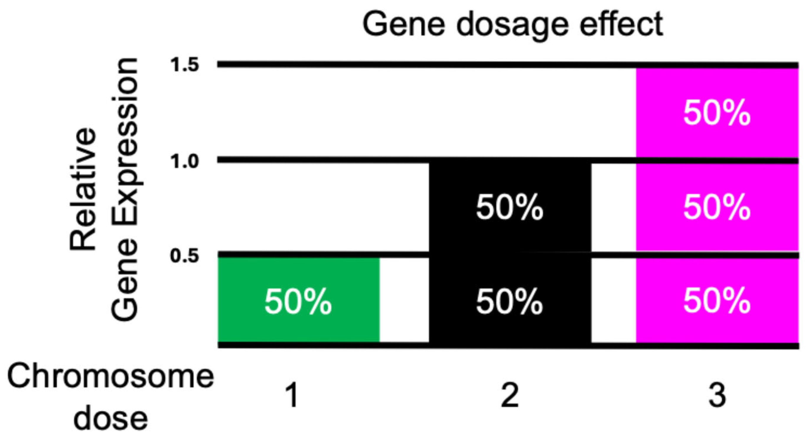
Figure 2.
Inverse Dosage Effect. When changing the dosage of an individual dosage sensitive regulatory gene or a chromosomal segment, effects on target genes across the genome can occur that are directly proportional to the copy number of the regulatory gene, or they can be inversely proportional. Because they act across the whole genome, they are referred to as trans-acting effects. The percentages represent the relative expression of a gene copy with the diploid set to 100%. Because the effects operate in trans, the unvaried chromosome number is two. The inverse effect is the more common response with hyperploidy, but a mix of direct and inverse effects occur with hypoploidy.
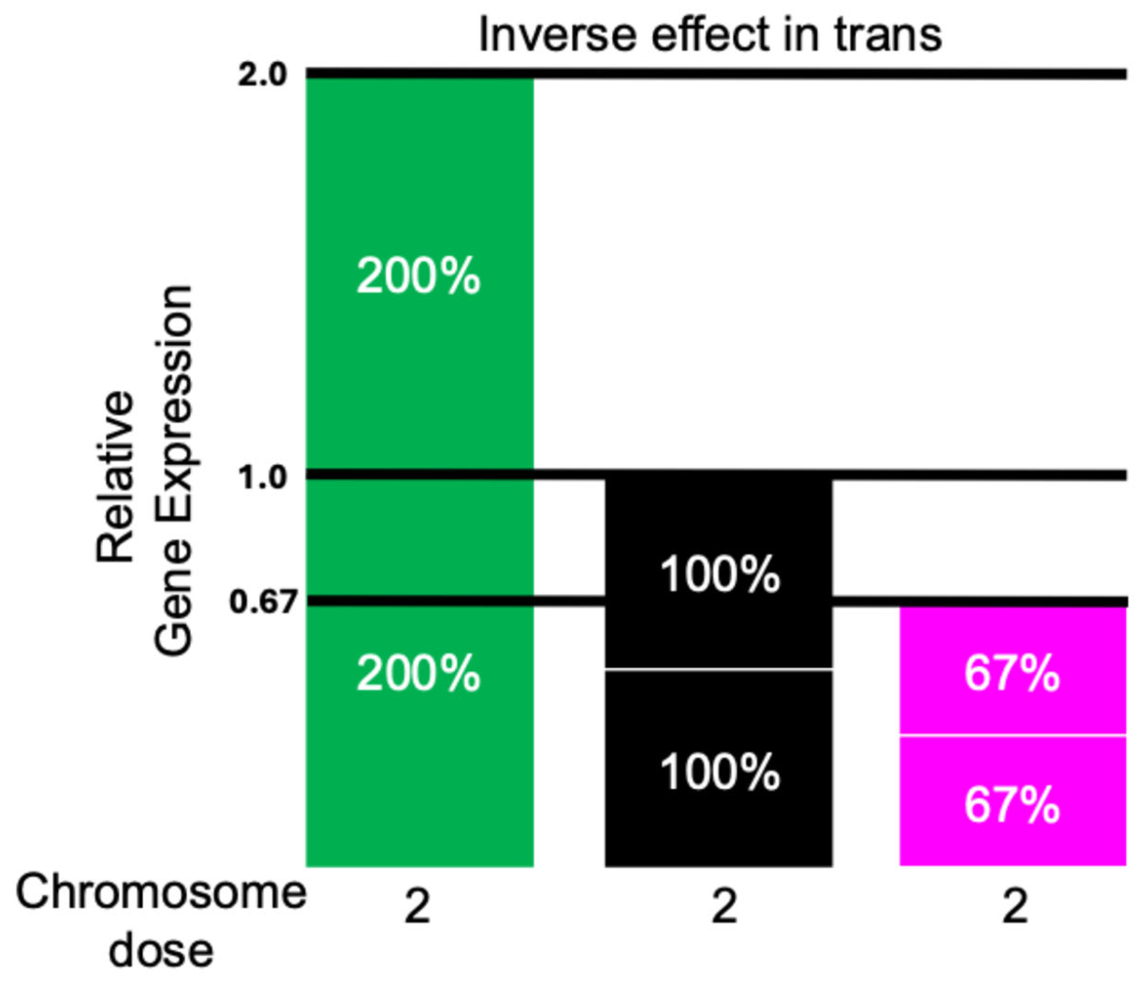
Figure 3.
Dosage Compensation Results from a Combination of a Gene Dosage Effect and an Inverse Effect Operating on the Same Gene. In a chromosomal dosage series, if a target gene is reduced to half the copy number compared to the normal diploid, but at the same time a regulatory gene that is also varied is inversely acting on it, the combination of effects will result in a total gene expression that is near the diploid level because the single copy is doubled in expression. When the chromosomal dosage is increased to three copies and the target gene is inversely affected, each copy would be expressed at 2/3rds the level in a diploid resulting in the total output being nearly equal to the diploid. The percentages represent the relative expression of each allele with a copy in a diploid set to 100%.
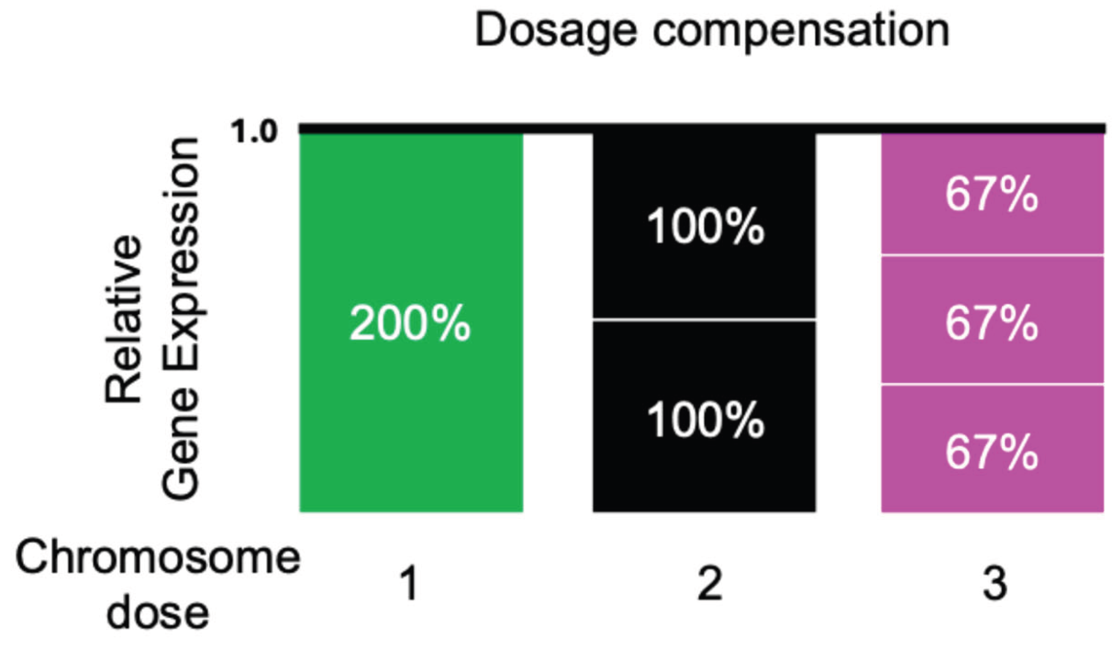
Figure 4.
Parameters of Sex Chromosome Dosage Compensation in Drosophila. Normal males have one X chromosome and normal females have two, but the total gene expression for X-linked genes is nearly equal. In females, producing genotypes that have only one copy of an X linked gene will reduce its output below the female and male level even though it is the same copy number as the latter. Increasing its dosage in a female also produces a gene dosage effect. A duplication of the gene in a male will double the expression. In other words, each copy in a male is expressed at about twice the level as in a female. Metafemales have three X chromosomes and the total output from them is similar to normal females and males, illustrating dosage compensation in the triple X genotype because each copy present is expressed at about two-thirds of the level in a normal female.
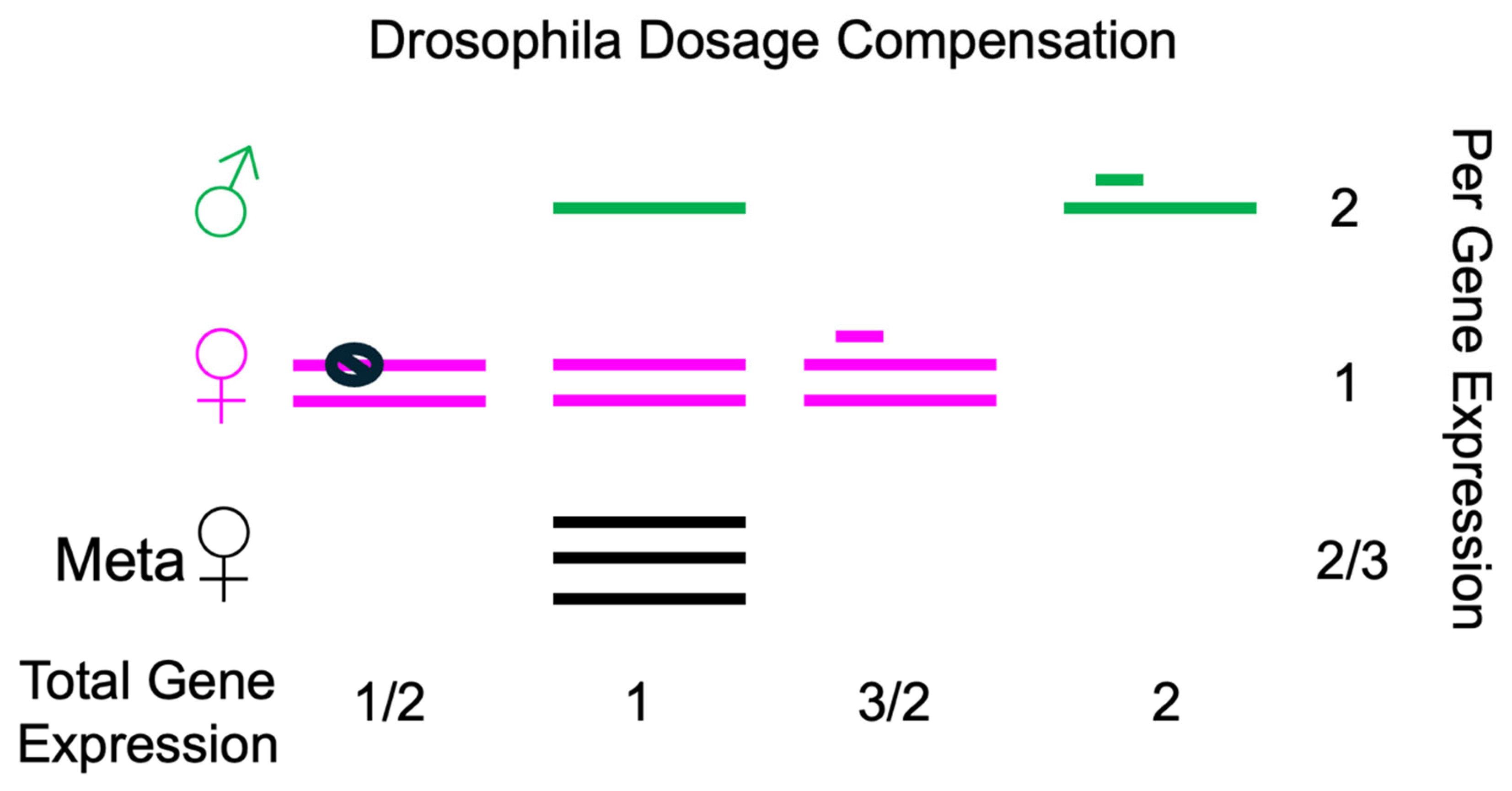
Figure 5.
Summary of Gene Expression Changes Affecting Dosage Compensation in Drosophila. In the top row is depicted the evolutionary progenitor state for the X (green) and the autosomes (magenta). The sex chromosomes evolve from a pair of homologues that would have similar levels of gene expression in both sexes. In the second row is depicted the current state of the male X chromosome that is hyperactivated compared to the progenitor by approximately two-fold with a similar expression of the autosomes. The triple X genotype (metafemales) is depicted with the three X chromosomes showing similar overall expression to the progenitor two X chromosomes while the autosomes are reduced to an approximately two-thirds level. In the third row, the situation that results from the loss of the MSL complex in males is shown. MOF and H4Lys16 acetylation become redistributed in the genome to be uniform producing an increase on the autosomes. The male X retains its two-fold hyperactivation while the autosomes show an increase. In the fourth row is shown genotypes that have been documented to lose dosage compensation of the male X with reductions to 50% of the normal male. These are the mof; mle/mle genotype and a maternal mof-minus scenario that has no deposition of MOF into eggs. These genotypes are embryonic lethal. In the fifth row is depicted the ectopic expression of MSL2 in females that will organize the MSL complex on the two X’s. There is no increase in expression of the X’s demonstrating a constraining activity on histone acetylation, but some autosomal genes are reduced in expression. Ectopic expression of MSL2 has also been performed in triple X metafemales with no increase of X expression but a further reduction in the expression of the autosomes. In the last row is depicted the situation in which genes implicated in the constraining activity are mutant, which results in male specific overexpression of the X chromosome. This overexpression resulting from the Mtor mutation is coincident with modulation of the male autosomes with increases and decreases in expression with the latter predominating.
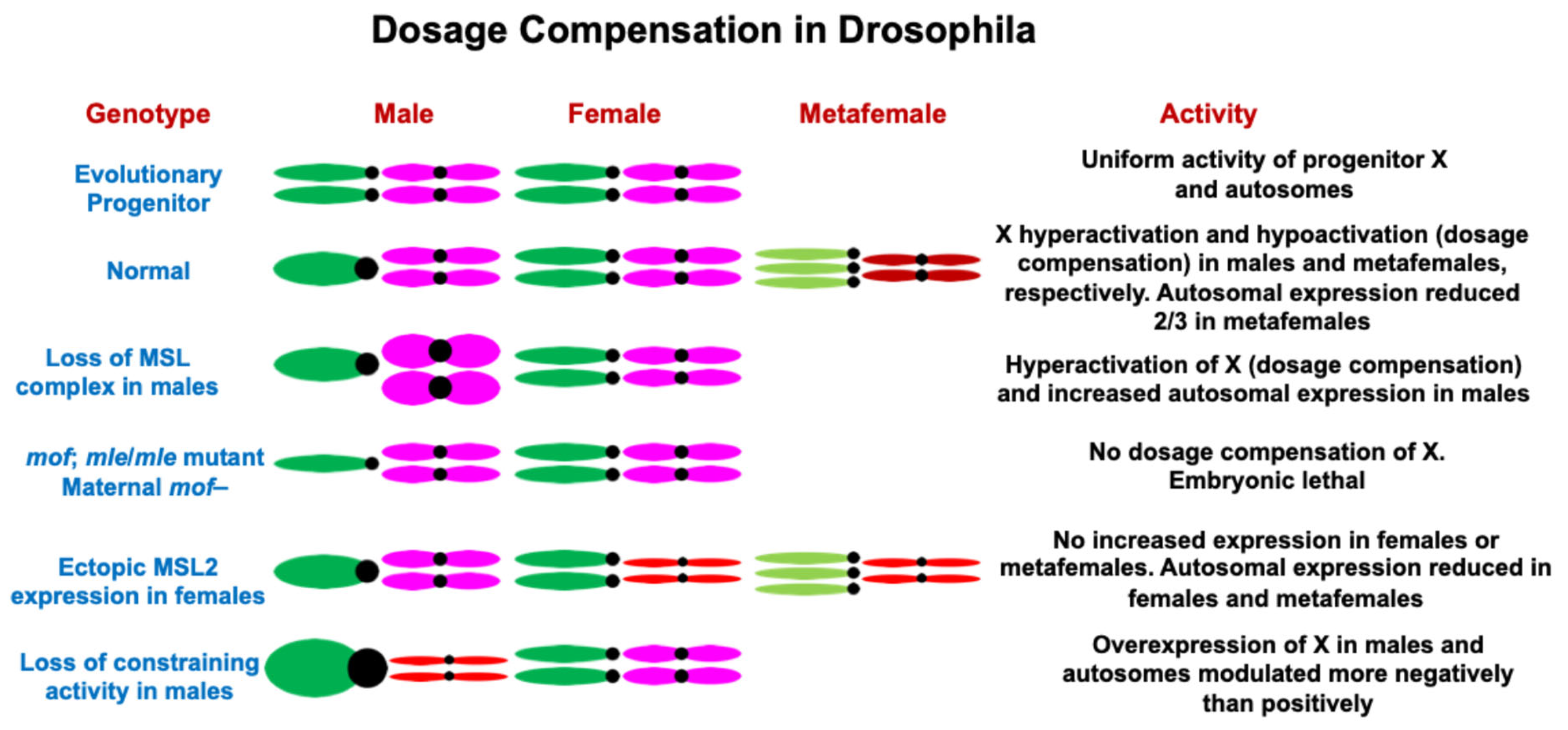
Disclaimer/Publisher’s Note: The statements, opinions and data contained in all publications are solely those of the individual author(s) and contributor(s) and not of MDPI and/or the editor(s). MDPI and/or the editor(s) disclaim responsibility for any injury to people or property resulting from any ideas, methods, instructions or products referred to in the content. |
© 2026 by the authors. Licensee MDPI, Basel, Switzerland. This article is an open access article distributed under the terms and conditions of the Creative Commons Attribution (CC BY) license (http://creativecommons.org/licenses/by/4.0/).
Copyright: This open access article is published under a Creative Commons CC BY 4.0 license, which permit the free download, distribution, and reuse, provided that the author and preprint are cited in any reuse.