

Submitted:
03 October 2026
Posted:
07 October 2026
You are already at the latest version
Abstract
Yellow fever virus (YFV) causes a systemic, frequently fatal infection in susceptible nonhuman primates (NHPs), which serve as important hosts and sentinels of viral circulation. However, susceptibility and pathological outcomes differ markedly between New World and Old World species. This narrative review comparatively examines the hepatic and extrahepatic histopathological spectrum of lesions associated with YFV infection in NHPs and explores their implications for pathogenesis, surveillance, conservation, and outbreak preparedness. The liver is the principal target organ, with characteristic findings including midzonal hepatocellular necrosis, Councilman bodies, microvesicular steatosis, sinusoidal congestion, hemorrhage, and relatively limited inflammatory infiltration. Extrahepatic lesions commonly involve the spleen, lymph nodes, kidneys, lungs, heart, and central nervous system, reflecting the systemic nature of severe infection. Highly susceptible Neotropical primates, particularly Alouatta spp., generally develop high viral burdens, extensive multiorgan damage, and high mortality. In contrast, many African Old World primates experience subclinical or milder infections with more limited tissue injury, possibly reflecting long-term host–virus coevolution and enhanced mechanisms of viral tolerance or control. Histopathology remains essential for investigating primate epizootics, although diagnostic accuracy improves with integration of immunohistochemistry, reverse-transcription polymerase chain reaction, in situ hybridization, and genomic methods. Standardized protocols for necropsy, sampling, lesion scoring, and reporting are needed to improve interspecies and interregional comparisons. Comparative primate pathology can strengthen early-warning surveillance, guide human vaccination strategies, support wildlife conservation, and advance integrated One Health preparedness.
Keywords:
yellow fever virus
; nonhuman primates
; comparative histopathology
; hepatic lesions
; extrahepatic involvement
; epizootic surveillance
; one Health
1. Introduction
Yellow fever (YF) is an acute hemorrhagic viral disease caused by the yellow fever virus (YFV), an arbovirus belonging to the genus Orthoflavivirus (family Flaviviridae), which continues to represent a major public health problem in tropical and subtropical regions of Africa and South America [1,2]. Despite the availability of a highly effective vaccine, the disease maintains a high epidemiological burden due to the persistence of jungle transmission cycles, the expansion of competent vectors, and the increasing interaction between human populations and forest ecosystems [3,4]. Non-human primates (NHPs) play a critical role in the ecoepidemiology of YFV, acting as amplifying hosts and biological sentinels of viral circulation in endemic areas [5].
The susceptibility of NHPs to YFV shows marked differences between species and geographical regions [6]. In the New World, several species of the families Atelidae and Cebidae, particularly the genera Alouatta, Ateles, and Lagothrix, are highly susceptible to infection and frequently develop fatal disease. The high mortality observed in these animals has made epizootic events essential tools for epidemiological surveillance and early detection of viral circulation [7,8]. In contrast, many African Old World NHPs, including species of the genera Cercopithecus, Papio, and Chlorocebus, tend to exhibit subclinical infections or less severe clinical / disease outcomes, suggesting evolutionary mechanisms of adaptation derived from prolonged coexistence with the virus in African ecosystems [9].
Differences in clinical susceptibility and mortality between New World and Old World primates have sparked considerable scientific interest in elucidating the pathogenic mechanisms underlying YFV infection. Among the most relevant findings are the histopathological lesions observed in target organs, especially in the liver, considered the main site of viral replication and tissue damage [10]. Liver injury characteristic of yellow fever includes predominantly midzonal hepatocellular necrosis, hepatocyte apoptosis with formation of Councilman’s acidophilic bodies, variable fat degeneration, sinusoidal congestion, and generally sparse inflammatory infiltrates [11,12]. These alterations reflect both the direct cytopathic viral effect and the complex interaction between the host’s immune response and the microvascular dysfunction induced by the infection [13,14].
In addition to hepatic damage, yellow fever produces significant alterations across multiple organs and systems. Several studies have documented involvement of lymph nodes, kidneys, lungs, heart, adrenal glands, and the central nervous system [1]. The most common extrahepatic lesions include splenic lymphoid depletion, acute renal tubular necrosis, pulmonary edema, multifocal hemorrhages, vascular congestion, and variable inflammatory tissue response [15,16,17]. These manifestations invariably lead to the development of multi-organ failure, one of the leading causes of death in severe YFV infections. However, the distribution, intensity, and pathogenic significance of these lesions can vary considerably between NHP species, reflecting intrinsic differences in susceptibility, tissue tropism, immune response, and evolutionary adaptation to the virus [18].
Comparative histopathology studies in NHP offer a unique opportunity to understand yellow fever pathogenesis from an evolutionary and translational perspective. Comparing highly susceptible New World species with relatively resistant Old World species allows us to identify lesion patterns associated with different clinical outcomes, as well as possible mechanisms of natural resistance to infection [19,20]. Likewise, these observations help improve the interpretation of findings observed during epizootics, strengthen surveillance systems based on primate mortality, and facilitate early detection of outbreaks that could potentially impact human health [21].
In recent years, the resurgence of large yellow fever epizootics in South America, particularly in Brazil, Colombia, and other countries in the region, has generated a significant amount of pathological information from different species of NHPs [22]. At the same time, the availability of historical and experimental studies carried out in African primates allows for more robust comparisons on the evolution of YFV-induced lesions [23,24]. However, information remains scattered, and few studies focus specifically on histopathological comparisons between New World and Old World primates.
Therefore, this study aims to comparatively analyze the main hepatic and extrahepatic lesions associated with yellow fever virus infection in susceptible non-human primate species from New and Old World settings, highlighting similarities and differences in the histopathological patterns described. This approach seeks to contribute to understanding the disease’s pathogenic mechanisms, provide relevant information for the surveillance of epizootics, and lay the groundwork for future research on the interaction between YFV and its primate hosts.
2. Diversity, Susceptibility, and Epidemiological Role of New and Old World Nonhuman Primates in Yellow Fever
Non-human primates play a central role in the ecology, epidemiology, and surveillance of yellow fever, an arbovirus caused by the yellow fever virus (YFV), a member of the genus Orthoflavivirus (family Flaviviridae) [25]. These animals actively participate in the jungle cycles of transmission, acting as susceptible vertebrate hosts and contributing to the maintenance of viral circulation in tropical ecosystems of Africa and South America [26]. However, the interaction between YFV and NHPs presents important biological and epidemiological differences between New World (Platyrrhini) and Old World (Catarrhini) species, reflecting different evolutionary processes that have conditioned the susceptibility, pathogenicity, and role of each group in the transmission dynamics of the virus [27].
New World primates comprise a great diversity of species distributed from southern Mexico to Argentina. Among them, the genera Alouatta, Sapajus, Callithrix, Saimiri, Ateles, and Lagothrix have been associated with naturally acquired YFV infections [28]. However, susceptibility varies considerably between taxa. Howler monkeys (Alouatta spp.) are recognized as the most vulnerable primates to infection and constitute the most frequently affected group during epizootics [29,30]. High mortality rates in these species, often close to 80–100% in exposed populations, have been documented in numerous regions of Brazil, Colombia, Venezuela, Peru, and Argentina [31]. Because of this extreme sensitivity, the detection of dead howler monkeys is usually one of the first indicators of viral circulation in a given area [20].
Other Neotropical primates exhibit different patterns of susceptibility. Robust capuchin monkeys (Sapajus spp.) can develop systemic infection and lesions consistent with yellow fever, although they generally show lower mortality rates than howler monkeys. Despite this, their ability to develop significant viremias makes them potential amplifiers of viral transmission [21]. Similarly, marmosets (Callithrix spp.), widely distributed in fragmented and peri-urban environments in Brazil, have acquired increasing epidemiological importance. During the large epizootics recorded between 2016 and 2019, multiple confirmed infections in Callithrix species showed that these primates can actively participate in transmission dynamics, especially in areas where urban expansion favors contact between vectors, wildlife, and humans [7].
In contrast, Old World primates show an older evolutionary relationship with YFV, which translates into generally lower clinical susceptibility [32]. In Africa, where the virus probably originated and evolved over thousands of years, species belonging to the genera Chlorocebus, Papio, Cercopithecus, and other cercopithecids usually develop subclinical infections or mild symptoms [26,33,34,35]. African green monkeys (Chlorocebus spp.) represent one of the best-known examples of hosts adapted to the virus [36]. Although they can become naturally infected and develop viremia sufficient to infect mosquito vectors, they rarely present severe disease or mass mortality [37]. Baboons (Papio spp.) and other African monkeys also contribute to the maintenance of jungle cycles through generally non-lethal infections, favoring the ecological persistence of the virus in forest environments [36,38].
These differences in susceptibility have important epidemiological implications. While in Africa, primates usually act as relatively resistant reservoirs or amplifiers, in the Americas, many species experience high mortality after infection [34]. This disparity has been interpreted as a consequence of the virus’s evolutionary history. African primates would have developed immunological and genetic mechanisms of adaptation during a prolonged coexistence with YFV, while American primates, exposed to the virus only since its historical introduction to the continent, would retain greater vulnerability to infection [39].
Historical and contemporary outbreaks of yellow fever clearly illustrate these differences. In Africa, the disease maintains complex enzootic cycles involving primates, jungle mosquitoes, and occasionally human populations [40]. Outbreaks in countries such as Uganda, Nigeria, Angola, the Democratic Republic of Congo, and Ghana have demonstrated the persistence of these natural cycles [41,42,43]. In South America, epizootics in NHPs have been fundamental to understanding the geographical expansion of the virus [12,44]. Particularly relevant were the outbreaks that occurred in Brazil between 2016 and 2019, considered among the largest in recent history, during which thousands of primates died in regions previously considered low-risk [45,46]. Similar situations, although of lesser magnitude, have been recorded in Colombia, Peru, Bolivia, and Venezuela, evidencing the capacity of the virus to expand into new ecological areas [47,48].
The epidemiological relevance of NHPs transcends their role as susceptible hosts. Currently, these animals constitute one of the most valuable surveillance systems for the early detection of YFV circulation [21,49]. The appearance of epizootics usually precedes the occurrence of human cases by weeks or months, allowing preventive measures to be activated before outbreaks are established in susceptible populations [5,39]. Investigation of primates found dead by necropsy, histopathology, immunohistochemistry, and molecular techniques provides direct evidence of viral circulation and contributes significantly to early warning systems [50,51].
From a One Health perspective, non-human primates represent an essential link between environmental, animal, and human health [52]. Systematic monitoring identifies risk areas, guides vaccination campaigns, and strengthens integrated epidemiological surveillance [53]. In addition, comparing New World and Old World species offers a unique opportunity to understand the biological mechanisms underlying differential susceptibility to YFV, information essential for interpreting histopathological findings across species and advancing knowledge of yellow fever pathogenesis [1,54].
3. Comparative Hepatic Histopathology in Yellow Fever: the Characteristic Organ of the Disease
Pathogenesis of Liver Injury
The liver is the main target organ for YFV infection and represents the tissue where the most characteristic histopathological lesions of the disease develop [55]. In both humans and NHPs, liver injury is responsible for a large part of the severe clinical manifestations and the high lethality observed in fulminant cases [56,57]. Viral replication in the liver parenchyma triggers a complex interaction between the direct cytopathic effect of the virus, the activation of apoptotic mechanisms, the inflammatory response of the host, and microvascular alterations, giving rise to a highly distinctive lesion pattern that has been considered for decades as one of the diagnostic features of yellow fever [58].
The most emblematic histopathological lesion of the disease is hepatocellular necrosis predominantly mediozonal (zone II of Rappaport’s hepatic acinus) [11]. This pattern is particularly characteristic because it differs from other viral hepatitis, in which periportal or centrolobular lesions usually predominate [59]. In yellow fever, hepatocytes located in the intermediate region between the portal spaces and the central veins show progressive cell degeneration, loss of tissue architecture, and extensive cell death [10]. Although the exact mechanisms responsible for this zonal distribution are not fully understood, it has been suggested that factors related to cellular metabolism, increased production of reactive oxygen species, oxidative stress, decreased antioxidant enzymes, differential expression of viral receptors, and susceptibility to apoptosis could explain the virus’s preference for this liver region [60].
Midzone necrosis has been consistently described in highly susceptible New World species, particularly in howler monkeys (Alouatta spp.), where it usually occurs in an extensive, diffuse form. In these animals, massive destruction of hepatocytes can compromise large areas of the liver parenchyma, contributing to the rapid development of acute liver failure [30]. Similar lesions have been documented in other Neotropical genera, including Sapajus and Callithrix, although in some cases the extent of damage is less severe [61]. In contrast, studies conducted on Old World African primates, such as Cercopithecus, have shown that mid-zonal lesions tend to be more focal or moderate, likely reflecting a greater ability to control viral replication and limit tissue damage [23].
Another classic histopathological finding is the presence of Councilman bodies, which are among the most representative microscopic markers of yellow fever. These structures, also known as acidophilic bodies, represent isolated apoptotic hepatocytes that appear as rounded, intensely eosinophilic bodies with pyknotic or absent nuclei. The abundance of Councilman bodies reflects massive activation of programmed cell death mechanisms induced by both viral replication and immune mediators [62], as seen in other acute-phase viral hepatitis. Experimental studies and immunohistochemical investigations have demonstrated the important involvement of caspase-dependent apoptotic pathways, as well as proinflammatory cytokines such as tumor necrosis factor-α (TNF-α) and various interferons [62]. In highly susceptible primates, these apoptotic bodies can be observed in large numbers distributed throughout the hepatic lobule, especially in the affected mid-zonal regions [12].
In addition to hepatocellular necrosis and apoptosis, yellow fever is associated with multiple additional morphological alterations. Hepatic steatosis, usually of the microvesicular type, is a common finding in advanced infections and reflects metabolic disorders secondary to virus-induced mitochondrial damage [11]. Sinusoidal congestion and focal hemorrhages are also common, as a result of vascular alterations, endothelial dysfunction, and coagulation disorders that characterize systemic disease. In severe cases, these alterations may coexist with edema, sinusoidal dilation, and partial collapse of the hepatic architecture [63]. The spectrum of histopathological findings in yellow fever also includes Kupffer cell hyperplasia and the presence of Vilela bodies. These are characteristic ochre-staining bodies, presumed to reflect a mixture of bile pigment and degradation products of Councilman bodies that have been phagocytosed within Kupffer cells [64,65,66]. In addition, a characteristic finding in experimental yellow fever models in rhesus monkeys is amorphous, eosinophilic intranuclear inclusions in hepatocytes, known as “Torres bodies.” These inclusions may occur synchronously with Councilman and Vilela bodies and are thought to result from a shared cytopathic viral insult [64,65,66].
A notable aspect of yellow fever histopathology is the relative scarcity of the inflammatory infiltrate, particularly compared with other viral hepatitis. Although lymphocytes, macrophages, and mononuclear inflammatory cells scattered in the portal spaces and parenchyma may be observed, the intensity of inflammation is usually surprisingly mild relative to the magnitude of necrosis [57]. This feature suggests that much liver damage results from direct cytopathic and apoptotic mechanisms rather than from an exuberant inflammatory response. However, the composition and magnitude of the infiltrate may vary between species and between different stages of infection [56,61]. Representative hepatic histopathological findings in naturally infected Neotropical NHPs, including midzonal necrosis, Councilman bodies, hepatocellular steatosis, and limited inflammatory infiltration, are shown in Figure 1, reproduced from previously reported fatal cases in southern Colombia [12].
Immunohistochemical studies have significantly expanded knowledge of YFV tissue distribution. Using antibodies against viral proteins, researchers have observed intense cytoplasmic immunoreactivity in hepatocytes within necrotic and perinecrotic areas. Viral antigens have also been detected in Kupffer cells, sinusoidal endothelial cells, and occasionally in infiltrating inflammatory cells [67]. These observations support the idea that the liver constitutes one of the main sites of viral replication during the acute phase of the disease.
Molecular techniques, particularly reverse transcriptase polymerase chain reaction (RT-PCR) and in situ hybridization, have confirmed the presence of high viral loads in liver tissues of infected [7]. Highly susceptible New World species typically show significantly elevated viral concentrations that correlate with severe histological lesions. In contrast, hepatic viral loads in Old World primates tend to be lower and more transient, which could explain the lower clinical severity observed in these hosts [23].
Together, midzone necrosis, Councilman’s bodies, steatosis, vascular congestion, and limited inflammatory response constitute the fundamental histopathological features of hepatic yellow fever [68]. Although these findings are shared by New and Old World primates, differences in the extent of damage, viral load, and intensity of tissue response reflect varying degrees of biological susceptibility [12]. These variations provide valuable information for understanding the disease’s comparative pathogenesis and the evolutionary mechanisms that shape the interaction between YFV and its primate hosts [56]. Table 1 summarizes the principal clinical, hepatic histopathological, virological, and host-response differences between New World and Old World NHPs.
4. Extrahepatic Histopathological Findings: Multiorgan Involvement in Yellow Fever
Although the liver is the main target organ for yellow fever virus (YFV) infection, the disease is essentially systemic and can produce lesions in multiple organs and tissues [57]. Viral replication, immune activation, endothelial dysfunction, microcirculatory alterations, and coagulation disorders contribute to multiorgan involvement that plays a determining role in the clinical course and mortality of severe cases [69]. In non-human primates (NHPs), both from the New and Old World, extrahepatic lesions show relatively consistent patterns, although with differences in frequency, intensity, and distribution that reflect varying degrees of susceptibility to the virus [70,71]. Representative gross hepatic and extrahepatic lesions in naturally infected Neotropical NHPs, including jaundice, hepatic necrosis, pulmonary edema, vascular congestion, and cardiac involvement, are shown in Figure 2, reproduced from previously reported fatal cases in southern Colombia [12].
4.1. Spleen: Lymphoid Depletion and Alterations of the White Pulp
The spleen is one of the most frequently affected lymphoid organs during YFV infection [72]. Histopathologically, the most characteristic lesion corresponds to a marked lymphoid depletion of the white pulp, accompanied by a reduction in size and disorganization of lymphoid follicles [73]. In severe cases, significant loss of lymphocytes is observed in the T- and B-cell-dependent areas, associated with extensive cellular apoptosis. These findings suggest profound immunosuppression induced by viral infection [74].
In highly susceptible New World primates, particularly Alouatta spp. (howler monkeys), yellow fever virus infection is associated with severe systemic disease and high mortality rates, often accompanied by marked lymphoid depletion and lymphocytolysis in the spleen [75]. In contrast, African Old World primates generally develop asymptomatic or milder infections [18]. Additional splenic findings may include vascular congestion, hemorrhage, follicular necrosis, and increased phagocytic activity in the red pulp, reflecting the systemic nature of the infection [21].
4.2. Lymph Nodes and Other Immune Tissues
Regional lymph nodes have alterations similar to those seen in the spleen. Among the most frequent findings are the depletion of cortical lymphocytes, the decrease of germinal centers, and the presence of scattered apoptotic bodies [73]. In some cases, sinus histiocytosis and macrophage activation are observed, associated with phagocytosis of cellular debris [74].
Tonsils, Peyer’s patches, and other mucosa-associated lymphoid tissues may also show reduced lymphoid cellularity. These changes suggest that YFV significantly affects host immune homeostasis, favoring the dysfunction of protective mechanisms that would normally limit viral replication [3]. Immunohistochemical studies have shown viral antigens in dendritic cells and macrophages of lymphoid organs, indicating that these tissues actively participate in systemic viral dissemination [65].
4.3. Kidney: Acute Tubular Injury and Infection-Associated Nephropathy
Renal involvement is one of the most relevant extrahepatic manifestations of yellow fever and is an important cause of morbidity and mortality in severe infections. The predominant lesion is consistent with acute tubular necrosis, particularly in the proximal convoluted tubules. Histologically, epithelial degeneration, cytoplasmic vacuolization, loss of the brush border, and cell desquamation towards the tubular lumen are observed [76].
Granular casts, interstitial edema, and vascular congestion may also be present. In the most severe cases, lesions compatible with acute renal failure develop, contributing to multiorgan failure syndrome. Several mechanisms are involved in the pathogenesis of these alterations, including tissue hypoperfusion, endothelial damage, hemodynamic alterations, and possible direct viral infection of renal cells [17].
Kidney lesions tend to be more extensive in highly susceptible New World species, whereas in Old World primates they are less severe and have less functional impact. However, basic histological findings are similar in both groups [77].
4.4. Heart: Myocarditis, Degeneration, and Inflammatory Infiltrates
Cardiac involvement has been documented in both natural and experimental YFV infections. The main alterations include degeneration of myocardial fibers, interstitial edema, and foci of mononuclear inflammatory infiltration. In some cases, focal areas of myocytic necrosis consistent with mild to moderate myocarditis are observed [15].
Inflammation is usually composed predominantly of lymphocytes and macrophages, distributed multifocally in the myocardial interstitium. Although cardiac lesions are generally less prominent than hepatic or renal lesions, they may contribute to the hemodynamic impairment seen in advanced infections [12]. Experimental studies suggest that a significant proportion of cardiac injury during severe yellow fever may result from immune-mediated mechanisms, endothelial dysfunction, and microvascular disturbances rather than from extensive viral replication within cardiomyocytes themselves [1,11,13,58,62].
4.5. Lung: Edema, Hemorrhage, and Congestion
The lungs frequently show alterations compatible with systemic vascular damage. Common findings include alveolar edema, diffuse capillary congestion, and focal or multifocal hemorrhages. In severe cases, thickening of the alveolar septa and accumulation of proteinaceous fluid in the airspaces may be observed [78,79].
These lesions reflect the combination of vascular permeability alterations, coagulation disorders, and systemic circulatory failure [80,81]. The presence of severe pulmonary edema may contribute significantly to the end-stage respiratory failure seen in animals with advanced disease [12]. In general, lung lesions are more pronounced in New World primates with fulminant infections than in relatively resistant African species [18,21].
4.6. Findings in the Brain and Central Nervous System
The central nervous system (CNS) is not considered a primary site of viral replication in yellow fever; however, various neuropathological alterations have been described in both humans and NHPs. Among the most frequent findings are vascular congestion, cerebral edema, petechial hemorrhages, and occasional mononuclear perivascular infiltrates [16].
The lesions are usually discrete compared to those seen in other neurotropic flaviviruses. However, in fatal cases, signs of encephalopathy associated with hepatic failure, systemic circulatory disturbances, and secondary metabolic disorders can be detected. Detection of viral antigens in the CNS is generally limited, supporting the idea that much neurological damage is indirect [82,83].
4.7. Comparison Between New and Old World Primates
Overall, extrahepatic histopathological patterns are similar in both primate groups, reflecting shared pathogenic mechanisms of YFV infection [61,82]. However, injury extent differs considerably. New World primates, particularly howler monkeys (Alouatta spp.), develop more extensive and severe lesions in lymphoid organs, kidneys, lungs, and other tissues, consistent with the high viral loads and high mortality observed during epizootics [12,44]. In contrast, Old World primates tend to have milder and more focal lesions, compatible with a greater ability to control viral replication and limit systemic damage [18]. These differences support the hypothesis that African hosts have evolved adaptations to YFV and are fundamental to understanding the comparative histopathology of yellow fever in susceptible species. Table 2 summarizes the principal extrahepatic histopathological lesions, their pathogenic mechanisms, clinical significance, and comparative patterns in New and Old World NHPs.
5. Comparative Pathogenesis, Diagnostic Pathology, and Future Lines of Research
Understanding the pathogenesis of yellow fever (YFV) in non-human primates (NHPs) has advanced significantly through the integration of histopathological, immunohistochemical, and molecular studies, which have enabled characterization of viral tropism, tissue distribution, and lesion patterns associated with infection [84]. However, important questions remain about the mechanisms that determine species-specific differential susceptibility, the tissue distribution of the virus, and the immunological responses associated with the clinical course of the disease [71]. Studies conducted during recent outbreaks in South America have demonstrated marked interspecific differences in viral load, mortality, and pathological findings among New World primates, highlighting gaps in the understanding of host–virus interactions [5]. Comparing findings in New World and Old World primates offers a unique opportunity to understand better interactions between yellow fever virus (YFV) and its natural hosts, as well as to optimize diagnostic and epidemiological surveillance strategies. [22,85].
Another rare phenomenon, infrequently recognized in humans, is YFV-associated hemophagocytic syndrome, characterized histopathologically by prominent hemophagocytic activity reflecting excessive macrophage activation in severe infection. In fatal YFV cases, including those reported during the 2017–2019 Brazilian outbreak, hemophagocytosis has been documented particularly within the pulmonary parenchyma, where activated macrophages containing erythrocytes and other hematopoietic elements may be observed in association with diffuse alveolar damage, pulmonary hemorrhage, and superimposed pneumonia [81]. Although less well characterized than the hepatic lesions that typify yellow fever, these findings suggest that macrophage-driven hyperinflammation may contribute to tissue injury in severe viscerotropic disease. In the appropriate clinical and histopathological context, prominent hemophagocytic activity may therefore represent secondary hemophagocytic lymphohistiocytosis (HLH) associated with YFV infection. This finding is important to recognize given the presumed increased susceptibility of New World nonhuman primates (NHPs) to severe YFV infection, which may potentially increase their risk of developing YFV-associated hemophagocytic syndrome compared with their Old World counterparts, paving the way for further research in this area [86].
5.1. Correlation Between Histopathology, Viral Tropism, and Viral Load
The lesions observed in yellow fever are closely related to YFV tissue tropism and the magnitude of viral replication across organs. Several studies have shown that the liver is the main site of viral replication, which explains the predominance of mid-zonal hepatocellular necrosis and the abundance of Councilman bodies in infected animals [87]. Detection of high viral loads in hepatocytes by RT-PCR, immunohistochemistry, and in situ hybridization consistently correlates with the severity of histological lesions [30,88].
Likewise, viral antigens and RNA in the spleen, lymph nodes, kidneys, lungs, and other organs confirm the infection’s systemic nature. However, the intensity of viral replication varies between tissues and species [84]. Highly susceptible New World primates, particularly howler monkeys (Alouatta spp.), typically have elevated viral loads in multiple organs, associated with extensive injury and fatal disease [12]. In contrast, Old World African primates generally develop less severe disease and lower mortality rates, likely reflecting long-term host–virus coevolution and greater resistance to the pathogenic effects of infection [24,35,39].
The correlation between viral load and lesion severity is not absolute, suggesting that additional immunopathological mechanisms play an important role in tissue damage. This observation has prompted research aimed at understanding how host responses modulate the evolution of the disease [11,62,89].
5.2. Immunopathological Mechanisms and Host Responses
Yellow fever pathogenesis results from a complex interaction between viral replication and the host immune response. After initial infection, YFV activates innate immune mechanisms mediated by type I interferons, dendritic cells, macrophages, and NK cells. Although these responses contribute to the control of viral replication, they can also participate in the generation of tissue damage when they occur in an excessive or dysregulated manner [90].
Apoptosis is one of the most important pathological mechanisms in YFV infection [91]. The abundant presence of Councilman bodies in the liver reflects the activation of apoptotic pathways induced by both direct viral infection and inflammatory mediators, including tumor necrosis factor-alpha (TNF-α), interferon gamma (IFN-γ), and various chemokines [92]. In parallel, the activation of macrophages and endothelial cells favors the production of proinflammatory cytokines that contribute to vascular dysfunction, hemorrhages, and multiorgan involvement [93].
The differences observed between New World and Old World primates may reflect evolutionary variation in these immune mechanisms. Some studies suggest that African primates have evolved immune tolerance strategies that limit excessive inflammation and tissue damage. In contrast, neotropical species exhibit less efficient responses to control viral replication without generating severe lesions. However, the specific molecular mechanisms responsible for these differences remain insufficiently characterized [21,39,94,95,96].
5.3. Histopathology Versus Immunohistochemistry, RT-PCR, and In Situ Hybridization
Conventional histopathology remains a fundamental tool for diagnosing yellow fever, especially in wildlife surveillance programs. Identifying mid-zonal necrosis, Councilman bodies, and compatible extrahepatic lesions provides highly suggestive evidence of YFV infection. However, these findings are not completely specific and may overlap with other viral or toxic liver diseases [12,68,97,98].
For this reason, complementary techniques have become increasingly important. Immunohistochemistry allows direct detection of viral proteins in tissues, providing visual evidence of the virus’s cellular location and establishing a direct correlation between viral presence and the observed lesions. This technique is particularly useful when samples have some degree of autolysis or when it is not possible to obtain fresh material for molecular analysis [61,99].
RT-PCR is currently the gold standard for diagnostic confirmation due to its high sensitivity and specificity. In addition to confirming the infection, it allows quantification of the viral load and study of the genetic diversity of circulating strains. More recently, in situ hybridization techniques have made it possible to identify viral RNA directly in tissues while preserving the histological architecture, providing valuable information on cell tropism and the spatial distribution of the virus within the affected organs [100,101,102,103].
5.4. Gaps in Knowledge About the Pathology of Old World Primates
Despite decades of research on yellow fever, information on New World and Old World primates remains markedly asymmetric. Most recent histopathological studies come from epizootics registered in South America, particularly in Brazil, where the high mortality facilitates the collection of samples for analysis [20,33,49,105,106].
In contrast, data on African species are relatively limited and, in many cases, come from historical research or experimental models conducted several decades ago. Important gaps remain regarding virus tissue distribution, local immune responses, mechanisms of natural resistance, and the detailed histopathological characteristics of numerous African species. The generation of new comparative studies using modern molecular tools is a priority to understand better host-virus evolution and the factors that determine differential susceptibility [107,108,109].
5.5. Standardization of Pathology Reports in Wildlife Surveillance
Surveillance of epizootics is one of the fundamental pillars of early detection of YFV circulation. However, heterogeneity in necropsy, sample processing, and histopathological description protocols makes it difficult to compare results across regions and countries [12,110].
Implementing standardized pathology reporting systems would improve the quality of data generated by surveillance programs. Such systems should include uniform criteria for describing hepatic and extrahepatic lesions, semi-quantitative severity scales, standardized immunohistochemistry and RT-PCR protocols, and the systematic integration of epidemiological and ecological information. This approach would facilitate the construction of internationally comparable databases and strengthen surveillance strategies under the One Health framework [52,111].
The integration of traditional histopathology with advanced molecular tools, coupled with greater standardization of pathological studies and strengthened research on African primates, will enable a deeper understanding of yellow fever pathogenesis and enhance the role of non-human primates as sentinels of viral circulation in tropical ecosystems [26,50].
6. Implications for One Health, Conservation and Outbreak Preparedness
Yellow fever is one of the most representative examples of a disease in which the human, animal, and environmental components that underpin the One Health concept converge closely. The persistence of YFV in tropical ecosystems depends on complex interactions between NHPs, mosquito vectors, ecological factors, and susceptible human populations [22,24]. In this context, the study of the histopathology and pathogenesis of infection in primates of the New and Old World not only provides relevant information to understand the biological mechanisms of the disease, but also has important implications for epidemiological surveillance, biodiversity conservation, and preparedness for future epidemic outbreaks [26,112].
Non-human primates play a dual role in yellow fever epidemiology. On the one hand, they serve as essential vertebrate hosts that maintain jungle transmission cycles. On the other hand, they act as extremely sensitive biological sentinels that allow viral circulation to be detected before human cases occur [21]. This sentinel function is particularly evident in South America, where highly susceptible species, such as howler monkeys (Alouatta spp.), develop high mortality rates during epizootics [113,114]. The appearance of sick or dead primates in jungle areas usually precedes the occurrence of human infections by weeks or even months, providing a critical window for the implementation of preventive measures [115,116].
Integrating epizootic surveillance with public health programs is one of the fundamental pillars of the One Health approach. The systematic investigation of cadavers through necropsy, histopathology, immunohistochemistry, and molecular techniques allows early identification of foci of viral circulation, guides vaccination campaigns, and strengthens prevention strategies in human populations at risk [117,118]. Likewise, the incorporation of geospatial surveillance systems and ecological modeling tools can improve the predictive capacity against the spread of the virus to new geographical areas [119,120].
From a conservation perspective, yellow fever poses a significant threat to many Neotropical primate species. Large epizootics recorded in Brazil between 2016 and 2019 showed that the disease can cause considerable population declines in highly susceptible species, particularly in Alouatta guariba and other representatives of the Atelidae family [22,121,122]. Drastic reductions in population densities have been reported in some regions, raising concerns about the long-term effects of the disease on ecosystems already affected by deforestation, habitat fragmentation, and other anthropogenic pressures [5].
Importantly, non-human primates are victims of the disease and not directly responsible for transmission to humans. However, during some outbreaks, events of persecution and intentional killing of monkeys have been recorded due to misperceptions about their epidemiological role [12,44]. These actions not only lack a scientific basis, but also compromise conservation efforts and weaken natural epidemiological surveillance systems. Accordingly, community education strategies should be an integral part of yellow fever control programs, promoting understanding of the ecological and health role of [123,124].
Environmental changes associated with climate change, agricultural expansion, urbanization, and modifications to natural ecosystems pose additional challenges for disease control [125,126]. Habitat alteration can modify vector distribution, increase wildlife-human contact, and encourage the emergence of new transmission scenarios. In this context, integrated surveillance of vectors, primates, and human populations is becoming increasingly important in anticipating changes in yellow fever epidemiological patterns [27].
Preparing for future outbreaks also requires strengthening diagnostic and rapid response capacities. The standardization of necropsy, sample processing, and histopathological analysis protocols will generate more comparable information between regions and improve the quality of surveillance systems [52,111]. Similarly, the routine incorporation of molecular, immunohistochemical, and genomic tools will facilitate early detection of viral circulation and monitoring of potential evolutionary changes in circulating strains [127,128].
Comparative research between New World and Old World primates also offers important opportunities to develop new preventive strategies. Understanding the biological mechanisms that allow certain African species to tolerate infection with little tissue damage could provide valuable information on natural resistance processes, immune regulation, and control of viral replication [39,129,130]. This knowledge could contribute to the design of new therapeutic or preventive approaches applicable both to human medicine and to the conservation of vulnerable species [131,132].
Finally, yellow fever highlights the need to strengthen interdisciplinary collaboration between doctors, veterinarians, pathologists, ecologists, conservation biologists, entomologists, and health authorities [133,134]. No single discipline can address the complexity of a disease that simultaneously involves human health, animal health, and ecosystem dynamics. The One Health approach provides an ideal conceptual framework for integrating this knowledge and developing sustainable surveillance, prevention, and response strategies [24,135,136,137].
7. Limitations
This review has several limitations. First, as a narrative synthesis, it did not follow a prespecified systematic review protocol or a formal risk-of-bias assessment; therefore, publication, availability, and language biases may have influenced study selection. Second, the evidence base is highly heterogeneous, combining natural epizootic cases, experimental infections, historical reports, autopsy studies, and investigations using different sampling procedures, diagnostic assays, and histopathological criteria. These differences preclude quantitative comparisons of lesion frequency or severity across species. Third, evidence is geographically and taxonomically imbalanced: recent data are concentrated in a limited number of highly susceptible Neotropical primates, especially Alouatta spp., whereas information on many African Old World species remains sparse and frequently derives from older experimental studies. Postmortem interval, tissue autolysis, incomplete organ sampling, and inconsistent use of immunohistochemistry or molecular confirmation may also have affected lesion detection. Furthermore, host age, sex, immune status, viral lineage, inoculum, and disease stage were not uniformly reported and could confound interspecies comparisons. Consequently, interpret proposed evolutionary and immunological explanations for differential susceptibility as biologically plausible hypotheses rather than definitive causal conclusions. Standardized, prospective, multispecies studies are needed.
8. Conclusions
Yellow fever produces a characteristic but multisystemic pathological pattern in nonhuman primates, dominated by midzonal hepatocellular necrosis, Councilman bodies, steatosis, sinusoidal congestion, and relatively limited inflammatory infiltration. Extrahepatic involvement, including lymphoid depletion, acute tubular injury, pulmonary edema and hemorrhage, myocardial damage, vascular alterations, and occasional central nervous system lesions, further demonstrates the systemic nature of severe infection.
Although New World and Old World primates share principal histopathological features, their extent and clinical consequences differ substantially by species. Highly susceptible Neotropical primates, particularly Alouatta spp., generally develop higher viral burdens, extensive multiorgan injury, and frequently fatal disease. In contrast, many African primates experience milder or subclinical infection with more limited tissue damage, supporting the possibility that long-term host–virus coexistence has favored mechanisms of viral control or tolerance. However, these evolutionary and immunological explanations require confirmation through standardized comparative studies.
Histopathology remains essential for yellow fever surveillance, particularly when investigating primate deaths during epizootics, but diagnostic accuracy improves when integrated with immunohistochemistry, RT-PCR, in situ hybridization, and genomic analysis. Standardized protocols for necropsy, tissue sampling, lesion scoring, and reporting would improve comparisons across species and regions.
Ultimately, comparative primate pathology contributes not only to understanding yellow fever pathogenesis but also to early outbreak detection, targeted human vaccination, wildlife conservation, and integrated One Health preparedness. Future research should prioritize underrepresented African species, prospective multispecies investigations, and the molecular mechanisms underlying differential susceptibility and resistance.
Author Contributions
Conceptualization, D.K.B.-A. and A.J.R.-M.; methodology, D.K.B.-A., J.L.B.-A. and A.J.R.-M.; resources, A.P.M. and A.J.R.-M.; writing—original draft preparation, D.K.B.-A., J.L.B.-A., A.P.M., L.I.Z., and A.J.R.-M.; writing—review and editing, D.K.B.-A., J.L.B.-A., A.P.M., L.I.Z., and A.J.R.-M.; supervision, D.K.B.-A., J.L.B.-A., A.P.M., L.I.Z., and A.J.R.-M. All authors have read and agreed to the published version of the manuscript.
Funding
The Instituto de Investigaciones One Health, Universidad Tecnológica Centroamericana (UNITEC), Tegucigalpa, MDC, Honduras, Central America (granted to Zambrano), funded the current article processing charges (publication fees).
Institutional Review Board Statement
Not applicable.
Informed Consent Statement
Not applicable.
Data Availability Statement
This study did not create or analyze new data.
Acknowledgments
This article has been registered in the Research Proposal Registration of the Coordination of Scientific Integrity and Surveillance of Universidad Cientifica del Sur, Lima, Peru. .
Conflicts of Interest
The authors declare no conflicts of interest.
References
- Monath, T.P.; Vasconcelos, P.F. Yellow fever. J. Clin. Virol. 2015, 64, 160–173. [Google Scholar] [CrossRef]
- Rodríguez-Morales, A.J.; Bonilla-Aldana, D.K.; Suárez, J.A.; Franco-Paredes, C.; Forero-Peña, D.A.; Mattar, S.; Villamil-Gómez, W.E.; Ruíz-Sáenz, J.; Cardona-Ospina, J.A.; Figuera, M.E.; et al. Yellow fever reemergence in Venezuela - Implications for international travelers and Latin American countries during the COVID-19 pandemic. Travel Med. Infect. Dis. 2021, 44, 102192. [Google Scholar] [CrossRef]
- Douam, F.; Ploss, A. Yellow Fever Virus: Knowledge Gaps Impeding the Fight Against an Old Foe. Trends Microbiol. 2018, 26, 913–928. [Google Scholar] [CrossRef]
- Reno, E.; Quan, N.G.; Franco-Paredes, C.; Chastain, D.B.; Chauhan, L.; Rodriguez-Morales, A.J.; Henao-Martínez, A.F. Prevention of yellow fever in travellers: an update. Lancet Infect. Dis. 2020, 20, e129–e137. [Google Scholar] [CrossRef]
- Bonilla-Aldana, D.K.; Bonilla-Aldana, J.L.; Castellanos, J.E.; Rodriguez-Morales, A.J. Importance of Epizootic Surveillance in the Epidemiology of Yellow Fever in South America. Curr. Trop. Med. Rep. 2025, 12, 16. [Google Scholar] [CrossRef]
- Goes de Jesus, J.; Gräf, T.; Giovanetti, M.; Mares-Guia, M.A.; Xavier, J.; Lima Maia, M.; Fonseca, V.; Fabri, A.; Dos Santos, R.F.; Mota Pereira, F.; et al. Yellow fever transmission in non-human primates, Bahia, Northeastern Brazil. PLoS Negl. Trop. Dis. 2020, 14, e0008405. [Google Scholar] [CrossRef]
- Sacchetto, L.; Silva, N.I.O.; Rezende, I.M.; Arruda, M.S.; Costa, T.A.; de Mello É, M.; Oliveira, G.F.G.; Alves, P.A.; de Mendonça, V.E.; Stumpp, R.; et al. Neighbor danger: Yellow fever virus epizootics in urban and urban-rural transition areas of Minas Gerais state, during 2017-2018 yellow fever outbreaks in Brazil. PLoS Negl. Trop. Dis. 2020, 14, e0008658. [Google Scholar] [CrossRef]
- Passos, P.H.O.; Ramos, D.G.; de Almeida, M.A.B.; Feijó, V.P.; Caraciolo, M.F.; Chame, M.; Pinter, A.; Romano, A.P.M.; de Albuquerque, L.C.; de Sousa, D.E.R.; et al. 25 Years of Non-Human Primates Surveillance contributing to Yellow Fever control in Brazil. Acta Trop. 2026, 108227. [Google Scholar] [CrossRef]
- Staples, J.E.; Diallo, M.; Janusz, K.B.; Manengu, C.; Lewis, R.F.; Perea, W.; Yactayo, S.; Sall, A.A. Yellow fever risk assessment in the Central African Republic. Trans. R Soc. Trop. Med. Hyg. 2014, 108, 608–615. [Google Scholar] [CrossRef]
- Lemos, F.O.; França, A.; Lima Filho, A.C.M.; Florentino, R.M.; Santos, M.L.; Missiaggia, D.G.; Rodrigues, G.O.L.; Dias, F.F.; Souza Passos, I.B.; Teixeira, M.M.; et al. Molecular Mechanism for Protection Against Liver Failure in Human Yellow Fever Infection. Hepatol. Commun. 2020, 4, 657–669. [Google Scholar] [CrossRef]
- Quaresma, J.A.; Barros, V.L.; Fernandes, E.R.; Pagliari, C.; Takakura, C.; da Costa Vasconcelos, P.F.; de Andrade, H.F., Jr.; Duarte, M.I. Reconsideration of histopathology and ultrastructural aspects of the human liver in yellow fever. Acta Trop. 2005, 94, 116–127. [Google Scholar] [CrossRef]
- Sanchez-Rojas, I.C.; Bonilla-Aldana, D.K.; Solarte-Jimenez, C.L.; Bonilla-Aldana, J.L.; Belisario-Tovar, M.; Ortega-Gómez, S.; Zambrano-Quenan, V.M.; Perafan-Gomez, J.C.; Gomez-Ocampo, C.H.; Delgado-Cajigas, M.; et al. Fatal yellow fever among captive non-human primates in southern Colombia, 2025. Front Vet. Sci. 2025, 12, 1655474. [Google Scholar] [CrossRef]
- Quaresma, J.A.; Pagliari, C.; Medeiros, D.B.; Duarte, M.I.; Vasconcelos, P.F. Immunity and immune response, pathology and pathologic changes: progress and challenges in the immunopathology of yellow fever. Rev. Med. Virol. 2013, 23, 305–318. [Google Scholar] [CrossRef]
- Rust, L.N.; Ricciardi, M.J.; Lutz, S.S.; Yusova, S.; Louw, J.J.; Yrizarry-Medina, A.; Biswas, S.; Fischer, M.; Barber-Axthelm, A.; Zilverberg, G.; et al. Prophylactic and therapeutic neutralizing monoclonal antibody treatment prevents lethal yellow fever infection. JCI Insight 2025, 10. [Google Scholar] [CrossRef]
- Giugni, F.R.; Aiello, V.D.; Faria, C.S.; Pour, S.Z.; Cunha, M.D.P.; Giugni, M.V.; Pinesi, H.T.; Ledesma, F.L.; Morais, C.E.; Ho, Y.L.; et al. Understanding yellow fever-associated myocardial injury: an autopsy study. EBioMedicine 2023, 96, 104810. [Google Scholar] [CrossRef]
- Frassetto, F.P.; Rosemberg, S. Neuropathology of yellow fever autopsy cases. Trop. Dis. Travel Med. Vaccines 2023, 9, 1. [Google Scholar] [CrossRef]
- Lopes, R.L.; Pinto, J.R.; Silva Junior, G.B.D.; Santos, A.K.T.; Souza, M.T.O.; Daher, E.F. Kidney involvement in yellow fever: a review. Rev. Inst. Med. Trop. Sao Paulo 2019, 61, e35. [Google Scholar] [CrossRef]
- Nederlof, R.A.; Virgilio, T.; Stemkens, H.J.J.; da Silva, L.C.C.P.; Montagna, D.R.; Abdussamad, A.M.; Chipangura, J.; Bakker, J. Yellow Fever in Non-Human Primates: A Veterinary Guide from a One Health Perspective. Vet. Sci. 2025, 12, 339. [Google Scholar] [CrossRef]
- Mares-Guia, M.; Furtado, M.C.; Chalhoub, F.L.L.; Portugal, M.D.; de Oliveira Coelho, J.M.C.; de Filippis, A.M.B.; Naveca, F.G. Coinfection with Canine Distemper Virus and Yellow Fever Virus in a Neotropical Primate in Brazil. Viruses 2024, 16. [Google Scholar] [CrossRef]
- Mares-Guia, M.; Horta, M.A.; Romano, A.; Rodrigues, C.D.S.; Mendonca, M.C.L.; Dos Santos, C.C.; Torres, M.C.; Araujo, E.S.M.; Fabri, A.; de Souza, E.R.; et al. Yellow fever epizootics in non-human primates, Southeast and Northeast Brazil (2017 and 2018). Parasit. Vectors 2020, 13, 90. [Google Scholar] [CrossRef]
- de Azevedo Fernandes, N.C.C.; Guerra, J.M.; Díaz-Delgado, J.; Cunha, M.S.; Saad, L.D.; Iglezias, S.D.; Ressio, R.A.; Dos Santos Cirqueira, C.; Kanamura, C.T.; Jesus, I.P.; et al. Differential Yellow Fever Susceptibility in New World Nonhuman Primates, Comparison with Humans, and Implications for Surveillance. Emerg. Infect. Dis. 2021, 27, 47–56. [Google Scholar] [CrossRef]
- Bonilla-Aldana, D.K.; Bonilla-Aldana, J.L.; Calle-Hernández, D.M.; Castellanos, J.E.; Zambrano, L.; Rodriguez-Morales, A.J. Yellow fever virus infection in non-human primates: a systematic review and meta-analysis of prevalence, seroprevalence, and epizootic case reports (1950–2025). Front. Vet. Sci. 2026, Volume 13 - 2026. [Google Scholar] [CrossRef]
- Hughes, T.P. The reaction of the African grivet monkey (Cercopithecus aethiops centralis) to yellow fever virus. Trans. R. Soc. Trop. Med. Hyg. 1943, 36, 339–346. [Google Scholar] [CrossRef]
- Nederlof, R.A.; Virgilio, T.; Stemkens, H.J.J.; da Silva, L.; Montagna, D.R.; Abdussamad, A.M.; Chipangura, J.; Bakker, J. Yellow Fever in Non-Human Primates: A Veterinary Guide from a One Health Perspective. Vet. Sci. 2025, 12. [Google Scholar] [CrossRef]
- Saad, L.D.C.; Chiaravalloti-Neto, F. Reemergence of yellow fever in the state of São Paulo: the structuring role of surveillance of epizootics in non-human primates in a one health approach. Rev. Bras. Epidemiol. 2024, 27, e240064. [Google Scholar] [CrossRef]
- Valentine, M.J.; Murdock, C.C.; Kelly, P.J. Sylvatic cycles of arboviruses in non-human primates. Parasit. Vectors 2019, 12, 463. [Google Scholar] [CrossRef]
- Srivastava, S.; Dhoundiyal, S.; Kumar, S.; Kaur, A.; Khatib, M.N.; Gaidhane, S.; Zahiruddin, Q.S.; Mohanty, A.; Henao-Martinez, A.F.; Krsak, M.; et al. Yellow Fever: Global Impact, Epidemiology, Pathogenesis, and Integrated Prevention Approaches. Infez. Med. 2024, 32, 434–450. [Google Scholar] [CrossRef]
- Silva, N.I.O.; Sacchetto, L.; de Rezende, I.M.; Trindade, G.S.; LaBeaud, A.D.; de Thoisy, B.; Drumond, B.P. Recent sylvatic yellow fever virus transmission in Brazil: the news from an old disease. Virol. J. 2020, 17, 9. [Google Scholar] [CrossRef]
- Holzmann, I.; Agostini, I.; Areta, J.I.; Ferreyra, H.; Beldomenico, P.; Di Bitetti, M.S. Impact of yellow fever outbreaks on two howler monkey species (Alouatta guariba clamitans and A. caraya) in Misiones, Argentina. Am. J. Primatol. 2010, 72, 475–480. [Google Scholar] [CrossRef]
- Leal, S.G.; Romano, A.P.; Monteiro, R.V.; Melo, C.B.; Vasconcelos, P.F.; Castro, M.B. Frequency of histopathological changes in Howler monkeys ( Alouatta sp.) naturally infected with yellow fever virus in Brazil. Rev. Soc. Bras. Med. Trop. 2016, 49, 29–33. [Google Scholar] [CrossRef]
- de Almeida, M.A.; Dos Santos, E.; da Cruz Cardoso, J.; da Fonseca, D.F.; Noll, C.A.; Silveira, V.R.; Maeda, A.Y.; de Souza, R.P.; Kanamura, C.; Brasil, R.A. Yellow fever outbreak affecting Alouatta populations in southern Brazil (Rio Grande do Sul State), 2008-2009. Am. J. Primatol. 2012, 74, 68–76. [Google Scholar] [CrossRef]
- Barrett, A.D.; Monath, T.P. Epidemiology and ecology of yellow fever virus. Adv. Virus Res. 2003, 61, 291–315. [Google Scholar] [CrossRef]
- Aliaga-Samanez, A.; Real, R.; Segura, M.; Marfil-Daza, C.; Olivero, J. Yellow fever surveillance suggests zoonotic and anthroponotic emergent potential. Commun. Biol. 2022, 5, 530. [Google Scholar] [CrossRef]
- Chippaux, J.P.; Chippaux, A. Yellow fever in Africa and the Americas: a historical and epidemiological perspective. J. Venom. Anim. Toxins Incl. Trop. Dis. 2018, 24, 20. [Google Scholar] [CrossRef]
- Oyono, M.G.; Kenmoe, S.; Abanda, N.N.; Takuissu, G.R.; Ebogo-Belobo, J.T.; Kenfack-Momo, R.; Kengne-Nde, C.; Mbaga, D.S.; Tchatchouang, S.; Kenfack-Zanguim, J.; et al. Epidemiology of yellow fever virus in humans, arthropods, and non-human primates in sub-Saharan Africa: A systematic review and meta-analysis. PLoS Negl. Trop. Dis. 2022, 16, e0010610. [Google Scholar] [CrossRef]
- Diallo, D.; Chen, R.; Diagne, C.T.; Ba, Y.; Dia, I.; Sall, A.A.; Weaver, S.C.; Diallo, M. Bloodfeeding patterns of sylvatic arbovirus vectors in southeastern Senegal. Trans. R Soc. Trop. Med. Hyg. 2013, 107, 200–203. [Google Scholar] [CrossRef]
- Mutebi, J.-P.; Gianella, A.; Travassos da Rosa, A.; Tesh, R.B.; Barrett, A.D.T.; Higgs, S. Yellow Fever Virus Infectivity for Bolivian Aedes aegypti Mosquitoes. Emerg. Infect. Dis. J. 2004, 10, 1657. [Google Scholar] [CrossRef]
- Taylor, R.M.; Haseeb, M.A.; Work, T.H. A regional reconnaissance on yellow fever in the Sudan; with special reference to primate hosts. Bull. World Health Organ 1955, 12, 711–725. [Google Scholar]
- Tappan, J. Wandering epizootics and zones of emergence: Constructing yellow fever endemicity in Africa. Health Place 2022, 77, 102770. [Google Scholar] [CrossRef]
- Mutebi, J.P.; Barrett, A.D. The epidemiology of yellow fever in Africa. Microbes Infect. 2002, 4, 1459–1468. [Google Scholar] [CrossRef]
- Nwaiwu, A.U.; Musekiwa, A.; Tamuzi, J.L.; Sambala, E.Z.; Nyasulu, P.S. The incidence and mortality of yellow fever in Africa: a systematic review and meta-analysis. BMC Infect. Dis. 2021, 21, 1089. [Google Scholar] [CrossRef]
- Otshudiema, J.O.; Ndakala, N.G.; Mawanda, E.K.; Tshapenda, G.P.; Kimfuta, J.M.; Nsibu, L.N.; Gueye, A.S.; Dee, J.; Philen, R.M.; Giese, C.; et al. Yellow Fever Outbreak - Kongo Central Province, Democratic Republic of the Congo, August 2016. MMWR Morb. Mortal. Wkly. Rep. 2017, 66, 335–338. [Google Scholar] [CrossRef]
- Vasconcelos, P.F.; Monath, T.P. Yellow Fever Remains a Potential Threat to Public Health. Vector Borne Zoonotic Dis. 2016, 16, 566–567. [Google Scholar] [CrossRef]
- Sanchez-Rojas, I.C.; Bonilla-Aldana, D.K.; Solarte-Jimenez, C.L.; Bonilla-Aldana, J.L.; Suárez, E.; Montealegre, J.L.; Suárez, M.; Rodriguez-Morales, A.J. Yellow fever epizootics among non-human primates in Huila, Colombia, 2025-2026. New Microbes New Infect. 2026, 71, 101762. [Google Scholar] [CrossRef]
- Cunha, M.D.P.; Duarte-Neto, A.N.; Pour, S.Z.; Ortiz-Baez, A.S.; Cerny, J.; Pereira, B.B.S.; Braconi, C.T.; Ho, Y.L.; Perondi, B.; Sztajnbok, J.; et al. Origin of the Sao Paulo Yellow Fever epidemic of 2017-2018 revealed through molecular epidemiological analysis of fatal cases. Sci. Rep. 2019, 9, 20418. [Google Scholar] [CrossRef]
- de Oliveira Figueiredo, P.; Stoffella-Dutra, A.G.; Barbosa Costa, G.; Silva de Oliveira, J.; Dourado Amaral, C.; Duarte Santos, J.; Soares Rocha, K.L.; Araujo Junior, J.P.; Lacerda Nogueira, M.; Zaza Borges, M.A.; et al. Re-Emergence of Yellow Fever in Brazil during 2016-2019: Challenges, Lessons Learned, and Perspectives. Viruses 2020, 12. [Google Scholar] [CrossRef]
- Rodriguez-Morales, A.J.; Chang-Cheng, B.; Gross, R.; Llanque-Espinoza, O.E.; Villamil-Macareno, J.; Pacheco-Jimenez, C.; Pineda-Bersoza, G.B.; Delgado-Torres, N.F.; Sanchez-Rojas, I.C.; Solarte-Jimenez, C.L.; et al. Clinical features of yellow fever in cases from Bolivia, Ecuador, Colombia, and Peru (2023-2025): A descriptive retrospective study. New Microbes New Infect. 2025, 68, 101651. [Google Scholar] [CrossRef]
- Rodriguez-Morales, A.J.; Navarro, J.C.; Forero-Peña, D.A.; Romero-Alvarez, D. Reemergence of yellow fever in Venezuela, 2025/2026. New Microbes New Infect. 2026, 70, 101737. [Google Scholar] [CrossRef]
- Cunha, M.S.; Tubaki, R.M.; de Menezes, R.M.T.; Pereira, M.; Caleiro, G.S.; Coelho, E.; Saad, L.D.C.; Fernandes, N.; Guerra, J.M.; Nogueira, J.S.; et al. Possible non-sylvatic transmission of yellow fever between non-human primates in Sao Paulo city, Brazil, 2017-2018. Sci. Rep. 2020, 10, 15751. [Google Scholar] [CrossRef]
- Bonilla-Aldana, D.K.; Bonilla-Aldana, J.L.; Zambrano, L.; Rodriguez-Morales, A.J. Revisiting the Status of Yellow Fever Epizootics and Its Surveillance in South America: New Non-Human Primates, Spillover and Ecological Drivers. Pathogens 2026, 15. [Google Scholar] [CrossRef]
- Fernandes, N.; Cunha, M.S.; Guerra, J.M.; Réssio, R.A.; Cirqueira, C.D.S.; Iglezias, S.D.; de Carvalho, J.; Araujo, E.L.L.; Catão-Dias, J.L.; Díaz-Delgado, J. Outbreak of Yellow Fever among Nonhuman Primates, Espirito Santo, Brazil, 2017. Emerg. Infect. Dis. 2017, 23, 2038–2041. [Google Scholar] [CrossRef]
- Organization, W.H.; Nations, F.a.A.O.o.t.U.; Programme, U.N.E.; Health, W.O.f.A. One Health Joint Plan of Action (2022–2026): Working together for the health of humans, animals, plants and the environment; World Health Organization: Geneva, 2022; ISBN 978-92-4-005913-9. [Google Scholar]
- Leandro, A.; Maciel-de-Freitas, R. Development of an Integrated Surveillance System to Improve Preparedness for Arbovirus Outbreaks in a Dengue Endemic Setting: Descriptive Study. JMIR Public Health Surveill. 2024, 10, e62759. [Google Scholar] [CrossRef]
- Barrett, A.D.; Higgs, S. Yellow fever: a disease that has yet to be conquered. Annu Rev. Entomol. 2007, 52, 209–229. [Google Scholar] [CrossRef]
- Song, A.T.W.; Carneiro D’Albuquerque, L.A. Acute Liver Failure Secondary to Yellow Fever: A Challenging Scenario. Clin. Liver Dis. 2019, 13, 58–61. [Google Scholar] [CrossRef]
- Ferreira, M.S.; Sousa, J.R.; Bezerra Júnior, P.S.; Cerqueira, V.D.; Oliveira Júnior, C.A.; Rivero, G.R.C.; Castro, P.H.G.; Silva, G.A.; Muniz, J.A.P.C.; da Silva, E.V.P.; et al. Experimental Yellow Fever in Squirrel Monkey: Characterization of Liver In Situ Immune Response. Viruses 2023, 15, 551. [Google Scholar] [CrossRef]
- Ribeiro, Y.P.; Falcão, L.F.M.; Smith, V.C.; de Sousa, J.R.; Pagliari, C.; Franco, E.C.S.; Cruz, A.C.R.; Chiang, J.O.; Martins, L.C.; Nunes, J.A.L.; et al. Comparative Analysis of Human Hepatic Lesions in Dengue, Yellow Fever, and Chikungunya: Revisiting Histopathological Changes in the Light of Modern Knowledge of Cell Pathology. Pathogens 2023, 12. [Google Scholar] [CrossRef]
- Quaresma, J.A.; Duarte, M.I.; Vasconcelos, P.F. Midzonal lesions in yellow fever: a specific pattern of liver injury caused by direct virus action and in situ inflammatory response. Med. Hypotheses 2006, 67, 618–621. [Google Scholar] [CrossRef]
- Dias, L.B., Jr.; Alves, V.A.; Kanamura, C.; Oikawa, R.T.; Wakamatsu, A. Fulminant hepatic failure in northern Brazil: morphological, immunohistochemical and pathogenic aspects of Lábrea hepatitis and yellow fever. Trans. R Soc. Trop. Med. Hyg. 2007, 101, 831–839. [Google Scholar] [CrossRef]
- Coelho Ferraz, A.; Bueno da Silva Menegatto, M.; Lameira Souza Lima, R.; Samuel Ola-Olub, O.; Caldeira Costa, D.; Carlos de Magalhães, J.; Maurício Rezende, I.; Desiree LaBeaud, A.; T, P.M.; Augusto Alves, P.; et al. Yellow fever virus infection in human hepatocyte cells triggers an imbalance in redox homeostasis with increased reactive oxygen species production, oxidative stress, and decreased antioxidant enzymes. Free Radic. Biol. Med. 2024, 213, 266–273. [Google Scholar] [CrossRef]
- Santos, D.O.D.; de Oliveira, A.R.; de Lucena, F.P.; de Mattos, S.A.; de Carvalho, T.P.; Costa, F.B.; Moreira, L.G.A.; Paixao, T.A.D.; Santos, R.L. Histopathologic Patterns and Susceptibility of Neotropical Primates Naturally Infected With Yellow Fever Virus. Vet. Pathol. 2020, 57, 681–686. [Google Scholar] [CrossRef]
- Quaresma, J.A.; Barros, V.L.; Pagliari, C.; Fernandes, E.R.; Guedes, F.; Takakura, C.F.; Andrade, H.F., Jr.; Vasconcelos, P.F.; Duarte, M.I. Revisiting the liver in human yellow fever: virus-induced apoptosis in hepatocytes associated with TGF-beta, TNF-alpha and NK cells activity. Virology 2006, 345, 22–30. [Google Scholar] [CrossRef]
- Olímpio, F.A.; Falcão, L.F.M.; Carvalho, M.L.G.; da Costa Lopes, J.; Mendes, C.C.H.; Filho, A.J.M.; da Silva, C.A.M.; Miranda, V.; Santos, L.C.D.; da Silva Vilacoert, F.S.; et al. Endothelium Activation during Severe Yellow Fever Triggers an Intense Cytokine-Mediated Inflammatory Response in the Liver Parenchyma. Pathogens 2022, 11. [Google Scholar] [CrossRef]
- Duarte-Neto, A.N.; Ho, Y.L.; Kallas, E.G.; Bailey, A.L. Update on the Pathophysiology of Yellow Fever Intoxication. Am. J. Trop. Med. Hyg. 2026, 10.4269/ajtmh.26–0276. [Google Scholar] [CrossRef]
- Engelmann, F.; Josset, L.; Girke, T.; Park, B.; Barron, A.; Dewane, J.; Hammarlund, E.; Lewis, A.; Axthelm, M.K.; Slifka, M.K.; et al. Pathophysiologic and transcriptomic analyses of viscerotropic yellow fever in a rhesus macaque model. PLoS Negl. Trop. Dis. 2014, 8, e3295. [Google Scholar] [CrossRef]
- Duarte-Neto, A.N.; Monteiro, R.A.A.; Johnsson, J.; Cunha, M.D.P.; Pour, S.Z.; Saraiva, A.C.; Ho, Y.L.; da Silva, L.F.F.; Mauad, T.; Zanotto, P.M.A.; et al. Ultrasound-guided minimally invasive autopsy as a tool for rapid post-mortem diagnosis in the 2018 Sao Paulo yellow fever epidemic: Correlation with conventional autopsy. PLoS Negl. Trop. Dis. 2019, 13, e0007625. [Google Scholar] [CrossRef]
- De Brito, T.; Siqueira, S.A.; Santos, R.T.; Nassar, E.S.; Coimbra, T.L.; Alves, V.A. Human fatal yellow fever. Immunohistochemical detection of viral antigens in the liver, kidney and heart. Pathol. Res. Pract. 1992, 188, 177–181. [Google Scholar] [CrossRef]
- Carvalho, M.L.G.; Falcao, L.F.M.; Lopes, J.D.C.; Mendes, C.C.H.; Olimpio, F.A.; Miranda, V.; Santos, L.C.D.; de Moraes, D.D.P.; Bertonsin Filho, M.V.; da Costa, L.D.; et al. Role of Th17 Cytokines in the Liver’s Immune Response during Fatal Yellow Fever: Triggering Cell Damage Mechanisms. Cells 2022, 11. [Google Scholar] [CrossRef]
- Duarte-Neto, A.N.; Cunha, M.D.P.; Marcilio, I.; Song, A.T.W.; de Martino, R.B.; Ho, Y.L.; Pour, S.Z.; Dolhnikoff, M.; Saldiva, P.H.N.; Duarte, M.I.S.; et al. Yellow fever and orthotopic liver transplantation: new insights from the autopsy room for an old but re-emerging disease. Histopathology 2019, 75, 638–648. [Google Scholar] [CrossRef]
- Denk, D.; Taylor, R.E.; Stidworthy, M.F.; Spiro, S. Entamoebiasis in zoological non-human primates in the United Kingdom. J. Comp. Pathol. 2025, 219, 21–30. [Google Scholar] [CrossRef]
- Passos, P.H.O.; Ramos, D.G.; Romano, A.P.; Cavalcante, K.; Miranda, L.H.M.; Coelho, J.; Barros, R.C.; Martins Filho, A.J.; Quaresma, J.A.S.; Macedo, I.L.; et al. Hepato-pathological hallmarks for the surveillance of Yellow Fever in South American non-human primates. Acta Trop. 2022, 231, 106468. [Google Scholar] [CrossRef]
- Sanchez-Rojas, I.C.; Solarte-Jimenez, C.L.; Jaramillo-Paz, J.J.; Mantilla-Bernal, A.M.; Rengifo-Castillo, L.F.; Bonilla-Aldana, J.L.; Bonilla-Aldana, D.K.; Rodriguez-Morales, A.J. Yellow fever in Caqueta, Colombia, 2024. Clin. Infect. Pract. 2026, 29, 100614. [Google Scholar] [CrossRef]
- da Costa Lopes, J.; Falcão, L.F.M.; Martins Filho, A.J.; Carvalho, M.L.G.; Mendes, C.C.H.; Olímpio, F.A.; do Socorro Cabral Miranda, V.; Dos Santos, L.C.; Chiang, J.O.; Cruz, A.C.R.; et al. Factors Involved in the Apoptotic Cell Death Mechanism in Yellow Fever Hepatitis. Viruses 2022, 14. [Google Scholar] [CrossRef]
- de Oliveira Bottino, F.; Euzebio Pereira Dias de Oliveira, B.C.; Rodrigues Dos Santos, J.P.; Barata Viana Tiradentes, M.; Rodrigues Maia de Souza, Y.; Silva Galdino de Paula, T.; da Silva Medeiros Elido, H.; Barbieri Dos Santos, I.; Pereira Ribeiro, I.; Bonaldo, M.C.; et al. Wild Brazilian yellow fever virus infection in Syrian hamsters (Mesocricetus auratus): Clinical and histopathological analyses. Virus Res. 2024, 350, 199505. [Google Scholar] [CrossRef]
- Wachtman, L.; Mansfield, K. Viral Diseases of Nonhuman Primates. In Nonhuman Primates in Biomedical Research, Second Edition ed.; Abee, C.R., Mansfield, K., Tardif, S., Morris, T., Eds.; Academic Press, 2012; pp. 1–104. [Google Scholar]
- Pierrotti, L.C.; Duarte-Neto, A.N.; Song, A.T.W.; Ventura, C.G.; David-Neto, E.; Azevedo, L.S. Fatal Yellow Fever in a Kidney Transplant Patient. Clin. Infect. Dis. 2020, 70, 144–148. [Google Scholar] [CrossRef]
- Melo, J.M.; Falcão, L.F.M.; da Ponte, L.C.T.; Silva, C.C.; Martins, L.C.; Chiang, J.O.; Martins Filho, A.J.; Franco, E.C.S.; Duarte, M.I.S.; Sousa, J.R.; et al. Emergence of New Immunopathogenic Factors in Human Yellow Fever: Polarisation of the M1/M2 Macrophage Response in the Renal Parenchyma. Viruses 2022, 14. [Google Scholar] [CrossRef]
- Garcia-Oliveira, G.F.; Guimarães, A.; Moreira, G.D.; Costa, T.A.; Arruda, M.S.; de Mello É, M.; Silva, M.C.; de Almeida, M.G.; Hanley, K.A.; Vasilakis, N.; et al. YELLOW ALERT: Persistent Yellow Fever Virus Circulation among Non-Human Primates in Urban Areas of Minas Gerais State, Brazil (2021-2023). Viruses 2023, 16. [Google Scholar] [CrossRef]
- Vasconcelos, D.B.; Falcão, L.F.M.; da Ponte, L.C.T.; Silva, C.C.; Martins, L.C.; Nunes, B.T.D.; Martins Filho, A.J.; Franco, E.C.S.; Duarte, M.I.S.; Sousa, J.R.; et al. New Insights into the Mechanism of Immune-Mediated Tissue Injury in Yellow Fever: The Role of Immunopathological and Endothelial Alterations in the Human Lung Parenchyma. Viruses 2022, 14. [Google Scholar] [CrossRef]
- Osman, S.R.; Messer, W.B. Endothelial leak and knowledge gaps: yellow fever virus non-structural protein 1 and the antibodies that bind it. Virol. J. 2025, 22, 338. [Google Scholar] [CrossRef]
- Duarte-Neto, A.N.; Dantas, K.C.; Ferreira, S.C.; Giugni, F.R.; Cunha, M.P.; Pour, S.Z.; Ledesma, F.L.; Ho, Y.L.; Nastri, A.C.S.; Borges, C.S.C.; et al. Pulmonary Complications in Fatal Yellow Fever, Brazil, 2017-2019. Emerg. Infect. Dis. 2026, 32, 211–224. [Google Scholar] [CrossRef]
- Oliveira, S.R.M.; Cruz, E.; Prestes, N.G.O.; da Silva, F.S.; de Araújo, M.T.F.; Amador Neto, O.P.; Lima, M.L.G.; de Alcântara, B.N.; Dias, D.D.; Sousa, J.R.; et al. Histopathological Changes and Immune Response Profile in the Brains of Non-Human Primates Naturally Infected with Yellow Fever Virus. Viruses 2025, 17. [Google Scholar] [CrossRef]
- Marinho, P.E.S.; Alvarenga, P.P.M.; Crispim, A.P.C.; Candiani, T.M.S.; Alvarenga, A.M.; Bechler, I.M.; Alves, P.A.; Dornas, F.P.; de Oliveira, D.B.; Bentes, A.A.; et al. Wild-Type Yellow Fever Virus RNA in Cerebrospinal Fluid of Child. Emerg. Infect. Dis. 2019, 25, 1567–1570. [Google Scholar] [CrossRef]
- Ferreira, M.S.; Júnior, P.S.B.; Cerqueira, V.D.; Rivero, G.R.C.; Júnior, C.A.O.; Castro, P.H.G.; Silva, G.A.D.; Silva, W.B.D.; Imbeloni, A.A.; Sousa, J.R.; et al. Experimental yellow fever virus infection in the squirrel monkey (Saimiri spp.) I: gross anatomical and histopathological findings in organs at necropsy. Mem. Inst. Oswaldo Cruz 2020, 115, e190501. [Google Scholar] [CrossRef]
- Bonilla-Aldana, D.K.; Bonilla-Aldana, J.L.; Sanchez-Rojas, I.C.; Rodriguez-Morales, A.J. Beyond primates: exploring the potential role of other mammalian hosts in yellow fever virus ecology. New Microbes New Infect. 2026, 71, 101753. [Google Scholar] [CrossRef]
- Garcia Thomas, M.E.; Van Denakker, T.; Grimes, Z.; Petersen, B.; Wright, S.; Sordillo, E.M.; Hussein, S.; Paniz Mondolfi, A.E. Viral-associated hemophagocytic syndrome: a guide for clinicians and pathologists. Diagn. Histopathol. 2026, 32, 475–487. [Google Scholar] [CrossRef]
- Siconelli, M.J.L.; de Almeida Dias, J.C.; Machado, E.F.; Cunha, M.S.; de Azevedo Fernandes, N.C.C.; Guerra, J.M.; Bonon, L.; Salomão, A.B.; Werther, K.; Bürger, K.P.; et al. Yellow Fever Virus Surveillance in Callithrix spp. Marmosets during Epizootic Outbreak, Brazil, 2024-2025. Emerg. Infect. Dis. 2026, 32, 997–1002. [Google Scholar] [CrossRef]
- Baruch, E.; Carbonell, L.; Weibel, J. Fine structure of Councilman bodies in the liver of Aluatta seniculus seniculus L. infected with yellow fever virus. Exp. Cell Res. 1963, 29, 50–53. [Google Scholar] [CrossRef]
- Cenci Dietrich, V.; Costa, J.M.C.; Oliveira, M.; Aguiar, C.E.O.; Silva, L.G.O.; Luz, M.S.; Lemos, F.F.B.; de Melo, F.F. Pathogenesis and clinical management of arboviral diseases. World J. Virol. 2025, 14, 100489. [Google Scholar] [CrossRef]
- Sanchez-Rojas, I.C.; Solarte-Jimenez, C.L.; Pabón-Cadena, N.A.; Rodriguez-Cardona, A.; Caicedo-Triana, D.; Bravo-Muñoz, L.C.; López-Delgado, D.S.; Solarte-Portilla, M.A.; Bonilla-Aldana, J.L.; Bonilla-Aldana, D.K.; et al. Yellow fever mimicking dengue and leptospirosis in Nariño, Colombia, 2024/2025. New Microbes New Infect. 2026, 72, 101785. [Google Scholar] [CrossRef]
- Brunaldi, M.O.; Silva, R.J.C.; Fabro, A.T.; de Almeida, E.A.D.C.; Basile-Filho, A.; Auxiliadora-Martins, M.; Menegueti, M.G.; Beloddi, M.I.; Alves Esposito, D.L.; Lopes da Fonseca, B.A. Case Report: Fatal Viscerotropic Disease in a Young Woman Following Yellow Fever Vaccination. Am. J. Trop. Med. Hyg. 2021, 105, 1803–1805. [Google Scholar] [CrossRef]
- Holanda, G.M.; Casseb, S.M.M.; Quaresma, J.A.S.; Vasconcelos, P.F.C.; Cruz, A.C.R. Yellow fever virus modulates cytokine mRNA expression and induces activation of caspase 3/7 in the human hepatocarcinoma cell line HepG2. Arch. Virol. 2019, 164, 1187–1192. [Google Scholar] [CrossRef]
- Djarma, O.M.; Elisee, D.; Bolti, M.A.; Sougoudi, D.A.; Diop, A.B.; Haggar, F.A.; Hidjab, A.; Chatte, A.; Mad-Toingue, J.; Fissou, H.; et al. Recrudescence of yellow fever in Chad: case report of the last confirmed case in the health district of Lai-Chad. Pan Afr. Med. J. 2021, 38, 248. [Google Scholar] [CrossRef]
- Hughes, H.R.; Kayiwa, J.; Mossel, E.C.; Lutwama, J.; Staples, J.E.; Lambert, A.J. Phylogeny of Yellow Fever Virus, Uganda, 2016. Emerg. Infect. Dis. 2018, 24, 1598–1599. [Google Scholar] [CrossRef]
- Ribeiro, A.F.; Brasil, L.; Prada, R.M.; Nogueira, J.S.; Maeda, A.Y.; Sztajnbok, J. Detection of Wild-type Yellow Fever Virus in Breast Milk. Pediatr. Infect. Dis. J. 2020, 39, 68–69. [Google Scholar] [CrossRef]
- Vermeij, A.; Slobbe, L.; GeurtsvanKessel, C.H.; van Nood, E.; Reusken, C.; van Genderen, P.J.J. Yellow fever in Brazil: the importance of vaccination. Ned. Tijdschr. Geneeskd. 2018, 162. [Google Scholar]
- de Oliveira Figueiredo, P.; Stoffella-Dutra, A.G.; Costa, G.B.; de Oliveira, J.S.; Amaral, C.D.; Alves, P.A.; Filho, J.D.A.; Paz, G.F.; Tonelli, G.B.; Kroon, E.G.; et al. Absence of yellow fever virus circulation in wildlife rodents from Brazil. Braz. J. Microbiol. 2022, 53, 647–654. [Google Scholar] [CrossRef]
- Fernandes, N.; Cunha, M.S.; Guerra, J.M.; Diaz-Delgado, J.; Ressio, R.A.; Cirqueira, C.S.; Kanamura, C.T.; Fuentes-Castillo, D.; Catao-Dias, J.L. Yellow Fever as Cause of Death of Titi Monkeys (Callicebus Spp.). Vet. Pathol. 2021, 58, 730–735. [Google Scholar] [CrossRef]
- Telles-de-Deus, J.; Claro, I.M.; Bertanhe, M.; Whittaker, C.; Port-Carvalho, M.; Rocha, E.C.; Coletti, T.M.; da Silva, C.A.M.; Valenca, I.N.; Lima-Camara, T.N.; et al. Evolution and spillover dynamics of yellow fever at the forest-urban interface in Brazil. Nat. Microbiol. 2026, 11, 877–891. [Google Scholar] [CrossRef]
- Basile, A.J.; Niedrig, M.; Lambert, A.J.; Meurant, R.; Brault, A.C.; Domingo, C.; Goodman, C.H.; Johnson, B.W.; Mossel, E.C.; Mulders, M.N.; et al. Laboratory evaluation of RealStar Yellow Fever Virus RT-PCR kit 1.0 for potential use in the global yellow fever laboratory network. PLoS Negl. Trop. Dis. 2022, 16, e0010770. [Google Scholar] [CrossRef]
- da Costa Faria, N.R.; Monteiro-Maia, R.; Nunes, P.C.G.; da Rocha Queiroz Lima, M.; de Bruycker-Nogueira, F. Conventional RT-PCR for Yellow Fever Virus. Methods Mol. Biol. 2025, 2913, 103–115. [Google Scholar] [CrossRef]
- Trindade, G.F.; Fernandes, A.G.; de Lima, S.M.B. A Fast One-Step RT-qPCR to Quantify Yellow Fever Virus. Methods Mol. Biol. 2025, 2913, 39–50. [Google Scholar] [CrossRef]
- Fischer, C.; Torres, M.C.; Patel, P.; Moreira-Soto, A.; Gould, E.A.; Charrel, R.N.; de Lamballerie, X.; Nogueira, R.M.R.; Sequeira, P.C.; Rodrigues, C.D.S.; et al. Lineage-Specific Real-Time RT-PCR for Yellow Fever Virus Outbreak Surveillance, Brazil. Emerg. Infect. Dis. 2017, 23, 1867–1871. [Google Scholar] [CrossRef]
- Adam, A.; Jassoy, C. Epidemiology and Laboratory Diagnostics of Dengue, Yellow Fever, Zika, and Chikungunya Virus Infections in Africa. Pathogens 2021, 10. [Google Scholar] [CrossRef]
- Hamer, D.H.; Angelo, K.; Caumes, E.; van Genderen, P.J.J.; Florescu, S.A.; Popescu, C.P.; Perret, C.; McBride, A.; Checkley, A.; Ryan, J.; et al. Fatal Yellow Fever in Travelers to Brazil, 2018. MMWR Morb. Mortal. Wkly. Rep. 2018, 67, 340–341. [Google Scholar] [CrossRef]
- Selemane, I. Epidemiological monitoring of the last outbreak of yellow fever in Brazil - An outlook from Portugal. Travel Med. Infect. Dis. 2019, 28, 46–51. [Google Scholar] [CrossRef]
- Bean, A.G.; Baker, M.L.; Stewart, C.R.; Cowled, C.; Deffrasnes, C.; Wang, L.F.; Lowenthal, J.W. Studying immunity to zoonotic diseases in the natural host - keeping it real. Nat. Rev. Immunol. 2013, 13, 851–861. [Google Scholar] [CrossRef]
- Wickenhagen, A.; van Tol, S.; Munster, V. Molecular determinants of cross-species transmission in emerging viral infections. Microbiol. Mol. Biol. Rev. 2024, 88, e0000123. [Google Scholar] [CrossRef]
- Markotter, W.; Coertse, J.; De Vries, L.; Geldenhuys, M.; Mortlock, M. Bat-borne viruses in Africa: a critical review. J. Zool. 2020, 311, 77–98. [Google Scholar] [CrossRef]
- Muller, N.F.D.; Lima, M.M.; Dos Santos, E.; Campos, A.A.S.; Menegazzi, T.R.; Melgarejo, A.S.; Paredes-Galarza, B.; Violet-Lozano, L.; Oliveira, M.T.; Oliveira, C.H.; et al. Entomo-virological investigation during the epizootic outbreak of sylvatic yellow fever in Rio Grande do Sul, Brazil, between 2021 and 2022. Mem. Inst. Oswaldo Cruz 2026, 121, e250169. [Google Scholar] [CrossRef]
- Health, W.O.f.A. Terrestrial Animal Health Code. Volume I: General Provisions; Paris, France, 2019; ISBN 978-92-95108-85-1. [Google Scholar]
- Bicca-Marques, J.C.; de Freitas, D.S. The Role of Monkeys, Mosquitoes, and Humans in the Occurrence of a Yellow Fever Outbreak in a Fragmented Landscape in South Brazil: Protecting Howler Monkeys is a Matter of Public Health. Trop. Conserv. Sci. 2010, 3, 78–89. [Google Scholar] [CrossRef]
- Yarto-Jaramillo, E.; Citaku, I.; Rodriguez, C.E.; Lewy Sanchez-Aldana, C.; Morales, M.C.; Moresco, A. Integrated approaches to howler monkey (Alouatta spp.) medicine in professional care and conservation. Front Vet. Sci. 2022, 9, 897404. [Google Scholar] [CrossRef]
- Tavares da Silva Fernandes, A.; Moreira, S.B.; Gaspar, L.P.; Simoes, M.; Cajaraville, A.; Pereira, R.C.; Gomes, M.P.B.; Linhares, J.H.R.; Santos, V.O.; Santos, R.T.; et al. Safety and immunogenicity of 17DD attenuated yellow fever vaccine in howler monkeys (Alouatta spp.). J. Med. Primatol. 2021, 50, 36–45. [Google Scholar] [CrossRef]
- Bonaldo, M.C.; Gomez, M.M.; Dos Santos, A.A.; Abreu, F.V.S.; Ferreira-de-Brito, A.; Miranda, R.M.; Castro, M.G.; Lourenco-de-Oliveira, R. Genome analysis of yellow fever virus of the ongoing outbreak in Brazil reveals polymorphisms. Mem. Inst. Oswaldo Cruz 2017, 112, 447–451. [Google Scholar] [CrossRef]
- Salas-Rojas, M.; de Oliveira-Filho, E.F.; Almazan-Marin, C.; Rodas-Martinez, A.Z.; Aguilar-Setien, A.; Drexler, J.F. Serological evidence for potential yellow fever virus infection in non-human primates, southeastern Mexico. One Health Outlook 2023, 5, 14. [Google Scholar] [CrossRef]
- Kaul, R.B.; Evans, M.V.; Murdock, C.C.; Drake, J.M. Spatio-temporal spillover risk of yellow fever in Brazil. Parasit. Vectors 2018, 11, 488. [Google Scholar] [CrossRef]
- das Gracas Dantas Andrade, V.; Ribeiro Adelino, T.E.; Fonseca, V.; Melo Farias Moreno, K.; Ribeiro Tome, L.M.; Pereira, L.A.; Lima de La-Roque, D.G.; Bispo de Filippis, A.M.; Garkauskas Ramos, D.; Brock Ramalho, D.; et al. Reemergence of yellow fever virus in forest and periurban settings in Brazil. Commun. Med. (Lond) 2025, 6, 35. [Google Scholar] [CrossRef]
- Romano, A.P.; Costa, Z.G.; Ramos, D.G.; Andrade, M.A.; Jayme Vde, S.; Almeida, M.A.; Vettorello, K.C.; Mascheretti, M.; Flannery, B. Yellow Fever outbreaks in unvaccinated populations, Brazil, 2008-2009. PLoS Negl. Trop. Dis. 2014, 8, e2740. [Google Scholar] [CrossRef]
- Jentes, E.S.; Poumerol, G.; Gershman, M.D.; Hill, D.R.; Lemarchand, J.; Lewis, R.F.; Staples, J.E.; Tomori, O.; Wilder-Smith, A.; Monath, T.P. The revised global yellow fever risk map and recommendations for vaccination, 2010: consensus of the Informal WHO Working Group on Geographic Risk for Yellow Fever. Lancet Infect. Dis. 2011, 11, 622–632. [Google Scholar] [CrossRef]
- Abreu, F.V.S.; de Andreazzi, C.S.; Neves, M.; Meneguete, P.S.; Ribeiro, M.S.; Dias, C.M.G.; de Albuquerque Motta, M.; Barcellos, C.; Romao, A.R.; Magalhaes, M.; et al. Ecological and environmental factors affecting transmission of sylvatic yellow fever in the 2017-2019 outbreak in the Atlantic Forest, Brazil. Parasit. Vectors 2022, 15, 23. [Google Scholar] [CrossRef]
- Caleiro, G.S.; Vilela, L.O.; Nuevo, K.M.B.; Tubaki, R.M.; de Menezes, R.M.T.; Mucci, L.F.; Telles-de-Deus, J.; Bergo, E.S.; Araujo, E.L.L.; Cunha, M.S. Yellow Fever Virus (YFV) Detection in Different Species of Culicids Collected During an Outbreak in Southeastern Brazil, 2016-2019. Trop. Med. Infect. Dis. 2025, 10. [Google Scholar] [CrossRef]
- Srivastava, S.; Jayaswal, N.; Gupta, P.; Sridhar, S.B.; Jaiswal, P.; Tariq, M.; Rao, G.; Mohanty, A.; Sah, S.; Mehta, R.; et al. The Yellow Fever Vaccine Journey: Milestones and Future Directions. Vaccines 2026, 14. [Google Scholar] [CrossRef]
- Pattanayak, S.K.; Kramer, R.A.; Vincent, J.R. Ecosystem change and human health: implementation economics and policy. Philos. Trans. R Soc. Lond. B Biol. Sci. 2017, 372. [Google Scholar] [CrossRef]
- Akello, W. Climate Change and Veterinary Medicine: A Call to Action for a Healthier Planet. F1000Res 2024, 13, 1360. [Google Scholar] [CrossRef]
- Aliaga-Samanez, A.; Romero, D.; Murray, K.; Cobos-Mayo, M.; Segura, M.; Real, R.; Olivero, J. Climate change is aggravating dengue and yellow fever transmission risk. Ecography 2024, 2024, e06942. [Google Scholar] [CrossRef]
- Faria, N.R.; Kraemer, M.U.G.; Hill, S.C.; Goes de Jesus, J.; Aguiar, R.S.; Iani, F.C.M.; Xavier, J.; Quick, J.; du Plessis, L.; Dellicour, S.; et al. Genomic and epidemiological monitoring of yellow fever virus transmission potential. Science 2018, 361, 894–899. [Google Scholar] [CrossRef]
- Delatorre, E.; de Abreu, F.V.S.; Ribeiro, I.P.; Gómez, M.M.; Dos Santos, A.A.C.; Ferreira-de-Brito, A.; Neves, M.; Bonelly, I.; de Miranda, R.M.; Furtado, N.D.; et al. Distinct YFV Lineages Co-circulated in the Central-Western and Southeastern Brazilian Regions From 2015 to 2018. Front Microbiol. 2019, 10, 1079. [Google Scholar] [CrossRef]
- Warren, C.J.; Yu, S.; Peters, D.K.; Barbachano-Guerrero, A.; Yang, Q.; Burris, B.L.; Worwa, G.; Huang, I.C.; Wilkerson, G.K.; Goldberg, T.L.; et al. Primate hemorrhagic fever-causing arteriviruses are poised for spillover to humans. Cell 2022, 185, 3980–3991.e3918. [Google Scholar] [CrossRef]
- Drumond, B.P.; Nogueira, M.L.; Vasilakis, N. Yellow fever beyond the Amazon: surveillance challenges and the role of Callithrix spp. in sylvatic cycles in Brazil. Lancet Reg. Health Am. 2026, 56, 101416. [Google Scholar] [CrossRef]
- Anshebo, M.; Megaze, A.; Dobamo, T. Investigating primate densities and human-primate conflict in Tembaro Community Conserved Forest, Omo River Basin, Ethiopia. BMC Zool. 2025, 10, 10. [Google Scholar] [CrossRef]
- Fominka, N.T.; Oliveira, H.F.M.; Taboue, G.C.T.; Luma, F.E.; Robinson, C.A.; Fokam, E.B. Conserving the forgotten: New insights from a Central African biodiversity hotspot on the anthropogenic perception of nocturnal primates (Mammalia: Strepsirrhini). Primates 2021, 62, 537–546. [Google Scholar] [CrossRef]
- Dorsey, A.F. Urbanization and Infectious Disease. Am. J. Hum. Biol. 2025, 37, e24197. [Google Scholar] [CrossRef]
- Escalera-Antezana, J.P.; Aviles-Sarmiento, J.L.; Montenegro-Narvaez, C.M.; Castro-Calderon, H.A.; Bonilla-Aldana, J.L.; Bonilla-Aldana, D.K.; Rodriguez-Morales, A.J. Yellow fever among captive non-human primates in La Paz, Bolivia, 2025. New Microbes New Infect. 2026, 69, 101695. [Google Scholar] [CrossRef]
- Zinsstag, J.; Schelling, E.; Waltner-Toews, D.; Tanner, M. From “one medicine” to “one health” and systemic approaches to health and well-being. Prev. Vet. Med. 2011, 101, 148–156. [Google Scholar] [CrossRef]
- Mackenzie, J.S.; Jeggo, M. The One Health Approach-Why Is It So Important? Trop. Med. Infect. Dis. 2019, 4. [Google Scholar] [CrossRef]
- Pettan-Brewer, C.; Martins, A.F.; de Abreu, D.P.B.; Brandão, A.P.D.; Barbosa, D.S.; Figueroa, D.P.; Cediel, N.; Kahn, L.H.; Brandespim, D.F.; Velásquez, J.C.C.; et al. From the Approach to the Concept: One Health in Latin America-Experiences and Perspectives in Brazil, Chile, and Colombia. Front Public Health 2021, 9, 687110. [Google Scholar] [CrossRef]
Figure 1.
Representative hepatic histopathological findings in yellow fever virus-infected New World non-human primates. Liver sections from an Ateles fusciceps case (A) and Aotus spp. cases (B, C). Hematoxylin and eosin staining; original magnification ×20 in A and ×40 in B and C. The sections show histopathological findings compatible with YFV-induced hepatitis, including midzonal hepatocellular necrosis, Councilman bodies representing apoptotic hepatocytes (arrows), limited inflammatory infiltration, and hepatocellular steatosis. Reproduced from Sanchez-Rojas et al. [12]© 2025 the authors, under the terms of the Creative Commons Attribution (CC BY) License.
Figure 1.
Representative hepatic histopathological findings in yellow fever virus-infected New World non-human primates. Liver sections from an Ateles fusciceps case (A) and Aotus spp. cases (B, C). Hematoxylin and eosin staining; original magnification ×20 in A and ×40 in B and C. The sections show histopathological findings compatible with YFV-induced hepatitis, including midzonal hepatocellular necrosis, Councilman bodies representing apoptotic hepatocytes (arrows), limited inflammatory infiltration, and hepatocellular steatosis. Reproduced from Sanchez-Rojas et al. [12]© 2025 the authors, under the terms of the Creative Commons Attribution (CC BY) License.
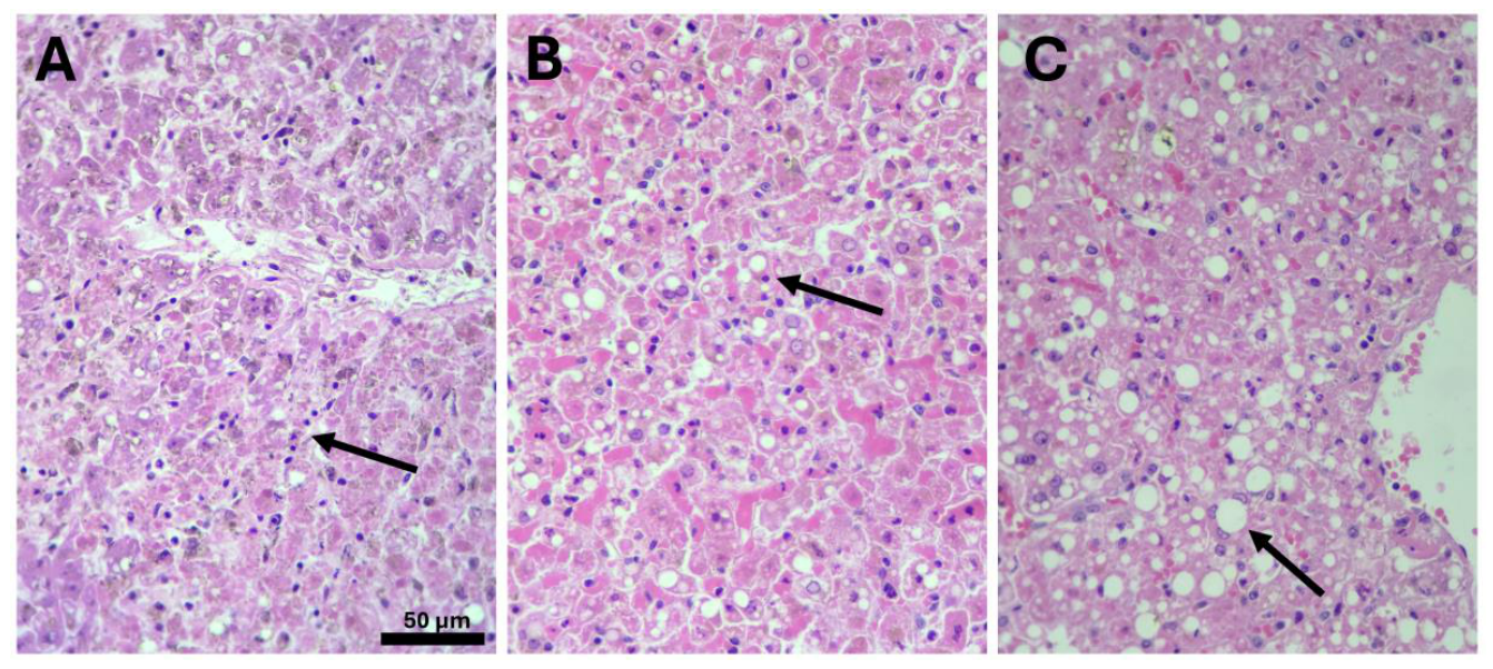
Figure 2.
Representative gross pathological findings in yellow fever virus-infected New World non-human primates. (A) Liver from a woolly monkey (Lagothrix lagotricha), showing hepatic necrosis and marked discoloration consistent with jaundice. (B) Lung from a juvenile white-fronted capuchin monkey (Cebus albifrons), showing prominent interstitial and intra-alveolar edema, generalized vascular congestion, and scattered necrotic foci. (C) Liver from a black-headed spider monkey (Ateles fusciceps), showing marked jaundice, focal vascular congestion, and architectural disruption. (D) Heart from an A. fusciceps, showing yellowish discoloration and generalized vascular congestion consistent with cardiac involvement. (E) Skin and subcutaneous tissue from an A. fusciceps, showing jaundice of the subcutaneous fat and fascial connective tissue. Reproduced from Sanchez-Rojas et al. [12]© 2025 the authors, under the terms of the Creative Commons Attribution (CC BY) License.
Figure 2.
Representative gross pathological findings in yellow fever virus-infected New World non-human primates. (A) Liver from a woolly monkey (Lagothrix lagotricha), showing hepatic necrosis and marked discoloration consistent with jaundice. (B) Lung from a juvenile white-fronted capuchin monkey (Cebus albifrons), showing prominent interstitial and intra-alveolar edema, generalized vascular congestion, and scattered necrotic foci. (C) Liver from a black-headed spider monkey (Ateles fusciceps), showing marked jaundice, focal vascular congestion, and architectural disruption. (D) Heart from an A. fusciceps, showing yellowish discoloration and generalized vascular congestion consistent with cardiac involvement. (E) Skin and subcutaneous tissue from an A. fusciceps, showing jaundice of the subcutaneous fat and fascial connective tissue. Reproduced from Sanchez-Rojas et al. [12]© 2025 the authors, under the terms of the Creative Commons Attribution (CC BY) License.
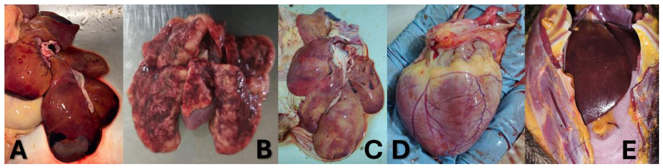

Table 1.
Comparative histopathological, virological, and clinical features of yellow fever in New and Old World non-human primates.
Table 1.
Comparative histopathological, virological, and clinical features of yellow fever in New and Old World non-human primates.
| Comparative domain | Histopathological feature | New World primates | Old World primates |
| Clinical susceptibility and outcomes | Susceptibility to YFV infection | Very high, especially in Alouatta spp. | Moderate to low; frequent relative clinical resistance. |
| Infection-associated mortality | High (up to 80–100% in some epizootics). | Generally low; infections are frequently subclinical or mild. | |
| Acute liver failure | Frequent in fatal infections. | Rare in natural infections. | |
| Hepatic histopathology | Distribution of liver injury | Predominantly extensive, multifocal midzone necrosis. | Mediozonal necrosis present, but usually more limited and focal. |
| Hepatocellular necrosis | Serious, extensive, and frequently confluent. | Mild to moderate; less extensive. | |
| Councilman Bodies | Very abundant and widely distributed. | Present, but in smaller numbers. | |
| Hepatocellular apoptosis | Intense activation of apoptotic pathways. | Lower relative apoptotic intensity. | |
| Hepatic steatosis | Frequent, mainly microvesicular; moderate to severe. | Variable; usually discreet or moderate. | |
| Sinusoidal congestion | Marked and diffuse in fatal cases. | Mild to moderate. | |
| Intrahepatic hemorrhages | Common in severe and terminal infections. | Less frequent and generally focal. | |
| Inflammatory infiltrate | Scarce to moderate; predominantly mononuclear. | Scarce; similar to that observed in neotropical primates. | |
| Kupffer cells | Frequent hyperplasia and hypertrophy. | Moderate activation. | |
| Alteration of lobular architecture | Frequent in advanced cases. | Usually preserved or minimally altered. | |
| Virological and tissue-detection findings | Liver viral load (RT-PCR) | High viral load in liver tissues. | Lower viral load and shorter replication duration. |
| Immunohistochemical detection | Marked immunoreactivity in hepatocytes and Kupffer cells. | Positivity present but generally less extensive. | |
| Viral antigen distribution | Wide distribution in mid-zonal areas and necrotic areas. | More focal and limited distribution. | |
| Host–virus interaction and epidemiological relevance | Host response | Less evolutionary adaptation to YFV; High tissue susceptibility. | Greater evolutionary adaptation and better control of viral replication. |
| Value as an epidemiological sentinel | Very high due to the high mortality. | Lower utility based on mortality; Greater ecological role. |
Table 2.
Extrahepatic histopathological lesions and their pathogenic significance in yellow fever virus infection of New and Old World non-human primates.
Table 2.
Extrahepatic histopathological lesions and their pathogenic significance in yellow fever virus infection of New and Old World non-human primates.
| Organ or system | Principal histopathological findings | Main pathogenic mechanisms | Clinical or functional significance | Comparative pattern and evidence gaps |
| Spleen | White-pulp lymphoid depletion; follicular disorganization; lymphocytolysis and apoptosis; vascular congestion; hemorrhage; follicular necrosis; increased red-pulp phagocytic activity. | Lymphocyte apoptosis, macrophage activation, and viral dissemination through lymphoid tissues. | Immune dysfunction and impairment of the antiviral response. | Usually marked in highly susceptible New World primates, especially Alouatta spp.; African species generally show less severe disease. |
| Lymph nodes and mucosa-associated lymphoid tissue | Cortical lymphocyte depletion; reduced germinal centers; scattered apoptotic bodies; sinus histiocytosis; macrophage activation; reduced lymphoid cellularity. | Infection and activation of dendritic cells and macrophages, with associated lymphocyte depletion. | Disruption of immune homeostasis and facilitation of systemic viral dissemination. | Likely more pronounced in susceptible Neotropical species, but direct comparative evidence remains limited. |
| Kidneys | Acute tubular injury or necrosis; epithelial degeneration; cytoplasmic vacuolization; loss of the brush border; tubular desquamation; granular casts; interstitial edema; vascular congestion. | Tissue hypoperfusion, endothelial injury, hemodynamic disturbance, and possible direct viral infection. | Acute kidney injury and contribution to multiorgan failure. | More extensive and functionally important in susceptible New World primates; usually milder in Old World species. |
| Heart | Myofiber degeneration; interstitial edema; mononuclear inflammatory infiltrates; focal myocytic necrosis; mild-to-moderate myocarditis. | Immune-mediated injury, endothelial dysfunction, and microvascular disturbance; direct cardiomyocyte infection appears limited. | Hemodynamic impairment in advanced or fatal disease. | Comparative species-specific evidence is limited. |
| Lungs | Alveolar edema; diffuse capillary congestion; focal or multifocal hemorrhage; alveolar septal thickening; proteinaceous intra-alveolar material. | Increased vascular permeability, endothelial dysfunction, coagulation abnormalities, and circulatory failure. | Respiratory dysfunction and terminal respiratory failure. | Generally, more pronounced in fulminant New World infections and milder in relatively resistant African species. |
| Central nervous system | Vascular congestion; cerebral edema; petechial hemorrhage; occasional mononuclear perivascular infiltrates. | Systemic circulatory dysfunction, hepatic failure, metabolic abnormalities, and limited direct viral involvement. | Encephalopathy and neurological manifestations in severe disease. | Direct viral-antigen detection is generally limited; comparative data are sparse. |
| Systemic vasculature and coagulation | Multiorgan congestion, edema, petechiae, and focal or multifocal hemorrhages. | Endothelial activation, increased vascular permeability, microcirculatory dysfunction, and coagulopathy. | Pathophysiological link between hepatic injury and pulmonary, renal, cardiac, and neurological damage. | Usually more extensive in highly susceptible species with fulminant infection. |
The intensity and distribution of extrahepatic lesions vary according to species, viral burden, disease stage, and the type and quality of available evidence.
Disclaimer/Publisher’s Note: The statements, opinions and data contained in all publications are solely those of the individual author(s) and contributor(s) and not of MDPI and/or the editor(s). MDPI and/or the editor(s) disclaim responsibility for any injury to people or property resulting from any ideas, methods, instructions or products referred to in the content. |
© 2026 by the authors. Licensee MDPI, Basel, Switzerland. This article is an open access article distributed under the terms and conditions of the Creative Commons Attribution (CC BY) license.
Copyright: This open access article is published under a Creative Commons CC BY 4.0 license, which permit the free download, distribution, and reuse, provided that the author and preprint are cited in any reuse.