

Submitted:
03 October 2026
Posted:
07 October 2026
You are already at the latest version
Abstract
Timely recognition of intraoperative bleeding can support visualization and hemostatic response. This narrative review organizes representative systems by temporal purpose - anticipation, detection, and post-hoc assessment - while treating input sources separately. Video classifiers, object detectors, segmentation-assisted models, and synchronized device - video pipelines demonstrate distinct capabilities, but their reported metrics are not interchangeable. One device-associated detector achieved 81.0% sensitivity with 4.6% precision, corresponding to roughly eight false alerts per operation, whereas other studies evaluated event counts, duration, or warning lead time. We distinguish offline testing, streaming feasibility, and prospective intraoperative evaluation, and relate these evidence levels to event-level accuracy, alert burden, and response time. Current evidence remains limited by small datasets, uncertain transfer across procedures and institutions, workflow integration, and surgeon interaction. Future studies should test transferable contextual cues and determine prospectively whether alerts improve recognition or hemostatic response without excessive interruption.
Keywords:
intraoperative hemorrhage
; bleeding detection
; surgical video
; computer vision
; context-aware AI
; real-time surgical assistance
; clinical translation
1. Introduction
Intraoperative bleeding is a time-critical surgical problem: recognition and control must occur while blood can obscure the operative field and interrupt ongoing dissection. Perioperative mortality studies underscore the clinical importance of hemorrhage and major bleeding complications [1,2]. For surgical assistance, the relevant question is not only how much blood has been lost, but whether an evolving event can be recognized early enough to support the surgical team’s response.
Existing reviews examine blood-loss estimation and hemorrhage prediction [3,4]. Eppler and colleagues synthesize automated detection across several intraoperative adverse events and identify heterogeneous definitions and reporting [5]. For bleeding assistance, a further distinction is essential: recognizing visible blood, predicting an event before onset, and delivering a useful alert are different achievements. Detection scores alone do not establish warning lead time or interruption burden. The anticipatory evidence included here comprises two small studies, underscoring the gap between interest in early warning and its validation [6,7]. This review connects temporal tasks and input sources to validation settings and alert-level endpoints, clarifying what existing results establish for intraoperative use.
We organize recognition by temporal purpose: anticipation before bleeding onset, detection of an ongoing event, and post-hoc assessment of its course or severity. Video-derived context and synchronized device signals are input characteristics within these tasks. Bounding boxes and segmentation are included when they support recognition or alerting; preoperative risk prediction, quantitative blood-loss estimation, detailed localization, and autonomous hemostasis are addressed in companion reviews. Conventional image-processing and severity-grading studies provide methodological and clinical comparators. One simulated suction-planning study is included only to illustrate externally supplied context at the recognition–response interface.
We combined a structured keyword search with citation searching. Three term sets—bleeding or hemorrhage prediction; robotic, open, or intraoperative setting; and deep learning, context-aware, generative, or agentic AI—yielded 24 query combinations, run on 28 April 2026 in Google Scholar and supplemented by PubMed/MEDLINE and Scopus. English-language reports published from 2020 onward were considered. Ninety-eight full-text reports were screened; ten met the core criteria for evaluated systems recognizing imminent or active bleeding from operative video or synchronized signals, including two reports from the same group, and three additional reports served as simulation or adjacent-task comparators. Most exclusions involved nonsurgical hemorrhage, preoperative risk models without operative video, blood-loss quantification, or non-bleeding intraoperative outcomes. The resulting evidence base therefore contains fewer than fifteen primary studies of AI recognition of intraoperative bleeding from operative video or synchronized signals. Preprints were considered when no archival version existed, and reports sharing development data were not treated as independent validation.
The review is intended for surgeons and clinical teams seeking to assess the usefulness and limitations of bleeding alerts, and for AI and surgical-engineering researchers seeking clear recognition targets and appropriate evaluation methods. By connecting task definitions, computational approaches, and deployment barriers, it also supports clinical–technical teams planning validation studies and integration into operative workflows.
2. Clinical and Operational Definitions of Bleeding
2.1. From Blood-Loss Thresholds to Visual Bleeding Events
Clinical grading based on accumulated blood loss addresses a different question from recognition of an event in surgical video [8]. A detector requires an operational definition of what is visible and when it becomes positive. Active flow, blood accumulation, and newly appearing blood after device activation are related but non-equivalent targets. For example, active-bleeding detection can explicitly distinguish flowing blood from pooling, whereas a blood-accumulation classifier may label the resulting collection of blood without establishing that flow continues [9,10]. Table 1 summarizes these distinctions and their annotation consequences.
Intervention-based definitions add clinical meaning but also introduce dependence on surgical practice. Electrosurgical bleeding labels can incorporate whether a hemostatic response is required, while severity grading can relate visible blood to the method used to control it [12,13]. These labels differ from active-flow annotations in both target and dependence on intervention decisions.
2.2. Procedural Context and Clinical Actionability
Bleeding recognition depends on the surrounding action as well as blood appearance. Abrupt instrument movement and electrosurgical activation provide distinct temporal cues, while the current dissection scene helps determine the significance of new blood [6,12]. These cues motivate context-sensitive recognition rather than a general account of surgical risk factors. A system must distinguish a change suggesting new bleeding from persistent staining or a previously recognized pool.
Clinical actionability also depends on whether bleeding compromises visualization or requires a change in operative behavior. Recognition of blood, recognition of active flow, and assessment of the need for intervention therefore represent distinct outputs. Their relationship depends on procedure-specific clinical labels.
2.3. How Often Recognition Fails, and How Long a Response Takes
Reliable estimates of missed or delayed intraoperative bleeding recognition remain scarce. Intraoperative adverse events are underreported when ascertainment relies on retrospective records rather than direct observation [5], although this evidence does not quantify real-time recognition failure by the operating team. Human assessment can also be imprecise: in video review of a cadaveric carotid-injury exercise, fellowship-trained experts underestimated blood loss by a mean of 131 mL and identified fewer than half of episodes exceeding 500 mL [14]. These findings establish a clinically relevant recognition gap without defining its frequency during live surgery.
The available response interval also depends on event severity. In the same simulation, successful management required hemostasis within five minutes [14], whereas Horita and colleagues annotated 254 bleeding events across 27 colectomies, or roughly nine events per operation [9]. Together, these studies illustrate substantial variation in bleeding context and frequency but do not establish a general onset-to-intervention threshold. The clinical value of the 0.662-s advance warning reported by Rahbar and colleagues therefore cannot yet be determined from available human-response data. Evaluations of early-warning systems should align bleeding onset, human recognition, alert delivery, and hemostatic intervention on a common timeline and report event severity alongside alert burden.
3. Bleeding Recognition: Tasks and Methods
Temporal purpose and input source provide separate ways to describe a recognition system. Table 2 places each representative study once, according to its primary task and actual inference inputs. Context extracted from video remains a video input; labels used during training do not constitute an additional runtime modality. Post-hoc assessment connects detected events to operative interpretation. No included recognition study used contextual information supplied outside the video or device stream at inference time; the column is retained to mark that absence.
Figure 1 connects these task distinctions to event matching and alert delivery; the evaluation settings are developed in Section 4.
We use three descriptive evidence levels in Table 3: L1, offline image or video evaluation; L2, an integrated streaming or interactive interface demonstrated on replayed or experimental inputs; and L3, prospective operation during surgery with evaluated outputs. The highest demonstrated setting is listed. These levels describe evaluation setting, not study quality or proven clinical benefit; prospective enrollment alone does not establish L3, and an exploratory L3 study is not a comparative outcome trial.
3.1. Anticipation Before Bleeding Onset
Anticipation predicts bleeding before its independently annotated onset. Rahbar and colleagues used local-entropy changes to identify abrupt tool motion, reporting a mean advance warning of 0.662 s in a small video series [6]. Checcucci and colleagues ran a neural-network alert application during 10 robotic prostatectomies and reviewed its outputs against recorded videos; they reported a next-second event after confidence exceeded 93–95% [7]. The latter provides exploratory intraoperative evidence (L3), although its timing framework was still under development. Together, these studies motivate short-horizon warning without resolving whether the signal reliably precedes visible onset across procedures.
3.2. Detection of Ongoing Bleeding
Video-based methods span classification, spatial detection, segmentation, and temporal event analysis. Horita and colleagues distinguished active flow from pooling with YOLOv7 (AP50 0.574; 48.5 FPS), while Sunakawa and colleagues reported a Dice coefficient of 0.77 for bleeding-region segmentation during hepatectomy [9,11]. Marullo and colleagues combined blood-accumulation classification with tool segmentation, and BLAIR extended video recognition to a replay interface evaluated against a urologist’s recognition times [10,16]. Earlier recognition relative to an observer remains distinct from prediction before independently annotated onset. Rus and colleagues additionally demonstrated an augmented-reality route for displaying detections [17].
Temporal models can summarize event course as well as presence. Hong and colleagues evaluated entire-video bleeding counts and duration, reporting three-fold mean absolute errors of 12.3–13.2 events and 5.09–5.83 min across two AMAGI variants [15]. These summaries do not identify which events were missed or when an alert would arrive.
Synchronized device signals can narrow the detection interval. Ribbens and colleagues combined video with generator activation times and reported fragment-level sensitivity of 81.0% with 4.6% precision [12]. Section 5 translates this precision into alert burden. Temporal association with device activation provides context for detection but does not itself establish causation.
3.3. Post-Hoc Assessment and Clinical Context
Post-hoc assessment relates recognized bleeding to its severity or management. Sawa and colleagues linked a red-pixel index to hemostatic management during robot-assisted pancreatoduodenectomy, analyzing 885 bleeding scenes and four conversion scenes [13]. This exploratory grading scheme is an operational comparator, rather than a learned detector. Hemostatic actions supply reference labels; they are not an additional model input. Such work helps define clinically meaningful targets, while transfer to other procedures remains an open question.
4. Evaluation and Reporting Requirements
4.1. Data, Annotation, and Validation
Dataset size requires counts of independent operations as well as images, clips, and events: adjacent frames are not independent clinical observations. Simulated resources such as SOCAL support method development, but validation of instrument detection or outcome prediction on these data does not establish clinical bleeding-alert performance [19]. Patient- or operation-level partitions reduce leakage; external testing examines transfer beyond the development setting. Confidence intervals need to account for clustering within operations.
Reproducible evaluation requires an explicit target, onset and offset rules, annotator expertise, disagreement resolution, and treatment of obscured intervals. Complete non-bleeding periods matter because selected bleeding clips can conceal false-alert burden. The analysis unit and test prevalence accompany each result, especially when comparing frames, activation fragments, and complete events.
4.2. From Image Scores to Event-Level Performance
Sensitivity and precision describe threshold-specific classification, AP50 bounding-box detection, and Dice or intersection over union (IoU) spatial overlap. AUROC and area under the precision–recall curve (AUPRC) summarize threshold ranges but do not define an acceptable alert threshold. Count and duration errors provide operative summaries, while low precision in device-associated detection shows why sensitivity alone can misrepresent alert utility [12,15]. Appendix A distinguishes reported measures from the review’s proposed priorities.
Event-level evaluation requires explicit onset, matching, and deduplication rules. Figure 1A distinguishes pre-onset warning lead time from post-onset detection delay. Event sensitivity should be paired with false alerts per operative hour, because repeat notifications may count as one matched event yet still interrupt the team. Comparisons therefore require common event rules, analysis units, and test prevalence.
4.3. Timing and the Delivered Alert
Figure 1B separates model throughput (FPS) from end-to-end alert latency. System latency spans capture to display, including buffering, processing, any future-frame waiting, and alert-handling delays; surgeon interpretation is separate. Thresholds, persistence, and suppression determine which outputs reach the display and when. Historical context does not necessarily add steady-state waiting, whereas future-frame dependence does. Horita and colleagues identified display delay as unevaluated despite fast inference [9]. Hardware, resolution, temporal dependencies, and alert-handling rules therefore accompany timing results.
5. Clinical Translation and Barriers to Deployment
5.1. What the Current Evidence Supports
The evidence levels in Table 3 separate recognition tests from interface demonstrations and intraoperative use. BLAIR provides replay-based evaluation, while the earlier Checcucci alert study reports prospective operation during surgery [7,16]. These settings support different claims. Neither an L3 designation nor favorable surgeon ratings establish reduced blood loss, faster hemostasis, or fewer complications without an appropriate clinical comparison.
5.2. Data and Generalization Barriers
Changes in camera appearance, instruments, bleeding prevalence, and hemostatic practice can alter both model inputs and the meaning of a positive label. Horita and colleagues identified procedure-dependent backgrounds and limited representation of severe bleeding as constraints on transfer [9]. External validation therefore needs routine and difficult cases from independent settings, with explicit separation of visual domain shift from differences in what clinicians label actionable. Local threshold adjustment cannot establish reliability for bleeding patterns absent from development data.
5.3. Technical and Workflow Integration Barriers
Deployment requires stable video access, synchronized auxiliary signals when used, and interpretable output at the point of care. In activation-associated detection, generator availability and timestamp alignment determine which video interval is examined [12]. Systems should communicate unavailable or unassessable states when inputs fail or image quality deteriorates because of smoke, contamination, occlusion, camera movement, or interrupted capture. Interface placement must also balance visibility with distraction and anatomical obstruction. These requirements make acquisition reliability, failure communication, and interface design part of clinical integration.
5.4. Human Factors and False-Alert Burden
Checcucci’s study reported a median of 85 anticipated bleeding episodes per patient (IQR 70–90) and a 3% false-positive percentage over a median operative time of 94 min, approximately one warning per minute [7]. The report does not provide the denominator or deduplication rule needed to interpret these figures. If 3% refers to distinct warnings, the implied number of genuine precursors is unusually high; if repeated warnings mark the same event, the reported percentage does not describe alert reliability. The study therefore establishes intraoperative feasibility more clearly than an interpretable alert rate.
Ribbens and colleagues analyzed 2,623 activation fragments from 45 operations, including 21 device-induced bleeding events [12]. At 81.0% sensitivity and 4.6% precision, approximately 17 true events would be detected alongside roughly 350 false positive classifications, or about eight per operation. Because analysis already excluded most non-activation periods, continuous deployment could face a larger alert denominator. This burden is more clinically informative than sensitivity alone.
Surgeon-rated overdetection provides a complementary measure of how outputs are perceived [9]. Alert persistence, dismissal, and meaning should fit the operative task and distinguish visible blood from a recommendation to intervene. Prospective interface studies should therefore evaluate interruption, learning, overreliance, and responses to failure in addition to model discrimination.
5.5. Regulatory Pathway
An intraoperative bleeding alert is software as a medical device, subject to US Food and Drug Administration oversight. The evidence levels used here map onto that framework only partially. L1 offline performance supports analytical validation but no clinical claim; L2 streaming demonstrations address integration rather than clinical validity; L3 prospective operation approaches submission-grade evidence only when intended use, alert threshold, and reference standard are fixed in advance rather than interpreted afterward. The claim itself sets the burden: a system that marks visible blood is a narrower claim than one indicating a need for hemostasis, and a narrower claim is easier to substantiate and less useful. Because retraining and site adaptation change performance, a predetermined change-control plan carries as much weight as the initial evaluation. Teams should therefore settle the intended-use statement before designing validation, since an exploratory study reinterpreted after the fact rarely satisfies reviewers or regulators.
6. Future Directions
6.1. Transferable Context and Precursors
The key question is which contextual inputs improve recognition beyond blood appearance. A direct test would hold the detector, dataset, and event definition fixed, add instrument activity, temporal history, operative phase, or synchronized signals one at a time, and compare event sensitivity with false alerts per operative hour. Inputs that do not improve this trade-off add complexity without demonstrated value. Externally supplied context remains largely untested for recognition; in simulated response planning, Zargarzadeh and colleagues combined a scene image with human-provided pool conditions and textual priority rules [20]. Cross-procedure testing can further distinguish transferable bleeding precursors from procedure-specific motion patterns, while surgeon disagreement about actionability can be modeled as uncertainty rather than forced into a binary label.
6.2. Shared Resources and Adaptation
A useful first benchmark is narrower than a general one. A workable specification is a single procedure, complete unedited operations rather than selected bleeding clips, onset and offset annotated by two surgeons under a declared disagreement rule, and a held-out institution fixed in advance. Results reported as event sensitivity and false alerts per operative hour on that set would be comparable across groups, which current reports are not. Controlled-access hosting, in which models are sent to the data and only metrics are returned, permits such comparison where video cannot be released. Open problems include adapting to new cameras or devices with few labels, detecting unfamiliar appearances, and deciding when a system must withhold an unreliable output. These questions extend beyond improving a score on the original dataset.
6.3. Prospective and Surgeon-Centered Studies
Prospective silent-mode studies can characterize live performance before alerts influence behavior, followed by surgeon-visible comparisons that test recognition, response, and interruption burden. Multicenter studies can then assess transfer across operators, procedures, and rare severe events.
Practical design constraints include data-use agreements for operative video, annotation cost for complete non-bleeding intervals, and the ethics of withholding a potentially useful but unvalidated alert. Controlled-access data environments can reduce sharing barriers, while prespecified interim analyses and stopping rules can govern progression from silent to visible deployment. These design choices should be planned with institutional review boards before prospective testing.
6.4. Limitations of This Review
This targeted narrative synthesis includes neither systematic risk-of-bias assessment nor quantitative pooling. Its literature base is small because the field is small, as the screening counts in Section 1 indicate, but selection among eligible reports was purposive and a systematic search might recover additional borderline work. The included studies span heterogeneous targets, datasets, and evaluation units, and related reports may share development data. The evidence levels describe testing settings rather than methodological quality. The review therefore provides a framework for interpreting representative evidence, not a ranking of systems or an estimate of the field’s overall maturity.
7. Conclusion
AI-based video analysis and synchronized signals support bleeding anticipation, detection, and post-hoc assessment at different stages of validation. Separating temporal purpose from input source clarifies what each system recognizes, while explicit evidence levels distinguish offline performance, streaming feasibility, and intraoperative evaluation. Event-level accuracy, false-alert burden, and response time connect technical outputs to clinical use. Generalization, reliable integration, and surgeon interaction remain barriers; prospective comparisons are needed to establish whether alerts improve surgical response.
Author Contributions
Conceptualization, J.S. and A.R.S.T; methodology, J.S.; software, J.S.; validation, J.S., A.R.S.T. and A.S; formal analysis, J.S.; investigation, J.S. and B.N.; resources, B.N.; data curation, J.S.; writing - original draft preparation, J.S., A.R.S.T, and A.S.; writing - review and editing, J.S., A.R.S.T, A.S., Y.C., A.L.N, J.S.T, X.A.C, L.Z., J.Z., R.J.R, and B.N.; visualization, J.S., A.R.S.T., A.S, and S.H.; supervision, B.N.; project administration, J.S. and B.N.; funding acquisition, B.N. All authors have read and agreed to the published version of the manuscript.
Funding
This study was informed by research conducted under grants from the National Institute on Aging, National Institutes of Health: “Closing the Gap Between Cognitive Health Span and Human Lifespan” (Grant No. 5U19AG065169) and
“A Multimodal Wearable Sensor for Early Detection of Cognitive Decline and Remote Monitoring of Cognitive–
Motor Decline Over Time” (Grant No. R44AG084487). These awards supported the authors’ broader program
in AI-enabled physiological monitoring, which informed the methodological framework presented here. This
specific review did not receive direct external funding.
Institutional Review Board Statement
Not applicable.
Informed Consent Statement
Not applicable.
Data Availability Statement
No new data were generated or analyzed for this narrative review. All studies discussed are cited in the reference list.
Conflicts of Interest
B.N. is serving as Editor-in-Chief for DHI. B.N. serves as a consultant for BioSensics LLC, unrelated to the scope of this manuscript. All other authors report no conflicts of interest.
Appendix A. Evaluation Metrics and Reporting Considerations
Table A1 links measures to relevant studies among the existing references. Event sensitivity and false-alert burden are proposed reporting endpoints; the cited studies do not consistently measure them. Reporting considerations summarize this review’s interpretation rather than a published consensus checklist.

Table A1.
Measures relevant to bleeding recognition and alert evaluation. TP, FP, TN, and FN denote true positives, false positives, true negatives, and false negatives at the declared analysis unit.
Table A1.
Measures relevant to bleeding recognition and alert evaluation. TP, FP, TN, and FN denote true positives, false positives, true negatives, and false negatives at the declared analysis unit.
| Measure | Definition / purpose | Reporting consideration |
|---|---|---|
| Sensitivity and specificity [9,12] | and . | Declare threshold and whether units are frames, clips, or events. High frame specificity can coexist with frequent alerts. |
| Precision (PPV) [12,19] | . | Depends on event prevalence and the analysis unit; report with sensitivity. |
| F1 score [15] | . | Omits true negatives and does not incorporate clinical costs or timing. |
| AUROC / AUPRC [12,15] | Area under receiver operating characteristic / precision–recall curves. | AUROC does not establish positive predictive value; AUPRC depends on positive-class prevalence. Neither selects an acceptable alert threshold. |
| AP50 [9] | Average precision using a bounding-box IoU threshold of 0.50. | Specify matching rules and classes; does not measure onset-to-alert delay. |
| Dice [12] | Pixel overlap: . | Identify the segmentation target and aggregation rule; neither establishes clinical actionability. |
| Event sensitivity [9,12,13,15,16] | Proportion of annotated events matched by a valid alert. | Define allowable timing and matching rules, including obscured intervals and repeated alerts. |
| Detection delay / warning lead time [6] | Alert time minus onset for detection; onset minus alert time for anticipation. | Report distributions and missed events separately. Warning must use only past information within a prespecified horizon. |
| False-alert burden [7,12,15] | Unmatched alerts per operative hour or case. | State denominator, threshold, suppression rules, and whether full non-bleeding intervals were evaluated. |
| FPS / Latency [9] | Processing throughput / elapsed time from acquisition to displayed output. | Report hardware, resolution, buffering, temporal context, and display delay; distinguish measured from estimated latency. |
| Surgeon-centered outcomes [7,9] | Recognition time, interruption burden, usability, and responses to errors. | Specify comparator, task, setting, and number of raters; acceptance does not establish clinical benefit. |
References
- Nuttall, G.A.; Merren, M.P.; Naranjo, J.; Portner, E.R.; Ambrose, A.R.; Rihal, C.S. Perioperative mortality: a retrospective cohort study of 75,446 noncardiac surgery patients. Mayo Clin. Proc. Innov. Qual. Outcomes 2024, 8, 435–442. [Google Scholar] [CrossRef] [PubMed]
- Spence, J.; LeManach, Y.; Chan, M.T.V.; et al. Association between complications and death within 30 days after noncardiac surgery. CMAJ 2019, 191, E830–E837. [Google Scholar] [CrossRef] [PubMed]
- Lin, Y.M.; Yu, C.; Xian, G.Z. Calculation methods for intraoperative blood loss: a literature review. BMC Surg. 2024, 24, 394. [Google Scholar] [CrossRef] [PubMed]
- Yan, S.; Zhang, P.; Qiao, W.; Xie, S.; Hu, H.; Gao, Y.; Xie, L.; Jing, J. Machine Learning for Intraoperative Bleeding Prediction in Patients Undergoing Surgery: Scoping Review. JMIR Med. Inform. 2026, 14, e80930. [Google Scholar] [CrossRef] [PubMed]
- Eppler, M.B.; Sayegh, A.S.; Maas, M.; Venkat, A.; Hemal, S.; Desai, M.M.; Hung, A.J.; Grantcharov, T.; Cacciamani, G.E.; Goldenberg, M.G. Automated capture of intraoperative adverse events using artificial intelligence: a systematic review and meta-analysis. J. Clin. Med. 2023, 12, 1687. [Google Scholar] [CrossRef] [PubMed]
- Daneshgar Rahbar, M.; Ying, H.; Pandya, A. Visual intelligence: Prediction of unintentional surgical-tool-induced bleeding during robotic and laparoscopic surgery. Robotics 2021, 10, 37. [Google Scholar] [CrossRef]
- Checcucci, E.; De Cillis, S.; Amparore, D.; Gabriele, V.; Piramide, F.; Piana, A.; Fiori, C.; Piazzolla, P.; Porpiglia, F. Artificial Intelligence alert systems during robotic surgery: a new potential tool to improve the safety of the intervention. Urol. Video J. 2023, 18, 100221. [Google Scholar] [CrossRef]
- Perri, G.; Sparrelid, E.; Siriwardena, A.K.; Marchegiani, G. Estimation of intraoperative blood loss in hepatopancreatobiliary surgery: a Delphi consensus process of the European–African Hepato-Pancreato-Biliary Association (E-AHPBA). Br. J. Surg. 2024, 111, znae256. [Google Scholar] [CrossRef] [PubMed]
- Horita, K.; Hida, K.; Itatani, Y.; Fujita, H.; Hidaka, Y.; Yamamoto, G.; Ito, M.; Obama, K. Real-time detection of active bleeding in laparoscopic colectomy using artificial intelligence. Surg. Endosc. 2024, 38, 3461–3469. [Google Scholar] [CrossRef] [PubMed]
- Marullo, G.; Tanzi, L.; Ulrich, L.; Porpiglia, F.; Vezzetti, E. A multi-task convolutional neural network for semantic segmentation and event detection in laparoscopic surgery. J. Pers. Med. 2023, 13, 413. [Google Scholar] [CrossRef] [PubMed]
- Sunakawa, T.; Kitaguchi, D.; Kobayashi, S.; Aoki, K.; Kujiraoka, M.; Sasaki, K.; Azuma, L.; Yamada, A.; Kudo, M.; Sugimoto, M.; et al. Deep learning-based automatic bleeding recognition during liver resection in laparoscopic hepatectomy. Surg. Endosc. 2024, 38, 7656–7662. [Google Scholar] [CrossRef] [PubMed]
- Ribbens, V.J.; Baltus, S.C.; Tan, C.O.; Broeders, I.A. Machine learning-based detection of electrosurgical device-induced bleeding in laparoscopic videos of diaphragmatic hernia repair. Surg. Endosc. 2025, 39, 6230–6238. [Google Scholar] [CrossRef] [PubMed]
- Sawa, Y.; Inoue, Y.; Kiritani, S.; Kobayashi, K.; Oba, A.; Ono, Y.; Ito, H.; Takahashi, Y. Proposal for grading of intraoperative bleeding and the assessment of hemostasis quality during robot-assisted pancreatoduodenectomy. Surg. Endosc. 2025, 39, 8754–8761. [Google Scholar] [CrossRef] [PubMed]
- Pangal, D.J.; Kugener, G.; Zhu, Y.; Sinha, A.; Unadkat, V.; Cote, D.J.; Strickland, B.; Rutkowski, M.; Hung, A.; Anandkumar, A.; et al. Expert surgeons and deep learning models can predict the outcome of surgical hemorrhage from 1 min of video. Sci. Rep. 2022, 12, 8137. [Google Scholar] [CrossRef] [PubMed]
- Hong, S.; Hong, S.; Jang, J.; Kim, K.; Hyung, W.J.; Choi, M.K. Amplifying action-context greater: image segmentation-guided intraoperative active bleeding detection. Comput. Methods Biomech. Biomed. Eng. Imaging Vis. 2023, 11, 1261–1270. [Google Scholar] [CrossRef]
- Checcucci, E.; Piazzolla, P.; Marullo, G.; Innocente, C.; Salerno, F.; Ulrich, L.; Moos, S.; Quarà, A.; Volpi, G.; Amparore, D.; et al. Development of bleeding artificial intelligence detector (BLAIR) system for robotic radical prostatectomy. J. Clin. Med. 2023, 12, 7355. [Google Scholar] [CrossRef] [PubMed]
- Rus, G.; Andras, I.; Vaida, C.; Crisan, N.; Gherman, B.; Radu, C.; Tucan, P.; Iakab, S.; Hajjar, N.A.; Pisla, D. Artificial intelligence-based hazard detection in robotic-assisted single-incision oncologic surgery. Cancers 2023, 15, 3387. [Google Scholar] [CrossRef] [PubMed]
- Kugener, G.; Zhu, Y.; Pangal, D.J.; Sinha, A.; Markarian, N.; Roshannai, A.; Chan, J.; Anandkumar, A.; Hung, A.J.; Wrobel, B.B.; et al. Deep neural networks can accurately detect blood loss and hemorrhage control task success from video. Neurosurgery 2022, 90, 823–829. [Google Scholar] [CrossRef] [PubMed]
- Kugener, G.; Pangal, D.J.; Cardinal, T.; Collet, C.; Lechtholz-Zey, E.; Lasky, S.; Sundaram, S.; Markarian, N.; Zhu, Y.; Roshannai, A.; et al. Utility of the simulated outcomes following carotid artery laceration video data set for machine learning applications. JAMA Netw. Open 2022, 5, e223177. [Google Scholar] [CrossRef] [PubMed]
- Zargarzadeh, S.; Mirzaei, M.; Ou, Y.; Tavakoli, M. From Decision to Action in Surgical Autonomy: Multi-Modal Large Language Models for Robot-Assisted Blood Suction. IEEE Robot. Autom. Lett. 2025, 10, 2598–2605. [Google Scholar] [CrossRef]
Figure 1.
Event matching and alert delivery in bleeding recognition. (A) A shared timeline illustrates early warnings, detection delay, unmatched alerts, and repeated notifications relative to annotated bleeding events. (B) The workflow connects capture, recognition, alert handling, and display to surgeon interpretation, distinguishing model throughput from end-to-end alert latency. Conceptual illustration; no empirical data.
Figure 1.
Event matching and alert delivery in bleeding recognition. (A) A shared timeline illustrates early warnings, detection delay, unmatched alerts, and repeated notifications relative to annotated bleeding events. (B) The workflow connects capture, recognition, alert handling, and display to surgeon interpretation, distinguishing model throughput from end-to-end alert latency. Conceptual illustration; no empirical data.
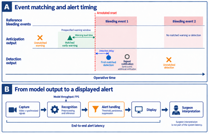
Table 1.
Operational bleeding targets and their implications for annotation. These targets may overlap but are not interchangeable.
Table 1.
Operational bleeding targets and their implications for annotation. These targets may overlap but are not interchangeable.
| Target | Positive finding | Annotation consideration |
|---|---|---|
| Accumulated blood loss | Estimated or measured volume lost over the operation [3,8]. | A whole-operation quantity, not a frame- or event-level label; estimation methods are not standardized, so thresholds do not transfer. |
| Active bleeding | Visible ongoing flow, distinguished from pooling [9]. | Define onset and how occlusion or uncertain flow is labeled. |
| Blood accumulation | Visible collection of blood in the operative scene [10]. | Accumulation does not necessarily imply continuing flow; define the classification unit. |
| Bleeding region | Pixels showing bleeding during liver transection [11]. | State what the region includes; overlap accuracy is distinct from event recognition. |
| Device-associated bleeding | New blood around a recorded activation, with study-specific hemostatic criteria [12]. | Specify the observation window and distinguish temporal association from causal proof. |
| Severity-linked bleeding | Image-derived severity related to hemostatic management [13]. | Treatment choice and procedure affect labels; thresholds require external validation. |
Table 2.
Temporal tasks and inference inputs of representative studies, with an adjacent response-planning example shown separately. Video-derived context remains in the visual-only column. Supplied context denotes additional human-provided information or rules, not training labels. Blank cells indicate no matching study in the literature reviewed here.
Table 2.
Temporal tasks and inference inputs of representative studies, with an adjacent response-planning example shown separately. Video-derived context remains in the visual-only column. Supplied context denotes additional human-provided information or rules, not training labels. Blank cells indicate no matching study in the literature reviewed here.
| Task | Images/video only | Video + device signals | Images/video + supplied context |
|---|---|---|---|
| Anticipation | Rahbar [6]; Checcucci (alert study) [7] | ||
| Detection | Horita [9]; Sunakawa [11]; Hong [15]; Marullo [10]; Checcucci (BLAIR) [16]; Rus [17] | Ribbens [12]: synchronized generator activations | |
| Post-hoc assessment | Sawa (grading comparator) [13]; Kugener [18] and Pangal [14], simulation comparators using the SOCAL dataset [19] |
Table 3.
Representative studies by result, evaluation unit, and evidence level. L1: offline evaluation; L2: integrated streaming or interactive interface demonstration on replayed or experimental inputs; L3: prospective intraoperative operation with evaluated outputs. Levels describe the highest demonstrated setting, not study quality or clinical benefit. Results across different targets are not a performance ranking. The final three rows are comparators rather than clinical bleeding-alert evidence: two evaluate simulated hemorrhage and one addresses response planning.
Table 3.
Representative studies by result, evaluation unit, and evidence level. L1: offline evaluation; L2: integrated streaming or interactive interface demonstration on replayed or experimental inputs; L3: prospective intraoperative operation with evaluated outputs. Levels describe the highest demonstrated setting, not study quality or clinical benefit. Results across different targets are not a performance ranking. The final three rows are comparators rather than clinical bleeding-alert evidence: two evaluate simulated hemorrhage and one addresses response planning.
| Study / task | Data and evaluation | Method / output | Result / evaluation unit | Level |
|---|---|---|---|---|
| Rahbar, 2021 [6]Anticipation | 17 videos; 11 with tool-induced bleeding. | Local-entropy changes identify abrupt tool motion. | Accuracy 88%; precision 90%; mean lead time 0.662 s in the timing analysis. | L1 |
| Checcucci, 2023 [7]Anticipation | 10 prospectively enrolled cases; intraoperative execution and subsequent video review. | Neural-network confidence used for warning. | Reported TP 98%, FP 3%; median 85 warnings per patient (IQR 70–90). Percentage denominators incompletely specified. | L3 |
| Checcucci, 2023 [16]Detection (BLAIR) | 10 recorded prostatectomies; replay compared with urologist annotations. | Multitask CNN and blood-accumulation interface. | Bleeding identification and timing compared with a human observer on replay. | L2 |
| Horita, 2024 [9]Detection | 27 colectomy videos; 34,117 images, 254 events; 20 surgeon raters. | YOLOv7 boxes for active bleeding and pooling. | Box AP50 0.574; 48.5 FPS; surgeon ratings of inference videos. | L1 |
| Sunakawa, 2024 [11]Detection | 2,203 images from 44 hepatectomy videos. | Bleeding-region segmentation. | Region precision 0.76; recall 0.79; Dice 0.77. | L1 |
| Hong, 2023 [15]Detection | 40 robotic gastrectomies; three-fold evaluation. | Spatial guidance with temporal recognition. | Whole-video count MAE 12.3–13.2 events; duration MAE 5.09–5.83 min across two variants. | L1 |
| Marullo, 2023 [10]Detection | Video-derived images and video tests. | Joint blood-accumulation classification and tool segmentation. | Image-level event accuracy 90.63%; tool-mask Dice 81.89%. | L1 |
| Rus, 2023 [17]Detection | Experimental hazard-detection interface. | YOLO detection and HoloLens overlay. | Streaming detection-to-overlay implementation. | L2 |
| Ribbens, 2025 [12]Detection | 45 patients; 34 images and 2,678 activation fragments, of which 2,623 were analyzed and 21 annotated as bleeding. | Blood-pixel random forest; activation-associated logistic regression. | Fragment sensitivity 81.0%; precision 4.6%; accuracy 78.2%. | L1 |
| Sawa, 2025 [13]Post-hoc comparator | 885 bleeding scenes and four conversion scenes. | Red-pixel index and management-based grading. | Scene-level association of visual severity with hemostatic management. | L1 |
| Kugener, 2022 [18]Post-hoc, simulation | 143 cadaveric-simulator trials of carotid-injury control (SOCAL) [19]. | Deep network predicting blood loss and task outcome from video. | Blood-loss RMSE 358 mL from video alone and 260 mL with instrument annotations; outcome accuracy 85–90%. | L1 |
| Pangal, 2022 [14]Post-hoc, simulation | 20 held-out SOCAL videos; four blinded expert raters given the first 60 s. | Model and expert prediction of outcome and blood loss. | Model 17/20 versus experts 14/20 correct; experts underestimated blood loss by 131 mL on average. | L1 |
| Zargarzadeh, 2025 [20]Response planning, adjacent | Simulated blood-suction scenes. | Multimodal language model ordering suction from a scene image with supplied pool conditions and priority rules. | Decision-level ordering in simulation; no bleeding-recognition metric reported. | L1 |
Disclaimer/Publisher’s Note: The statements, opinions and data contained in all publications are solely those of the individual author(s) and contributor(s) and not of MDPI and/or the editor(s). MDPI and/or the editor(s) disclaim responsibility for any injury to people or property resulting from any ideas, methods, instructions or products referred to in the content. |
© 2026 by the authors. Licensee MDPI, Basel, Switzerland. This article is an open access article distributed under the terms and conditions of the Creative Commons Attribution (CC BY) license (http://creativecommons.org/licenses/by/4.0/).
Copyright: This open access article is published under a Creative Commons CC BY 4.0 license, which permit the free download, distribution, and reuse, provided that the author and preprint are cited in any reuse.