

Submitted:
02 October 2026
Posted:
07 October 2026
You are already at the latest version
Abstract
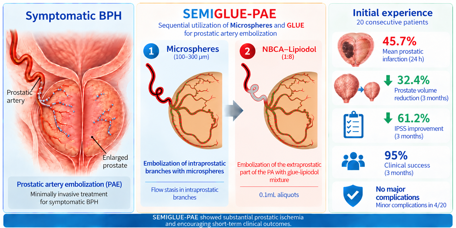
Background/Objectives: Prostatic artery embolization (PAE) with n-butyl cyanoacrylate (NBCA) glue-lipiodol mixture has several advantages over PAE with microspheres for the treatment of symptomatic benign prostatic hyperplasia (BPH). However, distal penetration of glue-lipiodol in intraprostatic vasculature is often not possible and proximal-only prostatic artery (PA) occlusion with glue may fail to induce significant prostatic infarction. To mitigate this limitation, the authors applied and evaluated SEquential utilization of MIcrospheres and GLUE for PAE (SEMIGLUE-PAE). Meth-ods: For SEMIGLUE-PAE, embolization began with microspheres (100-300μm) with the microcatheter tip at the distal extraprostatic part of the PA; intraprostatic advancement of the microcatheter and injection of additional microspheres was then at-tempted. When flow stasis in intraprostatic branches was observed, embolization continued with small aliquots (0.1ml) of glue-lipiodol mixture (1:8) with the same initial position of microcatheter tip, until the distal one half (to two thirds) of the extraprostatic PA was filled with the mixture. SEMIGLUE-PAE was applied in 20 consecutive patients with symptomatic BPH. Short-term improvement in imaging and clinical parameters and safety were evaluated. Results: Clinical success rate was 95%, 90% and 83.6% at 3, 6 and 12 months post SEMIGLUE-PAE, respectively. Three months post SEMIGLUE-PAE, mean reduction of prostate volume was 32.4% (p< 0.01) and mean improvement of international prostate symptom score was 61.2% (p< 0.01). Mean percentage of prostatic infarction (measured within 24 hours post PAE and compared to baseline PV) was 45.7%. Minor (only) complications were encountered in 4/20 patients. Conclusions: SEMIGLUE-PAE seems to be safe, effective and feasible, especially for operators with limited experience in embolization with glue.
Keywords:
N-butyl cyanoacrylate glue
; lipiodol
; microspheres
; prostatic artery embolization
; infarction
1. Introduction
During the last few years, n-butyl cyanoacrylate (NBCA) glue-lipiodol mixture has emerged as a promising embolic alternative to microparticles for prostatic artery embolization (PAE)[1]. Attractive features of glue-lipiodol as an embolic material for PAE (glue-PAE) include its potential for rapid and possibly durable vascular occlusion and its radiopacity, which enhances radiologic visualization of the embolic mixture during and after the procedure [1,2]. However, glue-PAE is even more technically demanding than the standard PAE procedure and requires significant operator expertise [2]; in addition to the usual limitations associated with glue as an embolic material (microcatheter blockade or microcatheter adherence to arterial wall, glue reflux and irreversible occlusion of non-target arteries, etc), distal penetration of glue into the prostate and adequate coverage of small intraprostatic branches may be extremely difficult and may require significant experience and application of special techniques[3]. Proximal (instead of distal) occlusion of the PA with glue is not likely to cause significant prostatic ischemia [4]; it will rather activate collaterals to the prostate and it will also permanently block subsequent attempts to reach distal prostatic branches with the microcatheter.
To address these challenges of glue-PAE, which may be more pronounced for less experienced operators, the authors applied the “SEquential utilization of MIcrospheres and GLUE” (“SEMIGLUE”) approach for PAE. The primary objective of this study is to describe this approach and to investigate its early clinical outcomes and its safety. The secondary objective is to describe imaging findings relevant to this approach.
2. Materials and Methods
2.1. Study Design and Inclusion and Exclusion Criteria
A retrospective review of the first patients who received SEMIGLUE-PAE for symptomatic BPH at a tertiary center (from March 2025 to November 2025) was conducted. Inclusion criteria were [5]: Moderate or severe lower urinary tract symptoms (International Prostate Symptom Score, IPSS≥8); Quality of Life (QoL) ≥3; failure of medical treatment; chronic urinary retention caused by BPH and managed with indwelling bladder catheter; prostate volume (PV)> 40 mL. Exclusion criteria were: Urinary tract infection; prostate or bladder cancer; neurogenic bladder; large (>2,5 cm) bladder diverticula or bladder stones; contraindications to angiographic procedures; clinical and imaging follow up of less than 3 months.
Written informed consent was obtained from all patients prior to treatment and the study was approved by the institutional review board.
2.2. Pre-Interventional Evaluation
All patients were clinically evaluated prior to treatment and IPSS and QoL scores were recorded. Contrast-enhanced pelvic computed tomography (CT) and CT angiography (CTA) were performed a few days prior to intervention, to evaluate prostate size and morphology, to detect any coexistent pathology and for vascular planning. Prostate volume was calculated with Artificial Intelligence-assisted segmentation and volumetry on axial CT images with dedicated software (“3D Slicer” with “nnInteractive” AI Segmentation addon) [6,7,8].
2.3. PAE Technique
Vascular access was obtained via the common femoral artery and catheterization of the PA was performed in the usual manner. A 1.9 Fr or 1.98 Fr microcatheter (Tellus or Parkway Soft, Asahi Intecc Co.) and a double angle microguidewire (0.016’’ Meister, Asahi, Japan) were the standard combination of devices for PA catheterization. When this combination failed, the standard microcatheter was replaced with a 2.4 Fr steerable microcatheter (Swiftninja, Merit Medical, USA). If possible, the microcatheter was advanced into the distal extraprostatic part of the PA, just proximal to the prostatic capsule. Embolization began from that point, using hydrophilic polymer (Polifit 70®) microspheres with a diameter of 100-300μm (Embosoft, Scitech Medical, Brazil). Distal advancement of the microcatheter in intraprostatic branches and additional injection of microspheres was then applied, if vascular anatomy was favorable. Embolization with microspheres stopped when complete flow stasis in intraprostatic vascular bed was achieved. Thereafter, glue embolization also began with the tip of the microcatheter at the distal extraprostatic part of the PA. An 1/8 glue/lipiodol mixture (Glubran2, GEM, Italy/Lipiodol Ultra Fluid, Guerbet, France) was routinely utilized and small aliquots (0.1ml) were injected with a 1ml syringe with luer-lock. Particularly with smaller PAs, a “blocked flow” technique (with the microcatheter “wedged” in the distal extraprostatic part of the PA) was initially attempted, although this was not aggressively pursued. The microcatheter was then slowly withdrawn while injecting the rest of the glue aliquot. The microcatheter was flushed with 5 ml of nonheparinized 5% dextrose solution before and after each glue injection (“sandwich” technique). Additional 0.1ml aliquots of glue/lipiodol were administered (especially in cases of PAs with a large diameter and a long trajectory), until the distal half (up to two thirds) of the PA were filled with glue. When the distal extraprostatic part of the PA could not be approached, due to unfavorable anatomy or poor support from the base catheter, embolization was performed with “free-flow” technique, with the microcatheter in the proximal part of the PA, first with microspheres and then with glue-lipiodol, with the same respective end-points as previously described.
Almost immediately after the procedure (15-20min after arterial closure) an unenhanced CT scan was applied to show the distribution of the embolic materials within and around the prostate. Radiation parameters (fluoroscopy time-FT and dose-area product-DAP) were recorded after each procedure. SEMIGLUE-PAE was performed by 2 interventional radiologists with approximately 10 years of experience in PAE with microspheres but no previous experience in glue-PAE.
2.4. Clinical Evaluation Post PAE
Diagnosis of clinical success of SEMIGLUE-PAE was based on reduction of IPSS (<18 with a decrease of at least 25% from baseline) and of QoL (≤ 3, with at least an 1 point decrease compared to baseline) or on the successful removal indwelling bladder catheter [5]. Patients were clinically re-evaluated during the first 3-4days post SEMIGLUE-PAE, one and three months later and then every three months. Complications were recorded and graded using the modified (for PAE) Clavien-Dindo system [9].
2.5. Imaging Evaluation Post PAE
Prostate infarction was calculated within 24 hours post PAE, with transabdominal contrast-enhanced ultrasound (CEUS). The volume of each prostatic infarction was calculated using the ellipsoid formula; the volumes of all prostatic infarcts were summed and divided by PV, thus calculating the percentage of Prostatic Infarction (pPI), in line with previous work [10]. Three months post SEMIGLUE-PAE, PV was measured again using CT and the aforementioned dedicated software, and prostate shrinkage was evaluated by comparing PV measurements at baseline and 3 months post-procedure. Post-void residual (PVR) was also measured 3 months post-procedure with transabdominal ultrasound and compared to baseline measurement. Angiographic evaluation with CTA 3 months post procedure (at the same session with the standard CT) and other imaging studies performed after 3 months were optional.
2.6. Statistics
Descriptive statistics were applied for the basic quantitative and qualitative parameters. The Wilcoxon Signed-Rank Test was used to evaluate the significance of the change of the outcome parameters before and after treatment. The Kaplan-Meier method was used to calculate the clinical success rates of SEMIGLUE PAE over time. Statistical significance was defined as a P value of <0.05. Statistical Package for Social Sciences (Version 26.0, SPSS Inc., Chicago, Illinois, USA) was used for the statistical analysis.
3. Results
3.1. Basic Population Features and Technical Outcomes
The “SEMIGLUE” approach was applied in 23 consecutive patients. Three of them did not return for the 3-month evaluation and were excluded; therefore, 20 patients were eventually studied (Figure 1, Table 1). Bilateral PAE was performed in 18/20 patients and unilateral PAE in 2 patients (totally, 38 embolized hemiprostates). From a total of 46 PAs (2.3 PAs per patient), 42 PAs were embolized with the aforementioned technique. Regarding procedural radiation parameters, mean FT was 41.8 ± 13.0 min and mean DAP was 22990 ± 11910μGy·m2.
3.2. Clinical Outcomes
Clinical success rate was 95%, 90% and 83.6% at 3, 6 and 12 months post SEMIGLUE-PAE, respectively (Figure 2). Short-term improvement of imaging and clinical parameters was statistically significant, with 32.4% mean reduction of PV and 61.2% mean improvement of IPSS (Table 2).
The majority of the patients (85%) experienced post embolization syndrome and 4/20 patients (20%) experienced minor complications: There were 2 grade I complications (urinary incontinence for 1.5-2 weeks, n=2) and 2 grade II complications (acute urinary retention managed with bladder catheter for 5-7 days, n=2). No major complication was encountered.
3.3. Imaging Outcomes
Non-enhanced CT (performed immediately post PAE in all 20 patients) showed hyperdensities of varying extent in both prostatic lobes in 18/20 patients (36/38 embolized hemiprostates), indicating intraprostatic retention of the contrast-microspheres mixture. Hyperdensities in only 1 prostatic lobe was noted in the 2 patients with unilateral embolization. Areas of dense intraprostatic retention corresponded to ischemic infarcts, as demonstrated by contrast-enhanced ultrasonography (CEUS), which was performed within 24 hours post PAE (Figure 3).
Mean percentage of prostatic infarction was 45.7%. In 2 cases, intraprostatic retention took the form of dense contrast “pooling” in the treated lobe and this finding was associated with long-standing necrosis (Figure 4).
Non-enhanced CT immediately post PAE, also confirmed opacification (with the glue-lipiodol mixture) of the distal extraprostatic part of the embolized PAs in all cases. Limited distribution of glue in few intraprostatic branches was additionally noted in 28/38 embolized hemiprostates. Notably, in 2 cases of duplicate PA in the same pelvic side, glue injection in the first PA was rapidly followed by reflux of glue in the second ipsilateral PA. In this way, both PAs of the same side were embolized (one antegradely and the other retrogradely) with only one catheterization (Figure 5).
Three months post SEMIGLUE-PAE, all 20 patients underwent follow-up with CT (with additional CTA in 8 patients). CT and CTA confirmed occlusion and pooling of the glue-lipiodol mixture in the distal extraprostatic part of all the studied PAs. Absence of significant enhancement was also observed in the proximal extraprostatic part in the majority of the studied PAs (Table 3, Figure 6).
4. Discussion
Prostatic ischemia is considered as one of the main mechanisms of the therapeutic action of PAE; ischemic infarcts, which are demonstrated on contrast-enhanced studies shortly post PAE can predict clinical success and prostate shrinkage [10,11,12]. The potential of the smaller microspheres (<=300μm) for deep penetration in the intraprostatic prostatic branches and for induction of extensive ischemia has been repeatedly demonstrated, both with imaging and histologically; there is also relevant clinical evidence [13,14]. Regarding glue-PAE, the clinical benefits of distal, intraprostatic distribution of glue are less clear [15]. Of course, adequate delivery of glue-lipiodol mixture in distal parenchymal arteries can be achieved, but most researchers agree that it requires a high level of expertise and/or advanced embolization techniques [2,3]. Moreover, the ischemic effects of glue-PAE on prostate have not been thoroughly evaluated, although there is evidence that proximal-only glue PAE cannot induce significant prostatic infarction [4].
The SEMIGLUE approach seeks to combine the advantages of microspheres and glue. A synergistic effect between the two embolic agents can be theoretically expected; the rapid and complete blockade of the extraprostatic PA with glue could enhance the ischemic effect of the microspheres and prevent early reperfusion and collateralization. This is probably supported by the CT finding of intraprostatic contrast pooling (not just contrast retention, as previously described after microsphere-PAE) [16] and by the associated persistent prostatic infarction. Moreover, the extent prostatic infarction post SEMIGLUE-PAE (pPI:45.7% in the present study) was significantly higher compared to PAE with microspheres of the same size (pPI:30.3%), almost reaching the ischemic effect of smaller (40-120μm) microspheres (pPI:48.3%) [13]. Regarding other imaging outcomes, although the herein presented results are very early and derived from a small group of patients, they also appear to be comparable, or even better than the respective outcomes of glue (alone)- or microsphere (alone)-PAE: Prostate shrinkage 3 months post SEMIGLUE-PAE reached 32.4%, versus only 11%-21% for glue-PAE alone [1] and 28.4% for PAE with microspheres of comparable size [13]. An original feature of the present study was the re-evaluation of PA patency 3 months post SEMIGLUE-PAE in a subgroup of the treated patients: It was confirmed that occlusion persisted in the distal extraprostatic part in all treated PAs and in the proximal part of most of them. This could be an indication of the efficacy of extraprostatic PA occlusion with glue and deserves long-term evaluation.
Clinical outcomes of SEMIGLUE-PAE (with short term improvement of IPSS, QoL and PVR exceeding 55%), are also satisfactory and comparable to PAE with either glue, or microspheres. Notably, in the present study, IPSS reduction reached 14.7 units at 3 months post SEMIGLUE-PAE; corresponding short-term reduction of IPSS post glue-PAE (from experienced operators) and post microsphere-PAE (with 100-300μm microspheres) was 10.6-11.3 units and 13.8 units, respectively [2,13].
The herein presented approach requires no advanced maneuvers for distal penetration of the glue; therefore, it is suitable for operators who have recently incorporated this liquid agent in their PAE practice and are not familiar with such maneuvers. Moreover, after injection of microspheres, glue may still be capable of reaching some distal branches and anastomoses that have escaped occlusion with microspheres. This is supported by the cases of simultaneous embolization of duplicated ipsilateral PAs after glue injection in only one of them.
Safety profile of SEMIGLUE-PAE appears to be satisfactory, with no major complications. Particularly the absence of non-target embolization events (caused by reflux of the embolic in non-prostatic branches) could be attributed to: i) the delivery of microspheres mainly in intraprostatic branches and ii) the administration of small aliquots of glue-lipiodol, far from the PA origin.
Previous experience with combination of liquid embolic and microspheres for PAE is very limited. In a study on repeat PAE, ethylene-vinyl alcohol copolymer (EVOH) was injected after 300-500μm microspheres, and resulted in significant short-term clinical improvement, moderate (11.3%) reduction of PV and 1 major complication (3.3%) [17]. The relatively larger size of microspheres in this study (300-500μm vs 100-300 μm in the current study) could have been the cause of the moderate prostate shrinkage. In a case report describing PAE in the context of a rare PA variant, NBCA-glue was successfully combined with hydrogel microspheres (250μm); significant prostate shrinkage (31.4%) and prostatic infarction were evident on magnetic resonance one month post PAE. Further technical details regarding utilization of the embolic materials from this case are not available [18]. There is also sparse evidence on the utilization of the “Embosoft” microsphere for PAE. In an earlier comparative study [19], PAE with “Embosoft 300-500” provided significant symptomatic improvement, although it was inferior to open simple prostatectomy in relieving bladder outlet obstruction. Notably, short-term PV reduction (20.9%) in this study was inferior to the herein reported result. Utilization of Embosoft 100-300 has been previously reported in a salvage case of PAE with rapid control of hematuria of prostatic origin [20].
At least two disadvantages of SEMIGLUE-PAE should be taken into account: First, since it combines two embolic agents, it is associated with higher cost than PAE with either glue, or microspheres. Second, the injection of the embolics requires significantly more time compared to glue-PAE, resulting in increased FT and RD. Indeed, FT (41.8 min)in the current series was significantly longer compared to glue-PAE studies (24.9-32.6 min) [1]; radiation dose was also significantly higher in SEMIGLUE-PAE compared to glue-PAE [1].
A major limitation of the present work is that it is a small, single-center study, with only short-term results. Moreover, some potentially important outcomes were not included in the study; for example uroflowmetry measurements were not systematically performed, neither was erectile function evaluated.
5. Conclusions
It is recognized that “SEMIGLUE” approach is not a novel approach, but rather a combination of two established techniques. Nevertheless, and despite the aforementioned disadvantages, “SEMIGLUE” PAE can easily induce prostatic ischemia and yield good short-term clinical results with high safety and with no need for advanced embolization techniques. The long-term efficacy of this technique and its place in the personalized, minimally invasive management of symptomatic BPH probably deserve further evaluation.
Author Contributions
“Conceptualization, H.M. and E.H; methodology, H.M. and E.H.; software H.M. and E.H; validation A.H.; Formal analysis, A.M., E.H. and H.M.; investigation, H.M. and E.H.; resources, K.S. and M.I.; data curation, E.H. and M-TH.M.; writing—original draft preparation, H.M.; writing—review and editing, H.M. and E.H.; supervision, H.M.; visualization, E.H., D.K. and M-TH.M; Supervision, H.M.; project administration, H.M.; graphical abstract, E.H. and M-TH.M.; funding acquisition, - . All authors have read and agreed to the published version of the manuscript.”.
Funding
This research received no external funding.
Institutional Review Board Statement
The study was conducted in accordance with the Declaration of Helsinki and approved by the Institutional Review Board (or Ethics Committee) of G.H.P.T. (protocol code (decision number) 5, approved on 24/02/2026). .
Informed Consent Statement
Informed consent was obtained from all subjects involved in the study. Written informed consent has been obtained from the patients to publish this paper.
Data Availability Statement
The data presented in this study are available on request from the corresponding author.
Acknowledgments
The authors are grateful to Konstantinos Chouchos for his assistance in the preparation of the manuscript. Konstantinos: Chouchos, MSc, Electronics Engineering with Medical Electronics, Department of Diagnostic & Interventional Radiology, School of Medicine, Democritus University of Thrace. During the preparation of this manuscript, the authors used OpenAI ChatGPT to assist with the visual presentation and layout of the graphical abstract. The Step 1 and Step 2 illustrations were drawn by the authors. All scientific content and data were provided and verified by the authors. The authors reviewed and edited the output and take full responsibility for the content of this publication.
Conflicts of Interest
The authors declare no conflicts of interest.
Abbreviations
The following abbreviations are used in this manuscript:
| PAE | Prostatic Artery Embolization |
| PA | Prostatic Artery |
| NBCA | n-butyl cyanoacrylate |
| BPH | Benign Prostatic Hyperplasia |
| SEMIGLUE - PAE | SEquential utilization of MIcrospheres and GLUE for PAE |
| glue-PAE | glue-lipiodol as an embolic material for PAE |
| QoL | Quality of Life |
| IPSS | International Prostate Symptom Score |
| PV | Prostate Volume |
| CT | Computed Tomography |
| CTA | CT angiography |
| CEUS | contrast-enhanced ultrasound |
| pPI | Prostatic Infarction |
| PVR | Post-void residual |
References
- Mohamed, N.S.A.; El Khatib, E.; Justaniah, A.I.; Fouad, M.E.M.; Loffroy, R. N-Butyl Cyanoacrylate Glue Application in Prostate Artery Embolization for Benign Prostatic Hyperplasia: A Systematic Review of Safety and Efficacy. CVIR Endovasc. 2025, 8, 98. [Google Scholar] [CrossRef] [PubMed]
- Loffroy, R.; Quirantes, A.; Tomassoni, M.T.; Fouad, M.; Comby, P.-O.; Aho-Glélé, L.S.; Chevallier, O. One-Year Outcomes of Glue Prostate-Artery-Embolization for Symptomatic Benign Prostatic Hyperplasia: A Single-Center Retrospective Cohort Study. Eur. Radiol. 2025, 36, 2317–2328. [Google Scholar] [CrossRef] [PubMed]
- Vizzuso, A.; Spina, A.; Bazzocchi, M.V.; Giampalma, E.; Renzulli, M. Safe Prostatic Artery Embolization with Cyanoacrylate: Representative Cases Demonstrating Non-Target Artery Preservation. Diagnostics 2026, 16, 1785. [Google Scholar] [CrossRef] [PubMed]
- Moschouris, H.; Şentürk, Ç.; Stamatiou, K. Commentary on “Glue Penetration Index Score for Use During Glue Prostate Artery Embolization.”. Cardiovasc. Interv. Radiol. 2025, 49, 458–459. [Google Scholar] [CrossRef] [PubMed]
- Cornelis, F.H.; Bilhim, T.; Hacking, N.; Sapoval, M.; Tapping, C.R.; Carnevale, F.C. CIRSE Standards of Practice on Prostatic Artery Embolisation. Cardiovasc. Interv. Radiol. 2020, 43, 176–185. [Google Scholar] [CrossRef] [PubMed]
- Fedorov, A.; Beichel, R.; Kalpathy-Cramer, J.; Finet, J.; Fillion-Robin, J.-C.; Pujol, S.; Bauer, C.; Jennings, D.; Fennessy, F.; Sonka, M.; et al. 3D Slicer as an Image Computing Platform for the Quantitative Imaging Network. Magn. Reson. Imaging 2012, 30, 1323–1341. [Google Scholar] [CrossRef] [PubMed]
- Isensee, F.; Rokuss, M.; Krämer, L.; Dinkelacker, S.; Ravindran, A.; Stritzke, F.; Hamm, B.; Wald, T.; Langenberg, M.; Ulrich, C.; et al. nnInteractive: Redefining 3D Promptable Segmentation 2025. [CrossRef]
- de Vente, C.; Venkadesh, K.V.; van Ginneken, B.; Sánchez, C.I. SlicerNNInteractive: A 3D Slicer Extension for nnInteractive 2025. [CrossRef]
- Moreira, A.M.; De Assis, A.M.; Carnevale, F.C.; Antunes, A.A.; Srougi, M.; Cerri, G.G. A Review of Adverse Events Related to Prostatic Artery Embolization for Treatment of Bladder Outlet Obstruction Due to BPH. Cardiovasc. Interv. Radiol. 2017, 40, 1490–1500. [Google Scholar] [CrossRef] [PubMed]
- Moschouris, H.; Stamatiou, K.; Malagari, K.; Marmaridou, K.; Kladis-Kalentzis, K.; Kiltenis, M.; Papadogeorgopoulos, N.; Tsavari, A.; Manoloudaki, K. The Value of Contrast-Enhanced Ultrasonography in Detection of Prostatic Infarction after Prostatic Artery Embolization for the Treatment of Symptomatic Benign Prostatic Hyperplasia. Diagn. Interv. Radiol. 2019, 25, 134–143. [Google Scholar] [CrossRef] [PubMed]
- Sun, F.; Lucas-Cava, V.; Sánchez-Margallo, F.M. Clinical Predictive Factors in Prostatic Artery Embolization for Symptomatic Benign Prostatic Hyperplasia: A Comprehensive Review. Transl. Androl. Urol. 2020, 9, 1754–1768. [Google Scholar] [CrossRef] [PubMed]
- Lin, Y.-T.; Amouyal, G.; Correas, J.-M.; Pereira, H.; Pellerin, O.; Del Giudice, C.; Déan, C.; Thiounn, N.; Sapoval, M. Can Prostatic Arterial Embolisation (PAE) Reduce the Volume of the Peripheral Zone? MRI Evaluation of Zonal Anatomy and Infarction after PAE. Eur. Radiol. 2016, 26, 3466–3473. [Google Scholar] [CrossRef] [PubMed]
- Moschouris, H.; Şentürk, Ç.; Stamatiou, K.; Mouroukis, A.; Stylianou, C.; Kontothanasis, D. Single-Center Retrospective Comparative Study of 40–120 Μm versus 100-300 Μm Trisacryl Gelatin Microspheres as the Main Embolic Agents for Prostatic Artery Embolization. Diagn. Interv. Radiol. 2026. [Google Scholar] [CrossRef] [PubMed]
- Moschouris, H.; Mouroukis, A.; Şentürk, Ç. Reply: “Single-Center Retrospective Comparative Study of 40–120 Μm versus 100–300 Μm Trisacryl Gelatin Microspheres as the Main Embolic Agents for Prostatic Artery Embolization.”. Diagn. Interv. Radiol. 2026. [Google Scholar] [CrossRef]
- Von Stempel, C.; Vigneswaran, G.; Little, M.; Quirantes, A.; Al-Nowfal, A.; Patel, S.; Beckett, D.; Loffroy, R.; Bent, C. Glue Penetration Index Score for Use during Glue Prostate Artery Embolization. Cardiovasc. Interv. Radiol. 2025, 48, 1355–1360. [Google Scholar] [CrossRef] [PubMed]
- Hakimé, A.; Tun, J.K.; Haab, F.; Sarrazin, J.L.; De Baere, T.; Tselikas, L.; Kuoch, V. Using Prostate Contrast Retention (PCR) as the Procedural Endpoint in Prostatic Artery Embolization for Benign Prostatic Hyperplasia. Eur. Radiol. 2021, 31, 9150–9160. [Google Scholar] [CrossRef] [PubMed]
- Guerra, X.; Sapoval, M.; Querub, C.; Al Ahmar, M.; Di Gaeta, A.; Dean, C.; Pellerin, O.; Boeken, T. Repeat Prostatic Artery Embolization with the Addition of a Liquid Embolic Agent: Short-Term Results. Acad. Radiol. 2024, 31, 1959–1967. [Google Scholar] [CrossRef] [PubMed]
- Tran, D.K.; Nguyen, T.A.; Tran, T.T.T.; Le, H.D.; Le, T.D.; Than, V.S. Prostatic Artery Embolization in a Rare Anatomical Variant Arising from the Superior Rectal Artery: Case Report. J. Korean Soc. Radiol. 2026, 87, 152. [Google Scholar] [CrossRef] [PubMed]
- Lebani, B.R.; Porto, D.D.D.S.; Silva, A.B.D.; Girotti, M.E.; Pinto, E.R.; Skaff, M.; Szejnfeld, D.; Almeida, F.G.D. Randomized Controlled Trial Comparing Open Simple Prostatectomy or Prostate Artery Embolization in Large Prostates: Clinical and Urodynamic Assessment — PoPAE Study. Urology 2024, 189, 94–100. [Google Scholar] [CrossRef] [PubMed]
- Moschouris, H.; Tsetsou, I.; Kaniaris, A.; Stamatiou, K. Transradial Prostatic Artery Embolization as a Salvage Procedure in a Patient With Severe Deformities and Refractory Hematuria of Prostatic Origin. Cureus 2024. [Google Scholar] [CrossRef] [PubMed]
Figure 1.
Flowchart of the patients of this study.
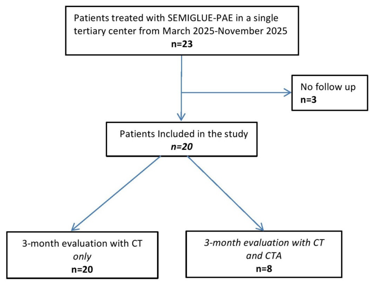
Figure 2.
Kaplan-Meier curve showing short-term clinical success rates of SEMIGLUE-PAE.
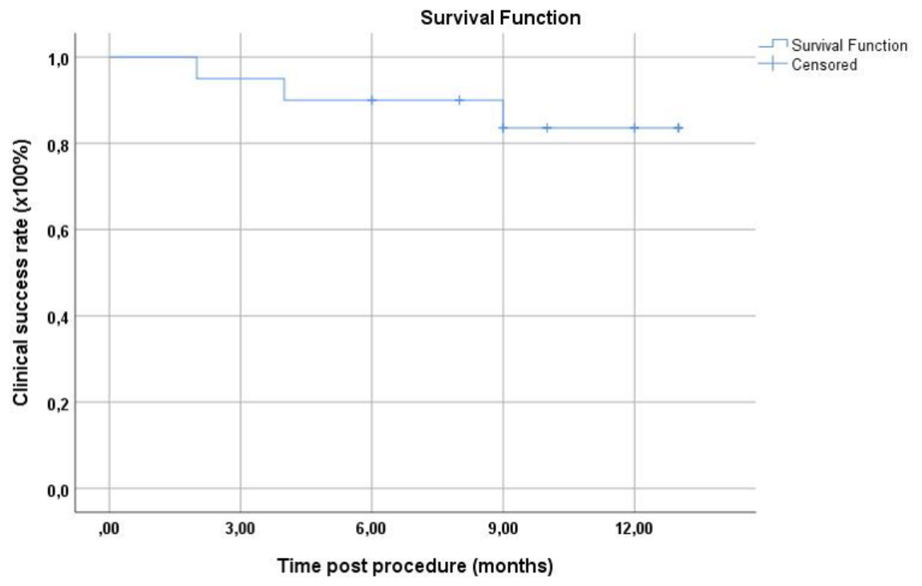
Figure 3.
Representative images post SEMIGLUE PAE. Coronal reformatted image from unenhanced CT immediately post procedure (a), shows parenchymal hyperdense appearance of both prostatic lobes (*). Filling of the distal extraprostatic part of the right PA with glue-lipiodol mixture can also be appreciated (arrow). Axial dual sonographic image acquired 18 hours post PAE (b), with reference, gray-scale image on the left and corresponding CEUS image on the right, shows non-enhancing infarcts of both prostatic lobes (*). Coronal reformatted image (c), from unenhanced CT performed 3 months post PAE, shows prostate shrinkage and resolution of the parenchymal hyperdensities that were noted on CT immediately post PAE. Filling of the distal extraprostatic part of the right PA can still be appreciated (arrow).
Figure 3.
Representative images post SEMIGLUE PAE. Coronal reformatted image from unenhanced CT immediately post procedure (a), shows parenchymal hyperdense appearance of both prostatic lobes (*). Filling of the distal extraprostatic part of the right PA with glue-lipiodol mixture can also be appreciated (arrow). Axial dual sonographic image acquired 18 hours post PAE (b), with reference, gray-scale image on the left and corresponding CEUS image on the right, shows non-enhancing infarcts of both prostatic lobes (*). Coronal reformatted image (c), from unenhanced CT performed 3 months post PAE, shows prostate shrinkage and resolution of the parenchymal hyperdensities that were noted on CT immediately post PAE. Filling of the distal extraprostatic part of the right PA can still be appreciated (arrow).
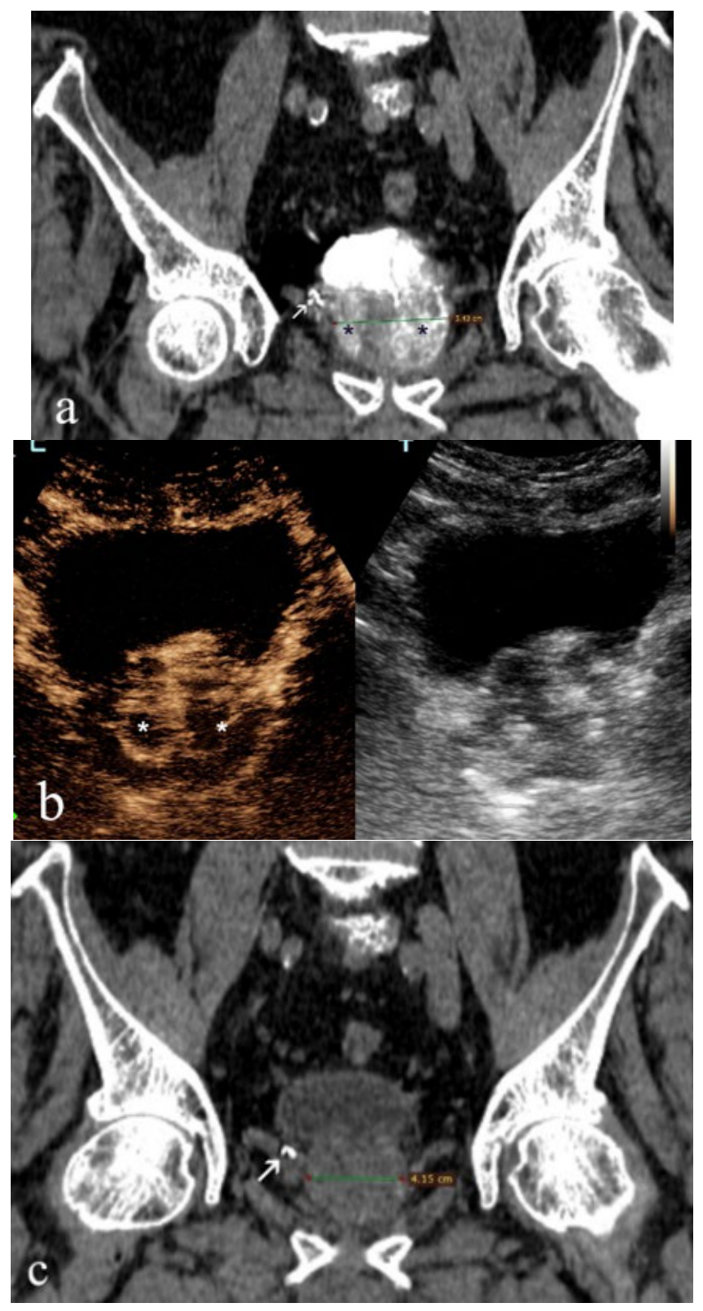
Figure 4.
Representative images from a case of SEMIGLUE PAE with persistent infarction. Unenhanced CT image immediately post PAE (a), shows dense contrast pooling (*) in the right hemiprostate. Corresponding contrast-enhanced US image, 5 months post PAE (b), shows a persistent enhancement defect at the same area (*).
Figure 4.
Representative images from a case of SEMIGLUE PAE with persistent infarction. Unenhanced CT image immediately post PAE (a), shows dense contrast pooling (*) in the right hemiprostate. Corresponding contrast-enhanced US image, 5 months post PAE (b), shows a persistent enhancement defect at the same area (*).
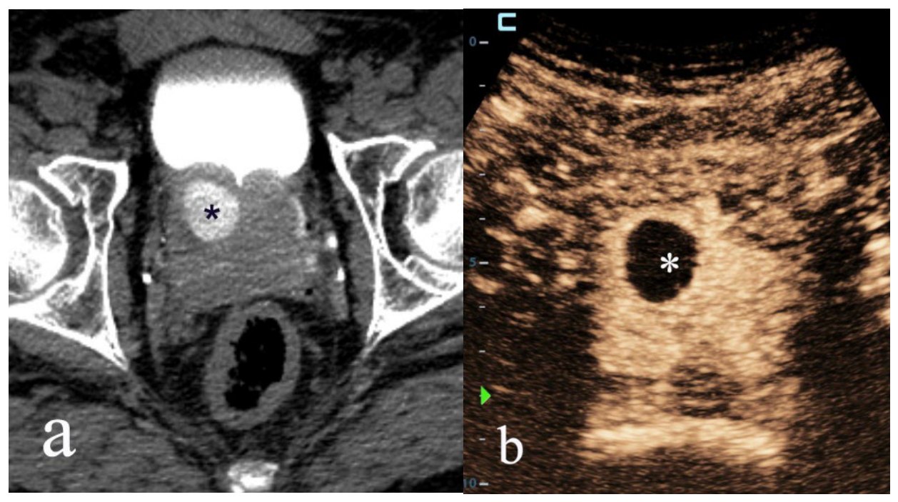
Figure 5.
Representative images from a case of SEMIGLUE PAE in duplicate left PA. Fluoroscopic anteroposterior image (a) during injection of glue-lipiodol with the microcatheter tip in the distal extraprostatic part of the lower PA (arrow), shows reflux of liquid embolic in the upper PA (dotted arrow) and retrograde filling of this artery. Sagittal oblique reformatted maximum intensity projection image from unenhanced CT immediately post PAE (b), shows filling of large extraprostatic part of both the lower (arrow) and the upper (dotted arrow) left PA with glue-lipiodol mixture. The opacified left ureter is also shown (open arrow). Axial dual sonographic image acquired 6 hours post PAE (c), with reference, gray-scale image on the left and corresponding CEUS image on the right, shows almost complete devascularization of the left prostatic lobe (*). Residual enhancement (arrow) of the right prostatic lobe.
Figure 5.
Representative images from a case of SEMIGLUE PAE in duplicate left PA. Fluoroscopic anteroposterior image (a) during injection of glue-lipiodol with the microcatheter tip in the distal extraprostatic part of the lower PA (arrow), shows reflux of liquid embolic in the upper PA (dotted arrow) and retrograde filling of this artery. Sagittal oblique reformatted maximum intensity projection image from unenhanced CT immediately post PAE (b), shows filling of large extraprostatic part of both the lower (arrow) and the upper (dotted arrow) left PA with glue-lipiodol mixture. The opacified left ureter is also shown (open arrow). Axial dual sonographic image acquired 6 hours post PAE (c), with reference, gray-scale image on the left and corresponding CEUS image on the right, shows almost complete devascularization of the left prostatic lobe (*). Residual enhancement (arrow) of the right prostatic lobe.
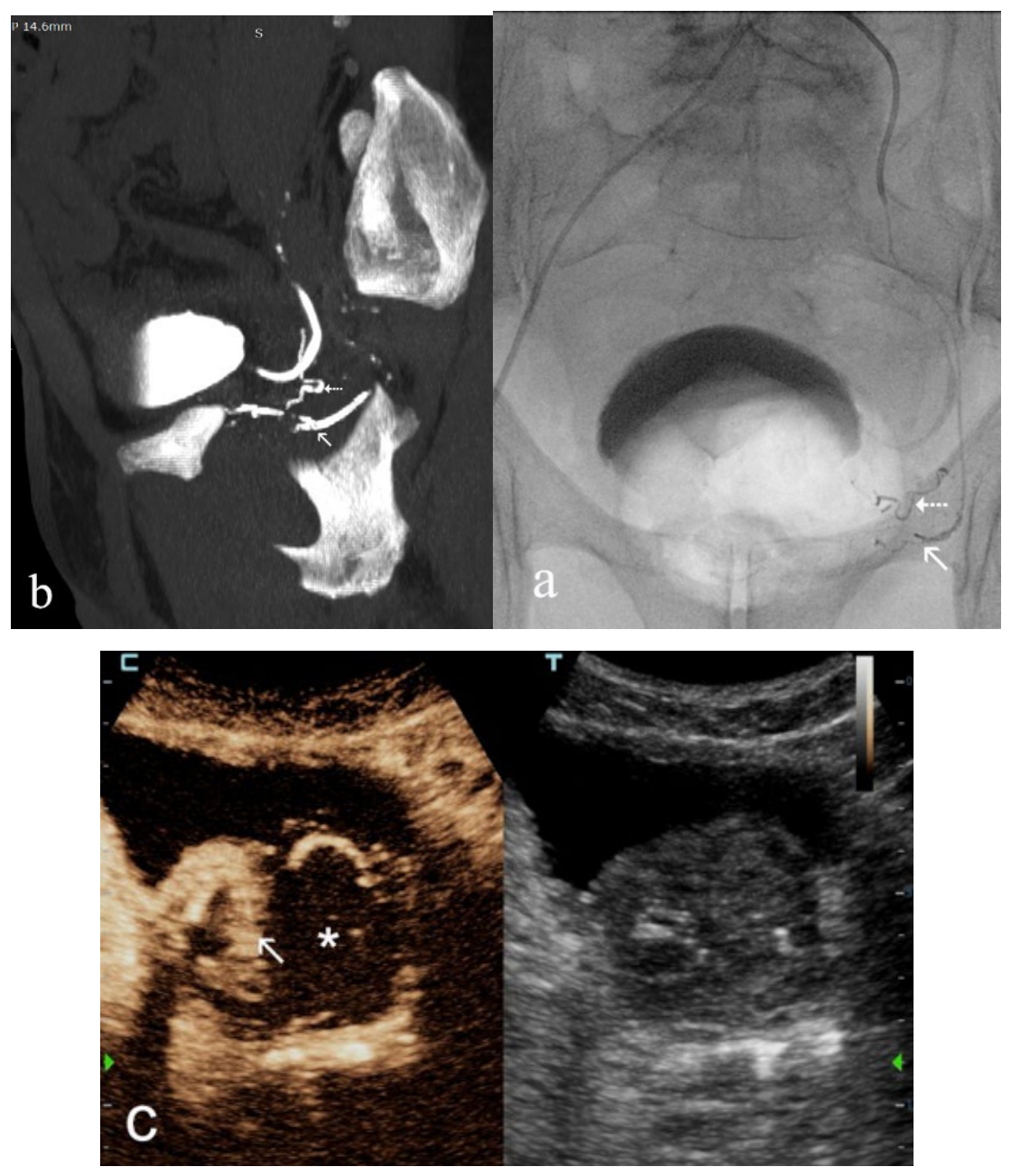
Figure 6.
Short-term follow-up of the extraprostatic left PA after SEMIGLUE-PAE. All images are sagittal oblique reformatted maximum intensity projections. CTA prior to intervention (a), shows a common origin of superior vesical artery (dotted arrow) and PA (arrow). Unenhanced CT immediately post SEMIGLUE-PAE (b), shows filling of the PA (arrow: proximal PA, double arrow: distal PA) by the glue-lipiodol mixture. The opacified ureter (open arrow) and atheromatous calcifications of inferior gluteal artery (arrowhead) are also shown. CTA 3 months post intervention shows persistent filling of the distal PA (double arrow) with glue-lipiodol mixture and absence of enhancement. There is an impression of minimal enhancement in a small proximal segment of the PA (arrow). Normal enhancement of the superior vesical artery (dotted arrow).
Figure 6.
Short-term follow-up of the extraprostatic left PA after SEMIGLUE-PAE. All images are sagittal oblique reformatted maximum intensity projections. CTA prior to intervention (a), shows a common origin of superior vesical artery (dotted arrow) and PA (arrow). Unenhanced CT immediately post SEMIGLUE-PAE (b), shows filling of the PA (arrow: proximal PA, double arrow: distal PA) by the glue-lipiodol mixture. The opacified ureter (open arrow) and atheromatous calcifications of inferior gluteal artery (arrowhead) are also shown. CTA 3 months post intervention shows persistent filling of the distal PA (double arrow) with glue-lipiodol mixture and absence of enhancement. There is an impression of minimal enhancement in a small proximal segment of the PA (arrow). Normal enhancement of the superior vesical artery (dotted arrow).
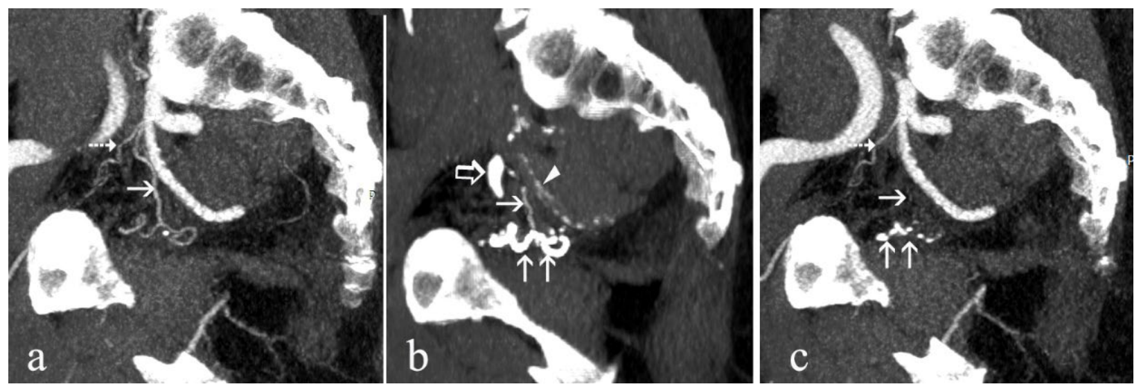

Table 1.
Demographic and clinical features of the first 20 patients treated with “SEMIGLUE” PAE.
| Variable | Value (Mean ± SD) | |
|---|---|---|
| Age (years) | 72.0±8.7 | |
| PV (ml) | 91.9±34.4 | |
| PVR (ml) | 201±91 | |
| IPSS (ml) | 24.0±2.6 | |
| QoL (ml) | 4.6±0.5 | |
| Indication for PAE | Proportion of patients | |
| -Moderate LUTS | 3/20 | |
| -Severe LUTS | 9/20 | |
| -IBC | 8/20 | |
| SD: standard deviation; PV: prostate volume; PVR: post void residual; IPSS: international prostate symptom score; QoL: quality of life; PAE: prostatic artery embolization; LUTS: lower urinary tract symptoms; IBC: indwelling bladder catheter | ||
Table 2.
Values of imaging and clinical parameters at short-term follow up after “SEMIGLUE” PAE and relevant improvement.
Table 2.
Values of imaging and clinical parameters at short-term follow up after “SEMIGLUE” PAE and relevant improvement.
| Variable | Value(Mean ± SD) | Improvement | p-valuec |
|---|---|---|---|
| pPIa (%) | 45.7±16.5 | ||
| PV (ml) | 62.1±30.3 | 32.4% | <0.01 |
| PVR (ml) | 77±44 | 61.7% | 0.01 |
| IPSS | 9.3±4.4 | 61.2% | <0.01 |
| QoL | 2±1.1 | 56.5% | <0.01 |
| pPI: percentage of prostatic infarction=volume of infarcts /total volume of prostate (infarcts were evaluated with contrast-enhanced ultrasound); SD: standard deviation; PV: prostate volume; PVR: post void residual; IPSS: international prostate symptom score; QoL: quality of life a: pPI was calculated the first day post PAE; all other variables were calculated approximately 3 months post PAE and compared with respective baseline measurements c: statistical significance was evaluated after comparison with baseline measurements. Wilcoxon Signed-Rank Test was used for the comparisons | |||
Table 3.
Imaging findings relevant to glue-lipiodol embolization of the PAs (extraprostatic part) of this study.
Table 3.
Imaging findings relevant to glue-lipiodol embolization of the PAs (extraprostatic part) of this study.
| PAs of the patients of the study (n) | 46 |
| PAs embolized with glue-lipiodol | 42 |
| PAs embolized antegradely | 40 |
| PAs embolized retrogradely | 2 |
| PAs not embolized | 4 |
| PAs studied with CT 3 months post PAE | 42 |
| PAs with retained glue-lipiodol | 42 |
| PAs studied with CTA 3 months post PAE | 19 |
| PAs with occlusion of their distal part | 19 |
| PAs with minimal or no enhancement of their proximal part | 13 |
| PAs with residual enhancement of their proximal part | 6 |
| PAs: prostatic arteries; PAE: prostatic artery embolization; CT: computed tomography; CTA: computed tomography-angiography. | |
Disclaimer/Publisher’s Note: The statements, opinions and data contained in all publications are solely those of the individual author(s) and contributor(s) and not of MDPI and/or the editor(s). MDPI and/or the editor(s) disclaim responsibility for any injury to people or property resulting from any ideas, methods, instructions or products referred to in the content. |
© 2026 by the authors. Licensee MDPI, Basel, Switzerland. This article is an open access article distributed under the terms and conditions of the Creative Commons Attribution (CC BY) license (http://creativecommons.org/licenses/by/4.0/).
Copyright: This open access article is published under a Creative Commons CC BY 4.0 license, which permit the free download, distribution, and reuse, provided that the author and preprint are cited in any reuse.