

Submitted:
03 October 2026
Posted:
06 October 2026
You are already at the latest version
Abstract
Geographic atrophy (GA) is a vision-threatening late manifestation of age-related macular degeneration (AMD) and is increasingly recognized as the atrophic endpoint toward which advanced dry and neovascular AMD may converge. This review examines the clinical and translational progression from early AMD to GA, focusing on the key pathogenic factors, affected tissues, and stage-specific therapeutic approaches. We performed a mechanism-oriented synthesis of clinical, translational, and preclinical evidence identified through search of Medline, Web of Science Core Collection, and Scopus conducted from February to September 2026. Aging, genetic susceptibility, oxidative stress, mitochondrial dysfunction, impaired heterophagy and autophagy, retinoid by-product accumulation, complement dysregulation, and Bruch’s membrane remodeling progressively disrupt outer-retinal homeostasis. Drusen and subretinal pigment epithelium deposits represent structural markers of this imbalance and contribute to a self-amplifying microenvironment of impaired transport, local inflammation, and tissue remodeling. As reparative capacity fails, regulated cell-death programs including ferroptosis, necroptosis, apoptosis, pyroptosis, and inflammation-associated injury, promote lesion formation and expansion of atrophic areas. Current therapies remain stage-specific: anti-vascular endothelial growth factor agents control exudation in neovascular AMD, whereas complement inhibitors slow GA enlargement without restoring lost RPE-photoreceptor architecture. GA should therefore be regarded as the final expression of cumulative metabolic, immune, vascular, and extracellular matrix failure, supporting earlier multifaceted therapeutic strategies.
Keywords:
geographic atrophy
; age-related macular degeneration
; outer retinal metabolism
; retinal immune dysregulation
; multifaceted therapeutic strategies
1. Introduction
1.1. Geographic Atrophy as a Distinct Late-Stage Phenotype of Age-Related Macular Degeneration
GA is an advanced and vision threatening form of AMD. It is defined by progressive loss of retinal pigment epithelium (RPE) integrity and function together with photoreceptor degeneration and remodeling of the outer retina-choroid interface. The most common clinical manifestation is the formation and the expansion of scotomas that lead to an irreversible central vision loss with consequent disability [1,2].
Although GA has historically been framed as the end stage of “dry” AMD, it is now considered as the atrophic endpoint of a broader AMD continuum. This shift has required standardized terminology and reproducible structural readouts to identify disease stages and support appropriate interventional trials across cohorts of patients [3,4]. This shift has been largely driven by advances in multimodal imaging; the Optical Coherence Tomography (OCT)-based consensus enables reproducible structural definitions of AMD-associated atrophy and supports earlier identification of transitional states that precede fully developed lesions [4,5]. In particular, it captures a spectrum from incomplete to complete RPE and retinal atrophy, enabling earlier detection of pre-GA structural transition [4]. Histopathology complements these imaging frameworks by demonstrating that the GA border shows active cellular remodeling regarding RPE dysmorphia, migration and layer rearrangements [5]. Together, modern clinical classification and imaging consensus position GA as a quantifiable late-stage outcome arising from long-lasting stress and dysregulation of the RPE-Bruch’s membrane-choriocapillaris complex, with measurable structural progression, central for therapeutic development and trial design [3,4,5,6].
1.2. Global Incidence, Projected Growth, and Societal Burden of GA
GA is embedded within the broader epidemiology of AMD, one of the leading causes of vision impairment in older adults globally [1]. A large global systematic review and meta-analysis have estimated that the number of people living with AMD will rise from 196 million in 2020 to 288 million by 2040, highlighting a major and expanding public health burden [7,8]. GA imposes a disproportionate impact because of the progressive macular atrophy, its structural hallmark that directly impairs reading vision, face recognition, and independence. Consequently, GA involves escalating personal and public costs through disability, increased care needs and reduced quality of life, even in cases where symptoms can be managed pharmacologically [6,9]. Focusing on this soon-to-be worldwide health challenge, GA should be considered not only as a clinical endpoint but also as a global problem whose prevalence and economic burden are destined to increase, reinforcing the urgency of earlier intervention in the AMD pathway [1,7,8].
1.3. GA in Relation to Dry and Wet AMD: Shared Origins, Divergent Development, Convergent Atrophy
GA is biologically and clinically embedded within the macular degeneration phenotype evolution, belonging to both late-stage non-neovascular (“dry”) and neovascular (“wet”) AMD, given their shared upstream susceptibility, environmental causes and aging drivers [1,10].
The progression of dry AMD is driven by the cumulative accumulation of extracellular deposits, including drusen and sub-RPE deposits. This chronic deposition exposes RPE cells to prolonged stress, ultimately impairing phagocytosis and clearance pathways. The resulting dysfunction of heterophagy, autophagy, and mitophagy further exacerbates inflammatory activation. The initial tissue damage eventually evolves into sharply demarcated atrophic loss, captured as GA during imaging [4,5,11,12]. Wet AMD is distinguished by choroidal neovascularization, exudation and fibrotic complications. Nevertheless, the strong genetic overlap between wet and dry AMD supports the concept that neovascular conversion is not an entirely separate disease, but rather an alternate trajectory emerging from a shared substrate of complement dysregulation, extracellular matrix alteration, retinoid stress and inflammatory signaling [10,13,14]. On this topic, multiple articles discuss that atrophic macular degeneration can be observed across all AMD phenotypes, emphasizing mechanisms convergence upon RPE-photoreceptor loss even when the clinical course includes neovascular events [6].
After a closer inspection of all the pathways discussed in GA literature, we can separate two courses that bias AMD toward predominantly wet against predominantly dry endpoints: the balance between pro- and anti-angiogenic signaling within the compromised Bruch’s membrane-choroid interface and the rate at which chronic stress exceeds RPE repair capability, inducing regulated death pathways and collapse of outer retinal support [10,15]. Both trajectories still converge on macular atrophy, staying consistent with histopathological evidence of active RPE remodeling at lesion borders and with imaging consensus definitions capturing progressive outer retinal RPE loss in both cases [4,5].
Rather than representing the failure of a single molecular pathway, geographic atrophy should be regarded as the clinical expression of the progressive collapse of the outer retinal ecosystem, where photoreceptors, retinal pigment epithelium, Bruch’s membrane and choriocapillaris lose their ability to function as an integrated biological unit (Figure 1).
2. Anatomical Determinants of Degeneration in AMD and Geographic Atrophy
Although the retina is organized into multiple neuronal layers, the anatomical compartment primarily affected in AMD and GA is the outer retinal interface formed by photoreceptors, the RPE, Bruch’s membrane, and the choriocapillaris [2,16,17] (Figure 2). Photoreceptors depend on the polarized RPE for outer-segment phagocytosis, retinoid processing, metabolic support, and maintenance of the outer blood-retina barrier [17,18,19]. The RPE, in turn, exchanges nutrients and waste products with the choroidal circulation across Bruch’s membrane, which also provides an extracellular platform for lipid deposition and local complement regulation [13,14,17,20]. Age-related dysfunction within this interconnected unit promotes oxidative and metabolic stress, impaired proteostasis, lipid and bisretinoid accumulation, and inflammatory activation, progressively destabilizing both the RPE and photoreceptors [12,13,20,21,22,23]. In GA, progressive RPE alteration and loss are accompanied by degeneration of the overlying photoreceptors; because these changes occur within the macula, their location and extension directly determine the severity of central visual impairment [4,5,24,25,26].
3. Metabolic Interdependence in the Outer Retina: photoreceptor-RPE-Bruch’s Membrane Crosstalk in the Maintenance of Central Vision
3.1. Metabolic Organization of the Neural Retina and Macular Specialization
The neural retina is among the most metabolically active tissues in the human body, reflecting its continuous demand for phototransduction, synaptic transmission and its need for cellular maintenance, processes required to support active vision. Photoreceptors, particularly cones concentrated in the macula and fovea, exhibit exceptionally high energy requirements due to sustained ion fluxes, membrane turnover and neurotransmitter release. They are characterized by a high metabolic demand that puts them, consequently, at risk of accumulating non-processed toxic intermediates [1,16]. Within photoreceptors, energy production reflects compartmental specialization: inner segments are mitochondria rich and support oxidative phosphorylation, whereas outer segments are optimized for phototransduction and continuous membrane renewal. Müller glia buffer extracellular K⁺ and contribute to metabolic coupling across layers, maintaining homeostasis during changes in illumination and activity [27]. These demands are met thanks to the outer retina being structurally integrated with a specialized support system, the RPE and Bruch’s membrane, which connects the neural retina to the choroidal vases, forming a continuous trophic axis from photoreceptors to systemic circulation [16]. This metabolic organization is built upon a bioenergetic symbiosis: while photoreceptors primarily metabolize glucose through aerobic glycolysis, generating high levels of lactate, the RPE maintains a predominantly oxidative metabolism, sparing glucose for the neural retina and instead utilizing the photoreceptors’ lactate and fatty acids released from the daily phagocytosis of outer segment (OS) tips [17,19]. Through mitochondrial ketogenesis, the RPE converts these substrates into β-hydroxybutyrate (β-HB), which is secreted apically via monocarboxylate transporters (MCT) to support the oxidative demands of the photoreceptors [17,19]. This metabolic cooperation is particularly critical due to macular specialization. In the human macula, the functional burden on the RPE is unique: each epithelial cell must manage and recycle the metabolites of approximately 30 to 33 photoreceptors. This extreme demand requires high efficiency in nutrient and waste exchange across Bruch’s membrane to sustain central vision and prevent the cellular degeneration associated with metabolic stress (Figure 3).
In the visual cycle, light-induced conformational changes in rhodopsin to metarhodopsin II activate a signal transduction cascade culminating in vision, accompanied by the isomerization of 11-cis-retinal (11-cRAL) to all-trans-retinal (atRAL). The compound atRAL can be reduced by retinol dehydrogenase 12 (RDH12) to all-trans-retinol (atROL) and subsequently transported to the RPE, where it is regenerated as 11-cRAL through the sequential actions of lecithin-retinol acyltransferase (LRAT), RPE65, and RDH5. Additionally, atRAL can form an adduct with phosphatidylethanolamine (PE), known as N-retinylidene-PE (N-ret-PE). The ABCA4 transporter mediates the clearance of N-ret-PE, thereby reducing the heterophagy of photoreceptor outer segments containing N-ret-PE and preventing the accumulation of bisretinoids, which otherwise contributes to the formation of lipofuscin granules.
The physiological photo-oxidative state of the RPE, resulting from light exposure, heterophagy, and mitochondrial metabolism, drives the production of reactive oxygen species (ROS). This production is counteracted by an antioxidant defense network consisting of catalase, superoxide dismutase 2 (SOD2), the glutathione system (GSH/GSSG), and melanosomes, which provide additional photoprotection. The production of ROS triggers the translocation of nuclear factor erythroid 2-related factor 2 (NRF2) into the nucleus, facilitating the transcription of antioxidant response elements (AREs) that contribute to the cellular antioxidant defense.
Complement factor H (CFH), produced by the RPE, exerts a dual protective function. CFH binds to the cell membrane, inhibiting the alternative pathway of the complement system and thereby preventing cellular damage. Furthermore, CFH can bind to malondialdehyde (MDA) epitopes, which are generated by lipid peroxidation on the RPE cell surface, thus inhibiting their recognition by macrophages.
Tight junction proteins, including zonula occludens-1 (ZO-1), occludin, and claudin, establish the integrity of the outer blood-retinal barrier, thereby maintaining an optimal environment for the efficient functioning of the retina. Created in https://BioRender.com
3.2. Photoreceptor Outer-Segment Renewal and RPE-Mediated Heterophagy
Photoreceptors undergo a continuous renewal of their outer segments during their physiological activity. Approximately 10% of rod outer-segment discs are shed daily and replaced by newly synthesized membranes. This process is coupled to the RPE specific phagocytosis, called heterophagy, during which apical RPE microvilli engulf spent outer-segment tips and route them to lysosomal degradation pathways [21].
Heterophagy requires coordinated cytoskeletal dynamics, lysosomal processing and efficient recycling of cellular components rich in lipids and proteins. In parallel, physiological autophagy and mitophagy contribute to organelle quality control, particularly in the RPE where high levels of phagocytosis and oxidative exposure coexist [8,21,22]. This metabolic cycle is a cornerstone of the retinal trophic axis: the RPE recycles lipids like docosahexaenoic acid (DHA) and arachidonic acid from the ingested membranes to fuel its own mitochondrial β-oxidation, ultimately providing ketogenic substrates to support the neural retina [17,19] (Figure 3). Ultimately, photoreceptors and the RPE should be regarded as an integrated physiological unit rather than as two anatomically adjacent but functionally separate tissues. Nevertheless, while their metabolic activities are highly cooperative under physiological conditions, this finely tuned balance is continually challenged by disturbances in metabolic homeostasis and antioxidant defense pathways.[2].
3.3. The Retinoid Cycle and Visual Chromophore Recycling
Phototransduction requires continuous regeneration of 11-cis-retinal, the chromophore needed for visual pigments. Vision is initiated when 11-cis-retinal, covalently bound to opsin, absorbs a photon and undergoes a light-driven photoisomerization to all-trans-retinal (atRAL). This process triggers conformational changes in the opsin protein (forming metarhodopsin II), which activates the G-protein transducin, initiating the signal transduction cascade that ultimately results in nerve signaling [17]. After the all-trans-retinal is generated in photoreceptor outer segments, it must be cleared and recycled efficiently to prevent cellular damage: as an aldehyde-bearing agent, free atRAL is highly reactive and potentially toxic, necessitating careful handling through compartmentalized sequestration and enzymatic reduction [12]. The classical retinoid cycle distributes this process across photoreceptors and RPE: in the photoreceptors, atRAL is reduced to all-trans-retinol by NADPH-dependent retinol dehydrogenases (RDHs) or transported across the disc membrane as a Schiff base adduct (-retinylidene-PE) by the ABCA4 transporter. These retinoid intermediates are then transferred to the RPE for enzymatic conversion. Within the RPE, all-trans-retinol is esterified by lecithin retinol acyltransferase (LRAT) and converted back to 11-cis-retinol by the RPE-specific isomerohydrolase: RPE65. Finally, the chromophore is oxidized and returned to the photoreceptors as 11-cis-retinal to reconstitute photosensitive pigments (Figure 3) [17,35,36].
This loop establishes a physiological metabolic cooperation by combining enzymatic activity, membrane transport and trafficking across cell boundaries, being linked to outer-segment renewal. Physiologically, handling of all-trans-retinal and related intermediates also limits the formation and accumulation of bisretinoid by-products within the RPE’s lysosomal compartment. Over time, these by-products form lipofuscin, which is why normal retinoid processing is considered a key homeostatic requirement for preserving the health of the outer retina and preventing the degenerative changes associated with age-related macular degeneration [12,28].
3.4. Transport, Barrier Function and Trophic Exchange Across the RPE-Bruch’s Membrane Interface
The RPE is a polarized epithelium that forms the outer blood-retina barrier and mediates vectorial transport. Through its strict apical-basal organization and tight junctions (TJs) maintained across each epithelial cell, characterized by proteins such as claudin-19, occludin, and ZO-1, the RPE regulates the movement of metabolites, ions, water, lipids and retinoids between the upper subretinal space and the bottom choroidal circulation (Figure 3) [1,2]. Multiomics and single-cell datasets further confirm that, under normal conditions, RPE cells express specialized transporters, antioxidant enzymes and immune modulators that together support outer-retinal homeostasis [15,29,30].
Bruch’s membrane provides the extracellular interface that enables this exchange while maintaining structural support and selective permeability. In physiological conditions, it supports diffusion and anchoring at the RPE-choriocapillaris boundary and hosts ECM associated regulators, including complement-associated factors, consistent with its role as an active transitional microenvironment that connects the deeper retinal layers with choroidal circulation [13,14]. Trophic exchange across this interface is highly specialized: GLUT1 transporters located on both the apical and basal RPE membranes facilitate the continuous flow of glucose to the photoreceptors. Simultaneously, the interface manages metabolic waste through a system of monocarboxylate transporters: MCT1 on the apical membrane imports the high levels of lactate produced by photoreceptor glycolysis, while MCT3 on the basal side exports it toward the choroid. Additionally, the RPE takes up all-trans-retinol (vitamin A) from systemic circulation via receptor-mediated endocytosis to initiate the visual cycle [17]. This vital exchange is increasingly compromised during aging and disease. The progressive accumulation of ApoB100-containing lipoproteins and lipid-rich debris, often termed the “oil spill” in the aging membrane, leads to the formation of basal linear (BLinD) and laminar (BLamD) deposits. These deposits, along with membrane thickening and calcification, increase the diffusion distance between the choriocapillaris and the outer retina. This physical barrier impairs the supply of oxygen and nutrients, inducing chronic hypoxia and metabolic stress that ultimately drive the degenerative changes seen in age-related macular degeneration [2,16,20].
3.5. Redox Homeostasis and Antioxidant Systems as Protection in a Light-Exposed Tissue
The outer retina operates in a physiological microenvironment characterized by a constant burden of ROS generated by high oxygen consumption rates needed for mitochondria metabolism and the continuous photochemical reactions triggered by light exposure. To counteract this inherent oxidative load, photoreceptors and the RPE employ a sophisticated network of antioxidant defenses designed to neutralize ROS before they inflict damage (Figure 3) [31,32]. The RPE deploys multiple protective systems, including glutathione cycling, superoxide dismutases, (specifically SOD2), and catalase. Central to this defense is the NRF2-regulated transcriptional program; as a master regulator, NRF2 translocate to the nucleus under oxidative stress to initiate the transcription of genes containing antioxidant response elements (AREs). This program maintains NADPH pools and mitochondrial integrity through the pentose phosphate pathway and metabolic shuttles (such as IDH), which provide the reducing power needed to neutralize lipid peroxidation products.[29]. Furthermore, complement factor H (CFH) and related proteins exert cell-autonomous antioxidant effects at the RPE surface, linking immune regulation with redox homeostasis. By specifically recognizing malondialdehyde (MDA) epitopes, toxic products of polyunsaturated fatty acid peroxidation, CFH contributes to the protection of RPE cells against oxidative injury and supports the maintenance of transcriptional homeostasis [33]. Complementing these enzymatic systems, the RPE is enriched with melanin and macular pigments (lutein and zeaxanthin), which serve as ROS scavengers and physical filters to intercept high-energy blue light [13,34,35]. Physiologically, these systems cooperate to buffer reactive oxygen species generated during phototransduction and mitochondrial respiration, preserving cellular viability across decades of visual activity [31,36].
Vision is therefore sustained not by individual retinal cells but by the integrity of their metabolic dialogue. AMD develops when this dialogue progressively fails, long before irreversible structural atrophy becomes clinically detectable.
4. Transition from Homeostatic Maintenance to Progressive and Degenerative Tissue Remodeling
4.1. Lifelong Metabolic Stress and the Age-Related Decline in Reparative Capacity
In the healthy eye the macula photoreceptors-RPE-Bruch’s membrane-choriocapillaris complex sustain the central vision through tightly coordinated renewal, transport, redox control and a heavy metabolic burden. With aging, this system operates continuously under accumulating metabolic byproducts, oxidative photochemical stress and repeated membrane turnover, while the efficiency of proteostasis, mitochondrial quality control and ECM renewal gradually declines. First hints of the disease emerge when homeostatic activity is no longer sufficient to buffer the cumulative metabolic load and this happens particularly in the macula, where energetic demand is highest [1,16,36].
Contemporary literature on AMD development frames the transition from physiological aging to pathological macular degeneration as a progressive failure of adaptive homeostatic mechanisms. These include antioxidant defense responses, controlled immune surveillance, and regulated lysosomal clearance, which gradually give way to persistent stress signaling, low-grade inflammation, and impaired homeostasis at the RPE-Bruch’s membrane interface [8,9,37,38].
Genetic association studies further underscore the central involvement of the RPE-Bruch’s membrane complex in the metabolic pathways outlined above. Collectively, these findings support the view that AMD arises from a maladaptive reprogramming of physiological homeostatic mechanisms, driven by cumulative burden and age-related decline in reparative capacity [7,10,16].
The transition from physiological aging to AMD should not be interpreted as the appearance of new pathological mechanisms, but rather as the progressive inability of retinal tissues to preserve metabolic resilience under lifelong stress.
4.2. Inefficient Maintenance Structures and Early Failures
Photoreceptors renew their outer segments continuously, aided by the RPE which exhibit high heterophagy flux and lysosomal digestion, simultaneously maintaining its own organelle homeostasis through autophagy and mitophagy. With aging and cumulative stress, lysosomal capacity, autophagic flux and proteostasis can become progressively altered, leading to incomplete degradation and gradual accumulation of unclear metabolic and cellular residue inside the RPE [2,21,22]. This stage is clinically silent yet biologically important because it increases the probability that secondary stressors such as oxidative bursts, inflammatory triggers and retinoid-reactive intermediates, will have disproportionate impact in the following phases [8].
In tandem, the retina is continuously exposed to light and high oxygen delivery, making redox control an indispensable requirement. Light damage can generate photochemical load, requiring robust antioxidant defenses and mitochondrial quality control. With the progressive weakening of these defensive mechanisms, oxidative stress shifts from an episodic challenge to a persistent pathological condition, thereby contributing to a chronic injury milieu in the outer retina [22,23,28,37]. The association between NRF2 deficiency, reduced NADPH availability, and increased mitochondrial vulnerability exemplifies how initially adaptive redox programs may become progressively constrained under chronic metabolic and oxidative burden [22,39].
In photoreceptor metabolism, the retinoid cycle is essential for sustaining phototransduction, but reactive intermediates such as atRAL require efficient clearance and recycling. When atRAL removal is incomplete, reactive stress may increase, promoting the formation and accumulation of bisretinoids and lipofuscin-related metabolites, including A2E, within RPE lysosomal compartments. Although this process is not sufficient to define disease on its own, it may increase vulnerability by adding phototoxic and pro-oxidant stimuli to a retinal environment already challenged by oxidative and proteostatic stress [12,40]. This stage creates a starting point for later extracellular deposit formation and immune activation (Figure 4).
4.3. Drusen Accumulation as a Driver of Microenvironmental Remodeling
The early-to-intermediate phases of AMD are characterized by the progressive deposition of extracellular material at the RPE-Bruch’s membrane interface, forming the lesions classically known as drusen. These deposits represent complex aggregates of cellular and extracellular debris, lipid-derived metabolites, and immune surveillance-related proteins. Their accumulation reflects defects in clearance and transport mechanisms, resulting in the retention of incompletely processed material within the sub-RPE compartment [9,13]. Drusen formation can be positioned as a mid-course event in clinical development of AMD symptoms. It follows prolonged subclinical stress and partial repair failure and precedes late outcomes by progressively altering the structural, inflammatory, immune-modulatory and transport properties of the RPE-Bruch’s membrane niche [3,16].
The same retinal metabolic processes used to form bisretinoids and lipofuscin and to process atRAL, play a pivotal role when incompletely processed material does accumulate, contributing to deposit biology. In this view, drusen can be considered as an extracellular readout of long-term imbalance between generation of by-products, including lipofuscin-related material and retinoid-derived stress products, and the capacity of the RPE-Bruch’s membrane unit to dispose of it [12,22,40].
Importantly, drusen should not be considered biologically inert deposits. Rather, complement activation and local inflammation contribute to their recognition as aberrant, danger-associated structures within the retinal microenvironment. These immune processes participate not only in deposit formation but also in the remodeling of the surrounding tissue milieu, establishing a positive feedback loop between drusen accumulation, immune activation, local inflammation, and extracellular matrix remodeling at the RPE-Bruch’s membrane interface [9,13,41]. Complement regulators, particularly members of the factor H family that localize to Bruch’s membrane, critically shape immune regulation and modulate the local response to drusen formation. Beyond complement control, these molecules have also been implicated in antioxidant protection at the RPE surface. Thus, immune regulation, redox buffering, and extracellular matrix stability at the RPE-Bruch’s membrane interface emerge as tightly interconnected processes [13,14,42] (Figure 4).
4.4. A Shared Pathogenic Substrate Underlying Divergent Dry and Wet AMD Trajectories
Dry and wet AMD can be viewed as late-divergent manifestations of macular degeneration arising from a common pathogenic substrate of retinal stress, altered tissue homeostasis, and chronic low-grade inflammation. Both phenotypes share several upstream drivers, including oxidative and mitochondrial stress, defective debris clearance, complement and innate immune activation, and extracellular matrix remodeling. Nevertheless, these shared mechanisms ultimately evolve along distinct degenerative trajectories [1,6,9].
At the molecular level, dry and wet AMD share several pathogenic pathways, including complement dysregulation involving the CFH/CFHR and C3/C5 axes, innate immune signaling, inflammatory mediator activation, oxidative stress and NRF2-dependent redox programs, autophagy-mitophagy and lysosomal dysfunction, altered lipid handling, extracellular matrix remodeling, and impaired retinoid processing with bisretinoid and lipofuscin accumulation. In addition, the DICER1/Alu RNA axis has been described as a disease-relevant mechanism linking RNA dysregulation to RPE stress and inflammatory activation in AMD [2,9,12,13,21,39].
Dry AMD is commonly presented as a progressive degenerative pathway arising from cumulative drusen accumulation and increasing RPE metabolic stress. In this setting, persistent deposit burden contributes to a pro-inflammatory microenvironment at the RPE-Bruch’s membrane interface, accompanied by oxidative stress, mitochondrial dysfunction, impaired autophagy and mitophagy, and lysosomal insufficiency. The resulting metabolic strain further compromises retinoid metabolism, enhances complement activation, and sustains chronic para-inflammation. Together, these processes drive extracellular matrix and Bruch’s membrane remodeling, thereby altering tissue architecture as well as diffusion and transport across the outer retinal interface. With disease progression, these alterations promote RPE dysfunction and cell death, ultimately leading to the development of atrophic areas [6,12,13,21,22,23].
Wet AMD is defined by macular neovascularization and exudation with risk of hemorrhage and fibrotic remodeling. The shared degenerative substrate is still present, but neovascular conversion shows angiogenic signaling and vascular barrier disruption that drives fluid accumulation and rapid symptom onset. Key processes include VEGF-driven pro-angiogenic signaling, inflammation-mediated increases in vascular permeability, and remodeling of the Bruch’s membrane-choroid interface, which together establish a microenvironment permissive to neovascular invasion [1,2,3,6,16].
4.5. Convergence of Terminal Structural Failure Toward Geographic Atrophy
Geographic atrophy represents an advanced pathological endpoint of AMD, characterized by the convergence of aberrant tissue remodeling, RPE loss, extracellular matrix disruption, and secondary photoreceptor degeneration. The progressive loss of RPE-derived trophic and metabolic support promotes expansion of atrophic lesions, clinically manifesting as enlarging scotomas and irreversible central vision loss [4,24].
Histology-based imaging classifications have shown that the transition from degenerative remodeling to established atrophy is characterized by distinct RPE phenotypes. These changes progress from focal remodeling of the RPE layer toward the development of continuous atrophic lesions, with an expanding border of active degeneration marking ongoing tissue loss [4,5]. Studies of wet AMD have increasingly recognized that neovascular disease may coexist with progressive atrophic remodeling, even in the presence of VEGF-driven exudation. This supports the concept that exudative and non-exudative AMD trajectories, although clinically divergent, may ultimately converge on macular atrophy [6].
4.6. The Need for Early Intervention Before Geographic Atrophy Becomes Established
A consistent implication across clinical and translational studies is that the most actionable and recoverable therapeutic window precedes widespread cell loss, when tissue remains viable but stressed and dysfunctional. This is particularly evident for dry AMD, which is more common and directly linked to GA development, highlighting the value of intervention prior to irreversible atrophic conversion [6,7,24,38].
4.7. Cell Death and Inflammatory Amplification as Emerging Areas of Investigation
In recent years, research has increasingly focused on defining the metabolic and degradative alterations that underlie the progression of macular degeneration. In this context, regulated cell death programs are emerging as critical degenerative amplifiers, linking chronic stress responses to irreversible tissue loss and inflammatory propagation. Ferroptosis represents one of the most extensively investigated mechanisms. This iron-dependent form of regulated cell death is driven by lipid peroxidation and is closely associated with age-related iron accumulation and oxidative stress. Experimental studies have implicated ferroptotic pathways in late-stage macular degeneration and geographic atrophy progression, particularly through models of oxidative stress-induced RPE cell death [31,32,34,35].
The second process is referred to necroptosis. It is a RIPK-dependent cell death program necessary for the digestion of dead cells and necrotic tissues. Given its metabolic scavenger role, necroptosis has been closely associated with tissue immune surveillance, suggesting a broader function in debris clearance and damage sensing. Alterations of necroptosis pathway have been proposed to be particularly relevant in retinal degeneration through coupling with neuroinflammatory processes and chronic degenerative retinal disease states [43,44].
Collectively, these regulated cell death programs may contribute to shaping the inflammatory microenvironment at the degenerative front. This supports the notion that effective disease-modifying strategies may need to address multiple converging mechanisms, rather than relying on the inhibition of single pathways [6,9,27].
4.8. From Imaging Definitions to Clinical Translation and Therapeutic Intervention
Patients with early and intermediate AMD may already exhibit progressive functional deficits, most notably impaired reading performance and reduced contrast sensitivity. In wet AMD, patients often report view distortion, metamorphopsia and rapid central vision decline, consistent with exudation and macular fluid accumulation. In dry AMD, patients commonly describe expanding central or paracentral scotomas with progressive impairment of reading and face recognition capabilities [3,16,24]. Standard evaluation includes visual acuity, dilated fundus examination and multimodal retinal imaging. Structural imaging does not merely document retinal damage; it provides a window into the metabolic state of the outer retina. The earliest OCT biomarkers should therefore be interpreted as signatures of metabolic failure rather than simple anatomical abnormalities. From this perspective, multimodal imaging becomes a tool for identifying tissue dysfunction before irreversible structural collapse occurs. This concept supports a shift from descriptive retinal imaging toward functional structural phenotyping, where imaging biomarkers are interpreted within the biological processes they reflect rather than as isolated morphological findings. OCT is central in identifying drusen, pigmentary changes, intraretinal or subretinal fluid, suggesting wet AMD, or structural atrophy, indicating dry AMD and GA. Fundus autofluorescence is widely employed to identify and delineate areas of atrophy, as well as surrounding zones of degenerative activity. By contrast, fluorescein angiography and OCT angiography are primarily used to detect and characterize macular neovascularization in suspected wet AMD [3,4,24]. Histological imaging provides biological context to imaging-defined boundaries by demonstrating that RPE phenotypes changes and border remodeling actively shapes the progression of atrophy [5].
5. Molecular Pathways and Metabolic Dynamics Involved in AMD and GA
5.1. Multifactorial Course for AMD and GA Pathology
Across literature, AMD and GA are consistently described as network diseases in which multiple insults mutually reinforce one another. Metabolic and mitochondrial stress, oxidative injury, defective proteostasis, ECM and Bruch’s membrane remodeling, RPE alteration, trophic support ineffectiveness and immune activation play a central role in their progression. Alterations in the same anatomical processes - namely RPE-Bruch’s membrane exchange and RPE support to photoreceptors- can lead to different clinical phenotypes in wet or dry AMD, while still sharing a core of molecular stress signatures [9,10,15,29,30,37,45,46,47].
5.2. Metabolic Dysfunction and Mitochondrial Remodeling in AMD Progression
Metabolic dysfunction is widely recognized as an upstream determinant of whether the outer retina preserves stress-compensatory homeostasis or shifts toward degenerative remodeling. Within the RPE, mitochondrial performance is central to redox buffering, lipid handling, and the maintenance of continuous heterophagy and transport functions. Age and stress-associated mitochondrial alteration are a source of ROS and a limiting factor for repairing processes, creating conditions in which chronic stress becomes self-amplifying [23,36]. Mitochondrial resilience and oxidative management are strictly dependent on NRF2-based redox pathways and NADPH availability. The decline in redox-buffering capacity enhances vulnerability to lipid peroxidation and inflammatory signaling, thereby connecting mitochondrial dysfunction with ferroptosis-related alterations and innate immune amplification [23,39].
This interconnected system based on metabolic impairment, mitochondrial damage and unregulated immune activation, has been demonstrated to be a key aspect of AMD in experimental models of oxidative defense perturbation. Mice models lacking SOD2 enzyme showed an excessive retain of superoxide radicals as metabolic byproducts. Mitochondrial oxidative stress was sufficient to drive early AMD-like pathology in experimental mice, coupling metabolic and mitochondrial dysfunction with immune recruitment in the retina [48]. Consistently, mice exposed to a high-fat diet showed increased accumulation of toxic lipid radicals, resulting in a shift toward a pro-oxidant retinal environment. In this context, suppression of the mitochondrial biogenesis regulator PGC-1α further impaired metabolic homeostasis, promoting mitochondrial distress and RPE dysfunction and culminating in AMD-like retinal changes [49]. Collectively, these findings support the concept that systemic metabolic status may shape retinal fate by modulating tissue homeostasis, stress resilience, and vulnerability to degeneration.
5.3. Oxidative-Inflammatory Coupling as a Central Axis in AMD Progression
Persistent sublethal oxidative stress may promote maladaptive inflammatory crosstalk, linking the failure of adaptive stress responses to metabolic dysfunction, inflammatory amplification, and degenerative tissue remodeling [37]. Oxidative stress in both RPE and its connected structures is heterogeneous in a mechanical sense. Comparative analyses of different oxidative stressors show distinct death phenotypes and signaling outputs, implying that the source of AMD and GA must not be limited to a single oxidative process.
Prolonged stress affecting one or more oxidative pathways shapes the downstream programs that drive the macular damage. These include apoptosis, necrotic injury and necroptosis, inflammasome activation, and ferroptosis associated with lipid peroxidation, which are the metabolic processes that govern AMD, and often act simultaneously within the same tissue [50]. Consistently, experimental models of retinal light damage and RPE phototoxicity indicate that environmental light exposure and photochemical alterations can promote ROS generation, thereby activating downstream injury pathways [28].
5.4. Lipid Toxicity and Ferroptotic Susceptibility
In maculopathies, lipid metabolic dysregulation encompasses intracellular lipotoxicity, extracellular lipid deposition, and membrane remodeling, collectively increasing vulnerability to lipid radical propagation. In this context, phospholipid composition represents a key mechanistic link between lipid metabolism, especially peroxidation-prone lipid pools, altered redox homeostasis, and susceptibility to ferroptosis. ge-related iron accumulation and lipid peroxidation are strongly implicated in AMD pathogenesis and ferroptosis activation, in line with the contribution of iron to oxidative cell death induced by retinal bisretinoids. In this context, retinoid by-products may act as risk amplifiers for lipid peroxidation-driven injury, while iron homeostasis and ACSL4-dependent phospholipid remodeling emerge as key checkpoints controlling oxidative damage, ferroptotic susceptibility, and lipid radical burden [31,34,51]. Bisretinoids and lipofuscin metabolism introduces lipid-reactive substrates that can intensify oxidative injury; iron has been associated with oxidative cell death caused by retinal bisretinoids, linking retinoid byproducts to lipid peroxidation-induced injury [12,52].
Experimental RPE models have demonstrated that oxidative stress can directly trigger ferroptotic cell death, supporting the involvement of ferroptosis in RPE loss during advanced atrophic remodeling. This mechanism may be particularly relevant in the setting of time-dependent iron accumulation, which increases susceptibility to lipid peroxidation-driven injury [32].
5.5. Impaired Heterophagy and Downstream Autophagy-Mitophagy Dysregulation
The RPE is continuously challenged by the daily heterophagic clearance of shed photoreceptor outer segment discs, a process that requires sustained lysosomal proteolysis, autophagic flux, and tightly coordinated metabolic regulation. Age-related decline in RPE autophagic capacity has been widely described and links proteostatic failure to AMD pathogenesis. Accordingly, alterations in proteostasis may represent a key mechanism connecting chronic oxidative stress, excessive ROS accumulation, and the transition toward irreversible retinal degeneration [21].
High heterophagic load and oxidative modifications increase the fraction of substrates that resist efficient degradation. Incomplete proteolysis and lysosomal stress impair autophagy flux, which secondarily worsens mitochondrial quality control, amplifies ROS and increases inflammation tone, leading to mitochondrial damage, dysfunction and progressive degradation through mitophagy. Together, these alterations establish a self-reinforcing loop that can drive tissue progression toward either atrophic degeneration or inflammatory injury, ultimately facilitating the expression of later-stage AMD phenotype [22,53]. Studies conducted on iPSCs focusing on the complement system have further deepened this concept by linking complement-related pathology to lysosomal and autophagic activity, reinforcing the evidence that immune biology and proteostasis are closely related to AMD initiation [54].
5.6. Retinoid-Driven Phototoxicity and Inflammasome Activation in AMD Progression
Retinoid cycling is essential for photoreceptor functionality yet inherently risky due to the production of reactive intermediates such as atRAL. Several studies have demonstrated that impaired atRAL handling and clearance can induce retinal degeneration phenotypes relevant to AMD biology, including light-induced retinopathy. Models of phototoxicity and disrupted retinoid clearance indicate that accumulation of unprocessed atRAL exacerbates oxidative stress in the outer retina, thereby amplifying downstream injury pathways [40]. A key mechanistic bridge to inflammatory amplification is the evidence that atRAL can activate NLRP3 inflammasome pathways in RPE and that this activation is associated with RPE cell death, providing a direct relation between retinoid stress and inflammasome dependent injury [55,56]. Bisretinoids accumulation and phototoxic exposure profiles further support a long-term model in which lipofuscin-associated metabolism can amplify oxidative stress and interact with iron-dependent lipid peroxidation, thus shaping an age-dependent abnormal remodeling of both RPE and photoreceptors towards an atrophy-committed destiny [12,28,52].
5.7. ECM-Bruch’s Membrane Remodeling as a Consequence of Trophic Failure and Immune Activation
Bruch’s membrane is framed as the structural and functional surface at which oxidative stress, metabolic alteration, trophism restrictions, lipid retention and immune activation converge and collectively drive the degradation of macular structure. The ECM remodeling that leads to Bruch’s membrane pathological change is an evolving process, in which stress signals from the RPE, meet the physiological immune surveillance activity, eliciting a damage-induced inflammatory response that reshapes Bruch’s membrane matrix. The altered immune cell reaction causes persistent local inflammation elicited by a plethora of damage signals in the upper retinal layers that remain unsolved, affecting the ECM, inhibiting the trophic functionality and consequently the release of tissue repair signals. he convergence of tissue damage, altered membrane function, and insufficient reparative capacity further promotes immune dysregulation. Although local inflammation may initially represent an attempt to restore microenvironmental homeostasis, its persistence can ultimately impair matrix regeneration and reinforce maladaptive remodeling. Genetic evidence further supports the pathogenic relevance of extracellular matrix deposit biology: the EFEMP1 R345W mutation induces AMD-like deposits in mice, indicating that altered ECM composition can directly drive deposit formation and retinal remodeling [57]. When considered alongside impaired intracellular proteolysis and age-related autophagic decline, these alterations support a staged model in which transport limitations and extracellular matrix remodeling favor the retention of oxidized and lipid-rich substrates. This process facilitates the formation and progressive deposition of drusen at the RPE-Bruch’s membrane interface. These deposits, in turn, promote immune activation and further skew the local tissue response toward maladaptive remodeling and degeneration, ultimately contributing to progression along either dry or wet AMD trajectories [8,21,53].
5.8. From Drusen Deposition to Downstream Remodeling at the RPE-Bruch’s Membrane Interface
Local inflammation contributes to drusen formation, reinforcing the view that drusen act not only as by-products of impaired clearance but also as active amplifiers of local immune responses. These deposits consist of cellular and extracellular debris retained at the RPE-Bruch’s membrane interface and progressively enriched with lipid-rich, oxidized, and immunogenic components. Evidence from retinoid and bisretinoid studies further suggests that inefficient RPE processing and transport can favor the accumulation of lipofuscin-related and atRAL-linked debris products, thereby linking defective visual-cycle by-product handling to drusen deposition and inflammatory propagation [12,40]. The downstream effect of deposits formation and the altered RPE spaces is the complement activation and persistent local immune recruitment, while simultaneously worsening of both diffusion and transport of metabolites and trophic elements, due to the strengthened ECM remodeling. This combination produces a stable and progressive pro-degenerative niche that increases local alterations shifting the tissue phenotype towards neovascular dominance in wet AMD or atrophic degeneration in dry AMD and GA [9,13].
5.9. Recruitment and Amplification of Complement Activation in AMD Progression
Complement dysregulation is widely recognized as a central contributor to AMD pathogenesis, irrespective of dry or wet disease phenotype. During drusen deposition, local immune surveillance pathways become activated, while stressed or damaged macular cells release danger-associated signals that promote immune recruitment and amplify inflammatory responses [10,13,45]. Anatomically, local inflammation forms complement factors-rich local niches, especially at Bruch’s membrane, where compartmentalized complement control plays an important role in immune-mediated macular environment reshape. The identification of FHL-1 as a predominant complement regulator in Bruch’s membrane supports a model in which local regulatory deficiency can amplify complement activity exactly at the RPE-Bruch’s membrane interface, where deposits and diffusion failure accumulate [14].
Studies focusing on Multiple CFH further support causal relevance between complement regulation and macular degeneration. CFH deficiency in aged mice causes retinal abnormalities and visual dysfunction, while expression of human CFH in CFH-KO mice drove regulation of AMD-like pathology, confirming that it is a functional determinant of tissue integrity and immune homeostasis, pivotal for AMD advance [58,59]. CFH is also implied in oxidative resilience at the RPE surface, linking complement regulation to redox vulnerability [42]. Human complement modeling in iPSC systems expressing CFH Y402H mutation and complement modulation studies that restore lysosomal function, demonstrated an integrated behavior between complement activity and proteostasis capacity, reinforcing complement-lysosome link as an important mechanism driving maculopathy [46,54].
5.10. Inflammasome-Mediated Innate Immune Activation in Tissue Damage and Cell Death
Studies centered on DICER1 have defined an upstream nucleic acid-stress axis that contributes to RPE oxidative toxicity and inflammatory activation. DICER1 deficiency promotes the accumulation of Alu RNA, which in turn triggers MyD88-dependent inflammatory signaling and NLRP3 inflammasome activation. These events converge on caspase-dependent cell death pathways, particularly caspase-8-mediated RPE death, thereby linking chronic stress, inflammatory amplification, and progressive RPE loss in AMD and geographic atrophy [60,61,62].
This inflammasome axis is further supported by works showing that oxidative stress activates NLRP3 pathway in RPE cell models and that atRAL can trigger NLRP3-associated RPE cell death, thereby integrating oxidative stress and retinoid-associated stress with inflammasome biology [55]. Rodent-eye models of AMD provide various evidence for activation of pyroptotic and apoptotic pathways associated with NLRP3, supporting inflammasome-linked inflammatory death as a plausible contributor to late-stage tissue remodeling and degeneration [56].
5.11. Interconnected Regulated Cell Death Programs as Drivers of Inflammatory Amplification
Within the AMD landscape, apoptosis is frequently implicated as part of RPE stress responses, but comparative stressor analyses conclude that apoptosis is often one of the activated pathways within a complex plethora of different cell response programs, acting together under chronic or severe injury [50,56]. Apoptosis is only one of the pathways responsible for limiting damage through induced cell death, and many studies have found that apoptosis factors are co-activated with other pathways responsible for cell death and clearance.
Necroptosis is strongly represented in both patients-derived samples and in retinal models. Since RIP1 kinase has been identified as a specific target of necrostatins, RIPK1 can be considered as a decisive control node of RPE damage response. Studies investigating the retina reported necrotic injury in RPE under oxidative stress with active necroptosis to dismantle dead materials. Indeed, sodium iodate-induced RPE necroptosis and in vivo protection with RIPK1 inhibition in retinal degeneration models support necroptosis as an inflammatory amplifier in atrophy-biased injury both in macula and at lesion margins [63,64,65]. Studies focused on necroptosis link this process to neuroinflammation in retinal degeneration, confirming that lytic forms of regulated cell death can propagate inflammatory remodeling beyond the initiating cell type [43,44,66]. RIP3-mediated microglial necroptosis promotes neuroinflammation and neurodegeneration, showing how necroptotic programs in immune cells can exacerbates retinal inflammatory injury [67].
Ferroptosis has been extensively characterized in the literature as an iron-dependent form of regulated cell death driven by lipid peroxidation. This process represents the central metabolic event underlying ferroptotic execution, whereas cellular susceptibility is shaped by membrane lipid composition, peroxidation-prone lipid pools, and the efficiency of redox-buffering defenses. Retina-focused studies linking age-related iron accumulation to lipid peroxidation, along with experimental evidence of oxidative stress-induced ferroptotic RPE cell death, support the involvement of ferroptosis as a plausible contributor to late-stage RPE loss and geographic atrophy expansion. This pathway may be especially relevant within iron-rich, lipid radical-prone microenvironments [31,32,34,51]. The bisretinoids-iron interaction further integrates retinoid and lipofuscin metabolism into ferroptosis and lipid peroxidation [12,52].
Pyroptosis is represented most directly via inflammasome-linked RPE injury and NLRP3-associated activation patterns under oxidative and retinoid stress, reinforcing that inflammatory death signaling can convert chronic stress into cytokine amplification and tissue remodeling [55,56,61].
Collectively, current evidence suggests that crosstalk among regulated cell death programs is highly dependent on disease stage and local microenvironmental context. In late-stage degeneration, ferroptosis may amplify oxidative damage through lipid radical propagation, whereas necroptosis may enhance inflammatory signaling through lytic cell death. These processes are regulated by upstream determinants such as iron load, lipid composition, caspase-dependent checkpoints, RIPK1/RIPK3 activity, and redox imbalance. Accordingly, targeting these initiating insults may offer a rationale for antioxidant, anti-inflammatory, immunomodulatory, and regenerative therapeutic strategies [23,47].
The retina should no longer be viewed as a collection of independent cellular compartments but as a highly coordinated metabolic ecosystem, in which failure of intercellular cooperation precedes anatomical degeneration by several years. Therapeutic strategies capable of preserving this cooperation may ultimately prove more effective than interventions directed against individual downstream pathways.
5.12. Adaptive Immunity as a Modulator of Disease Chronicity
Several studies have positioned adaptive immunity as a contributor to chronic inflammatory programming and cytokine remodeling in AMD. Within this broader inflammatory landscape, T- and B-cell responses, regulatory T cells, and antigen presentation may contribute to the persistence, chronicity, and tissue specificity of macular inflammation. Although innate immune pathways, including complement and inflammasome activation, represent central drivers of the slow degenerative process affecting the macula, adaptive immune responses may further modulate cytokine networks, immune memory, and microglial/macrophage behavior. In this context, adaptive immune-derived cytokines may influence AMD-related tissue remodeling and contribute to the divergence toward neovascular or atrophic disease phenotypes [29,30].
5.13. Wet and Dry AMD Phenotype Specificity and Convergence Toward GA
Across both wet and dry AMD phenotypes, a shared pathogenic substrate encompasses oxidative, metabolic, and mitochondrial stress; defective proteostasis and autophagic decline; extracellular matrix and Bruch’s membrane remodeling; drusen and sub-RPE deposit formation; and chronic complement-driven low-grade inflammation. This inflammatory milieu may be further shaped by chemokine-mediated signals derived from adaptive immune responses [2,22,45,50].
Despite a common pathological background, wet AMD is defined by the neovascular response driven by an overexpression of the main angiogenic inducer Vascular Endothelial Growth Factor (VEGF). In a microenvironment already shaped by inflammation and barrier dysfunction, angiogenic signaling emerges as the dominant response, driven by the excess of trophic factors associated with the stress response. Therefore, the neovascular phenotype can be considered as arising from the common chronic stress substrate rather than representing a separate disease, reflecting a physiological tissue response to injury, characterized by excessive trophic support [2].
Dry AMD phenotype is characterized by progressive RPE dysfunction, increasing failure of debris clearance and transports, and gradual engagement of regulated cell-death programs, leading to irreversible RPE loss and secondary photoreceptor degeneration. Structurally, it is represented by atrophic areas with enlarging lesion fronts, clinically classified as GA. Pharmacological models are employed in order to investigate the temporal sequence of RPE injury, complement activation and inflammatory remodeling relevant to GA pathology [21,56,64,68]. Histological descriptions of RPE state transitions at the edge of macular lesions support that late-stage degeneration in GA shows active remodeling toward gradual atrophy and necrosis, rather than passive lack of nutrients inducing tissue deterioration [5].
5.14. Environmental and Dietary Stressors as Modulators of Oxidative Stress and Retinal Vulnerability
Environmental and lifestyle factors have been identified as modulators of key features in AMD, such as oxidative burden, lipid metabolism, mitochondrial reserve and chronic inflammation. Studies investigating the relationship between diet and metabolic remodeling have demonstrated that high-fat diet exposure, particularly in the context of PGC-1α repression, can induce AMD-like phenotypes in mice. These findings support the concept that systemic metabolic status can influence local retinal pathology and link dysregulated lipid metabolism to increased degenerative risk [49]. Immunogenetic-, environmental- and aging-focused studies, integrate diet, systemic inflammation and cumulative exposures into AMD susceptibility. These findings are consistent with the multifactorial nature of macular degeneration, in which systemic context gradually influence disease progression, further exacerbated by external stimuli that enhance inflammation, oxidative stress amplification and metabolic collapse [23,36]. The future of AMD management will probably depend less on suppressing isolated pathways and more on preserving the biological cooperation that allows the outer retina to function as a single metabolic organ.
6. Current Treatments for Wet AMD, Dry AMD, and GA: Intervention on Pathway Modulation with Limited Structural Rescue
Current treatments mainly interfere with downstream disease manifestations. However, preventing retinal degeneration will likely require preservation of tissue resilience before irreversible failure of the outer retinal ecosystem occurs.
6.1. Therapeutic Targets and Clinical Intervention Strategies in AMD
The key pathological mechanisms contributing to AMD also represent potential targets for tailored therapeutic intervention. These targetable pathways can be grouped into the following categories: (i) the redox and mitochondrial buffering capacity through NRF2/NADPH rescue, ROS containment, and redox metabolism compensation, together with lipid radical detoxification; (ii) on the behalf of immune amplification checkpoints, CFH/FHL-1 regulation and complement activation balance are promising points for clinical regulation, being the innate immune system response to drusen accumulation, one of the driving factors for tissue degeneration; (iii) beyond broad inflammatory suppression, inflammasome-related therapeutic entry points may include the DICER1-Alu RNA axis, oxidized lipid-derived stimuli, and atRAL-associated stress signals; (iv) a further therapeutically relevant category includes the homeostatic systems that regulate exchange, clearance, and tissue maintenance, encompassing lysosomal and proteolytic function, autophagic flux, mitophagy-mediated quality control, and ECM/Bruch’s membrane permeability. Indeed, the impairment of the mentioned processes contributes to the transition from the first to intermediate stages of AMD, establishing the basis for AMD development. These points are essential for clinical intervention as they determine whether stress remains reversible, whether remodeling establishes a pro-degenerative niche, and whether the system develops into regulated death and irreversible atrophy [13,39,53,61].
6.2. Translating Mechanistic Insights Into Stage-Specific Clinical Intervention
Current clinical literature indicates that therapeutic approaches for AMD and geographic atrophy remain largely centered on stage-specific damage control. These interventions are applied at different time points along the progressive failure of the photoreceptor-RPE-Bruch’s membrane unit, rather than achieving true restoration of retinal function. In early-intermediate disease, management primarily aims at reducing risks and slow down the disease progression by altering the patient’s diet and lifestyle, together with an intensified clinical surveillance. In late disease, intervention is specific for the observed phenotype: anti-VEGF drugs to address neovascular conversion [69,70], and complement-pathway modulators for established atrophic phenotype/GA. Although current therapeutic strategies have improved in precision and efficacy, they remain primarily disease-modifying rather than restorative, aiming to slow AMD progression rather than reverse established tissue loss [1,2,6,36].
6.3. Clinical Intervention in Wet AMD
In wet AMD, VEGF-driven neovascularization and exudation are the key targetable processes. Accordingly, intravitreal injection of anti-VEGF drugs remains the main clinical strategy to preserve vision by stabilizing vascular leakage and neovascular activity [69]. From a mechanical point of view, this is only a downstream solution that does not affect the upstream root cause: limiting angiogenic injury does not normalize the pro-degenerative substrate of oxidative and metabolic stress, chronic inflammation, ECM/Bruch’s membrane dysfunction that origin of the macular damage. Despite exudative control, the long-term damage can degenerate into atrophic remodeling [1,6,9].
6.4. Clinical Intervention in Dry AMD
In dry AMD and geographic atrophy, therapeutic approaches increasingly focus on inhibition of the complement pathway, reflecting the central role of complement-driven inflammation in AMD progression. Current evidence suggests that complement modulation can slow the rate of atrophic lesion expansion; however, its therapeutic effect remains time-dependent and primarily disease-modifying. Thus, clinical benefit is expected to derive from delayed progression and preservation of residual tissue, rather than from restoration of already lost RPE and photoreceptor structures [13,25,46,71].
C3 inhibition with pegcetacoplan has been evaluated in phase 2 and phase 3 trials, which demonstrated a slowing of geographic atrophy lesion growth and suggested durable effects over extended follow-up. At the same time, these studies underscore the importance of monitoring phenotypic conversion patterns during late-stage AMD progression [71,72]. Reports assessing pegcetacoplan safety, associated its use with reduced photoreceptor degeneration beyond established GA borders, support the idea that complement suppression may influence perilesional tissue even when structural regeneration is not achieved [26]. C5 inhibition has been achieved using avacincaptad pegol. Similarly to pegcetacoplan, it is a complement pathway containment solution that reduces inflammatory amplification; trial “GATHER2” reports support a statistically significant reduction in GA growth rate, yet again only slowing atrophic signatures rather than regenerating injured tissues [73].
6.5. Alternative Pathway-Inhibitory Approaches in AMD and Geographic Atrophy
Consistent with the pathway-directed strategies discussed above, multiple therapies have been evaluated as potential inhibitors of AMD- and GA-associated mechanisms. These efforts underscore the expanding therapeutic landscape, while also highlighting the limitations of current approaches in achieving durable disease control or tissue restoration.
Among these approaches, lampalizumab was evaluated as an inhibitor of complement factor D, a rate-limiting enzyme required for activation and amplification of the alternative complement pathway.
Phase 1 and 2 trials supported the targeting of the alternative pathway, later followed by phase 3 trials that conversely did not demonstrate reduced GA enlargement compared with sham controls. These results, together with the previously described C3 and C5 inhibitors, show the translational gap between reasonable mechanisms and phase 3 efficacy: given the multifactorial nature of GA progression, the partial inhibition of complement response may not be sufficient in all patients [46,74,75].
Eculizumab represents another complement-targeted approach, acting as an inhibitor of terminal systemic complement activation. The limited clinical benefit achieved by selectively targeting individual complement pathway components has encouraged the exploration of broader strategies aimed at more comprehensive complement inhibition. The COMPLETE study, a phase 3 trial testing eculizumab, provides direct evidence that systemic terminal complement inhibition does not significantly slow GA growth. This evidence supports the previously proposed view that AMD and GA complement- related worsening is locally regulated within the ocular microenvironment and may not be adequately prevented by systemic intervention, especially when atrophy is already established [13,41].
Given the partial efficacy of complement-targeted interventions, other studies have turned toward upstream drivers of RPE-Bruch’s membrane dysfunction, particularly oxidative, metabolic, and mitochondrial processes. Although oxidative stress, mitochondrial vulnerability and chronic inflammation are the upstream drivers of RPE-Bruch’s dysfunction, the clinically established interventions that have found clinical use in trials are still predominantly supportive and preventive, encouraging changes in nutrition and lifestyle, rather than adopting a drug-driven approaches, with variable outcomes across candidates [6,23,36]. Alongside AMD causes resolutions, trials focused on neuroprotection have been attempted to preserve retinal neuronal function under ongoing stress, rescuing cell death. This damage limiting approach remains constrained by the limitation atrophy establishes, given the ineffective long-term effect of damage defense instead of tissue regeneration [27,76].
The discrepancy between compelling mechanistic rationale and limited clinical efficacy remains a central challenge in AMD and geographic atrophy research. Current therapeutic strategies have demonstrated a greater capacity to slow lesion expansion than to rescue the macula or restore lost retinal function.
6.6. Beyond Disease Modification: The Unmet Need for Retinal Regeneration
In light of the numerous trials that have failed to achieve meaningful clinical benefit, further research is needed to develop next-generation therapeutic strategies and trial designs aimed not only at slowing disease progression but also at promoting structural and functional recovery.
In this context, many studies have proposed targeting underexplored metabolic pathways critical for degeneration progression as a potential direction for future research, even though the single-pathway approach has been questioned, due to the multifactorial nature of AMD. Combinatorial approaches might potentially highlight that the multifactorial degeneration may require multimodal treatments [6,41,72,73,75].
7. Preclinical and Translational Models of AMD and GA
7.1. Distinct Model Systems for AMD and Geographic Atrophy
AMD and geographic atrophy result from the progressive breakdown of multiple interconnected systems across the macula. Because no single experimental model can fully reproduce the clinical and biological spectrum observed from early to intermediate and late-stage disease, contemporary research increasingly adopts a modular model-selection strategy: in vitro platforms prioritize cell autonomous stress biology and controllable metabolic perturbations; in vivo rodent models reproduce chronic tissue remodeling and immune-vascular-ECM crosstalk; nonhuman primates (NHPs) offer a valuable platform because of their human macula-like anatomy and clinically relevant imaging readouts; in parallel, human omics resources provide stage-resolved snapshots of cellular states, enabling characterization of disease-associated molecular changes across AMD progression. [29,30,77]. This plurality reflects the biological complexity, multifactorial origins, and heterogeneous progression of AMD and GA. Their pathobiology is shaped by intersecting processes whose anatomical placing and relevance shifts in time and differs between wet and dry phenotypes and terminal GA [21,22,23,31,49,59,61,78].
7.2. In Vitro RPE Systems for Stress-Response Modeling
In vitro RPE models represent essential platforms for investigating the mechanisms underlying AMD-related degeneration. These systems enable controlled manipulation of metabolic substrate availability, oxidative stress, complement exposure, and retinoid toxicity, thereby allowing mechanistic dissection of RPE stress responses under disease-relevant conditions. On the same level, they enable for readout of lysosomal activity, mitochondrial function and regulated cell death processes.
Among the current literature, RPE immortalized cell culture models, mostly ARPE-19 and telomerase-immortalized hTERT-RPE systems, are still the most used platforms in AMD/GA study environment. The emphasis on their value is posed on the possibility to control their growing conditions, hence to accurately assess the cell lines metabolic response These models provide scalability and experimental flexibility for investigating oxidative stress, inflammasome activation, and death pathways, while maintaining reproducibility and expandability. Other in vitro AMD models include primary human RPE and iPSC-derived RPE, obtained from both healthy subjects and patients, which provide increased fidelity for RPE differentiation states, polarization and patient related genotype defects [79].
In this framework, oxidative alterations have been used to induce and analyze responses to both acute and chronic metabolic stress, eliciting lethal or sublethal effects to model early to intermediate AMD stages, characterized by tissue impairment, as well as late AMD/GA, which shows cellular death and tissue degeneration. Multiple studies describe in vitro platforms as models for the transition from adaptive stress responses to failure of repair, with comparative studies directly contrasting distinct oxidative insults to determine which death programs dominate under different injury stimuli [50]. These platforms have been taken advantage of connecting ROS handling and lipid peroxidation with cellular degradation programs, including necrosis, ferroptosis and inflammasome activation, under defined redox conditions [31,32,52].
A major strength of in vitro systems is the ability to model retinoid stress and its association with inflammatory activation. Retinoid toxicity conditions allow mimicking of atRAL-linked injury and downstream inflammatory responses, providing a mechanistic connection between phototransduction byproducts, RPE stress and cell death programs that are interested during atrophic degeneration and lesion front advance [40,55]. Complement activation and regulatory responses can also be modeled using cell- based systems, allowing investigations of genotype-dependent response and genotype-based testing under defined conditions. Indeed, patient-specific CFH studies supported evidence of how complement dysregulation intersects with lysosomal function during proteostatic stress and cellular debris accumulation in RPE [54]. Finally, phenotypic screening approaches allow these RPE systems to be used to identify pathway nodes that support or hinder toxin/metabolism induced RPE death, keeping in consideration which aspect of AMD/GA these models approximate [80].
7.3. Complex In Vitro Systems for Retinal Tissue Niche Reconstruction
A major limitation of RPE monoculture models is their inability to recapitulate the Bruch’s membrane-choriocapillaris interface, immune surveillance cues, and extracellular matrix signals that shape transport function, inflammatory tone, and chronic tissue remodeling during macular degeneration. Emerging platforms seek to recapitulate these interfaces through human 3D iPSC-derived models of RPE-choriocapillaris complex and organoid systems, in order to investigate intercellular signaling, barrier function, its impairment and niche-specific stress amplification dynamics [81]. According to different reports, iPSC-RPE platforms recapitulate patient-specific disease feature and development, highlighting complement-related risk factors and allowing the exploration of genetic-related co-morbidities affecting cellular homeostasis, lysosomal competence and vulnerability to inflammatory or oxidative stimuli [54]. hiPSC-based 3D modeling of the RPE-choriocapillaris complex is described as a step toward capturing reciprocal epithelial-vascular signaling and niche-level inflammatory dynamics that are not accessible in monoculture, strengthening their value in modeling both atrophic and neovascular remodeling [29,30,81].
7.4. Sodium Iodate-Induced Retinal Injury as a Scalable Model of Geographic Atrophy
For in vivo modeling of selected features of geographic atrophy, sodium iodate (NaIO₃) represents a widely used pharmacological injury model. In rodents, NaIO₃ induces primary RPE damage followed by secondary photoreceptor degeneration and outer retinal atrophic remodeling, with lesion onset and progression tunable according to dose and experimental time frame. Although mice and rats lack a true macula and fovea, NaIO₃-induced models reproduce several structural and cellular features relevant to human GA and should therefore be regarded as models of GA-like RPE and outer-retinal atrophy rather than complete models of macular disease. Multiple studies have characterized the behavioral, anatomical, and morphological consequences of NaIO₃ exposure, including the temporal progression of RPE disruption, photoreceptor loss, retinal thinning, and functional impairment in both rats and mice [68,82,83]. Systemic NaIO₃ administration is among the best-characterized and most reproducible pharmacological approaches for investigating the expansion of RPE and outer-retinal atrophy, lesion-border dynamics, and therapeutic interventions in vivo, while acknowledging that it does not reproduce the macular topography or the chronic multifactorial pathogenesis of human GA [11]. NaIO₃ models are also used as systemic injection capable of inducing regulated cell death in RPE in which the relative contributions of apoptosis, necroptosis, inflammasome-linked pyroptosis and oxidative ferroptosis can be analyzed over time and across compartments as stand-alone phenomena or systemic response to altered conditions [64,84]. The translational value of NaIO₃ is therefore dual, being a tunable “atrophy generator” for late-AMD/GA study, and a stress test for cell death pathway and mechanics in oxidative metabolism alteration, all in an in vivo platform. Even considering the usefulness of sodium iodate in AMD/GA study, we must consider this model’s drawbacks: NaIO₃ shortens chronic decades long degeneration detectable in human macula, into days to weeks spanning alteration, turning a slow erosive process into an acute damaging one. It best recapitulates late-stage injury biology and death program’s role in animal subjects, rather than the slow accumulation of drusen-associated remodeling characteristic of intermediate AMD [77].
7.5. Genetic and Immune-Based Mouse Models
Mouse genetics provides the possibility of constructing specific causal modules based on selected gene variants. Notably, it has been applied on modeling altered complement regulation and innate immune reaction amplification. Multiple studies focused on CFH loss of function via genetic KO mice, analyzing the context for dysregulated complement activation and its phenotypic consequences in aging, including retinal abnormalities and visual dysfunction. CFH-deficient mice have been adopted to explore how environmental cues can modulate pathology severity, highlighting gene-environment interactions in complement altered retinal aging [74]. Complement rescue further defines this model’s validity by demonstrating that expression of human CFH in CFH-KO mice can mitigate AMD-like retinal damage, highlighting CFH as both a central node for AMD development and a target for translational studies [58,59]. The validity of this system relies on evaluating how complement activation/inhibition interacts with RPE stress resilience, including antioxidant cues relevant for the analysis of CFH biology.
Chronic inflammatory remodeling and microglial/macrophage contributions are modeled through chemokine pathway perturbations. The Ccl2/Cx3cr1-deficient mouse line with associated immunological profiling, are used to capture inflammatory lesions and protein expression changes that resemble aspects of AMD pathology, providing a platform to interrogate innate immune recruitment, chronic inflammation and downstream tissue modifications [85].
Deposit and drusen-like phenotypes are modeled through oxidative defense gene perturbations and ECM related mutations. SOD1-deficient mice are used to model drusen, choroidal neovascularization and RPE dysfunction, with related work assessing progressive retinal dysfunction and cell death. These studies have provided unprecedented support in SOD related oxidative imbalance as a driver of AMD deposit formation with both atrophic and neovascular features in mice [86]. On the same line, perturbation of mitochondrial antioxidant capacity using SOD2 knockdown mice, is presented as a reliable model for early AMD features, supporting mitochondrial redox imbalance and metabolic stress to address the upstream alteration that drive initial atrophic stages [48].
The NRF2-deficient model is similarly used to represent age-related retinopathy in the setting of impaired antioxidant response, providing a model to study oxidative to metabolic axis during redox defenses loss [39,87]. ECM deposit biology is modeled through EFEMP1 mutation: The R345W mutation is pathogenic and has been associated with AMD-like deposits in mice, providing a matrix-centered model for investigating Bruch’s membrane-ECM remodeling and deposit formation [57]. Finally, genetic background effects have been explored by variability assessment in Crb1rd8/rd8-related mutations. This model was useful to understand the severity of numerous and additional genetic factors in AMD/GA studies and their reciprocal interactions. The phenotypic variability observed in this model has prompted differential interpretation based on mutation penetrance, genetic modifier loci, and strain-specific background effects. This underscores the need for studies specifically designed to evaluate the model’s translational relevance in relation to the pathogenic factor being investigated [88].
7.6. Modeling Specific AMD/GA Trigger Factors
To reproduce the retinoid stress axis implicated in RPE injury and retinal degeneration, several models focus on atRAL toxicity and clearance defects or on acute light-induced retinopathy, where retinoid intermediates contribute to injury biology [40]. Modeling these specific degenerative pathways is invaluable for reproducing stage specific processes, like early photochemical and retinoid metabolism stress, intermediate inflammatory activation, and late cell death and structural loss. The availability of a reproducible and thorough system for degeneration studies, provides a relevant readout for interventions that aim to buffer retinoid toxicity or improve oxidative resilience in the human macula [50].
Another class of models focuses on DICER1 loss, Alu RNA dysmetabolism and inflammasome signaling as initiators and promoters of eye damage, being used to connect transcriptional stress to innate immune activation and RPE death programs relevant to GA outcomes.
Studies on this topic have managed to link DICER1-Alu pathways to NLRP3 inflammasome activation and MyD88 signaling, while also connecting Alu RNA dysmetabolism to caspase 8 and its cell death program, establishing a model for inflammasome-associated degenerative action, active beyond initial oxidative injury [60,61]. Additional inflammasome-focused works on ARPE-19 cell line, pd-iPSC cells and rodent eyes, showed a link between toxic oxidative triggers that activate NLRP3 and consequently pyroptotic and apoptotic pathways [55,56,78]. Nucleic acid driven degeneration is also represented by dsRNA-induced retinal degeneration models, where programmed necrosis is emphasized as the dominant mediator over apoptosis for cell loss and DAMP driven inflammation [89].
7.7. Lysosomal Metabolism and AMD Vulnerability
Because RPE homeostasis depends on continuous phagocytosis, heterophagy and lysosomal degradation, many preclinical systems explicitly evaluate autophagy flux, lysosomal integrity and age-dependent decline in clearance pathways. Mouse models replicate age-associated autophagy decline and protective effects of trehalose in retinal degeneration as an in vivo framework for clearance failure and RPE degeneration as vulnerable targets [21,22]. The intersection between complement risk and lysosomal-autophagic competence is highlighted by work showing that complement modulation can reverse pathology in a CFH Y402H RPE model by restoring lysosomal function, suggesting a mechanistic link between immune dysregulation and proteostasis failure that can be modeled in human iPSC systems [54].
7.8. Modeling Stress-Activated Regulated Cell Death Programs in the Macula
Both RPE cell lines and pd-iPSC have the ability of recapitulating AMD-associated cell death programs, allowing the identification of the dominant pathway under different stress conditions, in which two or more mechanisms are simultaneously active, rather than reducing cell loss to a single pathway outcome. Oxidative stress studies in RPE to mimic both acute and chronic damage provide platforms where necrotic death, apoptosis and inflammatory amplification can be compared across injury metabolisms [50].
Necroptosis focused evidence is represented both within NaIO₃ paradigms and across related in vitro necrosis models, with pharmacological inhibition of RIPK1 discussed in the context of in vivo protection in mice retinal degenerative disease models, supporting necroptosis as a targetable subject in preclinical pipelines for a translationally relevant solution [64,65,84]. RIP3 pathway is represented in mice systems to replicate microglial necroptosis, lipidic metabolic imbalance and neuroinflammation and in zebrafish to mimic photoreceptor death and to test rescue paradigms. Taken together, these evidence pave the way for studying RIPK-dependent necroptotic signaling in retinal neurodegeneration, the role of lipid accumulation and its crosstalk with inflammatory remodeling [67].
In vitro drug modeling of ferroptosis represents this process as an iron-dependent non-apoptotic cell death pathway supporting retinal evidence linking iron accumulation and lipid peroxidation to AMD degeneration, including bisretinoid oxidative death promoted by iron metabolism. These studies allow to understand mechanistic factors like lipid peroxidation, iron handling, redox collapse, that can be modulated in both RPE cultures and in vivo systems to evaluate candidate efficacy in shifting the tissue away from ferroptosis states or instead increase its metabolic damage [31,32,52].
7.9. Non-Human Primate Models for Clinical Translation
Because of the specialized architecture of the human macula, non-human primate models provide an invaluable platform for translational validation, especially when experimental findings must be aligned with clinical imaging features and functional measures. These models include spontaneous NHP models of inherited retinal degeneration, as well as experimentally induced outer retinal atrophy models that reproduce structural and functional deficits relevant to AMD and geographic atrophy. In translational research and clinical trials, these are considered precious resources that can strengthen anatomical observations, support clinical OCT mapping and serve as high level validation platforms for mechanisms and interventions previously emerging from rodent and in vitro systems [90]. In the context of AMD/GA, the value of NHP models is therefore less about hypothesis generation and more about testing whether candidate mechanisms and protective strategies preserve retinal integrity in a setting closer to the human macular anatomy.
7.10. Promising Experimental Models for AMD and Geographic Atrophy Research
Across the AMD and geographic atrophy literature, the most promising models are those that integrate three convergent criteria: human relevance, reflected by cell-state fidelity and genotype matching; faithful reconstruction of the RPE-Bruch’s membrane-choroid interface, including immune-vascular niche components; and predictive utility for identifying therapeutic targets, testing interventions, and informing clinical trial design.
Human multicellular platforms that recapitulate the RPE-Bruch’s membrane-choriocapillaris interface and integrate patient-specific risk factors, including CFH-related complement dysregulation, may provide powerful systems for modeling disease onset and early-to-intermediate remodeling [54,79,81]. In vivo, complement centered genetic models like CFH-KO and human CFH rescue and deposit-oriented models such as SOD1-deficiency and EFEMP1 R345W models, provide platforms for mirroring chronic remodeling and immune-ECM interactions that are difficult to reproduce in vitro, with dietary-metabolic-mitochondrial paradigms adding tunable and clinically relevant environmental modulators [48,49,57,58,59,86,87]. To reproduce GA-like end stage phenotypes, in vivo NaIO₃ model remains the most efficient for recreating lesion expansion and mechanistic analysis of regulated cell death [11,64,68,82,84].
Crucially, the above-mentioned models, although promising, have to be framed in the actual clinical reality previously highlighted: current therapies slow down AMD progression but do not restore atrophic tissues. This limitation allows us to select experimental models suitable to identify upstream and possibly reversible stress programs, discriminate endotypes responding differently to immune and oxidative modulation, and validate whether modifying a mechanistic node translates into restoring structural integrity. In this context, pd-iPSCs represent a practical, reproducible, expandable and reliable source of cell cultures that can mimic patients’ health microenvironment, allowing for the definition of personalized clinical interventions [29,30,77].
8. From Single-Pathway Suppression to Multifactorial Regeneration in AMD and Geographic Atrophy
8.1. Geographic Atrophy as a Rationale for Upstream AMD Prevention and Early Intervention
Given that geographic atrophy is expected to expand alongside the rising prevalence of AMD and represents a largely irreversible stage of retinal tissue loss, an essential therapeutic implication is the need to move intervention upstream. Rather than focusing exclusively on established atrophy, future strategies should aim to prevent the transition from early-to-intermediate AMD to late atrophic disease and, where possible, modulate the common pathogenic processes that contribute to both atrophy and neovascular conversion [1,7,8]. In wet AMD, therapeutic intervention remains primarily focused on suppressing pathological neovascular signaling. Nevertheless, long-term outcome studies underscore the need to monitor and address progressive degenerative remodeling, since control of exudative activity does not necessarily prevent progression toward atrophic endpoints [6,22,27]. For dry AMD, upstream intervention spans from risk factors modification and antioxidant defense support, targeting complement and innate immune amplification at the RPE-Bruch’s membrane interface, restoring proteostasis and autophagy-mitophagy to maintain RPE function. Moreover, mitigating retinoid phototoxicity and light-induced injury to interrupt terminal cell death programs such as ferroptosis or necroptosis can represent a possible therapeutic target [6,12,13,14,22,28,40,42,47]. Emerging strategies also include neuroprotective approaches, synergic drug combinations for retinal protection and metabolic state-based therapies, enabled by single-cell transcriptomic and surfaceome analysis [15,22].
8.2. Modulating Oxidative Metabolism to Address Upstream Drivers of AMD Progression
Alongside the previously discussed and validated inhibition of complement factor C3 and C5 using pegcetacoplan and avacincaptad pegol respectively, a second major line of therapeutic development is to address oxidative stress and metabolic impairement as upstream stress factors that fuel immune activation, barrier breakdown and death programs triggering in the RPE and retina [16,23,36].
Most of the several ongoing trials focused on the use and validation of components or drugs towards other mechanisms of AMD/GA pathobiology: Several therapeutic candidates have been investigated as upstream modulators of oxidative, metabolic, inflammatory, and structural stress pathways in AMD. APE1/Ref-1 and elamipretide have been studied for their capacity to preserve mitochondrial redox homeostasis and reduce ROS-associated injury, thereby limiting the progression from oxidative stress to mitochondrial dysfunction and defective mitophagy. Additional strategies include MRZ-99030, which has been proposed to interfere with drusen biology by targeting aggregate stability and promoting disassembly of drusen-associated components. CNTF has been evaluated for its neuroprotective effects on photoreceptors exposed to inflammatory and metabolic stress, whereas RAP1 is being explored for its role in preserving RPE tight junction integrity. Finally, celecoxib, a selective COX-2 inhibitor, has been investigated for its anti-inflammatory and anti-angiogenic effects at the RPE-Bruch’s membrane interface, potentially reducing immune recruitment and pyroptosis-associated inflammatory activation [1,23,50]. From a translational point of view, this approch may lead to the development of a combination of drugs to be included in multifactorial therapies targeting multiple factors of AMD/GA progression, reducing ROS burden while simultaneously limiting inflammatory escalation, while granting exogenous trophic support to the tissue [76]. Among these strategies, systemic redox readouts based on albumin oxidation states and Cys34-adduct chemistry have been proposed as clinically accessible biomarkers ofoxidative load, potentially supporting patient enrollement in future clinical trials [71,91,92,93].
8.3. Ferroptosis as an Emerging Driver and Therapeutic Target in AMD Progression
An emerging body of research focuses on iron homeostasis, lipid peroxidation, and susceptibility to ferroptotisis. The rationale is twofold: first, iron dyshomeostasis in retina and RPE can amplify oxidative damage and inflammatory signaling; second, iron-driven lipid peroxidation provides a direct correlation between chronic stress and death programs activation [35,52]. Within this framework, transferrin has been proposed as a therapeutic candidate to restore iron-buffering capacity and counteract ferroptosis-associated injury. Exogenous transferrin administration may represent a protective strategy, aimed at iron sequestration to reduce lipid toxicity due to excess,unprocessed iron, consistent with patient-derived observations ofaltered iron-binding capacity in early GA/dry AMD [34,94]. The theoretical basis aligns mechanistically with bisretinoid and iron metabolism observed in retinal oxidative injury models, supporting the hypothesis that ferroptosis-related intervention can be therapeutically addressable, especially as a rescue strategy for biochemical balance in the diseased RPE niche [35,52]. In parallel, necroptosis signaling remains a relevant research topic; however, no clinical trial have been established to date, likely due to its predominant association with late-stage AMD and GA. Nonetheless, it is a convergence point between inflammatory amplification and cell loss, suggesting that future interventions may try to anticipate pathway cross-talk to stop both death-related cellular atrophy and its associated factors that allow AMD progression into GA [43].
8.4. From Single-Target Approaches to Multifactorial Therapeutic Strategies
Collectively, the available clinical trial results and mechanistic studies converge on a central aspect for future translational studies: because AMD and geographic atrophy arise from multifactorial degenerative processes, single-target therapies may slow structural progression but are unlikely to restore the complex trophic, metabolic, and cellular support systems lost during advanced remodeling. Once the RPE-photoreceptor unit has entered an atrophic state, effective intervention may require strategies that combine disease modification with tissue preservation and regenerative support [6,9]. This consideration creates a strong rationale for pleiotropic approaches that simultaneously attenuate inflammatory amplification, reduce oxidative load, stabilize cell death execution thresholds and provide regenerative trophic cues.
Within this framework, the intraretinal injection of Cord Blood-derived Platelet-Rich Plasma (CB-PRP) has shown encouraging results. This multimodal therapeutic approach integrates immunomodulatory ligands, growth factors, extracellular matrix proteins, antioxidant metabolites, and iron-binding components within a clinically deliverable formulation [95,96,97,98]. Mechanistically, CB-PRP contains trophic and regenerative mediators released from platelets, including PDGF, bFGF, EGF, VEGF, and TGF-β among others, which can support epithelial cells survival programs and tissue remodeling in injured microenvironments [95,96,97]. Importantly for immune regulation, CB-PRP contains soluble NKG2D ligands, including sULBP1 and soluble MICA variants, which can modulate NK cells, CD4⁺ and CD8⁺ T-cell activation. This may dampen pathological immune reactivity in chronically inflamed tissues without compromising physiological immune surveillance [95]. Furthermore, other components of CB-PRP include albumin, transferrin, fibronectin, fibrinogen, minerals, adiponectin, vitamins A and E, fatty acids, which represent some of the most abundant factors interacting with metabolic pathways altered in AMD. These factors may collectively modulate oxidative buffering, extracellular matrix support, and iron homeostasis, thereby providing antioxidant support, structural ECM elements, and ferroptosis-relevant iron regulation, within a single therapeutic product [93,95].
Collectively, the diverse bioactive components of CB-PRP may engage multiple pathways implicated in AMD and geographic atrophy, potentially providing trophic, metabolic, antioxidant, and growth factor-mediated support to degenerating retinal tissues. In this framework, CB-PRP could contribute to macular tissue protection and, theoretically, promote reparative or regenerative responses. These properties position CB-PRP as a candidate multifactorial strategy for intervention within the complex pathogenic architecture of AMD and geographic atrophy [23,43,92].
8.5. Early Clinical Evaluation of CB-PRP and Future Translational Directions
Early clinical evaluation of intravitreal CB-PRP in patients with bilateral geographic atrophy has shown acceptable tolerability and encouraging evidence of slowed atrophic progression in treated eyes compared with fellow eyes. Although further validation is required, the observation of preliminary tissue-regenerative signals supports the rationale for continued investigation of CB-PRP as a promising multifactorial intervention for geographic atrophy [98]. Subretinal CB-PRP administration in GA patients has likewise been reported and is still under evaluation. Recent reports describe careful surgical delivery and safety profile, characterized by the absence of major inflammatory or infectious complications during follow-up, supporting CB-PRP as a promising translational therapeutic product with verified surgical protocols for future studies [99].
In addition, plasma enriched in growth factors (PRGF) has demonstrated antioxidant and cytoprotective effects in RPE experimental contexts, including modulation of canonical antioxidant programs such as KEAP1-NRF2 signaling after phototoxic stress, reinforcing the positive observation that platelet-derived therapies can deliver multifactorial trophic and antioxidative support to vulnerable RPE and macular systems [100].
9. Conclusions
Taken together, the emerging therapeutic landscape of AMD and geographic atrophy is increasingly shaped by promising ongoing trials adopting modular, pathway-directed strategies. These approaches aim to complement existing therapies such as pegcetacoplan and avacincaptad pegol by targeting complement activation, inflammatory amplification, metabolic and oxidative stress, iron dysregulation, and ferroptosis-related injury. By acting on upstream drivers of degeneration, next-generation interventions may not only slow geographic atrophy progression but also support trophic restoration, immune rebalancing, and regenerative processes within the macular environment [6,73,95].
Author Contributions
Research design, data acquisition and research execution L.Z., M.N.S., S.T., M.C.S., G.P.; writing - original draft preparation, L.Z., M.N.S., S.T., M.C.S., G.P.; writing - review and editing, all authors; supervision and project administration, M.C.S., G.P. All authors have read and agreed to the published version of the manuscript.
Funding
This research received no external funding.
Acknowledgments
Rafforzamento e potenziamento della ricerca biomedica del SSN, finanziato dall’Unione Europea - NextGeneration EU - CODICE PROGETTO: PNRR-MCNT2-2023-12377045; CUP: C53C23001160007. During the preparation of this work, the authors used ChatGPT (OpenAI; GPT-6 Astra) to assist with language refinement and readability; the authors critically reviewed and edited the AI-assisted content and take full responsibility for the scientific content and conclusions of the manuscript.
Conflicts of Interest
The authors declare no conflicts of interest.
Abbreviations
The following abbreviations are used in this manuscript:
| GA | Geographic Atrophy |
| AMD | Age-related Macular Degeneration |
| RPE | Retinal Pigment Epithelium |
| OCT | Optical Coherence Tomography |
| OS | Outer Segment |
| β-HB | β-Hydroxybutyrate |
| MCT | Monocarboxylate Transporters |
| TCA | Tricarboxylic Acid |
| 11-cRAL | 11-cis-Retinal |
| atRAL | all-trans-Retinal |
| RDH12 | Retinol Dehydrogenase 12 |
| atROL | all-trans-Retinol |
| LRAT | Lecithin-Retinol Acyltransferase |
| PE | Phosphatidylethanolamine |
| N-ret-PE | N-retinylidene-PE |
| ROS | Reactive Oxygen Species |
| SOD2 | Superoxide Dismutase 2 |
| NRF2 | Nuclear factor erythroid 2-Related Factor 2 |
| CFH | Complement Factor H |
| MDA | Malondialdehyde |
| ZO-1 | Zonula Occludens-1 |
| GLUT | Glucose Transporter |
| MCT | Monocarboxylate Transporter |
| TCA | Tricarboxylic Acid |
| RDH | Retinol Dehydrogenase |
| LRAT | Lecithin Retinol Acyltransferase |
| RPE65 | Retinal Pigment Epithelium-specific 65 kDa protein |
| ABCA4 | ATP-Binding Cassette subfamily A member 4 |
| GSH | reduced Glutathione |
| GSSG | oxidized Glutathione |
| DHA | Docosahexaenoic Acid |
| RDHs | Retinol Dehydrogenases |
| LRAT | Lecithin Retinol Acyltransferase |
| TJs | Tight Junctions |
| BLinD | Basal Linear Deposits |
| BLamD | Basal Laminar Deposits |
| AREs | Antioxidant Response Elements |
| CFH | Complement Factor H |
| MDA | Malondialdehyde |
| VEGF | Vascular Endothelial Growth Factor |
| NHPs | Nonhuman Primates |
| NaIO₃ | Sodium Iodate |
| CB-PRP | Cord Blood-derived Platelet-Rich Plasma |
| PRGF | Plasma enriched in Growth Factors |
References
- Amini, M.A.; Karbasi, A.; Vahabirad, M.; Khanaghaei, M.; Alizamir, A. Mechanistic Insight into Age-Related Macular Degeneration (AMD): Anatomy, Epidemiology, Genetics, Pathogenesis, Prevention, Implications, and Treatment Strategies to Pace AMD Management. Chonnam Med. J. 2023, 59, 143. [Google Scholar] [CrossRef] [PubMed]
- Jiang, F.; Ma, J.; Lei, C.; Zhang, Y.; Zhang, M. Age-Related Macular Degeneration: Cellular and Molecular Signaling Mechanisms. Int. J. Mol. Sci. 2025, 26. [Google Scholar] [CrossRef] [PubMed]
- Ferris, F.L.; Wilkinson, C.P.; Bird, A.; Chakravarthy, U.; Chew, E.; Csaky, K.; Sadda, S.R. Clinical Classification of Age-Related Macular Degeneration. Ophthalmology 2013, 120, 844–851. [Google Scholar] [CrossRef] [PubMed]
- Sadda, S.R.; Guymer, R.; Holz, F.G.; Schmitz-Valckenberg, S.; Curcio, C.A.; Bird, A.C.; Blodi, B.A.; Bottoni, F.; Chakravarthy, U.; Chew, E.Y.; et al. Consensus Definition for Atrophy Associated with Age-Related Macular Degeneration on OCT: Classification of Atrophy Report 3. Ophthalmology 2018, 125, 537–548. [Google Scholar] [CrossRef] [PubMed]
- Zanzottera, E.C.; Ach, T.; Huisingh, C.; Messinger, J.D.; Spaide, R.F.; Curcio, C.A. Visualizing Retinal Pigment Epithelium Phenotypes in the Transition to Geographic Atrophy in Age-Related Macular Degeneration. In Proceedings of the Retina; Lippincott Williams and Wilkins, 2016; Vol. 36, pp. S12–S25. [Google Scholar]
- Marchesi, N.; Capierri, M.; Pascale, A.; Barbieri, A. Different Therapeutic Approaches for Dry and Wet AMD. Int. J. Mol. Sci. 2024, 25. [Google Scholar] [CrossRef] [PubMed]
- Wong, W.L.; Su, X.; Li, X.; Cheung, C.M.G.; Klein, R.; Cheng, C.Y.; Wong, T.Y. Global Prevalence of Age-Related Macular Degeneration and Disease Burden Projection for 2020 and 2040: A Systematic Review and Meta-Analysis. Lancet Glob. Health 2014, 2. [Google Scholar] [CrossRef] [PubMed]
- Wong, J.H.C.; Ma, J.Y.W.; Jobling, A.I.; Brandli, A.; Greferath, U.; Fletcher, E.L.; Vessey, K.A. Exploring the Pathogenesis of Age-Related Macular Degeneration: A Review of the Interplay between Retinal Pigment Epithelium Dysfunction and the Innate Immune System. Front. Neurosci. 2022, 16. [Google Scholar] [CrossRef] [PubMed]
- Arrigo, A.; Aragona, E.; Bandello, F. The Role of Inflammation in Age-Related Macular Degeneration: Updates and Possible Therapeutic Approaches. Asia-Pac. J. Ophthalmol. 2023, 12, 158–167. [Google Scholar] [CrossRef] [PubMed]
- Fritsche, L.G.; Igl, W.; Bailey, J.N.C.; Grassmann, F.; Sengupta, S.; Bragg-Gresham, J.L.; Burdon, K.P.; Hebbring, S.J.; Wen, C.; Gorski, M.; et al. A Large Genome-Wide Association Study of Age-Related Macular Degeneration Highlights Contributions of Rare and Common Variants. Nat. Genet. 2016, 48, 134–143. [Google Scholar] [CrossRef] [PubMed]
- Anderson, B.D.; Bell, B.A.; Song, Y.; Lee, T.T.; Wang, T.; Dunaief, J.L. Systemic Sodium Iodate Injection as a Model for Expanding Geographic Atrophy. Transl. Vis. Sci. Technol. 2025, 14. [Google Scholar] [CrossRef] [PubMed]
- Sparrow, J.R.; Gregory-Roberts, E.; Yamamoto, K.; Blonska, A.; Ghosh, S.K.; Ueda, K.; Zhou, J. The Bisretinoids of Retinal Pigment Epithelium. Prog. Retin. Eye Res. 2012, 31, 121–135. [Google Scholar] [CrossRef] [PubMed]
- Wilke, G.A.; Apte, R.S. Complement Regulation in the Eye: Implications for Age-Related Macular Degeneration. J. Clin. Investig. 2024, 134. [Google Scholar] [CrossRef] [PubMed]
- Clark, S.J.; Schmidt, C.Q.; White, A.M.; Hakobyan, S.; Morgan, B.P.; Bishop, P.N. Identification of Factor H-like Protein 1 as the Predominant Complement Regulator in Bruch’s Membrane: Implications for Age-Related Macular Degeneration. J. Immunol. 2014, 193, 4962–4970. [Google Scholar] [CrossRef] [PubMed]
- Senabouth, A.; Daniszewski, M.; Lidgerwood, G.E.; Liang, H.H.; Hernández, D.; Mirzaei, M.; Keenan, S.N.; Zhang, R.; Han, X.; Neavin, D.; et al. Transcriptomic and Proteomic Retinal Pigment Epithelium Signatures of Age-Related Macular Degeneration. Nat. Commun. 2022, 13. [Google Scholar] [CrossRef] [PubMed]
- Fleckenstein, M.; Schmitz-Valckenberg, S.; Chakravarthy, U. Age-Related Macular Degeneration: A Review. JAMA 2024, 331, 147–157. [Google Scholar] [PubMed]
- Lakkaraju, A.; Umapathy, A.; Tan, L.X.; Daniele, L.; Philp, N.J.; Boesze-Battaglia, K.; Williams, D.S. The Cell Biology of the Retinal Pigment Epithelium. Prog. Retin. Eye Res. 2020, 78. [Google Scholar] [CrossRef] [PubMed]
- Adijanto, J.; Du, J.; Moffat, C.; Seifert, E.L.; Hurley, J.B.; Philp, N.J. The Retinal Pigment Epithelium Utilizes Fatty Acids for Ketogenesis Implications for Metabolic Coupling with the Outer Retina. J. Biol. Chem. 2014, 289, 20570–20582. [Google Scholar] [CrossRef] [PubMed]
- Reyes-Reveles, J.; Dhingra, A.; Alexander, D.; Bragin, A.; Philp, N.J.; Boesze-Battaglia, K. Phagocytosis-Dependent Ketogenesis in Retinal Pigment Epithelium. J. Biol. Chem. 2017, 292, 8038–8047. [Google Scholar] [CrossRef] [PubMed]
- Curcio, C.A.; Johnson, M.; Rudolf, M.; Huang, J.D. The Oil Spill in Ageing Bruch Membrane. Br. J. Ophthalmol. 2011, 95, 1638–1645. [Google Scholar] [CrossRef] [PubMed]
- Golestaneh, N.; Chu, Y.; Xiao, Y.Y.; Stoleru, G.L.; Theos, A.C. Dysfunctional Autophagy in RPE, a Contributing Factor in Age-Related Macular Degeneration. Cell Death Dis. 2017, 8. [Google Scholar] [CrossRef] [PubMed]
- Markitantova, Y.; Simirskii, V. Retinal Pigment Epithelium Under Oxidative Stress: Chaperoning Autophagy and Beyond. Int. J. Mol. Sci. 2025, 26. [Google Scholar] [CrossRef] [PubMed]
- Ochoa Hernández, M.E.; Lewis-Luján, L.M.; Burboa Zazueta, M.G.; Del Castillo Castro, T.; De La Re Vega, E.; Gálvez-Ruiz, J.C.; Trujillo-López, S.; López Torres, M.A.; Iloki-Assanga, S.B. Role of Oxidative Stress and Inflammation in Age Related Macular Degeneration: Insights into the Retinal Pigment Epithelium (RPE). Int. J. Mol. Sci. 2025, 26. [Google Scholar] [CrossRef] [PubMed]
- Fleckenstein, M.; Mitchell, P.; Freund, K.B.; Sadda, S.V.; Holz, F.G.; Brittain, C.; Henry, E.C.; Ferrara, D. The Progression of Geographic Atrophy Secondary to Age-Related Macular Degeneration. Ophthalmology 2018, 125, 369–390. [Google Scholar] [CrossRef] [PubMed]
- Heier, J.S.; Pieramici, D.; Chakravarthy, U.; Patel, S.S.; Gupta, S.; Lotery, A.; Lad, E.M.; Silverman, D.; Henry, E.C.; Anderesi, M.; et al. Visual Function Decline Resulting from Geographic Atrophy: Results from the Chroma and Spectri Phase 3 Trials. In Proceedings of the Ophthalmology Retina; Elsevier Inc., 1 July 2020; Vol. 4, pp. 673–688. [Google Scholar]
- Pfau, M.; Schmitz-Valckenberg, S.; Ribeiro, R.; Safaei, R.; McKeown, A.; Fleckenstein, M.; Holz, F.G. Association of Complement C3 Inhibitor Pegcetacoplan with Reduced Photoreceptor Degeneration beyond Areas of Geographic Atrophy. Sci. Rep. 2022, 12. [Google Scholar] [CrossRef] [PubMed]
- Johnson, T. V.; Di Polo, A.; Sahel, J.A.; Schuman, J.S. Neuroprotection, Neuroenhancement, and Neuroregeneration of the Retina and Optic Nerve. Ophthalmol. Sci. 2022, 2. [Google Scholar] [CrossRef] [PubMed]
- Arnault, E.; Barrau, C.; Nanteau, C.; Gondouin, P.; Bigot, K.; Viénot, F.; Gutman, E.; Fontaine, V.; Villette, T.; Cohen-Tannoudji, D.; et al. Phototoxic Action Spectrum on a Retinal Pigment Epithelium Model of Age-Related Macular Degeneration Exposed to Sunlight Normalized Conditions. PLoS ONE 2013, 8. [Google Scholar] [CrossRef] [PubMed]
- Farjood, F.; Nandakumar, S.; Bertucci, T.; Kiehl, T.; Lotz, S.; Wang, Y.; Black, J.; Sai, S.; Kozak, J.; Arduini, B.L.; et al. Single-Cell Transcriptome and Surfaceome Profiling of the Adult Human Retinal Pigment Epithelium. Stem Cell Rep. 2025, 20. [Google Scholar] [CrossRef] [PubMed]
- Wang, W.; Lin, P.; Wang, S.; Zhang, G.; Chen, C.; Lu, X.; Zhuang, Y.; Su, J.; Wang, H.; Xu, L. In-Depth Mining of Single-Cell Transcriptome Reveals the Key Immune-Regulated Loops in Age-Related Macular Degeneration. Front. Mol. Neurosci. 2023, 16. [Google Scholar] [CrossRef] [PubMed]
- Zhao, T.; Guo, X.; Sun, Y. Iron Accumulation and Lipid Peroxidation in the Aging Retina: Implication of Ferroptosis in Age-Related Macular Degeneration. Aging Dis. 2021, 12, 529–551. [Google Scholar] [CrossRef] [PubMed]
- Totsuka, K.; Ueta, T.; Uchida, T.; Roggia, M.F.; Nakagawa, S.; Vavvas, D.G.; Honjo, M.; Aihara, M. Oxidative Stress Induces Ferroptotic Cell Death in Retinal Pigment Epithelial Cells. Exp. Eye Res. 2019, 181, 316–324. [Google Scholar] [CrossRef] [PubMed]
- Weismann, D.; Hartvigsen, K.; Lauer, N.; Bennett, K.L.; Scholl, H.P.N.; Issa, P.C.; Cano, M.; Brandstätter, H.; Tsimikas, S.; Skerka, C.; et al. Complement Factor H Binds Malondialdehyde Epitopes and Protects from Oxidative Stress. Nature 2011, 478, 76–81. [Google Scholar] [CrossRef] [PubMed]
- Zhao, N.; Li, S.; Wu, H.; Wei, D.; Pu, N.; Wang, K.; Liu, Y.; Tao, Y.; Song, Z. Ferroptosis: An Energetic Villain of Age-Related Macular Degeneration. Biomedicines 2025, 13. [Google Scholar] [CrossRef] [PubMed]
- Hao, X.D.; Xu, W.H.; Zhang, X.; Xue, J. Targeting Ferroptosis: A Novel Therapeutic Strategy for the Treatment of Retinal Diseases. Front. Pharmacol. 2024, 15. [Google Scholar] [CrossRef] [PubMed]
- Vujosevic, S.; Limoli, C.; Kozak, I. Hallmarks of Aging in Age-Related Macular Degeneration and Age-Related Neurological Disorders: Novel Insights into Common Mechanisms and Clinical Relevance. Eye 2025, 39, 845–859. [Google Scholar] [CrossRef] [PubMed]
- Hollyfield, J.G.; Bonilha, V.L.; Rayborn, M.E.; Yang, X.; Shadrach, K.G.; Lu, L.; Ufret, R.L.; Salomon, R.G.; Perez, V.L. Oxidative Damage-Induced Inflammation Initiates Age-Related Macular Degeneration. Nat. Med. 2008, 14, 194–198. [Google Scholar] [CrossRef] [PubMed]
- Zarbin, M.A. Current Concepts in the Pathogenesis of Age-Related Macular Degeneration; 2004; Vol. 122. [Google Scholar]
- Cano, M.; Datta, S.; Wang, L.; Liu, T.; Flores-Bellver, M.; Sachdeva, M.; Sinha, D.; Handa, J.T. Nrf2 Deficiency Decreases NADPH from Impaired IDH Shuttle and Pentose Phosphate Pathway in Retinal Pigmented Epithelial Cells to Magnify Oxidative Stress-Induced Mitochondrial Dysfunction. Aging Cell 2021, 20. [Google Scholar] [CrossRef] [PubMed]
- Chen, Y.; Okano, K.; Maeda, T.; Chauhan, V.; Golczak, M.; Maeda, A.; Palczewski, K. Mechanism of All-Trans-Retinal Toxicity with Implications for Stargardt Disease and Age-Related Macular Degeneration. J. Biol. Chem. 2012, 287, 5059–5069. [Google Scholar] [CrossRef] [PubMed]
- Yehoshua, Z.; Alexandre De Amorim Garcia Filho, C.; Nunes, R.P.; Gregori, G.; Penha, F.M.; Moshfeghi, A.A.; Zhang, K.; Sadda, S.; Feuer, W.; Rosenfeld, P.J. Systemic Complement Inhibition with Eculizumab for Geographic Atrophy in Age-Related Macular Degeneration: The COMPLETE Study. Ophthalmology 2014, 121, 693–701. [Google Scholar] [CrossRef] [PubMed]
- Borras, C.; Canonica, J.; Jorieux, S.; Abache, T.; El Sanharawi, M.; Klein, C.; Delaunay, K.; Jonet, L.; Salvodelli, M.; Naud, M.C.; et al. CFH Exerts Anti-Oxidant Effects on Retinal Pigment Epithelial Cells Independently from Protecting against Membrane Attack Complex. Sci. Rep. 2019, 9. [Google Scholar] [CrossRef] [PubMed]
- Seo, J.; Nam, Y.W.; Kim, S.; Oh, D.B.; Song, J. Necroptosis Molecular Mechanisms: Recent Findings Regarding Novel Necroptosis Regulators. Exp. Mol. Med. 2021, 53, 1007–1017. [Google Scholar] [CrossRef] [PubMed]
- Tao, Y.; Murakami, Y.; Vavvas, D.G.; Sonoda, K.H. Necroptosis and Neuroinflammation in Retinal Degeneration. Front. Neurosci. 2022, 16. [Google Scholar] [CrossRef] [PubMed]
- Thomsen, A.K.; Steffensen, M.A.; Villarruel Hinnerskov, J.M.; Nielsen, A.T.; Vorum, H.; Honoré, B.; Nissen, M.H.; Sørensen, T.L. Complement Proteins and Complement Regulatory Proteins Are Associated with Age-Related Macular Degeneration Stage and Treatment Response. J. Neuroinflammation 2024, 21. [Google Scholar] [CrossRef] [PubMed]
- Vienne-Jumeau, A.; Bousquet, E.; Behar-Cohen, F. Complement System Modulation in Age-Related Macular Degeneration: Navigating Failures, Building Future Successes. Curr. Opin. Immunol. 2025, 96. [Google Scholar] [CrossRef] [PubMed]
- Galluzzi, L.; Vitale, I.; Aaronson, S.A.; Abrams, J.M.; Adam, D.; Agostinis, P.; Alnemri, E.S.; Altucci, L.; Amelio, I.; Andrews, D.W.; et al. Molecular Mechanisms of Cell Death: Recommendations of the Nomenclature Committee on Cell Death 2018. Cell Death Differ. 2018, 25, 486–541. [Google Scholar] [CrossRef] [PubMed]
- Justilien, V.; Pang, J.J.; Renganathan, K.; Zhan, X.; Crabb, J.W.; So, R.K.; Sparrow, J.R.; Hauswirth, W.W.; Lewin, A.S. SOD2 Knockdown Mouse Model of Early AMD. Invest. Ophthalmol. Vis. Sci. 2007, 48, 4407–4420. [Google Scholar] [CrossRef] [PubMed]
- Zhang, M.; Chu, Y.; Mowery, J.; Konkel, B.; Galli, S.; Theos, A.C.; Golestaneh, N. Pgc-1α Repression and High-Fat Diet Induce Age-Related Macular Degeneration-like Phenotypes in Mice. DMM Dis. Model. Mech. 2018, 11. [Google Scholar] [CrossRef] [PubMed]
- Tong, Y.; Wu, Y.; Ma, J.; Ikeda, M.; Ide, T.; Griffin, C.T.; Ding, X.Q.; Wang, S. Comparative Mechanistic Study of RPE Cell Death Induced by Different Oxidative Stresses. Redox Biol. 2023, 65. [Google Scholar] [CrossRef] [PubMed]
- Doll, S.; Proneth, B.; Tyurina, Y.Y.; Panzilius, E.; Kobayashi, S.; Ingold, I.; Irmler, M.; Beckers, J.; Aichler, M.; Walch, A.; et al. ACSL4 Dictates Ferroptosis Sensitivity by Shaping Cellular Lipid Composition. Nat. Chem. Biol. 2017, 13, 91–98. [Google Scholar] [CrossRef] [PubMed]
- Ueda, K.; Kim, H.J.; Zhao, J.; Song, Y.; Dunaief, J.L.; Sparrow, J.R. Iron Promotes Oxidative Cell Death Caused by Bisretinoids of Retina. Proc. Natl. Acad. Sci. U. S. A. 2018, 115, 4963–4968. [Google Scholar] [CrossRef] [PubMed]
- Ferrington, D.A.; Sinha, D.; Kaarniranta, K. Defects in Retinal Pigment Epithelial Cell Proteolysis and the Pathology Associated with Age-Related Macular Degeneration. Prog. Retin. Eye Res. 2016, 51, 69–89. [Google Scholar] [CrossRef] [PubMed]
- Cerniauskas, E.; Kurzawa-Akanbi, M.; Xie, L.; Hallam, D.; Moya-Molina, M.; White, K.; Steel, D.; Doherty, M.; Whitfield, P.; Al-Aama, J.; et al. Complement Modulation Reverses Pathology in Y402H-Retinal Pigment Epithelium Cell Model of Age-Related Macular Degeneration by Restoring Lysosomal Function. Stem Cells Transl. Med. 2020, 9, 1585–1603. [Google Scholar] [CrossRef] [PubMed]
- Liao, Y.; Zhang, H.; He, D.; Wang, Y.; Cai, B.; Chen, J.; Ma, J.; Liu, Z.; Wu, Y. Retinal Pigment Epithelium Cell Death Is Associated with NLRP3 Inflammasome Activation by All-Trans Retinal. Invest. Ophthalmol. Vis. Sci. 2019, 60, 3034–3045. [Google Scholar] [CrossRef] [PubMed]
- Gao, J.; Cui, J.Z.; To, E.; Cao, S.; Matsubara, J.A. Evidence for the Activation of Pyroptotic and Apoptotic Pathways in RPE Cells Associated with NLRP3 Inflammasome in the Rodent Eye. J. Neuroinflammation 2018, 15. [Google Scholar] [CrossRef] [PubMed]
- Fu, L.; Garland, D.; Yang, Z.; Shukla, D.; Rajendran, A.; Pearson, E.; Stone, E.M.; Zhang, K.; Pierce, E.A. The R345W Mutation in EFEMP1 Is Pathogenic and Causes AMD-like Deposits in Mice. Hum. Mol. Genet. 2007, 16, 2411–2422. [Google Scholar] [CrossRef] [PubMed]
- Ding, J.D.; Kelly, U.; Landowski, M.; Toomey, C.B.; Groelle, M.; Miller, C.; Smith, S.G.; Klingeborn, M.; Singhapricha, T.; Jiang, H.; et al. Expression of Human Complement Factor h Prevents Age-Related Macular Degeneration-like Retina Damage and Kidney Abnormalities in Aged Cfh Knockout Mice. Am. J. Pathol. 2015, 185, 29–42. [Google Scholar] [CrossRef] [PubMed]
- Toomey, C.B.; Kelly, U.; Saban, D.R.; Rickman, C.B. Regulation of Age-Related Macular Degeneration-like Pathology by Complement Factor H. Proc. Natl. Acad. Sci. U. S. A. 2015, 112, E3040–E3049. [Google Scholar] [CrossRef] [PubMed]
- Kaneko, H.; Dridi, S.; Tarallo, V.; Gelfand, B.D.; Fowler, B.J.; Cho, W.G.; Kleinman, M.E.; Ponicsan, S.L.; Hauswirth, W.W.; Chiodo, V.A.; et al. DICER1 Deficit Induces Alu RNA Toxicity in Age-Related Macular Degeneration. Nature 2011, 471, 325–332. [Google Scholar] [CrossRef] [PubMed]
- Tarallo, V.; Hirano, Y.; Gelfand, B.D.; Dridi, S.; Kerur, N.; Kim, Y.; Cho, W.G.; Kaneko, H.; Fowler, B.J.; Bogdanovich, S.; et al. DICER1 Loss and Alu RNA Induce Age-Related Macular Degeneration via the NLRP3 Inflammasome and MyD88. Cell 2012, 149, 847–859. [Google Scholar] [CrossRef] [PubMed]
- Kim, Y.; Tarallo, V.; Kerur, N.; Yasuma, T.; Gelfand, B.D.; Bastos-Carvalho, A.; Hirano, Y.; Yasuma, R.; Mizutani, T.; Fowler, B.J.; et al. DICER1/Alu RNA Dysmetabolism Induces Caspase-8-Mediated Cell Death in Age-Related Macular Degeneration. Proc. Natl. Acad. Sci. U. S. A. 2014, 111, 1682–1687. [Google Scholar] [CrossRef] [PubMed]
- Degterev, A.; Hitomi, J.; Germscheid, M.; Ch’en, I.L.; Korkina, O.; Teng, X.; Abbott, D.; Cuny, G.D.; Yuan, C.; Wagner, G.; et al. Identification of RIP1 Kinase as a Specific Cellular Target of Necrostatins. Nat. Chem. Biol. 2008, 4, 313–321. [Google Scholar] [CrossRef] [PubMed]
- Hanus, J.; Anderson, C.; Sarraf, D.; Ma, J.; Wang, S. Retinal Pigment Epithelial Cell Necroptosis in Response to Sodium Iodate. Cell Death Discov. 2016, 2. [Google Scholar] [CrossRef] [PubMed]
- Jang, K.H.; Do, Y.J.; Koo, T.S.; Choi, J.S.; Song, E.J.; Hwang, Y.; Bae, H.J.; Lee, J. hee; Kim, E. Protective Effect of RIPK1-Inhibitory Compound in in Vivo Models for Retinal Degenerative Disease. Exp. Eye Res. 2019, 180, 8–17. [Google Scholar] [CrossRef] [PubMed]
- Meng, Y.; Sandow, J.J.; Czabotar, P.E.; Murphy, J.M. The Regulation of Necroptosis by Post-Translational Modifications. Cell Death Differ. 2021, 28, 861–883. [Google Scholar] [CrossRef] [PubMed]
- Huang, Z.; Liang, J.; Chen, S.; Ng, T.K.; Brelén, M.E.; Liu, Q.; Yang, R.; Xie, B.; Ke, S.; Chen, W.; et al. RIP3-Mediated Microglial Necroptosis Promotes Neuroinflammation and Neurodegeneration in the Early Stages of Diabetic Retinopathy. Cell Death Dis. 2023, 14. [Google Scholar] [CrossRef] [PubMed]
- Zhao, S.; Voegele, F.; Tang, J.; Clark, S.J.; Tschulakow, A. V.; Julien, S. Time Course of Structural, Functional, Complement Changes and Inflammatory Processes in a Sodium Iodate Rat Model of Geographic Atrophy. FASEB J. 2025, 39. [Google Scholar] [CrossRef] [PubMed]
- MARCO A. ZARBIN, M.P.; CHRISTINA Y. WENG, M.M.; NIKOLAS J.S. LONDON, M.F.; ADRIAN HOCK CHUAN KOH, F.F.; ROBERTO GALLEGO-PINAZO, M.; VARUN CHAUDHARY, M.F.; AUDREY SOUVERAIN, P.M.; IVAYLO STOILOV, M.; PHILIPPE MARGARON, P. Disease Activity Criteria Impact Dosing Interval Assignment in Neovascular Age-Related Macular Degeneration Trials. Ophthalmol. Retin. 2025, Volume 9, 404–407. [Google Scholar] [CrossRef] [PubMed]
- Zarbin, M.A.; Hill, L.; Maunz, A.; Gliem, M.; Stoilov, I. Anti-VEGF-Resistant Subretinal Fluid Is Associated with Better Vision and Reduced Risk of Macular Atrophy. Br. J. Ophthalmol. 2021, 106, 1561–1566. [Google Scholar] [CrossRef] [PubMed]
- Dascalu, A.M.; Grigorescu, C.C.; Serban, D.; Tudor, C.; Alexandrescu, C.; Stana, D.; Jurja, S.; Costea, A.C.; Alius, C.; Tribus, L.C.; et al. Complement Inhibitors for Geographic Atrophy in Age-Related Macular Degeneration—A Systematic Review. J. Pers. Med. 2024, 14. [Google Scholar] [CrossRef] [PubMed]
- Heier, J.S.; Lad, E.M.; Holz, F.G.; Rosenfeld, P.J.; Guymer, R.H.; Boyer, D.; Grossi, F.; Baumal, C.R.; Korobelnik, J.-F.; Slakter, J.S.; et al. Articles Pegcetacoplan for the Treatment of Geographic Atrophy Secondary to Age-Related Macular Degeneration (OAKS and DERBY): Two Multicentre, Randomised, Double-Masked, Sham-Controlled, Phase 3 Trials. 2023, Vol. 402. [Google Scholar]
- Khanani, A.M.; Patel, S.S.; Staurenghi, G.; Tadayoni, R.; Danzig, C.J.; Eichenbaum, D.A.; Hsu, J.; Wykoff, C.C.; Heier, J.S.; Lally, D.R.; et al. Efficacy and Safety of Avacincaptad Pegol in Patients with Geographic Atrophy (GATHER2): 12-Month Results from a Randomised, Double-Masked, Phase 3 Trial. The Lancet 2023, 402, 1449–1458. [Google Scholar] [CrossRef] [PubMed]
- Yaspan, B.L.; Williams, D.F.; Holz, F.G.; Regillo, C.D.; Li, Z.; Dressen, A.; Van, M.; Campagne, L.; Le, K.N.; Graham, R.R.; et al. Targeting Factor D of the Alternative Complement Pathway Reduces Geographic Atrophy Progression Secondary to Age-Related Macular Degeneration. 2017. [Google Scholar] [CrossRef] [PubMed]
- Holz, F.G.; Sadda, S.R.; Busbee, B.; Chew, E.Y.; Mitchell, P.; Tufail, A.; Brittain, C.; Ferrara, D.; Gray, S.; Honigberg, L.; et al. Efficacy and Safety of Lampalizumab for Geographic Atrophy Due to Age-Related Macular Degeneration: Chroma and Spectri Phase 3 Randomized Clinical Trials. JAMA Ophthalmol. 2018, 136, 666–677. [Google Scholar] [CrossRef] [PubMed]
- Lin, J.B.; Murakami, Y.; Miller, J.W.; Vavvas, D.G. Neuroprotection for Age-Related Macular Degeneration. Ophthalmol. Sci. 2022, 2. [Google Scholar] [CrossRef] [PubMed]
- Soundara Pandi, S.P.; Ratnayaka, J.A.; Lotery, A.J.; Teeling, J.L. Progress in Developing Rodent Models of Age-Related Macular Degeneration (AMD). Exp. Eye Res. 2021, 203. [Google Scholar] [CrossRef] [PubMed]
- Tseng, W.A.; Thein, T.; Kinnunen, K.; Lashkari, K.; Gregory, M.S.; D’Amore, P.A.; Ksander, B.R. NLRP3 Inflammasome Activation in Retinal Pigment Epithelial Cells by Lysosomal Destabilization: Implications for Age-Related Macular Degeneration. Invest. Ophthalmol. Vis. Sci. 2013, 54, 110–120. [Google Scholar] [CrossRef] [PubMed]
- Bharti, K.; den Hollander, A.I.; Lakkaraju, A.; Sinha, D.; Williams, D.S.; Finnemann, S.C.; Bowes-Rickman, C.; Malek, G.; D’Amore, P.A. Cell Culture Models to Study Retinal Pigment Epithelium-Related Pathogenesis in Age-Related Macular Degeneration. Exp. Eye Res. 2022, 222. [Google Scholar] [CrossRef] [PubMed]
- Schustak, J.; Han, H.; Bond, K.; Huang, Q.; Saint-Geniez, M.; Bao, Y. Phenotypic High-Throughput Screening Identifies Aryl Hydrocarbon Receptor Agonism as Common Inhibitor of Toxin-Induced Retinal Pigment Epithelium Cell Death. PLoS ONE 2024, 19. [Google Scholar] [CrossRef] [PubMed]
- Manian, K. V.; Galloway, C.A.; Dalvi, S.; Emanuel, A.A.; Mereness, J.A.; Black, W.; Winschel, L.; Soto, C.; Li, Y.; Song, Y.; et al. 3D IPSC Modeling of the Retinal Pigment Epithelium-Choriocapillaris Complex Identifies Factors Involved in the Pathology of Macular Degeneration. Cell Stem Cell 2021, 28, 846–862.e8. [Google Scholar] [CrossRef] [PubMed]
- Enzmann, V.; Row, B.W.; Yamauchi, Y.; Kheirandish, L.; Gozal, D.; Kaplan, H.J.; McCall, M.A. Behavioral and Anatomical Abnormalities in a Sodium Iodate-Induced Model of Retinal Pigment Epithelium Degeneration. Exp. Eye Res. 2006, 82, 441–448. [Google Scholar] [CrossRef] [PubMed]
- Koster, C.; van den Hurk, K.T.; Ten Brink, J.B.; Lewallen, C.F.; Stanzel, B. V.; Bharti, K.; Bergen, A.A. Sodium-Iodate Injection Can Replicate Retinal Degenerative Disease Stages in Pigmented Mice and Rats: Non-Invasive Follow-Up Using OCT and ERG. Int. J. Mol. Sci. 2022, 23. [Google Scholar] [CrossRef] [PubMed]
- Upadhyay, M.; Bonilha, V.L. Regulated Cell Death Pathways in the Sodium Iodate Model: Insights and Implications for AMD. Exp. Eye Res. 2024, 238. [Google Scholar] [CrossRef] [PubMed]
- Chan, C.-C.; Ross, R.J.; Shen, D.; Ding, X.; Majumdar, Z.; Bojanowski, C.M.; Zhou, M.; Salem, N.; Bonner, R.; Tuo, J. Ccl2/Cx3cr1-Deficient Mice: An Animal Model for Age-Related Macular Degeneration. 2008. [Google Scholar] [CrossRef] [PubMed]
- Imamura, Y.; Noda, S.; Hashizume, K.; Shinoda, K.; Yamaguchi, M.; Uchiyama, S.; Shimizu, T.; Mizushima, Y.; Shirasawa, T.; Tsubota, K. Drusen, Choroidal Neovascularization, and Retinal Pigment Epithelium Dysfunction in SOD1-Deficient Mice: A Model of Age-Related Macular Degeneration; 2006; Vol. 103. [Google Scholar]
- Zhao, Z.; Chen, Y.; Wang, J.; Sternberg, P.; Freeman, M.L.; Grossniklaus, H.E.; Cai, J. Age-Related Retinopathy in NRF2-Deficient Mice. PLoS ONE 2011, 6. [Google Scholar] [CrossRef] [PubMed]
- Luhmann, U.F.O.; Carvalho, L.S.; Holthaus, S.M. kleine; Cowing, J.A.; Greenaway, S.; Chu, C.J.; Herrmann, P.; Smith, A.J.; Munro, P.M.G.; Potter, P.; et al. The Severity of Retinal Pathology in Homozygous Crb1rd8/Rd8 Mice Is Dependent on Additional Genetic Factors. Hum. Mol. Genet. 2015, 24, 128–141. [Google Scholar] [CrossRef] [PubMed]
- Murakami, Y.; Matsumoto, H.; Roh, M.; Giani, A.; Kataoka, K.; Morizane, Y.; Kayama, M.; Thanos, A.; Nakatake, S.; Notomi, S.; et al. Programmed Necrosis, Not Apoptosis, Is a Key Mediator of Cell Loss and DAMP-Mediated Inflammation in DsRNA-Induced Retinal Degeneration. Cell Death Differ. 2014, 21, 270–277. [Google Scholar] [CrossRef] [PubMed]
- Liu, Y. V.; Konar, G.; Aziz, K.; Tun, S.B.B.; Hua, C.H.E.; Tan, B.; Tian, J.; Luu, C.D.; Barathi, V.A.; Singh, M.S. Localized Structural and Functional Deficits in a Nonhuman Primate Model of Outer Retinal Atrophy. Invest. Ophthalmol. Vis. Sci. 2021, 62. [Google Scholar] [CrossRef] [PubMed]
- Di Marco, S.; Riccitelli, S.; Di Paolo, M.; Campos, E.; Buzzi, M.; Bisti, S.; Versura, P. Cord Blood Serum (CBS)-Based Eye Drops Modulate Light-Induced Neurodegeneration in Albino Rat Retinas. Biomolecules 2020, 10. [Google Scholar] [CrossRef] [PubMed]
- Grigoryan, H.; Li, H.; Iavarone, A.T.; Williams, E.R.; Rappaport, S.M. Cys34 Adducts of Reactive Oxygen Species in Human Serum Albumin. Chem. Res. Toxicol. 2012, 25, 1633–1642. [Google Scholar] [CrossRef] [PubMed]
- Tabata, F.; Wada, Y.; Kawakami, S.; Miyaji, K. Serum Albumin Redox States: More than Oxidative Stress Biomarker. Antioxidants 2021, 10. [Google Scholar] [CrossRef] [PubMed]
- Youale, J.; Bigot, K.; Jaworski, T.; Lebon, C.; Françon, A.; Delaunay, K.; Bénard, R.; De Bastard, T.; Daruich, A.; Kaddour, N.; et al. Transferrin Is a Drug Candidate for the Treatment of Dry Age-Related Macular Degeneration (AMD). Cell Death Dis. 2025, 16. [Google Scholar] [CrossRef] [PubMed]
- Samarkanova, D.; Cox, S.; Hernandez, D.; Rodriguez, L.; Casaroli-Marano, R.P.; Madrigal, A.; Querol, S. Cord Blood Platelet Rich Plasma Derivatives for Clinical Applications in Non-Transfusion Medicine. Front. Immunol. 2020, 11. [Google Scholar] [CrossRef] [PubMed]
- Tang, Y.; Li, Y.; Chen, H.; Huang, Y.; Huang, C.; Wei, W. Application of Cord Blood-Derived Platelet-Rich Plasma in the Treatment of Diseases. J. Int. Med. Res. 2024, 52. [Google Scholar] [CrossRef] [PubMed]
- Tong, T.H.; Do, X.H.; Nguyen, T.T.; Pham, B.H.; Le, Q.D.; Nguyen, X.H.; Hoang, N.T.M.; Nguyen, T.H.; Nguyen, N.H.; Than, U.T.T. Umbilical Cord Blood-Derived Platelet-Rich Plasma as a Coating Substrate Supporting Cell Adhesion and Biological Activities of Wound Healing. Eur. J. Med. Res. 2025, 30, 145. [Google Scholar] [CrossRef] [PubMed]
- Savastano, M.C.; Fossataro, C.; Berni, A.; Savastano, A.; Cestrone, V.; Giannuzzi, F.; Boselli, F.; Carlà, M.M.; Cusato, M.; Mottola, F.; et al. Intravitreal Injections of Cord Blood Platelet-Rich Plasma in Dry Age-Related Macular Degeneration: Regenerative Therapy. Ophthalmol. Sci. 2025, 5. [Google Scholar] [CrossRef] [PubMed]
- Rizzo, S.; Savastano, M.C.; Falsini, B.; Bernardinelli, P.; Boselli, F.; De Vico, U.; Carlà, M.M.; Giannuzzi, F.; Fossataro, C.; Gambini, G.; et al. Safety Results for Geographic Atrophy Associated with Age-Related Macular Degeneration Using Subretinal Cord Blood Platelet-Rich Plasma. Ophthalmol. Sci. 2024, 4. [Google Scholar] [CrossRef] [PubMed]
- Suárez-Barrio, C.; Del Olmo-Aguado, S.; García-Pérez, E.; de la Fuente, M.; Muruzabal, F.; Anitua, E.; Baamonde-Arbaiza, B.; Fernández-Vega-Cueto, L.; Fernández-Vega, L.; Merayo-Lloves, J. Antioxidant Role of PRGF on RPE Cells after Blue Light Insult as a Therapy for Neurodegenerative Diseases. Int. J. Mol. Sci. 2020, 21. [Google Scholar] [CrossRef] [PubMed]
Figure 1.
Evolution of Age-related Macular Degeneration (AMD) towards non-neovascular (or “dry”) and neovascular (or “wet”) AMD. Both AMD phenotypes share common pathogenic origins and may ultimately converge on geographic atrophy (GA), although the progression of dry and wet AMD is driven by distinct mechanisms: drusen deposition and accumulation in dry AMD, and pro-/antiangiogenic imbalance leading to neovascularization in wet AMD. Created in https://BioRender.com.
Figure 1.
Evolution of Age-related Macular Degeneration (AMD) towards non-neovascular (or “dry”) and neovascular (or “wet”) AMD. Both AMD phenotypes share common pathogenic origins and may ultimately converge on geographic atrophy (GA), although the progression of dry and wet AMD is driven by distinct mechanisms: drusen deposition and accumulation in dry AMD, and pro-/antiangiogenic imbalance leading to neovascularization in wet AMD. Created in https://BioRender.com.
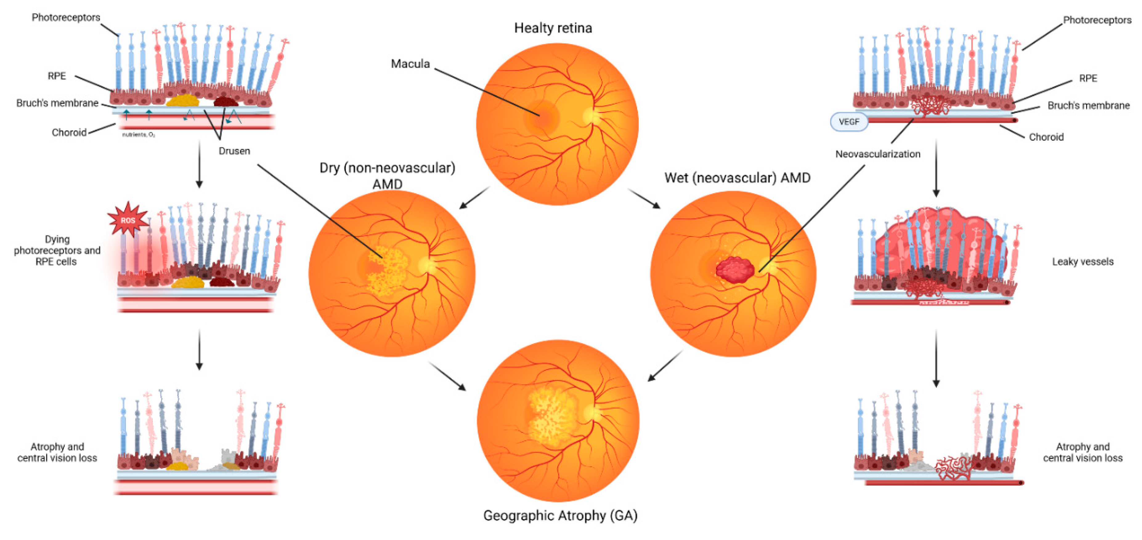
Figure 2.
Anatomy of the retina. The retina is a multilayered neural tissue organized into distinct cellular and structural layers. Created in https://BioRender.com.
Figure 2.
Anatomy of the retina. The retina is a multilayered neural tissue organized into distinct cellular and structural layers. Created in https://BioRender.com.
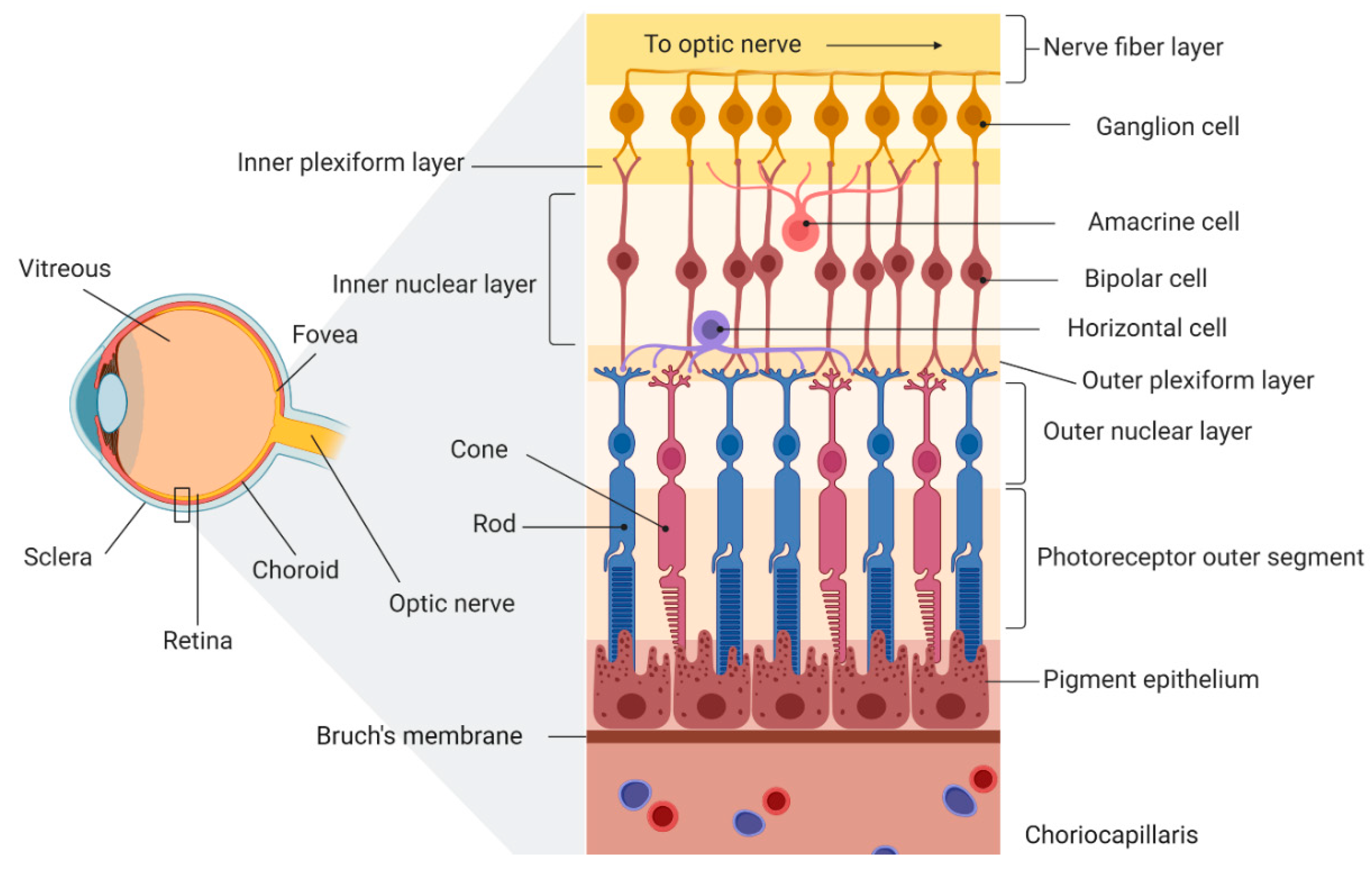
Figure 3.
Overview of RPE metabolism and associated cellular pathways. Glucose is transported from the choroidal circulation into the RPE via the glucose transporter GLUT1 and is subsequently supplied to photoreceptors, which predominantly rely on aerobic glycolysis for energy production, resulting in lactate generation. The lactate transporter MCT1 facilitates the uptake of lactate from the subretinal space into the RPE, where it is converted to pyruvate and integrated into the tricarboxylic acid (TCA) cycle. Excess lactate is effluxed from the RPE across the basolateral membrane by MCT3. The RPE also utilizes fatty acids derived from the heterophagy of shed photoreceptor outer segments to fuel β-oxidation and ketogenesis, ultimately generating β-HB, which is then utilized by photoreceptors to support oxidative metabolism.
Figure 3.
Overview of RPE metabolism and associated cellular pathways. Glucose is transported from the choroidal circulation into the RPE via the glucose transporter GLUT1 and is subsequently supplied to photoreceptors, which predominantly rely on aerobic glycolysis for energy production, resulting in lactate generation. The lactate transporter MCT1 facilitates the uptake of lactate from the subretinal space into the RPE, where it is converted to pyruvate and integrated into the tricarboxylic acid (TCA) cycle. Excess lactate is effluxed from the RPE across the basolateral membrane by MCT3. The RPE also utilizes fatty acids derived from the heterophagy of shed photoreceptor outer segments to fuel β-oxidation and ketogenesis, ultimately generating β-HB, which is then utilized by photoreceptors to support oxidative metabolism.
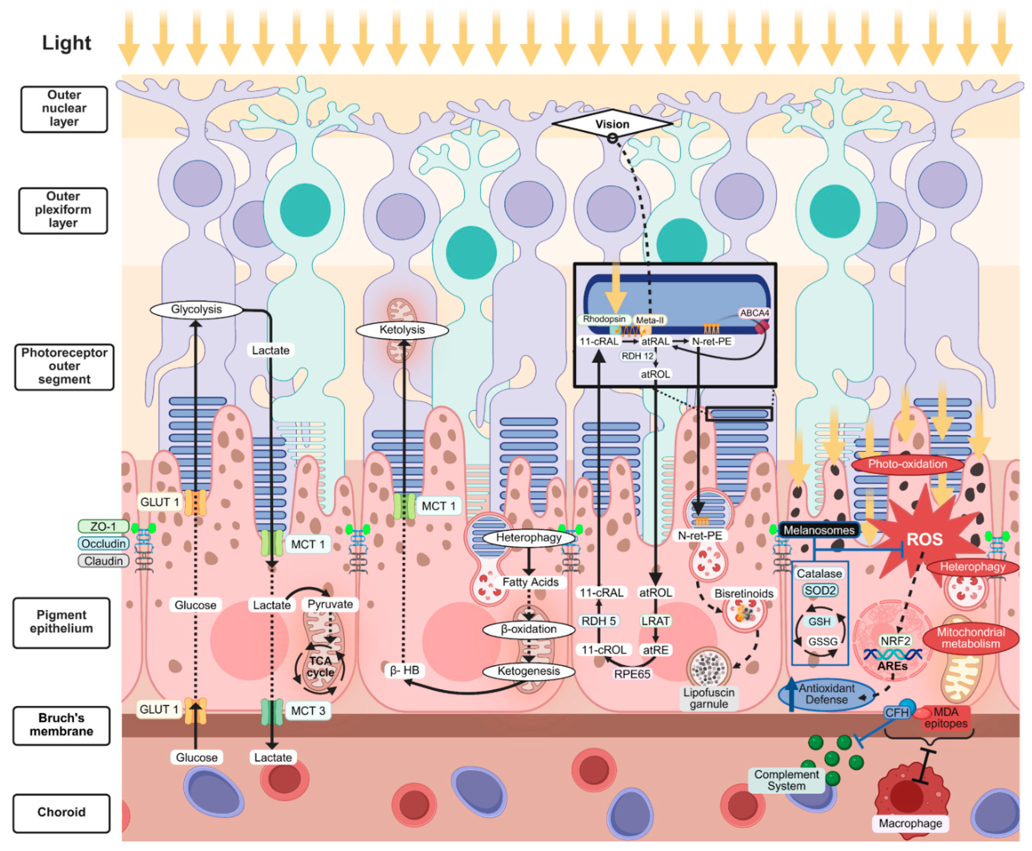
Figure 4.
Staged model of AMD progression. (a) Stage 0 represents the physiological, well-regulated compensation of metabolic demands and stressors. (b) Stage 1: chronic stress priming, characterized by increasing oxidative stress burden and baseline inflammatory activation. (c) Stage 2: impaired clearance, transport, and regenerative capacity, with the initial accumulation of intracellular debris. (d) Stage 3: extracellular matrix remodeling and drusen formation progressively restrict nutrients diffusion across Bruch’s membrane. (e) Stage 4: atrophic extracellular matrix remodeling and RPE dysfunction leads to activation of regulated cell death pathways. (f) Stage 5: the active front of atrophy tends to expand, culminating in irreversible vision loss and GA. Created in https://BioRender.com.
Figure 4.
Staged model of AMD progression. (a) Stage 0 represents the physiological, well-regulated compensation of metabolic demands and stressors. (b) Stage 1: chronic stress priming, characterized by increasing oxidative stress burden and baseline inflammatory activation. (c) Stage 2: impaired clearance, transport, and regenerative capacity, with the initial accumulation of intracellular debris. (d) Stage 3: extracellular matrix remodeling and drusen formation progressively restrict nutrients diffusion across Bruch’s membrane. (e) Stage 4: atrophic extracellular matrix remodeling and RPE dysfunction leads to activation of regulated cell death pathways. (f) Stage 5: the active front of atrophy tends to expand, culminating in irreversible vision loss and GA. Created in https://BioRender.com.
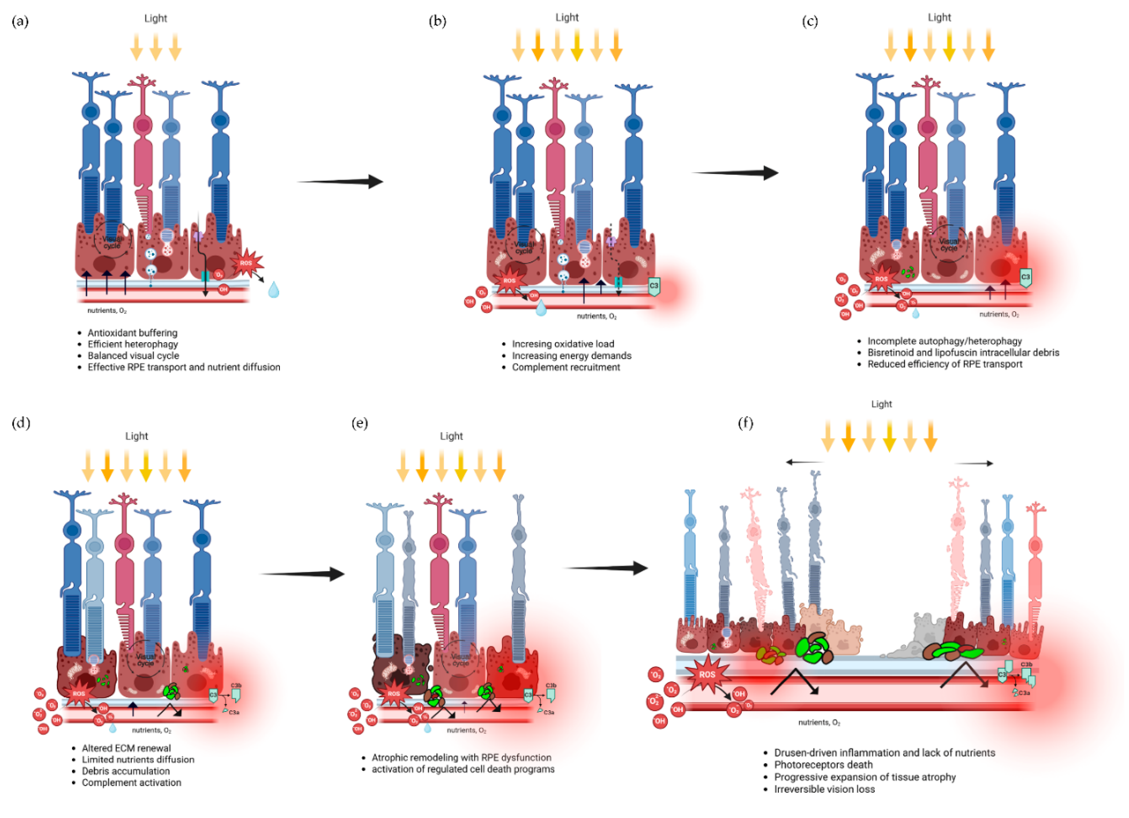
Disclaimer/Publisher’s Note: The statements, opinions and data contained in all publications are solely those of the individual author(s) and contributor(s) and not of MDPI and/or the editor(s). MDPI and/or the editor(s) disclaim responsibility for any injury to people or property resulting from any ideas, methods, instructions or products referred to in the content. |
© 2026 by the authors. Licensee MDPI, Basel, Switzerland. This article is an open access article distributed under the terms and conditions of the Creative Commons Attribution (CC BY) license (http://creativecommons.org/licenses/by/4.0/).
Copyright: This open access article is published under a Creative Commons CC BY 4.0 license, which permit the free download, distribution, and reuse, provided that the author and preprint are cited in any reuse.