

Submitted:
02 October 2026
Posted:
05 October 2026
You are already at the latest version
Abstract
Background/Objectives: Histone H3 K27-altered diffuse midline gliomas (DMGs) have poor outcomes and are characterized by global loss of H3K27 trimethylation (H3K27me3). Evidence from other H3K27me3-deficient tumors suggests a potential vulnerability to combined histone deacetylase (HDAC) and MEK inhibition. We examined a clinical experience with this combination and tested its activity in glioma models. Methods: We reviewed the course of an 11-year-old with neurofibromatosis type 1 and a thoracic spinal DMG treated with selumetinib and vorinostat after gross-total resection, without upfront radiotherapy. In post hoc viability assays, we tested HDAC and MEK inhibition in two H3K27M-mutant glioma lines, an isogenic K27-rescued derivative, and two H3-wildtype lines. Combination benefit was measured as the reduction in area under the viability curve relative to vorinostat alone. Results: The patient experienced 22 months of radiotherapy-free, progression-free survival before recurrence, with a postoperative enhancing focus resolving during treatment. Treatment was associated with grade 1 diarrhea and dermatitis. In vitro, combination benefit was greater in the H3K27M-mutant lines than in the H3-wildtype lines (mean difference, 13.2; 95% confidence interval, 8.1–18.3; p < 0.001). Benefit was also greater in DIPG17 than in its K27-rescued derivative (mean difference, 22.4; 95% confidence interval, 10.1–34.7; p = 0.015). Conclusions: This clinical observation and the post hoc validation experiments support prospective study of H3K27me3 loss as a candidate biomarker for combined HDAC and MEK inhibition.
Keywords:
diffuse midline glioma
; K27M mutation
; PRC2 complex
; MEK inhibitor
; biomarker
; high-grade glioma
; vorinostat
; diffuse glioma
; molecular targeted therapies
; H3K27me3
1. Introduction
Histone H3 lysine 27 (H3K27)-altered diffuse midline gliomas (DMG) represent one of the most lethal central nervous system (CNS) malignancies, with a median overall survival rate of 14- 20 months [1,2,3,4]. The pathognomonic histone H3.3 (H3-3A) mutation at lysine 27 (K27) results in a pathologic epigenome characterized by a global reduction of H3 K27 trimethylation (H3K27me3) [5]. While DMGs primarily affect children, they also occur in adolescent and young adult (AYA) populations [6]. Standard of care remains maximally safe surgical resection followed by radiotherapy. Although temozolomide is frequently administered concurrently and adjuvantly for radiosensitization, its efficacy remains unproven [7,8]. Radiotherapy is palliative, and disease progression typically occurs within 6 to 12 months. Emerging treatment modalities, including oncolytic virotherapy, dopamine receptor antagonists, and chimeric antigen receptor T-cell therapies have shown efficacy signals in early clinical experiences but long-term disease control remains elusive [6,9,10,11]. In this report, we describe an adolescent with Neurofibromatosis Type 1 (NF1) and a spinal cord H3K27-altered diffuse midline glioma who achieved 22 months of radiotherapy-free, progression-free survival (PFS) using a novel combination of the histone deacetylase (HDAC) inhibitor vorinostat and the MEK inhibitor selumetinib.
Neurofibromatosis Type 1 (NF1), a multisystem disorder phenotypically characterized by café-au-lait macules, is the most common tumor predisposition syndrome [12], though the development of high-grade gliomas in the adolescent and young adult NF1 population is rare [13,14]. More common NF1-associated neoplasms in the AYA age group include low-grade gliomas, plexiform neurofibromas, and malignant peripheral nerve sheath tumors (MPNST) [15]. NF1-related tumors are characterized by hyperactivation of the RAS-mitogen-activated protein kinase (MAPK) [16–21pathway due to loss of neurofibromin-mediated negative regulation of Ras signaling. MAPK pathway inhibitors, including MEK inhibitors, are now standard in managing various NF1-related and sporadic pediatric neoplastic pathologies [16,17,18,19,20,21]. High-grade MPNSTs evolve from plexiform neurofibromas via additional somatic alterations, including loss-of-function mutations in the Polycomb Repressive Complex 2 (PRC2) [22,23]. This loss results in a global depletion of (H3K27me3) creating an epigenetic landscape analogous to H3K27-altered DMGs, where the K27M substitution itself antagonizes PRC2 function [3,24,25]. Preclinical models of H3K27me3-deficient MPNST demonstrate profound synergy between HDAC and MAPK inhibition [26], a finding currently being translated in a phase 0 clinical trial (NCT06693284). Guided by this biological rationale, and published safety of HDAC inhibition in the DMG population [27], combination therapy with selumetinib and vorinostat was initiated as a personalized strategy for an adolescent with NF1 and a spinal DMG who sought to avoid radiotherapy due to the risk of exacerbated toxicity in the setting of baseline paraplegia.
2. Materials and Methods
2.1. Clinical Assessment and Treatment
The clinical course was reviewed retrospectively, including diagnostic pathology, germline testing, serial MRI, treatment exposure, and reported adverse events.
2.2. Cell Models and Culture
To investigate whether the notable clinical activity of dual HDAC and MEK inhibition was unique to the patient’s underlying germline NF1 genetics or represented a broader therapeutic vulnerability driven by the H3K27me3-deficient epigenetic phenotype, we conducted an in vitro validation study using two independently derived H3K27M-mutant glioma cell lines, DIPG17 and SF8628, together with the isogenic K27-rescued DIPGWT derivative of DIPG17 and two H3-wildtype pediatric-type high-grade glioma lines, SF9427 and SJ-HGGx2.
Glioma cell lines were maintained as serum-free adherent cultures using glioma tumor stem cell medium based on established protocols.28,29 The basal medium consisted of a 1:1 mixture of KnockOut DMEM/F-12 (Thermo Fisher Scientific, Cat# 12660012) and phenol red-free Neurobasal Medium (Thermo Fisher Scientific, Cat# 12348017). This base was supplemented with 1× MEM Non-Essential Amino Acids (Thermo Fisher Scientific, Cat# 11140050), GlutaMAX (Thermo Fisher Scientific, Cat# 35050061), sodium pyruvate (Thermo Fisher Scientific, Cat# 11360070), and 1M HEPES (Thermo Fisher Scientific, Cat# 15630080). The medium was enriched with B-27 Supplement (vitamin A-free) (Thermo Fisher Scientific, Cat# 12587010), N-2 Supplement (Thermo Fisher Scientific, Cat# 17502048), StemPro Neural Supplement (Thermo Fisher Scientific, Cat# A1050801), 0.2% heparin sodium salt (StemCell Technologies, Cat# 07980), and Primocin (InvivoGen, Cat# ant-pm-1). Recombinant human growth factors were added at final concentrations of 20 ng/mL epidermal growth factor (PeproTech, Cat# AF-100-15), 20 ng/mL basic fibroblast growth factor (PeproTech, Cat# 100-18B), 10 ng/mL platelet-derived growth factor-AA (Cell Guidance Systems, Cat# GFH16AF-100), and 10 ng/mL platelet-derived growth factor-BB (PDGF-BB; Cell Guidance Systems, Cat# GFH18AF-100). For adherent monolayer growth, culture vessels were pre-coated with Corning hESC-Qualified Matrigel matrix (Corning, Cat# 354277) diluted in chilled, HEPES-buffered DMEM/F-12 (Thermo Fisher Scientific, Cat# 11330032) and allowed to polymerize at 37 °C for 2 hours prior to cell seeding.
2.3. Drug-Response Assay
Glioma cells were seeded onto matrix-coated 96-well plates at 10,000 cells per well. Following overnight attachment, culture medium was replaced with treatment medium containing three-fold serial dilutions of mirdametinib (Selleckchem, Cat# S1036), vorinostat (Selleckchem, Cat# S1047), or their fixed-ratio combination. Maximum concentrations were 1,000 µM mirdametinib and 800 µM vorinostat, with the combination maintaining a fixed 1.25:1 mirdametinib:vorinostat ratio. This protocol mirrored the optimized experimental conditions for our MPNST experience [26]. Vehicle control wells were treated with a DMSO concentration equivalent to the highest concentration present in the drug dilutions.
Following 48 hours of continuous drug exposure, alamarBlue HS cell viability reagent (Thermo Fisher Scientific, Cat# DAL1025) was added to each well according to the manufacturer’s protocol. After a 4-hour incubation at 37 °C, absorbance at 570 and 600 nm was measured using a Cytation 5 cell imaging multi-mode reader (BioTek). Percent reduction of alamarBlue was calculated using the manufacturer’s dual-wavelength extinction coefficient method. Media-only background was subtracted from each measurement, and relative viability was normalized to the mean blank-corrected signal of vehicle-treated wells, defined as 100% viability. Six technical replicate wells at each concentration were averaged within each independent biological experiment.
2.4. Statistical Analysis
Combination benefit across the complete dose-response range was quantified for each independent biological experiment using the difference in normalized area under the viability curve (ΔAUC), defined as AUCvorinostat − AUCcombination. Larger positive ΔAUC values therefore indicate greater reduction in viability following addition of mirdametinib to vorinostat. First, ΔAUC was compared between DIPG17 and its isogenic K27-rescued DIPGWT derivative using a two-sided unpaired Welch t-test. Second, cross-background activity was evaluated using an equal-weight Welch-Satterthwaite fixed contrast comparing the mean of the two independently derived H3K27M lines, DIPG17 and SF8628, with the mean of the two independently derived H3-wildtype lines, SF9427 and SJ-HGGx2. Dose-response relationships were fit separately for each biological experiment using a four-parameter log-logistic model, with lower and upper asymptotes fixed at 0% and 100% viability, respectively. Statistical analyses and graphics were performed in R using the drc, ggplot2, and ggprism packages.
3. Results
3.1. Patient Presentation, Treatment, and Outcome
An 11-year-old with Neurofibromatosis Type 1 (germline pathogenic NF1 c.1586T>G (p.Leu529Arg) mutation) presented with scoliosis, neck pain, urinary hesitancy, and gait instability. MRI revealed a thoracic spinal cord mass (T2-T3 to T9) with a syrinx extending to C2 and surrounding edema (Figure 1A). In April 2021, the patient underwent a gross-total surgical resection. Post-operative baseline neurologic deficits included lower extremity paresis and neurogenic bladder. Histopathology confirmed a diffuse midline glioma, H3K27-altered (WHO grade 4). Immunohistochemistry was positive for H3K27M, SOX10, GFAP, S100, and Olig2, and negative for both H3K27me3 and MGMT promoter methylation. A neurofilament stain confirmed an infiltrative pattern of growth. Germline testing for EPCAM, MLH1, MSH2, MSH6, and PMS2 returned with no alterations, ruling out constitutional mismatch repair deficiency disorders [14].
Post-operative imaging showed trace intramedullary contrast enhancement, consistent with post-operatives changes or residual disease (Figure 1B). To avoid the toxicities of spinal radiotherapy in the context of NF1 and existing neurological compromise, the patient began selumetinib (25 mg/m2 BID) and vorinostat (230 mg/m2, days 1-7 and 15-21) in May 2021. The regimen was well-tolerated, with side effects limited to grade 1 diarrhea and grade 1 dermatitis. The patient maintained typical development, including normal linear growth and initiation of menses. Follow-up MRI at three and five months showed a contrast enhancing focus on the superior margin of the previously resected lesion, concerning for residual disease (Figure 1C, D). After eight months of therapy, this focus of enhancement resolved and there was no radiographic evidence of disease from January 2022 through February 2023 (Figure 1E,F).
In April 2023, 23 months after initiating therapy, the patient presented with back pain and worsening of her baseline neurologic deficits. MRI confirmed tumor recurrence, and she was initiated on dexamethasone for management of spinal cord compression. The family subsequently reported decreased medication adherence in the preceding months, involving pill-hiding behavior in the context of an underlying anxiety disorder. Despite a second resection and salvage radiotherapy, the disease proved refractory, and the patient passed away in September 2023.
3.2. In Vitro Combination Activity Across H3K27-Altered and Comparator Models
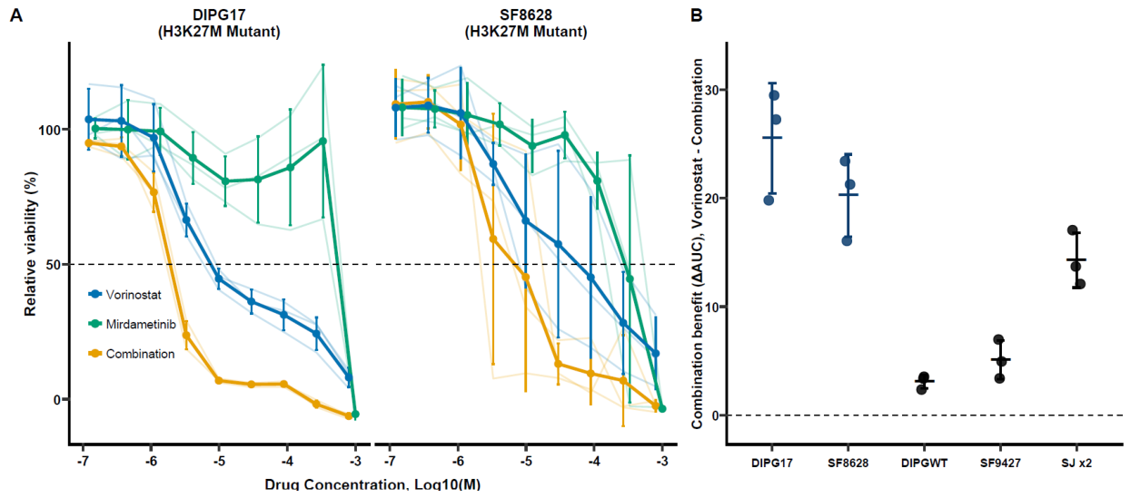
Biallelic NF1 tumor status was unable to be determined in this case owing to tissue availability limitations. To assess whether the combination effect extended beyond the NF1-/- genotype, we evaluated H3K27M-mutant, isogenic K27-rescued, and H3-wildtype glioma models (Figure 2) using the MEK inhibitor mirdametinib and HDAC inhibitor vorinostat.
In two independently derived H3K27M-mutant lines, DIPG17 and SF8628, addition of mirdametinib enhanced vorinostat activity across the dose-response range, despite relatively limited single-agent activity of mirdametinib. Mean ΔAUC was 25.5 in DIPG17 and 3.1 in its isogenic K27-rescued DIPGWT derivative (mean difference, 22.4; 95% CI, 10.1–34.7; Welch p = 0.015). Across independent cell-line backgrounds, mean ΔAUC was 22.9 for the two H3K27M lines and 9.7 for the two H3-wildtype lines (difference, 13.2; 95% CI, 8.1–18.3; Welch–Satterthwaite fixed contrast p < 0.001). Both comparisons remained significant after Holm correction.
4. Discussion
The clinical course of this patient is noteworthy, as a progression-free survival (PFS) exceeding 20 months without upfront radiotherapy is exceptional in the management of DMG. This prolonged disease control must be interpreted in the context of the surgical accessibility of the spinal lesion, whereas most DMGs involve more eloquent structures and are ineligible for near-total resection. However, maximal safe resection alone is insufficient for durable control of DMG, particularly in the presence of residual contrast-enhancing disease. It remains unknown whether the patient stopped adhering to therapy, permitting disease regrowth, or if she stopped therapy without seeking medical advice owing to the return of neurologic symptoms; thus, whether further PFS would have been observed remains conjecture. Another important caveat is that the disease response observed here was achieved prior to the institution of radiotherapy, which could in theory modulate cellular responses to targeted therapies in unpredictable ways. Our institutional experience evaluating the synergy of histone deacetylase and MEK inhibition for H3K27me3-deficient MPNST (NCT06693284), combined with this signal of efficacy in a patient with DMG, carries significant implications not only for DMGs but also for Posterior Fossa Group A (PFA) ependymomas, another lethal childhood malignancy characterized by a global loss of H3K27me3.
While global loss of H3K27me3 in MPNSTs typically arise from inactivating mutations in the PRC2 subunits SUZ12 or EED, PFA ependymomas and DMGs achieve a similar H3K27me3-deficient phenotype through EZHIP overexpression or H3K27 alterations, respectively [2,30,31,32]. This depletion of repressive trimethylation facilitates a reciprocal gain in acetylation (H3K27ac) at the same residue, which promotes an active, enhancer-driven chromatin state [32]. HDAC inhibitors have emerged as a potent therapeutic strategy to exploit this vulnerability. By blocking the removal of acetyl groups, HDAC inhibitors induce hyperacetylation, leading to transcriptional dysregulation [33,34].
Importantly, H3K27me3 loss converges with heightened MAPK signaling across these malignancies, linking the altered epigenome to a therapeutically targetable kinase pathway. In MPNSTs, the loss of PRC2 acts as a potent amplifier of Ras-driven transcription, synergizing with NF1 loss to drive oncogenesis [35,36]. A similar phenomenon is observed in DMGs and PFA ependymomas, with observed MAPK pathway activation. H3K27-altered DMGs frequently exhibit functional hyperactivation of the RAS/MAPK signaling cascade as a direct consequence of their defining epigenetic state [37]. This epigenetically driven MAPK activation is further augmented by the activity of commonly co-occurring activating alterations of receptor tyrosine kinases such as PDGFRA [38,39,40]. Intriguingly, PFA ependymoma exhibits some of the highest functional phosphorylated ERK scores across pediatric central nervous system tumors despite absence of genomic mutations in the MAPK pathway [38,41,42]. Together, these data identify recurrent MAPK pathway activation across H3K27me3-deficient nervous system tumors and provide a rationale for extending dual HDAC/MEK inhibition beyond MPNST.
The cell-line results support testing H3K27me3 deficiency as a candidate marker of combination benefit. The well-tolerated nature of these agents, coupled with proven safety profiles and blood-brain-barrier penetrance [18,43,44], warrants independent in vivo and prospective clinical validation. The co-occurrence of MAPK activation and H3K27me3 loss suggest that dual inhibition of HDACs and the MAPK pathway may provide a synergistic therapeutic approach for these aggressive malignancies.
5. Conclusions
An adolescent with H3K27-altered spinal diffuse midline glioma experienced prolonged radiotherapy-free disease control while receiving selumetinib and vorinostat after gross-total resection. In vitro, adding mirdametinib to vorinostat produced greater viability reduction in H3K27M-mutant models than in the K27-rescued and H3-wildtype comparators. Together, these observations justify prospective testing of H3K27me3 loss as a candidate biomarker for combined MEK and HDAC inhibition.
Author Contributions
Conceptualization, R.T.G., C.L.M., D.A.L. and K.W.; methodology, R.T.G., A.L., M.S., G.F., D.N., P.C.G., C.L.M., D.A.L. and K.W.; validation, R.T.G., A.L., M.S., G.F., D.N., C.H., E.C.N., G.T., C.L.M., D.A.L. and K.W.; formal analysis, R.T.G., G.F., D.N., C.L.M., D.A.L. and K.W.; investigation, R.T.G., A.L., M.S., G.F., D.N., C.L.M., D.A.L. and K.W.; resources, R.T.G., C.H., E.C.N., G.T., C.L.M., D.A.L. and K.W.; data curation, R.T.G., G.F., D.N., P.C.G. and C.L.M.; writing—original draft preparation, R.T.G., G.F., D.N. and P.C.G.; writing—review and editing, R.T.G., A.L., M.S., G.F., D.N., P.C.G., C.H., E.C.N., G.T., C.L.M., D.A.L. and K.W.; visualization, R.T.G., D.N. and K.W.; project administration, R.T.G., C.L.M., D.A.L. and K.W. All authors have read and agreed to the published version of the manuscript.
Funding
This research received no external funding.
Institutional Review Board Statement
Ethical review and approval were waived for this study due to non-human research designation.
Informed Consent Statement
The authors confirm that written consent for submission and publication of this case report, including the images and associated text, have been obtained from the legal authorized representative.
Data Availability Statement
Experimental data and protocols are available at https://osf.io/w4zf2/overview?view_only=58da12eacb1244e18e4a5577f6623670.
Acknowledgments
The DIPG17 and DIPGWT cell lines originated from Paul Knofler, UC Davis. The SJ-HGGX2 cell line and pediatric high-grade glioma culture protocol originated from the St. Jude Brain Tumor Portal and Suzanne Baker, respectively. The SF8628 and SF9427 were obtained from Edward Hinchcliffe, Hormel Institute.
Conflicts of Interest
Christopher L. Moertel: OX2 Therapeutics—Chief Medical Officer. OX2 is currently conducting a clinical trial for the treatment of diffuse midline glioma. Healx, Ltd.—Global Head of Clinical development. Healx is currently conducting a clinical trial using an HDAC inhibitor, domatinostat, in the treatment of metastatic/recurrent sarcoma. David A. Largaespada: Dr. Largaespada is the co-founder and co-owner of NeoClone Biotechnologies, Inc., Discovery Genomics, Inc. (recently acquired by Immunsoft, Inc.), B-MoGen Biotechnologies, Inc. (recently acquired by the Biotechne corporation), and Luminary Therapeutics, Inc. He consults for Styx Biotechnologies and Genentech, Inc., which has funded some of his research. The business of all these companies is unrelated to the contents of this manuscript. All other authors declare no competing interests..
Abbreviations
The following abbreviations are used in this manuscript:
| MDPI | Multidisciplinary Digital Publishing Institute |
| DOAJ | Directory of open access journals |
| TLA | Three letter acronym |
| LD | Linear dichroism |
References
- Ostrom, Q.T.; Cioffi, G.; Waite, K.; Kruchko, C.; Barnholtz-Sloan, J.S. CBTRUS Statistical Report: Primary Brain and Other Central Nervous System Tumors Diagnosed in the United States in 2014–2018. Neuro-Oncol. 2021, 23 (Supplement_3), iii1–iii105. [Google Scholar] [CrossRef] [PubMed]
- Mackay, A.; Burford, A.; Carvalho, D.; et al. Integrated Molecular Meta-Analysis of 1,000 Pediatric High-Grade and Diffuse Intrinsic Pontine Glioma. Cancer Cell 2017, 32(4), 520–537.e5. [Google Scholar] [CrossRef] [PubMed]
- Saratsis, A.M.; Knowles, T.; Petrovic, A.; Nazarian, J. H3K27M mutant glioma: Disease definition and biological underpinnings. Neuro-Oncol. 2024, 26 (Supplement_2), S92–S100. [Google Scholar] [CrossRef] [PubMed]
- Van Den Bent, M.; Saratsis, A.M.; Geurts, M.; Franceschi, E. H3 K27M-altered glioma and diffuse intrinsic pontine glioma: Semi-systematic review of treatment landscape and future directions. Neuro-Oncol. 2024, 26 (Supplement_2), S110–S124. [Google Scholar] [CrossRef] [PubMed]
- Wen, P.Y.; Packer, R.J. The 2021 WHO Classification of Tumors of the Central Nervous System: clinical implications. Neuro-Oncol. 2021, 23(8), 1215–1217. [Google Scholar] [CrossRef] [PubMed]
- Arrillaga-Romany, I.; Gardner, S.L.; Odia, Y.; et al. ONC201 (Dordaviprone) in Recurrent H3 K27M–Mutant Diffuse Midline Glioma. J. Clin. Oncol. 2024, 42(13), 1542–1552. [Google Scholar] [CrossRef] [PubMed]
- Cohen, K.J.; Heideman, R.L.; Zhou, T.; et al. Temozolomide in the treatment of children with newly diagnosed diffuse intrinsic pontine gliomas: a report from the Children’s Oncology Group. Neuro-Oncol. 2011, 13(4), 410–416. [Google Scholar] [CrossRef] [PubMed]
- Stupp, R.; Weller, M.; Belanger, K.; et al. Radiotherapy plus Concomitant and Adjuvant Temozolomide for Glioblastoma. N Engl J Med. Published online. 2005.
- Gállego Pérez-Larraya, J.; Garcia-Moure, M.; Labiano, S.; et al. Oncolytic DNX-2401 Virus for Pediatric Diffuse Intrinsic Pontine Glioma. N Engl. J. Med. 2022, 386(26), 2471–2481. [Google Scholar] [CrossRef] [PubMed]
- Hansford, J.R.; Manoharan, N.; Bouche, G.; Ramaswamy, V.; Jabado, N.; Bouffet, E. ONC201 (Dordaviprone): review of evidence to date in diffuse midline glioma, hope or hype? Expert Opin. Emerg. Drugs 2024, 29(4), 321–325. [Google Scholar] [CrossRef] [PubMed]
- Majzner, R.G.; Ramakrishna, S.; Yeom, K.W.; et al. GD2-CAR T cell therapy for H3K27M-mutated diffuse midline gliomas. Nature 2022, 603(7903), 934–941. [Google Scholar] [CrossRef] [PubMed]
- Legius, E. Revised diagnostic criteria for neurofibromatosis type 1 and Legius syndrome: an international consensus recommendation. Genet Med. 8.
- Garibotto, F.; Madia, F.; Milanaccio, C.; et al. Pediatric Diffuse Midline Gliomas H3 K27M-Mutant and Non-Histone Mutant Midline High-Grade Gliomas in Neurofibromatosis Type 1 in Comparison With Non-Syndromic Children: A Single-Center Pilot Study. Front Oncol. 2020, 10, 795. [Google Scholar] [CrossRef] [PubMed]
- Wimmer, K.; Kratz, C.P.; Vasen, H.F.A.; et al. Diagnostic criteria for constitutional mismatch repair deficiency syndrome: suggestions of the European consortium ‘Care for CMMRD’ (C4CMMRD). J. Med. Genet. 2014, 51(6), 355–365. [Google Scholar] [CrossRef] [PubMed]
- Galvin, R.; Watson, A.L.; Largaespada, D.A.; Ratner, N.; Osum, S.; Moertel, C.L. Neurofibromatosis in the Era of Precision Medicine: Development of MEK Inhibitors and Recent Successes with Selumetinib. Curr. Oncol. Rep. 2021, 23(4), 45. [Google Scholar] [CrossRef] [PubMed]
- de Blank, P.M.K.; Gross, A.M.; Akshintala, S.; et al. MEK inhibitors for neurofibromatosis type 1 manifestations: Clinical evidence and consensus. Neuro-Oncol. 2022, 24(11), 1845–1856. [Google Scholar] [CrossRef] [PubMed]
- Dombi, E.; Baldwin, A.; Marcus, L.J.; et al. Activity of Selumetinib in Neurofibromatosis Type 1–Related Plexiform Neurofibromas. N Engl. J. Med. 2016, 375(26), 2550–2560. [Google Scholar] [CrossRef] [PubMed]
- Fangusaro, J.; Onar-Thomas, A.; Poussaint, T.Y.; et al. A phase II trial of selumetinib in children with recurrent optic pathway and hypothalamic low-grade glioma without NF1: a Pediatric Brain Tumor Consortium study. Neuro-Oncol. 2021, 23(10), 1777–1788. [Google Scholar] [CrossRef] [PubMed]
- Weiss, B.D.; Wolters, P.L.; Plotkin, S.R.; et al. NF106: A Neurofibromatosis Clinical Trials Consortium Phase II Trial of the MEK Inhibitor Mirdametinib (PD-0325901) in Adolescents and Adults With NF1-Related Plexiform Neurofibromas. J. Clin. Oncol. 2021, 39(7), 797–806. [Google Scholar] [CrossRef] [PubMed]
- Fangusaro, J.; Onar-Thomas, A.; Young Poussaint, T.; et al. Selumetinib in paediatric patients with BRAF-aberrant or neurofibromatosis type 1-associated recurrent, refractory, or progressive low-grade glioma: a multicentre, phase 2 trial. Lancet Oncol. 2019, 20(7), 1011–1022. [Google Scholar] [CrossRef] [PubMed]
- Ryall, S.; Zapotocky, M.; Fukuoka, K.; et al. Integrated Molecular and Clinical Analysis of 1,000 Pediatric Low-Grade Gliomas. Cancer Cell 2020, 37(4), 569–583.e5. [Google Scholar] [CrossRef] [PubMed]
- Pemov, A.; Li, H.; Presley, W.; Wallace, M.R.; Miller, D.T. Genetics of human malignant peripheral nerve sheath tumors. Neuro-Oncol. Adv. 2020, 2 (Supplement_1), i50–i61. [Google Scholar] [CrossRef] [PubMed]
- Zhang, X.; Murray, B.; Mo, G.; Shern, J.F. The Role of Polycomb Repressive Complex in Malignant Peripheral Nerve Sheath Tumor. Genes. 2020, 11(3), 287. [Google Scholar] [CrossRef] [PubMed]
- Somatilaka, B.N.; Sadek, A.; McKay, R.M.; Le, L.Q. Malignant peripheral nerve sheath tumor: models, biology, and translation. Oncogene 2022, 41(17), 2405–2421. [Google Scholar] [CrossRef] [PubMed]
- Suppiah, S.; Mansouri, S.; Mamatjan, Y.; et al. Multiplatform molecular profiling uncovers two subgroups of malignant peripheral nerve sheath tumors with distinct therapeutic vulnerabilities. Nat. Commun. 2023, 14(1), 2696. [Google Scholar] [CrossRef] [PubMed]
- Shah, R.C.; Larsson, A.T.; Garana, B.B.; et al. Integrated ex vivo screening and transcriptomic profiling to prioritize drug combinations for rare cancers. bioRxiv 2026, 741264. [Google Scholar] [CrossRef]
- Monje, M.; Cooney, T.; Glod, J.; et al. Phase I trial of panobinostat in children with diffuse intrinsic pontine glioma: A report from the Pediatric Brain Tumor Consortium (PBTC-047). Neuro-Oncol. 2023, 25(12), 2262–2272. [Google Scholar] [CrossRef] [PubMed]
- Krug, B.; De Jay, N.; Harutyunyan, A.S.; et al. Pervasive H3K27 Acetylation Leads to ERV Expression and a Therapeutic Vulnerability in H3K27M Gliomas. Cancer Cell 2019, 35(5), 782–797.e8. [Google Scholar] [CrossRef] [PubMed]
- Pollard, S.M.; Yoshikawa, K.; Clarke, I.D.; et al. Glioma Stem Cell Lines Expanded in Adherent Culture Have Tumor-Specific Phenotypes and Are Suitable for Chemical and Genetic Screens. Cell Stem Cell 2009, 4(6), 568–580. [Google Scholar] [CrossRef] [PubMed]
- Hübner, J.M.; Müller, T.; Papageorgiou, D.N.; et al. EZHIP/CXorf67 mimics K27M mutated oncohistones and functions as an intrinsic inhibitor of PRC2 function in aggressive posterior fossa ependymoma. Neuro-Oncol. 2019, 21(7), 878–889. [Google Scholar] [CrossRef] [PubMed]
- Krug, B.; De Jay, N.; Harutyunyan, A.S.; et al. Pervasive H3K27 Acetylation Leads to ERV Expression and a Therapeutic Vulnerability in H3K27M Gliomas. Cancer Cell 2019, 35(5), 782–797.e8. [Google Scholar] [CrossRef] [PubMed]
- Wojcik, J.B.; Marchione, D.M.; Sidoli, S.; et al. Epigenomic Reordering Induced by Polycomb Loss Drives Oncogenesis but Leads to Therapeutic Vulnerabilities in Malignant Peripheral Nerve Sheath Tumors. Cancer Res. 2019, 79(13), 3205–3219. [Google Scholar] [CrossRef] [PubMed]
- Patel, A.B.; He, Y.; Radhakrishnan, I. Histone acetylation and deacetylation – Mechanistic insights from structural biology. Gene 2024, 890, 147798. [Google Scholar] [CrossRef] [PubMed]
- Algranati, D.; Oren, R.; Dassa, B.; et al. Dual targeting of histone deacetylases and MYC as potential treatment strategy for H3-K27M pediatric gliomas. eLife 2024, 13, RP96257. [Google Scholar] [CrossRef] [PubMed]
- De Raedt, T.; Beert, E.; Pasmant, E.; et al. PRC2 loss amplifies Ras-driven transcription and confers sensitivity to BRD4-based therapies. Nature 2014, 514(7521), 247–251. [Google Scholar] [CrossRef] [PubMed]
- Bhunia, M.M.; Stehn, C.M.; Jubenville, T.A.; et al. Multiomic analyses reveal new targets of polycomb repressor complex 2 in Schwann lineage cells and malignant peripheral nerve sheath tumors. Neuro-Oncol. Adv. 2024, 6(1), vdae188. [Google Scholar] [CrossRef] [PubMed]
- Koncar, R.F.; Dey, B.R.; Stanton, A.C.J.; et al. Identification of Novel RAS Signaling Therapeutic Vulnerabilities in Diffuse Intrinsic Pontine Gliomas. Cancer Res. 2019, 79(16), 4026–4041. [Google Scholar] [CrossRef] [PubMed]
- Selt, F.; Sigaud, R.; Korshunov, A.; et al. Association of phosphorylation status of ERK and genetic MAPK alterations in pediatric tumors. Sci. Rep. 2025, 15(1), 13498. [Google Scholar] [CrossRef] [PubMed]
- Pfaff, E.; Schramm, K.; Blattner-Johnson, M.; et al. Molecular characterization and clinical features of diffuse midline glioma in the pediatric precision oncology registry INFORM. Acta Neuropathol. (Berl) 2025, 150(1), 42. [Google Scholar] [CrossRef] [PubMed]
- Georgescu, M.M.; Islam, M.Z.; Li, Y.; et al. Global activation of oncogenic pathways underlies therapy resistance in diffuse midline glioma. Acta Neuropathol. Commun. 2020, 8(1), 111. [Google Scholar] [CrossRef] [PubMed]
- Bayliss, J.; Mukherjee, P.; Lu, C.; et al. Lowered H3K27me3 and DNA hypomethylation define poorly prognostic pediatric posterior fossa ependymomas. Sci. Transl. Med. 2016, 8(366). [Google Scholar] [CrossRef] [PubMed]
- Saleh, A.H.; Samuel, N.; Juraschka, K.; Saleh, M.H.; Taylor, M.D.; Fehlings, M.G. The biology of ependymomas and emerging novel therapies. Nat. Rev. Cancer 2022, 22(4), 208–222. [Google Scholar] [CrossRef] [PubMed]
- Su, J.M.; Kilburn, L.B.; Mansur, D.B.; et al. Phase I/II trial of vorinostat and radiation and maintenance vorinostat in children with diffuse intrinsic pontine glioma: A Children’s Oncology Group report. Neuro-Oncol. 2022, 24(4), 655–664. [Google Scholar] [CrossRef] [PubMed]
- Wang, K.; Yuan, M.; Graziano, G.; et al. ABSTRACT CITATION ID: NOAE064.440 LGG-49. EVALUATING THE EFFECTS OF MIRDAMETINIB IN NF1- MUTANT LOW-GRADE GLIOMAS USING BOTH CULTURED CELLS AND A NOVEL ZEBRAFISH XENOGRAFT MODEL. [PubMed]
Figure 1.
Response to selumetinib in combination with vorinostat in a patient with neurofibromatosis type 1 and diffuse midline glioma, H3K27-altered. Time points among the patient’s disease course are shown with representative sagittal T1 post-contrast MRI sequences. A) 4/16/21 T2 TSE sagittal, T1 TSE post contrast Dixon fat suppression. Presenting MRI shows a heterogeneously enhancing intramedullary mass from the T2-3 to the upper T9 level with severe hydromyelia superior to the tumor up to C2 and spinal cord edema both inferior to the tumor and superior to the hydromyelia extending to the lower pons. B) 4/30/21 STIR sagittal, T1 TSE post contrast Dixon Fat suppression. Post resection of the intramedullary mass, with marked improvement in hydromyelia, but with persistent spinal cord edema. C) 8/5/21, T1 TSE post contrast Dixon Fat suppression. Follow-up MRI 3 months from initiation of selumetinib and vorinostat demonstrates post-surgical changes and residual contrast enhancing lesion on the superior aspect of the lesion (arrow). D) 10/13/2021, T1 TSE post contrast Dixon Fat suppression. MRI continues to demonstrate a residual contrast enhancing foci at the T2 aspect of the previously resected lesion (arrow). E) 1/19/22, T1 TSE post contrast Dixon Fat suppression. Follow up imaging demonstrates post-surgical changes with trace residual contrast enhancement. F) 02/01/23, T1 TSE post contrast Dixon Fat suppression. Follow up imaging 22 months from initiation of therapy continues to demonstrate no radiographic evidence of disease recurrence. G) 03/29/2023, Sagittal T2-weighted TSE and T1-weighted TSE with Dixon Fat suppression. The patient presented with worsening neurologic symptoms; MRI showed disease recurrence with expansile contrast enhancing and intramedullary lesion in the thoracic cord (T4 – T7).
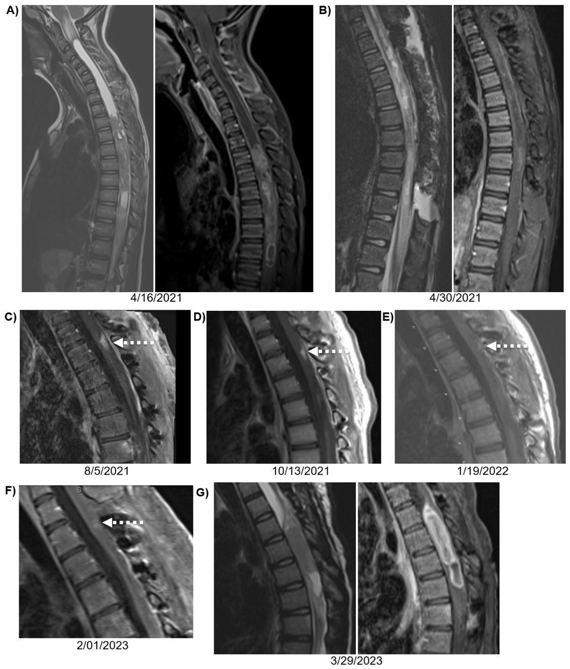
Figure 2.
H3K27me3 deficiency is associated with enhanced potentiation of vorinostat by MEK inhibition in DMG. A) Dose-response relationships for vorinostat, mirdametinib, and their fixed-ratio combination in two independently derived H3K27M-mutant glioma cell lines, DIPG17 and SF8628. Thin lines represent individual biological experiments; points and error bars represent mean ± SD across three independent biological experiments. Addition of mirdametinib enhanced vorinostat activity in both H3K27M models. B) Combination benefit quantified for each biological experiment as ΔAUC, defined as normalized AUCvorinostat—AUCcombination, with larger positive values indicating greater combination-associated reduction in viability. DIPG17 demonstrated greater combination benefit than its isogenic K27-rescued DIPGWT derivative (mean ΔAUC, 25.5 vs. 3.1; Welch p=0.015). The mean ΔAUC across the two H3K27M lines (DIPG17 and SF8628) was greater than across the two independent H3-wildtype lines (22.9 vs. 9.7; Welch-Satterthwaite fixed contrast, p<0.001).
Disclaimer/Publisher’s Note: The statements, opinions and data contained in all publications are solely those of the individual author(s) and contributor(s) and not of MDPI and/or the editor(s). MDPI and/or the editor(s) disclaim responsibility for any injury to people or property resulting from any ideas, methods, instructions or products referred to in the content. |
© 2026 by the authors. Licensee MDPI, Basel, Switzerland. This article is an open access article distributed under the terms and conditions of the Creative Commons Attribution (CC BY) license (http://creativecommons.org/licenses/by/4.0/).
Copyright: This open access article is published under a Creative Commons CC BY 4.0 license, which permit the free download, distribution, and reuse, provided that the author and preprint are cited in any reuse.