

Submitted:
02 October 2026
Posted:
05 October 2026
You are already at the latest version
Abstract
Pyocyanin is the blue green pigment produce by various strains of Pseudomonas aeruginosa .It has an ability to produce various pigment pyocyanin, pyomelanin, pyoverdine, pyorubin. Bacteria producing Pyocyanin were isolated from the laboratory instruments. CLM11 strain found to produce good amount of pyocyanin and molecular characterization revealed the bacteria is Pseudomonas aeruginosa. Ultra Violet Visible Spectrophotometry and FT-IR confirms the presence of aromatic and phenazine rings and characteristic absorption peaks of pyocyanin. Pyocyanin act as a natural antioxidant by helping to neutralize harmful free radical in the body this will support cellular health and reduce oxidative stress. CLM11pyocyanin had shown significant antioxidant and anti-microbial properties, in vivo toxicity studies in Zebra fish embryos confirms its non-toxic nature and the pigment had shown good dyeing ability to the cloths tested. This study advances creation of an environmentally safe and production process that may be used to replace synthetic dyes and also used as a dye in textile industries.
Keywords:
pyocyanin
; Pseudomonas aeruginosa
; antioxidant
; antimicrobial
Introduction
Pseudomonas aeruginosa has a remarkable virulence that determines its ability to resist different antibiotics, making it a life-threatening pathogen (Crone et al,. 2020) due to its unique and remarkable flexible microbes with metabolites that can adapt areas, sinks, sewage, hospital settings, and the majority of man-made ecosystems worldwide. flexible microbes with metabolites that can adapt to a wide range of biological niches areas, sinks, sewage, hospital settings, and the majority of man-made ecosystems worldwide. (Rossi al,. 2022) Pseudomonas aeruginosa has an amazing ability to synthesise secondary metabolite goods like pigments. In addition to being colorants, some pigments can have significant biological effects including antimicrobial, anticancer, antibiofilm, and antioxidant activity. These pigments have a substantial advantage as they hinder the growth of other possible microorganisms (Zhang et al,. 2020).
Pseudomonas aeruginosa as a cell factory for pyocyanin. It is gram-negative rod-shaped organism it produced various pigments like pyomelanin, pyoverdine, pyorubin and pyocyanin. The biosynthesis of pyocyanin is orchestrated by a cluster of genes known as the phz operons these genes guide the formation of phenazine-1-carboxylic acid which is converted into pyocyanin through enzymatic modifications [Mavrodi et al.,2001] Pyocyanin is a derivative of phenazine that comprises a heterocyclic nitrogen molecule of N-methyl-1-hydroxyphenazine, a secondary metabolite that is redox active and important for bacterial viability (Laxmi & Bhat, 2016.
Environmental influences on pyocyanin production are sensitive to various environmental factors. Optimal synthesis occurs under slightly alkaline pH condition [7.4 - 8.4] and at the temperature around 37ºC. Factors influencing pyocyanin production environmental factor like temperature, pH, nutrient availability and oxygen demand these are the factor affect the pyocyanin synthesis [Jayaseelan et al., 2014] Co-culturing with other microbes or adding specific carbon sources can enhance the production of pyocyanin of microorganisms. The cellular respiration of the species also depends on it. Pyocyanin is essential for the bacterium's ecological role. ecological and biological significance of pyocyanin also contributes to the ecological success of Pseudomonas aeruginosa in natural environment it plays a crucial role against other microorganisms by inhibiting the development of fungus and bacteria [Price-Whelan et al.,2006]. Pyocyanin helps in bio film formation a key survival for Pseudomonas aeruginosa by promoting extracellular DNA release and maintained redox balance within in bacterial communities.
Due to the adaptive nature of Pseudomonas aeruginosa the current study explores the presence of the bacteria in laboratory instruments like laminar air flow working space where UV treatment is done frequently. The pyocyanin production ability of the bacteria will be assessed.
Materials and Methods
Sample Collection
The sample was collected from laminar air flow cabinet working space. A sterile swab is used to wipe every part of laminar air flow including corners. The sample suspension was serially diluted with distilled water, and 100μl of the sample suspension was added into Nutrient agar plates. The plates were incubated. Colonies with different morphology and pigmented were selected for purification. Purification was done using quadrant streaking of the colonies in nutrient agar plates with 2% NaCl.
Screening Methods
Primary Screening
The isolated pure colonies were screened for pyocyanin production in tyrosine basal broth (Yabuuchi & Ohyama, 1972) with 1g/L L-tyrosine as the major source of nitrogen and carbon. 2% of isolates and basal broth were transferred in tubes with a 10mL medium, and 1% of L-tyrosine was separately added to the broth after autoclaving. The tubes were incubated for 2 to 3 days at 37ºC. Colour change in the medium was monitored the pyocyanin producing strains were selected for secondary screening in 100 mL of tyrosine basal broth medium.
Secondary Screening
Green colour producing isolates were utilized for mass production of pyocyanin in a 250mL conical flask with 100mL of Tyrosine basal broth and kept that medium in a shaking incubator for 2 to 3 days at 37ºC. Colour change in the medium were monitored daily.
Characterization of Pyocyanin Producing Bacterial Strain
Pyocyanin producing strain was characterized using Gram Staining, IMViC tests and catalase tests (Hemraj et al.,2013).
Antibiotic Sensitivity Tests
Antibiotic sensitivity tests which is explained below, was used to profile antibiotic sensitivity (Bauer et al., 1966). 3 mL of sterile physiological saline was used to hold one colony of the test strain. By either adding more sterile saline or adding new colonies. On a Mueller Hinton agar (HiMedia, India) plate, a consistent bacterial smear was created using a sterile cotton swab. Each plate could only contain four antibiotic discs. The discs were placed on the plate. Space was created between the discs to allow for the growth of the inhibition zone. to the examination Based on data from the manufacturer (Himedia, India).
Molecular Characterization of the Strain
Genomic DNA was isolated using the traditional phenol/chloroform extraction procedure, then used to amplify the 16S rRNA gene by PCR. PEG-NaCl precipitation was used to purify. During the sequencing process, each area was read at least twice from both ends using extra internal primers to guarantee accuracy. The Lasergene software package was used to process the assembled sequence data, and the EzBioCloud database was used to identify the bacteria.
Phylogenetic Analysis of Strain
BioEdit software was used for the multiple sequence alignment. Aligned sequences were put into version 5.0 of the MEGA5: Molecular Evolutionary Genetics Analysis (MEGA) software for further analysis (Tamura et al., 2007). The alignment's ends were cut to ensure that each sequence had the same length, and the aligned sequences were then put into MEGA format in order to do phylogenetic analysis. Utilizing the nucleotide-based TN84 evolutionary model for the construction of the phylogenetic tree, the neighbor-joining approach (Saitou and Nei, 1987) is utilized to calculate genetic distances using nucleotide alterations that are synonymous and nonsynonymous.
Extraction and Purification of Pigment
Centrifugation was used to extract Pseudomonas aeruginosa from the culture medium for 15 minutes at 7500 rpm. After discarding the bacterial pellet, the supernatant was moved to a conical flask in order to extract the pigment. Chloroform was used in a 1:2 ratio to extract the supernatant using a separating funnel. After carefully removing the aqueous layer, 1 N HCl was applied until a noticeable color shift was observed. A rotary vacuum evaporator (SARE-TE43 Model, SPAN Automation) was used to concentrate the resultant extract after it had been heated to 70°C and spun for a few minutes at 80 rpm. Purified pyocyanin was produced by this process, which also helped to concentrate the pigment and evaporate the chloroform (Abdelaziz, 2023).
Concentration of the pigment was estimated by
Concentration of pyocyanin =O.D520 x 17.072
Thin Layer Chromatography
The purity of pyocyanin is tested by thin-layer chromatography. The extracted pigment was spotted on a TLC plate and subjected to a solvent system including 75ml of methanol and 75ml of chloroform as a moving phase and wait for the pigment started to move spot towards the upper end with a gradual colour change after few hours remove the TLC from the glass container and left in room temperature for few hrs to dry as resolution front and the pigment retention factor was determined.
Characterization of Purified Pigment
UV Visible Spectrophotometry
A Ultra violet-visible spectrum was produced by scanning pyocyanin pigment a diverse range of UV and visible wavelengths. In the analysis Distilled water was used as the blank. To verify the pigment's authenticity, the spectrum produced was compared to a standard of pyocyanin (Marey et al., 2024).
FT-IR Spectrophotometry
IR-grade KBr (1:10 ratio) was combined with the purified bacteria and pyocyanin pigment, and the mixture was compressed into discs using a pellet machine under high pressure. A Thermo Nicolet Avatar 370 spectrophotometer and a KBr beam splitter, was used to record the FT-IR spectrum.
Bioactive Properties of the Pigment
DPPH Radical Scavenging Assay
Ascorbic acid served as the standard reference compound for comparison in the DPPH assay. In distilled water (1 mg/mL; w/v), the ascorbic acid stock solution was made. 200µL of a freshly made 60μM DPPH in methanol solution was combined with 50μL of CLM11 pyocyanin The DPPH solution alone, without the addition of pyocyanin or ascorbic acid, was utilized as the control. There was also a blank sample that contained 95% methanol (Munteanu & Apetrei, 2021).
Antimicrobial Activity of Pyocyanin by Well Diffusion Method
Microorganisms were used to assess the antibacterial activity of pure and plated in Mueller Hinton agar using well puncher to make a 5 wells control as streptomycin and 1st well load 100µl pyocyanin 2nd well 75µl of pyocyanin with 25µl of distilled water 3rd well 50µl of pyocyanin with 50µl of distilled water in 4th well 25µl of pyocyanin with 75µl of distilled water and in 5th well add 100µl of distilled water alone kept it in incubator for 24hrs and observe the zone (inhibition zone) using antibiotic zone scale to measure the diameter of the inhibition zone formed by the pyocyanin pigment (Gandhiraja,2009).
In Vivo Cytotoxicity of Pyocyanin
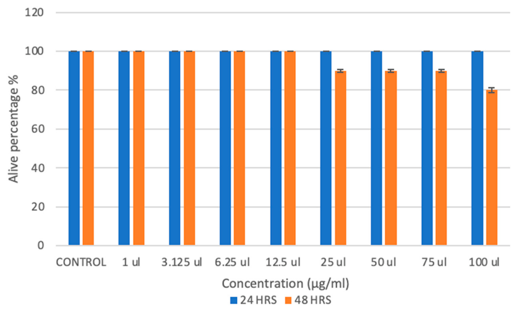
The embryo was purchased from the nearby aquarium and then we separated in 24 well plate as 5 embryo in each well and fill the well with the different concentration of the pigment from 1µl to 100µl concentration . The embryos was monitored for 24 hrs and 48 hrs by using microscope to observe the growth of the embryo present in the well plate to analyse the toxicity of the sample (Joshi & Katti, 2018).
Dyeing of Fabric with the Pigment
The dyeing ability of the pyocyanin in evaluated by using cotton cloth (Misra et al. 2017), The fabric that has been dyed with pyocyanin pigment produced by the CLM11to a period of 24 hours and then fabric washed to check the colour retaining ability (Sengupta & Bhowal, 2023)
Results and Discussion
Isolation of Bacteria from Laminar Air Flow Cabinet
From the spread plate method (Figure 1 a), 10 colonies were found to be grown. colonies were selected based on their color and morphological features for generating pure cultures; pure cultures formed in the fourth quadrant (Figure 1 b) in quadrant-streaked plates were transferred to fresh nutrient agar plates maintained at 4oC as temporary stock. Permanent glycerol stocks were prepared in 1:1 Ratio and stored at -80oC.
Screening for Pyocyanin Producing Bacteria
Primary Screening
Selected pure cultures of 10 colonies were screened for pyocyanin production in tyrosine basal broth. On the 3rd day, a color change was observed in some of the tubes, and at the end of the 5th day, we selected 1colony with better pyocyanin production, which is the strain CLM11 (Figure 2), showed very good pyocyanin production So, further research has been conducted using the CLM11strain.
Secondary Screening
CLM11 strain was selected for secondary screening, and the strain was inoculated in a flask containing tyrosine basal broth; the strain started producing pyocyanin from the 2nd day, and on the 4th day, complete green colour pyocyanin was produced. The production media is white (Figure 3 a) in color during inoculation, which turned green in colour on the 4th day (Figure 3 b).
Characterization of Pyocyanin Producing Strain CLM11
Gram Staining and Biochemical Characterization of Pyocyanin Producer
Gram staining was done to the strain CLM11 . The colonies appears pink colored rods which was found to be Gram negative in nature (Figure 4).
Biochemical characterization shows that CLM11 strain showed negative for indole, methyl red and Voges-Proskauer and organism growth from the stab line in motility medium its confirms its motile nature. The organism in kings B media produce green in color most notably pyocyanin produced by Pseudomonas aeruginosa (Table 1).
Antibiotics Susceptibility Test
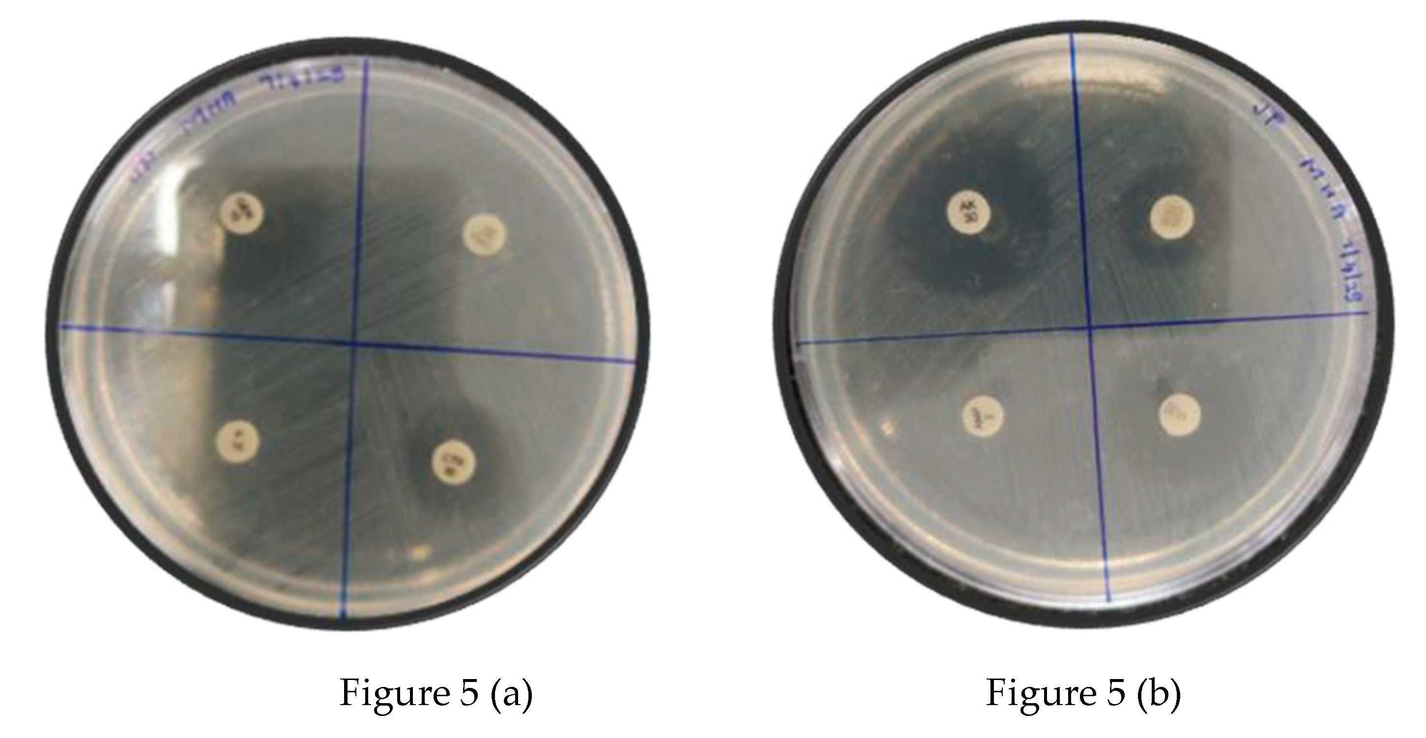
CLM11strain was swabbed over the agar plate 8 different antibiotic discs was placed in 2 plates the antibiotic disc forms clear zone against CLM11 stain without any contamination. Here, a total of 8 antibiotics were tested, and 3 antibiotic was found to be resistant (Table 2)
Figure 5.
Gentamycin [GEN10], Cefepime [CMP30], Ceftriaxone [CTR30], Penicillin [P10] antibiotics disc shows the zone of inhibition in the (Figure 5 a). Amikacin [ AK 30], Cefoperazone [CFS75/30], Streptomycin [S25], Ampicillin [AMP2] antibiotic disc and its zone of inhibition shown in (Figure 5 b).
Molecular Characterization of Strain CLM11
The 16S rDNA sequence is searched against GenBank database using BLAST. The strain had shown 94% similarity with Pseudomonas aeruginosa. which confirmed the strain CLM11 to be Pseudomonas aeruginosa. The accession number was obtained ( PV574319)
Phylogenetic Analysis of Strain CLM11
The phylogenetic tree was constructed by using 16S rDNA genes from 9 bacteria strains of which Staphylococcus aureus 41 served as the outgroup. Pseudomonas aeruginosa CLM11shared the same node with Pseudomonas aeruginosa strain. The strain showed similarity with other Pseudomonas species like Pseudomonas furukawaii, Pseudomonas yangonensis ,etc..as evident from a phylogenetic tree (Figure 6).
Extraction and Purification of the Pigment
Centrifugation was used to extract Pseudomonas aeruginosa from the culture medium for 15 minutes at 7500 rpm. After discarding the bacterial pellet, the supernatant was moved to a conical flask in order to extract the pigment. Chloroform was used in a 1:2 ratio to extract the supernatant using a separating funnel. After carefully removing the aqueous layer, 1 N HCl was applied until a noticeable color shift was observed. A rotary vacuum evaporator (SARE-TE43 Model, SPAN Automation) was used to concentrate the resultant extract after it had been heated to 70°C and spun for a few minutes at 80 rpm. Purified pyocyanin was produced by this process, which also helped to concentrate the pigment and evaporate the chloroform.
Figure 7.
In figure(a) the crude pyocyanin and chloroform in the separating funnel. Figure(b) the aqueous solution [chloroform] and pigment started to extract in the result the colour change was occur. In figure (c) chloroform was removed and collect in the glass container. In figure (d) the separated aqueous solution was treat with 1 N HCl and the red colour appeared. In Figure 8(e) after the red colour appeared transfer the pigment into round bottom flask in rotary vacuum evaporator for concentrating the pigment.
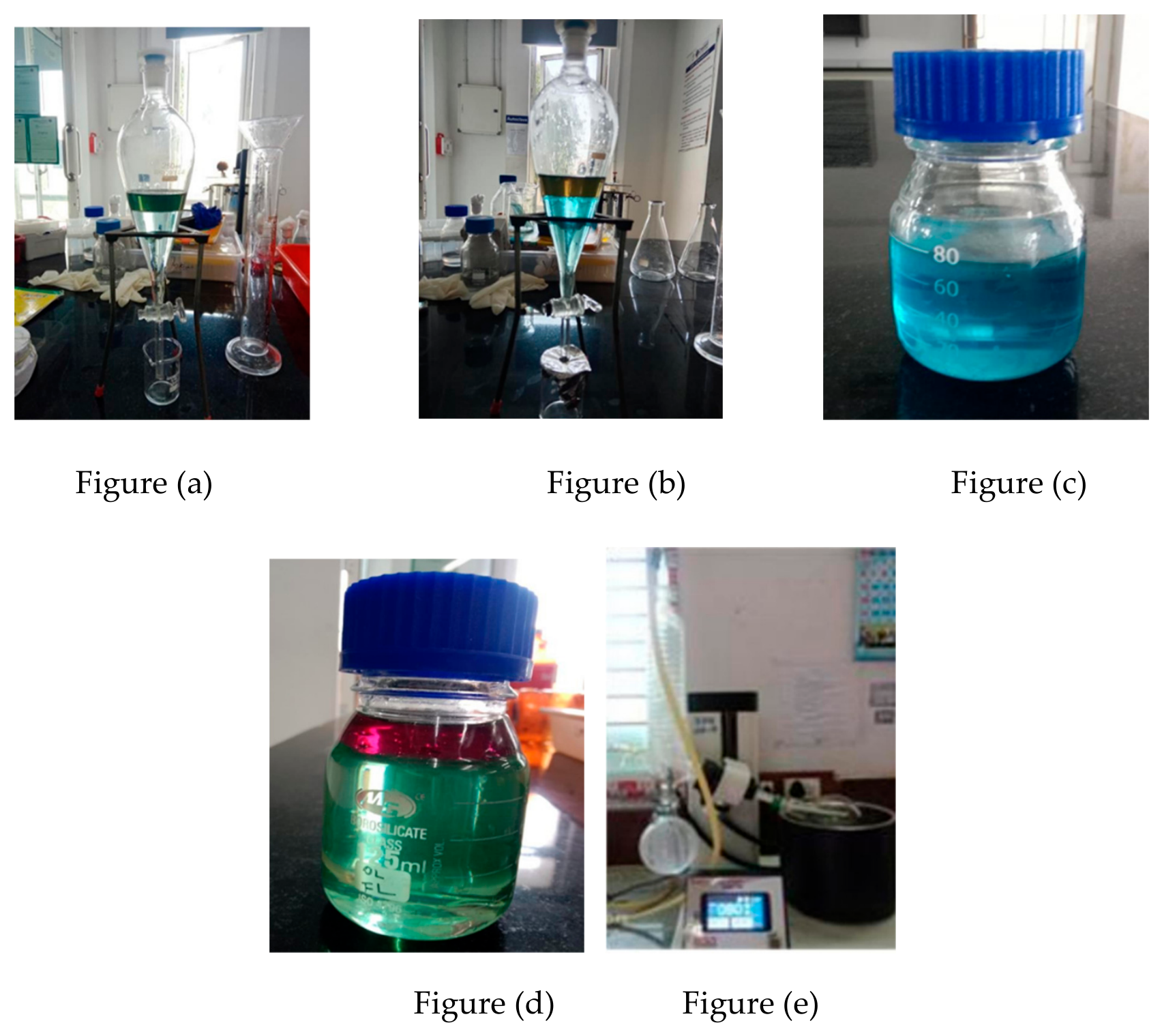
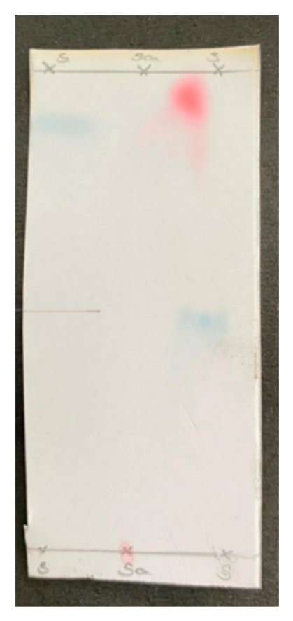
Figure 8.
Separation of Pyocyanin pigment using Thin Layer Chromatography.
Thin Layer Chromatography
The pyocyanin pigment which was extracted with chloroform was purified using thin layer chromatography and the value of RF=0.75 was found. The retention factor shown similar to that of the earlier reports (Ozdal, 2019)
Characterization of the Pigment
UV-Visible Spectrophotometry
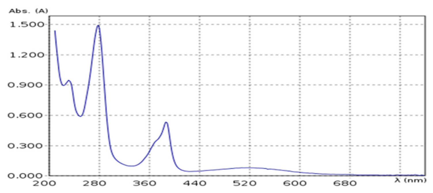
UV-visible spectrum examination ,CLM11 developed pyocyanin pigment dissolved in 0.2N of HCl and scanned using UV visible spectrometer at a wavelength range of 200-700nm. The spectrum had shown similar to the earlier reports (Marey et al., 2024).
Figure 9.
Shows the board absorption on the visible region.
FT-IR Spectrum
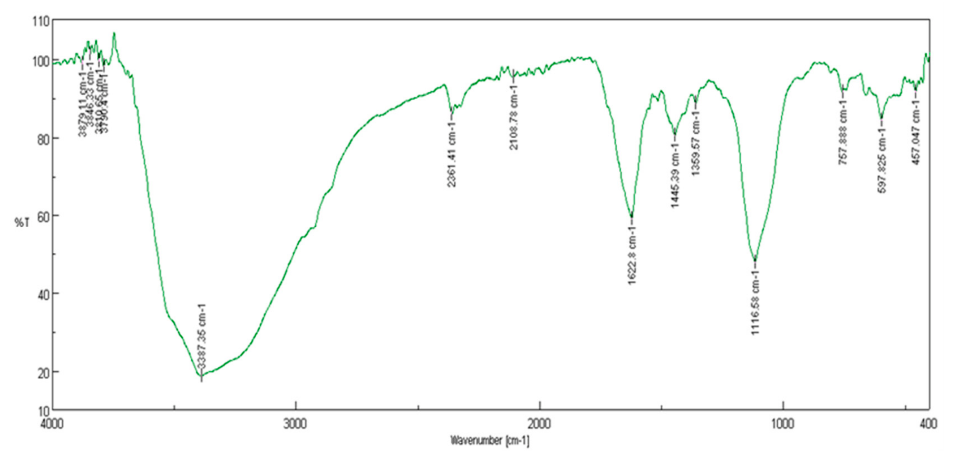
FT-IR results shows the identify functional group present in pigment and peaks indicate hydroxyl, carbonyl and aromatic group. Which confirm the presence of phenolic and heterocyclic compound in pigment. The spectrum was found similar to the earlier reports (Laxmi & Bhat, 2016).
Figure 10.
FT-IR results shows the identify functional group present in pigment CLM11 and the peaks indicate hydroxyl,carbonyl and aromatic group. Which confirm the presence of phenolic and heterocyclic compound in pigment.
Figure 10.
FT-IR results shows the identify functional group present in pigment CLM11 and the peaks indicate hydroxyl,carbonyl and aromatic group. Which confirm the presence of phenolic and heterocyclic compound in pigment.
Bioactive Properties of the Pigment
Antioxidant Activity of CLM11
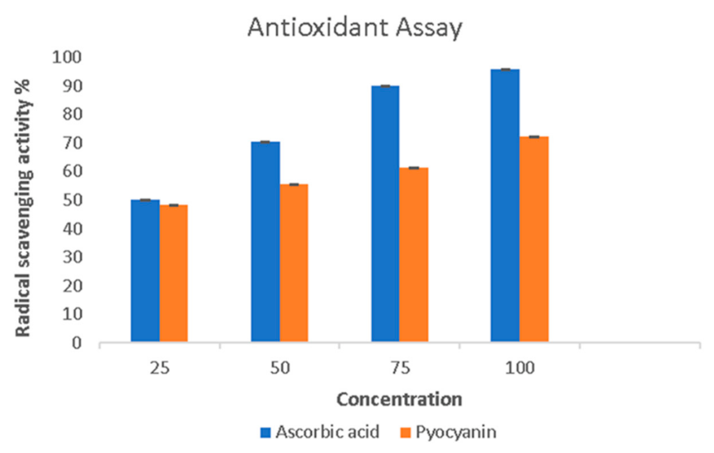
The antioxidant activity of pyocyanin was evaluated using the DPPH free radical scavenging method. pyocyanin showed concentration dependent scavenging activity.
Figure 11.
The represent the dose dependent antioxidant activity of pyocyanin measured by DPPH assay.
Figure 11.
The represent the dose dependent antioxidant activity of pyocyanin measured by DPPH assay.
Antimicrobial Activity of Pyocyanin Pigment
Pyocyanin pigment produced by the Pseudomonas aeruginosa and its antimicrobial activity. The result was found to be similar with earlier reports (Hamad et al, 2020).
Figure 12.
Pyocyanin shows strong antimicrobial activity which showing the significant zones of inhibition against different bacteria.
Figure 12.
Pyocyanin shows strong antimicrobial activity which showing the significant zones of inhibition against different bacteria.
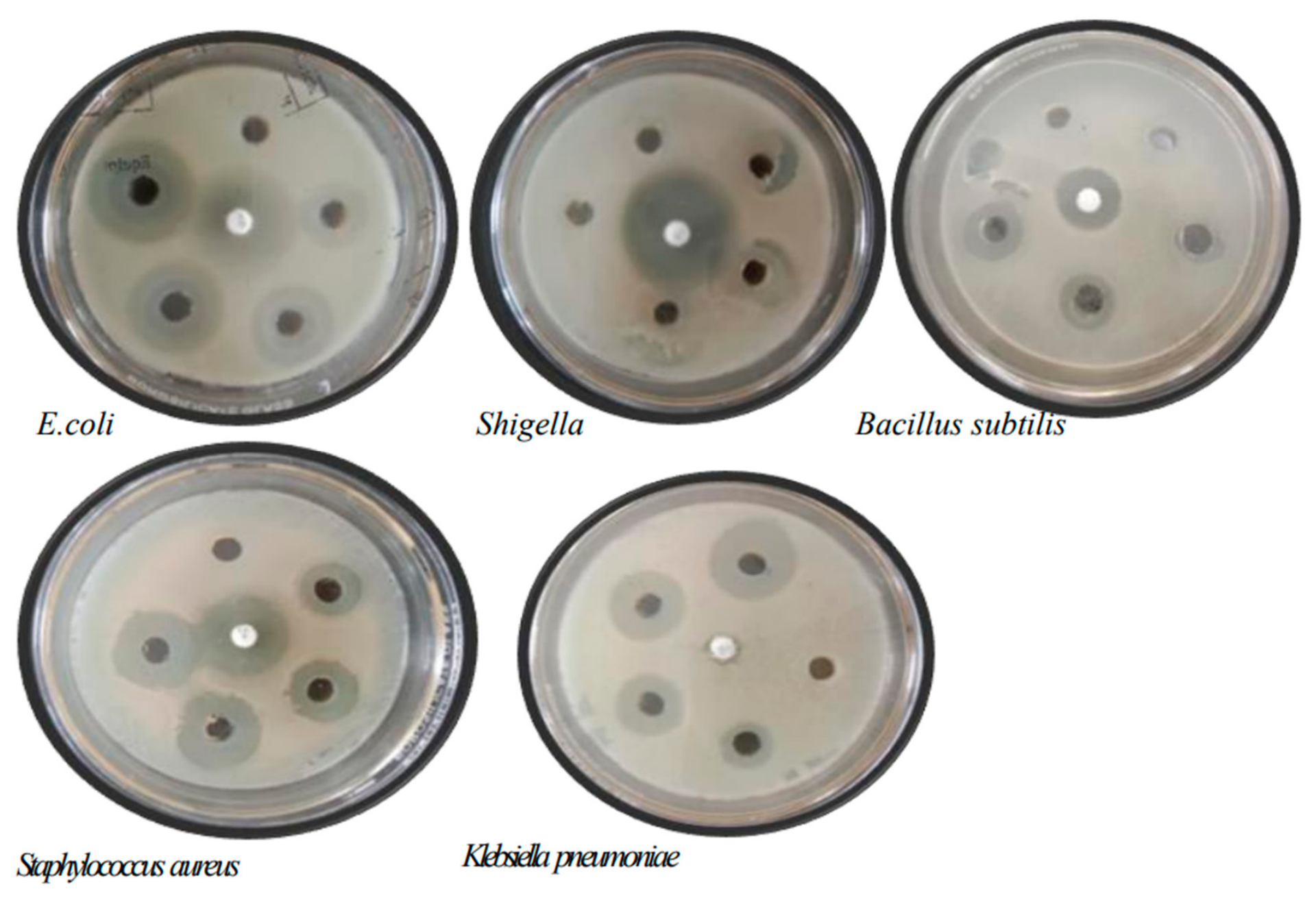
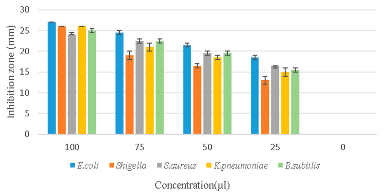
Figure 13.
The antimicrobial activity of pyocyanin was assessed against various test organism and the graph illustrate the zone of inhibition at different concentration.
Figure 13.
The antimicrobial activity of pyocyanin was assessed against various test organism and the graph illustrate the zone of inhibition at different concentration.
In Vivo Cytotoxicity of CLM11
In vivo cytotoxicity of pyocyanin in Danio rerio (zebra fish) is a valuable model to study the toxicological and developmental effects of this redox active pigment. CLM11 pyocyanin produced by the Pseudomonas aeruginosa was found to show less toxicity even in higher concentration, the outcome shows that the pigment is not harmful to Danio rerio embryos.
Figure 14.
Cytotoxicity graph represent how a pyocyanin compound affects embryo in different concentration.
Figure 14.
Cytotoxicity graph represent how a pyocyanin compound affects embryo in different concentration.
Dyeing of Fabric with the Pigment
Textiles dye application of extracted pyocyanin pigment on white cotton cloth and the cloth without the application of pyocyanin serve as the control . The cloth washed after the six hours , 12 hours and 24 hours intervals after the treatment. 24 hour treated cloth had shown higher accumulation of the pigment (Figure 15).
In conclusion, CLM11 pyocyanin pigment had shown good antimicrobial and dyeing potential. The pigment has shown very less toxicity on in vivo studies. More studies needed to confirm the potential and use it in textiles which could provide antimicrobial ability.
References
- Crone, S., Vives-Flórez, M., Kvich, L., Saunders, A. M., Malone, M., Nicolaisen, M. H., ... & Bjarnsholt, T. The environmental occurrence of Pseudomonas aeruginosa. Apmis 2020, 128(3), 220–231. [CrossRef]
- Rossi, E.; Ghoul, M.; La Rosa, R. (Eds.) Pseudomonas aeruginosa: pathogenesis.
- virulence, antibiotic tolerance and resistance, stress responses and host pathogen interactions. Front Cell Infect. Microb. 2022. [CrossRef]
- Zhang, C.; Chen, X.; Too, H.P. Microbial astaxanthin biosynthesis: recent achievements, challenges, and commercialization outlook. Appl. Microb. Biotech-Nol 2020. [Google Scholar] [CrossRef]
- Mavrodi, D. V.; Blankenfeldt, W.; Thomashow, L. S. Phenazine compounds in fluorescent Pseudomonas spp. biosynthesis and regulation. Annu. Rev. Phytopathol. 2001. [Google Scholar]
- Jayaseelan, S.; Ramaswamy, D.; Dharmaraj, S. Pyocyanin: Production, applications, and challenges. J. Pharm. Res. 2014. [Google Scholar] [CrossRef]
- Laxmi, M.; Bhat, S. G. Characterization of pyocyanin with radical scavenging and antibiofilm properties isolated from Pseudomonas aeruginosa strain BTRY1. 3 Biotech 2016, 6(1), 27. [Google Scholar] [CrossRef]
- Price-Whelan, A.; Dietrich, L. E.; Newman, D. K. Rethinking ‘secondary’ metabolism: physiological roles for phenazine antibiotics. In Nature Chemical Biology; 2006. [Google Scholar]
- YABUUCHI, E.; Ohyama, A. Characterization of “pyomelanin”-producing strains of Pseudomonas aeruginosa. Int. J. Syst. Evol. Microbiol. 1972, 22(2), 53–64. [Google Scholar] [CrossRef]
- Hemraj, V.; Diksha, S. Avneet. G. A review on commonly used biochemical test for bacteria. Innov. J. Life Sci. 2013, 1(1). [Google Scholar]
- Bauer, A. W.; Kirby, W. M.; Sherris, J. C.; Turck, M. Antibiotic susceptibility testing by a standardized single disk method. Am. J. Clin. Pathol. 1966, 45(4_ts), 493–496. [Google Scholar] [CrossRef]
- Tamura, K.; Dudley, J.; Nei, M.; Kumar, S. MEGA4: molecular evolutionary genetics analysis (MEGA) software version 4.0. Mol. Biol. Evol. 2007, 24(8), 1596–1599. [Google Scholar] [CrossRef]
- Saitou, N.; Nei, M. The neighbor-joining method: a new method for reconstructing phylogenetic trees. Mol. Biol. Evol. 1987, 4(4), 406–425. [Google Scholar] [CrossRef]
- Abdelaziz, A. A.; Kamer, A. M. A.; Al-Monofy, K. B.; Al-Madboly, L. A. Pseudomonas aeruginosa’s greenish-blue pigment pyocyanin: its production and biological activities. Microb. Cell Fact. 2023, 22(1), 110. [Google Scholar] [CrossRef]
- Marey, M. A.; Abozahra, R.; El-Nikhely, N. A.; Kamal, M. F.; Abdelhamid, S. M.; El-Kholy, M. A. Transforming microbial pigment into therapeutic revelation: extraction and characterization of pyocyanin from Pseudomonas aeruginosa and its therapeutic potential as an antibacterial and anticancer agent. Microb. Cell Fact. 2024, 23(1), 174. [Google Scholar] [CrossRef]
- Munteanu, I. G.; Apetrei, C. Analytical methods used in determining antioxidant activity: A review. Int. J. Mol. Sci. 2021, 22(7), 3380. [Google Scholar] [CrossRef]
- Gandhiraja, N.; Sriram, S.; Meenaa, V.; Srilakshmi, J. K.; Sasikumar, C.; Rajeswari, R. Phytochemical screening and antimicrobial activity of the plant extracts of Mimosa pudica L. against selected microbes. Ethnobot. Leafl. 2009, 2009(5), 8. [Google Scholar]
- Joshi, V.; Katti, P. Developmental toxicity assay for food additive tartrazine using zebrafish (Danio rerio) embryo cultures. Int. J. Toxicol. 2018, 37(1), 38–44. [Google Scholar] [CrossRef]
- Sengupta, S.; Bhowal, J. Characterization of a blue-green pigment extracted from Pseudomonas aeruginosa and its application in textile and paper dyeing. Environ. Sci. Pollut. Res. 2023, 30(11), 30343–30357. [Google Scholar] [CrossRef]
- Ozdal, M. A new strategy for the efficient production of pyocyanin, a versatile pigment, in Pseudomonas aeruginosa OG1 via toluene addition. 3 Biotech 2019, 9(10), 374. [Google Scholar] [CrossRef]
- Hamad, M. N.; Marrez, D. A.; El-Sherbieny, S. M. Toxicity evaluation and antimicrobial activity of purified pyocyanin from Pseudomonas aeruginosa. Biointerface Res. Appl. Chem. 2020, 10(6), 6974–90. [Google Scholar] [CrossRef]
Figure 1.
Isolation of bacteria from laboratory instruments (a) spread-plated plates (b) Selected Colonies were made pure by quadrant streaking.
Figure 1.
Isolation of bacteria from laboratory instruments (a) spread-plated plates (b) Selected Colonies were made pure by quadrant streaking.
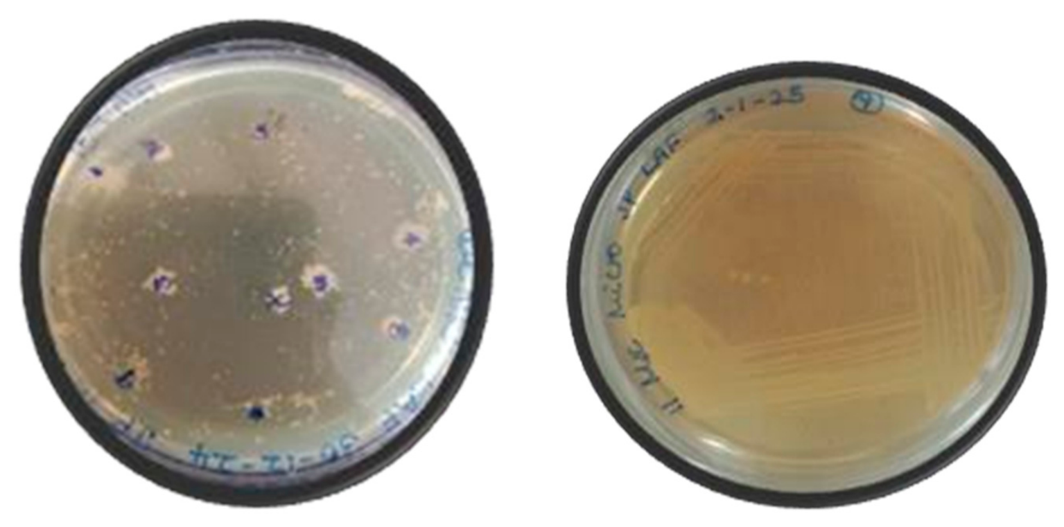
Figure 2.
Pyocyanin-producing bacteria in tyrosine basal broth in test tube.
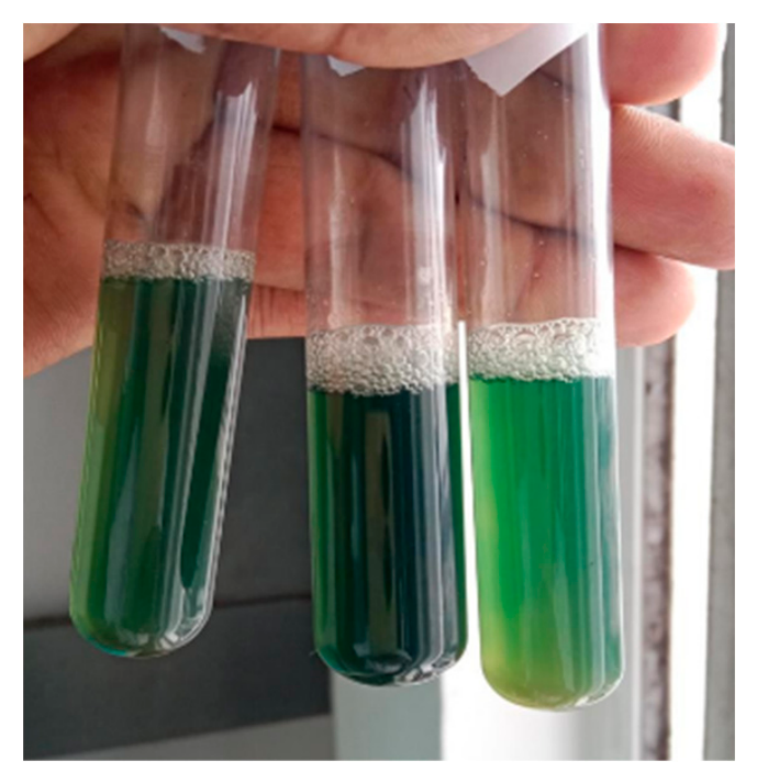
Figure 3.
Pyocyanin production by strain CLM11 in tyrosine basal broth (a) media before inoculation (b) media after 4 days of pyocyanin production.
Figure 3.
Pyocyanin production by strain CLM11 in tyrosine basal broth (a) media before inoculation (b) media after 4 days of pyocyanin production.
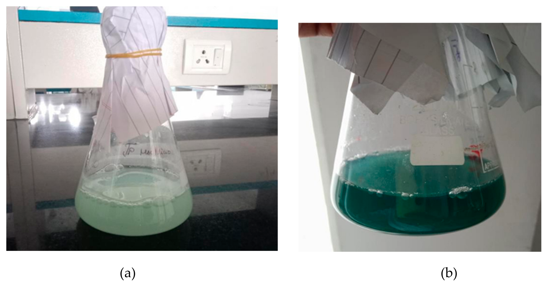
Figure 4.
Gram staining of CLM11 stain.
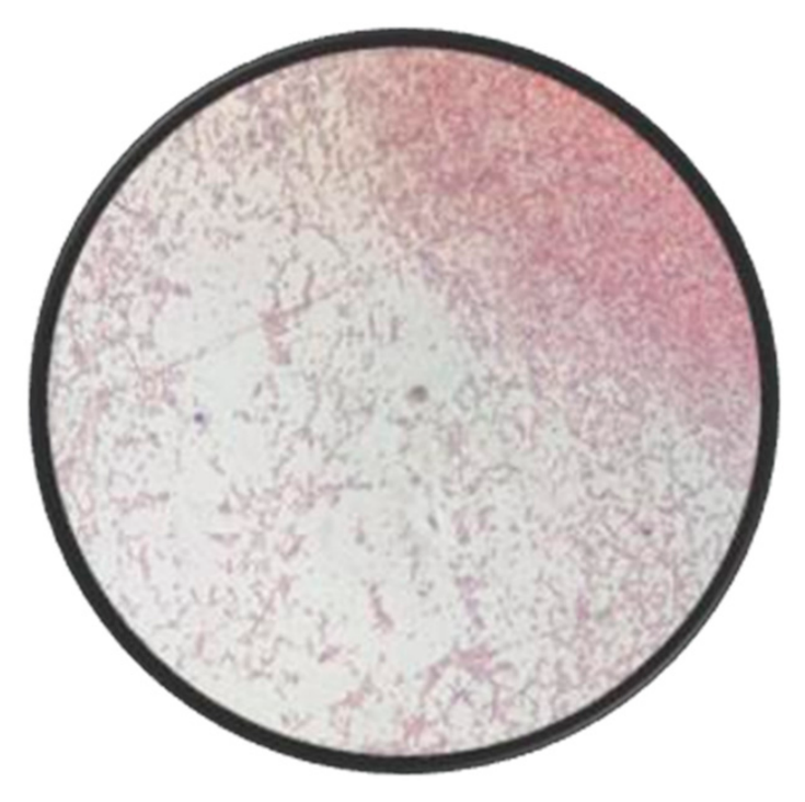
Figure 6.
Phylogenetic relationship of Pseudomonas aeruginosa CLM11against other Pseudomonas species.
Figure 6.
Phylogenetic relationship of Pseudomonas aeruginosa CLM11against other Pseudomonas species.
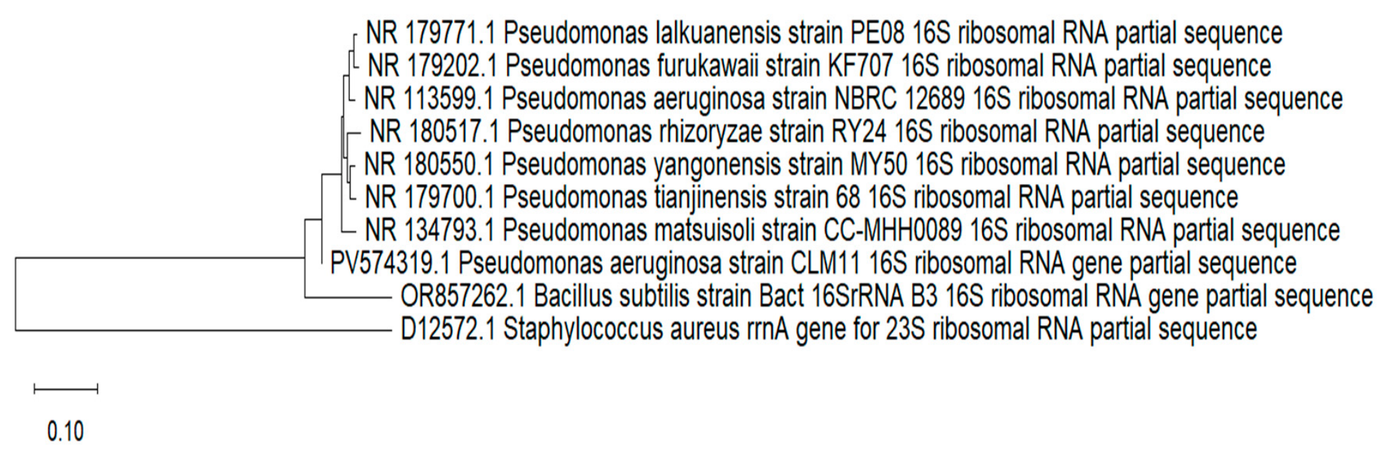
Figure 15.
The dyeing potential of pyocyanin pigment isolated from CLM 11.
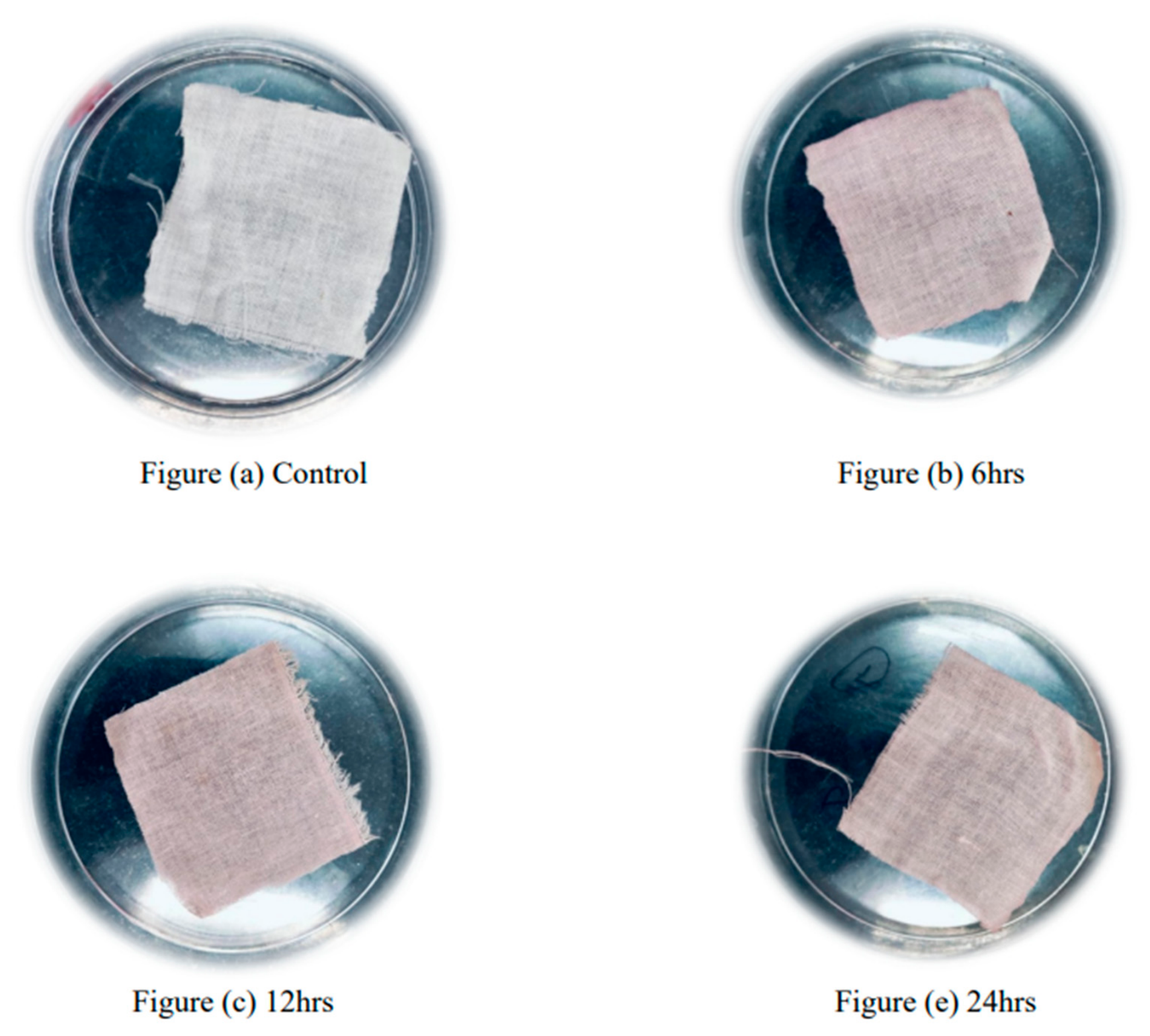

Table 1.
Biochemical Characterization of CLM11.
| Biochemical test | Results |
| Indole | Negative |
| Methyl red | Negative |
| Vogues-Proskauer | Negative |
| Simmons citrate | Positive |
| Catalase | Positive |
| Motility | Positive |
| Pigment production | Positive |
| Oxidase | Positive |
Table 2.
Different antibiotic and their zone of inhibition against CLM11.
| Antibiotics | Sensitive | Resistance | Intermediate |
|---|---|---|---|
| Penicillin G | - | Resistance | - |
| Gentamicin | Sensitive(20mm) | - | - |
| Ceftriaxone | - | - | Intermediate(18mm) |
| Ampicillin | - | Resistance | - |
| Streptomycin | - | - | Intermediate(23mm) |
| Amikacin | Sensitive(25mm) | - | - |
| Cefoperazone | - | - | Intermediate(17mm) |
| Cefepime | - | Resistance | - |
Disclaimer/Publisher’s Note: The statements, opinions and data contained in all publications are solely those of the individual author(s) and contributor(s) and not of MDPI and/or the editor(s). MDPI and/or the editor(s) disclaim responsibility for any injury to people or property resulting from any ideas, methods, instructions or products referred to in the content. |
© 2026 by the authors. Licensee MDPI, Basel, Switzerland. This article is an open access article distributed under the terms and conditions of the Creative Commons Attribution (CC BY) license.
Copyright: This open access article is published under a Creative Commons CC BY 4.0 license, which permit the free download, distribution, and reuse, provided that the author and preprint are cited in any reuse.