

Submitted:
02 October 2026
Posted:
05 October 2026
You are already at the latest version
Abstract
Congenital heart disease (CHD) is increasingly diagnosed before birth, but the benefit of prenatal diagnosis depends on the care pathway that follows detection. This narrative review traces fetal cardiac care from obstetric screening to neonatal stabilization and follow-up and proposes an organizational framework adapted to the Italian healthcare context. International guidelines, consensus statements, cohort studies, organizational surveys, and quality-improvement studies were interpreted alongside the experience of a fetal cardiology center in Rome. Published evidence supports structured pathways based on hub-and-spoke referral, a multidisciplinary Fetal Heart Team, lesion-specific surveillance, physiology-based Level of Care (LOC) stratification, and standardized delivery planning. A multi-institutional implementation study has associated standardized delivery planning with higher rates of vaginal delivery and delivery at ≥39 weeks. The Italian perinatal system presents specific challenges, including numerous birth centers, separation between delivery hospitals and cardiac surgical centers, and variable neonatal resuscitation readiness. Proposed adaptations include mapping LOC categories onto Italian Level-I and Level-II birth centers, telemedicine-supported regional networks, a dedicated cardiac care coordinator, minimum monitoring and simulation-training requirements, a three-tier model of parental psychological support, and a national outcome registry. These organizational hypotheses require prospective evaluation in multicenter Italian settings.
Keywords:
fetal echocardiography
; congenital heart disease
; delivery planning
; prenatal counseling
; level of care
; fetal heart team
; neonatal resuscitation
; health services organization
; telemedicine
; Italy
1. Introduction
Congenital heart disease (CHD) affects approximately 9 per 1,000 live births worldwide [1] and deaths in infancy account for nearly half of all CHD-related mortality [2]. The systematic inclusion of outflow tract and three-vessel views in mid-trimester screening has substantially increased prenatal detection, although population-level rates remain well below those achieved at experienced centers because of variability in screening protocols and sonographer training [3,4,5].
What has changed less is what happens after suspicion is raised. In many healthcare systems the path from a positive obstetric screen to confirmed diagnosis, structured counseling, and a coordinated delivery plan remains fragmented and dependent on local resources. This matters because the survival benefit of prenatal diagnosis depends on downstream planning: a meta-analysis found that prenatal identification of critical CHD reduces the risk of death from cardiovascular compromise before planned neonatal surgery [6], an advantage realized only when the pathway that follows detection functions well [7].
This article is a narrative review and organizational perspective. It follows the fetal cardiac care pathway from screening and red-flag recognition through referral, specialist diagnosis, Fetal Heart Team assessment, counseling and psychological support, gestational surveillance, LOC assignment, delivery planning, and neonatal stabilization and transport, to postnatal follow-up, registry, and audit. For each step it considers the international standard, the corresponding gap in the Italian system, a proposed organizational response, and a measurable quality indicator. Four elements are offered as original contributions: an analysis of how the Italian perinatal landscape requires adapted solutions; a proposed mapping of Level of Care categories onto the Italian classification of birth centers; a session-based counseling checklist; and a pragmatic proposal for integrating psychological support into counseling, addressing a gap identified across 95 programs in the Fetal Heart Society (FHS) benchmarking survey [8].
2. Methods
2.1. Narrative Review Methodology
This is a narrative, non-systematic review. PubMed/MEDLINE was searched through 15 September 2026 for English-language publications using combinations of the terms fetal echocardiography, prenatal diagnosis, congenital heart disease, delivery planning, level of care, fetal heart team, prenatal counseling, maternal hyperoxygenation, neonatal resuscitation, telemedicine, and Italy. The reference lists of retrieved articles and the guideline and consensus documents of the International Society of Ultrasound in Obstetrics and Gynecology (ISUOG), the American Heart Association (AHA), the American Society of Echocardiography (ASE), the European Society of Cardiology (ESC), the Fetal Heart Society (FHS), and the Italian Society of Neonatology (SIN) were hand-searched. Sources comprised international guidelines and consensus statements on fetal cardiac screening, fetal echocardiography, perinatal management of CHD, and cardiovascular disease in pregnancy; cohort studies and meta-analyses of perinatal outcomes; organizational and benchmarking surveys of fetal cardiac programs and of Italian neonatal resuscitation practice; and quality-improvement studies of standardized delivery planning. Sources were selected and weighted according to relevance to pathway organization, giving priority to guidelines, consensus statements, multicenter studies, and national surveys; no formal quality assessment was performed. Where evidence was heterogeneous or derived from expert consensus, this is stated explicitly, and author proposals are distinguished from published standards throughout.
2.2. Institutional Experience
The organizational perspective is informed by the author’s program, a fetal cardiology center in Rome that performed approximately 9,900 fetal echocardiograms between January 2021 and June 2026. The service-activity analysis presented in Section 11 reports approximate annual examination volumes for the five complete calendar years 2021–2025 and the first six months of 2026, drawn from routine service records. Referral urgency is assigned at the triage call according to the criteria described in Section 3.2. No patient-level clinical data, diagnoses, or outcomes are included, and no inferential analysis was performed.
3. Screening, Referral, and the Regional Network
3.1. First-Line Screening and Red Flags
The four-chamber view alone detects roughly half of significant CHD and misses conotruncal lesions, transposition, and arch anomalies; adding the outflow tract, three-vessel, and three-vessel-trachea views substantially improves detection, and current ISUOG and ASE guidelines include them in the standard mid-trimester examination [5,9]. Training sonographers and obstetricians to obtain these views reliably is arguably among the highest-leverage investments a healthcare system can make in prenatal cardiac care.
The critical competency is recognition of red flags – ventricular size asymmetry, abnormal cardiac axis, abnormal outflow tract crossing, or a dilated right ventricle – followed by immediate referral rather than repeated attempts at clarification in a non-specialist setting, since each additional non-diagnostic scan adds parental anxiety and delays counseling.
3.2. Referral Urgency and Triage
We propose that referral urgency be explicitly tiered as an organizational target rather than as an established international standard. This tiering applies to outpatient referrals: fetuses with signs of acute compromise are managed by obstetric or emergency services and fall outside the scope of the fetal cardiology referral pathway. Within that pathway, suspected complex CHD or fetal arrhythmia should prompt same-day contact with the fetal cardiology coordinator, who determines the timing of specialist evaluation on a case-by-case basis; other, non-urgent suspected cardiac abnormalities should be scheduled according to the specific defect suspected rather than within a fixed time window [7,10].
Italian gap. The Italian referral infrastructure is inherently fragmented: the national survey by Gizzi et al. was sent to the directors of all 418 Italian birth centers, 303 of which are Level-I units without a neonatal intensive care unit; among responding Level-I centers the median number of births was 737 per year (interquartile range 525–1,035) [11]. In that survey, which covered 258 centers and approximately 70% of Italian births, multidisciplinary antenatal counseling was routinely offered at 90% of Italian Level-II (NICU) birth centers but at only 57% of Level-I centers [11].
Proposed response. A structured same-day triage call between the referring obstetrician or maternal–fetal medicine (MFM) specialist and the fetal cardiology coordinator. In the author’s center, urgent referrals already reach a fetal cardiologist directly on the same day, while non-urgent referrals are scheduled by the unit’s administrative office; in the author’s experience, this facilitates rapid triage and may reduce the uncertainty experienced by families. A coordinator would bring both channels under a single point of contact, freeing specialist time and making the triage criteria explicit. The coordinator would determine the pathway at this call: same-day specialist contact for suspected complex CHD or fetal arrhythmia, and individualized scheduling for other non-urgent findings; signs of acute fetal compromise fall under obstetric/emergency management, outside this pathway. Indicator: time from suspicion to first fetal echocardiogram.
3.3. Hub-and-Spoke Network and Telemedicine
A regional network is the structural backbone of the proposed pathway (Figure 1). Peripheral maternity units screen and triage; the specialist fetal cardiac hub provides definitive diagnosis, counseling, and coordination of high-risk delivery, and returns structured care plans that support selected LOC 1–2 deliveries to be managed locally according to predefined criteria, minimizing family disruption and unnecessary centralization.
Telemedicine is increasingly central to this architecture. In the 2022 FHS survey, 34% of programs offered telehealth fetal cardiology services with remote interpretation of fetal echocardiograms, and half of these could review images in real time and guide the sonographer [8]. In the maternal cardiac field, the Erasmus MC pregnancy heart team, established in line with the 2018 and 2025 ESC guidelines on cardiovascular disease in pregnancy [13,14], reviews both its own patients and those referred from other hospitals at multidisciplinary meetings held every two weeks and issues individualized peripartum care plans covering timing, mode, and location of delivery [12]; the same organizational logic can be applied to a fetal cardiac network.
Italian gap and proposal. The recent Italian SICP survey, based on 45 responses among 548 members (8.2%), reported on-site pediatric cardiac surgery in only about one-third of responding programs and did not identify a standardized national hub-and-spoke pathway, highlighting the need for more clearly defined referral networks [15]. Given the low response rate, these findings should be interpreted as a description of participating centers rather than as a complete national census. We propose that each region designate one or more specialist fetal cardiac hubs, according to population, geography, and case volume, with defined spoke units, a shared electronic care plan, and scheduled virtual multidisciplinary review. Indicator: proportion of LOC 1–2 deliveries managed at the referring center with a hub-issued care plan.
4. The Fetal Heart Team
The Fetal Heart Team (FHT) is a structured multidisciplinary unit that meets regularly, makes collective decisions, and communicates them through a single documented plan (Table 1). In the FHS survey (median 1,000 fetal echocardiograms per program per year, interquartile range 580–2,100), neonatology (93%), MFM (88%), genetic counseling (88%), and social work (79%) were available in most programs, and cardiac surgery, palliative care, electrophysiology, single-ventricle care, and cardiac transplantation were each available in more than half, whereas psychology (38%) and psychiatry (16%) were not [8].
The operational core is the regular multidisciplinary case conference, at which new diagnoses are presented, follow-up cases reviewed with updated imaging, and delivery plans finalized before 36 weeks. Its output should be a written, dated document accessible in the electronic health record at all times, including out-of-hours when a woman may present in labor earlier than anticipated.
Italian context. Where the fetal cardiology program, delivery suite, and cardiac surgical service are located in different hospitals – the typical Italian arrangement – the case conference must explicitly include the delivering unit and the receiving surgical center, if necessary by video link. Indicator: proportion of LOC 3–4 cases with a documented multidisciplinary plan before 36 weeks.
5. Counseling and Psychological Support
5.1. Counseling
Initial counseling should follow the diagnostic echocardiogram, in a dedicated room, with the fetal cardiologist and MFM specialist present. The first session should deliver the diagnosis clearly, acknowledge its emotional impact, and arrange further counseling, as parents experiencing acute distress may have difficulty focusing on and fully attending to the medical information provided [7,16]. Further counseling sessions should be offered according to family needs and clinical evolution. In the proposed pathway, two sessions are fixed points: the initial session at diagnosis and a delivery-planning session at approximately 34–36 weeks; between them, one or more intermediate sessions are scheduled as needed to distribute information progressively and to introduce the neonatologist, cardiac surgeon, and, where needed, geneticist and palliative care team, with written summaries provided. Parents value knowing in advance which team will care for their child after birth [17], and consistently report wanting more information than they received [7,18]. A structured checklist, informed by Lee [7] and organized across these sequential sessions, is proposed in Table 2; it covers the anatomical diagnosis, expected postnatal physiology, diagnostic uncertainty, genetic associations, disease progression and the risk of intrauterine demise, surgical strategy with center-specific survival, neurodevelopmental risk [19], delivery planning, and support resources.
5.2. A Three-Tier Pathway for Access to Psychological Support: An Author Proposal
Mothers receiving a prenatal diagnosis of CHD show higher rates of post-traumatic stress, depression, and anxiety [7,20], and in fetuses with CHD maternal stress and anxiety have been associated with smaller hippocampal and cerebellar volumes on fetal magnetic resonance imaging, which has led to calls for universal screening of maternal psychological distress in this population [16,21]. Yet fewer than half of fetal cardiac programs have access to a psychologist [8]. In the recent Italian SICP survey, a psychologist was routinely present during counseling in only 11.6% of responding programs, although a reference psychologist could be activated in a further 65.1% [15].
For settings without a dedicated perinatal psychologist, we propose a simple three-tier referral pathway that makes access to existing mental-health services systematic rather than occasional. It is an author proposal and has not been externally validated.
Tier 1 – Systematic identification of distress. At the initial counseling session and again at the delivery-planning session, both parents are offered a brief distress screening instrument, administered by the cardiac care coordinator or trained staff, as is done for perinatal depression screening in obstetric care. The choice of instrument, the thresholds, and the referral pathway should be defined by psychology/psychiatry specialists; the instruments used in published fetal CHD studies have been research scales rather than brief clinical screeners [20,21], and the validity of any brief instrument in this population should be confirmed before adoption.
Tier 2 – Coordinator-mediated navigation. The cardiac care coordinator, with appropriate training and clinical governance, maintains structured follow-up contacts, connects families with peer support, and flags cases that meet the agreed referral criteria. This role is navigational, not therapeutic: it does not replace professional psychological care.
Tier 3 – Specialist care. Parents meeting the referral criteria, or showing persistent distress or difficulty engaging in the decision-making process, are referred to perinatal psychology or psychiatry, who lead assessment and treatment; telemedicine may be used where local services are unavailable. The evidence for mental-health interventions in parents of infants with CHD is limited to a few small trials, most showing a reduction in maternal anxiety [22].
This pathway is an interim measure that acknowledges resource constraints while providing a structured route to specialist care in settings where routine psychological screening is not currently available. Indicator: proportion of families offered screening at both time points. Its feasibility and effect should be evaluated prospectively, with mental-health specialists involved from the design stage.
6. Level of Care Assignment
Risk stratification is the pivot of delivery planning. Risk-stratification frameworks link fetal echocardiographic findings to the anticipated postnatal level of care according to the predicted risk of hemodynamic instability at birth and the urgency of intervention [3,9,23,24,25,26,27]. The 2014 AHA scientific statement incorporated a risk-stratified Level of Care framework, and subsequent publications and the 2023 ASE recommendations have continued to support image-based risk stratification paired with specific perinatal care plans [3,9,24,25,27]. For the purposes of this review, the delivery-planning framework is organized as LOC 1–4, with LOC P for pregnancies in which comfort-focused or palliative care has been chosen. Assignment is physiology-based, not diagnosis-based: the same anatomical lesion may receive a different LOC depending on atrial septal restriction, ductal characteristics, ventricular function, arrhythmia control, hydrops, pulmonary venous obstruction, and the expected timing of intervention (Table 3). Adoption is far from universal: in the FHS survey only 51% of programs used perinatal LOC guidelines and 13% were unfamiliar with them [8].
7. Gestational Surveillance and Selective Maternal Hyperoxygenation Testing
Surveillance intensity should be lesion-specific (Table 4): a balanced atrioventricular septal defect in trisomy 21 poses a different problem from evolving aortic stenosis with ventricular dysfunction or fetal tachycardia on transplacental therapy. The 2023 ASE recommendations support serial fetal echocardiographic follow-up when progression or postnatal compromise is anticipated, with intervals determined by lesion-specific risk and clinical evolution [9]. The cardiovascular profile (CVP) score may provide additional prognostic information in selected high-risk fetuses; a score ≤7 on the last pre-delivery examination has been associated with substantially higher perinatal mortality [23,30], but routine serial use as a universal surveillance standard has not been established.
In HLHS, the pulmonary venous Doppler pattern predicts the need for emergent atrial septostomy after birth: a forward/reverse velocity–time integral (VTI) ratio below 5 was the strongest predictor in the original Cincinnati series, and a threshold of 3 or less optimizes specificity [32,33]. Acute maternal hyperoxygenation (MHO) testing – assessment of fetal pulmonary vasoreactivity during 10–15 minutes of high-flow oxygen – is a selective adjunct rather than a routine test. It is intended to answer specific questions in the late third trimester, typically at around 34 weeks in published protocols: whether the atrial septum in HLHS is likely to be severely restrictive at birth, and whether pulmonary venous obstruction is present in total anomalous pulmonary venous return (TAPVR). Its purpose is to refine perinatal risk stratification and delivery planning, including the need for immediate intervention. In 2022, 35% of FHS programs offered MHO testing for clinical and/or research use, and a consensus protocol has since been published [8,23,31]. A 2026 case series of three fetuses with TAPVR reported that MHO unmasked occult pulmonary venous obstruction in one case and amplified abnormal flow in another [34]. This application is promising but remains investigational and is currently supported by very limited clinical data. Evidence and protocols continue to evolve, and the test should be performed in experienced centers according to a defined protocol. Fetal MRI may be considered when airway or lung concerns coexist.
8. Delivery Planning
8.1. Standardized Delivery Planning
Standardized Clinical Assessment and Management Plans (SCAMPs) make the recommended action at each decision point explicit while documenting deviations. In a multi-institutional University of California implementation (496 maternal–fetal dyads), a fetal CHD SCAMP with vaginal delivery and term birth (≥39 weeks) as defaults was associated with a later gestational age at delivery (38.9 vs 38.1 weeks) and a higher rate of vaginal delivery (61% vs 50%, p = 0.03); among pregnancies with major cardiac lesions (n = 373), deliveries at ≥39 weeks increased from 33% to 48% (p = 0.004) and vaginal birth from 51% to 64% (p = 0.008) [35]. Cesarean delivery carries higher maternal mortality and morbidity than vaginal birth and increases the risk of uterine rupture and abnormal placentation in subsequent pregnancies [36], and birth at 37–38 weeks is independently associated with worse outcomes after neonatal cardiac surgery [37]. Table 5 provides a delivery planning worksheet integrating LOC, team requirements, equipment, and stabilization strategy.
Italian gap and proposal. Italy has no nationally adopted LOC-based delivery protocol. We propose that the LOC framework be mapped onto the Italian perinatal classification, with explicit criteria for which categories may be delivered at Italian Level-I and Level-II birth centers and which require a cardiac surgical center (Table 5). Because this requires consensus rather than new infrastructure, it may be implementable without major additional infrastructure. Indicators: term delivery rate and cesarean delivery without obstetric indication in LOC 1–2.
8.2. Timing, Mode, and Site of Delivery
For most CHD lesions, including ductal-dependent lesions and most LOC 3 cases, vaginal delivery is generally safe and preferred: with multidisciplinary coordination, fetuses with prenatally diagnosed CHD have cesarean rates and delivery-room compromise similar to those of unaffected fetuses, and what improves neonatal outcome is place of birth and team preparedness rather than the mode of delivery itself [35,38]. Elective early-term delivery should be avoided, and delivery at or beyond 39 weeks is the default whenever clinically feasible [37]. Planned induction at ≥39 weeks is used in many programs for LOC 3–4 fetuses to align the birth with team availability at the appropriate site. Cesarean delivery should be reserved for obstetric indications and for exceptional cardiac scenarios in which immediate coordinated intervention requires a controlled birth; it should not be chosen for organizational convenience. For LOC 4 fetuses, timing and mode require individualized multidisciplinary planning that weighs the predicted instability at placental separation against the risks of earlier delivery.

Table 6.
Diagnosis-specific oxygen and stabilization considerations in the delivery room.
| Cardiac physiology | Expected transitional physiology | Stabilization considerations (according to the pre-delivery plan) | Rationale |
|---|---|---|---|
| Structurally normal heart | Pre-ductal SpO2 ≥90% by 10 minutes of life | Standard neonatal resuscitation algorithm | Normal transitional physiology |
| Single ventricle (HLHS, HRHS) | Lower-than-normal saturations may be expected; target individualized according to systemic perfusion and pulmonary/systemic flow balance | Avoid supplemental oxygen aimed solely at normalizing saturation; PGE1 if ductal-dependent; assess systemic perfusion and pulmonary/systemic flow balance | Excessive reduction in pulmonary vascular resistance may increase pulmonary flow at the expense of systemic output |
| d-TGA (parallel circulations) | Saturation depends on the degree of mixing; may be very low with restrictive atrial septum | PGE1 and ventilatory support as planned; assess adequacy of intercirculatory mixing; preparation for urgent balloon atrial septostomy when mixing is inadequate; supplemental oxygen as clinically indicated | Adequate intercirculatory mixing is essential; atrial septostomy is required when restriction causes severe hypoxemia or low systemic oxygen delivery |
| Ductal-dependent pulmonary flow (PA/IVS, severe TOF) | Cyanosis may persist despite adequate ventilation; target individualized according to lesion and systemic oxygen delivery | PGE1 to maintain ductal patency when indicated; oxygen titrated to clinical need rather than to normalization of SpO2 | Pulmonary blood flow depends on ductal patency in critical obstruction; oxygen alone cannot correct inadequate pulmonary blood flow |
| Ductal-dependent systemic flow (critical CoA, HLHS) | Saturation alone may not reflect systemic perfusion; target individualized according to perfusion and pulmonary/systemic flow balance | PGE1; oxygen titrated to hypoxemia and systemic/pulmonary flow balance according to the pre-delivery plan | Avoid an excessive fall in pulmonary vascular resistance that may increase pulmonary flow at the expense of systemic output |
| Obstructed TAPVR | Profound hypoxemia possible; no definitive medical therapy | Ventilatory support and immediate surgical referral; urgent relief of the obstruction is required; supplemental oxygen and PGE1 have limited benefit | Definitive anatomical relief is urgently required, usually by surgical repair |
| Ebstein anomaly / functional pulmonary atresia | Variable; circular shunt possible | Management depends on the dominant physiology. Support ventilation and systemic perfusion; measures that reduce pulmonary vascular resistance may be appropriate when forward pulmonary flow is limited. In the presence of pulmonary regurgitation and a circular shunt, PGE1 and aggressive pulmonary vasodilation should be used cautiously according to the pre-delivery plan | Reducing right ventricular afterload may help when pulmonary blood flow is limited, whereas a circular shunt can worsen systemic steal and is associated with high perinatal mortality [29] |
SpO2 = pre-ductal (right hand); HRHS = hypoplastic right heart syndrome. These considerations are informed by the 2014 AHA statement and subsequent reviews of fetal-to-neonatal transition and delivery-room management [3,23,24,25,40,41]. They are not validated universal numerical standards; targets and interventions should be pre-specified for each fetus according to predicted physiology and updated after postnatal assessment.
9. Neonatal Stabilization, Transport, and the Italian Delivery Room
9.1. Physiology-Guided Stabilization
The 2025 AHA/AAP neonatal resuscitation guidelines remain the starting point for every newborn with CHD, with their emphasis on team preparation, assisted ventilation, and titrated oxygen therapy [39]; what differs for these newborns is the interpretation of oxygen saturation and the physiology-specific strategy that follows [25,40,41]. In mixing or parallel circulations, oxygen saturation should be interpreted in the context of lesion-specific physiology. In single-ventricle circulation, indiscriminate escalation of supplemental oxygen to normalize SpO2 may disturb the balance between pulmonary and systemic blood flow; oxygen targets should therefore be individualized and pre-specified in the delivery plan [23,25,40]. The expected physiology and intended stabilization strategy should be documented in the delivery worksheet (Table 5), and the resuscitating team should know them before birth. Diagnosis-specific considerations – ventilation, systemic perfusion, mixing, prostaglandin therapy, and lesion-specific stabilization – are summarized in Table 6; they derive largely from institutional protocols, reviews, and expert consensus and should be applied according to the individual pre-delivery plan rather than as universal numerical targets.
9.2. Readiness of Italian Birth Centers
Evidence. The national survey by Gizzi et al. found high overall adherence to resuscitation guidelines but gaps relevant to CHD: an anesthesiologist was part of the high-risk resuscitation team in 79% of Italian Level-I centers versus 27% of Level-II centers; only 7% of Level-I centers rated their intubation skills as excellent, compared with 56% of Level-II centers; an end-tidal CO2 detector was used to confirm tube placement in about 20% of centers at both levels; a three-lead ECG monitor was used for heart-rate assessment during resuscitation in 31% of Level-I and 42% of Level-II centers; and a team briefing before resuscitation was routine in 43% of Level-I centers versus 64% of Level-II centers [11]. The survey therefore suggests important differences in procedural competence and team practices between Italian Level-I and Level-II centers, alongside relevant monitoring and equipment gaps.
Proposal. We propose (a) that end-tidal CO2 detection and three-lead ECG monitoring be minimum requirements for any unit designated to conduct LOC 2 or higher deliveries; (b) simulation-based training in CHD-specific delivery-room scenarios, with periodic reassessment, at all centers receiving fetal cardiac referrals, allowing selected Italian Level-I centers to be designated “CHD-ready”; (c) a protocolized pre-delivery briefing using the completed worksheet; and (d) telemedicine-supported delivery-room coaching for unexpected presentations. Indicator: proportion of LOC ≥2 deliveries preceded by a documented team briefing.
9.3. Failure Modes and Mitigation
Anticipating failure is a prerequisite for resilience. Drawing on the author’s organizational experience and published series, four scenarios appear most consequential.
Precipitous delivery before planned LOC 3–4 management. A fetus with d-transposition is delivered at 36 weeks at a peripheral center without catheterization capability. A worksheet completed and distributed electronically by 34 weeks gives that team pre-specified stabilization targets, prostaglandin dosing, and a transport activation protocol; without it, the team improvises and delays compound.
Missed prenatal diagnosis. Lesions with a normal four-chamber view, such as transposition and coarctation, are detected prenatally less often than those with four-chamber abnormalities [4,5,42]. The safety net is universal newborn pulse-oximetry screening, an effective, simple, and inexpensive test that identifies part of the critical CHD missed prenatally [43], together with a low threshold for postnatal echocardiography.
Transport delay. Where delivery hospital and cardiac surgical center are separate, the interval from birth to arrival at the surgical center is modifiable: for HLHS, greater distance and driving time to the surgical center are associated with higher neonatal, and particularly presurgical, mortality [23,44]. Pre-activating the transport team at the onset of labor and notifying the receiving surgeon before the birth are pragmatic organizational mitigations, particularly in Italy, where regional transport systems vary substantially.
Communication failure. The plan exists but the on-call neonatologist has not read it; prostaglandin is available but staff are unfamiliar with dosing; the target saturation is specified but the monitor alarm remains at default. In the proposed model, the coordinator is the redundancy layer intended to ensure that the plan is enacted, not merely written.
10. Postnatal Follow-up, Registry, and Quality Audit
An integrated feto-neonatal model, in which the same cardiology team provides prenatal counseling and postnatal follow-up, is proposed to address the prenatal-to-postnatal transition directly by preserving continuity of information and of the family’s relationship with the team [17]. The pathway closes with postnatal confirmation of the diagnosis, structured follow-up, and feedback of outcomes into practice through an outcome registry audited against defined indicators (Table 7). The cardiac care coordinator is central to this loop. In the FHS survey, 79% of programs had one or more fetal cardiac coordinators, in most cases a registered nurse, with a median of one full-time equivalent per program, and 78% maintained an independent fetal database; in univariate analysis, programs with a fetal cardiac coordinator (p = 0.003) and programs with a formal fetal database (p = 0.003) were each more likely to have a formal quality-improvement program, as were larger-volume programs (p = 0.03) [8]. The coordinator’s role spans scheduling and triage, family navigation and Tier 1–2 support, distribution of the delivery plan, transport liaison, and registry management.
Italian context. Italy lacks a directly equivalent, nationally standardized advanced-practice-provider role comparable to the North American model; the function could be filled by a specialist nurse, a midwife with cardiac training, or a clinically trained case manager – the combination of clinical knowledge, communication skills, and organizational capacity matters more than the title. We further propose a national registry of LOC 3–4 delivery outcomes stratified by transport time and distance from the surgical center. Indicators: dedicated coordinator in post; proportion of cases entered in the registry.
11. Institutional Experience: Illustrative Service-Activity Data
To illustrate the scale of the program in an Italian setting, approximate aggregated service-activity data from the author’s center are reported for January 2021–June 2026. These data describe examination volumes rather than clinical outcomes and are subject to the limitations discussed in Section 13. Across the five complete calendar years, annual fetal echocardiography volume increased from approximately 1,448 examinations in 2021 to 1,982 in 2025 (2022: 1,604; 2023: 1,725; 2024: 1,776). A further 1,349 examinations were performed between January and June 2026, resulting in a cumulative total of approximately 9,884 examinations from January 2021 to June 2026. For context, the annual volume lies above the median of the programs in the FHS benchmarking survey (median 1,000 fetal echocardiograms per year, interquartile range 580–2,100) [8], although volume alone says nothing about quality of care. The program currently operates a two-channel intake: for suspected complex CHD or fetal arrhythmias, the referring clinician contacts a fetal cardiologist directly, with specialist evaluation timed on a case-by-case basis; non-urgent referrals are scheduled through the unit’s administrative office according to the specific defect suspected; signs of acute fetal compromise are managed by obstetric/emergency services outside this pathway. The single coordinator-managed triage call described in Section 3.2, which would bring both channels under one point of contact, is a proposed evolution rather than current practice. Figure 2 summarizes the proposed organizational pathway and should be interpreted as a target model rather than as a representation of a fully implemented system.
12. A Proposal for National Implementation
The models reviewed above derive largely from North American and Northern European centers with co-located obstetric and cardiac services, high-volume surgical programs, and well-resourced transport systems; 91% of the programs in the FHS survey were North American [8]. The Italian landscape differs in the number and size of birth centers, the separation of delivery and cardiac surgical services, and the variability of resuscitation readiness documented by national survey [11]; importing international guidelines wholesale is unlikely to address these differences.
We propose that the Italian Society of Neonatology (SIN), the Italian Society of Pediatric Cardiology (SICP), and the Italian Society of Obstetric and Gynecological Ultrasound (SIEOG) jointly develop a national fetal cardiac LOC pathway, to be incorporated into the next revision of the SIN organizational standards for perinatal care [45]. The pathway should comprise mapping of LOC categories onto Italian Level-I and Level-II birth centers and cardiac surgical centers; regional designation of one or more hubs according to population, geography, and case volume, with shared electronic care plans and telemedicine; minimum monitoring and simulation-training requirements for LOC ≥2 delivery sites; a transport activation standard; a dedicated coordinator role; and a national outcome registry. Each element should be introduced with a measurable indicator and evaluated prospectively.
13. Limitations
This review is narrative, and the selection and weighting of evidence were not systematic; relevant studies may have been omitted. The published organizational models are heterogeneous and derive predominantly from North American and Northern European programs, so their transferability to Italy is inferred rather than demonstrated. Direct evidence for several delivery-room protocols, including diagnosis-specific oxygen strategies and MHO testing, remains limited and rests largely on institutional protocols and expert consensus. The national and international surveys cited are self-reported and subject to response bias and incomplete coverage. The SICP survey received 45 responses among 548 members (8.2%), limiting its representativeness [15]; in the SIN survey, only 49% of Level-I centers responded [11]. The association between a coordinator and quality-improvement activity in the FHS survey is cross-sectional and unadjusted [8], and the transport-time data for HLHS derive from a single US state registry [44].
The institutional figures are single-center, aggregated service-activity data reported descriptively; they do not include diagnostic accuracy or clinical outcomes, which would require a formally approved study, and they are presented as illustration rather than as evidence of effectiveness. The experience of a high-volume urban center may not generalize to smaller or geographically isolated Italian programs. The organizational proposals, including the tiered referral triage, the session-based counseling checklist, the three-tier psychological-support model, and the mapping of LOC onto Italian birth-center levels, are author proposals that require prospective multicenter evaluation.
14. Conclusions
The evidence reviewed supports a limited number of priorities. Physiology-based LOC stratification provides a practical shared language for obstetric, neonatal, and cardiac teams, and its routine use should be prospectively evaluated within standardized pathways. Standardized delivery planning that defaults to vaginal birth at or beyond 39 weeks, with individualized exceptions, has been associated with higher rates of vaginal delivery and more deliveries at ≥39 weeks. Regional hub-and-spoke organization supported by telemedicine, a dedicated cardiac care coordinator, simulation-based neonatal preparation, and outcome registries with continuous quality improvement are recurrent features of structured fetal cardiac programs and provide a framework for coordinated quality improvement.
For Italy, we recommend national consensus, led jointly by SIN, SICP, and SIEOG, on a LOC-based pathway for incorporation into the next revision of the national perinatal standards and mapped onto the existing perinatal classification; regional networks with defined transport activation; minimum monitoring and training requirements for designated delivery sites; and integrated parental psychological support, for which the three-tier model proposed here offers a starting point.
Several elements require prospective evaluation: the feasibility and effect of the psychological-support model, the safety of LOC 2 delivery at designated Italian Level-I centers, the relationship between transport time and outcome in Italian LOC 3–4 deliveries, and achievable benchmarks for referral interval and prenatal–postnatal diagnostic concordance across Italian centers. A national registry would make these questions answerable.
Author Contributions
Conceptualization, methodology, investigation, writing—original draft preparation, writing—review and editing, and visualization, G.P. All authors have read and agreed to the published version of the manuscript.
Funding
This research received no external funding.
Institutional Review Board Statement
Not applicable. Only aggregated, non-identifiable service-activity data collected as part of routine service evaluation are reported; no patient-level clinical data or outcomes were analyzed.
Informed Consent Statement
Not applicable.
Data Availability Statement
No new patient-level data were created or analyzed in this study. The aggregated service-activity figures reported are contained within the article.
Acknowledgments
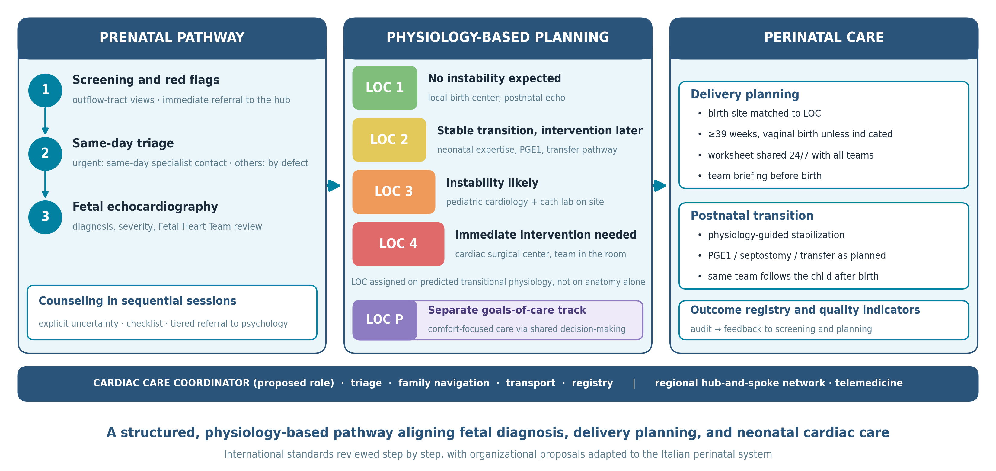
The author thanks Francesca Gallini and Luigi Orfeo for their institutional support, and Riccardo Riccardi, Valerio Romano, Matteo Pavan, Alessandra Piersanti, Michela Cammerata, and Roberto Chioma for their contribution to the clinical activity of the program described in this article. During the preparation of this manuscript, the author used Claude (Anthropic, Claude) for formatting, for grammar checking, and for the preparation of the code used to draw the graphical abstract, which is an original schematic and not AI-generated imagery. The author has reviewed and edited the output and takes full responsibility for the content of this publication.
Conflicts of Interest
The author declares no conflicts of interest.
References
- van der Linde, D.; Konings, E.E.M.; Slager, M.A.; Witsenburg, M.; Helbing, W.A.; Takkenberg, J.J.M.; Roos-Hesselink, J.W. Birth prevalence of congenital heart disease worldwide: a systematic review and meta-analysis. J Am Coll Cardiol 2011, 58, 2241–2247. [CrossRef]
- Gilboa, S.M.; Salemi, J.L.; Nembhard, W.N.; Fixler, D.E.; Correa, A. Mortality resulting from congenital heart disease among children and adults in the United States, 1999 to 2006. Circulation 2010, 122, 2254–2263. [CrossRef]
- Donofrio, M.T.; Moon-Grady, A.J.; Hornberger, L.K.; Copel, J.A.; Sklansky, M.S.; Abuhamad, A.; Cuneo, B.F.; Huhta, J.C.; Jonas, R.A.; Krishnan, A.; Lacey, S.; Lee, W.; Michelfelder, E.C.; Rempel, G.R.; Silverman, N.H.; Spray, T.L.; Strasburger, J.F.; Tworetzky, W.; Rychik, J. Diagnosis and treatment of fetal cardiac disease: a scientific statement from the American Heart Association. Circulation 2014, 129, 2183–2242. [CrossRef]
- Quartermain, M.D.; Pasquali, S.K.; Hill, K.D.; Goldberg, D.J.; Huhta, J.C.; Jacobs, J.P.; Jacobs, M.L.; Kim, S.; Ungerleider, R.M. Variation in prenatal diagnosis of congenital heart disease in infants. Pediatrics 2015, 136, e378–e385. [CrossRef]
- Carvalho, J.S.; Axt-Fliedner, R.; Chaoui, R.; Copel, J.A.; Cuneo, B.F.; Goff, D.; Gordin Kopylov, L.; Hecher, K.; Lee, W.; Moon-Grady, A.J.; Mousa, H.A.; Munoz, H.; Paladini, D.; Prefumo, F.; Quarello, E.; Rychik, J.; Tutschek, B.; Wiechec, M.; Yagel, S. ISUOG Practice Guidelines (updated): fetal cardiac screening. Ultrasound Obstet Gynecol 2023, 61, 788–803. [CrossRef]
- Holland, B.J.; Myers, J.A.; Woods, C.R. Prenatal diagnosis of critical congenital heart disease reduces risk of death from cardiovascular compromise prior to planned neonatal cardiac surgery: a meta-analysis. Ultrasound Obstet Gynecol 2015, 45, 631–638. [CrossRef]
- Lee, C.K. Prenatal counseling of fetal congenital heart disease. Curr Treat Options Cardiovasc Med 2017, 19, 5. [CrossRef]
- Patel, S.; McBrien, A.; Michelfelder, E.; Kavanaugh-McHugh, A.; Samples, S.; Laternser, C.; Hornberger, L.; Moon-Grady, A.; Srivastava, S.; Shuplock, J. Current trends in fetal cardiology: results from an international benchmarking survey of fetal cardiac programs through the Fetal Heart Society Research Collaborative. Pediatr Cardiol 2026, 47, 1218–1228. [CrossRef]
- Moon-Grady, A.J.; Donofrio, M.T.; Gelehrter, S.; Hornberger, L.; Kreeger, J.; Lee, W.; Michelfelder, E.; Morris, S.A.; Peyvandi, S.; Pinto, N.M.; Pruetz, J.; Sethi, N.; Simpson, J.; Srivastava, S.; Tian, Z. Guidelines and recommendations for performance of the fetal echocardiogram: an update from the American Society of Echocardiography. J Am Soc Echocardiogr 2023, 36, 679–723. [CrossRef]
- Sharma, C.; Burns, J.; Joshi, K.; Gupta, M.; Singh, H.; Agarwal, A. Advances in the prenatal management of fetal cardiac disease. Children (Basel) 2022, 9, 812. [CrossRef]
- Gizzi, C.; Trevisanuto, D.; Gagliardi, L.; Vertecchi, G.; Ghirardello, S.; Di Fabio, S.; Moretti, C.; Mosca, F. Neonatal resuscitation practices in Italy: a survey of the Italian Society of Neonatology (SIN) and the Union of European Neonatal and Perinatal Societies (UENPS). Ital J Pediatr 2022, 48, 81. [CrossRef]
- Rijs, K.; van der Marel, C.D.; van der Knijff-van Dortmont, A.L.M.J.; Dons-Sinke, I.J.J.; Adriaens, V.F.R.; Cornette, J.M.J.; Mulders, A.G.M.G.J.; Duvekot, J.J.; Driessen, M.M.P.; van den Bosch, A.E.; Cuypers, J.A.A.E.; Kauling, R.M.; Roos-Hesselink, J.W. How to set up a pregnancy heart team. Int J Cardiol Congenit Heart Dis 2025, 22, 100635. [CrossRef]
- Regitz-Zagrosek, V.; Roos-Hesselink, J.W.; Bauersachs, J.; Blomström-Lundqvist, C.; Cífková, R.; De Bonis, M.; Iung, B.; Johnson, M.R.; Kintscher, U.; Kranke, P.; Lang, I.M.; Morais, J.; Pieper, P.G.; Presbitero, P.; Price, S.; Rosano, G.M.C.; Seeland, U.; Simoncini, T.; Swan, L.; Warnes, C.A. 2018 ESC Guidelines for the management of cardiovascular diseases during pregnancy. Eur Heart J 2018, 39, 3165–3241. [CrossRef]
- De Backer, J.; Haugaa, K.H.; Hasselberg, N.E.; de Hosson, M.; Brida, M.; Castelletti, S.; Cauldwell, M.; Cerbai, E.; Crotti, L.; de Groot, N.M.S.; Estensen, M.-E.; Goossens, E.S.; Haring, B.; Kurpas, D.; McEniery, C.M.; Peters, S.A.E.; Rakisheva, A.; Sambola, A.; Schlager, O.; Schoenhoff, F.S.; Simoncini, T.; Steinbach, F.; Sudano, I.; Swan, L.; Valente, A.M. 2025 ESC Guidelines for the management of cardiovascular disease and pregnancy. Eur Heart J 2025, 46, 4462–4568. [CrossRef]
- Balducci, A.; Morelli, C.; Baldinelli, A.; Campanale, C.M.; Cerutti, A.; Codazzi, A.C.; Consigli, V.; Culeddu, C.; Marcora, S.A.; Lerose, C.C.; Palleri, D.; Pasquini, L.; Toscano, A.; Tuo, G.; Favilli, S.; Rinelli, G. Current foetal cardiology practice in Italian centres: a survey among SICP members. Int J Cardiol Congenit Heart Dis 2026, 24, 100659. [CrossRef]
- Tacy, T.A.; Kasparian, N.A.; Karnik, R.; Geiger, M.; Sood, E. Opportunities to enhance parental well-being during prenatal counseling for congenital heart disease. Semin Perinatol 2022, 46, 151587. [CrossRef]
- Piacentini, G. From Communication to Continuity: A Structural Response to Parents’ Priorities in Prenatal CHD. Pediatr Cardiol 2026, online ahead of print. [CrossRef]
- Arya, B.; Glickstein, J.S.; Levasseur, S.M.; Williams, I.A. Parents of children with congenital heart disease prefer more information than cardiologists provide. Congenit Heart Dis 2013, 8, 78–85. [CrossRef]
- Marino, B.S.; Lipkin, P.H.; Newburger, J.W.; Peacock, G.; Gerdes, M.; Gaynor, J.W.; Mussatto, K.A.; Uzark, K.; Goldberg, C.S.; Johnson, W.H.; Li, J.; Smith, S.E.; Bellinger, D.C.; Mahle, W.T. Neurodevelopmental outcomes in children with congenital heart disease: evaluation and management: a scientific statement from the American Heart Association. Circulation 2012, 126, 1143–1172. [CrossRef]
- Rychik, J.; Donaghue, D.D.; Levy, S.; Fajardo, C.; Combs, J.; Zhang, X.; Szwast, A.; Diamond, G.S. Maternal psychological stress after prenatal diagnosis of congenital heart disease. J Pediatr 2013, 162, 302–307.e1. [CrossRef]
- Wu, Y.; Kapse, K.; Jacobs, M.; Niforatos-Andescavage, N.; Donofrio, M.T.; Krishnan, A.; Vezina, G.; Wessel, D.; du Plessis, A.; Limperopoulos, C. Association of maternal psychological distress with in utero brain development in fetuses with congenital heart disease. JAMA Pediatr 2020, 174, e195316. [CrossRef]
- Kasparian, N.A.; Kan, J.M.; Sood, E.; Wray, J.; Pincus, H.A.; Newburger, J.W. Mental health care for parents of babies with congenital heart disease during intensive care unit admission: systematic review and statement of best practice. Early Hum Dev 2019, 139, 104837. [CrossRef]
- Patel, S.R.; Michelfelder, E. Prenatal diagnosis of congenital heart disease: the crucial role of perinatal and delivery planning. J Cardiovasc Dev Dis 2024, 11, 108. [CrossRef]
- Donofrio, M.T.; Levy, R.J.; Schuette, J.J.; Skurow-Todd, K.; Sten, M.-B.; Stallings, C.; Pike, J.I.; Krishnan, A.; Ratnayaka, K.; Sinha, P.; duPlessis, A.J.; Downing, D.S.; Fries, M.I.; Berger, J.T. Specialized delivery room planning for fetuses with critical congenital heart disease. Am J Cardiol 2013, 111, 737–747. [CrossRef]
- Ali, N.; Donofrio, M.T. Delivery room and early postnatal management of neonates with congenital heart disease. Prenat Diagn 2024, 44, 915–924. [CrossRef]
- Słodki, M.; Respondek-Liberska, M.; Pruetz, J.D.; Donofrio, M.T. Fetal cardiology: changing the definition of critical heart disease in the newborn. J Perinatol 2016, 36, 575–580. [CrossRef]
- Donofrio, M.T.; Skurow-Todd, K.; Berger, J.T.; McCarter, R.; Fulgium, A.; Krishnan, A.; Sable, C.A. Risk-stratified postnatal care of newborns with congenital heart disease determined by fetal echocardiography. J Am Soc Echocardiogr 2015, 28, 1339–1349. [CrossRef]
- Pyle, A.K.; George, T.N.; Cummings, J.J.; Laventhal, N.T.; Committee on Bioethics; Section on Neonatal-Perinatal Medicine; Committee on Fetus and Newborn. Guidance for Caring for Infants and Children With Trisomy 13 and Trisomy 18: Clinical Report. Pediatrics 2025, 156, e2025072719. [CrossRef]
- Freud, L.R.; Escobar-Diaz, M.C.; Kalish, B.T.; Komarlu, R.; Puchalski, M.D.; Jaeggi, E.T.; Szwast, A.L.; Freire, G.; Levasseur, S.M.; Kavanaugh-McHugh, A.; Michelfelder, E.C.; Moon-Grady, A.J.; Donofrio, M.T.; Howley, L.W.; Tierney, E.S.S.; Cuneo, B.F.; Morris, S.A.; Pruetz, J.D.; van der Velde, M.E.; Kovalchin, J.P.; Ikemba, C.M.; Vernon, M.M.; Samai, C.; Satou, G.M.; Gotteiner, N.L.; Phoon, C.K.; Silverman, N.H.; McElhinney, D.B.; Tworetzky, W. Outcomes and predictors of perinatal mortality in fetuses with Ebstein anomaly or tricuspid valve dysplasia in the current era: a multicenter study. Circulation 2015, 132, 481–489. [CrossRef]
- Wieczorek, A.; Hernandez-Robles, J.; Ewing, L.; Leshko, J.; Luther, S.; Huhta, J. Prediction of outcome of fetal congenital heart disease using a cardiovascular profile score. Ultrasound Obstet Gynecol 2008, 31, 284–288. [CrossRef]
- Madan, N.; Donofrio, M.T.; Szwast, A.; Moon-Grady, A.J.; Patel, S.R. Acute maternal hyperoxygenation protocol: consensus opinion from the Fetal Heart Society. Ultrasound Obstet Gynecol 2024, 64, 567–573. [CrossRef]
- Michelfelder, E.; Gomez, C.; Border, W.; Gottliebson, W.; Franklin, C. Predictive value of fetal pulmonary venous flow patterns in identifying the need for atrial septoplasty in the newborn with hypoplastic left ventricle. Circulation 2005, 112, 2974–2979. [CrossRef]
- Divanović, A.; Hor, K.; Cnota, J.; Hirsch, R.; Kinsel-Ziter, M.; Michelfelder, E. Prediction and perinatal management of severely restrictive atrial septum in fetuses with critical left heart obstruction: clinical experience using pulmonary venous Doppler analysis. J Thorac Cardiovasc Surg 2011, 141, 988–994. [CrossRef]
- Mathevosian, M.; Wang, S.; Davtyan, A.; Detterich, J.; Votava-Smith, J.; Pruetz, J. Maternal Hyperoxygenation for Prenatal Risk Stratification of Pulmonary Venous Obstruction in Total Anomalous Pulmonary Venous Return. Pediatr Cardiol 2026, online ahead of print. [CrossRef]
- Afshar, Y.; Hogan, W.J.; Conturie, C.; Sunderji, S.; Duffy, J.Y.; Peyvandi, S.; Boe, N.M.; Melber, D.; Fajardo, V.M.; Tandel, M.D.; Holliman, K.; Kwan, L.; Satou, G.; Moon-Grady, A.J. Multi-institutional practice-patterns in fetal congenital heart disease following implementation of a standardized clinical assessment and management plan. J Am Heart Assoc 2021, 10, e021598. [CrossRef]
- Sandall, J.; Tribe, R.M.; Avery, L.; Mola, G.; Visser, G.H.A.; Homer, C.S.E.; Gibbons, D.; Kelly, N.M.; Kennedy, H.P.; Kidanto, H.; Taylor, P.; Temmerman, M. Short-term and long-term effects of caesarean section on the health of women and children. Lancet 2018, 392, 1349–1357. [CrossRef]
- Costello, J.M.; Pasquali, S.K.; Jacobs, J.P.; He, X.; Hill, K.D.; Cooper, D.S.; Backer, C.L.; Jacobs, M.L. Gestational age at birth and outcomes after neonatal cardiac surgery: an analysis of the Society of Thoracic Surgeons Congenital Heart Surgery Database. Circulation 2014, 129, 2511–2517. [CrossRef]
- Adams, A.D.; Aggarwal, N.; Fries, M.H.; Donofrio, M.T.; Iqbal, S.N. Neonatal and maternal outcomes of pregnancies with a fetal diagnosis of congenital heart disease using a standardized delivery room management protocol. J Perinatol 2020, 40, 316–323. [CrossRef]
- Lee, H.C.; Strand, M.L.; Finan, E.; Illuzzi, J.; Kamath-Rayne, B.D.; Kapadia, V.; Mahgoub, M.; Niermeyer, S.; Schexnayder, S.M.; Schmölzer, G.M.; Weglarz, J.; Williams, A.L.; Weiner, G.M.; Wyckoff, M.; Yamada, N.K.; Szyld, E. Part 5: Neonatal Resuscitation: 2025 American Heart Association and American Academy of Pediatrics Guidelines for Cardiopulmonary Resuscitation and Emergency Cardiovascular Care. Circulation 2025, 152, S385–S423. [CrossRef]
- Peyvandi, S.; Donofrio, M.T. Circulatory changes and cerebral blood flow and oxygenation during transition in newborns with congenital heart disease. Semin Pediatr Neurol 2018, 28, 38–47. [CrossRef]
- Donofrio, M.T. Predicting the future: delivery room planning of congenital heart disease diagnosed by fetal echocardiography. Am J Perinatol 2018, 35, 549–552. [CrossRef]
- Bakker, M.K.; Bergman, J.E.H.; Krikov, S.; Amar, E.; Cocchi, G.; Cragan, J.; de Walle, H.E.K.; Gatt, M.; Groisman, B.; Liu, S.; Nembhard, W.N.; Pierini, A.; Rissmann, A.; Chidambarathanu, S.; Sipek, A.; Szabova, E.; Tagliabue, G.; Tucker, D.; Mastroiacovo, P.; Botto, L.D. Prenatal diagnosis and prevalence of critical congenital heart defects: an international retrospective cohort study. BMJ Open 2019, 9, e028139. [CrossRef]
- Narayen, I.C.; Blom, N.A.; Ewer, A.K.; Vento, M.; Manzoni, P.; te Pas, A.B. Aspects of pulse oximetry screening for critical congenital heart defects: when, how and why? Arch Dis Child Fetal Neonatal Ed 2016, 101, F162–F167. [CrossRef]
- Morris, S.A.; Ethen, M.K.; Penny, D.J.; Canfield, M.A.; Minard, C.G.; Fixler, D.E.; Nembhard, W.N. Prenatal diagnosis, birth location, surgical center, and neonatal mortality in infants with hypoplastic left heart syndrome. Circulation 2014, 129, 285–292. [CrossRef]
- Baronciani, D.; Bellù, R.; Franchi, M.; Gargano, G.; Jorizzo, G.; Viora, E.; Zanini, R.; Agostiniani, R.; Bertelè, P.; Bracchi, F.; Buccella, I.; Costamagna, G.; Da Rin della Mora, R.; Dasso, N.; del Vecchio, A.; Gennarelli, F.; Masella, C.; Portanova, A.; Simonetto, C.; Rinaldo, D.; Romeo, N.; Vicario, M.; Zucconi, G. Obiettivi di Sistema, Modello Organizzativo Generale e Standard Organizzativi per la Rete di Assistenza alla Madre e al Neonato. In Standard Organizzativi per l’Assistenza Perinatale; Mosca, F., Ed.; IdeaCpa Editore: Rome, Italy, 2021; pp. 46–102.
Figure 1.
Hub-and-spoke referral network model for fetal cardiac care. LOC = Level of Care. Referrals (solid arrows) flow from peripheral community and regional birth centers to the specialist fetal cardiac hub, where the Fetal Heart Team provides definitive fetal echocardiography, diagnosis and severity assessment, LOC assignment, counseling, delivery planning, and the outcome registry; structured care plans (dashed arrows) flow from the hub back to the periphery, allowing LOC 1–2 deliveries to be managed locally. The cardiac care coordinator (triage, family navigation, transport, registry) is the communication node across all units (plain line), and telemedicine support (double-headed arrow) links the hub with peripheral centers through remote image review and real-time guidance. In the Fetal Heart Society survey, 34% of programs offered telehealth fetal cardiology services [8]; the Erasmus MC pregnancy heart team model reviews referred patients at multidisciplinary meetings held every two weeks [12]. The regional designation of hubs and spokes is an author proposal for the Italian context.
Figure 1.
Hub-and-spoke referral network model for fetal cardiac care. LOC = Level of Care. Referrals (solid arrows) flow from peripheral community and regional birth centers to the specialist fetal cardiac hub, where the Fetal Heart Team provides definitive fetal echocardiography, diagnosis and severity assessment, LOC assignment, counseling, delivery planning, and the outcome registry; structured care plans (dashed arrows) flow from the hub back to the periphery, allowing LOC 1–2 deliveries to be managed locally. The cardiac care coordinator (triage, family navigation, transport, registry) is the communication node across all units (plain line), and telemedicine support (double-headed arrow) links the hub with peripheral centers through remote image review and real-time guidance. In the Fetal Heart Society survey, 34% of programs offered telehealth fetal cardiology services [8]; the Erasmus MC pregnancy heart team model reviews referred patients at multidisciplinary meetings held every two weeks [12]. The regional designation of hubs and spokes is an author proposal for the Italian context.
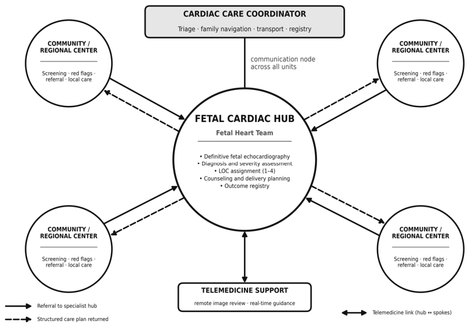
Figure 2.
Proposed integrated fetal cardiac care pathway: operational flowchart. Diamonds indicate decision points (red-flag identification, urgency triage, LOC assignment). After screening (Phase 1) and red-flag recognition (Phase 2), the coordinator triages referrals at a same-day call: suspected complex CHD or fetal arrhythmia receives same-day contact with specialist evaluation timed case-by-case, other suspected abnormalities are scheduled according to the suspected defect, and signs of acute fetal compromise fall under obstetric/emergency management, outside this pathway. Outpatient referrals then proceed to specialist diagnosis and Fetal Heart Team review (Phases 3–4). LOC assignment separates two tracks: Track A (LOC 1–2; standard surveillance, pre-delivery worksheet, local or regional delivery) and Track B (LOC 3–4, hatched boxes; intensive surveillance, delivery board, delivery at the hub or cardiac surgical center—LOC 3 at a center with pediatric cardiology and catheterization capability, LOC 4 at a cardiac surgical center). Both tracks converge on postnatal follow-up and the quality registry (Phase 8); the dashed arrow indicates the feedback loop of outcome data, audit, and improvement from postnatal follow-up back to screening. In the proposed model, the cardiac care coordinator operates continuously from referral to postnatal follow-up.
Figure 2.
Proposed integrated fetal cardiac care pathway: operational flowchart. Diamonds indicate decision points (red-flag identification, urgency triage, LOC assignment). After screening (Phase 1) and red-flag recognition (Phase 2), the coordinator triages referrals at a same-day call: suspected complex CHD or fetal arrhythmia receives same-day contact with specialist evaluation timed case-by-case, other suspected abnormalities are scheduled according to the suspected defect, and signs of acute fetal compromise fall under obstetric/emergency management, outside this pathway. Outpatient referrals then proceed to specialist diagnosis and Fetal Heart Team review (Phases 3–4). LOC assignment separates two tracks: Track A (LOC 1–2; standard surveillance, pre-delivery worksheet, local or regional delivery) and Track B (LOC 3–4, hatched boxes; intensive surveillance, delivery board, delivery at the hub or cardiac surgical center—LOC 3 at a center with pediatric cardiology and catheterization capability, LOC 4 at a cardiac surgical center). Both tracks converge on postnatal follow-up and the quality registry (Phase 8); the dashed arrow indicates the feedback loop of outcome data, audit, and improvement from postnatal follow-up back to screening. In the proposed model, the cardiac care coordinator operates continuously from referral to postnatal follow-up.
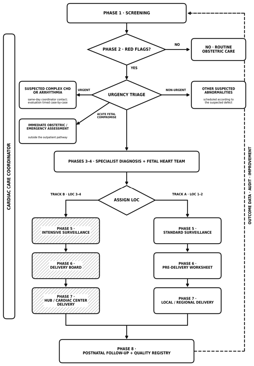
Table 1.
Fetal Heart Team: composition, core roles, and escalation responsibilities.
| Specialist | Core responsibilities | Escalation role |
|---|---|---|
| Fetal / pediatric cardiologist | Fetal echocardiography, hemodynamic assessment, counseling, LOC assignment, delivery planning | Fetal cardiac intervention where available; neonatal echocardiography at delivery |
| Maternal–fetal medicine specialist | Obstetric management, biometry, Doppler, maternal complications, mode and timing of delivery | Decision on earlier delivery, including in response to hydrops, severe pre-eclampsia, or fetal growth restriction; fetal cardiac intervention where available |
| Neonatologist / intensivist | Delivery-room stabilization, PGE1 protocol, airway strategy, NICU coordination | Advanced resuscitation; ECMO activation/coordination where available; early transfer |
| Clinical geneticist | Syndromic assessment, invasive testing, microarray/exome, recurrence counseling | Postnatal confirmation and completion of the genetic workup; translation of the diagnosis into a syndrome-specific surveillance and management plan |
| Cardiac surgeon | Surgical feasibility; counseling on operative strategy | Operating room at delivery for LOC 4; palliation versus biventricular repair decision |
| Cardiac care coordinator | Scheduling, triage, family navigation, registry, multidisciplinary logistics, Tier 1–2 psychological support (Section 5.2) | Crisis communication; transport liaison; Tier 3 referral to specialist psychology/psychiatry (Section 5.2) |
| Palliative care | End-of-life planning, family support for LOC P | Bereavement support; comfort-care coordination at delivery |
| Psychology | Parental psychological support, decision-making facilitation (Tiers 2–3, Section 5.2) | Referral to psychiatry for depression, post-traumatic stress, or other diagnosable conditions; decision-making paralysis addressed through intensified psychological support or multidisciplinary/ethics discussion |
Availability benchmarks from the Fetal Heart Society survey [8]. Psychological-support tiers refer to the author-proposed model in Section 5.2. FGR = fetal growth restriction; NICU = neonatal intensive care unit; ECMO = extracorporeal membrane oxygenation.
Table 2.
Proposed structured counseling checklist across sequential sessions.
| Domain | Items | S1 | S2 | S3 |
|---|---|---|---|---|
| Diagnosis and physiology | Diagram of normal vs abnormal anatomy; expected physiology before and after birth; diagnostic limitations and residual uncertainty | ● | ○ | |
| Etiology and genetics | Known causes and risk factors; genetic associations; referral for genetic counseling | ● | ○ | |
| Prognosis | Prognosis and mortality risk, with center-specific data when available; outcomes at the counseling and surgical centers | ● | ○ | |
| Disease progression | Possible progression during pregnancy; risk of intrauterine demise; surveillance plan | ● | ○ | |
| Pregnancy options | Continuation with a care plan; termination within the applicable legal framework; comfort care; fetal intervention if applicable | ● | ||
| Postnatal course | Expected intervention or surveillance-only pathway; what will happen at birth; NICU course; the diagnosis may be revised after birth | ● | ○ | ● |
| Neurodevelopment | Introduced as a topic; risk factors; planned developmental follow-up | ● | ● | |
| Modifiers of outcome | Extracardiac anomalies (with MFM); prematurity; growth restriction; low birth weight; associated syndromes | ● | ||
| Long-term issues and quality of life | Reinterventions; physical limitations; adult follow-up; heart failure or transplant if relevant; expected quality of life | ● | ||
| Delivery planning | Timing (≥39 weeks unless indicated), mode, and place; planned level of care; delivery-room plan; whether parents can hold the baby; timing of transfer | ● | ||
| Hospital course and discharge | Expected hospital course; feeding and breastfeeding plan; discharge planning; follow-up | ● | ||
| Family and resources | Parental leave, housing, siblings; written materials; vetted websites; parent groups; tour of the delivery suite and NICU; primary care pediatrician identified | ○ | ● | |
| Next steps | Date of the next consultation; written summary provided | ● | ● | ● |
S1, initial session at diagnosis; S2, intermediate session(s), scheduled as needed according to family needs and clinical evolution; S3, delivery-planning session (approximately 34–36 weeks). ● = addressed; ○ = revisited or introduced briefly. Author-proposed checklist, informed by Lee [7]; content and timing should be individualized. Throughout all sessions: risks stated as natural frequencies rather than percentages; both sides of a probability given; “we do not know yet” distinguished from “this cannot be predicted”; an explicit map of planned reassessments; language kept consistent across professionals; uncertainty documented in the report in the same words used in counseling. A structured handover to the postnatal team links this checklist to the continuity model described in Section 10.
Table 3.
Level of Care (LOC) classification for perinatal cardiac management, based on predicted postnatal physiology.
Table 3.
Level of Care (LOC) classification for perinatal cardiac management, based on predicted postnatal physiology.
| LOC | Definition (predicted postnatal physiology) | Illustrative lesions* | Delivery site and team | Delivery-room plan |
|---|---|---|---|---|
| P | CHD for which comfort-focused or palliative care has been chosen following individualized multidisciplinary counseling and shared decision-making | Severe CHD with major genetic or multisystem disease when goals of care are palliative; conditions associated with physiological futility | Local birth center; family support and palliative care | Comfort care; no cardiac intervention planned |
| 1 | No predicted hemodynamic instability at birth or in the first days of life | Small VSD; balanced AVSD; mild TOF | Local birth center (Italian Level-I or Level-II); routine delivery-room care | Postnatal echocardiogram before discharge (or early outpatient); cardiology follow-up |
| 2 | Stable transition expected; postnatal intervention or ductal support anticipated in the first days | Ductal-dependent lesions with stable predicted transition: HLHS without RAS, critical CoA, severe AS, IAA, PA/IVS, severe TOF | Center with neonatal expertise, vascular access, PGE1 available when indicated, and a defined transfer pathway (Italian Level-II) | Neonatologist present; umbilical venous access and PGE1 according to the plan; saturation strategy per the delivery worksheet (Section 8.1); planned transfer to cardiac center |
| 3 | Instability likely; immediate specialist cardiac care required | d-TGA; complete heart block with heart failure; poorly controlled arrhythmia | Center with pediatric cardiology and catheterization capability; neonatologist and cardiologist in delivery room; catheterization laboratory on standby | Physiology-guided stabilization per plan; balloon atrial septostomy preparation for d-TGA; PGE1 as indicated. Planned delivery at ≥39 weeks where feasible |
| 4 | Severe instability expected at placental separation; immediate catheter or surgical intervention required | HLHS with RAS/IAS; d-TGA with restrictive septum ± abnormal ductus; obstructed TAPVR; Ebstein anomaly with hydrops or circular shunt; TOF-APV with severe airway compromise | Cardiac surgical center; full cardiac team; operating room / catheterization laboratory immediately available; ECMO where available | Immediate intervention as pre-planned; timing and mode of delivery individualized by the Fetal Heart Team |
*LOC is assigned according to the predicted postnatal physiology, not the anatomical diagnosis alone: the same lesion may receive a different LOC depending on atrial septal restriction, ductal characteristics, ventricular function, arrhythmia control, hydrops, pulmonary venous obstruction, and the expected need and timing of intervention. Because the need for intervention cannot be predicted reliably in d-TGA, all fetuses with d-TGA should be assigned LOC 3 or 4 [27]. LOC P assignment depends on the agreed goals of care rather than on a specific genetic diagnosis: a 2025 AAP clinical report emphasizes that trisomy 13 and trisomy 18 are not uniformly lethal and that treatment decisions should follow individualized shared decision-making rather than the genetic diagnosis alone [28]. The mapping onto Italian Level-I/II birth centers is an author proposal. Adapted from Donofrio et al. [3,24,27], Ali and Donofrio [25], Słodki et al. [26], Moon-Grady et al. [9], and Patel and Michelfelder [23]; Ebstein anomaly with pulmonary regurgitation (circular shunt) carries a particularly high perinatal mortality [29]. AS = aortic stenosis; AVSD = atrioventricular septal defect; CoA = coarctation of the aorta; d-TGA = d-transposition of the great arteries; HLHS = hypoplastic left heart syndrome; IAA = interrupted aortic arch; IAS/RAS = intact/restrictive atrial septum; PA/IVS = pulmonary atresia with intact ventricular septum; PGE1 = prostaglandin E1; TAPVR = total anomalous pulmonary venous return; TOF(-APV) = tetralogy of Fallot (with absent pulmonary valve); VSD = ventricular septal defect.
Table 4.
Illustrative lesion-specific framework for gestational surveillance.
| CHD category | Fetal echocardiography | Obstetric monitoring | Additional assessments |
|---|---|---|---|
| LOC 1: stable shunt lesions, mild valve disease | Every 4–8 weeks or as clinically indicated | Standard obstetric schedule; growth assessment according to obstetric indications | Postnatal echocardiogram before discharge or early outpatient evaluation, according to the lesion |
| LOC 2: ductal-dependent lesions with stable predicted transition | Every 4–8 weeks; shorter interval if the lesion is evolving | Serial growth assessment according to obstetric indications; umbilical artery Doppler if FGR | Late-third-trimester reassessment of predicted transitional physiology; delivery worksheet completed before delivery |
| LOC 3–4: d-TGA, HLHS ± RAS, obstructed TAPVR, Ebstein anomaly | Every 2–4 weeks or according to lesion-specific risk; additional late-third-trimester assessment | Serial growth assessment; additional fetal surveillance according to obstetric and hemodynamic findings | Targeted assessment of atrial septum, ductal flow, pulmonary venous flow, and/or airway-lung findings according to lesion; selective MHO testing in the late third trimester, typically at around 34 weeks; fetal MRI if airway or lung concern; multidisciplinary review before delivery |
| Progressive lesions / arrhythmia (CHB, SVT, TOF-APV) | Every 2–4 weeks; closer intervals for fetal tachycardia unless well controlled; more frequently if unstable | Fetal heart-rate and hydrops surveillance; biophysical testing when clinically indicated | Pharmacological review; comprehensive cardiac functional assessment when heart failure is a concern; earlier delivery considered individually if hydrops or fetal compromise develops |
Intervals are indicative and should be individualized. This table is an author-proposed synthesis informed by the lesion-specific follow-up framework in the 2023 ASE recommendations [9] and by published fetal cardiac surveillance literature [3,23]. It is not intended as a validated universal schedule. CVP = cardiovascular profile score (a score ≤7 on the last pre-delivery examination is associated with higher perinatal mortality [30]); FGR = fetal growth restriction; MHO = acute maternal hyperoxygenation testing, a selective adjunct to be performed in experienced centers according to a defined protocol [31]; CHB = complete heart block; SVT = supraventricular tachycardia.
Table 5.
Fetal cardiac delivery planning worksheet.
| Element | Detail required |
|---|---|
| Diagnosis and genetics | Confirmed anatomical diagnosis; karyotype/microarray; extracardiac anomalies |
| LOC assignment | LOC P–4 on the basis of predicted postnatal physiology (Table 3), with the modifiers considered |
| Delivery site | LOC 1: any birth center with access to postnatal echocardiography before discharge. LOC 2: center with neonatal expertise, vascular access, PGE1 available when indicated, and a defined transfer pathway. LOC 3: center with pediatric cardiology and catheterization capability. LOC 4: cardiac surgical center with immediate intervention capability |
| Timing | Default ≥39 weeks; avoid elective early-term delivery. Earlier delivery only for obstetric or fetal indications, documented. LOC 4: individualized by the Fetal Heart Team |
| Mode | Vaginal delivery when obstetrically appropriate. Cesarean delivery for obstetric indications or for exceptional cardiac scenarios in which immediate coordinated intervention requires a controlled birth; not for organizational convenience |
| Delivery-room team | LOC 1: routine; LOC 2: neonatologist, umbilical venous access; LOC 3: + cardiologist, catheterization standby; LOC 4: full cardiac team, operating room, ECMO where available |
| Equipment checklist | PGE1 infusion protocol (neonatal weight-based dosing after birth); echocardiography; intubation kit; umbilical venous/arterial lines; inhaled nitric oxide if indicated; transport incubator |
| Stabilization strategy | Expected transitional physiology and intended saturation strategy pre-specified for this fetus (Table 6) |
| Transfer plan | Transport team on standby from onset of labor; receiving center and surgeon alerted before birth; parents briefed |
| Distribution | Completed by 34 weeks for LOC 3–4. Copies to delivering hospital, transport service, cardiac center, and family; 24/7 access in the electronic health record |
Table 7.
Quality indicators for fetal cardiac care programs.
| Domain | Indicator | Suggested target | Basis |
|---|---|---|---|
| Access | Time from suspicion to first fetal echocardiogram | Same-day coordinator contact for suspected complex CHD or fetal arrhythmia; interval from suspicion to first echocardiogram recorded and reported by urgency category | Author-proposed organizational target, informed by [7,10]* |
| Diagnostic accuracy | Prenatal–postnatal concordance (primary diagnosis) | Local baseline and prospective benchmark to be established | Author-proposed quality indicator; no externally validated universal threshold |
|
Delivery quality |
Term delivery (≥39 weeks), LOC 1–2 | Local baseline and prospective benchmark to be established | Author-proposed indicator; SCAMP, major lesions: 48% vs 33% [35] |
| Cesarean delivery without obstetric indication, LOC 1–2 | Local baseline and prospective benchmark to be established | Author-proposed indicator; SCAMP: vaginal delivery 61% vs 50% [35] | |
| Neonatal safety | Preoperative mortality, critical CHD with prenatal diagnosis | Monitor; lower with prenatal diagnosis | Meta-analysis [6] |
| Program structure | Dedicated cardiac care coordinator | Presence/absence and full-time-equivalent staffing recorded | FHS survey: 79% of programs had ≥1 coordinator [8] |
| Family experience | Psychological distress screening rate | Local baseline and prospective benchmark to be established | Author proposal (Section 5.2); no externally validated universal threshold |
| Data | Cases entered into outcome registry | Local baseline and prospective benchmark to be established | FHS survey: 78% maintained an independent fetal database [8] |
*The access and quality-improvement indicators are author-proposed and are not externally validated universal standards. Where no external benchmark exists, programs should establish a local baseline and evaluate prospective improvement rather than treat the suggested indicators as fixed performance thresholds. In the FHS survey, 46 of 80 responding programs had a formal quality-improvement program and 22 participated in national-level quality-improvement initiatives; the most cited barrier was lack of human resources [8]. SCAMP = Standardized Clinical Assessment and Management Plan.
Disclaimer/Publisher’s Note: The statements, opinions and data contained in all publications are solely those of the individual author(s) and contributor(s) and not of MDPI and/or the editor(s). MDPI and/or the editor(s) disclaim responsibility for any injury to people or property resulting from any ideas, methods, instructions or products referred to in the content. |
© 2026 by the authors. Licensee MDPI, Basel, Switzerland. This article is an open access article distributed under the terms and conditions of the Creative Commons Attribution (CC BY) license (http://creativecommons.org/licenses/by/4.0/).
Copyright: This open access article is published under a Creative Commons CC BY 4.0 license, which permit the free download, distribution, and reuse, provided that the author and preprint are cited in any reuse.