

Submitted:
01 October 2026
Posted:
05 October 2026
You are already at the latest version
Abstract
We report two cases of equine encephalomyelitis associated with the recently emerged European Shamonda virus type 1 (SHAV-EU1). Shamonda virus has not yet been described in horses but has caused an unprecedented outbreak in cattle in Western Europe this summer (2026). Both horses showed neurological deficits and lymphocytic cerebrospinal fluid pleocytosis associated with encephalomyelitis, vasculitis and hemorrhages in brain and spinal cord. Pan-Simbu RT-qPCR confirmed viral RNA in brain tissue, and Metatranscriptomic sequencing identified full-genome SHAV-EU1 sequences, proofing correlation to the outbreak in Swiss cattle.
Keywords:
encephalitis
; vasculitis
; neurology
; horses
; Shamonda virus
; orthobunyavirus
; Switzerland
Text
Orthobunyaviruses of the Simbu serogroup are arthropod-borne viruses of veterinary importance, several of which are associated with acute disease, reproductive disorders, fetal infection, and congenital abnormalities in ruminants [1]. They possess a segmented, negative-sense RNA genome comprising a large (L), a medium (M), and a small (S) segment, encoding the viral RNA-dependent RNA polymerase, glycoproteins, and nucleocapsid protein, respectively [1].
In July 2026, Shamonda virus (SHAV) emerged in Switzerland in association with a widespread outbreak of fever, diarrhea, and reduced milk production in dairy cattle. Viral RNA was detected in blood samples of affected animals and in brain and spleen of aborted bovine fetuses, raising concerns about transplacental infection and reproductive disease [2]. Concurrent detections in cattle in other European countries indicated geographic spread [3,4] and the presence of two SHAV subtypes provisionally designated as SHAV Europe 1 (EU1) and SHAV Europe 2 (EU2) [5].
During the same period, the Swiss equine disease surveillance system Equinella, a veterinary-based voluntary syndromic surveillance network for the early detection of infectious diseases and other emerging health threats in equids [6], recorded an increase in reports of horses with neurologic disease from multiple regions.
Here, we report the detection of SHAV-EU1 in brain tissues from horses with encephalomyelitis and vasculitis, providing evidence for a previously unrecognized neuroinvasive potential of SHAV in horses.
The Study
In late July and early August 2026, two horses with severe acute neurologic signs were presented to the Swiss Institute of Equine Medicine, University of Bern (case 1: 30.07.2026,), and the Clinic for Equine Internal Medicine, University of Zurich (case 2: 04.08.2026) (Table 1). Hematologic and biochemical analyses revealed no substantial abnormalities. Molecular and serologic testing for common neurotropic pathogens yielded negative or inconclusive results in both horses (Appendix, Table 1). Cerebrospinal fluid (CSF) cytology revealed lymphocytic pleocytosis in both horses, consistent with CNS inflammation and suggestive of a viral infection (Table 1). Both animals were euthanized due to deterioration of the health status 3 days (case 1) and 5 days (case 2) after onset of clinical disease.

Table 1.
Clinical presentation and diagnostic investigations performed in the two affected horses.
| Case | Signalment* | Clinical signs | Cytology cerebrospinal fluid |
| 1 | FM, f, 16y | Fever, apathy, ataxia (grade 4/5†), head hyperesthesia, muscle fasciculations muzzle, head tilt, horizontal nystagmus, episodes of somnolence, seizures | Xanthochromia, lymphocytic pleocytosis (96/µl, 94.5% lymphocytes) |
| 2 | KWPN, mc, 19y | Fever, low head carriage/ tilt, reduced lateral flexion of the neck, ataxia (grade 4/5†), cauda equina syndrome, muscle fasciculations muzzle, episodes of somnolence, head pressing, and compulsive wandering | Lymphocytic pleocytosis (56/µL, 96% lymphocytes) |
* Abbreviations: f, female; FM, Franches-Montagnes; KWPN, Koninklijk Warmbloed Paardenstamboek Nederland (Dutch Warmblood); mc, male castrated; y, years. †The severity of ataxia was graded using the Mayhew Ataxia Grading Scale.
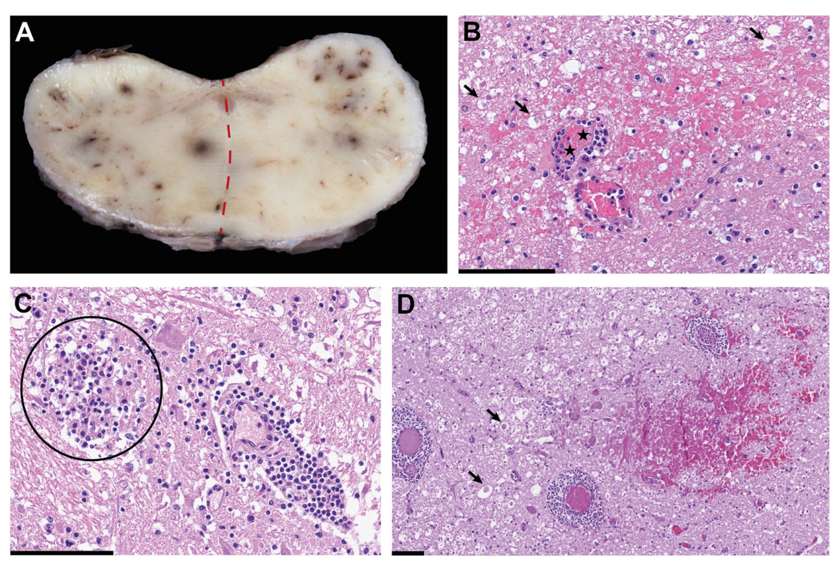
Post-mortem examination revealed multifocal hemorrhages in the brain and spinal cord, and histopathology identified a multifocal to disseminated, severe, lymphoplasmacytic and histiocytic vasculitis with hemorrhages distributed throughout the brain and spinal cord, most severely affecting the brainstem in both cases (Figure 1). No evidence of vasculitis or other lesions was found in other organs.
Because these findings supported a viral etiology, we subjected total RNA extracted from fresh-frozen brain samples of both horses to metatranscriptomic high-throughput sequencing (HTS) using short-read Illumina technology and a bioinformatics pipeline for virus discovery (Appendix). In case 1, we identified 3 contigs (899 nucleotide (nt), 4,295 nt, and 6,863 nt) sharing high nucleotide identity with the S (97.6% identity, 97.0% coverage), M (92.6% identity, 99.6% coverage), and L (93.1% identity, 100% coverage) segments, respectively, of the SHAV reference strain Ib An 5550 (L: NC_018463, S: NC_018464, M: NC_018467; Appendix Table 2). In case 2, we identified an 822-nt contig sharing 97.4% nucleotide identity with the S segment (88.7% coverage), as well as 22 and 31 reads mapping to the M and L segments, covering 29.7% (94.1% identity) and 30.1% (93.8% identity) of the respective reference segments (Appendix Table 2). We designated these virus strains as SHAV 2026/NE/CHE (case 1) and SHAV 2026/AG/CHE (case 2). We detected no other significant viral sequences in either dataset.
Based on these findings, we tested serum, whole blood, CSF, and several brain regions from both cases by pan-Simbu RT-qPCR [7]. Olfactory bulb and spinal cord were only available from case 2. Viral RNA was detected in all brain regions and the spinal cord, matching the sequencing data and confirming the presence of SHAV RNA in the CNS, whereas serum, whole blood, and CSF samples were negative (Table 2). Sera and CSF of both horses were strongly positive in a multi-species Schmallenberg ELISA that detects antibodies against SHAV (Appendix). Phylogenetic analysis based on the L, M, and S segments obtained in case 1 and the S segment obtained in case 2 placed the equine SHAV strains 2026/NE/CHE and 2026/AG/CHE within the SHAV-EU1 clade, together with bovine SHAV strains detected during the 2026 outbreak in Switzerland, Germany and France (Figure 2). These sequences shared >99.2% nucleotide sequence identity in the L segment, >99.8% in the M segment, and >99.7% in the S segment with other SHAV-EU1 strains.
Figure 2.
Phylogenetic trees of the L (A), M (B) and S (C) segments of SHAV. The trees were constructed with the fastTree approximately-maximum-likelihood method. Branch lengths are square root transformed. Historic publicly available SHAV sequences are labelled as "legacy" whereas sequences from the 2026 outbreak are called "Outbreak". Grey boxes represent nonclassified sequences based on the 2026 classification. The here described Swiss equine SHAV sequences are highlighted in bold and marked with black dots. Rooting of the tree was made using SBV isolate F6 (KC355457.1 - KC355459.1) as an outgroup as defined in the ICTV taxonomy. IDs correspond to GenBank entries.
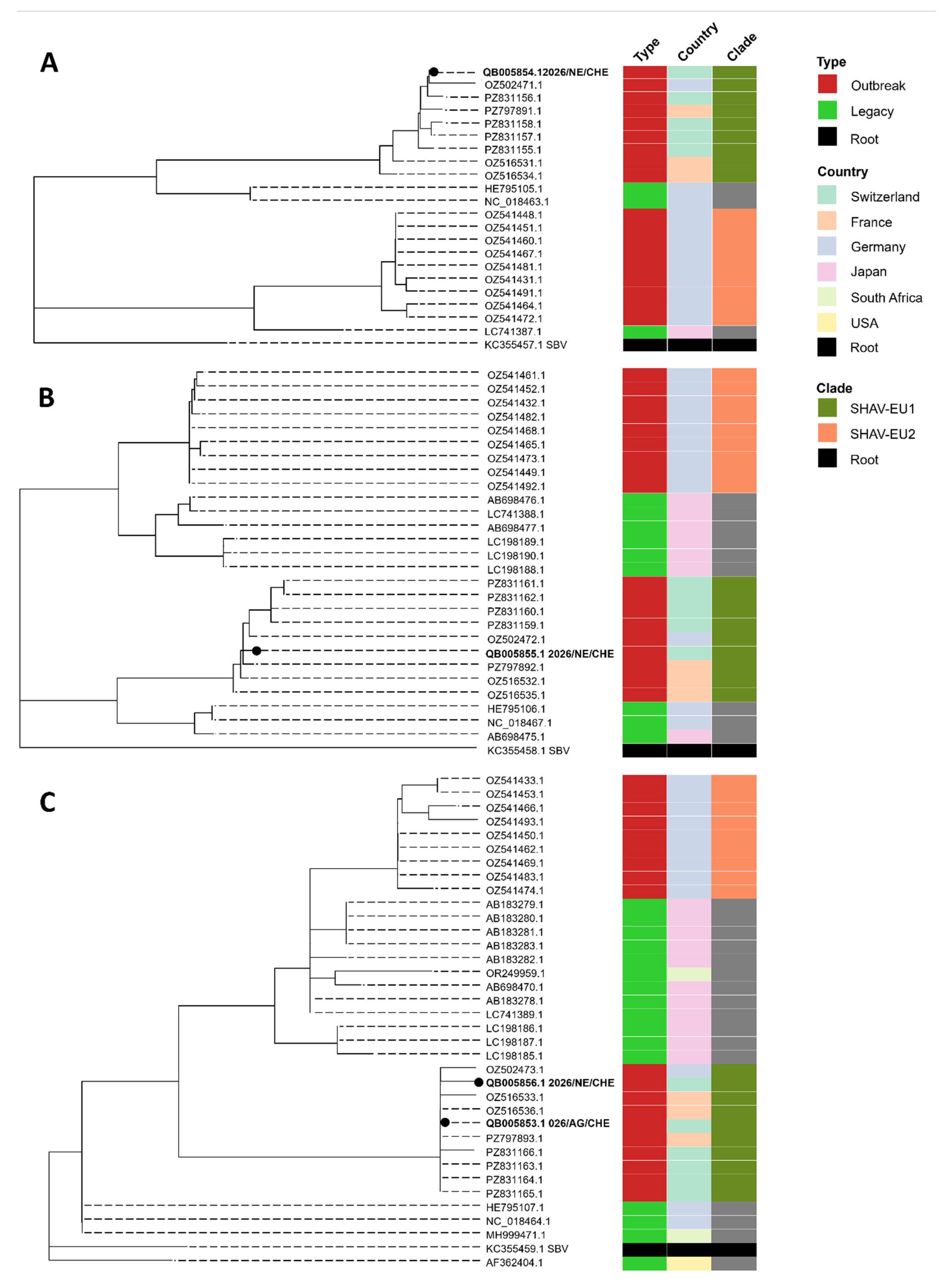
Table 2.
Results of the Pan-Simbu RT-qPCR (Quantification cycle, Cq) for brain tissues, blood, and cerebrospinal fluid samples of the two affected horses.
Table 2.
Results of the Pan-Simbu RT-qPCR (Quantification cycle, Cq) for brain tissues, blood, and cerebrospinal fluid samples of the two affected horses.
| Tissue / body fluid sample | Case 1 | Case 2 |
| Medulla oblongata | 30 | 37.3 |
| Cerebrum | 25.7 | 30.8 |
| Cerebellum | 22.7 | 30.5 |
| Olfactory bulb | n.a. | 26.3 |
| Spinal cord | n.a. | 37 |
| Whole blood | negative | negative |
| Serum | negative | negative |
| Cerebrospinal fluid | negative | negative |
Abbreviation: n.a. - sample not available.
Conclusions
Our findings provide evidence that SHAV-EU1 can infect the CNS of horses causing encephalomyelitis associated with vasculitis. Both affected horses had cerebrospinal lymphocytic pleocytosis and developed acute neurologic signs. Detection of SHAV RNA by metatranscriptomic sequencing and subsequent RT-qPCR in multiple CNS regions in both horses, together with the absence of other relevant viral sequences, and the presence of antibodies against orthobunyaviruses of the Simbu serogroup supports an association between SHAV-EU1 infection and the observed disease. However, localization of viral RNA or antigen within histologic lesions would provide additional evidence for a causal relationship.
From a clinical perspective, important differential diagnoses for the observed neurological signs include infections with neurotropic flaviviruses, such as West Nile virus, tick-borne encephalitis virus, and equine herpesvirus type 1 myeloencephalopathy, as well as Borna virus infection, neuroborreliosis and anaplasmosis, alongside non-infectious causes such as hyperammonemic encephalopathy [8,9,10]. In both horses, relevant differential diagnoses were largely ruled out (Appendix, Table 1).
Neurologic disease associated with Simbu serogroup viruses has been reported previously. Shuni virus has been associated with severe and sometimes fatal neurologic disease and encephalitis in horses in southern Africa [11,12,13], and Akabane virus was shown to cause postnatal encephalomyelitis in cattle in Japan [14,15]. Thus, CNS infection and associated neurological disease may be part of the pathogenic spectrum of some Simbu serogroup orthobunyaviruses.
Phylogenetic analysis placed the equine SHAV within the SHAV-EU1 clade comprising strains detected during the 2026 European outbreak. The high nucleotide sequence identities across all three genome segments further demonstrate the close genetic relationship between the equine virus and contemporaneously circulating bovine strains. Together with the temporal overlap of the equine cases and the SHAV-EU1 outbreak in Swiss cattle, these findings suggest that infection of horses was part of the same ongoing European epizootic rather than circulation of a distinct equine SHAV lineage. Thus, the emergence of neurologic disease in horses appears to reflect an expansion of the recognized host and clinical spectrum of the circulating SHAV-EU1 rather than the emergence of a genetically distinct virus.
Notably, SHAV RNA was detected in multiple CNS regions but not in whole blood, serum, or cerebrospinal fluid. Testing of blood or cerebrospinal fluid alone might therefore be insufficient to detect SHAV infection in horses with neurologic disease intra-vitam, except when sampling happens to coincide with the presumably short-lived viremic phase.
Although our observations are limited to two cases and do not establish the incidence of SHAV-EU1 infection or the risk for neurologic disease among infected horses, the consistent clinicopathologic findings warrant increased awareness and surveillance. SHAV should be considered in the differential diagnosis of acute encephalomyelitis in horses in regions where the virus is circulating. Further studies are needed to improve intra-vitam diagnostics, determine the prevalence, pathogenesis, and clinical spectrum of SHAV infection in horses.
Acknowledgments
We thank the Next Generation Sequencing Platform of the University of Bern for performing the high-throughput sequencing experiments.
About the Authors
PD Dr. med. Vet. PhD Bachofen is a veterinarian specialized in veterinary virology, leads the department of Diagnostics and Development at the Institute of Virology and Immunology (IVI) in Mittelhäusern, Switzerland, and is a lecturer at the Vetsuisse Faculty, University of Bern. As head of the Swiss national reference laboratory for viral epizootic diseases, her research interests include molecular epidemiology and surveillance of emerging and zoonotic animal viruses.
Appendix A
Materials and Methods
Serological Testing
The ID Screen® Schmallenberg virus Indirect Multi-species antibody detection ELISA from Innovative Diagnostics was used to determine antibodies against Shamonda virus in serum and CSF. This Schmallenberg ELISA was previously shown to cross-react with antibodies against Shamonda virus in cattle [3]. The test performed used according to the manufacturers’ instructions.
High-Throughput Sequencing and Bioinformatics
We extracted total RNA from fresh-frozen brain tissue with Trizol Reagent (Thermo Fisher Scientific) according to the instructions of the manufacturer. We assessed the quantity and quality of the purified total RNA using a Qubit 4.0 fluorometer with the Qubit RNA BR Assay Kit (Thermo Fisher Scientific) and an Advanced Analytical Fragment Analyzer System using a Fragment Analyzer RNA Kit (Agilent), respectively. We prepared cDNA libraries using the CORALL RNA-Seq V2 Library Prep Kit (Lexogen) and evaluated the resulting cDNA libraries with the Qubit 4.0 fluorometer and the Qubit dsDNA HS Assay Kit (Thermo Fisher Scientific) and the Agilent Fragment Analyzer (Agilent) with a HS NGS Fragment Kit (Agilent), respectively. We sequenced cDNA libraries 150bp paired-end using the NovaSeq 6000 SP Reagent Kit v1.5 (illumina) on a NovaSeq 6000 instrument (illumina). High-throughput sequencing reads for case 1 and case 2 are available at the NCBI sequence read archive under the BioProject accession number PRJNA1527475.
We subjected raw reads to a quality control process using fastqc (v.0.11.7). Reads were then quality selected and trimmed with fastp (v.0.12.5) at the 5' and 3' ends with a Phred quality control score less than 25 to remove low-quality reads and molecular identifiers, which were added during the library preparation. We mapped the trimmed reads to the horse reference genome EquCab 3.0 [1] using STAR (v.2.7.3a). We retained unmapped reads and analyzed them by the Genome detective platform (v.2.25.0) [2] (www.genomedetective.com) for the presence of viral sequences. Virus sequences were further analyzed by Geneious prime (v.2025.03, Dotmatics). The final sequences were uploaded to NCBI Genbank and are available under the following accession numbers: Case 2: 2026/AG/CHE (segment S), QB005853; case 1: 2026/NE/CHE, QB005854-6 (segments L, M, S).
Phylogenetic Analysis
Publicly available SHAV sequences were retrieved from NCBI GenBank (11 September 2026). Phylogenetic analysis was performed as described previously [3]. Briefly, sequences were aligned with MAFFT with option --op 5 (v.7.490, 4). The resulting alignment was visually inspected ahead of phylogenetic inference using default fasttree (v.2.1.11, 5). Visualization of the tree was performed using the libraries ggplot2 (3.5.2, 6), ggtree (v.3.10.1, 7) and ggnewscale (v.0.5.2, 8). NCBI GenBank was utilized as the source of country, sequences from 2026 are classified as outbreak and older sequences were classified as legacy data (see Table 3). Rooting of the tree was accomplished using isolate F6 from the ICTV taxonomy (MSL #41, https://ictv.global/taxonomy). For visualization purposes, branch lengths were square-root transformed to improve the resolution of differences between closely related sequences.
Table 1.
Molecular and serological testing results for pathogens associated with neurological disease in horses in central Europe.
Table 1.
Molecular and serological testing results for pathogens associated with neurological disease in horses in central Europe.
| Pathogen | Sample matrix | Method | Result | |
| Case 1 | Case 2 | |||
| EHV-1, EHV-4 | Nasal swab | PCR | Negative | Negative |
| Blood (EDTA) | PCR | N.d. | Negative | |
| WNV | Serum | ELISA | IgG: negative IgM: negative |
IgG: negative IgM: negative |
| CSF | RT-qPCR | Negative | Negative | |
| Serum | RT-qPCR | Negative | Negative | |
| TBEV | Serum | EIA | IgM: negative IgG: equivocal (68.8 U/mL) (63 - 126 U/mL: equivocal, > 126 U/mL: positive) |
IgM: negative IgG: equivocal (69.4 U/mL) (63 - 126 U/mL: equivocal, > 126 U/mL: positive) |
| CSF | EIA |
N.d. |
IgG: elevated (26.9 U/mL) | |
| RT-PCR | Negative | Negative | ||
| Borrelia | Serum | IFT | IgM: negative IgG: positive (1:256) (reference range < 1:64) |
N.d. N.d. |
| CSF | PCR | Negative | N.d. | |
| Anaplasma | Blood (EDTA) | PCR | Negative | N.d. |
Abbreviations: CSF, cerebrospinal fluid; EDTA, ethylenediaminetetraacetic acid; EIA, enzyme immunoassay; IFT, immunofluorescence test; N.d., not done; PCR, polymerase chain reaction; RT-PCR, reverse-transcription polymerase chain reaction TBEV, tick-borne encephalitis virus; WNV, West Nile Virus.
Table 2.
Results of high-throughput sequencing and the virus discovery pipeline. Data are referring to the comparison with the best hit (Shamonda virus isolate Ib An 5550, (L: NC_018463, S: NC_018464, M: NC_018467)).
Table 2.
Results of high-throughput sequencing and the virus discovery pipeline. Data are referring to the comparison with the best hit (Shamonda virus isolate Ib An 5550, (L: NC_018463, S: NC_018464, M: NC_018467)).
| Case ID | Segment | Number reads mapped | Contig size | Depths of coverage | Segment coverage |
| Case 1 | |||||
| S | 3877 | 839 nt | 488.4 | 97% | |
| M | 1324 | 4295 nt | 29.9 | 99.6% | |
| L | 1619 | 6820 nt | 35.4 | 100% | |
| Case 2 | |||||
| S | 199 | 823 nt | 29 | 88.7% | |
| M | 31 | - | - | 29.6% | |
| L | 22 | - | - | 30% |
Table 3.
Metadata of viral sequences retrieved from GenBank for phylogenetic comparison.
| ID | Type | Country | Clade | Segment |
| QB005854 | Outbreak | CH | SHAV-EU1 | L |
| PZ831157.1 | Outbreak | CH | SHAV-EU1 | L |
| PZ831156.1 | Outbreak | CH | SHAV-EU1 | L |
| PZ831155.1 | Outbreak | CH | SHAV-EU1 | L |
| PZ831158.1 | Outbreak | CH | SHAV-EU1 | L |
| OZ502471.1 | Outbreak | Germany | SHAV-EU1 | L |
| OZ516534.1 | Outbreak | France | SHAV-EU1 | L |
| OZ516531.1 | Outbreak | France | SHAV-EU1 | L |
| PZ797891.1 | Outbreak | France | SHAV-EU1 | L |
| NC_018463.1 | legacy | Germany | L | |
| HE795105.1 | legacy | Germany | L | |
| KC355457.1 | Root | Root | Root | L |
| OZ541491.1 | Outbreak | Germany | L | |
| OZ541481.1 | Outbreak | Germany | SHAV-EU2 | L |
| OZ541467.1 | Outbreak | Germany | SHAV-EU2 | L |
| OZ541460.1 | Outbreak | Germany | SHAV-EU2 | L |
| OZ541451.1 | Outbreak | Germany | SHAV-EU2 | L |
| OZ541448.1 | Outbreak | Germany | SHAV-EU2 | L |
| OZ541431.1 | Outbreak | Germany | SHAV-EU2 | L |
| OZ541472.1 | Outbreak | Germany | SHAV-EU2 | L |
| OZ541464.1 | Outbreak | Germany | SHAV-EU2 | L |
| LC741387.1 | legacy | Japan | L | |
| QB005855 | Outbreak | Switzerland | SHAV-EU1 | M |
| OZ516535.1 | Outbreak | France | SHAV-EU1 | M |
| OZ516532.1 | Outbreak | France | SHAV-EU1 | M |
| PZ831162.1 | Outbreak | Switzerland | SHAV-EU1 | M |
| PZ831161.1 | Outbreak | Switzerland | SHAV-EU1 | M |
| PZ831160.1 | Outbreak | Switzerland | SHAV-EU1 | M |
| PZ831159.1 | Outbreak | Switzerland | SHAV-EU1 | M |
| PZ797892.1 | Outbreak | France | SHAV-EU1 | M |
| OZ502472.1 | Outbreak | Germany | SHAV-EU1 | M |
| NC_018467.1 | legacy | Germany | M | |
| HE795106.1 | legacy | Germany | M | |
| AB698475.1 | legacy | Japan | M | |
| OZ541492.1 | Outbreak | Germany | SHAV-EU2 | M |
| OZ541468.1 | Outbreak | Germany | SHAV-EU2 | M |
| OZ541452.1 | Outbreak | Germany | SHAV-EU2 | M |
| OZ541482.1 | Outbreak | Germany | SHAV-EU2 | M |
| OZ541432.1 | Outbreak | Germany | SHAV-EU2 | M |
| OZ541449.1 | Outbreak | Germany | SHAV-EU2 | M |
| OZ541473.1 | Outbreak | Germany | SHAV-EU2 | M |
| OZ541465.1 | Outbreak | Germany | SHAV-EU2 | M |
| OZ541461.1 | Outbreak | Germany | SHAV-EU2 | M |
| LC741388.1 | legacy | Japan | M | |
| AB698476.1 | legacy | Japan | M | |
| AB698477.1 | legacy | Japan | M | |
| LC198190.1 | legacy | Japan | M | |
| LC198189.1 | legacy | Japan | M | |
| LC198188.1 | legacy | Japan | M | |
| KC355458.1 | Root | Root | Root | M |
| QB005853 | Outbreak | Switzerland | SHAV-EU1 | S |
| OZ516536.1 | Outbreak | France | SHAV-EU1 | S |
| OZ516533.1 | Outbreak | France | SHAV-EU1 | S |
| QB005856 | Outbreak | Switzerland | SHAV-EU1 | S |
| LC741389.1 | legacy | Japan | S | |
| KC355459.1 | Root | Root | Root | S |
| OZ541493.1 | Outbreak | Germany | S | |
| OZ541469.1 | Outbreak | Germany | SHAV-EU2 | S |
| OZ541462.1 | Outbreak | Germany | SHAV-EU2 | S |
| OZ541450.1 | Outbreak | Germany | SHAV-EU2 | S |
| OZ541483.1 | Outbreak | Germany | SHAV-EU2 | S |
| OZ541474.1 | Outbreak | Germany | SHAV-EU2 | S |
| OZ541453.1 | Outbreak | Germany | SHAV-EU2 | S |
| OZ541433.1 | Outbreak | Germany | SHAV-EU2 | S |
| OZ541466.1 | Outbreak | Germany | SHAV-EU2 | S |
| LC198187.1 | legacy | Japan | S | |
| LC198186.1 | legacy | Japan | S | |
| LC198185.1 | legacy | Japan | S | |
| AB698470.1 | legacy | Japan | S | |
| AB183278.1 | legacy | Japan | S | |
| AB183281.1 | legacy | Japan | S | |
| AB183280.1 | legacy | Japan | S | |
| AB183279.1 | legacy | Japan | S | |
| AB183282.1 | legacy | Japan | S | |
| AB183283.1 | legacy | Japan | S | |
| PZ831166.1 | Outbreak | Switzerland | SHAV-EU1 | S |
| PZ831165.1 | Outbreak | Switzerland | SHAV-EU1 | S |
| PZ831164.1 | Outbreak | Switzerland | SHAV-EU1 | S |
| PZ831163.1 | Outbreak | Switzerland | SHAV-EU1 | S |
| OZ502473.1 | Outbreak | Germany | SHAV-EU1 | S |
| PZ797893.1 | Outbreak | France | SHAV-EU1 | S |
| MH999471.1 | legacy | South Africa | S | |
| AF362404.1 | legacy | USA | S | |
| NC_018464.1 | legacy | Germany | S | |
| HE795107.1 | legacy | Germany | S | |
| OR249959.1 | legacy | South Africa | S |
References
- Kalbfleisch TS, Rice ES, DePriest MS Jr, Walenz BP, Hestand MS, Vermeesch JR, et al. Improved reference genome for the domestic horse increases assembly contiguity and composition. Commun Biol. 2018;1:197. [CrossRef]
- Vilsker M, Moosa Y, Nooij S, Fonseca V, Ghysens Y, Dumon K, et al. Genome Detective: an automated system for virus identification from high-throughput sequencing data. Bioinformatics. 2019;35:871–3. [CrossRef]
- Wyler M, Darpel K, Berset R, Renzullo S, Huber H, Bauer J, et al. First identification, isolation and genomic characterization of an emerging Shamonda virus variant associated with bovine disease in Switzerland. Preprints. 2026 [cited 2026 Sep 15]. [CrossRef]
- Katoh K, Toh H. Recent developments in the MAFFT multiple sequence alignment program. Brief Bioinform. 2008;9:286–98. [CrossRef]
- Wickham H. ggplot2: Elegant Graphics for Data Analysis. New York: Springer-Verlag; 2016. [CrossRef]
- Price MN, Dehal PS, Arkin AP. FastTree 2—approximately maximum-likelihood trees for large alignments. PLoS One. 2010;5:e9490. [CrossRef]
- Yu G, Smith DK, Zhu H, Guan Y, Lam TT-Y. ggtree: an R package for visualization and annotation of phylogenetic trees with their covariates and other associated data. Methods Ecol Evol. 2017;8:28–36. [CrossRef]
- Campitelli E. ggnewscale: Multiple Fill and Colour Scales in 'ggplot2'. R package version 0.5.2 [cited 2026 Sep 15]. [CrossRef]
References
- O'Connor TW, Hick PM, Finlaison DS, Kirkland PD, Toribio J. Revisiting the Importance of Orthobunyaviruses for Animal Health: A Scoping Review of Livestock Disease, Diagnostic Tests, and Surveillance Strategies for the Simbu Serogroup. Viruses. 2024;16(2):294. [CrossRef]
- Wyler M, Darpel K, Berset R, Renzullo S, Huber H, Bauer J, et al. First Identification, Isolation and Genomic Characterization of an Emerging Shamonda Virus Variant Associated with Bovine Disease in Switzerland. Preprints. 2026. [CrossRef]
- Wernike K, Hoffmann B, Link EK, Rotheneder S, Eshak M, Pfaff F, et al. It happened again – emergence of a Shamonda-like orthobunyavirus in Central Europe, 2026. bioRxiv. 2026. [CrossRef]
- Kellecil M, Fusade-Boyer M, Chrétien D, Durand E, Mircovich M, Sécula A, et al. Detection of a novel Shamonda in dairy cattle, France, June 2026. Eurosurveillance. 2026;31(33): 2600689. [CrossRef]
- Wernike K, Pfaff F, Zeiske S, Höper D, Beer M. Two incursions, two viruses: Emergence of a second novel Shamonda virus clade in Central Europe, 2026. bioRxiv. 2026. [CrossRef]
- Ozcelik R, Graubner C, Remy-Wohlfender F, Durr S, Faverjon C. Evaluating 5.5 Years of Equinella: A Veterinary-Based Voluntary Infectious Disease Surveillance System of Equines in Switzerland. Front Vet Sci. 2020;7:327. [CrossRef]
- Golender N, Bumbarov VY, Erster O, Beer M, Khinich Y, Wernike K. Development and validation of a universal S-segment-based real-time RT-PCR assay for the detection of Simbu serogroup viruses. J Virol Methods. 2018;261:80-5. [CrossRef]
- Cavalleri JV, Korbacska-Kutasi O, Leblond A, Paillot R, Pusterla N, Steinmann E, et al. European College of Equine Internal Medicine consensus statement on equine flaviviridae infections in Europe. J Vet Intern Med. 2022;36(6):1858-71. [CrossRef]
- Aleman M, Vedavally U, Pusterla N, Wensley F, Berryhill E, Madigan JE. Common and atypical presentations of Anaplasma phagocytophilum infection in equids with emphasis on neurologic and muscle disease. J Vet Intern Med. 2024;38(1):440-8. [CrossRef]
- Johnstone LK, Engiles JB, Aceto H, Buechner-Maxwell V, Divers T, Gardner R, et al. Retrospective Evaluation of Horses Diagnosed with Neuroborreliosis on Postmortem Examination: 16 Cases (2004-2015). J Vet Intern Med. 2016;30(4):1305-12. [CrossRef]
- van Eeden C, Williams JH, Gerdes TGH, van Wilpe E, Viljoen A, Swanepoel R, et al. Shuni Virus as Cause of Neurologic Disease in Horses. Emerg Infect Dis. 2012;18(2):318-21. [CrossRef]
- Motlou TP, Williams J, Venter M. Epidemiology of Shuni Virus in Horses in South Africa. Viruses-Basel. 2021;13(5):937. [CrossRef]
- Rakaki ME, van der Walt M, Williams J, Venter M. Detection of Novel Orthobunyavirus Reassortants in Fatal Neurologic Case in Horse and Culicoides Biting Midges, South Africa. Emerg Infect Dis. 2025;31(7):1455-9. [CrossRef]
- Kono R, Hirata M, Kaji M, Goto Y, Ikeda S, Yanase T, et al. Bovine epizootic encephalomyelitis caused by Akabane virus in southern Japan. BMC Vet Res. 2008;4:20. [CrossRef]
- Uchida K, Murakami T, Sueyoshi M, Tsuda T, Inai K, Acorda JA, et al. Detection of Akabane viral antigens in spontaneous lymphohistiocytic encephalomyelitis in cattle. J Vet Diagn Invest. 2000;12(6):518-24. [CrossRef]
Figure 1.
Pathological lesions, scale bar = 100 μm. A. Cross section of the brain stem with multiple asymmetrical hemorrhages within the parenchyma and shift of the midline (dashed line) due to edema. Formalin-fixed material, case 1. B. Severe lymphoplasmacytic infiltration of blood vessel wall (vasculitis) with endothelial cell necrosis, loss of endothelium, and intravascular formation of fibrin thrombi (stars). In the surrounding parenchyma, proteinaceous edema, hemorrhages and axonal degeneration (arrows) are present. H&E stain, case 1. C. Formation of glial nodule (circle) in proximity of affected blood vessels. H&E stain, case 1. D. Severe perivascular cuffing with hemorrhages and axonal swellings (ischemic damage, arrows) in the cervical spinal cord. H&E stain, case 2.
Disclaimer/Publisher’s Note: The statements, opinions and data contained in all publications are solely those of the individual author(s) and contributor(s) and not of MDPI and/or the editor(s). MDPI and/or the editor(s) disclaim responsibility for any injury to people or property resulting from any ideas, methods, instructions or products referred to in the content. |
© 2026 by the authors. Licensee MDPI, Basel, Switzerland. This article is an open access article distributed under the terms and conditions of the Creative Commons Attribution (CC BY) license.
Copyright: This open access article is published under a Creative Commons CC BY 4.0 license, which permit the free download, distribution, and reuse, provided that the author and preprint are cited in any reuse.