Preprint
Review

This version is not peer-reviewed.

Atractylodes macrocephala in Ulcerative Colitis: Bioactives, Microbiota, and Translational Gaps

Submitted:

01 October 2026

Posted:

05 October 2026

You are already at the latest version

Abstract

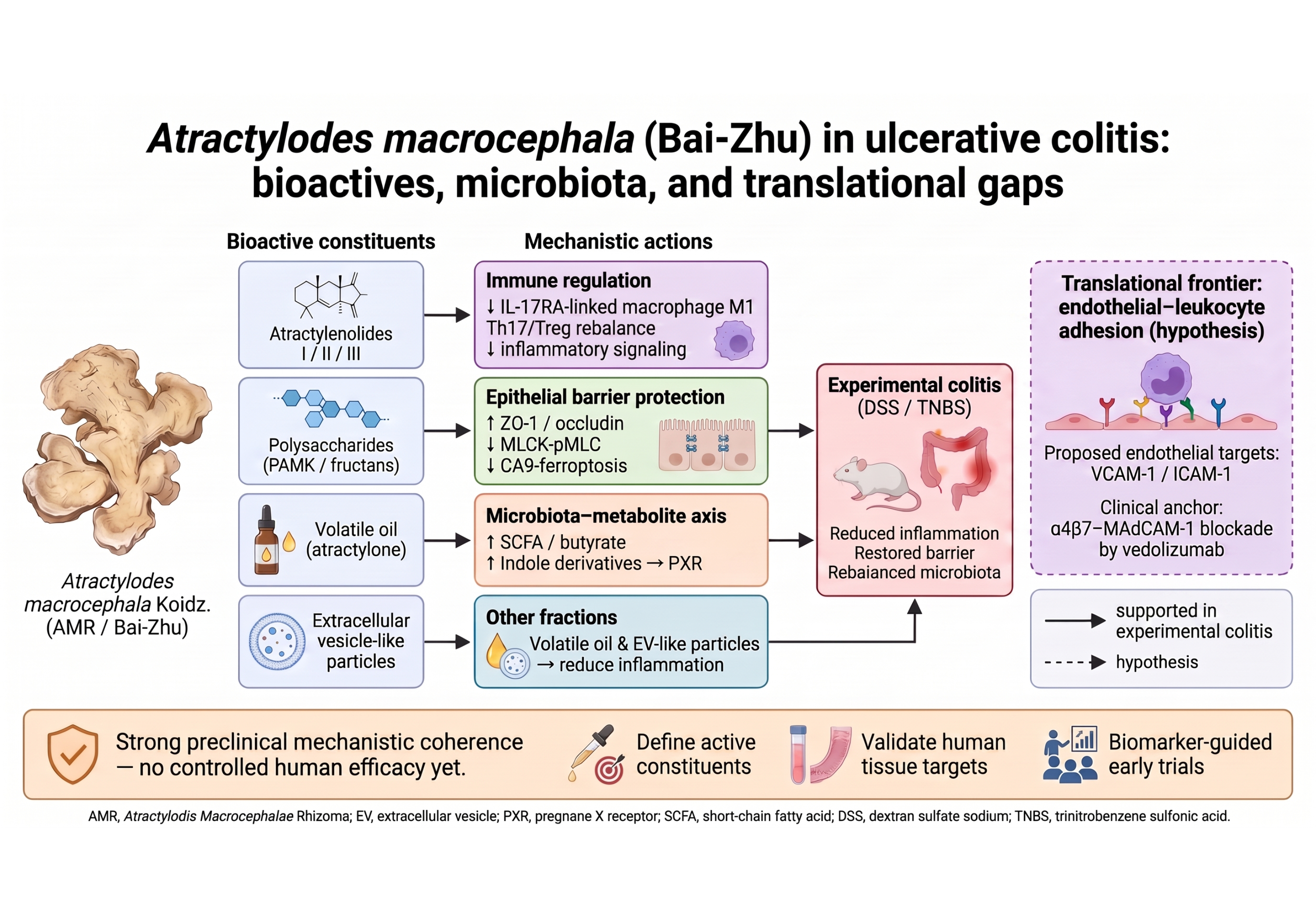
Atractylodes macrocephala Koidz. rhizome (Atractylodis Macrocephalae Rhizoma, AMR; Bai-Zhu) is a widely used medicinal plant, and its specialized metabolites are now being mapped onto defined molecular targets. Here we review, constituent by constituent, how AMR acts in ulcerative colitis (UC). The sesquiterpenoid lactones account for much of the activity: atractylenolide I protects the epithelial barrier (RhoA/ROCK/MLC; S100A9/AMPK/mTOR), reshapes intestinal metabolism and the microbiota (SPHK1/B4GALT2) and limits carbonic anhydrase IX-mediated ferroptosis, whereas atractylenolide II drives Nrf2-dependent antioxidant responses (in non-colitis models), and interleukin-17 receptor A is nominated as a candidate direct target of atractylenolide III—awaiting independent replication—through which it may curb macrophage M1 polarization. The polysaccharides rebalance Th17/Treg immunity and remodel the gut microbiota and its tryptophan metabolites through the pregnane X receptor, while the volatile oil and vesicle-like particles are likewise protective. We grade each mechanism by the strength of the underlying colitis-model evidence and stay explicit about what remains extrapolation. Controlled human trials are still lacking, with only very preliminary clinical observation to date. Finally, we single out endothelial-leukocyte adhesion—already exploited by anti-adhesion biologics—as the most tractable next step for translating these constituents toward the clinic.
Keywords: 
;  ;  ;  ;  ;  

1. Introduction

Ulcerative colitis (UC) is a chronic, relapsing inflammatory bowel disease (IBD) of unknown cause, marked by continuous mucosal and submucosal inflammation of the colon and rectum [1]. Long regarded as a Western disease, it is now rising steadily across newly industrialized regions [2]; in Taiwan, population-based data show both incidence and prevalence climbing over the past two decades [3]. Current therapy—5-aminosalicylates (5-ASA), corticosteroids, thiopurines, anti-TNF, anti-integrin and anti-IL-12/23 biologics, and small-molecule JAK inhibitors—has improved outcomes considerably [4,5,6], yet a substantial proportion of patients respond poorly at induction or lose response over time, and some still come to colectomy [1]. Prolonged corticosteroid and immunosuppressant use brings familiar toxicity, and the price of advanced agents limits access in many settings. It is against this backdrop that safer complementary options continue to attract attention.
In traditional Chinese medicine (TCM), UC-like illness has long been described as ‘jiu li’ (chronic dysentery) or ‘xie xie’ (protracted diarrhea) and attributed to spleen deficiency with accumulated dampness. The rhizome of Atractylodes macrocephala Koidz. (Atractylodis Macrocephalae Rhizoma; AMR; Bai-Zhu), a qi-tonifying and dampness-drying herb already recorded in the Shennong Bencao Jing [7], remains one of the most commonly used single herbs for chronic diarrheal disease in East Asia, and it anchors classical formulas such as Shen-Ling-Bai-Zhu-San, whose formula-level pharmacology has been reviewed elsewhere [8]. Recent mechanistic studies of such formulas in experimental UC keep returning to the same themes: remodeling of the gut microbiota, macrophage repolarization, and repair of the epithelial barrier [9,10]. The traditional use of AMR is, moreover, now backed by mechanistic data of steadily improving quality [11]. Here we bring that evidence together for AMR and its individual constituents in UC, organizing it by molecular axis and closing on an under-explored frontier, vascular endothelial activation and leukocyte adhesion, that we argue deserves priority. Existing reviews are broader in scope: TCM, gut microbiota and UC signaling [12]; natural products, IBD and gut microbes [13]; a general 2025 account of AMR [14]; and AMR polysaccharides in intestinal disorders [15]. Ours is deliberately narrower, being single-herb, UC-specific and constituent-resolved. A 2026 systematic review recently catalogued the traditional uses, phytochemistry, pharmacology and structure-activity relationships of the whole genus Atractylodes across hundreds of compounds [16]; by contrast, we stay disease-focused rather than genus-focused, restricting the scope to A. macrocephala in UC, grading each mechanism by its experimental context, mapping study-level treatment regimens, and spelling out disease-specific translational gaps and an endothelial anti-adhesion agenda that a genus-wide catalogue does not take up. Relative to the closest prior work, a 2024 review confined to the atractylenolides [11], the present synthesis differs in five concrete ways: it spans the full phytochemical range of AMR (atractylenolides, polysaccharides, volatile oil and vesicle-like particles); it adds primary studies from 2025-2026; it labels every mechanism as direct experimental-colitis evidence, extrapolation from non-UC or non-equivalent disease contexts, or general mechanistic biology (Table 1); it tabulates study-level models, doses and regimens for the direct-evidence studies (Table 2); and it sets out, without overstatement, an endothelial anti-adhesion research agenda.

2. Search Strategy

PubMed, Embase and Web of Science were searched to August 2026 using combinations of ‘Atractylodes macrocephala’, ‘Atractylodis Macrocephalae Rhizoma’, ‘Bai-Zhu’, ‘atractylenolide’, ‘polysaccharide’, ‘ulcerative colitis’, ‘colitis’, ‘inflammatory bowel disease’, ‘gut microbiota’, ‘NF-κB’ and ‘adhesion molecule’; the final search was updated on 30 August 2026, and reference lists of eligible primary studies and relevant reviews were hand-searched for additional records. Selection prioritized (i) target-identification and mechanistic studies in validated colitis models (dextran sulfate sodium [DSS], trinitrobenzene sulfonic acid [TNBS]) [17], (ii) landmark clinical evidence defining the UC therapeutic landscape, and (iii) recent (2020-2026) primary studies of AMR constituents, supplemented by authoritative reviews for context. Preprints and retracted or withdrawn articles were excluded, as were in vitro reports lacking a defined extract source, dosage or appropriate controls, and purely in-silico (network-pharmacology) predictions without experimental validation. Throughout, mechanisms are graded as direct experimental-colitis evidence, extrapolated evidence from non-UC or non-equivalent disease contexts, or general mechanistic biology (Table 1, dagger notation); we emphasize that dextran sulfate sodium and TNBS are experimental colitis models that inform, but do not equal, human UC. The search strategy was designed for evidence mapping and critical mechanistic synthesis rather than exhaustive quantitative meta-analysis; accordingly, and in keeping with the narrative design, no formal PRISMA flow or formal risk-of-bias scoring was applied. The final synthesis drew on the verified references cited herein, and findings from chemically induced models were interpreted in light of the known limitations of such models [17].

Use of AI-Assisted Tools

During the preparation of this manuscript, a large language model (Claude, Anthropic) assisted with language editing and with organizing and cross-checking the cited literature; it was not used to generate data, figures, or original scientific content. All statements, citations, mechanistic interpretations, and evidence gradings were checked against the primary sources by the authors, who take full responsibility for the content.

3. Bridging Traditional Theory and Modern Pharmacology

In TCM theory, AMR ‘fortifies the spleen and dries dampness’, targeting the ‘spleen-deficiency with dampness’ pattern that maps clinically onto chronic relapsing diarrhea with mucus, fatigue and poor appetite, a presentation that partially overlaps clinically with mild-to-moderate UC. The rhizome contains sesquiterpenoid lactones, polysaccharides, volatile oil and polyacetylenes as its principal bioactive classes [7]. Modern pharmacology reframes the classical actions as restoration of epithelial barrier function, resolution of mucosal immune activation, and correction of dysbiosis; the multi-component, multi-target character of the herb is no longer a rhetorical claim but an experimentally tractable property, as detailed below [11].

4. Bioactive Constituents of Atractylodis Macrocephalae Rhizoma and Their Molecular Targets

4.1. Atractylenolides: Receptor-Proximal Anti-Inflammatory Sesquiterpenoids

The sesquiterpenoid lactones atractylenolide I-III (AT-I, AT-II, AT-III) are the best characterized AMR constituents [18], and a growing panel of target-identification studies now assigns them distinct, receptor-proximal mechanisms relevant to UC [11] (Figure 1).
Table 1. Principal mechanisms of Atractylodis Macrocephalae Rhizoma constituents relevant to ulcerative colitis, graded by evidence tier.
Constituent Model / system Principal mechanism Evidence tier Reference
Atractylenolide I Colitis models; macrophage models; gastric injury; colitis-associated CRC† MD-2/TLR4-MyD88-NF-κB antagonism; SPHK1/B4GALT2 targeting; RhoA/ROCK/MLC and S100A9/AMPK/mTOR barrier protection; CA9-mediated ferroptosis inhibition (GPX4/SLC7A11); NLRP3/pyroptosis restraint (incl. Drp1/mitochondrial fission)† Direct + extrapolated† [19,20,21,25,26,27,28,30,31]
Atractylenolide II Oxidative injury (radiation)† Nrf2 activation; ROS clearance (HO-1 axis plausible in UC) Extrapolated (non-colitis)† [33,34]
Atractylenolide III DSS & TNBS colitis; macrophages; CRC cells† IL-17RA antagonism → ⊣ M1 polarization; ⊣ NF-κB-MLCK-pMLC (barrier); antioxidant/flora regulation; AMPK/SIRT1/PGC-1α; Bax/Bcl-2 pro-apoptotic† Direct + extrapolated† [35,39,40,41,42]
Polysaccharides (PAMK/AMP1) DSS colitis Th17/Treg rebalance (IL-6/STAT3); microbiota & tryptophan reshaping (PXR-mediated); IL-17RA signaling; bile acid-cAMP/TGR5-JNK axis; tight-junction/barrier preservation(lncRNA ITSN1-OT1) Direct (colitis model) [43,44,45,46,47,48,49,50,51,52]
Volatile oil & EV-like particles Acute DSS colitis Microbiota-metabolite regulation; microecological remodeling by vesicle-like particles Direct (colitis model) [55,56,57]
Microbiota-metabolite axis Colitis models Indole-PXR signaling (direct for PAMK); SCFA-GPR43 (general SCFA biology); barrier reinforcement; ↓ LPS translocation Direct (PXR) + general biology [45,58,61,63]
Evidence tiers: Direct (colitis model) = demonstrated in validated experimental-colitis models (DSS/TNBS), which inform but do not equal human UC; Extrapolated (†) = evidence derived from non-colitis models or disease contexts not equivalent to uncomplicated UC—including ovarian cancer and macrophage systems, gastric injury, radiation damage, colorectal cancer cells, and AOM/DSS colitis-associated colorectal cancer—biologically plausible but unconfirmed in uncomplicated UC. The proposed endothelial-leukocyte adhesion axis, which as yet has no published AMR-specific evidence, is not tabulated here and is discussed as a future direction in the emerging-frontier section (Figure 2). AMR, Atractylodis Macrocephalae Rhizoma (Bai-Zhu, the rhizome of Atractylodes macrocephala Koidz., distinct from Atractylodis Rhizoma/Cang-Zhu); PAMK, polysaccharides from Atractylodes macrocephala Koidz.; SCFA, short-chain fatty acid; LPS, lipopolysaccharide; CRC, colorectal cancer. References are cited by number in order of appearance; full entries appear in the numbered reference list.
Atractylenolide I. AT-I binds the MD-2/TLR4 complex and blocks MyD88-dependent signaling, thereby suppressing NF-κB nuclear translocation and MAPK cascade activation—evidence obtained in ovarian cancer and macrophage systems rather than colitis [19]—with consequent down-regulation of TNF-α, IL-6, IL-1β and COX-2 in lipopolysaccharide-stimulated macrophages, relative to lipopolysaccharide stimulation alone [20]. In vivo, AT-I alleviates experimental colitis at least in part by restraining RhoA/ROCK/MLC-mediated epithelial barrier dysfunction [21]; a published correction to one figure of that study does not affect its conclusions [22]. AT-I also inhibits NLRP3-ASC-Caspase-1 inflammasome assembly in a gastric (indomethacin-induced) mucosal injury model—an extrapolation beyond colitis that is nonetheless mechanistically relevant, because NLRP3 activation is a documented driver of IBD in both mice and humans [23,24,25]; in an azoxymethane (AOM)/DSS colitis-associated colorectal cancer model, AT-I likewise suppresses NLRP3 activation by inhibiting Drp1-mediated mitochondrial fission, though this tumor-context setting is indirect evidence for uncomplicated UC [26]. In colitis models, AT-I additionally targets SPHK1 and B4GALT2, regulating intestinal metabolism and flora composition to improve inflammation [27], restores intestinal barrier function in DSS-induced colitis by engaging the S100A9/AMPK/mTOR axis [28], and restrains ferroptosis—a documented mode of epithelial cell death in human UC [29]—in DSS colitis by targeting carbonic anhydrase IX (CA9), up-regulating GPX4 and SLC7A11 while down-regulating COX-2 and ACSL4, an effect reversed by the ferroptosis inducer erastin, abolished by AAV-mediated CA9 silencing, and corroborated by a cellular thermal shift assay (CETSA), although this single-study finding awaits independent replication [30], and mitigates antibiotic-induced dysbiosis of the intestinal microbiome, hinting at a microbiota-dependent component of its action [31]. Whether these targets act in parallel or converge on a shared cascade—for example, SPHK1 acting upstream of PI3K/AKT—remains undefined; importantly, the barrier-protective (RhoA/ROCK/MLC; S100A9/AMPK/mTOR) and microbiota-metabolic (SPHK1/B4GALT2) arms are those directly demonstrated in colitis, whereas the MD-2/TLR4 arm derives from non-colitis models and the NLRP3 arm from a gastric-injury model and an AOM/DSS colitis-associated colorectal cancer setting, so that both should be read as indirect evidence for uncomplicated UC. Atractylenolide I was also nominated as a candidate active constituent contributing to a processing-dependent action: rice-water-processed AMR exhibits enhanced anti-colitic efficacy through inhibition of LCN2-mediated pyroptosis, giving the traditional pao-zhi of the rhizome a defined molecular readout and illustrating how processing can amplify a constituent-level mechanism [32].
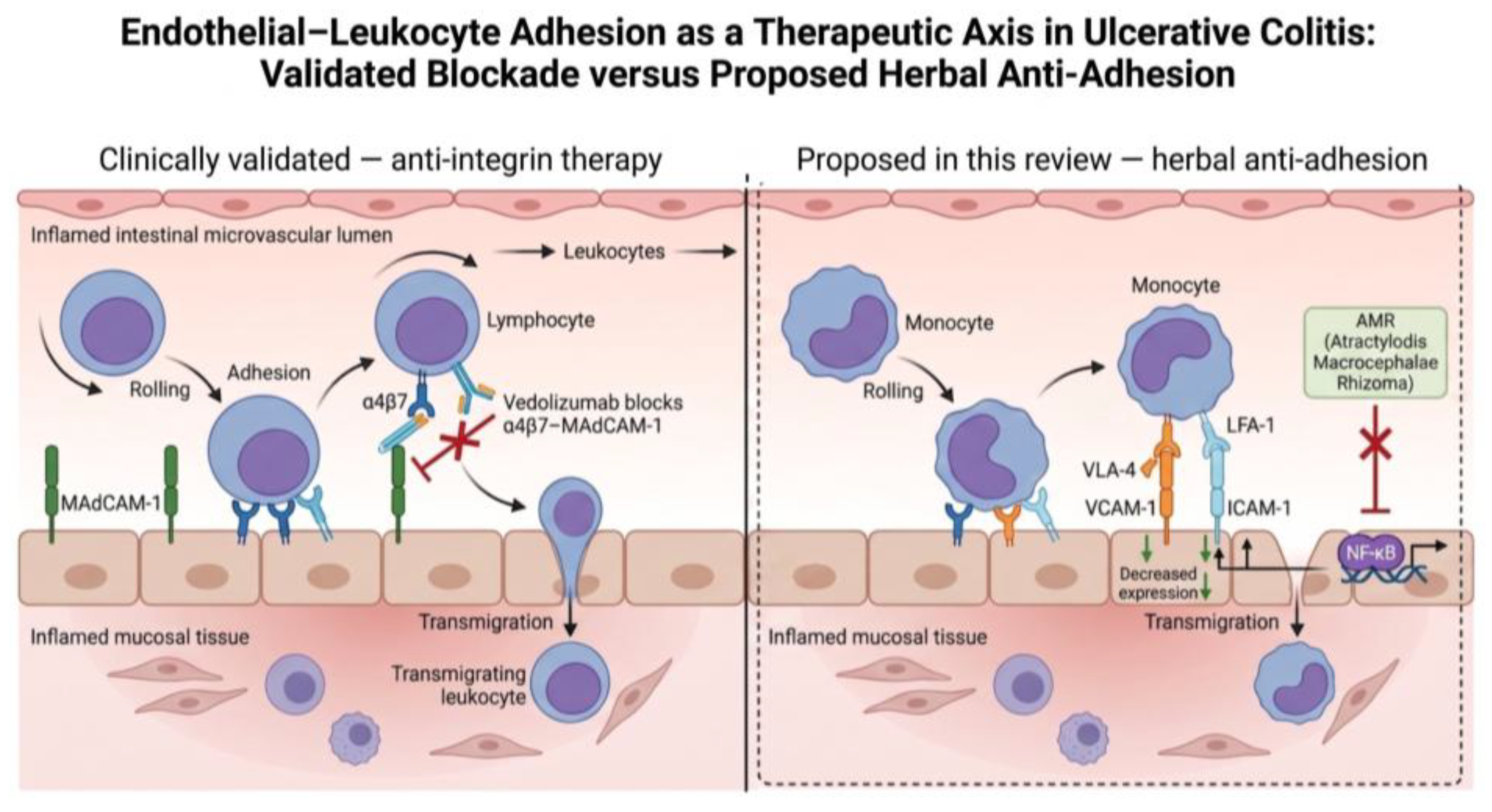
Figure 2. Endothelial-leukocyte adhesion as a therapeutic axis in ulcerative colitis: validated blockade versus proposed herbal anti-adhesion. Left (clinically validated): vedolizumab blocks α4β7 integrin-MAdCAM-1-dependent lymphocyte gut homing. Right (hypothesis proposed in this review, dashed outline): the rhizome of Atractylodes macrocephala Koidz. (Atractylodis Macrocephalae Rhizoma, AMR) is proposed to suppress NF-κB-driven endothelial expression of VCAM-1 and ICAM-1, thereby reducing VLA-4- and LFA-1-mediated leukocyte adhesion and transmigration. Selectin-mediated rolling is omitted for clarity. Leukocyte adhesion via VLA-4/LFA-1—engaging both myeloid and lymphoid cells—is depicted to contrast with the gut-selective α4β7-MAdCAM-1 homing axis targeted by vedolizumab, emphasizing that the proposed AMR effect concerns a broader, non-gut-selective arm of the adhesion cascade. This panel is hypothetical and awaits experimental validation. Created in BioRender. Huang, S. (2026) https://BioRender.com/dld5lfy.
Preprints 236200 g002
Atractylenolide II. In contrast to the receptor antagonism of AT-I, AT-II engages cytoprotective programs, activating Nrf2 signaling and reactive-oxygen-species clearance in models of oxidative tissue injury (radiation damage) [33]. Because HO-1—a canonical Nrf2 target gene—itself ameliorates murine colitis and rebalances Th17/Treg immunity [34], an AT-II-Nrf2-HO-1 axis in UC is biologically plausible, although it awaits direct demonstration in colitis models.
Atractylenolide III. Among the three congeners, AT-III currently carries the most direct—though still preliminary—target evidence. Target-fishing studies combining molecular docking, surface plasmon resonance and cellular thermal shift assays nominate IL-17RA as a candidate direct target of AT-III: by antagonizing IL-17RA, AT-III suppresses downstream NF-κB/MAPK signaling and restrains macrophage M1 polarization in ulcerative colitis models [35]—consistent with the broader recognition of macrophage polarization as a tractable TCM target in UC [36]—although independent replication of this recent finding is awaited. Caution is also warranted when extrapolating IL-17 pathway interference to IBD: systemic neutralization of IL-17A with secukinumab unexpectedly worsened Crohn’s disease in a randomized trial [37], underscoring tissue-protective roles of IL-17—including IL-17RA signaling in Lgr5+ intestinal stem cells that supports epithelial secretory-lineage commitment and barrier integrity [38]—and the need to distinguish receptor-level modulation by a natural compound from wholesale cytokine blockade. Independently, AT-III improves DSS-induced colitis by suppressing NF-κB-mediated MLCK-pMLC signaling to restore tight-junction integrity [39], ameliorates TNBS-induced intestinal inflammation by reducing oxidative stress and regulating the intestinal flora [40], and protects colonic epithelium by activating AMPK/SIRT1/PGC-1α-dependent mitochondrial biogenesis [41]. AT-III also induces apoptosis of colorectal cancer cells through the Bax/Bcl-2-regulated mitochondrial (intrinsic) apoptotic axis, with downstream caspase-9/caspase-3 activation, in vitro and in vivo [42], suggesting an anti-carcinogenic dimension relevant to colitis-associated neoplasia [11]; this pro-apoptotic activity appears directed at damaged or transformed cells and is therefore not at odds with its epithelial-protective actions [41]. The convergence of immune (IL-17RA/M1), barrier (MLCK) and metabolic (mitochondrial) mechanisms within a single molecule exemplifies the polypharmacology that makes AMR attractive—and challenging—to develop.

4.2. Polysaccharides: Immunometabolic and Microbiota-Directed Actions

Atractylodes macrocephala polysaccharides (PAMK) constitute the principal macromolecular fraction of aqueous AMR extracts. In DSS-induced colitis, PAMK restores the pathogenic Th17/Treg imbalance in mesenteric lymph nodes and spleen, with down-regulation of IL-6/STAT3 signaling [43], and ameliorates disease through extensive modification of the gut microbiota and host metabolism [44]. A recent study further links the polysaccharide fraction to microbiota-derived ligand signaling, demonstrating amelioration of murine colitis via reshaped gut microbiota and tryptophan metabolism [45]. Remarkably, a defined polysaccharide fraction (AMP1) ameliorates ulcerative colitis by regulating the gut microbiota and IL-17RA signaling [46]—converging on the same receptor axis nominated for atractylenolide III—albeit by a distinct route, since the polysaccharide effect is microbiota-dependent whereas the atractylenolide III effect is proposed to be direct receptor engagement—suggesting that this receptor node recurs across chemically distinct fractions of the herb—and its structural characterization links a defined α-glucan architecture to epithelial-barrier protection and concurrent microbiota-metabolite remodeling, although microbiota causality was not directly established in that study [47]. Complementary work shows that AMR polysaccharide also alleviates DSS-induced colitis through coordinated regulation of lipid metabolism, the bile acid-cAMP/TGR5-JNK axis and intestinal barrier function [48]. More recent studies reinforce this microbiota-dependent paradigm: a novel AMR fructan ameliorates ulcerative colitis through gut microbiota-mediated PI3K/Akt signaling [49], and a structurally defined soluble AMR fructan raises short-chain fatty acid levels and enhances butyrate transport via monocarboxylate transporter-1 (MCT-1) with concomitant HDAC2 inhibition to relieve murine colitis [50], extending the fraction-level evidence into 2026. Earlier studies had already established the barrier-protective action of AMR polysaccharides in DSS injury—operating through the novel long non-coding RNA ITSN1-OT1 [51] and attenuating intestinal mucosal injury with preserved tight junctions [52]—providing a foundation for the receptor- and metabolite-level mechanisms above. This barrier- and microbiota-directed paradigm parallels that of other medicinal-plant polysaccharides in experimental colitis, such as Lycium barbarum polysaccharide [53]. Bran-frying, the classical processing (pao zhi) of AMR, alters polysaccharide solubility and fermentability by human intestinal flora, providing a scientific rationale for processing-dependent efficacy [54].

4.3. Volatile Oil and Plant-Derived Extracellular Vesicle-Like Particles

The volatile oil fraction now carries direct evidence in colitis: Atractylodes macrocephala volatile oil relieves acute ulcerative colitis in mice by regulating the gut microbiota and its metabolites [55]; its sesquiterpene constituent atractylone, characterized in AMR essential oil, exerts anti-inflammatory activity and epithelial-barrier protection with amelioration of DSS colitis [56]. In addition, AMR-derived extracellular vesicle-like particles enhance recovery from experimental ulcerative colitis by remodeling intestinal microecological balance [57], extending the herb’s therapeutic repertoire to a nanoscale, cross-kingdom modality; independent replication and standardized isolation protocols remain needed.
Table 2. Selected study-level summary of direct in vivo evidence for Atractylodis Macrocephalae Rhizoma and its constituents in experimental colitis models relevant to ulcerative colitis.
Study Constituent Model & regimen Dose · route · duration Comparator / controls Key readouts → principal finding (tier)
Chen 2025 AT-I DSS 2.5%, 7 d + 3 d recovery; C57BL/6 (F) 1/3/10 mg/kg; i.p.; 10 d Model; butyrate 200 mg/kg DAI, colon length, ZO-1/occludin/claudin-1, FD4, MPO → barrier restored via S100A9/AMPK/mTOR (Direct colitis model)
Wang 2026 AT-I DSS 3% (acute); C57BL/6 (M); +erastin & AAV-shCA9 10/20/40 mg/kg; i.p.; once daily, d0-10 Model; SASP 200 mg/kg; erastin 40 mg/kg; AAV-shCA9 Histology, colon length, spleen index, cytokines, FITC-dextran, GPX4/SLC7A11/COX-2/ACSL4, claudin-1/occludin → CA9-mediated ferroptosis inhibition (Direct colitis model; causal erastin/shCA9)
Gao 2025 AT-I DSS 3.5%, 7 d (acute); BALB/c (M), 8 wk; +Caco-2 25/50 mg/kg; gavage; 7 d (co-treat) Model; SASP 250 mg/kg; fasudil (in vitro) DAI, colon length, spleen index, F-actin/TJ; RhoA/ROCK/MLC/p-MLC → barrier restored (Direct colitis model)
Qu 2022 AT-I DSS 3.5%, 7 d (acute); BALB/c (M), 6-8 wk 25/50 mg/kg; gavage; 7 d Model; SASP 250 mg/kg DAI, colon length, TNF-α/IL-6/IL-1β, MUC2/ZO-1/occludin, 16S → SPHK1 & B4GALT2 targeting (Direct colitis model)
Dai 2025 AT-III DSS 3%, 7 d (acute); C57BL/6J (M), 7 wk; +Caco-2/TNF-α 10/20 mg/kg; gavage; 7 d Model; SASP 200 mg/kg; PDTC (in vitro) DAI, TEER, FD-4, ZO-1/occludin, MLCK/p-MLC → NF-κB-MLCK-pMLC suppression (Direct colitis model)
Shentu 2025 AT-III Chronic DSS 2.5% (5 d/wk × 6 wk); C57BL/6 (M), 6-8 wk 10/20 mg/kg; gavage; 7 d (wk 6, therapeutic) Model; 5-ASA 100 mg/kg; brodalumab 0.5 mg/kg i.p. DAI, colon length, M1 markers, NF-κB/MAPK; SPR/CETSA/DARTS → IL-17RA direct target (Direct colitis model)
Ren 2021 AT-III TNBS 150 mg/kg intra-anal; C57BL/6 (M), 8 wk 5/10/20 mg/kg; gavage; d8-14 (therapeutic) Model; no positive control DAI, MPO, SOD/GSH-Px/MDA, Nrf2/FPR1, 16S → antioxidant + flora regulation (Direct colitis model, TNBS)
Yang 2022 PAMK (crude) DSS 3%; C57BL/6J (M) 100/200/400 mg/kg; gavage Model DAI, colon length, IL-6/STAT3, Th17/Treg → Th17/Treg rebalance (Direct colitis model)
Zhang 2025 (BJP) PAMK (crude) DSS 3%; C57BL/6J (M), 7-8 wk; +ABX & FMT arms 100/200/400 mg/kg; i.g.; d8-14 (therapeutic) Model; ABX-depleted; FMT Th17/Treg, 16S, Trp metabolites, PXR/AhR → PXR (not AhR), microbiota-dependent (Direct colitis model; causal ABX/FMT)
Mao 2025 AMP1 (fraction) DSS; C57BL/6; +ABX & FMT Dose-dependent (mg/kg NR); gavage Model; ABX; FMT UC symptoms, 16S, IL-17RA → microbiota-dependent IL-17RA down-regulation (Direct colitis model; causal)
Li 2026 AMP (2.2-3.2 kDa) DSS 3%, 1 wk (acute); ICR (M), n=50 100/200/400 mg/kg; gavage; 7-d pretreatment (wk 2), then DSS wk 3 Model; no positive control Histology, serum/fecal metabolomics, bile acids, TJ, NF-κB → lipid & bile acid-cAMP/TGR5-JNK axis (Direct colitis model)
Hao 2026 AMRP fructan (~3.3 kDa) DSS 3%, 7 d (acute); mice 100/200/400 mg/kg; gavage; 10 d Model; mesalazine 100 mg/kg DAI, colon length, IL-6/IL-1β/TNF-α, claudin-1, fecal SCFA, MCT-1/HDAC2 → butyrate-MCT1-HDAC2 axis (Direct colitis model)
Wu 2026 AMP fructan (3.56 kDa) DSS 3%, 7 d (acute); C57BL/6 (M), 8-10 wk; +ABX & FMT 200/400 mg/kg; oral; d5-12 Model; 5-ASA 200 mg/kg; ABX; FMT DAI, colon length, goblet cells, ZO-1/occludin/claudin/MUC2, 16S, Olink → microbiota-dependent PI3K/Akt (Direct colitis model; causal ABX/FMT)
Meng 2026 AMP1 glucan (7.98 kDa) DSS 2.5%, 10 d (acute); C57BL/6 (M), 6-8 wk 100/200 mg/kg; route NR; 10 d (concurrent) Model; 5-ASA 100 mg/kg DAI, histology, goblet cells (AB-PAS), ZO-1/occludin, serum LPS/D-lactate, shotgun metagenomics, targeted metabolomics → barrier protection + microbiota-metabolite remodeling; causality not established (Direct colitis model)
Cheng 2023 Volatile oil (AVO) DSS 2.5% (d4-11); C57BL/6J (M) 0.8 uL/10 g; oral; ~8 d Model Colon length, IL-2/4/17/IFN-γ, 16S, metabolomics → microbiota-metabolite remodeling (Direct colitis model)
Tan 2025 EV-like particles DSS 2.5%, 5 d; C57BL/6J (F) 0.5/2 mg/kg; gavage; ~5 d Model DAI, colon length, TJ, 16S, Trp/indole → microbiota + Th17 remodeling (Direct colitis model)
Zhang 2026 Raw vs rice-water-processed AMR DSS 2.5%, 18 d; Kunming (M), 6-8 wk R-AMR 1.82/3.64/7.28 g/kg; gavage; 14 d Model; mesalazine (0.0182 g/mL; ~364 mg/kg by gavage volume, see note); raw AMR 7.28 g/kg DAI, colon length, IL-1β/6/18/TNF-α, ZO-1/claudin-1/MUC2, LCN2/NLRP3/Caspase-1/GSDMD → processing ↑ efficacy via LCN2-pyroptosis inhibition (Direct colitis model)
Studies are restricted to those demonstrating efficacy in validated experimental-colitis models (direct colitis-model evidence). This table lists representative in vivo studies; their full texts were reviewed and any parameter unavailable in the source is marked NR. Further in vivo reports (e.g., Feng 2020, Han 2022) are discussed in the text. The seventeen studies tabulated here used predominantly male animals; two used females (Chen 2025, Tan 2025) and two did not report sex, an imbalance that may limit generalizability. Parameters were extracted from the primary full texts. NR, not reported in the source; DSS, dextran sulfate sodium; TNBS, 2,4,6-trinitrobenzenesulfonic acid; F, female; M, male; i.p., intraperitoneal; SASP, sulfasalazine; 5-ASA, 5-aminosalicylic acid (mesalazine); PDTC, pyrrolidine dithiocarbamate (NF-κB inhibitor, in vitro comparator); DAI, disease activity index; TJ, tight junction; TEER, transepithelial electrical resistance; FD4/FD-4, FITC-dextran 4 kDa; MPO, myeloperoxidase; SCFA, short-chain fatty acid; ABX, antibiotic-mediated microbiota depletion; FMT, fecal microbiota transplantation; PAMK/AMP/AMP1/AMRP, Atractylodes macrocephala polysaccharide/fructan fractions; AVO, A. macrocephala volatile oil; Trp, tryptophan; R-AMR, rice-water-processed AMR. Doses for Zhang 2026 are crude water-extract equivalents (g/kg); that source reports internally inconsistent dosing (its stated mesalazine concentration of 0.0182 g/mL at 0.2 mL/10 g equates to ~364 mg/kg), so its dose figures should be read with caution. All others are purified compound or fraction doses (mg/kg unless noted). Where a route or duration is unavailable it is marked NR. Unless otherwise stated, readouts are reported relative to the untreated colitis (model) group. AB-PAS, Alcian Blue-Periodic Acid-Schiff; Olink, a proximity extension assay-based targeted proteomics platform; SPR, surface plasmon resonance; CETSA, cellular thermal shift assay; DARTS, drug affinity responsive target stability.

5. The Gut Microbiota-Metabolite-Host Receptor Axis

UC is characterized by expansion of pro-inflammatory taxa (e.g., Escherichia coli, Ruminococcus gnavus) and loss of anti-inflammatory, butyrate-producing organisms such as Faecalibacterium prausnitzii [58,59]. AMR fractions partially reverse this dysbiotic architecture: PAMK reshapes community structure and its tryptophan metabolome [44,45], the volatile oil corrects microbiota metabolism in acute colitis [55], AT-I mitigates antibiotic-induced dysbiosis [31], and processing modulates the fermentability of the polysaccharide fraction by human flora [54]. Enrichment of beneficial taxa thickens the mucus layer and up-regulates tight-junction proteins (ZO-1, occludin, claudin-1), limiting lipopolysaccharide translocation, in keeping with the broader barrier-repair paradigm of TCM in UC [60]. Mechanistically, two metabolite classes dominate. First, short-chain fatty acids (SCFAs)—particularly butyrate—generated by treatment-expanded taxa signal through G-protein-coupled receptors such as GPR43 to restrain inflammatory responses and support colonic regulatory T-cell homeostasis [61,62]—a general property of SCFA biology that has not yet been validated with AMR-specific receptor experiments—reinforcing epithelial energetics and tempering TLR4/NF-κB and NLRP3 activation; a 2026 study adds an epigenetic layer specific to AMR, showing that an AMR fructan raises butyrate and enhances its uptake via monocarboxylate transporter-1 (MCT-1) with concomitant inhibition of histone deacetylase-2 (HDAC2) [50]. Second, microbial tryptophan catabolites (indole-3-propionic and indole-3-acetic acids) are best known as ligands of the aryl hydrocarbon receptor (AhR), whose disruption promotes colitis and whose restoration is protective [63,64]. Notably, however, the direct evidence for AMR polysaccharide is PXR-mediated: PAMK increased these metabolites with nuclear translocation and target-gene induction of the pregnane X receptor, but not AhR [45]; consistent with a protective role for this receptor, PXR activation ameliorates experimental colitis through inhibition of NF-κB target-gene expression [65]. AhR engagement by AMR should therefore be regarded as biologically plausible rather than demonstrated. This ‘prebiotic-like’ mode of action situates AMR within the broader paradigm of TCM-microbiota interaction in IBD [58,66,67].

6. An Emerging Frontier: Endothelial Activation and Leukocyte Adhesion

Leukocyte recruitment across activated microvascular endothelium—rolling, firm adhesion and transmigration—is an obligatory, rate-limiting step in colitis, governed by the canonical adhesion cascade [68]. Classic blocking studies established that VCAM-1 and ICAM-1 mediate leukocyte-endothelial adhesion in experimental colitis [69], VCAM-1 immunoblockade ameliorates DSS-induced colitis [70], and ICAM-1-deficient mice are protected from severe disease [71]. Crucially, these adhesion molecules are demonstrably up-regulated on the microvascular endothelium of human inflamed intestine: VCAM-1 and ICAM-1 expression is increased in IBD mucosa [72], ICAM-1 up-regulation is documented in situ specifically in ulcerative colitis [73], and MAdCAM-1 is induced on mucosal endothelium in IBD tissue [74,75], with colonic MAdCAM-1 expression tracking mucosal inflammation and predicting subsequent relapse in UC [76], establishing the human relevance of the pathway. Human intestinal microvascular endothelial cells (HIMEC) isolated from IBD tissue display constitutively enhanced leukocyte binding [77], pathological angiogenesis is a recognized component of IBD pathogenesis [78], mucosal VEGF expression and microvessel density correlate with disease behavior in UC [79], and the intestinal microvasculature has accordingly been proposed as a therapeutic target [80]. The concept is clinically validated: MAdCAM-1 is gut-selective [81], and vedolizumab, which blocks α4β7-integrin-MAdCAM-1-dependent gut lymphocyte homing, is an established induction and maintenance therapy for UC [82,83]. It should be emphasized that VCAM-1 and ICAM-1 engage the α4β1 (VLA-4) and LFA-1 axes, respectively—related to, but distinct from, the α4β7-MAdCAM-1 axis targeted by vedolizumab (Figure 2).
Strikingly, despite the breadth of mechanistic work summarized above, no published study has yet attributed the anti-colitic activity of AMR or its constituents to suppression of intestinal endothelial activation or endothelial-leukocyte adhesion. Indirect evidence makes this hypothesis plausible: NF-κB—constitutively activated in macrophages and epithelial cells of inflamed IBD mucosa [84]—is the master transcriptional driver of endothelial VCAM-1/ICAM-1/E-selectin expression, and the atractylenolides suppress NF-κB signaling in multiple cell types [11,19,35]; HO-1 induction—linked to AT-II—has been associated with reduced endothelial activation in other inflammatory settings [33,34]; and microbiota-derived short-chain fatty acids such as butyrate restrain inflammation through GPR43 signaling [61]. Moreover, other Chinese herbal medicines have been shown to suppress pathological angiogenesis in experimental UC [85], lending precedent to a vascular mode of action for botanical anti-inflammatories. Circulating soluble adhesion molecules (sVCAM-1/sICAM-1/sE-selectin) are measurable disease biomarkers in IBD [86,87], offering a ready-made translational readout. Confirmation in HIMEC, functional adhesion assays under flow, molecular loss-of-function experiments, and in vivo imaging of leukocyte rolling are required to establish causality. We propose this ‘herbal anti-adhesion’ hypothesis—AMR as a modulator of the gut endothelial-leukocyte interface—as a testable, translationally anchored agenda that would connect TCM pharmacology to a mechanism already validated by biologic therapy [82,83]. Mechanistically, the VCAM-1/ICAM-1 axis proposed here engages both myeloid and lymphoid leukocytes through VLA-4 and LFA-1, complementing rather than duplicating the gut-selective α4β7-MAdCAM-1 axis targeted by vedolizumab; a botanical modulator of endothelial activation might, in principle, act at this broader, non-gut-selective adhesion step.

7. Safety, Quality Control and Processing

AMR enjoys a long history of dietary and medicinal use with a favorable safety reputation [7], and the available preclinical literature on the atractylenolides has not flagged major organ toxicity at the doses used in efficacy studies [18]; systematic, dose-ranging toxicological characterization is nonetheless lacking, and the translational package remains thin. Key gaps include: (i) extract standardization and chemical fingerprinting (HPLC quantification of atractylenolides [88]; polysaccharide characterization); (ii) the pharmacokinetics and oral bioavailability of the atractylenolides—quantified in rodent plasma after oral dosing of Atractylodes rhizome preparations [89,90,91], yet still incompletely defined and not established for A. macrocephala specifically or in humans [18]; (iii) systematic comparison of raw versus bran-fried AMR and of classical pao-zhi processing against modern standardized extraction [54]—a question directly addressed by recent work showing that rice-water processing of the AMR rhizome enhances its anti-colitic efficacy by inhibiting LCN2-mediated pyroptosis [32]; (iv) herb-drug interaction studies, particularly with the immunosuppressants and biologics used in UC; (v) contaminant surveillance (heavy metals, pesticide residues) and batch-to-batch variability across regional pharmacopoeial sources, since the geographic origin and harvest of A. macrocephala measurably influence its atractylenolide and polysaccharide profiles [92,93]; and (vi) allometric translation of rodent doses to humans. In practical terms, a minimum chemical-fingerprinting standard—quantifying atractylenolide I, II and III together with defined polysaccharide content, and specifying acceptance ranges for their relative proportions—should be established and validated before any early-phase clinical evaluation, to control the batch-to-batch and provenance-driven variability noted above. Formal toxicology, formulation development and early-phase safety studies should precede any add-on clinical trial.

8. Limitations of the Current Evidence and Future Directions

Four limitations temper enthusiasm. First, most mechanistic data derive from chemically induced rodent colitis, which models epithelial injury better than the immune dysregulation of human UC [17]—in particular, DSS injury is driven chiefly by epithelial-barrier disruption and innate immune activation, whereas human UC additionally involves dysregulated adaptive immunity, so efficacy in these models does not guarantee activity against the adaptive-immune component of human disease and further reinforces the need for controlled human trials—indeed, to our knowledge AMR has not yet been evaluated in adaptive-immunity-driven colitis models such as T-cell transfer or IL-10-deficient mice, which we highlight as a specific preclinical priority; several links summarized here additionally derive from non-UC models (gastric injury, radiation damage and colorectal cancer cells) and should be regarded as biologically plausible extrapolations pending confirmation in UC-relevant systems. Second, controlled human trials of whole-herb AMR in UC are lacking; most clinical experience involves AMR-containing compound formulas, and the only human data are an exploratory two-patient observation of AMR-derived extracellular vesicle-like particles [57]—far short of controlled proof—so adequately powered herb-level trials with endoscopic endpoints are needed. Third, dose translation between crude extracts, defined fractions and single molecules is rarely addressed. Fourth, causality along the microbiota axis requires antibiotic-depletion, fecal transplantation and ultimately gnotobiotic confirmation—designs made realistic by the clinical maturity of fecal microbiota transplantation in UC [94]—and several ‘emerging’ mechanisms (extracellular vesicle-like particles; processing-derived metabolite chains) rest on preliminary or unreplicated reports. Priority directions include: (i) target validation of IL-17RA and MD-2/TLR4 engagement in human tissue; (ii) the endothelial anti-adhesion program outlined in the emerging-frontier section, including HIMEC-based flow assays and in vivo leukocyte-rolling imaging by intravital microscopy, which is indispensable for establishing causality within the adhesion cascade; (iii) comparison of single-herb AMR against its classical compound formulas (e.g., Shen-Ling-Bai-Zhu-San) with formula dissection to define the contribution of AMR; and (iv) biomarker-stratified early-phase trials—for example, standardized AMR extract added to 5-ASA in mild-to-moderate UC, stratified by baseline microbiome composition, with fecal calprotectin, standardized endoscopic indices (e.g., the Mayo endoscopic subscore) and circulating sVCAM-1/sICAM-1 [87] as pharmacodynamic endpoints, ideally enriching for patients with elevated baseline soluble adhesion molecules to align the trial with the proposed anti-adhesion mechanism. Microbiota-dependence should first be established preclinically—through antibiotic-mediated microbiota depletion, fecal transfer and ultimately gnotobiotic models in animals—while human studies can draw on the clinical maturity of fecal microbiota transplantation in UC for design precedent [94]. Finally, the low oral bioavailability of the atractylenolides motivates delivery innovation: polysaccharide-based, inflammation-responsive nanocarriers—an emerging platform that exploits the biocompatibility and modifiability of polysaccharides to target the UC microenvironment (elevated reactive oxygen species, low pH)—offer a rational strategy to bridge this gap, though this remains a general concept not yet demonstrated for AMR specifically [95].

9. Conclusions

Preclinical work has begun to pin down how AMR and its constituents act in experimental colitis. Several mechanisms are now shown directly in colitis models: atractylenolide III restores the epithelial barrier and suppresses macrophage M1 polarization, with IL-17RA nominated as a candidate direct target (awaiting independent replication); atractylenolide I targets SPHK1/B4GALT2 and restrains CA9-dependent ferroptosis; the polysaccharides rebalance Th17/Treg immunity, engage IL-17RA signaling (the AMP1 fraction), and drive PXR-mediated tryptophan-metabolite signaling; and both the volatile oil and the vesicle-like particles are anti-colitic. Other links are relevant to UC but extrapolated from different settings: the MD-2/TLR4 antagonism (ovarian cancer and macrophage systems) and NLRP3/pyroptosis restraint (gastric injury and AOM/DSS colitis-associated colorectal cancer) of atractylenolide I, the Nrf2-dependent cytoprotection of atractylenolide II (radiation injury), and general SCFA-GPR43 biology; AhR engagement remains hypothetical. What is still missing is human evidence: no adequately powered, controlled trial of whole-herb AMR in UC exists, the only clinical data being a two-patient exploratory observation, so rigorous trials should now take priority. Among the open questions, the intestinal endothelial-leukocyte adhesion interface stands out as the most conspicuous gap, and, because anti-adhesion biologics already validate this axis in the clinic, a particularly tractable hypothesis to test next for this ancient remedy—one that, at present, remains unproven for AMR. More broadly, the path taken here, isolating defined constituents from classical AMR-containing formulas such as Shen-Ling-Bai-Zhu-San and subjecting them to receptor-level validation, offers a generalizable route for the evidence-based modernization of traditional Chinese medicine.

Author Contributions

Conceptualization, S.-H.H., Y.-H.W. and J.-H.S.P.; investigation (literature search and evidence appraisal), S.-H.H., C.-C.L. and S.-F.C.; writing—original draft preparation, S.-H.H.; writing—review and editing, Y.-H.W. and J.-H.S.P.; supervision, Y.-H.W. and J.-H.S.P.; funding acquisition, J.-H.S.P. All authors have read and agreed to the published version of the manuscript.

Funding

This research was funded by the Chang Gung Medical Research Program, Chang Gung Memorial Hospital, grant number CMRPG3N0842.

Institutional Review Board Statement

Not applicable. This is a narrative review of previously published literature; no new studies involving humans or animals were performed by the authors.

Data Availability Statement

No new data were created or analyzed in this study. Data sharing is not applicable to this article; all data discussed are available in the cited published studies.

Acknowledgments

During the preparation of this manuscript, the authors used a large language model (Claude, Anthropic) for the purposes of language editing and for organizing and cross-checking the cited literature. The authors have reviewed and edited the output and take full responsibility for the content of this publication.

Conflicts of Interest

The authors declare no conflicts of interest.

References

  1. Ungaro, R.; Mehandru, S.; Allen, P.B.; Peyrin-Biroulet, L.; Colombel, J.F. Ulcerative colitis. Lancet 2017, 389, 1756–1770. [Google Scholar] [CrossRef]
  2. Ng, S.C.; Shi, H.Y.; Hamidi, N.; Underwood, F.E.; Tang, W.; Benchimol, E.I.; Panaccione, R.; Ghosh, S.; Wu, J.C.Y.; Chan, F.K.L.; Sung, J.J.Y.; Kaplan, G.G. Worldwide incidence and prevalence of inflammatory bowel disease in the 21st century: a systematic review of population-based studies. Lancet 2017, 390, 2769–2778. [Google Scholar] [CrossRef]
  3. Yen, H.H.; Weng, M.T.; Tung, C.C.; Wang, Y.T.; Chang, Y.T.; Chang, C.H.; Shieh, M.J.; Wong, J.M.; Wei, S.C. Epidemiological trend in inflammatory bowel disease in Taiwan from 2001 to 2015: a nationwide population-based study. Intest. Res. 2019, 17, 54–62. [Google Scholar] [CrossRef]
  4. Rutgeerts, P.; Sandborn, W.J.; Feagan, B.G.; Reinisch, W.; Olson, A.; Johanns, J.; Travers, S.; Rachmilewitz, D.; Hanauer, S.B.; Lichtenstein, G.R.; de Villiers, W.J.S.; Present, D.; Sands, B.E.; Colombel, J.F. Infliximab for induction and maintenance therapy for ulcerative colitis. N. Engl. J. Med. 2005, 353, 2462–2476. [Google Scholar] [CrossRef]
  5. Sandborn, W.J.; Su, C.; Sands, B.E.; D’Haens, G.R.; Vermeire, S.; Schreiber, S.; Danese, S.; Feagan, B.G.; Reinisch, W.; Niezychowski, W.; Friedman, G.; Lawendy, N.; Yu, D.; Woodworth, D.; Mukherjee, A.; Zhang, H.; Healey, P.; Panés, J. Tofacitinib as induction and maintenance therapy for ulcerative colitis. N. Engl. J. Med. 2017, 376, 1723–1736. [Google Scholar] [CrossRef]
  6. Wei, S.C.; Chang, T.A.; Chao, T.H.; Chen, J.S.; Chou, J.W.; Chou, Y.H.; Chuang, C.H.; Hsu, W.H.; Huang, T.Y.; Hsu, T.C.; Lin, C.C.; Lin, H.H.; Lin, J.K.; Lin, W.C.; Ni, Y.H.; Shieh, M.J.; Shih, I.L.; Shun, C.T.; Tsang, Y.M.; Wang, C.Y.; Wang, H.Y.; Weng, M.T.; Wu, D.C.; Wu, W.C.; Yen, H.H.; Wong, J.M. Management of ulcerative colitis in Taiwan: consensus guideline of the Taiwan Society of Inflammatory Bowel Disease. Intest. Res. 2017, 15, 266–284. [Google Scholar] [CrossRef]
  7. Zhu, B.; Zhang, Q.L.; Hua, J.W.; Cheng, W.L.; Qin, L.P. The traditional uses, phytochemistry, and pharmacology of Atractylodes macrocephala Koidz.: a review. J. Ethnopharmacol. 2018, 226, 143–167. [Google Scholar] [CrossRef]
  8. Ma, Q.; Ouyang, Y.; Meng, F.; Noolvi, M.N.; Avvaru, S.P.; More, U.A.; Aminabhavi, T.M.; Du, M.; Liu, H.; Zhuang, Y.; Pang, M.; Cai, T.; Cai, Y. A review of pharmacological and clinical studies on the application of Shenling Baizhu San in treatment of ulcerative colitis. J. Ethnopharmacol. 2019, 244, 112105. [Google Scholar] [CrossRef]
  9. Xu, F.; Hu, J.; Li, Y.; Cheng, C.; Au, R.; Tong, Y.; Wu, Y.; Cui, Y.; Fang, Y.; Chen, H.; Zhu, L.; Shen, H. Qin-Yu-Qing-Chang decoction reshapes colonic metabolism by activating PPAR-γ signaling to inhibit facultative anaerobes against DSS-induced colitis. Chin. Med. 2024, 19, 130. [Google Scholar] [CrossRef]
  10. Zhang, S.; Li, B.; Han, X.; Ruan, L.; Fu, X.; Chen, Y.; Wan, H.; Zhu, X.; Liu, D.; Yi, B. Sanren decoction ameliorates ulcerative colitis by modulating gut microbiota and macrophage polarization to enhance intestinal barrier function. Chin. Med. 2025, 20, 128. [Google Scholar] [CrossRef]
  11. Qian, H.; Ye, Z.; Hu, Y.; Wu, M.; Chen, L.; Li, L.; Hu, Z.; Zhao, Q.; Zhang, C.; Yang, M.; Wen, X.; Ye, Q.; Qin, K. Molecular targets associated with ulcerative colitis and the benefits of atractylenolides-based therapy. Front. Pharmacol. 2024, 15, 1398294. [Google Scholar] [CrossRef]
  12. Zhang, Y.; Zhu, M.; Dai, Y.; Gao, L.; Cheng, L. Research progress in ulcerative colitis: the role of traditional Chinese medicine on gut microbiota and signaling pathways. Am. J. Chin. Med. 2024, 52, 2277–2336. [Google Scholar] [CrossRef]
  13. Cheng, W.W.; Liu, B.H.; Hou, X.T.; Meng, H.; Wang, D.; Zhang, C.H.; Yuan, S.; Zhang, Q.G. Natural products on inflammatory bowel disease: role of gut microbes. Am. J. Chin. Med. 2024, 52, 1275–1301. [Google Scholar] [CrossRef]
  14. Luo, W.; Zhang, K.; Wang, Y.; Ye, M.; Zhang, Y.; Xu, W.; Chen, L.; Li, H. The rhizome of Atractylodes macrocephala Koidz.: a comprehensive review on the traditional uses, phytochemistry and pharmacology. Chem. Biodivers. 2025, 22, e202401879. [Google Scholar] [CrossRef]
  15. Li, M.; Ng, C.Y.J.; Chen, H.; Lam, W.C.; Zhong, L. Polysaccharides from Atractylodes macrocephala: a review of mechanistic and therapeutic insights into intestinal disorders. Nutrients 2025, 17, 3722. [Google Scholar] [CrossRef]
  16. Luo, S.; Jiang, J.; Zhang, D.; Lin, L.; Zhang, Q. A systematic review on the application, phytochemistry and pharmacological activities of the genus Atractylodes. Chin. Med. 2026, 21, 211. [Google Scholar] [CrossRef]
  17. Wirtz, S.; Neufert, C.; Weigmann, B.; Neurath, M.F. Chemically induced mouse models of intestinal inflammation. Nat. Protoc. 2007, 2, 541–546. [Google Scholar] [CrossRef]
  18. Deng, M.; Chen, H.; Long, J.; Song, J.; Xie, L.; Li, X. Atractylenolides (I, II, and III): a review of their pharmacology and pharmacokinetics. Arch. Pharm. Res. 2021, 44, 633–654. [Google Scholar] [CrossRef]
  19. Liu, H.; Zhang, G.; Huang, J.; Ma, S.; Mi, K.; Cheng, J.; Zhu, Y.; Zha, X.; Huang, W. Atractylenolide I modulates ovarian cancer cell-mediated immunosuppression by blocking MD-2/TLR4 complex-mediated MyD88/NF-κB signaling in vitro. J. Transl. Med. 2016, 14, 104. [Google Scholar] [CrossRef]
  20. Ji, G.; Chen, R.; Zheng, J. Atractylenolide I inhibits lipopolysaccharide-induced inflammatory responses via mitogen-activated protein kinase pathways in RAW264.7 cells. Immunopharmacol. Immunotoxicol. 2014, 36, 420–425. [Google Scholar] [CrossRef]
  21. Gao, Z.; Yu, X.; Su, W.; Huang, P.; Li, Z.; Lin, Y.; Chen, L.L.; Cao, Y.; Liu, Y.; Chen, J.; Yang, D.; Cao, G. Atractylenolide-1 alleviates ulcerative colitis via restraining RhoA/ROCK/MLC pathway-mediated intestinal barrier dysfunction. J. Agric. Food Chem. 2025, 73, 12690–12701. [Google Scholar] [CrossRef]
  22. Gao, Z.; Yu, X.; Su, W.; Huang, P.; Li, Z.; Lin, Y.; Chen, L.L.; Cao, Y.; Liu, Y.; Chen, J.; Yang, D.; Cao, G. Correction to ‘Atractylenolide-1 alleviates ulcerative colitis via restraining RhoA/ROCK/MLC pathway-mediated intestinal barrier dysfunction’. J. Agric. Food Chem. 2025, 73, 18045. [Google Scholar] [CrossRef]
  23. Liu, L.; Dong, Y.; Ye, M.; Jin, S.; Yang, J.; Joosse, M.E.; Sun, Y.; Zhang, J.; Lazarev, M.; Brant, S.R.; Safar, B.; Marohn, M.; Mezey, E.; Li, X. The pathogenic role of NLRP3 inflammasome activation in inflammatory bowel diseases of both mice and humans. J. Crohns Colitis 2017, 11, 737–750. [Google Scholar] [CrossRef]
  24. Wang, P.; Tang, C.T.; Li, J.; Huang, X.; Jin, R.; Yin, F.; Liu, Z.; Chen, Y.; Zeng, C. The E3 ubiquitin ligase RNF31 mediates the development of ulcerative colitis by regulating NLRP3 inflammasome activation. Int. Immunopharmacol. 2023, 125, 111194. [Google Scholar] [CrossRef]
  25. Yuan, C.; Yu, C.; Sun, Q.; Xiong, M.; Ren, B.; Zhong, M.; Peng, Q.; Zeng, M.; Meng, P.; Li, L.; Song, H. Atractylenolide I alleviates indomethacin-induced gastric ulcers in rats by inhibiting NLRP3 inflammasome activation. J. Agric. Food Chem. 2024, 72, 14165–14176. [Google Scholar] [CrossRef]
  26. Qin, Y.; Yu, Y.; Yang, C.; Wang, Z.; Yang, Y.; Wang, C.; Zheng, Q.; Li, D.; Xu, W. Atractylenolide I inhibits NLRP3 inflammasome activation in colitis-associated colorectal cancer via suppressing Drp1-mediated mitochondrial fission. Front. Pharmacol. 2021, 12, 674340. [Google Scholar] [CrossRef]
  27. Qu, L.; Shi, K.; Xu, J.; Liu, C.; Ke, C.; Zhan, X.; Xu, K.; Liu, Y. Atractylenolide-1 targets SPHK1 and B4GALT2 to regulate intestinal metabolism and flora composition to improve inflammation in mice with colitis. Phytomedicine 2022, 98, 153945. [Google Scholar] [CrossRef]
  28. Chen, C.; Sun, B.; Chen, K.; Bao, H.; Tao, Y.; Zhou, J.; Yuan, X.; He, L.; Lu, Z.; Chen, K.; Li, Y.; Yu, C.; Chen, Y.; Zhang, Y. Atractylenolide I restores intestinal barrier function by targeting the S100A9/AMPK/mTOR signaling pathway. Front. Pharmacol. 2025, 16, 1530109. [Google Scholar] [CrossRef]
  29. Xu, M.; Tao, J.; Yang, Y.; Tan, S.; Liu, H.; Jiang, J.; Zheng, F.; Wu, B. Ferroptosis involves in intestinal epithelial cell death in ulcerative colitis. Cell Death Dis. 2020, 11, 86. [Google Scholar] [CrossRef]
  30. Wang, Y.; Liang, M.; Guo, H.; Sun, Z.; Sun, C.; Chen, M.; Xu, L. Targeting carbonic anhydrase IX-mediated ferroptosis by atractylenolide I alleviates DSS-induced colitis in mice. Front. Pharmacol. 2026, 17, 1860254. [Google Scholar] [CrossRef]
  31. Liu, P.; Zhao, G.; Zhang, L.; Gong, Y.; Gu, Y. Atractylenolide I inhibits antibiotic-induced dysbiosis of the intestinal microbiome. Ann. Transl. Med. 2021, 9, 1539. [Google Scholar] [CrossRef]
  32. Zhang, A.; Wen, R.; Zheng, H.; Liu, Z.; Wang, J.; Xue, J.; Yu, H.; Li, C. Enhancing the efficacy of ulcerative colitis treatment by inhibiting LCN2-mediated pyroptosis through traditional processing techniques: with the rhizome of Atractylodes macrocephala Koidz. as an example. J. Ethnopharmacol. 2026, 370, 122037. [Google Scholar] [CrossRef]
  33. Xiao, C.; Xu, C.; He, N.; Liu, Y.; Wang, Y.; Zhang, M.; Ji, K.; Du, L.; Wang, J.; Wang, Q.; Liu, Q. Atractylenolide II prevents radiation damage via MAPKp38/Nrf2 signaling pathway. Biochem. Pharmacol. 2020, 177, 114007. [Google Scholar] [CrossRef]
  34. Zhang, L.; Zhang, Y.; Zhong, W.; Di, C.; Lin, X.; Xia, Z. Heme oxygenase-1 ameliorates dextran sulfate sodium-induced acute murine colitis by regulating Th17/Treg cell balance. J. Biol. Chem. 2014, 289, 26847–26858. [Google Scholar] [CrossRef]
  35. Shentu, C.; Mao, M.; Zhu, J.; Meng, Q.; Qian, H.; Li, X.; Zhang, S.; Ding, B.; Dai, S.; Yuan, X. Atractylenolide III ameliorates ulcerative colitis by targeting IL-17RA to suppress macrophage M1 polarization. J. Agric. Food Chem. 2025, 73, 30761–30776. [Google Scholar] [CrossRef]
  36. Yang, Z.; Lin, S.; Feng, W.; Liu, Y.; Song, Z.; Pan, G.; Zhang, Y.; Dai, X.; Ding, X.; Chen, L.; Wang, Y. A potential therapeutic target in traditional Chinese medicine for ulcerative colitis: macrophage polarization. Front. Pharmacol. 2022, 13, 999179. [Google Scholar] [CrossRef]
  37. Hueber, W.; Sands, B.E.; Lewitzky, S.; Vandemeulebroecke, M.; Reinisch, W.; Higgins, P.D.R.; Wehkamp, J.; Feagan, B.G.; Yao, M.D.; Karczewski, M.; Karczewski, J.; Pezous, N.; Bek, S.; Bruin, G.; Mellgard, B.; Berger, C.; Londei, M.; Bertolino, A.P.; Tougas, G.; Travis, S.P.L.; for, T.S.I.C.D.S.G. Secukinumab, a human anti-IL-17A monoclonal antibody, for moderate to severe Crohn’s disease: unexpected results of a randomised, double-blind placebo-controlled trial. Gut 2012, 61, 1693–1700. [Google Scholar] [CrossRef]
  38. Lin, X.; Gaudino, S.J.; Jang, K.K.; Bahadur, T.; Singh, A.; Banerjee, A.; Beaupre, M.; Chu, T.; Wong, H.T.; Kim, C.K.; Kempen, C.; Axelrad, J.; Huang, H.; Khalid, S.; Shah, V.; Eskiocak, O.; Parks, O.B.; Berisha, A.; McAleer, J.P.; Good, M.; Hoshino, M.; Blumberg, R.; Bialkowska, A.B.; Gaffen, S.L.; Kolls, J.K.; Yang, V.W.; Beyaz, S.; Cadwell, K.; Kumar, P. IL-17RA-signaling in Lgr5+ intestinal stem cells induces expression of transcription factor ATOH1 to promote secretory cell lineage commitment. Immunity 2022, 55, 237–253.e8. [Google Scholar] [CrossRef]
  39. Dai, T.T.; Fang, W.; Zhu, W.T.; Han, Z.L.; Sun, N.X.; Yin, G.; Wang, D.L. Atractylenolide III ameliorates DSS-induced colitis by improving intestinal epithelial barrier via suppressing the NF-κB-mediated MLCK-pMLC signaling pathway. Food Chem. Toxicol. 2025, 196, 115158. [Google Scholar] [CrossRef]
  40. Ren, Y.; Jiang, W.; Luo, C.; Zhang, X.; Huang, M. Atractylenolide III ameliorates TNBS-induced intestinal inflammation in mice by reducing oxidative stress and regulating intestinal flora. Chem. Biodivers. 2021, 18, e2001001. [Google Scholar] [CrossRef]
  41. Han, J.; Li, W.; Shi, G.; Huang, Y.; Sun, X.; Sun, N.; Jiang, D. Atractylenolide III improves mitochondrial function and protects against ulcerative colitis by activating AMPK/SIRT1/PGC-1α. Mediat. Inflamm. 2022, 2022, 9129984. [Google Scholar] [CrossRef]
  42. Zhang, D.; Li, X.; Song, D.; Chen, S.; Zhang, Z.; Cao, S.; Liu, M. Atractylenolide III induces apoptosis by regulating the Bax/Bcl-2 signaling pathway in human colorectal cancer HCT-116 cells in vitro and in vivo. Anticancer Drugs 2022, 33, 30–47. [Google Scholar] [CrossRef]
  43. Yang, M.; Zhang, Q.; Taha, R.; Abdelmotalab, M.I.; Wen, Q.; Yuan, Y.; Zhao, Y.; Li, Q.; Liao, C.; Huang, X.; Jiang, Z.; Chu, C.; Jiao, C.; Sun, L. Polysaccharide from Atractylodes macrocephala Koidz. ameliorates DSS-induced colitis in mice by regulating the Th17/Treg cell balance. Front. Immunol. 2022, 13, 1021695. [Google Scholar] [CrossRef]
  44. Feng, W.; Liu, J.; Tan, Y.; Ao, H.; Wang, J.; Peng, C. Polysaccharides from Atractylodes macrocephala Koidz. ameliorate ulcerative colitis via extensive modification of gut microbiota and host metabolism. Food Res. Int. 2020, 138, 109777. [Google Scholar] [CrossRef]
  45. Zhang, Q.W.; Yang, M.J.; Liao, C.Y.; Taha, R.; Li, Q.Y.; Abdelmotalab, M.I.; Zhao, S.Y.; Xu, Y.; Jiang, Z.Z.; Chu, C.H.; Huang, X.; Jiao, C.H.; Sun, L.X. Atractylodes macrocephala Koidz polysaccharide ameliorates DSS-induced colitis in mice by regulating the gut microbiota and tryptophan metabolism. Br. J. Pharmacol. 2025, 182, 1508–1527. [Google Scholar] [CrossRef]
  46. Mao, M.; Meng, Q.; Shentu, C.; Dai, S.; Zhu, N.; Hu, C.; Wu, Y.; Yuan, X. Atractylodes macrocephala polysaccharides 1 ameliorate ulcerative colitis by regulating gut microbiota and IL-17RA signaling pathway. Food Sci. Hum. Wellness 2025. [Google Scholar] [CrossRef]
  47. Meng, Q.; Mao, M.; Shentu, C.; Dai, S.; Qian, H.; Hao, J.; Chen, B.; Ding, B.; Zhang, Z.; Yuan, X. Structural characterization of Atractylodes macrocephala polysaccharide 1 and its gut microbiota-mediated alleviation of ulcerative colitis. Carbohydr. Polym. 2026, 377, 124863. [Google Scholar] [CrossRef]
  48. Li, R.; Li, X.; Tong, Q.; Li, C.; Tang, Q.; Xu, C.; Xia, L. Atractylodes macrocephala polysaccharide alleviates DSS-induced colitis through coordinated regulation of lipid metabolism, the bile acid-cAMP-JNK axis, and intestinal barrier function. J. Ethnopharmacol. 2026, 358, 120971. [Google Scholar] [CrossRef]
  49. Wu, J.; Xu, Q.; Yang, Y.; Yang, Z.; Li, Z.; Chen, L.; Song, Y.; Che, H.; Wang, G.; Lv, Q.; Han, J. A novel fructan from Atractylodes macrocephala ameliorates ulcerative colitis through gut microbiota-mediated PI3K/Akt signaling. Carbohydr. Polym. 2026, 375, 124764. [Google Scholar] [CrossRef]
  50. Hao, S.; Wang, Q.; Liu, S.; Chen, N.; Zhu, W.; Fang, W.; Sun, N.; Wang, D. Structural analysis of a soluble polysaccharide from the root of Atractylodis Macrocephala Rhizoma and its mechanism in alleviating ulcerative colitis in mice. Int. Immunopharmacol. 2026, 179, 116634. [Google Scholar] [CrossRef]
  51. Zong, X.; Xiao, X.; Kai, L.; Cheng, Y.; Fu, J.; Xu, W.; Wang, Y.; Zhao, K.; Jin, M. Atractylodis macrocephalae polysaccharides protect against DSS-induced intestinal injury through a novel lncRNA ITSN1-OT1. Int. J. Biol. Macromol. 2021, 167, 76–84. [Google Scholar] [CrossRef]
  52. Kai, L.; Zong, X.; Jiang, Q.; Lu, Z.; Wang, F.; Wang, Y.; Wang, T.; Jin, M. Protective effects of polysaccharides from Atractylodes macrocephalae Koidz. against dextran sulfate sodium induced intestinal mucosal injury on mice. Int. J. Biol. Macromol. 2022, 195, 142–151. [Google Scholar] [CrossRef]
  53. Li, Z.Y.; Lin, L.H.; Liang, H.J.; Li, Y.Q.; Zhao, F.Q.; Sun, T.Y.; Liu, Z.Y.; Zhu, J.Y.; Gu, F.; Xu, J.N.; Hao, Q.Y.; Zhou, D.S.; Zhai, H.H. Lycium barbarum polysaccharide alleviates DSS-induced chronic ulcerative colitis by restoring intestinal barrier function and modulating gut microbiota. Ann. Med. 2023, 55, 2290213. [Google Scholar] [CrossRef]
  54. He, H.; Huang, S.; Yu, C.; Wang, Q.; Liu, Y.; Chen, L.; Hu, Y.; Wang, F.; Chen, H. In vitro digestion of Atractylodes macrocephala polysaccharide and fermentation of human intestinal flora as affected by bran frying. Int. J. Biol. Macromol. 2025, 320, 145913. [Google Scholar] [CrossRef]
  55. Cheng, H.; Zhang, D.; Wu, J.; Liu, J.; Tan, Y.; Feng, W.; Peng, C. Atractylodes macrocephala Koidz. volatile oil relieves acute ulcerative colitis via regulating gut microbiota and gut microbiota metabolism. Front. Immunol. 2023, 14, 1127785. [Google Scholar] [CrossRef]
  56. Li, L.; He, Y.; Wang, N.; Li, Y.; Du, Y.; He, N.; Wang, B.; Zhang, T. Atractylone in the Atractylodes macrocephala rhizoma essential oil and its anti-inflammatory activity. Molecules 2023, 28, 7340. [Google Scholar] [CrossRef]
  57. Tan, X.; Gao, B.; Xu, Y.; Zhao, Q.; Jiang, J.; Sun, D.; Zhang, Y.; Zhou, S.; Fan, J.B.; Zhang, M.; Zhao, K. Atractylodes macrocephala-derived extracellular vesicles-like particles enhance the recovery of ulcerative colitis by remodeling intestinal microecological balance. J. Nanobiotechnol. 2025, 23, 433. [Google Scholar] [CrossRef]
  58. Yang, Y.; Wang, Y.; Zhao, L.; Wang, F.; Li, M.; Wang, Q.; Luo, H.; Zhao, Q.; Zeng, J.; Zhao, Y.; Du, F.; Chen, Y.; Shen, J.; Wei, S.; Xiao, Z.; Wu, X. Chinese herbal medicines for treating ulcerative colitis via regulating gut microbiota-intestinal immunity axis. Chin. Herb. Med. 2023, 15, 181–200. [Google Scholar] [CrossRef]
  59. Zhang, Z.; Hu, X.; Ma, Y. Gut microbiota and ulcerative colitis: a bibliometric analysis of knowledge structure, research hotspots, and future directions. Front. Microbiol. 2026, 17, 1765748. [Google Scholar] [CrossRef]
  60. Zong, Y.; Meng, J.; Mao, T.; Han, Q.; Zhang, P.; Shi, L. Repairing the intestinal mucosal barrier of traditional Chinese medicine for ulcerative colitis: a review. Front. Pharmacol. 2023, 14, 1273407. [Google Scholar] [CrossRef]
  61. Maslowski, K.M.; Vieira, A.T.; Ng, A.; Kranich, J.; Sierro, F.; Yu, D.; Schilter, H.C.; Rolph, M.S.; Mackay, F.; Artis, D.; Xavier, R.J.; Teixeira, M.M.; Mackay, C.R. Regulation of inflammatory responses by gut microbiota and chemoattractant receptor GPR43. Nature 2009, 461, 1282–1286. [Google Scholar] [CrossRef]
  62. Smith, P.M.; Howitt, M.R.; Panikov, N.; Michaud, M.; Gallini, C.A.; Bohlooly-Y, M.; Glickman, J.N.; Garrett, W.S. The microbial metabolites, short-chain fatty acids, regulate colonic Treg cell homeostasis. Science 2013, 341, 569–573. [Google Scholar] [CrossRef]
  63. Lamas, B.; Richard, M.L.; Leducq, V.; Pham, H.P.; Michel, M.L.; Da Costa, G.; Bridonneau, C.; Jegou, S.; Hoffmann, T.W.; Natividad, J.M.; Brot, L.; Taleb, S.; Couturier-Maillard, A.; Nion-Larmurier, I.; Merabtene, F.; Seksik, P.; Bourrier, A.; Cosnes, J.; Ryffel, B.; Beaugerie, L.; Launay, J.M.; Langella, P.; Xavier, R.J.; Sokol, H. CARD9 impacts colitis by altering gut microbiota metabolism of tryptophan into aryl hydrocarbon receptor ligands. Nat. Med. 2016, 22, 598–605. [Google Scholar] [CrossRef]
  64. Alvik, K.; Shao, P.; Hutin, D.; Baglole, C.J.; Grant, D.M.; Matthews, J. Increased sensitivity to chemically induced colitis in mice harboring a DNA-binding deficient aryl hydrocarbon receptor. Toxicol. Sci. 2022, 191, 321–331. [Google Scholar] [CrossRef]
  65. Shah, Y.M.; Ma, X.; Morimura, K.; Kim, I.; Gonzalez, F.J. Pregnane X receptor activation ameliorates DSS-induced inflammatory bowel disease via inhibition of NF-κB target gene expression. Am. J. Physiol. Gastrointest. Liver Physiol. 2007, 292, G1114–G1122. [Google Scholar] [CrossRef]
  66. Zhu, M.; Song, Y.; Xu, Y.; Xu, H. Manipulating microbiota in inflammatory bowel disease treatment: clinical and natural product interventions explored. Int. J. Mol. Sci. 2023, 24, 11004. [Google Scholar] [CrossRef]
  67. Zeng, S.H.; Jiang, X.Y.; Lin, D.R.; Zhang, W.J.; Wu, Y.Q.; Xu, L.; Guo, S.J. Mechanisms and therapeutic potential of traditional Chinese medicine for inflammatory bowel disease. World J. Gastroenterol. 2026, 32, 115821. [Google Scholar] [CrossRef]
  68. Ley, K.; Laudanna, C.; Cybulsky, M.I.; Nourshargh, S. Getting to the site of inflammation: the leukocyte adhesion cascade updated. Nat. Rev. Immunol. 2007, 7, 678–689. [Google Scholar] [CrossRef]
  69. Sans, M.; Panés, J.; Ardite, E.; Elizalde, J.I.; Arce, Y.; Elena, M.; Palacín, A.; Fernández–Checa, J.C.; Anderson, D.C.; Lobb, R.; Piqué, J.M. VCAM-1 and ICAM-1 mediate leukocyte-endothelial cell adhesion in rat experimental colitis. Gastroenterology 1999, 116, 874–883. [Google Scholar] [CrossRef]
  70. Soriano, A.; Salas, A.; Salas, A.; Sans, M.; Gironella, M.; Elena, M.; Anderson, D.C.; Piqué, J.M.; Panés, J. VCAM-1, but not ICAM-1 or MAdCAM-1, immunoblockade ameliorates DSS-induced colitis in mice. Lab. Investig. 2000, 80, 1541–1551. [Google Scholar] [CrossRef]
  71. Bendjelloul, F.; Malý, P.; Mandys, V.; Jirkovská, M.; Prokešová, L.; Tučková, L.; Tlaskalová-Hogenová, H. Intercellular adhesion molecule-1 (ICAM-1) deficiency protects mice against severe forms of experimentally induced colitis. Clin. Exp. Immunol. 2000, 119, 57–63. [Google Scholar] [CrossRef]
  72. Koizumi, M.; King, N.; Lobb, R.; Benjamin, C.; Podolsky, D.K. Expression of vascular adhesion molecules in inflammatory bowel disease. Gastroenterology 1992, 103, 840–847. [Google Scholar] [CrossRef]
  73. Vainer, B.; Nielsen, O.H.; Horn, T. Comparative studies of the colonic in situ expression of intercellular adhesion molecules (ICAM-1, -2, and -3), β2 integrins (LFA-1, Mac-1, and p150,95), and PECAM-1 in ulcerative colitis and Crohn’s disease. Am. J. Surg. Pathol. 2000, 24, 1115–1124. [Google Scholar] [CrossRef]
  74. de Souza, H.S.; Elia, C.C.; Spencer, J.; MacDonald, T.T. Expression of lymphocyte-endothelial receptor-ligand pairs, α4β7/MAdCAM-1 and OX40/OX40 ligand in the colon and jejunum of patients with inflammatory bowel disease. Gut 1999, 45, 856–863. [Google Scholar] [CrossRef]
  75. Arihiro, S.; Ohtani, H.; Suzuki, M.; Murata, M.; Ejima, C.; Oki, M.; Kinouchi, Y.; Fukushima, K.; Sasaki, I.; Nakamura, S.; Matsumoto, T.; Torii, A.; Toda, G.; Nagura, H. Differential expression of mucosal addressin cell adhesion molecule-1 (MAdCAM-1) in ulcerative colitis and Crohn’s disease. Pathol. Int. 2002, 52, 367–374. [Google Scholar] [CrossRef]
  76. Uchiyama, K.; Takagi, T.; Mizushima, K.; Hirai, Y.; Asaeda, K.; Sugaya, T.; Kajiwara-Kubota, M.; Kashiwagi, S.; Minagawa, Y.; Hotta, Y.; Tanaka, M.; Inoue, K.; Katada, K.; Kamada, K.; Ishikawa, T.; Yasuda, H.; Konishi, H.; Kishimoto, M.; Naito, Y.; Itoh, Y. Mucosal addressin cell adhesion molecule 1 expression reflects mucosal inflammation and subsequent relapse in patients with ulcerative colitis. J. Crohns Colitis 2023, 17, 786–794. [Google Scholar] [CrossRef]
  77. Binion, D.; West, G.; Ina, K.; Ziats, N.; Emancipator, S.; Fiocchi, C. Enhanced leukocyte binding by intestinal microvascular endothelial cells in inflammatory bowel disease. Gastroenterology 1997, 112, 1895–1907. [Google Scholar] [CrossRef]
  78. Danese, S.; Sans, M.; de la Motte, C.; Graziani, C.; West, G.; Phillips, M.H.; Pola, R.; Rutella, S.; Willis, J.; Gasbarrini, A.; Fiocchi, C. Angiogenesis as a novel component of inflammatory bowel disease pathogenesis. Gastroenterology 2006, 130, 2060–2073. [Google Scholar] [CrossRef]
  79. Hanabata, N.; Sasaki, Y.; Tanaka, M.; Tsuji, T.; Hatada, Y.; Hada, R.; Munakata, A. Vascular endothelial growth factor expression and microvessel parameters of colonic mucosa correlate with sensitivity to steroid in patients with ulcerative colitis. Scand. J. Gastroenterol. 2005, 40, 188–193. [Google Scholar] [CrossRef]
  80. Hatoum, O.A.; Heidemann, J.; Binion, D.G. The intestinal microvasculature as a therapeutic target in inflammatory bowel disease. Ann. N. Y. Acad. Sci. 2006, 1072, 78–97. [Google Scholar] [CrossRef]
  81. Briskin, M.; Winsor-Hines, D.; Shyjan, A.; Cochran, N.; Bloom, S.; Wilson, J.; McEvoy, L.M.; Butcher, E.C.; Kassam, N.; Mackay, C.R.; Newman, W.; Ringler, D.J. Human mucosal addressin cell adhesion molecule-1 is preferentially expressed in intestinal tract and associated lymphoid tissue. Am. J. Pathol. 1997, 151, 97–110. [Google Scholar]
  82. Feagan, B.G.; Rutgeerts, P.; Sands, B.E.; Hanauer, S.; Colombel, J.F.; Sandborn, W.J.; Van Assche, G.; Axler, J.; Kim, H.J.; Danese, S.; Fox, I.; Milch, C.; Sankoh, S.; Wyant, T.; Xu, J.; Parikh, A. Vedolizumab as induction and maintenance therapy for ulcerative colitis. N. Engl. J. Med. 2013, 369, 699–710. [Google Scholar] [CrossRef]
  83. Wyant, T.; Fedyk, E.; Abhyankar, B. An overview of the mechanism of action of the monoclonal antibody vedolizumab. J. Crohns Colitis 2016, 10, 1437–1444. [Google Scholar] [CrossRef]
  84. Rogler, G.; Brand, K.; Vogl, D.; Page, S.; Hofmeister, R.; Andus, T.; Knuechel, R.; Baeuerle, P.A.; Schölmerich, J.; Gross, V. Nuclear factor kappaB is activated in macrophages and epithelial cells of inflamed intestinal mucosa. Gastroenterology 1998, 115, 357–369. [Google Scholar] [CrossRef]
  85. Wei, Y.; Liu, L.; Bu, S.; Yan, J.; Wang, Y.; Miao, Z.; Xu, Y. Sanguisorba officinalis L. and Sophora japonica L. inhibit angiogenesis in ulcerative colitis. J. Gastroenterol. Hepatol. 2025, 40, 1991–2006. [Google Scholar] [CrossRef]
  86. Nielsen, O.H.; Brynskov, J.; Vainer, B. Increased mucosal concentrations of soluble intercellular adhesion molecule-1 (sICAM-1), sE-selectin, and interleukin-8 in active ulcerative colitis. Dig. Dis. Sci. 1996, 41, 1780–1785. [Google Scholar] [CrossRef]
  87. Przęczek, K.; Cibor, D.; Zwolińska-Wcisło, M.; Owczarek, D. Circulating cell adhesion molecules as biomarkers in inflammatory bowel disease: a systematic review and meta-analysis. Front. Immunol. 2025, 16, 1680317. [Google Scholar] [CrossRef]
  88. Gu, Z.; Nie, X.; Guo, P.; Lu, Y.; Chen, B. Simultaneous analysis of hydrophobic atractylenolides, atractylon and hydrophilic sugars in Bai-Zhu using a high-performance liquid chromatography column tandem technique. Foods 2023, 12, 3931. [Google Scholar] [CrossRef]
  89. Li, Y.; Zhang, Y.; Wang, Z.; Zhu, J.; Tian, Y.; Chen, B. Quantitative analysis of atractylenolide I in rat plasma by LC-MS/MS method and its application to pharmacokinetic study. J. Pharm. Biomed. Anal. 2012, 58, 172–176. [Google Scholar] [CrossRef]
  90. Yan, H.; Sun, Y.; Zhang, Q.; Yang, M.; Wang, X.; Wang, Y.; Yu, Z.; Zhao, Y. Simultaneous determination and pharmacokinetic study of atractylenolide I, II and III in rat plasma after intragastric administration of Baizhufuling extract and Atractylodis extract by UPLC-MS/MS. J. Chromatogr. B Anal. Technol. Biomed. Life Sci. 2015, 993–994, 86–92. [Google Scholar] [CrossRef]
  91. Xu, S.; Qi, X.; Liu, Y.; Liu, Y.; Lv, X.; Sun, J.; Cai, Q. UPLC-MS/MS of atractylenolide I, atractylenolide II, atractylenolide III, and atractyloside A in rat plasma after oral administration of raw and wheat bran-processed Atractylodis Rhizoma. Molecules 2018, 23, 3234. [Google Scholar] [CrossRef]
  92. Chang, Y.Y.; Wu, H.L.; Wang, T.; Chen, Y.; Yang, J.; Fu, H.Y.; Yang, X.L.; Li, X.F.; Zhang, G.; Yu, R.Q. Geographical origin traceability of traditional Chinese medicine Atractylodes macrocephala Koidz. by using multi-way fluorescence fingerprint and chemometric methods. Spectrochim. Acta A Mol. Biomol. Spectrosc. 2022, 269, 120737. [Google Scholar] [CrossRef]
  93. Yang, R.; Wang, Y.; Wang, J.; Guo, X.; Zhao, Y.; Zhu, K.; Zhu, X.; Zou, H.; Yan, Y. Geographical origin traceability of Atractylodis Macrocephalae Rhizoma based on chemical composition, chromaticity, and electronic nose. Molecules 2024, 29, 4991. [Google Scholar] [CrossRef]
  94. Paramsothy, S.; Kamm, M.A.; Kaakoush, N.O.; Walsh, A.J.; van den Bogaerde, J.; Samuel, D.; Leong, R.W.L.; Connor, S.; Ng, W.; Paramsothy, R.; Xuan, W.; Lin, E.; Mitchell, H.M.; Borody, T.J. Multidonor intensive faecal microbiota transplantation for active ulcerative colitis: a randomised placebo-controlled trial. Lancet 2017, 389, 1218–1228. [Google Scholar] [CrossRef]
  95. Wu, S.J.; Feng, R.; Meng, R.; Ji, Q.Y.; Tao, H.; Xu, B.C.; Zhang, B. Polysaccharide-based inflammatory microenvironment-responsive nanocarriers for ulcerative colitis: advances in design, mechanisms, and therapeutic applications. Carbohydr. Polym. 2026, 380, 125069. [Google Scholar] [CrossRef]
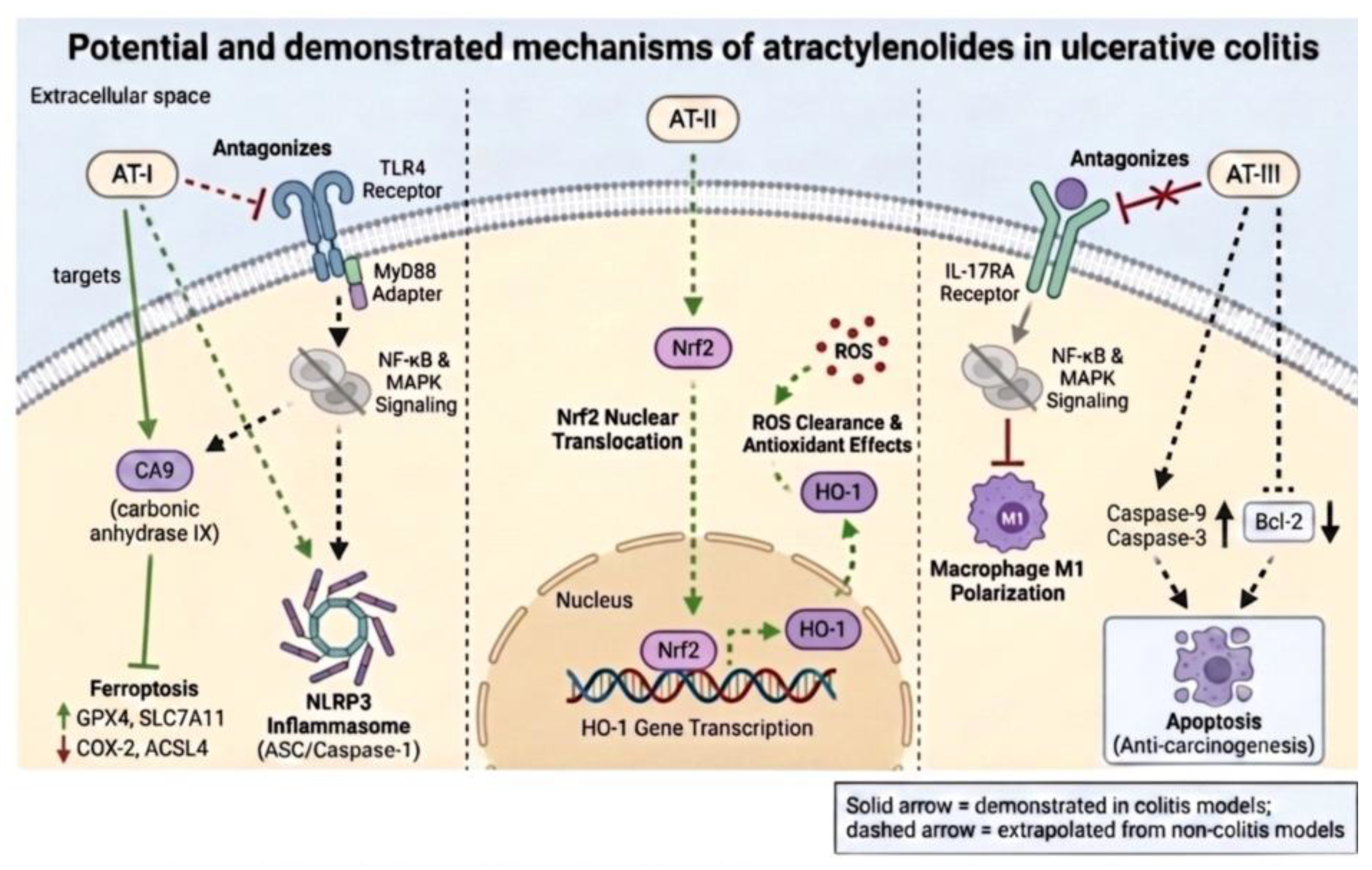
Figure 1. Potential and demonstrated mechanisms of atractylenolides relevant to ulcerative colitis. Atractylenolide I (AT-I) antagonizes the Toll-like receptor 4 (TLR4)/MyD88 axis, suppressing nuclear factor-κB (NF-κB) and mitogen-activated protein kinase (MAPK) signaling and pro-inflammatory cytokine output (tumor necrosis factor-α, interleukin-1β and interleukin-6), and restrains NLRP3 inflammasome (ASC/Caspase-1)-mediated pyroptosis; AT-I additionally targets carbonic anhydrase IX (CA9) to restrain ferroptosis, up-regulating GPX4 and SLC7A11 while down-regulating COX-2 and ACSL4 (a mechanism demonstrated in colitis, shown with solid arrows). AT-II activates nuclear factor erythroid 2-related factor 2 (Nrf2), which translocates into the nucleus and drives heme oxygenase-1 (HO-1) gene transcription, mediating reactive oxygen species clearance and antioxidant effects. AT-III antagonizes the interleukin-17 receptor A (IL-17RA), suppressing NF-κB/MAPK signaling and macrophage M1 polarization, and promotes apoptosis of damaged cells through up-regulation of caspase-9 and caspase-3 with down-regulation of Bcl-2 (anti-carcinogenic potential). Solid arrows denote mechanisms demonstrated in colitis models; dashed arrows denote mechanisms extrapolated from non-UC or non-equivalent disease contexts—the MD-2/TLR4 antagonism (ovarian cancer and macrophage systems), NLRP3/pyroptosis restraint (gastric injury and AOM/DSS colitis-associated colorectal cancer), Nrf2/HO-1 cytoprotection (radiation injury) and the pro-apoptotic Bax/Bcl-2-caspase arm (colorectal cancer cells); see the Atractylenolides section and Table 1. This figure depicts selected representative mechanisms of the atractylenolides; the complete constituent-level mechanism set is given in Table 1. AT: Atractylenolide. Created in BioRender. Huang, S. (2026) https://BioRender.com/c7boi0u.
Preprints 236200 g001
Disclaimer/Publisher’s Note: The statements, opinions and data contained in all publications are solely those of the individual author(s) and contributor(s) and not of MDPI and/or the editor(s). MDPI and/or the editor(s) disclaim responsibility for any injury to people or property resulting from any ideas, methods, instructions or products referred to in the content.
Copyright: This open access article is published under a Creative Commons CC BY 4.0 license, which permit the free download, distribution, and reuse, provided that the author and preprint are cited in any reuse.