
Submitted:
30 September 2026
Posted:
02 October 2026
You are already at the latest version
Abstract
Surgical interventions in cirrhotic patients are a relevant problem due to the high rates of perioperative morbidity and mortality. The purpose of this review is to provide up–to-date information on the current principles of extrahepatic abdominal surgery in cirrhotic patients. They are based on stratification of patients by risk groups and a multidisciplinary individual approach to the choice of treatment strategy. The implementation of these provisions includes the use of scores for predicting perioperative risk, diagnosis and possible correction of clinically significant portal hypertension, prophylaxis of infectious complications and postoperative venous thromboembolism, monitoring of coagulopathy and cardiovascular disorders, nutritional support and etiological therapy. When choosing the optimal method of surgical treatment in urgent cases, one should strive to use minimally invasive technologies. If elective operations are considered, a thorough examination of patients is necessary to determine the risk factors for adverse outcome, resolve issues of preoperative preparation, and choice of the scope and timing of the surgical intervention. It can be assumed that the thoughtful implementation of these principles in clinical practice and, in prospect, the use of robotics and artificial intelligence will improve the safety of extrahepatic abdominal surgery in cirrhotic patients.
Keywords:
liver cirrhosis
; extrahepatic abdominal surgery
; current principles
; risk factors
; perioperative management
; treatment strategy
1. Introduction
Liver cirrhosis (LC) is one of the most severe chronic somatic diseases, accounting for 2.4% of deaths worldwide in 2019 [1]. International epidemiological data indicate a significant increase in its prevalence over the past two decades, and the number of patients with compensated LC has almost doubled, from 66 million in 1990 to 112 million in 2017 [2].
Cirrhotic patients usually come into the view of surgeons in the case of gastroesophageal variceal bleeding, however, the presence of another abdominal pathology may require its surgical treatment. Historically, 10% of cirrhotic patients undergo surgery during the last two years of their lives. It is predicted that their number will grow, and high rates of perioperative morbidity and mortality with existing risk factors will persist [3]. Indeed, surgical interventions represent a serious danger to cirrhotic patients due to the threat of developing hepatic decompensation, and sometimes acute-on-chronic liver failure [4]. Their results may also be influenced by the presence of clinically significant portal hypertension (CSPH) and comorbidity, the patient’s age, the nature, complexity and urgency of surgical care, the need for intraoperative blood transfusions, and a number of other factors [5].
The purpose of this narrative review is to provide up–to-date information on the current principles of extrahepatic abdominal surgery in cirrhotic patients. The PubMed and EMBASE databases, the Web of Science platform, the Google Scholar retrieval system, the Cochrane Database of Systematic Reviews, and the reference lists from related articles were used to search for relevant publications. They were identified using the following terms: “liver cirrhosis”, “extrahepatic abdominal surgery”, “perioperative management of cirrhotic patients”, “ventral hernia repair in cirrhotic patients”, “biliary surgery in cirrhotic patients”, “colorectal surgery in cirrhotic patients”, “appendectomy in cirrhotic patients”, “gastric surgery in cirrhotic patients”, “pancreatic surgery in cirrhotic patients”, “bariatric surgery in cirrhotic patients”. The articles published in 2007-2026, as well as current guidelines, which describe the standards of perioperative management and the features of extrahepatic abdominal surgery in cirrhotic patients, are analyzed.
A schematic representation of the current principles of extrahepatic abdominal surgery in cirrhotic patients is shown in Figure 1.
2. Perioperative Management of Cirrhotic Patients
One of the most important tasks to reduce perioperative morbidity and mortality in cirrhotic patients is their thorough examination to determine the risk factors for adverse outcome, resolve issues of preoperative preparation, and choice of the scope and timing of the surgical intervention [6].
2.1. Prediction of Perioperative Risk
To predict the perioperative risk in cirrhotic patients, several scores were recommended, which, in combination with clinical data, to one degree or another allow to make an informed decision on the expediency of surgery [7].
The CTP (Child-Turcotte-Pugh) score was originally proposed in 1964 by C.G. Child and J.G. Turcotte to select cirrhotic patients with gastroesophageal variceal bleeding for portosystemic shunting. It has undergone several modifications and currently includes two qualitative and three quantitative indicators that make it possible to assess the severity of liver disease and determine the operational risk depending on the amount of points (CTP class A, B or C) (Table 1) [8].
The MELD (Model for End Stage Liver Disease) score was originally intended to predict the three-month survival of cirrhotic patients who underwent Transjugular Intrahepatic Portosystemic Shunt (TIPS). In 2002, the United Network for Organ Sharing (UNOS) approved it to prioritize patients awaiting liver transplantation. Later versions of the MELD score: MELD-Na and MELD 3.0 were created to optimize the prognosis of mortality and a more objective distribution of organs among cirrhotic patients who are on the waiting list for liver transplantation (Table 2) [9].
A systematic review and meta-analysis of 119 studies compared the discriminative ability of CTP score vs MELD score to assess the prognosis of cirrhotic patients. In five studies, which included cirrhotic patients who underwent surgery, the CTP score had a significantly higher specificity than MELD score, because the 95% confidence intervals (CIs) were not overlapped among them and the lower limit of 95%CI of CTP score was higher than the upper limit of 95%CI of MELD score (0.79 > 0.73) [10]. It should be noted that the CTP and MELD scores do not take into account the type and complexity of surgical intervention, the presence of concomitant diseases, or the general functional state of cirrhotic patients. It has been shown that cirrhotic patients CTP class A with low values of the MELD score (< 10) and/or without hepatic decompensation (i.e., without ascites, encephalopathy, or gastroesophageal variceal bleeding) can safely tolerate most elective and emergency operations of low risk [11].
The ADOPT-LC (Appropriate Operative Treatment for Liver Cirrhosis) score was developed in 2017 as a result of a national survey from Japan. It effectively predicted hospital mortality after elective operations and allowed help to make a decision about the possibility of surgical intervention in cirrhotic patients based on a quantitative risk assessment [12]. The ADOPT-LC score is calculated using an online calculator [https://apps.apple.com/uy/app/adopt-lc-score/id976986669]. Further multicenter prospective studies are needed to validate the ADOPT-LC score.
The MRS score (Mayo Risk Score) was, until recently, the only validated method for determining the risk of a specific operation in cirrhotic patients recommended by the American Gastroenterological Association [13]. It was developed based on the results of a retrospective single-center study by Teh et al. [14], which lasted more than 25 years and was completed in 2005, i.e., before the introduction of most modern minimally invasive surgical technologies. The MRS score is calculated using an online calculator [https://www.mayoclinic.org/medical-professionals/transplant-medicine/calculators/post-operative-mortality-risk-in-patients-with-irrhosis/itt-20434721] [15].
Mahmud et al. [16] performed an external validation of the MRS score and proposed a new predictive VPS score (VOCAL-Penn Score) using data at the population level. The results of their retrospective multicenter study showed that the efficacy of the VPS score was superior to the CTP, MELD, MELD-Na, and MRS scores. To calculate it, the authors proposed an online calculator [www.vocalpennscore.com].
2.2. Diagnosis of CSPH and Its Preoperative Correction
The presence of CSPH in cirrhotic patients is an important prognostic factor, as it leads to an increased risk of first and further decompensation, promotes the development of life-threatening complications such as gastroesophageal variceal bleeding, ascites, hepatic encephalopathy, and also serves as a predictor of adverse outcome after surgical interventions [17]. The gold standard of its diagnosis is the hepatic venous pressure gradient (HVPG) values ≥ 10 mmHg [18]. The HVPG values > 16 mmHg are related with postoperative mortality, and the HVPG values ≥ 20 mmHg are associated with a very high risk of mortality in cirrhotic patients who have undergone extrahepatic abdominal surgery [19].
In addition to HVPG measurement, CSPH can be excluded or identified by liver stiffness values in combination with platelet count, and by spleen stiffness values. According to the Baveno VIII consensus workshop criteria, in virus- and/or alcohol-related compensated LC as well as non-obese (BMI < 30 kg/m²) metabolic dysfunction-associated steatotic liver disease (MASLD)-related compensated LC, liver stiffness values by transient elastography of more than 25 kPa indicates the presence of CSPH, whereas with liver stiffness values of less than 15 kPa and a normal platelet count, it is unlikely. A CSPH is also indicated by spleen stiffness values (100 Hz) greater than 55 kPa. Cirrhotic patients with liver stiffness values between 20 and 25 kPa and platelet count less than 150 × 109/L or with liver stiffness values between 15 and 20 kPa and platelet count less than 110 × 109/L have a risk of developing CSPH with a probability of about 60%. They need additional screening [20].
An alternative to HVPG measurement can be endoscopic ultrasound (EUS)-guided portal pressure gradient (PPG) measurement, which is executed with the U.S. Food and Drug Administration–approved EchoTip Insight system. EUS-guided PPG measurement is a novel technique demonstrated to be safe and feasible [21]. In a prospective single-center study by Lim et al. [22], ten patients underwent preoperative EUS-guided PPG and HVPG measurements. EUS-guided PPG measurement was successful in nine patients (90%), with the median pressure gradient strongly correlated between the EUS-guided PPG (median = 6.65 mmHg; interquartile range (IQR): 1.2-15.3) and HVPG measurements (median = 6.5 mmHg; IQR: 4.3-12), (R = 0.895; P = 0.001). Three patients had CSPH based on EUS-guided PPG measurement and six patients underwent uncomplicated surgeries, including one patient with EUS-guided PPG proven CSPH who underwent pre-surgical TIPS. There was no 90-day mortality. Patients preferred the EUS-guided PPG measurement over the HVPG measurement, with significantly higher visual analog scale (9.0 (IQR: 8.8-10) vs 5.0 (IQR: 4.0-6.0); P < 0.01).
Non-selective β-blockers are able to effectively reduce portal pressure by acting on several links in the pathogenesis of portal hypertension in LC [23]. However, no special studies have been conducted on their use in cirrhotic patients before abdominal surgery [6].
Successful decompression of the portal system can be achieved through TIPS, which is why this endovascular procedure is recommended for the treatment of a number of complications of portal hypertension, in particular, gastroesophageal variceal bleeding, refractory ascites, and hepatic hydrothorax [24]. The prospects of its use in cirrhotic patients with CSPH before high-risk surgery are also being studied [25]. A systematic review and meta-analysis of 27 studies including 426 cirrhotic patients undergoing elective and emergency surgery with preoperative TIPS showed significantly lower odds of postoperative ascites [odds ratio (OR) = 0.40, 95%CI: 0.22-0.72, I2 = 28%]. At the same time, there were no significant differences in 90-day mortality (OR = 0.76, 95%CI: 0.33-1.77, I2 = 18.2%), perioperative blood transfusions requirement (OR = 0.89, 95%CI: 0.28-2,84, I2 = 70.1%), postoperative hepatic encephalopathy (OR = 0.97, 95%CI 0.35-2.69, I2 = 0%), and postoperative acute-on-chronic liver failure (OR = 1.02, 95%CI 0.15-6.8, I2 = 78.9%) [26].
Preoperative TIPS can theoretically enhance the efficacy of extrahepatic abdominal surgery in cirrhotic patients with portal hypertension [27]. However, given the risk of possible complications (ascites, encephalopathy, infection, etc.) [28], the current guidelines consider it inappropriate to perform preoperative TIPS in cirrhotic patients CTP class A, even in the presence of a CSPH. At the same time, it can be considered to improve postoperative outcomes in cirrhotic patients CTP classes B and C [6].
2.3. Prophylaxis of Postoperative Infectious Complications
Although cirrhotic patients are at high risk of infection, no study has evaluated the potential benefit of antibiotic prophylaxis before or during extrahepatic abdominal surgery [6]. Nevertheless, it seems reasonable to adhere to the already approved guidelines on antibiotic prophylaxis in cirrhotic patients. In particular, it is performed for both primary and secondary prevention of spontaneous bacterial peritonitis in cirrhotic patients with ascites and low ascites protein levels (< 15 g/l) with advanced liver failure (CTP score ≥ 9 points with serum bilirubin level ≥ 3 mg/dl) or impaired renal function (serum creatinine level ≥ 1.2 mg/dl, blood urea nitrogen level ≥ 25 mg/dl, or serum sodium level ≤ 130 mmol/L). For this purpose, they are prescribed norfloxacin (400 mg/day orally), or third-generation cephalosporins such as ceftriaxone (1 g/24 h intravenously) [29].
2.4. Prophylaxis of Postoperative Venous Thromboembolism
Due to the high risk of venous thromboembolism, the problem of its prophylaxis in cirrhotic patients is relevant [30], but poorly studied in those who have undergone extrahepatic abdominal surgery [6]. Nevertheless, the available literature data did not show that thromboprophylaxis is accompanied by an increased risk of bleeding in cirrhotic patients [31]. Probably, the decision on its use and regimen before surgery should be made in accordance with the current standards for the general population [32].
2.5. Perioperative Coagulopathy Monitoring
The current guidelines warn against the ingrained practice of perioperative coagulopathy monitoring in cirrhotic patients through conventional laboratory tests and in case of detected deviations from routine use before surgery of freshly frozen plasma, coagulation factor concentrates or platelets [6]. For a more objective application of hemostatic therapy and a reduction in the amount of transfused blood products, in addition to the standard hemostatic assays, it is recommended to resort to viscoelastic hemostatic tests: rotational thromboelastometry (ROTEM) or thromboelastography (TEG) [33].
2.6. Perioperative Monitoring of Cardiovascular Disorders
For perioperative monitoring of cardiovascular disorders in cirrhotic patients, preference should be given to dynamic indicators of preload, for example, mean arterial pressure, quantify pulse pressure variation or stroke volume variation, as well as values of transthoracic echocardiography. On the contrary, central venous pressure, being a static parameter, does not always correctly reflect the actual blood volume, especially in the presence of ascites, diastolic dysfunction, and hypoalbuminemia [34].
Due to the high cardiovascular risk, contraindications to elective extrahepatic abdominal surgery in cirrhotic patients are severe valvular heart disease, advanced pulmonary arterial hypertension (mean pulmonary artery pressure > 45 mmHg or pulmonary vascular resistance > 5 units by Wood), coronary artery disease (≥ 50% luminal stenosis in ≥ 1 segment of the three major coronary arteries, or stenosis ≥ 70% in at least moderate-size branch vessels and no revascularization possible), as well as uncontrolled symptomatic ventricular arrhythmias [6].
2.7. Nutritional Support
Both malnutrition (BMI < 18.5 kg/m2) and severe obesity (BMI > 40 kg/m2), as well as sarcopenia in cirrhotic patients, increase the risk of morbidity and mortality after major abdominal surgery [35]. Ideally, at least 2 weeks before surgery, they are recommended a high-calorie diet of 30-35 kcal/kg/day and 1.25–1.5 g/kg of protein per day, calculated based on ideal body weight. In the presence of hepatic encephalopathy, protein intake should not be limited, and a late snack in the evening can improve its overall level in the body by reducing periods of nocturnal fasting [7]. According to the indications, it is necessary to provide probe nutrition, including to cirrhotic patients with non-bleeding gastroesophageal varices [36].
2.8. Etiological Therapy
Currently, it has been established that etiological therapy of diseases, the natural history of which is accompanied by liver fibrosis, is an effective method not only for their prevention, but also for the reversal of histological disorders with the restoration of liver structure and function to a normal state, contributing to a reduction in intrahepatic vascular resistance underlying the development of portal hypertension. Therefore, before elective surgical procedures, it seems advisable to consider the possibility of its implementation. For example, patients with alcohol-related LC should be advised to long-term alcohol abstinence, and in patients with HBV-related and HCV-related LC, an antiviral therapy regimen should be considered [37]. However, the question of whether etiological therapy should be given priority and elective surgery postponed has not been definitively resolved [6].
3. Features of Extrahepatic Abdominal Surgery in Cirrhotic Patients
Any surgical interventions, including abdominal operations, are a serious stress for cirrhotic patients. Therefore, they are recommended to be conducted in hospitals with relevant experience [6]. Cirrhotic patients should avoid “major abdominal surgery” whenever possible [38].
3.1. Ventral Hernia Repair
Ventral hernias occur in 20-40% of cirrhotic patients. The previously existing “wait-and-see” tactics, which provided for surgical interventions in cirrhotic patients with ventral hernias only in the presence of complications, has now been radically revised in favor of elective hernia repair after a thorough examination [39]. In a prospective observational study including 246 cirrhotic patients with ventral hernias, elective hernia repair was performed in 57 patients. Conservative care was received by 189 patients, of which 43 (22.7%) developed complications that required emergency hernia repair. Elective surgery provided a higher five-year survival rate compared to conservative care (80% vs 62%; P = 0.012). Multivariate analysis identified multiple hernias [hazard ratio (HR) = 6.7, P < 0.001] and conservative non-operative care (HR = 3.62, P = 0.005) as risk factors for mortality. Among patients who underwent surgical intervention, a multivariate analysis revealed that a MELD score > 11 (HR = 7.8; P = 0.011) and emergency hernia repair (HR = 5.35; P = 0.005) are independent risk factors for 30-day mortality [40].
An active surgical approach significantly improved treatment outcomes, since emergency hernia repair in cirrhotic patients is accompanied by numerous complications and an 8-fold increased risk of death, compared to elective surgery [41]. At the same time, early elective hernia repair in cirrhotic patients should be advocated considering the hepatic reserve and patient’s condition [42]. The morbidity and mortality rates following ventral hernia repair in cirrhotic patients are presented in Table 3.
Laparoscopic hernia repair, which has become widespread, is currently being actively introduced in cirrhotic patients. Laparoscopic inguinal hernia repair showed similar morbidity and mortality rates when compared to open repairs [54]. Laparoscopic umbilical hernia repair, by a minimally invasive and tension-free technique, can realize less hernia recurrences, lower morbidity, and better prognosis in cirrhotic patients. In addition, laparoscopic hernia repair presents some other advantages over open repair, which are especially important in the cirrhotic condition: preventing interruption or rupture of large collateral veins; avoiding the exposure of abdominal viscera and restricting electrolytic and protein loss; promoting ascites assimilation and avoiding ascites leakage from the unhealed wound; reducing the risks of perioperative bleeding and wound or mesh infection; minimizing postoperative pain and recovery time [55]. In a study by Juo et al. [56], in comparison with open ventral hernia repair, laparoscopic ventral hernia repair was associated with similar short-term outcomes except for lower wound-related complications and shorter length of stay in cirrhotic patients. However, in the presence of ascites, laparoscopic ventral hernia repair is associated with higher mortality, systemic complications, and unplanned reoperation.
Another important issue is recurrence after ventral hernia repair especially patients having high intraabdominal pressure liked cirrhotic patients. Mesh use might be a solution for this problem, but ascitic leakage from the incision may be inevitable and may compromise patient comfort and disturbs the patients comfort. Some studies showed that the use of mesh, especially in large defects, reduces recurrence rates, but it increases various complications such as surgical site infection, seroma, and adhesion in the postoperative period [57].
In laparoscopic intraperitoneal onlay mesh repair (IPOM), there is a possibility of mesh infection due to its contact with ascitic fluid, especially in patients with weakened immune systems [58]. In addition, the IPOM method is fraught with the development of adhesions, intestinal obstruction and the formation of intestinal fistulas. To minimize these risks, it is proposed to use anti-adhesive composite meshes. Also, techniques with placement of the mesh in the retromuscular and preperitoneal spaces to exclude its contact with internal organs, such as laparoscopic totally extraperitoneal (TEP) repair and laparoscopic extended totally extraperitoneal (eTEP) repair, allow avoiding complications [59]. In a retrospective single-center study by Wang et al. [60], including 17 cirrhotic patients with inguinal hernias who underwent TEP repair, the mean operation time was 54.23 ± 10.51 minutes for unilateral hernia and 101.25 ± 13.77 minutes for bilateral hernias. The complication rate was 17.6%. In two cases, they were associated with Child class C and large ascites. During a follow-up of 19.29 ± 9.01 months, no patients had recurrence and chronic pain, but 2 patients died because of the progression of underlying liver disease.
Ventral hernias are common in cirrhotic patients with ascites. For example, umbilical hernias, which occur in ~3% of the population at large, are present in ~20% of cirrhotic patients with ascites [61]. Ascites is a serious problem of ventral hernia surgery in cirrhotic patients, since even after successful hernia repair, its presence is associated with a risk of hernia recurrence, as well as the high rates of postoperative morbidity and mortality [55]. In this regard, elective ventral hernias surgery among cirrhotic patients with ascites is controversial [62]. In a randomized controlled trial (CRUCIAL trial), cirrhotic patients with ascites and umbilical hernia were randomly assigned to receive either elective repair or conservative treatment. This trial could not show a relevant difference in overall morbidity after 24 months of follow-up in favor of elective umbilical hernia repair, because of the limited number of patients included [63]. According to the European and American Hernia Societies’ guideline, elective hernia repair is recommended for cirrhotic patients as early as possible after optimal treatment of ascites, particularly for patients with MELD score < 15 and serum albumin > 3 g/dL [64]. At the same time, there is currently insufficient data to determine the choice of the optimal method and the timing of ascites correction (before or after ventral hernia surgery) [65]. In accordance with the current guidelines, a sodium-restricted diet, diuretics, and, in case of their inefficiency, large-volume paracentesis (LVP) are prescribed for its treatment at the initial stage [29]. In case of refractory ascites, medical treatments and LVP are used, and TIPS [66,67], peritoneovenous shunting [68] and implantation of the automated low-flow ascites pump (Alfapump®) system [69,70] can be considered as a second-line measure. A serious problem after TIPS placement or LVP in cirrhotic patients with refractory ascites is the incarceration of pre-existing asymptomatic umbilical hernias, which is associated with a reduction in the volume of ascitic fluid and a narrowing of the hernia orifices [71]. The issue of abdominal drainage after hernia repair in cirrhotic patients with ascites is decided individually based on intraoperative data, in particular, the volume of ascitic fluid, the likelihood of its leakage (formation of the so-called “ascitic fistula”), as well as the estimated risk of postoperative complications [57]. Thus, there is a need to develop novel surgical approaches to the treatment of ventral hernias in cirrhotic patients with refractory ascites.
3.2. Biliary Surgery
Cholelithiasis prevalence in cirrhotic patients ranges between 25% and 30%, being at least twice that in the general population [72]. Symptomatic cholelithiasis is an indication for surgery [73]. Since the 1980s, laparoscopic cholecystectomy has become the preferred technique even to cirrhotic patients CTP classes A and B [6]. In a retrospective study by Pavlidis et al. [74] including 38 cirrhotic patients (CTP classes A and B) with symptomatic cholelithiasis who underwent laparoscopic cholecystectomy, both the conversion rate (15.78%) and the duration of hospital stay were higher than in the non-cirrhotic group, but without significant differences. Major complications occurred more often in cirrhotic patients (P = 0.027), increasing morbidity; however, the mortality was zero.
The optimal treatment strategy for acute cholecystitis in cirrhotic patients has not yet been determined, however a generally accepted clinical decision algorithm has been described in the current guidelines. Antibiotics and cholecystectomy are the standard of care for acute cholecystitis. Laparoscopic cholecystectomy for acute cholecystitis is generally safe in cirrhotic patients CTP classes A and B, but is associated with significantly higher rates of postoperative morbidity and mortality in cirrhotic patients CTP class C [7]. In a systematic review and meta-analysis of 42 studies including 24,575 cirrhotic patients (CTP class A—71.2%, CTP class B—26%, and CTP class C—2.8%) undergoing laparoscopic cholecystectomy, an emergency operation was performed in 28.7% of cases. Postoperative hepatic decompensation occurred in 7.1% (95%CI: 0.9-13.3) of patients, superficial surgical site infection in 1.1% (95%CI: 0.6-1.5), organ space surgical site infection in 3.7% (95%CI: 2.0-5.5), bile leak in 0.7% (95%CI: 0.2-1.1), venous thromboembolism in 1.8% (95%CI: 0.7-2.9), pneumonia in 1.9% (95%CI: 1.2-2.6) and blood transfusions in 4% (95%CI: 2.7-5.2). Postoperative mortality was 1.8% (95%CI: 1.2-2.4). Among cirrhotic patients CTP classes A and B, postoperative mortality was 0.7% (2 out of 201) and 2.0% (2 out of 98), respectively. Of the nine cirrhotic patients CTP class C, seven (77.8%) died after surgery [75].
Studies have shown that laparoscopic cholecystectomy can be safely performed in a well-selected group of patients with compensated LC. However, it can be challenging in many aspects and poses more difficulty and thus should be performed by experienced surgical teams, who follow the recommendations and take into account the special precautions which are requested to increase the safety of the operation and avoid or reduce the morbidity and mortality, and also who are able to tackle the more frequent intraoperative incidents or complications [76].
The CO2 pneumoperitoneum in laparoscopic cholecystectomy can result in ischemia-reperfusion injury to the internal organs, which may aggravate damage to the hepatic function [77]. That is the reason why it should be established with lower pressure and released slowly. During the operation, cirrhotic patients may experience serious problems, primarily due to the presence of dilated collateral veins around the gallbladder, in the greater omentum soldered to it and the liver surface, and in the umbilical region, damage to which can provoke dangerous bleeding. In this situation, using electrocoagulation near the hepatic hilum and especially extrahepatic biliary tract should be avoided as much as possible in order to prevent potential thermal damage. When separating the gallbladder, it is advisable to apply a harmonic scalpel that uses ultrasound waves to cut and coagulate tissue, and to install a laparoscopic trocar infra-umbilical. It should be noted that the harmonic scalpel has many advantages, such as facilitated dissection, minimal lateral thermal tissue damage, greater precision, and less smoke production, leading to reduced operating time and decreased conversion to open surgery [78]. Open (Hasson) laparoscopy can also be performed. In case of difficulties in diverting the cirrhotic liver upward, a retractor is inserted through an additional port immediately to the right of the epigastric port, with which the right lobe is lifted. By placing the retractor on the gallbladder body, it can also displace the Hartmann’s pouch for better visualization of the Calot’s triangle. If there is a threat of uncontrollable bleeding from the gallbladder bed, laparoscopic subtotal cholecystectomy type I is performed, when the posterior wall of the gallbladder is left in situ followed by mucosal cauterization. If it is difficult to identify anatomical structures in the hepatic hilum, laparoscopic subtotal cholecystectomy type II is allowed, during which the gallbladder is removed at the neck level as close as possible to the cystic duct [79,80].
Cirrhotic patients at high surgical risk (CTP class C or MELD score > 13) should be initially treated with antibiotics, and in the case of failed medical management, percutaneous cholecystostomy, endoscopic transpapillary gallbladder drainage with cystic duct stenting, and EUS-guided gallbladder drainage using a lumen-apposing metal stent are alternative procedural options [6,7,81,82]. In a study by Vedachalam et al. [83], data from cirrhotics and non-cirrhotics patients with acute calculous cholecystitis who underwent initial percutaneous cholecystostomy (with or without follow-up cholecystectomy) or cholecystectomy were examined using queries to the National Database. Out of 919,189 patients with acute calculous cholecystitis, 13,283 (1.4%) had cirrhosis. Among cirrhotics, cholecystectomy was performed in 12,790 (96.3%) and percutaneous cholecystostomy in the remaining 493 (3.7%). Multivariate analyses showed increased early readmissions (OR = 2.12, 95%CI: 1.43-3.13, P < 0.001), length of stay (effect ratio = 1.39, 95%CI: 1.20-1.61, P < 0.001), calendar-year hospital cost (effect ratio = 1.34, 95%CI: 1.28-1.39, P < 0.001) and calendar-year mortality (HR = 1.89, 95%CI: 1.07-3.29, P = 0.030) in cirrhotic patients undergoing initial percutaneous cholecystostomy compared to cholecystectomy. Decompensated LC (OR = 2.25, 95%CI: 1.67-3.03, P < 0.001) had the highest odds of getting initial percutaneous cholecystostomy. LC, regardless of compensated (OR = 0.56, 95%CI: 0.34-0.90, P = 0.020) or decompensated (OR = 0.28, 95%CI: 0.14-0.59, P < 0.001), reduced the chances of getting a subsequent cholecystectomy. In a retrospective single-center study by Cooper et al. [84], of the 55 cirrhotic patients undergoing endoscopic transpapillary gallbladder drainage with cystic duct stenting, 52 patients (mean MELD-Na score—16) successfully underwent the procedure (38 inpatient, 14 outpatient). Most patients (n = 48) experienced immediate symptomatic relief. Short-term adverse event rate was 7.7% and 30-day mortality was 1.9%. At 6 months, 86.5% of patients had a favorable outcome; unfavorable outcomes included death (n = 2), recurrent cholecystitis and/or need for urgent cholecystectomy (n = 5). At the end of the study, 80% of patients experienced a favorable outcome including five patients bridged to liver transplant and 20 who had their stent in place with adequate symptom control. Of those with imaging, 65% of patients experienced resolution of cholelithiasis. Recurrent cholecystitis (P = 1.00) and stone resolution (P = 0.35) did not differ in patients with one or two cystic duct stents in place.
For cirrhotic patients with cholecystitis and common bile duct stones, endoscopic sphincterotomy or balloon extraction is preferable to laparoscopic or open common bile duct exploration [7]. However, the increased risk of post-procedural complications, including acute pancreatitis, bleeding, infection, and perforation in cirrhotic patients, especially those with a decompensated stage of the disease, should be considered [85,86].
3.3. Colorectal Surgery and Appendectomy
Despite some positive trends that have emerged in recent years, colorectal surgery in cirrhotic patients is still associated with significant postoperative morbidity and mortality. In a recent systematic review, the overall complication rate associated with colorectal surgery in cirrhotic patients ranged from 29% to 75%, minor complication ranged 14.5-37% and major complication ranged 6.7-59.3%. The mortality rates ranged from 0 to 37% [87]. In a study by van Beekum et al. [88] showed that most common complications of colorectal surgery in cirrhotic patients were bleeding requiring blood transfusions (51.1%) and cardiocirculatory instability due to bleeding or sepsis (44.4%). In 53.7% of patients, an anastomosis was created without a protective ostomy. Anastomotic leakage occurred in 20.7% of these patients. Multivariate analysis showed that a primary anastomosis without a protective ostomy was the strongest risk factor for major complications (P = 0.042). As it turned out, LC is an important risk factor for anastomotic leakage after colorectal surgery [89]. In a study by Shahait et al. [90] including 731 cirrhotic patients undergoing colorectal surgery, 57.5% of whom were operated on as an emergency, complications occurred in 48.2% of cases, and 30-d mortality was 24.8%. Malignant neoplasm was the most common indication (25.4%). Laparoscopic approach was performed in 22.4%, with shorter operative time, less blood transfusions, shorter length of stay, and lower morbidity compared to open approach. Overall, MELD score was an independent factor of mortality, while laparoscopic approach had a protective effect on morbidity. In addition, cirrhotic patients who require emergent colorectal surgery have a high risk of postoperative complications and death. Increasing MELD-Na score is associated with mortality and can be used to risk stratify individuals [91].
LC is a known risk factor for colorectal cancer [92]. A systematic review and meta-analysis of five studies including 2,485 patients showed that colorectal cancer surgery in cirrhotic patients are associated with an increased postoperative major complication rate, a higher rate of re-operation, a higher short-term mortality rate, and poor overall survival [93]. In a retrospective single-center study by Ku et al. [94] including 2,523 patients examined the surgical outcomes of elective laparoscopic colorectal cancer resection in 55 cirrhotic patients compared to non-cirrhotic patients. The open conversion rate was significantly higher in the LC group than in the non-LC group (9.1% vs 1.3%, P = 0.001). Advanced LC, higher ASA classes, and larger tumor size were identified as independent risk factors for conversion, with LC CTP class B being the strongest predictor (OR = 40.671, 95%CI: 6.763-244.585, P < 0.001). The LC group also exhibited worse intraoperative outcomes and a higher postoperative complication rate (43.6% vs 24.8%, P = 0.002). However, long-term oncologic outcomes including overall survival and recurrence-free survival did not differ between the two groups.
In a retrospective single-center study by Pei et al. [95] data from 2,523 cirrhotic patients with ascites who underwent laparoscopic or open partial colectomy with diagnosis of colon cancer were analyzed. Two hundred and five cirrhotic patients (9.53%) with ascites underwent laparoscopic colectomy. There was no significant difference in operative time between laparoscopic versus open surgery (145 vs 146 min, P = 0.69). Laparoscopic colectomy was associated with decreased likelihood of overall complications (adjusted OR = 0.7, 95%CI: 0.4-1.0, P = 0.046) and shorter hospital length of stay (9 days vs 15 days, adjusted β = -4.2, 95%CI: -7.7 to -0.7, P = 0.018). There was no difference in 30-day mortality (adjusted OR 0.82, 95%CI: 0.50-1.35, P = 0.429).
In early colon cancer (stage 0—Tis N0 M0), organ-preserving treatment strategies are recommended, such as endoscopic mucosal resection and endoscopic submucosal dissection [96]. As a minimally invasive method of treating acute left-sided malignant colonic obstruction, the current guidelines recommend the endoscopic placement of a self-expandable metal stent through tumor stenosis [97].
Appendectomy in cirrhotic patients is accompanied by significant postoperative morbidity and mortality, which is associated with the bleeding, infections and hepatic decompensation [98]. In a systematic review and meta-analysis of four studies including 923 cirrhotic patients undergoing appendectomy, in-patient mortality ranged from 0 to 1.7%, and the thirty-day mortality was 9%. In patients with compensated LC, mortality following laparoscopic appendectomy (0.5%) was significantly lower than open appendectomy (3.2%). The meta-analysis highlighted a tenfold increase in perioperative mortality in cirrhotic patients (OR = 9.92, 95%CI: 4.67-21.06, I2 = 28%). All studies reported an increased length of stay in cirrhotic patients [99].
3.4. Gastric Surgery
The prevalence of peptic ulcer disease in cirrhotic patients is higher relative to the general population [100]. Its presence is associated with severe complications. For example, cirrhotic patients have a high risk of ulcerative bleeding, which significantly worsens their prognosis [101]. Mortality after emergency operations due to complications of peptic ulcer disease in cirrhotic patients remains high, especially those with a decompensated stage of the disease [79]. At the same time, the introduction of minimally invasive endoscopic technologies into clinical practice has decreased the need for resection procedures and reduced the rate of fatal outcomes [102,103].
The significant incidence of gastric cancer in cirrhotic patients is well known [104]. Radical gastrectomy with extended lymph node dissection is considered to be the standard surgical treatment for gastric cancer [105]. Open or laparoscopic radical gastrectomy with D1 or D2 lymph node dissection can be tolerated in cirrhotic patients CTP class A and only with D1 lymph node dissection in patients CTP class B. Radical gastrectomy is very dangerous, even fatal for cirrhotic patients CTP class C, and therefore are not recommended [106]. The morbidity and mortality rates following radical gastrectomy in cirrhotic patients are presented in Table 4.
Endoscopic mucosal resection and endoscopic submucosal dissection may be used in cirrhotic patients for the treatment of early gastric cancer (stage 0—Tis N0 M0) [113]. The results of the conducted studies show that endoscopic mucosal resection and endoscopic submucosal dissection are suitable for the treatment of early gastric cancer, and the main procedure-associated complications are bleeding and perforation. The risk of bleeding during endoscopic procedures in cirrhotic patients is increasing with the rise of portal pressure and worsening of hemostasis disorders, and is statistically significantly higher in CTP class C or MELD score > 11.5 [114].
3.5. Pancreatic Surgery
According to the current guidelines, pancreatic resection in cirrhotic patients can be considered only for malignant neoplasms or premalignant conditions and exclusively in patients CTP class A without CSPH [6]. In a systematic review and meta-analysis of eight studies including 722 cirrhotic patients (CTP class A—82.1%, CTP class B/C—17.9%) who underwent surgery for pancreatic cancer, the overall postoperative morbidity rate was 66% (51-80%). Infections (26%) and ascites formation/worsening (23%) were the most common postoperative complications, followed by anastomotic leak/fistula (17%). An analysis according to the CTP classification demonstrated higher postoperative mortality rate in cirrhotic patients CTP class B/C vs cirrhotic patients CTP class A: 15.2% and 6.2%, respectively (OR = 0.16, 95%CI: 0.04–0.77) but did not reveal any statistically significant difference in terms of postoperative morbidity rate (cirrhotic patients CTP class A: 47.7% vs cirrhotic patients CTP class B/C: 56.2% (OR = 0.72, 95%CI: 0.47–1.09) [115].
A systematic review and meta-analysis of 11 studies including 18,184 patients showed that LC increased the risk of postoperative mortality (OR = 3.94, P < 0.00001), major complications (OR = 2.25, P = 0.0002), and pancreatic fistula (OR = 1.73, P = 0.03) after pancreatoduodenectomy; it resulted in more blood loss [mean difference (MD) = 204.74 ml, P = 0.0003] and longer hospital stay (MD = 2.05 days, P < 0.00001). LC did not affect delayed gastric emptying (OR = 1.33, P = 0.21), postoperative bleeding (OR = 1.28, P = 0.42), and operative time (MD = 3.47 minutes, P = 0.51). Among the cirrhotic patients, CTP class B or C increased blood loss (MD = 293.33 ml, P < 0.00001), and portal hypertension increased postoperative mortality (OR = 2.41, P = 0.01); the other outcomes were not affected [116].
In fact, resection of the falciform and round ligaments of the liver, pancreaticoduodenal veins, and tributaries of the right gastroepiploic vein during pancreatoduodenectomy in cirrhotic patients with portal hypertension can lead to a dramatic raising portal pressure. It increases the risk of intraoperative bleeding associated with resection and release of vascular structures from the tumor, and in the postoperative period worsen ascites, right-sided cardiac overload, hepatic encephalopathy, and provoke variceal bleeding [117].
Preoperative management of portal hypertension in clinically resectable cirrhotic patients with pancreatic cancer requiring pancreatoduodenectomy continues to be a challenge and is currently not standardized [118].
In a retrospective single-center observational study by Shaker et al. [119], of the 1,140 patients in a pancreatic resection database, four had portal hypertension, for which they underwent TIPS before pancreatoduodenectomy. Three patients had LC, and one had portal thrombosis. Following TIPS, the mean HVPG decreased from 13 mmHg to 2.5 mmHg. The median estimated blood loss and operative time were 275 mL and 267 min, respectively. Postoperatively, one patient experienced dehydration, urinary tract infection, and bacteremia, another had delayed gastric emptying, and three developed hepatic encephalopathy at a median of 98 days.
3.6. Bariatric Surgery
Although obesity is inextricably linked to MASLD, it also affects the natural history and progression of other chronic liver diseases, including LC. This leads to an increase rates of hepatic decompensation events, the development of acute-on-chronic liver failure, infection, and in-hospital mortality in cirrhotic patients, and increased liver transplant waitlist mortality [120]. In this regard, it seems logical to consider the expediency of bariatric surgery in both obese cirrhotic patients and MASLD patients with obesity.
In a systematic review and meta-analysis of 30 studies including 3,134 MASLD patients with obesity, the changes in MRI-determined hepatic proton density fat fraction (MRI-PDFF) and nonalcoholic fatty liver disease activity score (NAS) after bariatric surgery or intragastric balloon/gastric banding was evaluated. Bariatric interventions significantly reduced BMI (ratio of means, 0.79) and showed 72% reduction of intrahepatic fat on MRI-PDFF at 6 months after bariatric intervention (ratio of means, 0.28). Eight studies revealed that NAS was reduced by 60% at 3-6 months compared to baseline, 40% at 12-24 months, and 50% at 36-60 months. Nineteen studies revealed that the proportion of patients with steatosis decreased by 44% at 3-6 months, 37% at 12-24 months, and 29% at 36-60 months; lobular inflammation by 36% at 12-24 months and 51% at 36-60 months; ballooning degeneration by 38% at 12-24 months; significant fibrosis (≥ F2) by 18% at 12-24 months and by 17% at 36-60 months after intervention [121].
In a systematic review and meta-analysis of 26 studies including 22,489 patients with compensated LC and obesity, the efficacy and safety of bariatric and metabolic surgery were evaluated. Mortality was 3.33% (95%CI: 2.06-5.35, I2 = 96.7%). Early postoperative complications were 14.0% (95%CI: 7.56-24.49, I2 = 90.3%). Obese cirrhotic patients had a significant weight loss after 1 year of follow-up (MD -43.06 kg, 95%CI [-70.93; -15.19], P < 0.0001, I2 = 94.0%), BMI (MD -10.79 kg/m2, 95%CI [-14.45-7.12], P < 0.01, I2 = 66.2%), percentage of excess weight loss (%EWL) (62.81%, 95%CI: 52.56-73.96, I2 = 78%) and percentage of total weight loss (%TWL) (26.43%, 95%CI: 23.89-28.96, I2 = 69.3%). The length of stay was 3.77 days (95%CI: 3.13-4.42, I2 = 99.1%) [122].
However, in a systematic review of 21 studies bariatric surgery (mainly sleeve gastrectomy and Roux-en-Y gastric bypass) in obese cirrhotic patients were accompanied by high rates of postoperative hepatic decompensation events, morbidity, and mortality [123].
Therefore, it is extremely important that bariatric interventions be performed only in carefully selected obese cirrhotic patients, in whom their benefits outweigh the risk of adverse outcome. Based on this, they are recommended only for obese cirrhotic patients CTP class A without CSPH, and laparoscopic sleeve gastrectomy is considered the most optimal. At the same time, the presence of gastroesophageal varices is a contraindication for endoscopic insertion of an intragastric balloon [6,124].
4. Future Prospects
4.1. Robotic Extrahepatic Abdominal Surgery in Cirrhotic Patients
Over the past two decades, the steady development of robotic abdominal surgery has not only improved the accessibility, efficiency, and safety of treatment but has also decreased resource consumption linked to prolonged patient hospitalization [125]. To reduce the risk of postoperative complications in cirrhotic patients, current guidelines advise using minimally invasive interventions whenever feasible, including robotic surgery [6]. However, there are presently very few reports of the use of robotic surgery in cirrhotic patients.
In a study by Amara et al. [126] was presented the first retrospective analysis of data from cirrhotic patients who underwent elective or emergent robotic hernia repair. A total of seven patients were included with a median MELD-Na of 17 (IQR, 14-22) and the majority of whom (6 of 7, 85.7%) had ascites at the time of surgery. Three patients required emergent or urgent operations. No drains were required at the time of surgery. There were no Clavien-Dindo grade 3 or higher complications, no patients had leakage of ascites from their incisions, and no patients developed hernia recurrence (median follow-up 173 days). There were two complications: one superficial skin infection treated with antibiotics and one case of urinary retention. This small study showed that robotic hernia repair is technically feasible and safe in a select group of cirrhotic patients including those with ascites.
In a retrospective single-center study by McCabe et al. [127], data from 61 cirrhotic patients who underwent laparoscopic cholecystectomy were compared with data from 31 cirrhotic patients who underwent robotic cholecystectomy, without significant difference in MELD score or elective status (all P > 0.05). The primary outcome was rate of Clavien-Dindo grade 3a or higher complication. There was not a significant difference in major morbidity rate by approach (4.8% vs 6.5%, P > 0.9), while estimated blood loss (10 vs 25 mL, P < 0.001) and open conversion rate (0 vs 12.9%, P = 0.049) were significantly lower for robotic cholecystectomy. Robotic cholecystectomy was associated with equivalent perioperative outcomes to laparoscopic cholecystectomy in cirrhotic patients in this study.
Li et al. [128] described the use of robotic cholecystectomy in a patient with decompensated LC and portal hypertension complicated by acute cholecystitis, in part due to limited visualization and space constraints. The authors noted that in preparation for surgery, it was important to use imaging to assess anatomical features and mark the organ borders to inform trocar placement and surgical approach.
4.2. Artificial Intelligence in Abdominal Surgery
In recent years, there has been active development of artificial intelligence (AI) technologies, which is leading to their widespread adoption in various areas of human activity, including healthcare, and in particular in surgery. The application of AI in abdominal surgery can be divided into three key directions: preoperative planning, intraoperative guidance, and surgical robotics [129]. There are four main areas of development in AI: machine learning (ML), artificial neural networks (ANNs), natural language processing (NLP), and computer vision (CV) [130]. Deep learning is a subset of ML that, thanks to the multi-layer architecture of interconnected neural networks, enables feature extraction and pattern recognition of highly complex and large-volume data [131].
4.2.1. Preoperative Planning
The preoperative planning in abdominal surgery is aimed at reducing the number of complications, improving the accuracy of surgical intervention, and optimizing patient selection. The preoperative implementation of AI has made it possible to create risk prediction models that surpass traditional assessment systems in speed, diagnostic accuracy, personalization, and clinical significance [132].
A study by Ammann et al. [133] showed that tools used preoperatively aim to predict therapeutic outcomes and pathological findings to better identify and stratify patient cohorts that may benefit from a specific therapy setting. For example, ANNs and ML make it possible to predict several clinical outcomes, such as surgical site infection and hernia recurrence in patients who have undergone hernia repair. CT, MRI, and PET radiomics were able to predict histological findings, therapeutic response, and postoperative outcomes in patients with gastric cancer. AI algorithms make it possible to diagnose colorectal cancer and predict intraoperative result, histological characteristics, and clinical outcomes using CV-guided CT, MRI, and PET-CT radiomics. ML algorithms can optimize patient management before surgery. Several ML algorithms were able to predict and potentially reduce intraoperative complications, as well as enhance postoperative recovery. AI tools used postoperatively aim to predict outcomes after surgery and aid decision-making during short- and long-term postoperative patient management. ML, ANNs, and NLP algorithms have been reported to predict postoperative morbidity and mortality. ML was used to develop an algorithm that accurately predicted patient risk of developing complications following liver, pancreatic, or colorectal surgery [134]. AI and ML are increasingly being integrated into metabolic and bariatric surgery, opening up opportunities to improve the decision-making process, optimize perioperative care, and personalize treatment outcomes. Preoperative applications are aimed at patient selection, anatomical prediction, and risk stratification, including models that predict the success of weight loss, the development of gastroesophageal reflux disease, and hiatal hernia. Intraoperative research is devoted to predicting the duration of the operation, workflow optimization, and automated video analysis for assessing skills and recognizing the stages of the operation. Postoperative models are designed to predict complications, monitor nutrition, and make long-term predictions of weight dynamics [135].
4.2.2. Intraoperative Guidance
The integration of AI into operating rooms and endoscopic suites is a significant achievement in abdominal surgery. These tools, which provide real-time data for anatomical identification and determining the nature of injuries, reduce the cognitive burden on surgeons when dealing with complex cases and improve patient safety [132]. The inroads made by AI include procedure duration prediction, gesture recognition, intraoperative cancer detection, intraoperative video analysis, workflow recognition, endoscopic navigation systems, knot tying, and others. The human factor is currently a more significant source of dangerous errors than technical failures. For a long time, it was believed that the operating room was the most common source of errors in hospital systems; more than half of these errors could have been prevented. One of the serious obstacles in analyzing incident data from operating rooms is that it consists of large volumes of data from various sources. With the advent of AI, it is possible that a more efficient method of processing, analyzing, and evaluating data will emerge to achieve better results [136].
CV helps to identify and analyze surgical workflows in highly complex surgical cases, and it has also made it possible to effectively distinguish between anatomical structures and surgical instruments during laparoscopic and robotic surgeries [133]. Studies have shown that anatomical recognition based on AI helps surgeons make decisions during laparoscopic or robotic gastrectomy [137,138]. According to the literature, 3D reconstruction makes it possible to create individual interactive models of patients, which significantly improve surgeons’ understanding of tumor resectability and vascular anatomy, thereby reducing the duration of the operation and blood loss. Based on this, AI algorithms can automate image segmentation for 3D modeling, increase diagnostic accuracy, and offer predictive analytics to forecast postoperative complications, such as liver failure [139]. The development of minimally invasive pancreatic surgery makes a large amount of intraoperative data available for further processing using AI applications. For example, by analyzing intraoperative data and using it in combination with preoperative features (from clinical data and radiomics), it is possible to predict postoperative morbidity and mortality, as well as provide feedback for surgeons and even inform them about what other surgeons would have done in their situation. AI enables surgeons to visualize tumors and their relationship with intraparenchymal vascular structures in real time, using 3D augmented reality intraoperative images (SmartLiverTM) created from 2D images. Cross-AI systems enable the recognition of landmarks during laparoscopic procedures, especially cholecystectomies. Deep learning is used alongside datasets of video data on laparoscopic cholecystectomy, creating learning models that recognize various stages of the operation and anatomical structures. This helps surgeons prevent errors by protecting key structures from damage and identifying “forbidden zones”. The use of AI along with augmented reality during trocar placement helps to better identify safety landmarks in real time. This method is based on the integration of deep learning with a laparoscopic instrument and an alert system. Analysis of surgical videos showing the use of Optiview Endopath Xcel trocar helps to recognize the various abdominal wall layers, which enables surgeons to operate safely [140].
4.2.3. Surgical Robotics
Recent advances in AI and ML have also been aimed at expanding the capabilities of surgical robots and augment the surgical experience in the operating room. Surgical robots operate based on data obtained from sensors and images, and this plethora of data capture is the key driver behind AI innovations in robotic surgery [141]. It has been shown that the use of artificial intelligence in robotic surgery increases the success rate of surgical interventions and reduces the duration of the operation. According to these studies, the integration of AI into robotic surgery and computer-assisted planning significantly improves surgical outcomes by increasing accuracy and reducing the incidence of complications. AI-assisted systems provide real-time predictive analytics, helping surgeons to anticipate and minimize potential risks. At the same time, 3D planning tools enable precise visualization and forecasting of the outcomes of surgical interventions, which makes it possible to develop more effective individual treatment plans. Overall, these technologies help reduce recovery times, increase the success rate of surgical interventions, and enable more efficient use of healthcare resources, which indicates a significant improvement in the quality and safety of surgical care [142].
5. Conclusions
An increase in extrahepatic abdominal surgery in cirrhotic patients is predicted in the near future. To reduce perioperative morbidity and mortality, their stratification by risk groups and a multidisciplinary individual approach to the choice of treatment strategy are required. It can be assumed that the thoughtful implementation of these principles in clinical practice and, in prospect, the use of robotics and AI will improve the safety of extrahepatic abdominal surgery in this growing and vulnerable category of patients.
Author Contributions
Garbuzenko DV contributed to the conception, design, acquisition, analysis, interpretation of data, wrote the manuscript and approved the final version.
Funding
This research received no external funding.
Institutional Review Board Statement
Not applicable.
Informed Consent Statement
Not applicable.
Data Availability Statement
Not applicable.
Conflicts of Interest
The author declare no conflicts of interest.
Abbreviations
The following abbreviations are used in this manuscript:
LC Liver cirrhosis
CSPH Clinically significant portal hypertension
CTP Child-Turcotte-Pugh
MELD Model for end stage liver disease
TIPS Transjugular intrahepatic portosystemic shunt
ADOPT-LC Appropriate operative treatment for liver cirrhosis
MRS Mayo risk score
VPS VOCAL-Penn score
HVPG Hepatic venous pressure gradient
BMI Body mass index
MASLD Metabolic dysfunction-associated steatotic liver disease
EUS Endoscopic ultrasound
PPG Portal pressure gradient
IPOM Intraperitoneal onlay mesh
LVP Large-volume paracentesis
ASA American Society of Anesthesiologists
MRI-PDFF MRI-determined hepatic proton density fat fraction
NAS Nonalcoholic fatty liver disease activity score
AI Artificial intelligence
ML Machine learning
ANNs Artificial neural networks
NLP Natural language processing
CV Computer vision
CT Computed tomography
MRI Magnetic resonance imaging
PET Positron emission tomography
PET Positron emission tomography
References
- Huang, D.Q.; Terrault, N.A.; Tacke, F.; Gluud, L.L.; Arrese, M.; Bugianesi, E.; Loomba, R. Global epidemiology of cirrhosis—aetiology, trends and predictions. Nat. Rev. Gastroenterol. Hepatol. 2023, 20, 388–398. [Google Scholar] [CrossRef] [PubMed]
- GBD 2017 Cirrhosis Collaborators. The global, regional, and national burden of cirrhosis by cause in 195 countries and territories, 1990-2017: a systematic analysis for the Global Burden of Disease Study 2017. Lancet Gastroenterol. Hepatol. 2020, 5, 245–266. [Google Scholar] [CrossRef] [PubMed]
- Morris, S.M.; Abbas, N.; Osei-Bordom, D.C.; Bach, S.P.; Tripathi, D.; Rajoriya, N. Cirrhosis and non-hepatic surgery in 2023—a precision medicine approach. Expert Rev. Gastroenterol. Hepatol. 2023, 17, 155–173. [Google Scholar] [CrossRef] [PubMed]
- Klein, L.M.; Chang, J.; Gu, W.; Manekeller, S.; Jansen, C.; Lingohr, P.; Praktiknjo, M.; Kalf, J.C.; Schulz, M.; Spengler, U.; et al. The Development and Outcome of Acute-on-Chronic Liver Failure After Surgical Interventions. Liver Transpl. 2020, 26, 227–237. [Google Scholar] [CrossRef] [PubMed]
- Johnson, K.M.; Newman, K.L.; Green, P.K.; Berry, K.; Cornia, P.B.; Wu, P.; Beste, L.A.; Itani, K.; Harris, A.H.S.; Kamath, P.S.; Ioannou, G.N. Incidence and Risk Factors of Postoperative Mortality and Morbidity After Elective Versus Emergent Abdominal Surgery in a National Sample of 8193 Patients With Cirrhosis. Ann. Surg. 2021, 274, e345–e354. [Google Scholar] [CrossRef] [PubMed]
- European Association for the Study of the Liver. EASL Clinical Practice Guidelines on extrahepatic abdominal surgery in patients with cirrhosis and advanced chronic liver disease. J. Hepatol. 2025, 83, 768–789. [Google Scholar] [CrossRef] [PubMed]
- Mahmud, N.; Fricker, Z.P.; McElroy, L.M.; Qayed, E.; Wong, R.J.; Ioannou, G.N. ACG Clinical Guideline: Perioperative Risk Assessment and Management in Patients With Cirrhosis. Am. J. Gastroenterol. 2025, 120, 1968–1984. [Google Scholar] [CrossRef] [PubMed]
- Da, B.; Wu, W.; Guo, W.; Xiong, K.; Chen, C.; Ke, Q.; Zhang, M.; Li, T.; Xiao, J.; Wang, L.; et al. External validation of the modified CTP score based on ammonia to predict survival in patients with cirrhosis after TIPS placement. Sci. Rep. 2024, 14, 13886. [Google Scholar] [CrossRef] [PubMed]
- Kim, W.R.; Mannalithara, A.; Heimbach, J.K.; Kamath, P.S.; Asrani, S.K.; Biggins, S.W.; Wood, N.L.; Gentry, S.E.; Kwong, A.J. MELD 3.0: The Model for End-Stage Liver Disease Updated for the Modern Era. Gastroenterology 2021, 161, 1887–1895. [Google Scholar] [CrossRef] [PubMed]
- Peng, Y.; Qi, X.; Guo, X. Child-Pugh Versus MELD Score for the Assessment of Prognosis in Liver Cirrhosis: A Systematic Review and Meta-Analysis of Observational Studies. Medicine 2016, 95, e2877. [Google Scholar] [CrossRef] [PubMed]
- Durkin, C.; Mahmud, N. Pre-Operative Assessment of Patients with Cirrhosis for Extrahepatic Surgery. Curr. Hepatol. Rep. 2025, 24, 25. [Google Scholar] [CrossRef] [PubMed]
- Sato, M.; Tateishi, R.; Yasunaga, H.; Horiguchi, H.; Matsui, H.; Yoshida, H.; Fushimi, K.; Koike, K. The ADOPT-LC score: a novel predictive index of in-hospital mortality of cirrhotic patients following surgical procedures, based on a national survey. Hepatol. Res. 2017, 47, E35–E43. [Google Scholar] [CrossRef] [PubMed]
- Northup, P.G.; Friedman, L.S.; Kamath, P.S. AGA Clinical Practice Update on Surgical Risk Assessment and Perioperative Management in Cirrhosis: Expert Review. Clin. Gastroenterol. Hepatol. 2019, 17, 595–606. [Google Scholar] [CrossRef] [PubMed]
- Teh, S.H.; Nagorney, D.M.; Stevens, S.R.; Offord, K.P.; Therneau, T.M.; Plevak, D.J.; Talwalkar, J.A.; Kim, W.R.; Kamath, P.S. Risk factors for mortality after surgery in patients with cirrhosis. Gastroenterology 2007, 132, 1261–1269. [Google Scholar] [CrossRef] [PubMed]
- Abbas, N.; Fallowfield, J.; Patch, D.; Stanley, A.J.; Mookerjee, R.; Tsochatzis, E.; Leithead, J.A.; Hayes, P.; Chauhan, A.; Sharma, V.; et al. Guidance document: risk assessment of patients with cirrhosis prior to elective non-hepatic surgery. Frontline Gastroenterol. 2023, 14, 359–370. [Google Scholar] [CrossRef] [PubMed]
- Mahmud, N.; Fricker, Z.; Hubbard, R.A.; Ioannou, G.N.; Lewis, J.D.; Taddei, T.H.; Rothstein, K.D.; Serper, M.; Goldberg, D.S.; Kaplan, D.E. Risk Prediction Models for Post-Operative Mortality in Patients With Cirrhosis. Hepatology 2021, 73, 204–218. [Google Scholar] [CrossRef] [PubMed]
- Newman, K.L.; Johnson, K.M.; Cornia, P.B.; Wu, P.; Itani, K.; Ioannou, G.N. Perioperative Evaluation and Management of Patients With Cirrhosis: Risk Assessment, Surgical Outcomes, and Future Directions. Clin. Gastroenterol. Hepatol. 2020, 18, 2398–2414. [Google Scholar] [CrossRef] [PubMed]
- Xie, X.Y.; Benmassaoud, A. Advances in the diagnosis and management of clinically significant portal hypertension in cirrhosis: A narrative review. World J. Hepatol. 2025, 17, 104761. [Google Scholar] [CrossRef] [PubMed]
- Reverter, E.; Cirera, I.; Albillos, A.; Debernardi-Venon, W.; Abraldes, J.G.; Llop, E.; Flores, A.; Martínez-Palli, G.; Blasi, A.; Martínez, J.; et al. The prognostic role of hepatic venous pressure gradient in cirrhotic patients undergoing elective extrahepatic surgery. J. Hepatol. 2019, 71, 942–950. [Google Scholar] [CrossRef] [PubMed]
- Faculty, Baveno, VIII. Baveno VIII—Advancing Consensus in Portal Hypertension. J. Hepatol. (Epub ahead of print). 2026. [Google Scholar] [CrossRef] [PubMed]
- Laleman, W.; Vanderschueren, E.; Mehdi, Z.S.; Wiest, R.; Cardenas, A.; Trebicka, J. Endoscopic procedures in hepatology: Current trends and new developments. J. Hepatol. 2024, 80, 124–139. [Google Scholar] [CrossRef] [PubMed]
- Lim, A.H.; Litwin, P.S.; Dionela, W.; Tse, E.; Selvanderan, S.; Chan, K.; Chen, J.; Thomson, J.; Neo, E.; Madigan, D.; et al. Endoscopic Ultrasound Guided Portal Pressure Gradient Measurement in Predicting Outcomes of Abdominal Surgery in Patients With Chronic Liver Disease. J. Gastroenterol. Hepatol. 2026, 41, 286–292. [Google Scholar] [CrossRef] [PubMed]
- Garbuzenko, D.V. Contemporary concepts of the medical therapy of portal hypertension under liver cirrhosis. World J. Gastroenterol. 2015, 21, 6117–6126. [Google Scholar] [CrossRef] [PubMed]
- Boike, J.R.; Thornburg, B.G.; Asrani, S.K.; Fallon, M.B.; Fortune, B.E.; Izzy, M.J.; Verna, E.C.; Abraldes, J.G.; Allegretti, A.S.; Bajaj, J.S.; et al. Advancing Liver Therapeutic Approaches (ALTA) Consortium. North American Practice-Based Recommendations for Transjugular Intrahepatic Portosystemic Shunts in Portal Hypertension. Clin. Gastroenterol. Hepatol. 2022, 20, 1636–1662. [Google Scholar] [CrossRef] [PubMed]
- Melandro, F.; Parisse, S.; Ginanni Corradini, S.; Cardinale, V.; Ferri, F.; Merli, M.; Alvaro, D.; Pugliese, F.; Rossi, M.; Mennini, G.; et al. Transjugular Intrahepatic Portosystemic Shunt as a Bridge to Abdominal Surgery in Cirrhosis. J. Clin. Med. 2024, 3, 2213. [Google Scholar] [CrossRef] [PubMed]
- Manzano-Nunez, R.; Jimenez-Masip, A.; Chica-Yanten, J.; Ibn-Abdelouahab, A.; Sartelli, M.; de’Angelis, N.; Moore, E.E.; García, A.F. Unlocking the potential of TIPS placement as a bridge to elective and emergency surgery in cirrhotic patients: a meta-analysis and future directions for endovascular resuscitation in acute care surgery. World J. Emerg. Surg. 2023, 18, 30. [Google Scholar] [CrossRef] [PubMed]
- Haider, M.; Nur, Y.A.; Syed, H.; Khan, K. A systematic review of preoperative transjugular intrahepatic portosystemic shunt prior to extrahepatic, abdominal surgery in patients with cirrhosis. Saudi J. Gastroenterol. 2024, 30, 275–282. [Google Scholar] [CrossRef] [PubMed]
- Lahat, E.; Lim, C.; Bhangui, P.; Fuentes, L.; Osseis, M.; Moussallem, T.; Salloum, C.; Azoulay, D. Transjugular intrahepatic portosystemic shunt as a bridge to non-hepatic surgery in cirrhotic patients with severe portal hypertension: a systematic review. HPB 2018, 20, 101–109. [Google Scholar] [CrossRef] [PubMed]
- European Association for the Study of the Liver. EASL Clinical Practice Guidelines for the management of patients with decompensated cirrhosis. J. Hepatol. 2018, 69, 406–460. [Google Scholar] [CrossRef] [PubMed]
- Ambrosino, P.; Tarantino, L.; Di Minno, G.; Paternoster, M.; Graziano, V.; Petitto, M.; Nasto, A.; Di Minno, M.N. The risk of venous thromboembolism in patients with cirrhosis. A systematic review and meta-analysis. Thromb. Haemost. 2017, 117, 139–148. [Google Scholar] [CrossRef] [PubMed]
- Pasta, A.; Calabrese, F.; Labanca, S.; Marenco, S.; Pieri, G.; Plaz Torres, M.C.; Intagliata, N.M.; Caldwell, S.H.; Giannini, E.G. Safety and efficacy of venous thromboembolism prophylaxis in patients with cirrhosis: A systematic review and meta-analysis. Liver Int. 2023, 43, 1399–1406. [Google Scholar] [CrossRef] [PubMed]
- Kakkos, S.K.; Gohel, M.; Baekgaard, N.; Bauersachs, R.; Bellmunt-Montoya, S.; Black, S.A.; Ten Cate-Hoek, A.J.; Elalamy, I.; Enzmann, F.K.; Geroulakos, G.; et al. Editor’s Choice—European Society for Vascular Surgery (ESVS) 2021 Clinical Practice Guidelines on the Management of Venous Thrombosis. Eur. J. Vasc. Endovasc. Surg. 2021, 61, 9–82. [Google Scholar] [CrossRef] [PubMed]
- Hartmann, J.; Murphy, M.; Dias, J.D. Viscoelastic Hemostatic Assays: Moving from the Laboratory to the Site of Care-A Review of Established and Emerging Technologies. Diagnostics 2020, 10, 118. [Google Scholar] [CrossRef] [PubMed]
- Canillas, L.; Pelegrina, A.; Álvarez, J.; Colominas-González, E.; Salar, A.; Aguilera, L.; Burdio, F.; Montes, A.; Grau, S.; Grande, L.; et al. Clinical Guideline on Perioperative Management of Patients with Advanced Chronic Liver Disease. Life 2023, 13, 132. [Google Scholar] [CrossRef] [PubMed]
- European Association for the Study of the Liver. EASL Clinical Practice Guidelines on nutrition in chronic liver disease. J. Hepatol. 2019, 70, 172–193. [Google Scholar] [CrossRef] [PubMed]
- Lai, J.C.; Tandon, P.; Bernal, W.; Tapper, E.B.; Ekong, U.; Dasarathy, S.; Carey, E.J. Malnutrition, Frailty, and Sarcopenia in Patients With Cirrhosis: 2021 Practice Guidance by the American Association for the Study of Liver Diseases. Hepatology 2021, 74, 1611–1644. [Google Scholar] [CrossRef] [PubMed]
- Garbuzenko, D.V. Role of etiological therapy in achieving recompensation of decompensated liver cirrhosis. World J. Hepatol. 2025, 17, 105127. [Google Scholar] [CrossRef] [PubMed]
- Courtney, A.; Clymo, J.; Dorudi, Y.; Moonesinghe, S.R.; Dorudi, S. Scoping review: The terminology used to describe major abdominal surgical procedures. World J. Surg. 2024, 48, 574–584. [Google Scholar] [CrossRef] [PubMed]
- Salamone, G.; Licari, L.; Guercio, G.; Campanella, S.; Falco, N.; Scerrino, G.; Bonventre, S.; Geraci, G.; Cocorullo, G.; Gulotta, G. The abdominal wall hernia in cirrhotic patients: a historical challenge. World J. Emerg. Surg. 2018, 13, 35. [Google Scholar] [CrossRef] [PubMed]
- Pinheiro, R.S.; Andraus, W.; Waisberg, D.R.; Nacif, L.S.; Ducatti, L.; Rocha-Santos, V.; Diniz, M.A.; Arantes, R.M.; Lerut, J.; D’Albuquerque, L.A.C. Abdominal hernias in cirrhotic patients: Surgery or conservative treatment? Results of a prospective cohort study in a high volume center: Cohort study. Ann. Med. Surg. (Lond.) . 2019, 49, 9–13. [Google Scholar] [CrossRef] [PubMed]
- Bennett, S.; Flemming, J.A.; Djerboua, M.; Wiseman, V.; Moore, J.; Szasz, P.; Nanji, S. Perioperative risks and risk factors in patients with cirrhosis undergoing abdominal wall hernia repair: A population-level analysis. Surgery 2025, 184, 109447. [Google Scholar] [CrossRef] [PubMed]
- Choi, S.B.; Hong, K.D.; Lee, J.S.; Han, H.J.; Kim, W.B.; Song, T.J.; Suh, S.O.; Kim, Y.C.; Choi, S.Y. Management of umbilical hernia complicated with liver cirrhosis: an advocate of early and elective herniorrhaphy. Dig. Liver Dis. 2011, 43, 991–995. [Google Scholar] [CrossRef] [PubMed]
- Park, J.K.; Lee, S.H.; Yoon, W.J.; Lee, J.K.; Park, S.C.; Park, B.J.; Jung, Y.J.; Kim, B.G.; Yoon, J.H.; Kim, C.Y.; et al. Evaluation of hernia repair operation in Child-Turcotte-Pugh class C cirrhosis and refractory ascites. J. Gastroenterol. Hepatol. 2007, 22, 377–382. [Google Scholar] [CrossRef] [PubMed]
- Oh, H.K.; Kim, H.; Ryoo, S.; Choe, E.K.; Park, K.J. Inguinal hernia repair in patients with cirrhosis is not associated with increased risk of complications and recurrence. World J. Surg. 2011, 35, 1229–1233. [Google Scholar] [CrossRef] [PubMed]
- Eker, H.H.; van Ramshorst, G.H.; de Goede, B.; Tilanus, H.W.; Metselaar, H.J.; de Man, R.A.; Lange, J.F.; Kazemier, G. A prospective study on elective umbilical hernia repair in patients with liver cirrhosis and ascites. Surgery 2011, 150, 542–546. [Google Scholar] [CrossRef] [PubMed]
- Cho, S.W.; Bhayani, N.; Newell, P.; Cassera, M.A.; Hammill, C.W.; Wolf, R.F.; Hansen, P.D. Umbilical hernia repair in patients with signs of portal hypertension: surgical outcome and predictors of mortality. Arch. Surg. 2012, 147, 864–869. [Google Scholar] [CrossRef] [PubMed]
- Odom, S.R.; Gupta, A.; Talmor, D.; Novack, V.; Sagy, I.; Evenson, A.R. Emergency hernia repair in cirrhotic patients with ascites. J. Trauma Acute Care Surg. 2013, 75, 404–409. [Google Scholar] [CrossRef] [PubMed]
- Hew, S.; Yu, W.; Robson, S.; Starkey, G.; Testro, A.; Fink, M.; Angus, P.; Gow, P. Safety and effectiveness of umbilical hernia repair in patients with cirrhosis. Hernia 2018, 22, 759–765. [Google Scholar] [CrossRef] [PubMed]
- Licari, L.; Salamone, G.; Ciolino, G.; Campanella, S.; Parinisi, Z.; Sabatino, C.; Carfì, F.; Bonventre, S.; Gulotta, G. The abdominal wall incisional hernia repair in cirrhotic patients. G. Chir. 2018, 39, 20–23. [Google Scholar] [CrossRef] [PubMed]
- Banu, P.; Socea, B.; Carâp, A.; Motofei, I.; Bălălău, C.; Bobic, S.; Constantin, V. The Current Surgical Managemnt of the Parietal Deffects in Patients with Liver Cirrhosis—Options and Limits. Our Experience and Review of Literature. Chirurgia (Bucur) 2019, 114, 29–38. [Google Scholar] [CrossRef] [PubMed]
- Kim, S.W.; Kim, M.A.; Chang, Y.; Lee, H.Y.; Yoon, J.S.; Lee, Y.B.; Cho, E.J.; Lee, J.H.; Yu, S.J.; Yoon, J.H.; et al. Prognosis of surgical hernia repair in cirrhotic patients with refractory ascites. Hernia 2020, 24, 481–488. [Google Scholar] [CrossRef] [PubMed]
- Sidhu, A.; Cabalag, C.; Lee, E.; Liew, C.H.; Young, A.; Christophi, C. Outcomes of inguinal hernia repair in cirrhotics: a single tertiary centre experience. ANZ J. Surg. 2020, 90, 772–775. [Google Scholar] [CrossRef] [PubMed]
- Mantaka, A.; Demetriou, G.; Lasithiotakis, K.; Papatzelou, I.; Panayiotou, S.; Kavousanaki, M.; Samonakis, D.N. Prospective analysis of outcomes in umbilical hernia repair for patients with decompensated cirrhosis. Ann. Gastroenterol. 2025, 38, 1–10. [Google Scholar] [CrossRef] [PubMed]
- Pei, K.Y.; Liu, F.; Zhang, Y. A matched comparison of laparoscopic versus open inguinal hernia repair in patients with liver disease using propensity score matching. Hernia 2018, 22, 419–426. [Google Scholar] [CrossRef] [PubMed]
- Guo, C.; Liu, Q.; Wang, Y.; Li, J. Umbilical Hernia Repair in Cirrhotic Patients With Ascites: A Systemic Review of Literature. Surg. Laparosc. Endosc. Percutan. Tech. 2020, 31, 356–362. [Google Scholar] [CrossRef] [PubMed]
- Juo, Y.Y.; Skancke, M.; Holzmacher, J.; Amdur, R.L.; Lin, P.P.; Vaziri, K. Laparoscopic versus open ventral hernia repair in patients with chronic liver disease. Surg. Endosc. 2017, 31, 769–777. [Google Scholar] [CrossRef] [PubMed]
- Aksoy, F.; Buyukpolat, B.; Balcin, O.; Polatkan, V.; Kaya, E. Mesh use does not affect the outcome of elective open hernia repair in cirrhotic patients. Front. Surg. 2025, 12, 1654774. [Google Scholar] [CrossRef] [PubMed]
- Habib Fady, M.; Sallam Ahmed, M.; Gertallah Loay, M. Mesh or not in the repair of complicated umbilical hernia in cirrhotic patients with decompensated liver cell failure. Egypt J. Surg. 2017, 36, 440–445. [Google Scholar] [CrossRef] [PubMed]
- Sambommatsu, M.; Matsubara, T.; Shimada, G.; Kaido, T. Hybrid Extended Totally Extraperitoneal Transversus Abdominis Release for Ruptured Incisional Hernia Etiologically Very Similar to Flood Syndrome: A Case Report. Surg. Case. Rep. 2025, 11, 24–00447. [Google Scholar] [CrossRef] [PubMed]
- Wang, H.; Fu, J.; Qi, X.; Sun, J.; Chen, Y. Laparoscopic totally extraperitoneal (TEP) inguinal hernia repair in patients with liver cirrhosis accompanied by ascites. Medicine 2019, 98, e17078. [Google Scholar] [CrossRef] [PubMed]
- Ecker, B.L.; Bartlett, E.K.; Hoffman, R.L.; Karakousis, G.C.; Roses, R.E.; Morris, J.B.; Kelz, R.R. Hernia repair in the presence of ascites. J. Surg. Res. 2014, 190, 471–477. [Google Scholar] [CrossRef] [PubMed]
- Cassidy, D.E.; Shao, Z.; Howard, R.; Englesbe, M.J.; Dimick, J.B.; Telem, D.A.; Ehlers, A.P. Variability in surgical approaches to hernias in patients with ascites. Surg. Endosc. 2024, 38, 735–741. [Google Scholar] [CrossRef] [PubMed]
- de Goede, B.; van Rooijen, M.M.J.; van Kempen, B.J.H.; Polak, W.G.; de Man, R.A.; Taimr, P.; Lange, J.F.; Metselaar, H.J.; Kazemier, G. Conservative treatment versus elective repair of umbilical hernia in patients with liver cirrhosis and ascites: results of a randomized controlled trial (CRUCIAL trial). Langenbecks Arch. Surg. 2021, 406, 219–225. [Google Scholar] [CrossRef] [PubMed]
- Henriksen, N.A.; Kaufmann, R.; Simons, M.P.; Berrevoet, F.; East, B.; Fischer, J.; Hope, W.; Klassen, D.; Lorenz, R.; Renard, Y.; et al. on behalf of the European Hernia Society and the Americas Hernia Society. EHS and AHS guidelines for treatment of primary ventral hernias in rare locations or special circumstances. BJS Open 2020, 4, 342–353. [Google Scholar] [CrossRef] [PubMed]
- Myers, S.P.; Kulkarni, S.S.; Malik, S.M.; Tevar, A.D.; Neal, M.D. Hernia Management in Cirrhosis: Risk Assessment, Operative Approach, and Perioperative Care. J. Surg. Res. 2019, 235, 1–7. [Google Scholar] [CrossRef] [PubMed]
- Triantos, C.K.; Kehagias, I.; Nikolopoulou, V.; Burroughs, A.K. Surgical repair of umbilical hernias in cirrhosis with ascites. Am. J. Med. Sci. 2011, 341, 222–226. [Google Scholar] [CrossRef] [PubMed]
- Garbuzenko, D.V.; Arefyev, N.O. Current approaches to the management of patients with cirrhotic ascites. World J. Gastroenterol. 2019, 25, 3738–3752. [Google Scholar] [CrossRef] [PubMed]
- Umemura, A.; Suto, T.; Sasaki, A.; Fujita, T.; Endo, F.; Wakabayashi, G. Laparoscopic umbilical hernia repair in a cirrhotic patient with a peritoneovenous shunt. Asian J. Endosc. Surg. 2015, 8, 212–215. [Google Scholar] [CrossRef] [PubMed]
- Nguyen-Khac, E.; Sarba, R.; Spahr, L.; Staszewicz, W.; DeGottardi, A.; Storni, F.; Elkrief, L.; Dokmak, S.; Valla, D.; Pricope, D.; et al. Combined treatment of refractory ascites with an alfapump® plus hernia repair in the same surgical session: A retrospective, multicentre, European pilot study in cirrhotic patients. J. Visc. Surg. 2021, 158, 27–37. [Google Scholar] [CrossRef] [PubMed]
- Sioda, N.A.; Vargas, H.E.; Weidknecht, J.L.; Harold, K.L. Using the Alfapump® to control ascites enabling elective umbilical hernia repair: A case report. Hernia 2025, 29, 209. [Google Scholar] [CrossRef] [PubMed]
- Vissa, A.; Far, A.T.; Lai, J.C.; Mehta, N.J.; Kolli, K.P.; Ge, J. Patients who underwent TIPS face increases in hernia incarceration rates compared to patients managed with paracentesis: An analysis of prevalence and risk factors 10.1097/LVT.0000000000000799. Liver Transpl. 2025. [Google Scholar] [CrossRef] [PubMed]
- Acalovschi, M. Gallstones in patients with liver cirrhosis: incidence, etiology, clinical and therapeutical aspects. World J. Gastroenterol. 2014, 20, 7277–7285. [Google Scholar] [CrossRef] [PubMed]
- European Association for the Study of the Liver (EASL). EASL Clinical Practice Guidelines on the prevention, diagnosis and treatment of gallstones. J. Hepatol. 2016, 65, 146–181. [Google Scholar] [CrossRef] [PubMed]
- Pavlidis, T.E.; Symeonidis, N.G.; Psarras, K.; Skouras, C.; Kontoulis, T.M.; Ballas, K.; Rafailidis, S.F.; Marakis, G.N.; Sakantamis, A.K. Laparoscopic cholecystectomy in patients with cirrhosis of the liver and symptomatic cholelithiasis. JSLS 2009, 13, 342–345. [Google Scholar] [PubMed]
- Wiseman, V.; Moore, J.; McKeown, S.; Lunsky, I.; Flemming, J.; Nanji, S.; Bennett, S. Post-operative morbidity and mortality of laparoscopic cholecystectomy in patients with cirrhosis: a systematic review and meta-analysis. HPB 2025, 27, 1131–1141. [Google Scholar] [CrossRef] [PubMed]
- Abdelrahman, T.M. Laparoscopic cholecystectomy in patients with liver cirrhosis: review of intraoperative measures and recommendations. Int. Surg. J. 2021, 8, 2860–2865. [Google Scholar] [CrossRef]
- Rajasurya, V.; Surani, S. Abdominal compartment syndrome: Often overlooked conditions in medical intensive care units. World J. Gastroenterol. 2020, 26, 266–278. [Google Scholar] [CrossRef] [PubMed]
- Gad, E.H.; Kamel, Y.; Alsebaey, A.; Mohammed, A.; Abdelsamee, M.A. Laparoscopic cholecystectomy in patients with liver cirrhosis: 8 years experience in a tertiary center. A retrospective cohort study. Ann. Med. Surg. (Lond) . 2020, 51, 1–10. [Google Scholar] [CrossRef] [PubMed]
- Garbuzenko, D.V. The risk factors of extrahepatic abdominal operations in patients with liver cirrhosis. Khirurgiia (Mosk) 2013, 8, 86–92. [Google Scholar] [PubMed]
- Pinheiro, R.S.; Waisberg, D.R.; Lai, Q.; Andraus, W.; Nacif, L.S.; Rocha-Santos, V.; D’Albuquerque, L.A.C. Laparoscopic cholecystectomy and cirrhosis: patient selection and technical considerations. Ann. Laparosc. Endosc. Surg. 2017, 2, 35. [Google Scholar] [CrossRef]
- Hanna, K.; Zangbar, B.; Kirsch, J.; Bronstein, M.; Okumura, K.; Gogna, S.; Shnaydman, I.; Prabhakaran, K.; Con, J. Non-operative management of cirrhotic patients with acute calculous cholecystitis: How effective is it? Am. J. Surg. 2023, 226, 668–674. [Google Scholar] [CrossRef] [PubMed]
- Faisal, M.S.; Harris, K.B.; Faisal, M.S.; Ashraf, T.; Shahzil, M.; Khan, M.Z.; Chaudhary, A.J.; Watson, A.; Dang, D.; Pompa, R.; et al. Cystic Duct Stenting Versus Other Treatment Modalities for the Management of Acute Cholecystitis in Patients with Decompensated Cirrhosis. Dig. Dis. Sci. 2025, 70, 3929–3936. [Google Scholar] [CrossRef] [PubMed]
- Vedachalam, S.; Jalil, S.; Krishna, S.G.; Porter, K.; Li, N.; Kelly, S.G.; Conteh, L.; Mumtaz, K. Call for action: Increased healthcare utilization with growing use of percutaneous cholecystectomy tube over initial cholecystectomy in cirrhotics. Hepatobiliary Pancreat. Dis. Int. 2022, 21, 56–62. [Google Scholar] [CrossRef] [PubMed]
- Cooper, K.M.; Winkie, M.; Achebe, I.; Devuni, D.; Mehta, S. Endoscopic transpapillary gallbladder drainage is associated with favorable long-term outcomes in patients with advanced liver disease. Endosc. Int. Open 2025, 13, a24724256. [Google Scholar] [CrossRef] [PubMed]
- Mashiana, H.S.; Dhaliwal, A.S.; Sayles, H.; Dhindsa, B.; Yoo, J.W.; Wu, Q.; Singh, S.; Siddiqui, A.A.; Ohning, G.; Girotra, M.; et al. Endoscopic retrograde cholangiopancreatography in cirrhosis—a systematic review and meta-analysis focused on adverse events. World J. Gastrointest. Endosc. 2018, 10, 354–366. [Google Scholar] [CrossRef] [PubMed]
- Yoo, T.; Epistola, R.; Epistola, J.; Ku, L.; Fleischman, M.W.; Reicher, S.; Eysselein, V.E.; Hou, L.A. Evaluating the risk of adverse events with interventional endoscopic retrograde cholangiopancreatography and endoscopic ultrasound procedures in cirrhotic patients. World J. Gastrointest. Endosc. 2019, 11, 523–530. [Google Scholar] [CrossRef] [PubMed]
- Ng, Z.Q.; Tan, P.; Theophilus, M. Colorectal Surgery in Patients with Liver Cirrhosis: A Systematic Review. World J. Surg. 2023, 47, 2519–2531. [Google Scholar] [CrossRef] [PubMed]
- van Beekum, C.J.; Beckmann, C.; Semaan, A.; Manekeller, S.; Matthaei, H.; Braun, L.; Willis, M.A.; Kalff, J.C.; Vilz, T.O. Predictors of Morbidity and Mortality After Colorectal Surgery in Patients With Cirrhotic Liver Disease-A Retrospective Analysis of 54 Cases at a Tertiary Care Center. Front. Med. . 2022, 9, 886566. [Google Scholar] [CrossRef] [PubMed]
- Käser, S.A.; Hofmann, I.; Willi, N.; Stickel, F.; Maurer, C.A. Liver Cirrhosis/Severe Fibrosis Is a Risk Factor for Anastomotic Leakage after Colorectal Surgery. Gastroenterol. Res. Pract. 2016, 2016, 1563037. [Google Scholar] [CrossRef] [PubMed]
- Shahait, A.; Pearl, A.; Saleh, K.J. Outcomes of Colectomy in United States Veterans With Cirrhosis: Predicting Outcomes Using Nomogram. J. Surg. Res. 2024, 293, 570–577. [Google Scholar] [CrossRef] [PubMed]
- Zhang, L.; Brennan, K.; Flemming, J.A.; Nanji, S.; Djerboua, M.; Merchant, S.J.; Caycedo-Marulanda, A.; Patel, S.V. Emergency Colorectal Surgery in Those with Cirrhosis: A Population-based Study Assessing Practice Patterns, Outcomes and Predictors of Mortality. J. Can. Assoc. Gastroenterol. 2023, 7, 160–168. [Google Scholar] [CrossRef] [PubMed]
- Xiang, Z.; Li, Y.; Zhu, C.; Hong, T.; He, X.; Zhu, H.; Jiang, D. Gastrointestinal Cancers and Liver Cirrhosis: Implications on Treatments and Prognosis. Front. Oncol. 2021, 11, 766069. [Google Scholar] [CrossRef] [PubMed]
- Cheng, Y.X.; Tao, W.; Zhang, H.; Peng, D.; Wei, Z.Q. Does liver cirrhosis affect the surgical outcome of primary colorectal cancer surgery? A meta-analysis. World J. Surg. Oncol. 2021, 19, 167. [Google Scholar] [CrossRef] [PubMed]
- Ku, G.Y.; Ryoo, S.B.; Kim, M.J.; Park, J.W.; Jeong, S.Y.; Park, K.J. Laparoscopic surgery for colorectal cancer in patients with liver cirrhosis: a comparative study on risk of open conversion. Am. J. Surg. 2026, 261, 117153. [Google Scholar] [CrossRef] [PubMed]
- Pei, K.Y.; Asuzu, D.T.; Davis, K.A. Laparoscopic colectomy reduces complications and hospital length of stay in colon cancer patients with liver disease and ascites. Surg. Endosc. 2018, 32, 1286–1292. [Google Scholar] [CrossRef] [PubMed]
- Labianca, R.; Nordlinger, B.; Beretta, G.D.; Mosconi, S.; Mandalà, M.; Cervantes, A.; Arnold, D. ESMO Guidelines Working Group. Early colon cancer: ESMO Clinical Practice Guidelines for diagnosis, treatment and follow-up. Ann. Oncol. 2013, 24 Suppl 6, vi64–72. [Google Scholar] [CrossRef] [PubMed]
- van Hooft, J.E.; Veld, J.V.; Arnold, D.; Beets-Tan, R.G.H.; Everett, S.; Götz, M.; van Halsema, E.E.; Hill, J.; Manes, G.; Meisner, S.; et al. Self-expandable metal stents for obstructing colonic and extracolonic cancer: European Society of Gastrointestinal Endoscopy (ESGE) Guideline—Update 2020. Endoscopy 2020, 52, 389–407. [Google Scholar] [CrossRef] [PubMed]
- Adiamah, A.; Rashid, A.; Crooks, C.J.; Hammond, J.S.; Jepsen, P.; West, J.; Humes, D.J. Outcomes after emergency appendicectomy in patients with liver cirrhosis: a population-based cohort study from England. Langenbecks Arch. Surg. 2023, 408, 362. [Google Scholar] [CrossRef] [PubMed]
- Rashid, A.; Gupta, A.; Adiamah, A.; West, J.; Grainge, M.; Humes, D.J. Mortality Following Appendicectomy in Patients with Liver Cirrhosis: A Systematic Review and Meta-Analysis. World J. Surg. 2022, 46, 531–541. [Google Scholar] [CrossRef] [PubMed]
- Wei, L.; Ding, H.G. Helicobacter pylori infection and peptic ulcer disease in cirrhotic patients: An updated meta-analysis. World J. Clin. Cases. 2021, 9, 7073–7084. [Google Scholar] [CrossRef] [PubMed]
- Gado, A.; Ebeid, B.; Axon, A. Prevalence and outcome of peptic ulcer bleeding in patients with liver cirrhosis. Alex. J. Med. 2014, 50, 143–148. [Google Scholar] [CrossRef]
- Demetiou, G.; Augoustaki, A.; Kalaitzakis, E. Endoscopic management and outcome of non-variceal bleeding in patients with liver cirrhosis: A systematic review. World J. Gastrointest. Endosc. 2022, 14, 163–175. [Google Scholar] [CrossRef] [PubMed]
- Vu, J.; Pecha, R.L.; Yen, A.W.; Rachakonda, V. Fully Covered Self-Expandable Metal Stent Placement for Management of a Perforated Duodenal Ulcer With Persistent Leak in a Decompensated Cirrhotic. Case Rep. Gastrointest. Med. 2025, 2025, 1608984. [Google Scholar] [CrossRef] [PubMed]
- Morais, R.; Moreira, J.; Gaspar, R.; Santos-Antunes, J.; Marques, M.; Coelho, R.; Alves, R.; Ferreira-Silva, J.; Dias, E.; Pereira, P.; et al. Higher frequency of gastric neoplasia in advanced chronic liver disease patients: Impact of screening endoscopy in an intermediate-high risk country. Dig. Liver Dis. 2024, 56, 2133–2142. [Google Scholar] [CrossRef] [PubMed]
- Wang, F.H.; Zhang, X.T.; Li, Y.F.; Tang, L.; Qu, X.J.; Ying, J.E.; Zhang, J.; Sun, L.Y.; Lin, R.B.; Qiu, H.; et al. The Chinese Society of Clinical Oncology (CSCO): Clinical guidelines for the diagnosis and treatment of gastric cancer, 2021. Cancer Commun. (Lond) . 2021, 41, 747–795. [Google Scholar] [CrossRef] [PubMed]
- Guo, F.; Ma, S.; Yang, S.; Dong, Y.; Luo, F.; Wang, Z. Surgical strategy for gastric cancer patients with liver cirrhosis: a retrospective cohort study. Int. J. Surg. 2014, 12, 810–814. [Google Scholar] [CrossRef] [PubMed]
- Jang, H.J.; Kim, J.H.; Song, H.H.; Woo, K.H.; Kim, M.; Kae, S.H.; Lee, J.; Cho, J.W.; Kang, J.H.; Lee, S.I.; et al. Clinical outcomes of patients with liver cirrhosis who underwent curative surgery for gastric cancer: a retrospective multi-center study. Dig. Dis. Sci. 2008, 53, 399–404. [Google Scholar] [CrossRef] [PubMed]
- Xu, Q.; Gu, L.; Luo, M.; Wu, Z.Y. Surgical modalities for gastric carcinoma with concurrent portal hypertension: a retrospective study. Hepatogastroenterology 2011, 58, 1632–1637. [Google Scholar] [CrossRef] [PubMed]
- Jin, Z.X.; Ma, Y.Y.; Wang, X.Y.; Li, L.J.; Zheng, Z.Q. Treatment selection for gastric cancer with portal hypertension: clinical management. Gastric Cancer 2014, 17, 302–309. [Google Scholar] [CrossRef] [PubMed]
- Zhang, Z.Y.; Zhou, Z.Q.; Zhou, G.W. Outcomes of surgical approaches for gastric cancer with portal hypertension. Eur. J. Gastroenterol. Hepatol. 2014, 26, 1348–1352. [Google Scholar] [CrossRef] [PubMed]
- Alshahrani, A.S.; Gong, G.S.; Yoo, M.W. Comparison of long-term survival and immediate postoperative liver function after laparoscopic and open distal gastrectomy for early gastric cancer patients with liver cirrhosis. Gastric Cancer 2017, 20, 744–751. [Google Scholar] [CrossRef] [PubMed]
- Kim, D.J.; Park, C.H.; Kim, W.; Jin, H.M.; Kim, J.J.; Lee, H.H.; Lee, J.H. Safety of laparoscopic radical gastrectomy in gastric cancer patients with liver cirrhosis. Surg. Endosc. 2017, 31, 3898–3904. [Google Scholar] [CrossRef] [PubMed]
- Ono, H.; Yao, K.; Fujishiro, M.; Oda, I.; Uedo, N.; Nimura, S.; Yahagi, N.; Iishi, H.; Oka, M.; Ajioka, Y.; et al. Guidelines for endoscopic submucosal dissection and endoscopic mucosal resection for early gastric cancer (second edition). Dig. Endosc. 2021, 33, 4–20. [Google Scholar] [CrossRef] [PubMed]
- Simon, K.; Orłowska, I.; Pazgan-Simon, M. The risk of complications of endoscopic procedures in patients with liver cirrhosis. Clin. Exp. Hepatol. 2017, 3, 135–140. [Google Scholar] [CrossRef] [PubMed]
- Schizas, D.; Peppas, S.; Giannopoulos, S.; Lagopoulou, V.; Mylonas, K.S.; Giannopoulos, S.; Moris, D.; Felekouras, E.; Toutouzas, K. The Impact of Cirrhosis on Pancreatic Cancer Surgery: A Systematic Review and Meta-Analysis. World J. Surg. 2021, 45, 562–570. [Google Scholar] [CrossRef] [PubMed]
- Hajibandeh, S.; Hajibandeh, S.; Puthiyakunnel Saji, A.; Ashabi, A.; Brown, C.; Mowbray, N.G.; Mortimer, M.; Shingler, G.; Kambal, A.; Al-Sarireh, B. Short-term Outcomes of Pancreatoduodenectomy in Patients with Liver Cirrhosis: A Systematic Review and Meta-analysis. Ann. Surg. Open. 2024, 5, e454. [Google Scholar] [CrossRef] [PubMed]
- González Abós, C.; Martínez Escalante, E.; Salgado Muñoz, F.; Charles Cantú, H.; Navarrete Espinosa, D.; Landi, F.; Martínez-Mifsud, B.; Baiges, A.; Ausania, F. Pancreaticoduodenectomy in a patient with severe portal hypertension: the role of preoperative TIPS. Rev. Esp. Enferm. Dig. 2025, 117, 353–355. [Google Scholar] [CrossRef] [PubMed]
- Klose, J.; Ronellenfitsch, U.; Kleeff, J. Management problems in patients with pancreatic cancer from a surgeon’s perspective. Semin. Oncol. 2021, 48, 76–83. [Google Scholar] [CrossRef] [PubMed]
- Shaker, A.H.; Vasudevan, S.S.; Verastegui, A.; Fajardo, D.N.; Stauffer, J.A. Use of Transjugular Intrahepatic Portosystemic Shunt (TIPS) to Provide for Safe Pancreaticoduodenectomy in Patients with Portal Hypertension. J. Gastrointest. Cancer. 2025, 56, 59. [Google Scholar] [CrossRef] [PubMed]
- Watt, K.D.; Paul, S.; Khan, M.Q.; Siddiqui, M.; Lam, J.; Diwan, T.S.; Camilleri, M. Obesity management for the hepatologist-What to do, how to do it and why? Hepatology 2025, 81, 1607–1620. [Google Scholar] [CrossRef] [PubMed]
- Hwang, J.; Hwang, H.; Shin, H.; Kim, B.H.; Kang, S.H.; Yoo, J.J.; Choi, M.Y.; Lee, D.E.; Jun, D.W.; Cho, Y. Bariatric intervention improves metabolic dysfunction-associated steatohepatitis in patients with obesity: A systematic review and meta-analysis. Clin. Mol. Hepatol. 2024, 30, 561–576. [Google Scholar] [CrossRef] [PubMed]
- Fonseca, P.; Pereira, L.; Braga, J.G.; Scremin, G.M.; de Araujo, L.; Alves, J.; de França, G.; Bregion, P.; Rego, R.; Farias, M.; et al. Efficacy and Safety of Bariatric Surgery in Well-Compensated Liver Cirrhosis: A Systematic Review and a Single-Arm Meta-analysis. Obes. Surg. 2025, 35, 4246–4263. [Google Scholar] [CrossRef] [PubMed]
- Ahmed, S.; Pouwels, S.; Parmar, C.; Kassir, R.; de Luca, M.; Graham, Y.; Mahawar, K. Global Bariatric Research Collaborative. Outcomes of Bariatric Surgery in Patients with Liver Cirrhosis: a Systematic Review. Obes. Surg. 2021, 31, 2255–2267. [Google Scholar] [CrossRef] [PubMed]
- Patton, H.; Heimbach, J.; McCullough, A. AGA Clinical Practice Update on Bariatric Surgery in Cirrhosis: Expert Review. Clin. Gastroenterol. Hepatol. 2021, 19, 436–445. [Google Scholar] [CrossRef] [PubMed]
- Torabi, M.; Aghanouri, M.; Hadavandsiri, F.; Goodarzi, M. Comprehensive Comparison of Features of Robotic Surgery Systems in Abdominal Surgeries: A Narrative Review Study. Health Sci. Rep. 2026, 9, e71894. [Google Scholar] [CrossRef] [PubMed]
- Amara, D.; Shaik, F.; Olivarez-Kidwell, R.; Lin, M.; Soriano, I.; Roll, G.; Syed, S. Pilot Case Series of Robotic-Assisted Hernia Repair in Patients With Cirrhosis. J. Abdom. Wall Surg. 2025, 4, 14226. [Google Scholar] [CrossRef] [PubMed]
- McCabe, M.; Cheung, Z.; Mungara, T.; Loszko, A.; Jimenez-Soto, C.; Rocque, B.; Qi, Y.; Nair, A.; Bekki, Y.; Hernandez-Alejandro, R.; et al. Laparoscopic vs. robotic cholecystectomy for patients with cirrhosis: A propensity score-matched, single center analysis. J. Robot. Surg. 2026, 20, 611. [Google Scholar] [CrossRef] [PubMed]
- Li, M.J.; Sprinkles, K.R.; Elfedaly, M.; Soliman, B. A Case of Robotic Cholecystectomy in a Patient With Decompensated Cirrhosis and Portal Hypertension. Cureus 2024, 16, e68315. [Google Scholar] [CrossRef] [PubMed]
- Mithany, R.H.; Aslam, S.; Abdallah, S.; Abdelmaseeh, M.; Gerges, F.; Mohamed, M.S.; Manasseh, M.; Wanees, A.; Shahid, M.H.; Khalil, M.S.; et al. Advancements and Challenges in the Application of Artificial Intelligence in Surgical Arena: A Literature Review. Cureus 2023, 15, e47924. [Google Scholar] [CrossRef] [PubMed]
- Hashimoto, D.A.; Rosman, G.; Rus, D.; Meireles, O.R. Artificial Intelligence in Surgery: Promises and Perils. Ann. Surg. 2018, 268, 70–76. [Google Scholar] [CrossRef] [PubMed]
- Morris, M.X.; Rajesh, A.; Asaad, M.; Hassan, A.; Saadoun, R.; Butler, C.E. Deep Learning Applications in Surgery: Current Uses and Future Directions. Am. Surg. 2023, 89, 36–42. [Google Scholar] [CrossRef] [PubMed]
- Tasci, B.; Dogan, S.; Tuncer, T. Artificial intelligence in gastrointestinal surgery: A systematic review. World J. Gastrointest. Surg. 2025, 17, 109463. [Google Scholar] [CrossRef] [PubMed]
- Ammann, Y.; Chatziisaak, D.; Bischofberger, S.; Steffen, T. Artificial intelligence in visceral surgery: A comprehensive narrative review and future outlook. Heliyon 2026, 12, e45165. [Google Scholar] [CrossRef]
- Merath, K.; Hyer, J.M.; Mehta, R.; Farooq, A.; Bagante, F.; Sahara, K.; Tsilimigras, D.I.; Beal, E.; Paredes, A.Z.; Wu, L.; et al. Use of Machine Learning for Prediction of Patient Risk of Postoperative Complications After Liver, Pancreatic, and Colorectal Surgery. J. Gastrointest. Surg. 2020, 24, 1843–1851. [Google Scholar] [CrossRef] [PubMed]
- Vitiello, A.; Berardi, G.; Spagnuolo, M.; Peltrini, R.; Pilone, V. Artificial intelligence and machine learning in bariatric surgery: a comprehensive systematic review. Langenbecks Arch. Surg. 2026, 411, 133. [Google Scholar] [CrossRef] [PubMed]
- Hamilton, A. The Future of Artificial Intelligence in Surgery. Cureus 2024, 16, e63699. [Google Scholar] [CrossRef] [PubMed]
- Aoyama, Y.; Matsunobu, Y.; Etoh, T.; Suzuki, K.; Fujita, S.; Aiba, T.; Fujishima, H.; Empuku, S.; Kono, Y.; Endo, Y.; et al. Artificial intelligence for surgical safety during laparoscopic gastrectomy for gastric cancer: Indication of anatomical landmarks related to postoperative pancreatic fistula using deep learning. Surg. Endosc. 2024, 38, 5601–5612. [Google Scholar] [CrossRef] [PubMed]
- Ishibayashi, K.; Inaki, N.; Ikeda, J.; Doden, K.; Takeuchi, M.; Hayashi, K.; Matsui, R.; Matsuda, S.; Tsuji, T.; Kinoshita, J.; et al. Artificial intelligence-based anatomical recognition improves surgeon decision-making during robotic gastrectomy. Gastric Cancer 2026, 29, 874–881. [Google Scholar] [CrossRef] [PubMed]
- Panagakis, A.; Katsaros, I.; Sotiropoulou, M.; Mylonakis, A.; Despotidis, M.; Sourgiadakis, A.; Sakarellos, P.; Kapiris, S.; Vergadis, C.; Schizas, D.; et al. Artificial Intelligence and 3D Reconstruction in Complex Hepato-Pancreato-Biliary (HPB) Surgery: A Comprehensive Review of the Literature. J. Pers. Med. 2025, 15, 610. [Google Scholar] [CrossRef] [PubMed]
- Ducas, A.; Mangano, A.; Borgioli, L.; Cassiani, J.; Lopez, P.; Giulianotti, P.C. The role of artificial intelligence in pancreatic surgery: Current and future perspectives. Ann. Hepatobiliary Pancreat. Surg. 2025, 29, 1–4. [Google Scholar] [CrossRef] [PubMed]
- Knudsen, J.E.; Ghaffar, U.; Ma, R.; Hung, A.J. Clinical applications of artificial intelligence in robotic surgery. J. Robot. Surg. 2024, 18, 102. [Google Scholar] [CrossRef] [PubMed]
- Tasdemirci, C.; Alam, M.; Jones, R.; Pierscionek, B.K. Review on the utility of artificial intelligence in robotic surgery. Curr. Probl. Surg. 2026, 74, 101941. [Google Scholar] [CrossRef] [PubMed]
Figure 1.
Current principles of extrahepatic abdominal surgery in cirrhotic patients. CSPH: clinically significant portal hypertension.
Figure 1.
Current principles of extrahepatic abdominal surgery in cirrhotic patients. CSPH: clinically significant portal hypertension.
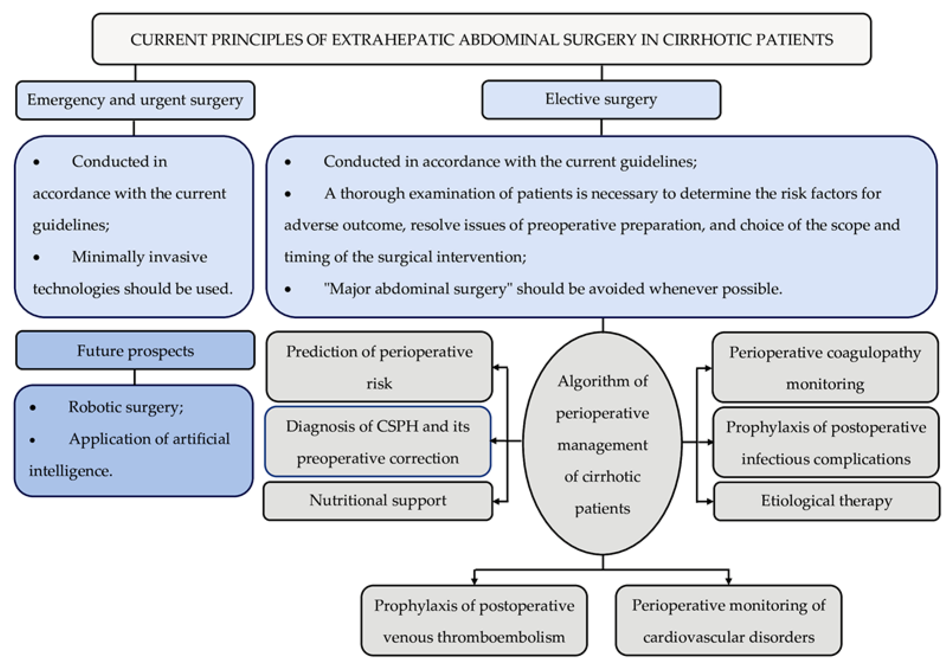

Table 1.
Child-Turcotte-Pugh score.
| Indicators | Degree of abnormality (points*) | ||
| 1 | 2 | 3 | |
| Ascites | None | Mild (or controlled by diuretics) | At least moderate despite diuretic treatment |
| Encephalopathy (grade†) | None | 1-2 | 3-4 |
| Total bilirubin | < 2 mg/dL (34 µmol/L) |
2–3 mg/dL (34– 51 µmol/L) |
> 3 mg/dL (51 µmol/L) |
| Albumin | > 3.5 g/dL (35 g/L) |
2.8–3.5 g/dL (28–35 g/L) |
< 2.8 g/dL (28 g/L) |
| PT (seconds prolonged) or, instead of PT INR |
< 4 < 1.7 |
4–6 1.7–2.3 |
> 6 > 2.3 |
| * Operational risk (grade) is based on the total number of points: Low (A): 5–6 Moderate (B): 7–9 High (C): 10–15 | |||
| † Encephalopathy is graded based on symptoms: 1: Sleep disturbances; impaired concentration; depression, anxiety, or irritability 2: Drowsiness, disorientation, poor short-term memory, uninhibited behavior 3: Somnolence; confusion; amnesia; anger, paranoia, or other bizarre behavior 4: Coma | |||
PT: prothrombin time; INR: international normalized ratio.
Table 2.
Formulas for calculating different versions of the MELD score.
| MELD = 9.57 × logе (creatinine, mg/dL) + 3.78 × logе (total bilirubin, mg/dL) + 11.20 × logе (INR) + 6.43 (etiology of LC). The result is rounded to the nearest integer. |
| MELD-Na = MELD + 1.32 × (137-Na, mmol/L) − [0.033 × MELD × (137-Na, mmol/L)]. The result is rounded to the nearest integer. |
| MELD 3.0 = 1.33 (if a woman) + 4.56 × logе (total bilirubin, mg/dL) + 0.82 × (137-Na, mmol/L) − 0.24 × (137-Na, mmol/L) × logе (total bilirubin, mg/dL) + 9.09 × logе (INR) + 11.14 × logе (creatinine, mg/dL) + 1.85 × (3.5– albumin, mg/dL) − 1.83 × (3.5– albumin, mg/dL) × logе (creatinine, mg/dL) + 6. The result is rounded to the nearest integer. |
MELD: Model for End Stage Liver Disease; INR: international normalized ratio; LC: liver cirrhosis.
Table 3.
Morbidity and mortality rates following ventral hernia repair in cirrhotic patients.
| Ref. |
Research design, number of patients |
CTP/ MELD scores, number of patients (%) |
Preoperative signs of portal hypertension, number of patients (%) |
Hernia type |
Type of operation |
Post- operative morbidity/ mortality, number of patients (%) |
Main results |
|
Park et al. [43] |
Retrospective single-center N = 53 |
CTP A—45 (34.9%) CTP B—27 (51%) CTP C—9 (17%) |
Refractory ascites—17 (32%) | Umbilical—7 (13%) Inguinal—42 (79%) Incisional—2 (4%) Combination—2 (4%) |
Elective—46 (87%) Emergency—7 (13%) |
N/A/ 1 (1.9%) |
Recurrence of hernia occurred in one patient CTP class B. The only patient CTP class C who died had septic shock related to preoperative incarceration. The ventral hernia repair could be done safely in patients CTP classes A and B with low rate of recurrences, and there was no definitive increase in the operative risk in class C. In addition, refractory ascites did not increase operative risk and recurrence rate. |
|
Oh et al. [44] |
Retrospective single-center N = 129 |
CTP A—17 (32%) CTP B—66 (51.1%) CTP C—18 (14%) |
Ascites—81 (62.8%) | Inguinal | Elective—127 (98.4%) Emergency—2 (1.6%) |
14 (10.9%)/ 2 (11.6%) |
The complication rate was unrelated to CTP class (A: B: C = 11.1%: 9.1%: 16.7%; P = 0.69) and was not higher than among non-cirrhotic patients (6.8%; P = 0.11). Two cirrhotic patients (1.6%) of CTP class C died postoperatively from liver failure. In cirrhotic patients, median follow-up was 22.9 months and recurrence developed in three (2.3%). Cumulative recurrence rates were not significantly different between the cirrhotic and non-cirrhotic patients (P = 0.87). |
|
Eker et al. [45] |
Retrospective single-center N = 30 |
CTP A—6 (20%) CTP B—9 (63%) CTP C—5 (17%) Median MELD score—12 (IQR, 8-16) |
Ascites | Umbilical | Elective | 2 (6.7%)/ 0 | A total of 2 patients suffered recurrence. Postoperative complications included pneumonia and decompensation of cirrhosis (1 case each). After a median follow-up period of 25 months (IQR, 14-34), 2 (7%) of the 30 patients died; neither of the deaths were attributable to the umbilical hernia repair. |
|
Cho et al. [46] |
Prospective multicenter N = 390 |
Median MELD score—15 | GEV+ascites—81 (20.8%) GEV—35 (8.9%) Ascites—274 (70.3%) |
Umbilical | Elective—243 (62.3%) Emergency—147 (37.7%) |
51 (13.1%)/ 20 {5.1%) | The mortality rate after elective surgeries among patients with a MELD score > 15 was 11.1%, compared to 1.3% among patients with a MELD score ≤ 15. In 37.7% of cases, patients with ascites and/or GEV underwent emergency surgical interventions. The complication rate for emergency surgeries was 20.8%, and the mortality rate was 7.4%. Age > 65 years, MELD score > 15, albumin level < 3.0 g/dL, and the presence of sepsis at the time of admission were the most significant predictors of postoperative mortality. |
|
Odom et al. [47] |
Retrospective single-center N = 69 |
Median MELD score—17 (IQR, 13.3-19.7) | Ascites | Umbilical—43 (62.3%) Inguinal, incisional or a combination—26 (37.7%) |
Emergency | 12 (17.4%)/ 2 (2.9%) (both MELD score > 20) |
Multivariate analysis revealed MELD score (18% increase in risk with each point of MELD), preoperative anemia (a 7-fold increase in risk), and preoperative small bowel obstruction (a 9-fold increase in risk) as predictive factors of major complication. In patients with MELD score > 10, morbidity was > 50%, and major morbidity was > 12% when MELD score > 20. |
|
Hew et al. [48] |
Prospective single-center N = 79 |
CTP A—7 (8.9%) CTP B—48 (60.7%) CTP C—24 (30.4%) Median MELD score—13 (IQR, 6-28) |
Ascites: controlled—26 (32.9) refractory—45 (56.9) |
Umbilical | Elective—65 (82%) Emergency—14 (18%) |
25 (31.7%)/ 1 (1.3%) |
The recurrence rate observed was of 2.7% (n = 2). The postoperative morbidity rate was 18.8% in patients CTP classes B and 66.7% in patients CTP classes C. One patient patients CTP classes C and MELD score of 22, who underwent emergency hernia repair, died 23 days after the operation from renal failure. |
|
Licari et al. [49] |
Retrospective single-center N = 27 |
CTP A—9 (33.3%) CTP B—8 (29.7%) CTP C—10 (37%) |
Ascites | Incisional | Elective—17 (63%) Emergency—10 (37%) |
13 (48.1%)/ 5 (18.5%) |
The recurrence rate observed was of 11% (n = 3), one after elective surgery and two after urgency surgery. The most common complication was seroma. Four out of five patients died after emergency surgical surgery. The causes of death were multiple organ failure due to sepsis (n = 3) and major ascites (n = 1). In the group of planned surgeries, death occurred due to heart attack (n = 1). |
|
Banu et al. [50] |
Retrospective single-center N = 65 |
CTP A—24 (36.9%) CTP B—30 (46.2%) CTP C—11 (16.9%) |
Ascites | Umbilical—32 (49.2%) Inguinal—18 (27.7%) Incisional—11 (16.9%) Epigastric—4 (6.2%) |
Elective—29 (44.6%) Emergency—36 (55.4%) |
37 (56.9%)/ 13 (20%) |
The postoperative morbidity rate was 45.8% in patients CTP classes A, 56.7% in patients CTP classes B and 81.8% in patients CTP classes C. The mortality rate was 4.2% in patients CTP classes A (one death), 13.3% in patients CTP classes B (4 deaths) and 72.7% in patients CTP classes C (8 cases). The highest morbidity and mortality rates have been registered in patients that underwent emergency surgical intervention. |
|
Kim et al. [51] |
Retrospective single-center N = 107 |
With refractory ascites: CTP B—18 (58.1%) CTP C—13 (41.9%) Median MELD score—13 (IQR, 11.5-17.0) Without refractory ascites: CTP A—31 (40.8%) CTP B—40 (52.6%) CTP C—5 (6.6%) Median MELD score—11 (IQR, 9.0-13.0) |
Refractory ascites—31 (29%) | With refractory ascites: Umbilical—26 (83.9%) Incisional—4 (12.9%) Combination—1 (3.2%) Without refractory ascites: Umbilical—27 (35.5%) Inguinal—7 (9.2%) Incisional—35 (46.1%) Femoral—2 (2.6%) Parietal wall—3 (3.9%) Combination—2 (2.6%) |
With refractory ascites: Elective—29 (93.5%) Emergency—2 (6.5%) Without refractory ascites: Elective—76 (100%) Emergency—0 |
With refractory ascites: N/A/ 1 (3.2%) Without refractory ascites: N/A/0 |
Recurrence of hernia occurred in one of the cirrhotic patients with refractory ascites and in 9 of the cirrhotic patients without refractory ascites (3.2% vs 11.8%, P = 0.31). The 30-day mortality rate (3.2% vs 0%, P = 0.64) and the risk of recurrence (hazard ratio (HR) = 0.41, 95%CI: 0.05–3.22, P = 0.39) did not differ significantly between cirrhotic patients with refractory ascites and cirrhotic patients without refractory ascites. |
|
Sidhu et al. [52] |
Retrospective single-center N = 31 |
CTP A—6 (19.4%) CTP B—19 (61.3%) CTP C—6 (19.4%) Median MELD score—11 (IQR, 7-36) |
Ascites—20 (64%) | Inguinal | Elective—29 (93.5%) Emergency—2 (6.5%) |
9 (29%)/ 1 (3.2%) |
The most common complications were postoperative encephalopathy (12.9%), followed by hematoma, wound infection, and ascites leak. There was one mortality in a patient with MELD score of 36 who presented with a strangulated hernia. |
|
Mantaka et al. [53] |
Prospective single-center N = 19 |
CTP B—13 (68.4%) CTP C—6 (31.6%) Median MELD score—15 (IQR, 11-39) |
Ascites | Umbilical | Elective—3 (15.8%) Emergency—16 (84.2%) |
0/5 (26.3%) | No patient had umbilical hernia recurrence. No association was found between survival and type of surgery, type of anesthesia, preoperative use of diuretics, ascites grade or laboratory findings. Survival rates at 30 days (P = 0.086), 3 months (P = 0.022), and 12 months (P = 0.031) postoperatively were better in patients who underwent emergent umbilical hernia repair. |
CTP: Child-Turcotte-Pugh; MELD: Model for End Stage Liver Disease; IQR: interquartile range; GEV: Gastroesophageal varices; N/A: Not available.
Table 4.
Morbidity and mortality rates following radical gastrectomy in cirrhotic patients.
| Ref. |
Research design, number of patients |
CTP score, number of patients (%) |
Preoperative signs ofPH, number of patients (%) |
Post- operative morbidity/ mortality, number of patients (%) |
Main results |
|
Jang et al. [107] |
Retrospective multicenter N = 57 |
CTP A—46 (81%) CTP B/C—11 (19%) |
GEV—18 (32%) Ascites—7 (13%) |
22 (39%)/ 5 (9%) |
The most common complications were ascites (23%), followed by wound infection and hepatic encephalopathy. Postoperative ascites occurred more frequently in patients CTP classes B/C than in patients CTP class A (63.6% vs 13%, P = 0.001). Postoperative mortality rate in patients CTP classes B/C was significantly higher than in patients CTP class A (27.2% vs 4.3%, P = 0.045). The estimated 5-year survival rate for all patients was 54%. |
|
Xu et al. [108] |
Retrospective single-center N = 30 |
CTP A—15 (50%) CTP B—8 (26.7%) CTP C—4 (13.3%) |
GEV History of GEV bleeding Ascites Hypersplenism |
16 (53.3%)/ 5 (16.7%) |
The rates of postoperative complications in patients CTP class A were 26.7%, in patients CTP class B—72.7%, and in patients CTP class C—100%. One patient died due to pulmonary embolism, two due to anastomosis leakage, and two due to multiple organ dysfunction syndrome. The median survival was 15.0 months. |
|
Jin et al. [109] |
Retrospective single-center N = 22 |
CTP A—12 (54.5%) CTP B—10 (45.5%) |
GEV History of GEV bleeding Ascites Hypersplenism |
11 (50%)/ 2 (9%) |
The rate of postoperative ascites in patients CTP class A was much lower than in patients CTP class B (P < 0.05). There was no significant difference in liver function deterioration rate between patients CTP class A and patients CTP class B (P > 0.05). The incidence of complications in patients with portal hypertension was significantly higher than in patients without it (P < 0.05). The survival time of patients CTP class A was significantly longer than that of patients CTP class B (P < 0.05). |
|
Zhang et al. [110] |
Retrospective, single-center, N = 60 |
CTP A—30 (50%) CTP B—30 (50%) |
GEV History of GEV bleeding: CTP A—9 (30%) CTP B—10 (33.3%) |
38 (63.3%)/ 2 (3.3%) |
The most common postoperative complications were severe ascites, pleural effusion, acute liver failure, anastomosis leakage, wound infection, and left inferior phrenic sepsis. Two patients CTP class B died in the perioperative period, among whom one died from severe ascites and acute liver failure and another from multiple organ failure due to postoperative anastomosis leakage. |
|
Alshahrani et al. [111] |
Retrospective single-center N = 75 (ODG—48, LDG -27) |
ODG: CTP A—43 (89.6%), CTP B—5 (10.4%) LDG: CTP A—24 (88.9%), CTP B—3 (11.1%) |
N/A |
ODG: 9 (18.8%)/ 1 (2.1%) LDG: 4 (14.8%)/ 0 |
The most common complications after ODG were intra-abdominal bleeding (8.3%), followed by wound infection, pneumonia, and anastomosis leakage. The most common complications after LDG were wound infection and pneumonia. The only patient who died after ODG had postoperative bleeding and liver failure. |
|
Kim et al. [112] |
Retrospective single-center N = 75 (OG—39, LG -36) |
OG: CTP A—31 (79.5%), CTP B—8 (20.5%) LG: CTP A—31 (86.1%), CTP B—4 (11.1%) CTP C—1 (2.8%) |
N/A |
OG: 10 (25.6%)/0 LG: 7 (19.4%)/ 1 (2.8%) |
The rates of postoperative complications after OG in patients CTP class A were 29.0%, and in patients CTP class B—12.5%. The rates of postoperative complications after LG in patients CTP class A were 16.1%, and in patients CTP class B—25.0%, and in patients CTP class C—100%. The most common complications after OG were gastric stasis, wound infection, intra-abdominal infection, intra-abdominal bleeding, anastomosis stricture, ascites, and pneumonia. The most common complications after LG were gastric stasis, wound infection, anastomosis leakage, anastomosis stricture, ascites, pneumonia, and hepatic failure. The only patient who died after LG had postoperative liver failure. |
CTP: Child-Turcotte-Pugh; PH: portal hypertension; GEV: Gastroesophageal varices; ODG: Open distal gastrectomy; LDG: Laparoscopic distal gastrectomy; OG: Open gastrectomy; LG: Laparoscopic gastrectomy; N/A: Not available.
Disclaimer/Publisher’s Note: The statements, opinions and data contained in all publications are solely those of the individual author(s) and contributor(s) and not of MDPI and/or the editor(s). MDPI and/or the editor(s) disclaim responsibility for any injury to people or property resulting from any ideas, methods, instructions or products referred to in the content. |
© 2026 by the authors. Licensee MDPI, Basel, Switzerland. This article is an open access article distributed under the terms and conditions of the Creative Commons Attribution (CC BY) license (http://creativecommons.org/licenses/by/4.0/).
Copyright: This open access article is published under a Creative Commons CC BY 4.0 license, which permit the free download, distribution, and reuse, provided that the author and preprint are cited in any reuse.