

Submitted:
30 September 2026
Posted:
01 October 2026
You are already at the latest version
Abstract
Background/Objectives: Hidradenitis suppurativa (HS) is a chronic inflammatory skin disease characterized by dysregulated immune responses. This study aimed to assess the expression of IL-1β, IL-8, IL-10, and TNF-α in HS lesioned and non-lesioned skin and to examine their associations with selected clinical characteristics. Methods: Skin samples from HS lesions and non-lesioned skin were obtained from 52 patients. Histopathological changes were assessed using hematoxylin and eosin staining, and cytokine expression was evaluated by immunohistochemistry and quantified using ImageJ. Cytokine expression was analyzed according to Hurley stage and selected demographic and clinical characteristics. Results: Of 52 patients, 24 (46%) were female. Patient’s median age was 35.5 (IQR 26-44) years; 28 (54%) patients were categorized as Hurley stage 2 and 24 (46%) as stage 3. IL-1β, IL-8, IL-10, and TNF-α expression was significantly higher in lesioned than in non-lesioned skin (all p<0.05). IL-1β expression was higher in Hurley stage 2 than stage 3 (p=0.042), whereas IL-8 expression was higher in Hurley stage 3 (p=0.039). No significant differences were found according to sex, BMI, smoking, or biological therapy. TNF-α expression was significantly higher in patients with a positive family history of HS (p=0.049). Conclusions: HS lesions demonstrate increased expression of both pro-inflammatory and regulatory cytokines. Differences in IL-1β and IL-8 expression across Hurley stages suggest that inflammatory pathways may vary with disease stage. The association between TNF-α expression and positive family history may indicate a potential genetic influence on the inflammatory phenotype of HS.
Keywords:
cytokines
; hidradenitis suppurativa
; interleukins
; TNF- α
1. Introduction
Hidradenitis suppurativa (HS) is a chronic, recurrent inflammatory skin disease characterized by the formation of painful inflammatory nodules, abscesses, and fistulous tracts, most commonly in intertriginous areas [1,2,3]. It occurs in areas with the highest density of apocrine glands-the armpits, groin, areas under the breasts, and the perianal and genital regions [3].
The clinical presentation and severity of HS are influenced by several demographic and clinical factors, which may also contribute to differences in disease phenotype and progression. The disease most commonly develops after puberty and predominantly affects women, although more severe disease has been reported in men in some cohorts. Increasing body mass index (BMI) and cigarette smoking have also been consistently associated with HS and, in several studies, with greater disease severity [4]. In a large retrospective cohort of 846 patients, male sex, longer disease duration, higher BMI, and greater cumulative smoking exposure were independently associated with higher Hurley stage [5]. Similarly, Sartorius et al. demonstrated that both smoking and increasing BMI were associated with greater clinical severity of HS [6]. More recent evidence from a systematic review and meta-analysis further supports significant associations between HS and obesity and smoking [7].
The clinical course of HS is heterogeneous, and disease severity may also be influenced by age of onset, disease duration, treatment exposure, and associated comorbidities. Early-onset disease has been associated with a higher frequency of positive family history and more widespread involvement, suggesting a stronger genetic predisposition in this subgroup [8]. Approximately one-third of patients with HS report a positive family history, further supporting the contribution of genetic susceptibility to disease development [9]. HS is also associated with a broad spectrum of comorbidities, including obesity, diabetes mellitus, metabolic syndrome, cardiovascular disease, depression, inflammatory bowel disease, and spondylarthritis [10]. Because systemic comorbidities, smoking, BMI, disease duration, and treatment may influence the inflammatory milieu, these variables should be considered when interpreting differences in histopathological findings and local cytokine expression between HS patients and non-lesioned skin.
Histopathological features of HS reflect the complexity of the inflammatory process and change depending on the stage of the disease. Dermatologists typically assess the clinical signs and symptoms of HS using the Hurley scale [1], the categories of which were developed based on a clinical assessment of disease severity. The Hurley classification system divides HS patients into three groups, primarily based on the presence and extent of abscesses, sinus tracts, and scarring [11,12,13,14,15]: (a) Hurley stage 1 – Single or multiple isolated abscesses without sinus tracts or scarring; (b) Hurley stage 2 – Recurrent abscesses forming sinus tracts and scars, but without involvement of the intervening skin; and (c) Hurley stage 3 – Multiple abscesses with multiple, interconnected sinus tracts across the entire affected area, with no healthy skin between them [16].
Although the pathogenesis of the disease is complex and not yet fully elucidated, HS is currently considered a disease in which follicular epithelial abnormalities, dysregulation of innate and adaptive immune responses, and the increased production of various inflammatory mediators play a significant role in its onset and progression [2].
The pathophysiology of HS is based on the occlusion of the upper part of the follicle due to follicular hyperkeratosis with the consequent dilatation and rupture of the follicle, and an inflammatory response [1,3]. The rupture of the follicular structure results in the release of keratin and other follicular contents into the surrounding dermis, thereby initiating and amplifying a local immune response. This response involves the recruitment and activation of various immune cells and the release of multiple inflammatory mediators, with cytokines playing a particularly important role in the initiation and perpetuation of inflammation. The pro-inflammatory cytokines interleukin-1β (IL-1β) and tumor necrosis factor alpha (TNF-α) are associated with the activation and maintenance of the local inflammatory response in the skin of patients with HS [17]. TNF-α also plays a key role in maintaining the inflammatory reaction and stimulates the expression of numerous chemokines and the activation of endothelial cells, which further favors the infiltration of inflammatory cells into the skin.
Kelly et al (2015) showed an increased expression of IL-1β and TNF-α in the skin of patients with HS [18]. The increased expression of IL-1β was not limited exclusively to clinically visible lesions, but was also present in the perilesional skin, which points to the existence of a subclinical inflammatory process. The authors also showed that cells of monocyte-macrophage origin are an important source of IL-1β and that caspase-1 activation is associated with increased production of IL-1β and IL-18.
More recent work by Manzo Margiotta et al (2024) further supports the importance of the IL-1 family in the pathogenesis of HS. A study comparing HS patients with healthy controls revealed alterations in the serum concentrations of several members of the IL-1 family and their soluble receptors; specifically, levels of IL-18 and free IL-18 were significantly higher in HS patients, and these values correlated with disease severity [19]. These results further underscore the importance of dysregulated IL-1 signaling in the pathogenesis and maintenance of chronic inflammation in HS.
In addition to pro-inflammatory mediators, anti-inflammatory mechanisms also play a significant role in HS. Interleukin-10 (IL-10) is a key anti-inflammatory cytokine that limits immune cell activation and the production of other pro-inflammatory cytokines. Interestingly, previous studies have observed increased IL-10 expression in HS lesions, despite the presence of marked pro-inflammatory activity [18,20].
According to Johnston et al. (2021), the expression levels of the aforementioned cytokines in the affected skin of HS patients varied. Specifically, the authors reported conflicting results, with the expression of certain cytokines being increased in some studies, decreased in others, and unchanged in others [21]. These discrepancies may be related to differences in study populations, anatomical sites and disease stages, as well as methodological differences in the assessment of cytokine expression. The co-expression of pro-inflammatory (IL-1β, TNF-α, IL-8) and anti-inflammatory (IL-10) cytokines suggests a complex and imbalanced inflammatory response in HS, which may contribute to the chronicity and recurrence of the disease [22,23].
Although previous studies have demonstrated alterations in cytokine expression and described histopathological features in HS, the relationship between local cytokine expression and tissue-level morphological changes remains incompletely characterized [18,21,24,25]. Furthermore, the available literature is heterogeneous with regard to patient populations, tissue sampling, analytical methods, and clinical characterization. A systematic review of immunohistochemical studies in HS identified substantial methodological heterogeneity and conflicting findings across studies, with many studies including relatively small patient cohorts and limited clinical stratification [25]. Similarly, a systematic review of cytokine studies highlighted considerable heterogeneity in the reported expression of individual cytokines, including IL-1β, IL-8, IL-10 and TNF-α, and emphasized the need for more consistent methodologies and better assessment of clinical factors that may influence inflammatory profiles [21]. Only a limited number of studies have directly compared lesioned and clinically non-lesioned skin obtained from the same patients, further limiting the characterization of local inflammatory changes in HS [21,25].
Accordingly, the primary aim of this study was to characterize the histopathological changes and the local expression of IL-1β, TNF-α, IL-8, and IL-10 in HS-affected skin and subcutaneous tissue and in paired clinically non-lesioned tissue samples obtained from the same patients. The secondary aim was to investigate the associations between histopathological findings, cytokine expression levels, and selected demographic and clinical characteristics, including age, sex, BMI, smoking status, disease duration, Hurley stage, treatment exposure, associated comorbidities, and family history of HS. It was hypothesized that HS lesioned tissue would exhibit characteristic histopathological changes accompanied by altered local expression of IL-1β, TNF-α, IL-8, and IL-10 compared with paired clinically non-lesioned tissue from the same patients. Given the heterogeneous findings reported in previous studies [21,25], it was further hypothesized that cytokine expression patterns and the extent of histopathological changes would vary according to selected demographic and clinical characteristics of patients with HS, particularly disease severity, disease duration, BMI, smoking status, treatment exposure, associated comorbidities, and family history.
2. Materials and Methods
2.1. Patients
A total of 52 patients with HS, treated at the Department of Plastic and Reconstructive Surgery of the Clinic of Surgery at the Clinical Hospital Centre (CHC) Rijeka, Croatia, participated in this study; samples of both lesioned and non-lesioned tissue was collected.
Inclusion criteria were: patients over 18 years of age, BMI > 18.5 kg/m² (according to World Health Organization classification for the European population), and data on prior HS therapy (systemic, topical, or no prior therapy).
Exclusion criteria were: pregnancy or breastfeeding patients and patients under 18 years of age. Further inclusion and exclusion criteria (with photographic examples) were described by Jemec et al. [26].
For all patients included in the planned study, the following data was recorded: age, sex, body mass index (BMI), history of tobacco smoking, Hurley stage of HS, prior HS therapy (systemic non-biological, biological, topical, or no prior therapy), illness duration and localization as well as comorbidities.
Experimental group consisted of skin samples affected by HS, involving the sampling of skin and subcutaneous tissue from the affected site (lesioned biopsy). The control group consisted of non-lesioned skin samples obtained from the same sample of patients, from whom we collected a 1 x 1 cm sample of skin and subcutaneous of an unaffected site.
2.2. Methodology
2.2.1. Histologycal Staining
At the Clinical Institute of Pathology and Cytology of CHC Rijeka, Croatia, following the standard procedure of tissue pathohistological processing after tissue fixation with 10% buffered formalin and embedding in paraffin, tissue sections stained with standard hematoxylin and eosin (HE) staining were analyzed using a light microscope in such a way as to determine the type and amount of inflammatory infiltrate, the amount of granulation tissue and fibrosis, and the epithelial changes of the surface and follicular epithelium.
2.2.2. Immunohistochemical Method
The aforementioned formalin-fixed, paraffin-embedded tissue samples were used to prepare tissue sections for immunohistochemical staining. Paraffin sections with a thickness of 5 µm were dried overnight and deparaffinized and rehydrated in the usual way [27]. Appropriate pretreatment was applied to detect antigenic epitopes of molecules of interest [28]. To display the molecules of interest, primary antibodies against the cytokines IL-1β, IL-8, IL-10 and TNF- α was used using the envision immunohistochemical method [29] in an automated immunostainer (DakoCytomation Colorado Inc, Fort Collins, CO, USA) according to the manufacturer's instructions. The ChemMateTM kit (K 5001 and K5007, DakoCytomation, Glostrup, Denmark) was used to visualize the binding specificity of the primary antibody. For each sample tested, a negative control was prepared using a tissue section where the secondary antibody is replaced by an antibody diluent (ChemMate antibody diluent, S 2022, DakoCytomation, Glostrup, Denmark). Normal structures within the examined tissue samples served as the positive control. Tissue dehydration was performed using ethyl alcohol in increasing concentrations (75%, 96%, and 100%). Following clearing with xylene, the specimen was mounted using an appropriate medium and a coverslip.
Immunohistochemical staining in the cell cytoplasm was analyzed using the ImageJ software program based on the percentage of stained cells [30].
2.3. Statistical Analysis
Sample size was calculated with the program G power 3.1.9.73. with an assumed power of 0.80. α=0.05 and a medium effect size for the calculation of the differences between the 2 groups, which amounts to 52 patients per group [31].
Categorical variables were presented as frequencies (n) and relative frequencies (%).
Quantitative variables normality was tested with Kolomogorov-Smirnof test. As half variables had non-normal distribution, variables were presented as median and interquartile range (IQR) and were compared between groups using the Mann Whitney U test. BMI was divided into 2 categories: normal BMI with values 18.9-24.9 and high BMI with values >24.9. Two patients with BMI<18.9 were excluded from the analysis.
Correlations were calculated with Spearman coefficient of correlation. The strength of the correlations was interpreted according to Schober et al. as follows: negligible (<0.1), weak (0.1–0.39), moderate (0.4–0.69), strong (0.7–0.89), and very strong (≥0.9) [32].
P values <0.05 were considered statistically significant. All analyses were done using the MedCalc statistical software version 23.7.1 (MedCalc Software Ltd. Ostend. Belgium).
2.4. Ethical Considerations
The study was conducted in accordance with the ethical principles of the Declaration of Helsinki and was approved by the Ethics Committee of the Faculty of Medicine, University of Rijeka and the Ethics Committee of Clinical Hospital Centre Rijeka, Croatia (Classification No.: 641-01/23-01/225; Registration No.: 2170-1-42-09-8-23-2). All participants have signed written informed consent.
3. Results
3.1. Sample Characteristics
There were 52 patients included in this study of which 24 (46%) female (Table 1). Patients were median 35.5 (IQR 26-44) years old, mostly smoking [41 (79%)] and with elevated BMI [median 27.3 (IQR 24.3-30.7)]. There were 28 (54%) patients in HS stage 2 and 24 (46%) in stage 3. One fifth [11 (21%)] of the patients were on biological therapy, they were mostly male (n=8) and in the HS stage 3 (n=9).
3.2. Histology and Immunohistochemistry
Histopathological examination of H&E-stained sections demonstrated the characteristic morphological changes associated with HS, with particular attention to the type, distribution, and extent of the inflammatory infiltrate within the dermis and subcutaneous tissue. The inflammatory response was assessed according to the predominant inflammatory cell populations, including neutrophils, lymphocytes, histiocytes/macrophages, and plasma cells, as well as their focal or diffuse distribution. The sections were also evaluated for follicular hyperkeratosis, follicular dilatation and rupture, perifollicular inflammation, abscess formation, and sinus tract formation. The extent of granulation tissue and dermal fibrosis was assessed as indicators of tissue repair and chronic remodeling. In addition, alterations of the surface epidermis and follicular epithelium, including hyperkeratosis, acanthosis, epithelial hyperplasia, ulceration, and structural disruption, were recorded and compared with the corresponding findings in non-lesioned control skin.
Immunohistochemical analysis revealed cytoplasmic expression of IL-1β, IL-8, IL-10, and TNF-α in lesional skin, with differences in the extent and intensity of immunoreactivity between samples. The staining was predominantly observed in inflammatory and stromal cells within the affected tissue, reflecting the local activation of both pro-inflammatory and regulatory immune pathways. As illustrated in Figure 1, higher immunoreactivity is shown in panels A–D, whereas panels A₁–D₁ demonstrate lower expression of the respective cytokines. The corresponding control sections (a–d) show absent or minimal specific immunoreactivity. The arrows indicate representative cells exhibiting distinct cytoplasmic cytokine expression. Overall, the immunohistochemical findings demonstrate increased local expression of the investigated cytokines in HS lesional skin compared with non-lesioned control tissue, supporting the presence of an active local inflammatory and regulatory response.
3.3. Comparisons of Inflammatory Cytokines
Expression of IL-1β. IL-8. IL-10 and TNF-α was significantly higher in HS lesioned skin then non-lesioned skin for all comparisons (all p<0.05) (Figure 2). There were no differences in the expression according to sex, BMI, smoking and biological therapy (all p>0.05) (Supplement 1. Tables 2–5).
3.3.1. Comparisons of Inflammatory Cytokines According to Hurley Stage System Grades
Expression of IL-1β was significantly higher in HS stage 2 then HS stage 3 (p=0.042). Expression of Il-8 was lower in HS stage 2 then in HS stage 3 (p=0.039). There were no differences in the median values of expression in Il-10 and TNF-α according to the HS stage (Figure 3).
3.3.2. Comparisons of Inflammatory Cytokines According to Other Relevant Parameters
There were no differences in inflammatory cytokines according to: sex (all p>0.05, S1 Table 2), BMI (all p>0.05, S1 Table 3), smoking (all p>0.05, S1 Table 4) and biological therapy (all p>0.05, S1 Table 5).
There were also no differences in the expression of cytokines according to family history for IL-1β, Il-8 and Il-10 (all p>0.05, S1 Table 6). However, there was a significant difference in the expression of TNF-α according to family history (p=0.049), where patients with positive family history (median=71.4, IQR 38.1-80.9)) had significantly higher expression of TNF-α than patients with no family history ((median=53.2, IQR 30.91-64.1)) (S1 Table 6).
4. Discussion
This study analyzed the expression of IL-1β, IL-8, IL-10, and TNF-α in lesioned skin from patients with HS. The study's most significant finding is the significantly higher expression of all four investigated cytokines in HS lesioned skin. This result confirms that cytokine-mediated inflammatory mechanisms are highly active in the skin lesions of HS patients. This finding aligns with previous evidence suggesting that HS is not merely a consequence of local follicular occlusion, but a disease characterized by immune response dysregulation and chronic inflammation [33].
Increased IL-1 expression may be linked to activation of the innate immune system. IL-1, particularly IL-1β, is a key pro-inflammatory mediator in HS [34]. Damage to the follicular structure and the release of cellular contents into the dermis trigger immune cell activation and the production of inflammatory mediators, thereby initiating and sustaining the inflammatory response. IL-1β can further stimulate the activation of keratinocytes and other skin cells, contributing to the formation of the inflammatory microenvironment characteristic of HS [21,35]. Elevated IL-1β expression in lesioned skin is therefore biologically expected and supports the fact that the innate immune activation plays a significant role in the pathogenesis and persistence of the disease.
IL-8 also plays a particularly important role in neutrophil recruitment. Neutrophilic inflammation is a recognized feature of active HS lesions, and IL-8 can contribute to attracting neutrophils to the site of inflammation, thereby intensifying the local inflammatory response [21,24]. The increased IL-8 expression observed in HS skin may thus reflect the active neutrophilic component of the inflammation. Chronic activation of this mechanism can contribute to sustained inflammation and tissue damage, especially in advanced lesions. Current models of HS pathogenesis describe interconnected feedback loops involving keratinocytes, neutrophils, macrophages, and lymphocytes, with various cytokines contributing to disease progression and chronicity [21,36].
Significantly elevated expression of TNF-α in lesioned skin is also consistent with the known role of this cytokine in HS. TNF-α is one of the key proinflammatory mediators that participate in the activation of various immune cells and the maintenance of chronic inflammation [33]. Its importance is further confirmed by therapeutic experience, since inhibition of TNF-α is one of the important approaches in the treatment of patients with moderate to severe HS. Therefore, the increased local expression of TNF-α in our study further supports the concept that TNF-α is an important part of the inflammatory network of HS, although the level of its expression alone does not necessarily reflect the clinical severity of the disease.
The finding of increased expression of IL-10 in the lesioned skin is also interesting. As a rule, IL-10 is considered a regulatory or anti-inflammatory cytokine, so its increased expression may at first glance seem unexpected in a chronic inflammatory disease [34]. However, the presence of IL-10 in HS lesions may reflect an attempt by the organism to limit the intensity of the inflammatory response. In HS, there are simultaneously strong proinflammatory signals and regulatory mechanisms, which points to a complex balance between activation and suppression of the immune response. Therefore, increased expression of IL-10 should not necessarily be interpreted as evidence of reduced inflammation, but as a possible compensatory response to chronic and intense activation of the immune system.
The higher expression of IL-8 in patients with Hurley stage 3 is particularly interesting. Hurley stage 3 is characterized by more extensive lesions, interconnected sinus tracts and more pronounced tissue destruction. Higher expression of IL-8 in this group may indicate greater activation of neutrophil-mediated inflammatory mechanisms in advanced disease. Since IL-8 plays an important role in neutrophil chemotaxis, its increased expression in stage 3 could be associated with chronic and more intense inflammatory processes characteristic of severe forms of HS [36,37]. However, it should be emphasized that this finding shows an association, and does not prove that an increase in IL-8 causes progression from stage 2 to stage 3. The opposite pattern was observed for IL-1β, whose expression was higher in stage 2 than in stage 3. This result is not entirely intuitive, as one would expect the proinflammatory cytokine to be more pronounced in a more severe clinical stage. However, HS is a heterogeneous disease and the Hurley classification primarily describes the anatomical extent of the disease and the presence of sinus tracts, not necessarily the current intensity of inflammation [17,34]. A stage 3 patient may have extensive fibrosis and chronic sinus tracts, but relatively less active inflammatory lesions at the time of biopsy. Therefore, a higher Hurley stage does not necessarily mean a higher expression of each individual proinflammatory cytokine. No difference was found between Hurley stage 2 and 3 for IL-10 and TNF-α. This finding may indicate that the expression of these cytokines does not necessarily follow a linear relationship with anatomical disease severity as assessed by the Hurley classification. TNF-α is involved in inflammatory processes throughout different stages of HS, while IL-10 may represent a regulatory response that is not directly proportional to clinical disease severity [17,34].
Regarding BMI, our findings should be interpreted in the context of the well-established association between obesity and HS. Increased BMI has consistently been associated with the occurrence and clinical severity of HS, and obesity may contribute to disease activity through several mechanisms, including chronic low-grade systemic inflammation, adipose tissue dysfunction, and increased production of pro-inflammatory mediators [4,6,7]. In the present study, however, BMI was not associated with a consistent pattern of differences in the expression of the investigated cytokines. Thus, although increased BMI represents an important clinical factor in HS, its effect on the local inflammatory milieu may not be reflected by a uniform increase in the expression of individual cytokines. The association between TNF-α expression and a positive family history of HS observed in our study is also of particular interest. A positive family history has been reported in a substantial proportion of patients with HS and supports the contribution of genetic susceptibility to disease development [8,9]. Familial HS may reflect inherited differences in immune regulation and inflammatory responses, which could contribute to interindividual variation in disease phenotype. The association observed in our study between positive family history and TNF-α expression may therefore suggest that genetic predisposition influences the local inflammatory phenotype of HS. However, this finding should be interpreted cautiously, as family history is an indirect measure of genetic susceptibility and the observed association does not establish a specific genetic mechanism responsible for altered TNF-α expression. Further studies incorporating genetic or molecular analyses are needed to determine whether inherited factors directly influence TNF-α-related inflammatory pathways in HS.
Overall, our results support the concept of HS as a disease characterized by complex and multidirectional inflammatory activation. The increased expression of IL-1β, IL-8, IL-10, and TNF-α in lesioned skin compared to non-lesioned skin indicates the simultaneous activation of various inflammatory and regulatory mechanisms. This finding aligns with the current understanding of HS as a disease involving the interplay of innate and adaptive immunity, neutrophil and macrophage activation, and various cytokine pathways [33]. It is particularly important to emphasize that our results do not support a simple model in which an increase in all proinflammatory cytokines linearly parallels the increase in clinical disease severity. Instead, the results regarding IL-1β and IL-8 suggest that the qualitative composition of the inflammatory response changes across different stages of the disease. In this sense, advanced HS may not merely be a "more intense" version of an earlier stage, but rather a condition involving a shift in the cellular and molecular composition of the chronic inflammation. Current models of pathogenesis also highlight that cellular and molecular processes-including inflammation, extracellular matrix remodeling, and fibrosis-undergo changes throughout the various phases of the disease [38]. The results of this study are significant as they further confirm the presence of local inflammatory activity in HS and point to potential differences in cytokine profiles across different clinical stages. The finding of increased IL-8 expression in Hurley stage 3 is particularly interesting as it may indicate the importance of neutrophil-mediated inflammation in advanced disease, whereas higher IL-1β expression in stage 2 demonstrates that the relationship between individual cytokines and clinical severity is not linear. The association of TNF-α with a positive family history further suggests that genetic predisposition may influence the individual inflammatory phenotype of the disease.
Several limitations of this study should be acknowledged. Cytokine expression was assessed at a single time point, which prevents evaluation of longitudinal changes in the inflammatory profile during disease progression or treatment. This cross-sectional design allows the identification of associations between cytokine expression and clinical or histopathological characteristics, but does not permit conclusions regarding causality or temporal relationships. HS is a heterogeneous disease, and cytokine expression may vary according to the anatomical site of the lesion, the stage and activity of individual lesions, and the presence of systemic or local factors. In this context, the Hurley classification primarily reflects the anatomical extent of disease and the presence of sinus tracts and scarring, rather than the intensity of the inflammatory response at the exact time and site of biopsy. Furthermore, immunohistochemical assessment provides information on the tissue distribution and relative expression of cytokines; however, it does not necessarily reflect their functional activity or allow for definitive identification of their cellular sources. Finally, patient-related factors, including previous or current treatment, smoking, obesity, and comorbidities, may represent potential confounding factors that could influence cytokine expression.
5. Conclusions
The results of this study confirm significant local activation of inflammatory and regulatory mechanisms in HS. The significant increase in IL-1β, IL-8, IL-10, and TNF-α in lesioned skin compared to non-lesioned skin supports their involvement in the disease's pathogenesis. Differences in IL-1β and IL-8 expression across Hurley stages suggest that specific inflammatory pathways may be activated differently during the development and progression of the disease. At the same time, the lack of association between cytokine expression and factors such as gender, BMI, smoking, and biological therapy indicates that the relationship between clinical factors and local immune activity is complex.
Finally, the association of higher TNF-α expression with a positive family history is an intriguing finding that may point to a potential genetic component underlying a distinct inflammatory phenotype; however, due to the sample size and borderline statistical significance, this requires confirmation in future studies. Overall, these results further support the concept of HS as a heterogeneous, chronic immuno-inflammatory disease in which different inflammatory pathways may play varying roles depending on the disease stage and individual patient characteristics.
Supplementary Materials
The following supporting information can be downloaded at the website of this paper posted on Preprints.org.
Author Contributions
Conceptualization, D.J., A.T.J.R., K.B. and K.M.I.; methodology, D.J., A.T.J.R., K.B. and H.O.; software, K.B. and H.O.; validation, D.J., A.T.J.R., K.B. and K.M.I.; formal analysis, A.T.J.R., K.B.; investigation, D.J., A.T.J.R., K.M.I.; resources, D.J. and K.M.I.; data curation, A.C.P.; writing-original draft preparation, D.J., A.T.J.R.,; writing-review and editing, D.J., A.T.J.R., K.B., H.O., A.C.P. and K.M.I.; visualization, A.T.J.R. and K.B.; supervision, K.M.I.; project administration, D.J. and A.T.J.R.; funding acquisition. All authors have read and agreed to the published version of the manuscript.
Funding
This research received no external funding.
Institutional Review Board Statement
The study was conducted in accordance with the Declaration of Helsinki, and approved by the Ethics Committee of the Faculty of Medicine, University of Rijeka and the Ethics Committee of Clinical Hospital Centre Rijeka, Croatia (Classification No.: 641-01/23-01/225; Registration No.: 2170-1-42-09-8-23-2).
Informed Consent Statement
Written informed consent has been obtained from the patients to publish this paper.
Data Availability Statement
The data presented in this study are available on request from the corresponding author.
Acknowledgments
During the preparation of this manuscript, the authors used Grammarly 14.1328.0 for spell checking. The authors reviewed and edited the generated content and take full responsibility for the final content of the manuscript.
Conflicts of Interest
The authors declare no conflicts of interest.
References
- Sabat, R.; Jemec, G.B.E.; Matusiak, Ł.; Kimball, A.B.; Prens, E.; Wolk, K. Hidradenitis suppurativa. Nat. Rev. Dis. Primers 2020, 6(1), 18. [Google Scholar] [CrossRef] [PubMed]
- Goldburg, S.R.; Strober, B.E.; Payette, M.J. Hidradenitis suppurativa: epidemiology, clinical presentation, and pathogenesis. J. Am. Acad. Dermatol. 2020, 82(5), 1045–1058. [Google Scholar] [CrossRef] [PubMed]
- Jenkins, T.; Isaac, J.; Edwards, A.; Okoye, G.A. Hidradenitis suppurativa. Dermatol. Clin. 2023, 41(3), 471–479. [Google Scholar] [CrossRef] [PubMed]
- Ingram, J.R. The epidemiology of hidradenitis suppurativa. Br. J. Dermatol. 2020, 183(6), 990–998. [Google Scholar] [CrossRef]
- Schrader, A.M.; Deckers, I.E.; van der Zee, H.H.; Boer, J.; Prens, E.P. Hidradenitis suppurativa: a retrospective study of 846 Dutch patients to identify factors associated with disease severity. J. Am. Acad. Dermatol. 2014, 71(3), 460–467. [Google Scholar] [CrossRef] [PubMed]
- Sartorius, K.; Emtestam, L.; Jemec, G.B.E.; Lapins, J. Objective scoring of hidradenitis suppurativa reflecting the role of tobacco smoking and obesity. Br. J. Dermatol. 2009, 161(4), 831–839. [Google Scholar] [CrossRef] [PubMed]
- Elzawawi, K.E.; Elmakaty, I.; Habibullah, M.; et al. Hidradenitis suppurativa and its association with obesity, smoking, and diabetes mellitus: a systematic review and meta-analysis. Int. Wound J. 2024, 21(9), e70035. [Google Scholar] [CrossRef]
- Deckers, I.E.; van der Zee, H.H.; Boer, J.; Prens, E.P. Correlation of early-onset hidradenitis suppurativa with stronger genetic susceptibility and more widespread involvement. J. Am. Acad. Dermatol. 2015, 72(3), 485–488. [Google Scholar] [CrossRef] [PubMed]
- Bruinsma, R.L.; Fajgenbaum, K.; Yang, Y.; Del Mar Melendez-Gonzalez, M.; Mohlke, K.L.; Li, Y.; Sayed, C.J. Assessment of familial risk in patients with hidradenitis suppurativa. Br. J. Dermatol. 2021, 184(4), 753–754. [Google Scholar] [CrossRef] [PubMed] [PubMed Central]
- Garg, A.; Malviya, N.; Strunk, A.; Wright, S.; Alavi, A.; Alhusayen, R.; et al. Comorbidity screening in hidradenitis suppurativa: evidence-based recommendations from the US and Canadian Hidradenitis Suppurativa Foundations. J. Am. Acad. Dermatol. 2022, 86(5), 1092–1101. [Google Scholar] [CrossRef] [PubMed] [PubMed Central]
- Johnston, L.; Dupuis, E.; Lam, L.; Poelman, S. Understanding Hurley Stage III hidradenitis suppurativa patients' experiences with pain: a cross-sectional analysis. J. Cutan. Med. Surg. 2023, 27(5), 487–492. [Google Scholar] [CrossRef] [PubMed] [PubMed Central]
- McKenzie, S.A.; Harview, C.L.; Truong, A.K.; Grogan, T.R.; Shi, V.Y.; Bennett, R.G.; Hsiao, J.L. Physical symptoms and psychosocial problems associated with hidradenitis suppurativa: correlation with Hurley stage. Dermatol. Online J. 2020, 26(9), 13030/qt4rm8w7kn. [Google Scholar] [CrossRef] [PubMed]
- Daoud, M.; Suppa, M.; Benhadou, F.; Heudens, S.; Sarkis, A.S.; Njimi, H.; et al. Factors associated with severe hidradenitis suppurativa, using Hurley staging and Metascore. Dermatology 2024, 240(5-6), 713–731. [Google Scholar] [CrossRef] [PubMed]
- Ingram, J.R.; Marzano, A.V.; Prens, E.; Schneider-Burrus, S.; Warren, R.B.; Keal, A.; et al. Hidradenitis suppurativa with and without draining tunnels: a real-world study characterizing differences in treatment and disease burden. J. Eur. Acad. Dermatol. Venereol. 2025, 39(8), 1431–1441. [Google Scholar] [CrossRef] [PubMed] [PubMed Central]
- Heath, C.R.; Usatine, R.P. Hidradenitis suppurativa. J. Fam. Pract. 2022, 71(7), E17–E18. [Google Scholar] [CrossRef] [PubMed]
- Gill, L.; Williams, M.; Hamzavi, I. Update on hidradenitis suppurativa: connecting the tracts. F1000Prime Rep. 2014, 6, 112. [Google Scholar] [CrossRef] [PubMed] [PubMed Central]
- Scala, E.; Cacciapuoti, S.; Garzorz-Stark, N.; et al. Hidradenitis suppurativa: where we are and where we are going. Cells 2021, 10(8), 2094. [Google Scholar] [CrossRef]
- Kelly, G.; Hughes, R.; McGarry, T.; van den Born, M.; Adamzik, K.; Fitzgerald, R.; et al. Dysregulated cytokine expression in lesional and nonlesional skin in hidradenitis suppurativa. Br. J. Dermatol. 2015, 173(6), 1431–1439. [Google Scholar] [CrossRef] [PubMed]
- Manzo Margiotta, F.; Gambino, G.; Pratesi, F.; Michelucci, A.; Pisani, F.; Rossi, L.; et al. Interleukin-1 family cytokines and soluble receptors in hidradenitis suppurativa. Exp. Dermatol. 2024, 33(9), e15179. [Google Scholar] [CrossRef] [PubMed]
- van der Zee, H.H.; de Ruiter, L.; van den Broecke, D.G.; Dik, W.A.; Laman, J.D.; Prens, E.P. Elevated levels of tumour necrosis factor (TNF)-α, interleukin (IL)-1β and IL-10 in hidradenitis suppurativa skin: a rationale for targeting TNF-α and IL-1β. Br. J. Dermatol. 2011, 164(6), 1292–1298. [Google Scholar] [CrossRef]
- Johnston, D.G.W.; Kirby, B.; Tobin, D.J. Hidradenitis suppurativa: a folliculotropic disease of innate immune barrier dysfunction? Exp. Dermatol. 2021, 30(10), 1554–1568. [Google Scholar] [CrossRef] [PubMed]
- Agnese, E.R.; Tariche, N.; Sharma, A.; Gulati, R. The pathogenesis and treatment of hidradenitis suppurativa. Cureus 2023, 15(11), e49390. [Google Scholar] [CrossRef] [PubMed] [PubMed Central]
- Goldburg, S.R.; Strober, B.E.; Payette, M.J. Hidradenitis suppurativa: current and emerging treatments. J. Am. Acad. Dermatol. 2020, 82(5), 1061–1082. [Google Scholar] [CrossRef] [PubMed]
- Dajnoki, Z.; Somogyi, O.; Medgyesi, B.; et al. Primary alterations during the development of hidradenitis suppurativa. J. Eur. Acad. Dermatol. Venereol. 2022, 36(3), 462–471. [Google Scholar] [CrossRef]
- Frew, J.W.; Hawkes, J.E.; Krueger, J.G. A systematic review and critical evaluation of immunohistochemical associations in hidradenitis suppurativa. F1000Res Published. 2018, 7, 1923. [Google Scholar] [CrossRef]
- Jemec, G.B. Clinical practice. Hidradenitis suppurativa. N Engl. J. Med. 2012, 366(2), 158–164. [Google Scholar] [CrossRef] [PubMed]
- Lillie, R.D. Histopathologic Technic and Practical Histochemistry, 3rd ed.; McGraw-Hill: New York, 1965. [Google Scholar]
- Cattoretti, G.; Suurmeijer, A.J.H. Antigen unmasking on formalin-fixed, paraffin-embedded tissues using microwaves: a review. Adv. Anat. Pathol. 1994, 1(2), 2–9. [Google Scholar] [CrossRef]
- Boenisch, T. Immunochemical Staining Methods; Dako Corporation: Carpinteria, CA, 2001. [Google Scholar]
- Schneider, C.A.; Rasband, W.S.; Eliceiri, K.W. NIH Image to ImageJ: 25 years of image analysis. Nat. Methods 2012, 9(7), 671–675. [Google Scholar] [CrossRef] [PubMed] [PubMed Central]
- Faul, F.; Erdfelder, E.; Lang, A.G.; Buchner, A. G*Power 3: a flexible statistical power analysis program for the social, behavioral, and biomedical sciences. Behav. Res. Methods 2007, 39(2), 175–191. [Google Scholar] [CrossRef] [PubMed]
- Schober, P.; Boer, C.; Schwarte, L.A. Correlation coefficients: appropriate use and interpretation. Anesth. Analg. 2018, 126(5), 1763–1768. [Google Scholar] [CrossRef] [PubMed]
- Melchor, J.; Prajapati, S.; Pichardo, R.O.; Feldman, S.R. Cytokine-Mediated Molecular Pathophysiology of Hidradenitis Suppurativa: A Narrative Review. Skin. Appendage Disord. 2024, 10(3), 172–179. [Google Scholar] [CrossRef]
- Wolk, K.; Join-Lambert, O.; Sabat, R. Aetiology and pathogenesis of hidradenitis suppurativa. Br. J. Dermatol. 2020, 183(6), 999–1010. [Google Scholar] [CrossRef]
- Hotz, C.; Boniotto, M.; Guguin, A.; et al. Intrinsic Defect in Keratinocyte Function Leads to Inflammation in Hidradenitis Suppurativa. J. Invest Dermatol. 2016, 136(9), 1768–1780. [Google Scholar] [CrossRef]
- Narla, S.; Azzam, M.; Townsend, S.; et al. Identifying key components and therapeutic targets of the immune system in hidradenitis suppurativa with an emphasis on neutrophils. Br. J. Dermatol. 2021, 184(6), 1004–1013. [Google Scholar] [CrossRef]
- Lima, A.L.; Karl, I.; Giner, T.; Poppe, H.; Schmidt, M.; Presser, D.; et al. Keratinocytes and neutrophils are important sources of proinflammatory molecules in hidradenitis suppurativa. Br. J. Dermatol. 2016, 174(3), 514–521. [Google Scholar] [CrossRef] [PubMed]
- Lolli, M.G.; Sanna, L.; Belli, R.; et al. Hidradenitis suppurativa: epidemiology, diagnosis, molecular pathogenesis and therapy. Mol. Biomed. 2026, 7(1), 104. [Google Scholar] [CrossRef]
Figure 1.
Immunohistochemical expression of IL-1β, IL-8, IL-10, and TNF-α in lesioned and control skin. Representative images showing high expression (A–D) and low expression (A₁–D₁) of IL-1β, IL-8, IL-10, and TNF-α, respectively, in lesioned skin. Images a–d represent the corresponding control skin samples. Arrowheads indicate cells showing distinct immunohistochemical expression. All images were captured at ×200 magnification.
Figure 1.
Immunohistochemical expression of IL-1β, IL-8, IL-10, and TNF-α in lesioned and control skin. Representative images showing high expression (A–D) and low expression (A₁–D₁) of IL-1β, IL-8, IL-10, and TNF-α, respectively, in lesioned skin. Images a–d represent the corresponding control skin samples. Arrowheads indicate cells showing distinct immunohistochemical expression. All images were captured at ×200 magnification.
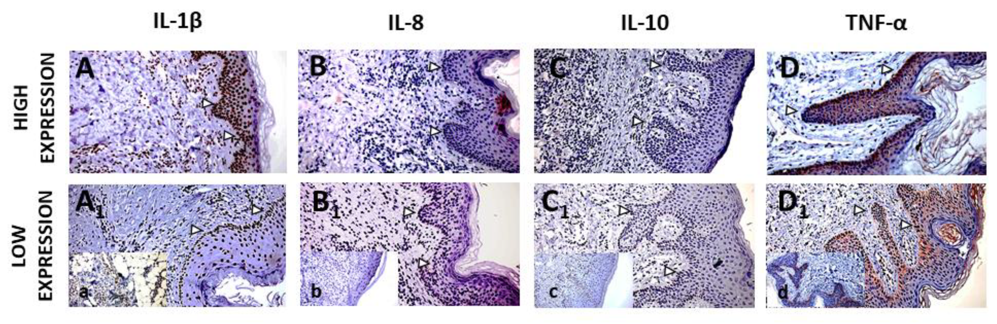
Figure 2.
Expression of IL-1β (A). IL-8 (B). IL-10 (C) and TNF α (D) lesioned and non-lesioned skin (all p<0.001) (Legend: Ex – experimental group, H – control group).
Figure 2.
Expression of IL-1β (A). IL-8 (B). IL-10 (C) and TNF α (D) lesioned and non-lesioned skin (all p<0.001) (Legend: Ex – experimental group, H – control group).
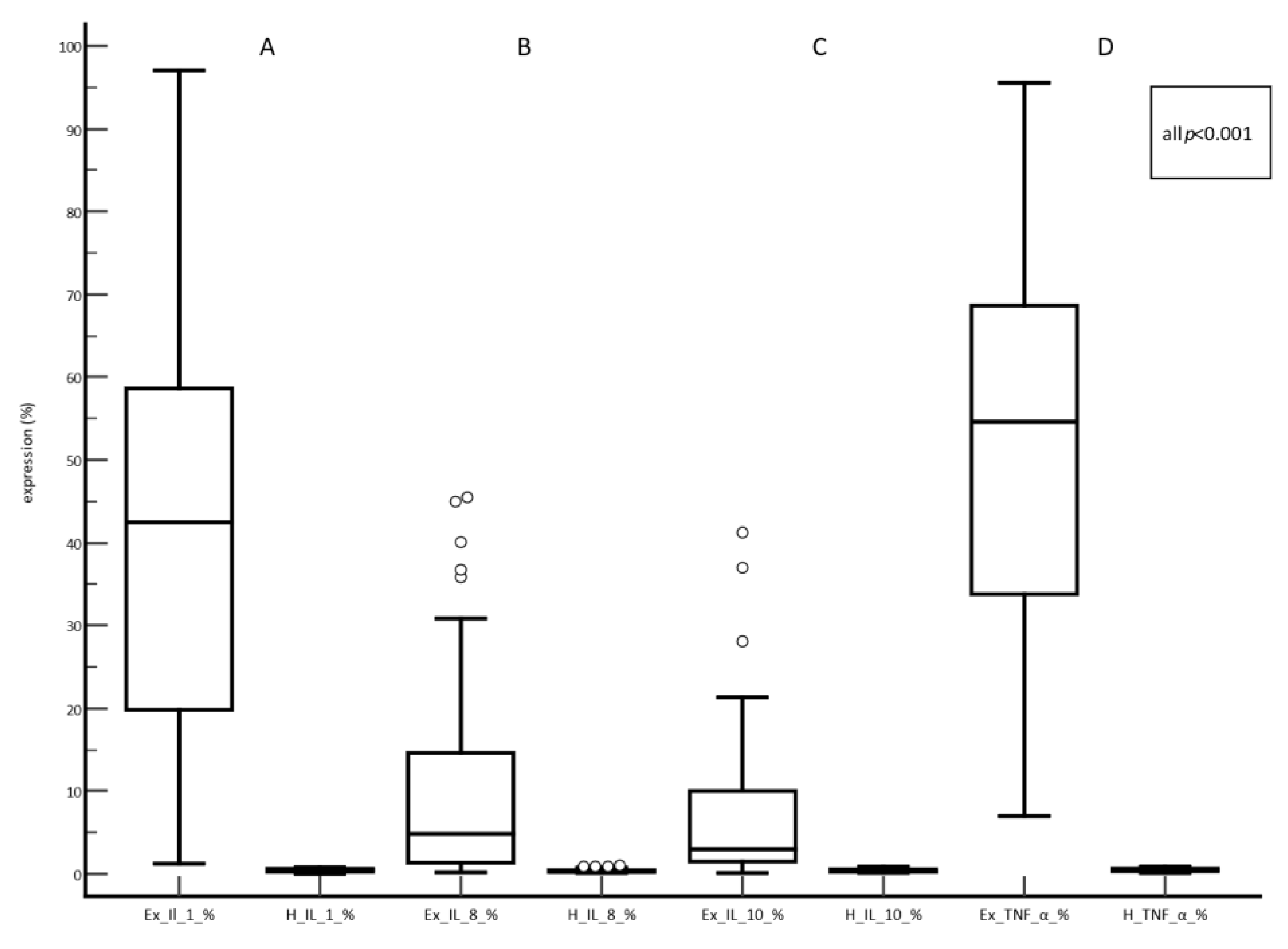
Figure 3.
Expression of IL-1β (A). IL-8 (B). IL-10 (C) and TNF α (D) in HS stage 2 and 3 (Legend: ex -experimental group; HS- Hurley stage).
Figure 3.
Expression of IL-1β (A). IL-8 (B). IL-10 (C) and TNF α (D) in HS stage 2 and 3 (Legend: ex -experimental group; HS- Hurley stage).
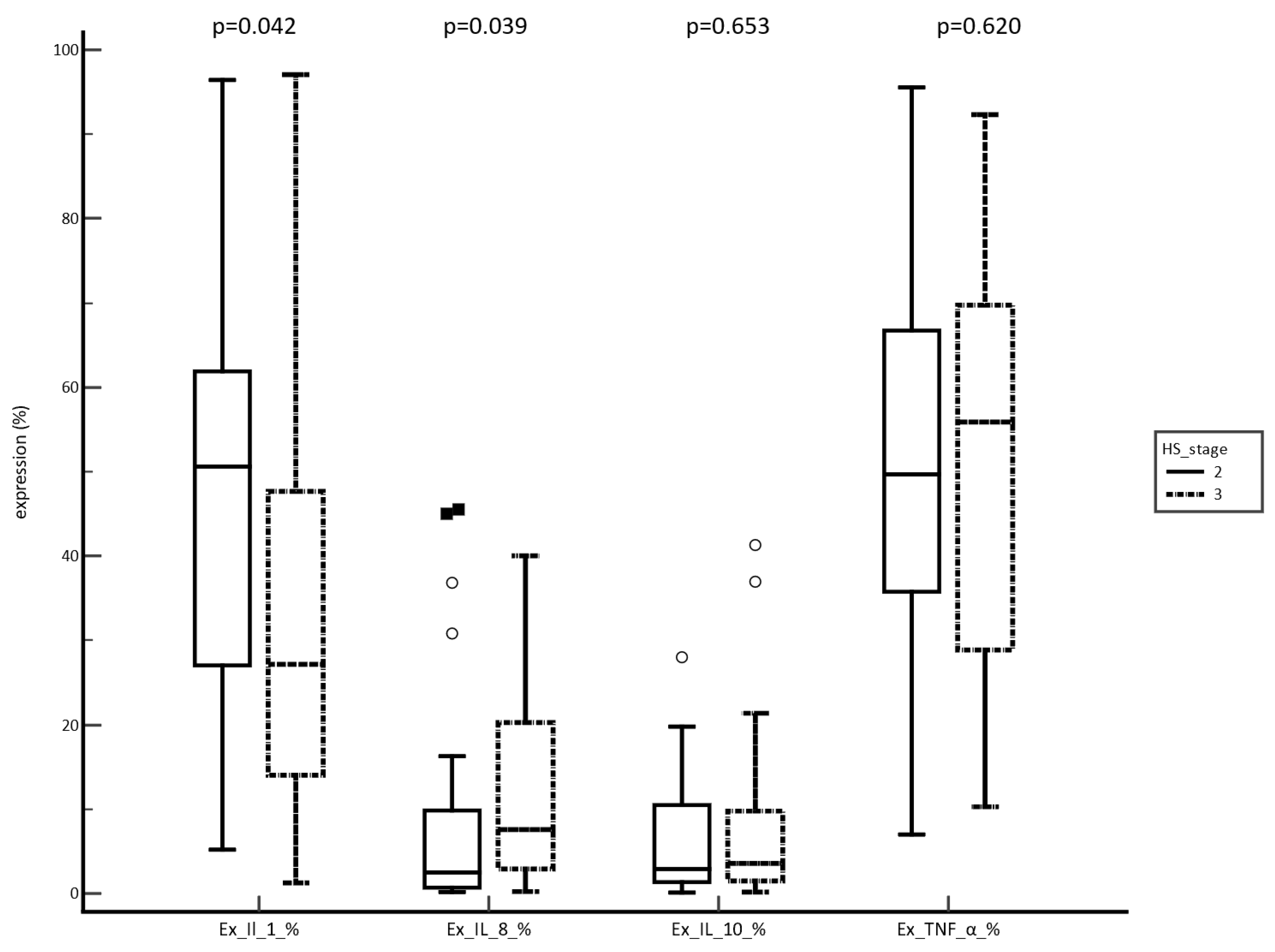

Table 1.
Patient’s characteristics.
| Variable (n=52) | n (%) or C (IQR) |
| Sex male female |
28 (54) 24 (46) |
| Age (years) | 35.5 (26-44) |
| Smoking | 41 (79) |
| BMI | 27.3 (24.3-30.7) |
| Hurley Stages system Stage 2 male female Stage 3 male female |
28 (54) 10 (19) 18 (35) 24 (46) 18 (35) 6 (11) |
| Illness duration | 8.0 (4.0-13.5) |
| Biological therapy male female |
11 (21) 8 (15) 3 (6) |
| Biological therapy Hurley stage 2 Hurley stage 3 |
11 (21) 2 (4) 9 (17) |
| Disease localization Axilla Inguinal region Gluteal Axilla, Inguinal region and gluteal region Other regions |
14 (26.9) 17 (32.7) 3 (5.8) 16 (30.8) 2 (3.8) |
| Positive family history | 15 (28.8) |
| Comorbidities Without comorbidities Other dermatological diseases Hormonal disbalance Depression and anxiety GIT (gastritis, GERB) Rheumatological diseases Obesity Cardiovascular disease Other |
17 (32.7) 9 (17.3) 7 (13.5) 4 (7.7) 4 (7.7) 3 (5.8) 3 (5.8) 2 (3.8) 3 (5.8) |
Legend: C (IQR): median (interquartile range).
Disclaimer/Publisher’s Note: The statements, opinions and data contained in all publications are solely those of the individual author(s) and contributor(s) and not of MDPI and/or the editor(s). MDPI and/or the editor(s) disclaim responsibility for any injury to people or property resulting from any ideas, methods, instructions or products referred to in the content. |
© 2026 by the authors. Licensee MDPI, Basel, Switzerland. This article is an open access article distributed under the terms and conditions of the Creative Commons Attribution (CC BY) license (http://creativecommons.org/licenses/by/4.0/).
Copyright: This open access article is published under a Creative Commons CC BY 4.0 license, which permit the free download, distribution, and reuse, provided that the author and preprint are cited in any reuse.