

Submitted:
29 September 2026
Posted:
30 September 2026
You are already at the latest version
Abstract
Raccoon dog and fox amdoparvovirus (RFAV), classified as Amdoparvovirus carnivoran3, was recently identified in domestic dogs, though its clinical significance remains unclear. This study describes RFAV detection in a 4-year-old female Doberman Pinscher with multisystemic disease, including progressive weight loss, poor coat condition, ocular lesions, hyperkeratosis of the nasal planum and footpads, and marked icterus. Clinical progression and laboratory parameters were monitored during hospitalisation, followed by postmortem pathological assessment. Antemortem and postmortem specimens were tested for amdoparvovirus DNA. PCR products were sequenced and subjected to phylogenetic analysis. Amdoparvovirus DNA was detected in nasal, conjunctival, and rectal swabs, whole blood, urine, and multiple postmortem tissues. Sequencing confirmed the virus as Amdoparvovirus carnivoran3, with the Croatian sequence closely related to a strain recently reported in clinically affected Doberman Pinschers in Serbia. Additionally, 60 archived canine rectal swab samples collected earlier in 2026 were screened for amdoparvoviruses. All retrospectively screened samples tested negative. This study documents the first detection of an amdoparvovirus in Croatia and provides new clinical and histopathological data on RFAV in domestic dogs.
Keywords:
Amdoparvovirus carnivoran3
; raccoon dog and fox amdoparvovirus
; domestic dog
; croatia
; clinical signs
; histopathology
; molecular detection
; phylogenetic analysis
1. Introduction
Amdoparvovirus is one of 11 currently recognised genera within the subfamily Parvovirinae, family Parvoviridae [1], which comprises small, non-enveloped viruses with single-stranded DNA [2]. The amdoparvovirus genome typically encodes two structural proteins, VP1 and VP2, and three nonstructural proteins, NS1, NS2, and NS3 [3]. Amdoparvoviruses are predominantly associated with carnivorans but have also been detected in rodents and chiropterans, suggesting a broader host range than previously recognised [3].
Amdoparvovirus carnivoran1 is the most extensively studied member of its genus and the causative agent of Aleutian disease in mink. Disease manifestation and cellular tropism vary by age, resulting in distinct clinical forms in adults and kits. In adult minks, the virus primarily infects macrophages and establishes a persistent infection that can progress to multisystemic disease. Clinical signs commonly observed include weight loss, anorexia, lethargy, and deterioration of coat condition. Histopathological changes include widespread lymphocytic and plasmacytic infiltration and immune-mediated lesions affecting multiple organs. Significant plasma cell proliferation may lead to lymphadenopathy, splenomegaly, and hypergammaglobulinemia, while immune-complex deposition contributes to necrotising arteritis and proliferative glomerulonephritis. In mink kits, viral replication occurs predominantly in type II pneumocytes and may result in acute interstitial pneumonia with high morbidity and mortality [4, 5].
Raccoon dog and fox amdoparvovirus (RFAV), a member of the species Amdoparvovirus carnivoran3, was first identified during disease outbreaks in farmed raccoon dogs (Nyctereutes procyonoides) and Arctic foxes (Vulpes lagopus) in China in 2012 and 2013. The initial outbreaks predominantly affected juvenile animals and were associated with anorexia, weight loss, growth retardation, chronic diarrhoea, poor coat condition, and high mortality [6]. Subsequent reports of RFAV have remained limited to China, where the virus has been detected in metagenomic studies of farmed raccoon dogs [7. 8]. Most recently, RFAV was reported for the first time in domestic dogs in Serbia after being detected in clinically affected Doberman Pinschers in March and April 2026 [9].
Because RFAV infection has only recently been documented in domestic dogs, its clinical significance and associated pathological findings are still largely unknown. This study aimed to describe Amdoparvovirus carnivoran3 infection in a clinically ill domestic dog and assess its potential association with the observed disease. Specifically, we aimed to document the clinical course, obtain the viral sequence, evaluate the diagnostic utility of different antemortem samples, and describe the histopathological findings observed in the affected dog. We also screened previously collected canine samples to determine whether the virus had been circulating locally before its detection in the present case.
2. Materials and Methods
2.1. Case Description
On 22 July 2026, a four-year-old intact female Doberman Pinscher was admitted to the Veterinary Teaching Hospital at the Faculty of Veterinary Medicine, University of Zagreb. The dog presented with a one-month history of ocular discharge, nasal planum fissuring, dry footpads, progressive weight loss, reduced appetite, and poor coat quality. In addition, the owner had noticed jaundice approximately two weeks before admission. According to the medical records, the dog was up to date with routine vaccinations and received regular antiparasitic prophylaxis. The epidemiological history revealed travel to Serbia, where the dog had stayed with a professional dog trainer and participated in dog shows.
On admission, the dog was ambulatory but visibly weak, with a body condition score of 3/9. The coat was dry and dull (Figure 1A-B). The skin was scaly, with areas of thickening over the muzzle and elbows. The nasal planum and all four footpads were markedly hyperkeratotic (Figure 1D-F). Bilateral purulent ocular discharge, conjunctival hyperaemia, and chemosis were present. The submandibular, prescapular, and popliteal lymph nodes were mildly enlarged, with more pronounced bilateral enlargement of the zygomatic lymph nodes. The skin and visible mucous membranes were markedly icteric (Figure 1C), and mild dehydration was suspected based on delayed skin turgor and dry gingivae. Thoracic auscultation and abdominal palpation were unremarkable, and the dog was normothermic.
The initial diagnostic evaluation included a complete blood count (CBC), serum biochemistry profile, C-reactive protein (CRP), coagulation testing, urinalysis, capillary serum protein electrophoresis, and an ophthalmic examination. Diagnostic imaging comprised thoracic, abdominal, and skull radiography, as well as abdominal ultrasonography. Infectious disease screening included a rapid immunochromatographic antibody test for Leishmania spp. (Leishmania Rapydtest®, Apacor Ltd., Wokingham, United Kingdom) a microscopic agglutination test for Leptospira spp.; antibody testing for Anaplasma spp., Ehrlichia spp., and Borrelia burgdorferi and antigen testing for Dirofilaria immitis (SNAP 4Dx Plus, IDEXX Laboratories, Inc., Westbrook, ME, USA); and a semiquantitative antibody test for canine distemper virus and canine parvovirus type 2 (FASTest® CDV-CPV Ab, MEGACOR Diagnostik GmbH, Hörbranz, Austria).
Conjunctival, nasal, and rectal swabs, urine, and whole blood were also collected for molecular testing. Real-time polymerase chain reaction was performed for canine parvovirus type 2 (CPV-2) and canine distemper virus (CDV), whereas conventional polymerase chain reaction (PCR) was used for canine adenovirus 1 (CAdV-1), canine adenovirus 2 (CAdV-2), canine minute virus (CMV), and canine herpesvirus (CHV). All assays were performed using routine diagnostic protocols employed by the Virology Laboratory, Faculty of Veterinary Medicine, University of Zagreb. In addition, samples were tested for amdoparvoviruses using a semi-nested PCR assay, as described below.
Initial haematological evaluation revealed mild anaemia and thrombocytopenia, while the leucogram remained within reference intervals. C-reactive protein was mildly increased. Serum biochemistry showed marked hepatobiliary abnormalities, including alanine aminotransferase (ALT) activity above the upper limit of measurement and hyperbilirubinaemia, with concurrent increases in total protein and globulin concentrations (Table 1). Serum protein electrophoresis showed a reduced relative albumin fraction, increased α- and β-globulin fractions, and a low albumin-to-globulin ratio (Table 2). Urinalysis revealed concentrated urine with marked bilirubinuria, bilirubin crystalluria, and proteinuria. Skull and thoracic radiographs were unremarkable. Abdominal ultrasonography revealed splenomegaly, increased hepatic echogenicity, gallbladder wall thickening, and mild enlargement of the iliac lymph nodes. Ophthalmic examination revealed bilateral conjunctivitis with anterior uveitis.
All infectious disease screening tests were negative, whereas semiquantitative testing showed moderate antibody levels against both CDV and CPV-2. In contrast, the semi-nested PCR assay for amdoparvoviruses was positive.
Based on the clinical and diagnostic findings, supportive treatment was initiated, including fluid therapy, hepatoprotective therapy and topical ophthalmic therapy. The dog was hospitalised for three days. CBC and serum biochemistry profiles were repeated daily. During hospitalisation, the clinical condition deteriorated, with complete loss of appetite and the development of diarrhoea, while laboratory findings showed no improvement. Humane euthanasia was therefore elected on the third day. A complete necropsy was subsequently performed, and tissue samples were collected for histopathological examination.
2.2. Samples, DNA Extraction and Polymerase Chain Reaction
Swab samples were collected in viral transport medium (VTM) comprising Dulbecco’s modified Eagle medium (DMEM; PAN-Biotech GmbH, Aidenbach, Germany) supplemented with an antibiotic-antimycotic solution (Corning Inc., Corning, NY, USA). Whole blood was collected in EDTA tubes, and urine was collected in sterile containers. Tissue samples collected at necropsy were placed in sterile containers. All samples were transported to the laboratory within two hours of collection and stored at −80 °C until further analysis.
Samples collected in VTM were vortexed briefly, centrifuged, and filtered through a 0.22 µm filter. Tissue samples were suspended in phosphate-buffered saline and mechanically homogenised using a Bead Ruptor 4 homogeniser (Omni International, Kennesaw, GA, USA). Total nucleic acids were then extracted from swab samples, urine, whole blood and tissues using the IndiSpin Pathogen Kit (QIAGEN, Hilden, Germany) following the manufacturer’s instructions.
The semi-nested PCR assay for amdoparvoviruses was performed according to a previously published protocol [6], using TaKaRa Taq™ DNA Polymerase Hot Start Version (Takara Bio Inc., Shiga, Japan). Supplementary Table S1 provides the complete primer sequences, cycling conditions, and expected amplicon sizes.
PCR products were submitted to Macrogen Europe (Amsterdam, the Netherlands) for purification and Sanger sequencing in both directions on an ABI 3730xl DNA Analyzer (Applied Biosystems, Foster City, CA, USA).
2.3. Sequence and Phylogenetic Analysis
Sequences were aligned using MEGA11 [10] and compared with available sequences using the Basic Local Alignment Search Tool (BLAST) at the National Centre for Biotechnology Information (NCBI). For phylogenetic analysis, representative amdoparvovirus sequences were downloaded from GenBank (Supplementary Table S2). A maximum-likelihood phylogenetic tree was constructed in IQ-TREE 2 [11], with the optimal nucleotide substitution model selected automatically. Branch support was assessed using 1,000 ultrafast bootstrap replicates, and the resulting tree was visualised using Interactive Tree Of Life (iTOL) [12].
2.4. Virus Isolation and Cell Culture
Low-passage Crandell–Rees feline kidney (CRFK; ATCC CCL-94) cells were maintained in DMEM (PAN-Biotech GmbH, Aidenbach, Germany) supplemented with 10% fetal bovine serum (FBS) (Capricorn Scientific GmbH, Ebsdorfergrund, Germany), 100 U/mL penicillin, and 100 μg/mL streptomycin (Capricorn Scientific GmbH, Ebsdorfergrund, Germany) at 37 °C in a humidified atmosphere containing 5% CO₂. Two days before inoculation, cells were seeded into 16-well tissue culture plates and infected when monolayers reached approximately 70% confluence.
Before inoculation, samples were thawed and filtered through sterile 0.22-µm syringe filters. A 100 µL volume of whole blood and urine was inoculated onto the corresponding CRFK monolayer and allowed to adsorb for 1 h at 37 °C and 5% CO₂. Following adsorption, inocula were removed from wells, and the monolayers were washed once with Hanks' Balanced Salt Solution (Capricorn Scientific GmbH, Ebsdorfergrund, Germany) to reduce sample-associated cytotoxicity. DMEM supplemented with 2% FBS was then added to all inoculated wells and uninoculated negative control cultures.
Cultures were examined daily for three days for cytopathic effect (CPE). On day 3 post-inoculation, cultures underwent one freeze–thaw cycle, and 100 µL of the resulting lysate was transferred to freshly prepared CRFK monolayers. Four blind passages were performed in total. Following the fourth passage, nucleic acid was extracted from the culture lysates and tested by semi-nested PCR as described above.
2.5. Retrospective Molecular Screening
An additional 60 archival canine rectal swab samples, originally submitted to the Virology Laboratory, Faculty of Veterinary Medicine, University of Zagreb, for routine diagnostic testing earlier in 2026, were retrospectively screened for amdoparvoviruses. We performed nucleic acid extraction and semi-nested PCR as described above.
3. Results
3.1. Molecular Detection of Amdoparvovirus
Amdoparvovirus DNA was detected in all antemortem samples tested, including nasal, conjunctival, and rectal swabs, whole blood, and urine. Viral DNA was also detected in all examined postmortem tissues, including the liver, spleen, lungs, small intestine, and kidney. Amplicons of approximately 800 bp and 300 bp were obtained in the first and second PCR rounds, respectively (Figure 2). Sanger sequencing generated high-quality sequences in both forward and reverse directions. The longer first-round amplicon was used for subsequent analysis. BLAST analysis identified the sequence as Amdoparvovirus carnivoran3, with 100.0% nucleotide identity to sequence PZ348355 obtained from a Doberman Pinscher in Serbia.
3.2. Phylogenetic Analysis
Phylogenetic analysis placed the Croatian sequence within the Amdoparvovirus carnivoran3 cluster. The sequence grouped closely with the Serbian sequence PZ348355, with strong branch support (SH-aLRT 99.4%; UFBoot 100%) (Figure 3).
3.3. Necropsy and Histopathological Findings
Grossly, the mucous membranes of the conjunctiva and mouth, as well as the subcutaneous tissue and the intima of blood vessels, appeared yellow due to jaundice. Hyperkeratosis was visible on the skin of the nose, paw pads, and elbows. The liver and spleen were slightly enlarged with rounded edges. The intestines contained a small amount of reddish liquid content, and the small intestinal mucosa was reddened.
Histopathological examination revealed the most significant changes in the liver, kidneys, small intestine and lung. Lesions in the liver were characterised by moderate to severe multifocal lymphoplasmacytic and necrotising hepatitis. Infiltrates of lymphocytes and plasma cells with a small number of neutrophils were most prominent in the interstitium of portal triads, but randomly distributed infiltrates were also present between hepatocytes, mostly associated with necrotic areas; apoptosis of lymphocytes was occasionally noticed (Figure 4A). In kidneys, moderate interstitial nephritis was present, characterised by infiltrates composed mostly of lymphocytes and plasma cells, mostly between tubules (especially at the junction of the cortex and the renal medulla) and less around glomeruli (Figure 4B). A moderate multifocal lymphoplasmacytic, partially eosinophilic enteritis with apoptosis of lymphocytes was present in the small intestine (Figure 4C). Within the lung, multifocal to diffuse mild to moderate interstitial pneumonia was present, characterised by expansion of alveolar walls with lymphocytes, rare neutrophils and macrophages with intraalveolar fluid exudation. In affected areas of skin on the nose and paw pads, severe orthokeratosis with scant lymphoplasmacytic dermatitis was visible. However, lesions on the elbow skin were accompanied by severe dermatitis with furunculosis and secondary bacterial infection (Figure 4D). The spleen showed moderate to severe infiltration of lymphocytes and plasma cells in the red pulp. Changes in other organs were of lesser intensity and included diffuse neutrophilic and lymphocytic cystitis and mild multifocal lymphoplasmacytic meningitis.
3.4. Virus Isolation in Cell Culture
No cytopathic effect was observed in any of the infected cell cultures during the incubation period across all passages. Following the third and fourth passage, viral DNA was successfully detected by semi-nested PCR in culture lysates derived from both whole blood and urine samples.
3.5. Retrospective Molecular Screening
A total of 60 canine rectal swab samples collected earlier in 2026 were retrospectively screened for amdoparvoviruses using the same semi-nested PCR assay as in the clinical case. All samples were negative for amdoparvovirus DNA, providing no evidence of virus circulation in this sample set before detection of the present case.
4. Discussion
The study reports, the first detection of an amdoparvovirus in Croatia, specifically RFAV (Amdoparvovirus carnivoran3), in a clinical ill domestic dog. This is especially significant given a recent report describing an RFAV outbreak in Doberman Pinschers in Serbia [9]. This case provides detailed clinical, laboratory, and histopathological information linked to RFAV detection in a domestic dog.
At the initial presentation, the disease was multisystemic with pronounced signs of liver disease (LD), which is in line with histopathological findings of an acute to subacute lymphoplasmacytic and necrotising hepatitis with multiorgan involvement. Most commonly, LDs in dogs result from infections, toxicoses, metabolic diseases, or are immune mediated [13]. Anamnestic data, multisystemic involvement, and lymphoplasmacytic infiltration in multiple organs argue against toxic injury and towards infection. Specific infectious causes of hepatitis in dogs are CAdV-1, CMV, CHV, leptospirosis and other bacterial infections [14]. Testing for these viral agents was negative, as was testing for leptospirosis. Doberman Pinschers are predisposed to chronic hepatitis, including copper-associated hepatitis [15], which could account for the marked hepatobiliary abnormalities and hepatic lesions observed in the present dog. However, it would not readily explain the concurrent inflammatory changes affecting multiple extrahepatic organs.
There are no established diagnostic criteria for immune-mediated hepatitis in dogs [16]. In human cases, high γ-globulin concentration suggests an immune-mediated mechanism in LD, which was not the case here. Eosinophilic predominance is described in drug-induced human cases but was not described in this dog [17].
The clinical presentation closely resembled that reported in RFAV-positive Doberman Pinschers in Serbia, including progressive weight loss, poor coat condition, ocular and nasal abnormalities, jaundice, and hyperkeratotic lesions [9]. Similarities are also evident when compared with the original description of RFAV infection in farmed raccoon dogs and Arctic foxes in China, in which affected animals commonly showed anorexia, emaciation, growth retardation, chronic diarrhoea, and poor coat condition [6].
Histopathological examination revealed multisystemic lymphoplasmacytic inflammation affecting multiple organs, including the liver, kidneys, spleen, small intestine, skin, and meninges. A similar predominance of lymphocytic and plasmacytic inflammation has been described in Aleutian disease in mink [18], in skunks infected with skunk amdoparvovirus [19], and in red pandas infected with red panda amdoparvovirus [20].
Taken together, the pathological similarities with other amdoparvovirus infections, the multisystemic distribution of lesions, and the widespread detection of viral DNA support the possibility that RFAV caused the disease observed in this dog.
Notably, all canine RFAV cases reported to date have involved Doberman Pinschers, raising the question of whether breed-related factors may influence susceptibility or clinical expression. Doberman Pinschers have historically been considered among breeds potentially predisposed to a weaker humoral response to CPV-2, although this association remains incompletely established [21]. The dog described in this study had detectable antibodies against CPV-2, indicating that it was capable of mounting a humoral response. Whether a comparable response develops following RFAV infection remains unknown and warrants further investigation.
Phylogenetic analyses indicated that the virus identified in the Croatian dog belonged to Amdoparvovirus carnivoran3 and was closely related to a sequence obtained from a Doberman Pinscher in Serbia. This genetic relationship, combined with the dog’s prior travel to Serbia, suggests a potential epidemiological link; however, it remains unclear whether infection occurred through direct contact with an infected animal or indirectly through contaminated fomites or the environment. Given the well-documented environmental stability of parvoviruses [22], the duration of RFAV infectivity in the environment remains an important knowledge gap. In the present case, amdoparvovirus DNA was detected in whole blood, urine, and conjunctival, nasal, and rectal swabs, all of which can be readily collected during routine clinical examination. The availability of multiple easily accessible samples may facilitate early diagnosis and timely implementation of infection-control measures, which is particularly important given the current lack of specific antiviral therapy or vaccine for RFAV infection.
The retrospective screening included canine rectal swab samples collected throughout 2026, covering the period before, during, and after the RFAV outbreak reported in Serbia. The screened samples represented multiple dog breeds, including breeds other than Doberman Pinschers, and all tested negative for amdoparvovirus DNA. These findings provide no evidence of RFAV circulation, although low-level circulation or isolated cases may have remained undetected in a screening of this size.
Several limitations should be considered. This report describes a single clinical case, and the molecular analysis was based on a partial viral sequence rather than a complete genome. In addition, although amdoparvovirus DNA was detected in all examined tissues, the virus was not directly demonstrated within histological lesions; therefore, a causal relationship between RFAV and the observed pathological changes cannot be confirmed.
5. Conclusions
This study documents the first known detection of RFAV (Amdoparvovirus carnivoran3) in Croatia and provides additional clinical, laboratory, histopathological, and molecular information from a naturally infected dog. The multisystemic clinical and pathological findings, together with the widespread detection of viral DNA, support a possible role of RFAV in the observed disease. Additional studies are needed to clarify its epidemiology, host response, transmission dynamics, and role in disease in domestic dogs.
Supplementary Materials
The following supporting information can be downloaded at the website of this paper posted on Preprints.org, Table S1: Complete primer sequences, cycling conditions, and expected amplicon sizes. Table S2: List of nucleotide sequences retrieved from NCBI GenBank and used in the analyses.
Author Contributions
Conceptualisation, I.C. and V.S.; methodology, I.C. and G.M.; software, I.C.; formal analysis, S.K., A.S., A.G.K., M.B.M., Z.P., I.B., I.Z.; resources, LJ.B., V.S.; data curation, I.C.; writing—original draft preparation, I.C.; writing—review and editing, G.M, V.S. All authors have read and agreed to the published version of the manuscript.
Funding
The sequences have been obtained through the Sequencing Service of the Animal Production and Health Sub-Programme of the Joint FAO/IAEA Centre in Vienna, Austria. This research was partially funded by the European Union NextGenerationEU project, supported by the Ministry of Science and Education of the Republic of Croatia, grant number NPOO-17, Faculty of Veterinary Medicine, University of Zagreb.
Institutional Review Board Statement
The animal study protocol was approved by the Ethics Committee of the Faculty of Veterinary Medicine, University of Zagreb (protocol code 640-01/25-17/121, approved on 27 November 2025).
Informed Consent Statement
Written informed consent was obtained from the owner for the use of the animal’s clinical data, diagnostic samples, and images for research and publication purposes.
Data Availability Statement
The sequence data presented in the study are openly available in NCBI’s GenBank under the accession number: QB046874.
Conflicts of Interest
The authors declare no conflicts of interest.
Abbreviations
The following abbreviations are used in this manuscript:
| RFAV | Raccoon dog and fox amdoparvovirus |
| LD | Liver disease |
References
- Black, E.J.; Powell, C.S.; Dempsey, D.M.; Hendrickson, R.C.; Mims, L.R.; Lefkowitz, E.J. Virus taxonomy: The database of the International Committee on Taxonomy of Viruses. Nucleic Acids Res. 2026, 54, D776–D789. [Google Scholar] [CrossRef] [PubMed]
- Cotmore, S.F.; Agbandje-McKenna, M.; Canuti, M.; Chiorini, J.A.; Eis-Hubinger, A.-M.; Hughes, J.; Mietzsch, M.; Modha, S.; Ogliastro, M.; Pénzes, J.J.; et al. ICTV Virus Taxonomy Profile: Parvoviridae. J. Gen. Virol. 2019, 100, 367–368. [Google Scholar] [CrossRef] [PubMed]
- Canuti, M.; Pénzes, J.J.; Lang, A.S. A new perspective on the evolution and diversity of the genus Amdoparvovirus (family Parvoviridae) through genetic characterization, structural homology modeling, and phylogenetics. Virus Evol. 2022, 8, veac056. [Google Scholar] [CrossRef] [PubMed]
- Bloom, M.E.; Kanno, H.; Mori, S.; Wolfinbarger, J.B. Aleutian mink disease: Puzzles and paradigms. Infect. Agents Dis. 1994, 3, 279–301. [Google Scholar] [PubMed]
- Best, S.M.; Bloom, M.E. Pathogenesis of Aleutian mink disease parvovirus and similarities to B19 infection. J. Vet. Med. B Infect. Dis. Vet. Public Health 2005, 52, 331–334. [Google Scholar] [CrossRef] [PubMed]
- Shao, X.-Q.; Wen, Y.-J.; Ba, H.-X.; Zhang, X.-T.; Yue, Z.-G.; Wang, K.-J.; Li, C.-Y.; Qiu, J.; Yang, F.-H. Novel amdoparvovirus infecting farmed raccoon dogs and Arctic foxes. Emerg. Infect. Dis. 2014, 20, 2085–2088. [Google Scholar] [CrossRef] [PubMed]
- Yang, S.; He, Y.; Chen, X.; Kalim, U.; Wang, Y.; Yang, S.; Qi, H.; Cheng, H.; Lu, X.; Wang, X.; Shen, Q.; Zhang, W. Viral metagenomics reveals diverse viruses in the feces samples of raccoon dogs. Front. Vet. Sci. 2021, 8, 693564. [Google Scholar] [CrossRef] [PubMed]
- Li, W.; Li, X.; Cheng, J.; Liu, J.; Liu, J.; Wang, Y.; Yuan, W.; Ren, E. Lung microbiota of raccoon dogs (Nyctereutes procyonoides) using high-throughput sequencing. Front. Microbiol. 2025, 16, 1677761. [Google Scholar] [CrossRef] [PubMed]
- Gajdov, V.; Pusic, I.; Savic, S.; Lazic, G.; Zekic, M.; Polacek, V.; Petrovic, T. Natural infection of domestic dogs with raccoon dog and fox amdoparvovirus during a severe disease outbreak. Animals 2026, 16, 2618. [Google Scholar] [CrossRef] [PubMed]
- Tamura, K.; Stecher, G.; Kumar, S. MEGA11: Molecular Evolutionary Genetics Analysis version 11. Mol. Biol. Evol. 2021, 38, 3022–3027. [Google Scholar] [CrossRef] [PubMed]
- Minh, B.Q.; Schmidt, H.A.; Chernomor, O.; Schrempf, D.; Woodhams, M.D.; von Haeseler, A.; Lanfear, R. IQ-TREE 2: New models and efficient methods for phylogenetic inference in the genomic era. Mol. Biol. Evol. 2020, 37, 1530–1534. [Google Scholar] [CrossRef] [PubMed]
- Letunic, I.; Bork, P. Interactive Tree of Life (iTOL) v6: Recent updates to the phylogenetic tree display and annotation tool. Nucleic Acids Res. 2024, 52, W78–W82. [Google Scholar] [CrossRef] [PubMed]
- Lester, C.; Cooper, J.; Peters, R.M.; et al. Retrospective evaluation of acute liver failure in dogs (1995–2012): 49 cases. J. Vet. Emerg. Crit. Care 2016, 26, 559–567. [Google Scholar] [CrossRef] [PubMed]
- Rothuizen, J.; Bunch, S.E.; Charles, J.A.; Cullen, J.M.; Desmet, V.J.; Szatmari, V.; Twedt, D.C.; van den Ingh, T.S.G.A.M.; van Winkle, T.J.; Washabau, R.J. WSAVA Standards for Clinical and Histological Diagnosis of Canine and Feline Liver Disease; Saunders Elsevier: Edinburgh, UK, 2006. [Google Scholar]
- Mandigers, P.J.J.; van den Ingh, T.S.G.A.M.; Bode, P.; Teske, E.; Rothuizen, J. Association between liver copper concentration and subclinical hepatitis in Doberman Pinschers. J. Vet. Intern. Med. 2004, 18, 647–650. [Google Scholar] [CrossRef] [PubMed]
- Webster, C.R.L.; Center, S.A.; Cullen, J.M.; Penninck, D.G.; Richter, K.P.; Twedt, D.C.; Watson, P.J. ACVIM consensus statement on the diagnosis and treatment of chronic hepatitis in dogs. J. Vet. Intern. Med. 2019, 33, 1173–1200. [Google Scholar] [CrossRef] [PubMed]
- Anand, A.C.; Nandi, B.; Acharya, S.K.; et al. Indian National Association for the Study of Liver consensus statement on acute liver failure (Part 1): Epidemiology, pathogenesis, presentation and prognosis. J. Clin. Exp. Hepatol. 2020, 10, 339–376. [Google Scholar] [CrossRef] [PubMed]
- Jensen, T.H.; Chriél, M.; Hansen, M.S. Progression of experimental chronic Aleutian mink disease virus infection. Acta Vet. Scand. 2016, 58, 35. [Google Scholar] [CrossRef] [PubMed]
- Alex, C.E.; Watson, K.D.; Schlesinger, M.; Jackson, K.; Mete, A.; Chu, P.; Pesavento, P.A. Amdoparvovirus-associated disease in striped skunks (Mephitis mephitis). Vet. Pathol. 2023, 60, 438–442. [Google Scholar] [CrossRef] [PubMed]
- Yang, L.; Nie, L.; Ren, H.; He, H.; Yuan, H.; Yang, Z.; Yang, K.; Li, W.; Wang, X.; Zhang, X.-X.; et al. Isolation and characterization of a red panda amdoparvovirus causing fatal respiratory disease: Identification of VP2-S447 as a key determinant of acute lung injury. Virulence 2026, 17, 2658906. [Google Scholar] [CrossRef] [PubMed]
- Decaro, N.; Buonavoglia, C.; Barrs, V.R. Canine parvovirus vaccination and immunisation failures: Are we far from disease eradication? Vet. Microbiol. 2020, 247, 108760. [Google Scholar] [CrossRef] [PubMed]
- Gordon, J.C.; Angrick, E.J. Canine parvovirus: Environmental effects on infectivity. Am. J. Vet. Res. 1986, 47, 1464–1467. [Google Scholar] [CrossRef]
Figure 1.
Clinical appearance of the affected Doberman Pinscher at presentation. (A, B) General appearance of the dog, demonstrating poor body condition. (C) Marked icterus of the gingiva. (D) Hyperkeratosis of the footpads. (E) Thickening of the nasal planum, accompanied by scaly skin, poor coat condition, and purulent ocular discharge. (F) Hyperkeratosis of the nasal planum.
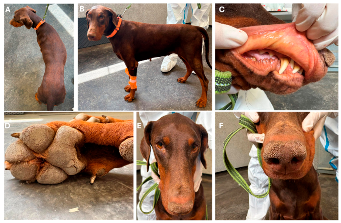
Figure 2.
Agarose gel electrophoresis of semi-nested PCR products. (A) First-round PCR products. (B) Second-round semi-nested PCR products. Sample lanes, from left to right, represent postmortem tissues (liver, spleen, kidney, small intestine, and lung), followed by antemortem samples (whole blood, urine, rectal swab, conjunctival swab, and nasal swab). NC, negative control; PC, positive control; M, 100-bp DNA ladder.
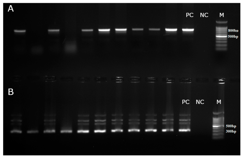
Figure 3.
Maximum-likelihood phylogenetic tree based on partial nucleotide sequences of amdoparvoviruses. The tree was inferred using IQ-TREE2 with the best-fit nucleotide substitution model selected by ModelFinder. Branch support was assessed using 1,000 SH-aLRT replicates and 1,000 ultrafast bootstrap replicates, shown as SH-aLRT/UFBoot (%). The tree was rooted using Amdoparvovirus chiropteran1 (NC_135456) as the rooting taxon. The sequence obtained in this study is shown in bold red, whereas the closely related Serbian sequence PZ348355 is shown in bold black letters. Sequences belonging to Amdoparvovirus carnivoran3 are highlighted in pink. Species assignments are indicated next to the corresponding sequences. GenBank accession numbers, country codes, and sample collection years are shown at the tips. The scale bar represents nucleotide substitutions per site.
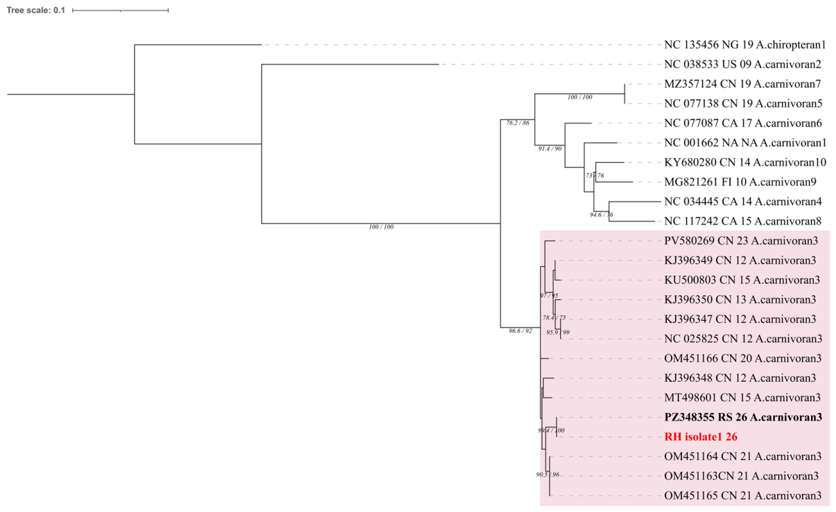
Figure 4.
(A) Liver; multifocal infiltrates of lymphocytes and plasma cells that particularly infiltrate the periportal areas (arrows); area of coagulation necrosis of hepatocytes (arrowhead) (HEx10). Insert: higher magnification of the necrotic area and inflammatory infiltrate (HEx40). (B) Kidney; multifocal to confluent lymphoplasmacytic interstitial nephritis most prominent at the junction of the cortex and the renal medulla. (HEx2). (C) Small intestine; lymphoplasmacytic, partially eosinophilic enteritis characterised by a more abundant infiltrate of inflammatory cells in the superficial parts of the intestinal villi (asterisks) and in the deeper parts of the lamina propria at the transition to the muscularis mucosae (arrows) (HEx10). (D) Skin from the elbow; severe pyogranulomatous, partially ulcerative dermatitis; the infundibular and isthmus portions of the hair follicles are dilated with visible hyperkeratosis and thickened epithelium. (HEx4).
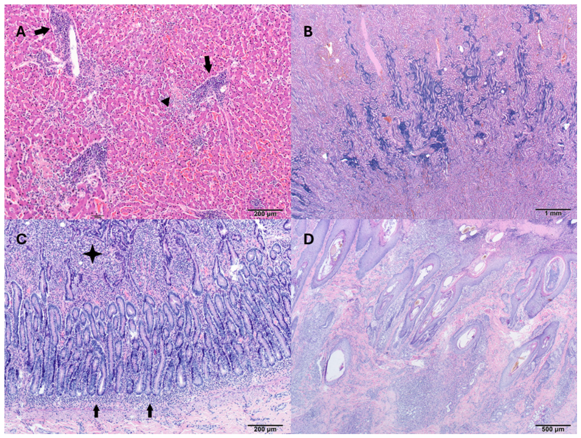

Table 1.
Haematological and serum biochemical findings during the three-day hospitalisation period.
| Parameter | Day 1 | Day 2 | Day 3 | Reference interval |
|---|---|---|---|---|
| Erythrocytes [×10¹²/L] | 5.0 | 5.0 | 4.7 | 5.5 - 8.5 |
| Hematocrit [%] | 33 | 33 | 30 | 37 – 55 |
| Haemoglobin [g/L] | 110 | 111 | 103 | 120 – 180 |
| MCH [pg] | 22 | 22 | 22 | 19 – 23 |
| MPV [fL] | 9 | 9 | 9 | 6.7 – 11 |
| MCV [fL] | 66 | 65 | 65 | 60 – 70 |
| MCHC [g/L] | 336 | 342 | 340 | 320 – 360 |
| Leukocytes [×10⁹/L] | 12.5 | 9.7 | 9.6 | 6 – 17 |
| Segmented neutrophils [×10⁹/L] | 11.25 | 7.86 | 7.01 | 3.00 – 11.50 |
| Band neutrophils [×10⁹/L] | ND | 0.29 | 0.38 | 0 – 0.3 |
| Lymphocytes [×10⁹/L] | 1 | 1.26 | 1.44 | 1.00 – 4.80 |
| Monocytes [×10⁹/L] | 0.25 | 0.1 | 0.58 | 0.15 – 1.35 |
| Eosinophils [×10⁹/L] | ND | 0.19 | 0.19 | 0.1 – 1.25 |
| Platelets [×10⁹/L] | 152 | 149 | 110 | 170 - 500 |
| Reticulocytes [/1000 E] | 5 | / | / | / |
| aPTT [s] | 73 | / | / | 72 - 102 |
| PT [s] | 14 | / | / | 11 - 17 |
| CRP [mg/L] | 13.9 | / | / | 0 – 10.7 |
| Lactate [mmol/L] | 2.2 | / | / | / |
| Glucose [mmol/L] | 5.22 | 5.48 | 4.78 | 4.11 - 7.95 |
| SDMA [µg/dL] | 14 | / | / | 0 - 14 |
| Creatinine [µmol/L] | 73 | 80 | 76 | 44 - 159 |
| Urea [mmol/L] | 6.2 | 6.3 | 3.0 | 2.5 - 9.6 |
| Phosphorus [mmol/L] | 1.56 | 1.30 | 1.51 | 0.81 - 2.20 |
| Calcium [mmol/L] | 2.33 | 2.34 | 2.30 | 1.98 - 3.00 |
| Total Protein [g/L] | 89 | 80 | 82 | 52 - 82 |
| Albumin [g/L] | 28 | 24 | 24 | 23 - 40 |
| Globulin [g/L] | 61 | 56 | 58 | 25 - 45 |
| A:G ratio | 0.5 | 0.4 | 0.4 | / |
| ALP [U/L] | >2000 | >2000 | >2000 | 23 - 212 |
| ALT [U/L] | >1000 | >1000 | >1000 | 10 - 125 |
| AST [U/L] | 396 | 326 | 245 | 0 - 50 |
| GGT [U/L] | 136 | 161 | 175 | 0 - 11 |
| LDH [U/L] | 502 | 426 | 357 | 40 - 400 |
| TBIL [µmol/L] | 162 | 156 | 156 | 0 - 15 |
| Cholesterol [mmol/L] | 10.45 | 9.72 | 9.97 | 2.84 - 8.26 |
| Amylase [U/L] | 713 | 680 | 1249 | 500 - 1,500 |
| Lipase [U/L] | 300 | 300 | 517 | 200 - 1,800 |
| Sodium [mmol/L] | 150 | 150 | / | 144 - 160 |
| Potassium [mmol/L] | 4.8 | 5.2 | / | 3.5 - 5.8 |
| Chloride [mmol/L] | 108 | 107 | / | 109 - 122 |
Table 2.
Serum protein fractions determined by capillary electrophoresis.
| Parameter | Result | Reference interval |
|---|---|---|
| Albumin [%] | 24.1 | 45.5–66.2 |
| α-globulins [%] | 34.2 | 9.9–19.5 |
| α1-globulins [%] | 16.8 | 3.8–8.0 |
| α2-globulins [%] | 17.4 | 5.0–12.8 |
| β-globulins [%] | 29.9 | 12.8–23.7 |
| β1-globulins [%] | 8.2 | 5.1–14.3 |
| β2-globulins [%] | 21.7 | 9.1–20.5 |
| γ-globulins [%] | 11.8 | 5.1–17.8 |
| A/G ratio | 0.32 | / |
Disclaimer/Publisher’s Note: The statements, opinions and data contained in all publications are solely those of the individual author(s) and contributor(s) and not of MDPI and/or the editor(s). MDPI and/or the editor(s) disclaim responsibility for any injury to people or property resulting from any ideas, methods, instructions or products referred to in the content. |
© 2026 by the authors. Licensee MDPI, Basel, Switzerland. This article is an open access article distributed under the terms and conditions of the Creative Commons Attribution (CC BY) license.
Copyright: This open access article is published under a Creative Commons CC BY 4.0 license, which permit the free download, distribution, and reuse, provided that the author and preprint are cited in any reuse.