

Submitted:
29 September 2026
Posted:
30 September 2026
You are already at the latest version
Abstract
Background: Metabolic dysfunction-associated steatotic liver disease (MASLD) and metabolic dysfunction-associated steatohepatitis (MASH) arise from coupled disturbances in lipid handling, insulin sensitivity, mitochondrial function, immune signaling, gut-liver communication, and fibrogenesis. Plant extracts and isolated compounds can act across several of these processes, but variable composition, uncertain active constituents, and limited oral exposure constrain their development. Purpose: This review compares source-plant extracts, defined bioactive compounds, and plant-derived exosome-like nanoparticles (PELNs) as related but distinct forms of plant pharmacology for MASLD/MASH. Study design: A narrative review integrating PELNs, source-plant extracts, isolated compounds, and clinical studies.Methods: A structured two-stage search of PubMed, Web of Science, Scopus, and Embase was conducted to identify evidence relevant to MASLD/MASH. Results: Evidence from the corresponding plants supports effects on lipogenesis, fatty-acid oxidation, oxidative stress, inflammasome signaling, intestinal barrier function, gut microbiota, hepatocyte injury, and hepatic stellate-cell activation. Direct PELN evidence is much narrower: 12 preclinical studies cover 11 plant sources, and most by a single report. Selected PELNs have shown intestinal, metabolic, inflammatory, or antifibrotic activity, but vesicle identity, functional cargo, oral fate, and tissue exposure are often incompletely defined. Engineered vesicle hybrids may improve cargo loading or distribution, although their pharmacology cannot be attributed to native PELNs alone. Clinical studies have examined several source-plant preparations, but no purified PELN formulation has entered clinical evaluation for MASLD/MASH. Conclusion:Progress will require standardized preparation, separation of vesicle-mediated effects from co-isolated constituents, causal cargo validation, quantitative biodistribution, direct comparison with source extracts and free compounds, and repeated-dose safety testing. At present, PELNs remain a preclinical research platform rather than an established therapeutic class.
Keywords:
MASLD
; MASH
; plant-derived exosome-like nanoparticles
; extracellular vesicles
; natural products
; gut-liver axis
1. Introduction
Metabolic dysfunction-associated steatotic liver disease (MASLD) affects an estimated 30%-38% of adults worldwide and is now the most common chronic liver disease(Younossi et al., 2023; Younossi et al., 2025). Its spectrum extends from isolated steatosis to metabolic dysfunction-associated steatohepatitis (MASH), progressive fibrosis, cirrhosis, and hepatocellular carcinoma(Kanwal et al., 2018; Younossi et al., 2023; Younossi et al., 2025). MASLD commonly coexists with obesity, type 2 diabetes, cardiovascular disease, and chronic kidney disease, and may overlap with other liver diseases, including chronic viral hepatitis(John et al., 2026; Liang et al., 2022b; Mantovani et al., 2022; Tao et al., 2024). These comorbidities and overlapping etiologies substantially increase its clinical burden. In 2023, a multisociety consensus replaced the terms non-alcoholic fatty liver disease and non-alcoholic steatohepatitis with MASLD and MASH, respectively(Rinella et al., 2023). The revised nomenclature places metabolic dysfunction at the center of diagnosis rather than defining disease mainly by the absence of alcohol exposure.
The sequential “two-hit” model no longer captures the biology of MASLD. Current evidence supports a multiple-hit process in which insulin resistance, excess fatty-acid delivery, de novo lipogenesis, lipotoxic lipid species, mitochondrial and endoplasmic reticulum stress, immune activation, intestinal barrier dysfunction, microbial dysbiosis, and fibrogenic cell–cell interactions develop in parallel(Buzzetti et al., 2016; Loomba et al., 2021; Targher et al., 2025; Wang et al., 2025; Wang et al., 2023a). Disease progression depends not simply on hepatic triglyceride content, but on lipid composition and handling, hepatocyte adaptation to metabolic stress, and the inflammatory and fibrotic responses of non-parenchymal cells. The approval of resmetirom and semaglutide for selected adults with noncirrhotic MASH and moderate-to-advanced fibrosis has expanded the therapeutic landscape (Harrison et al., 2024; Newsome and Loomba, 2025; Petta et al., 2024; Sanyal et al., 2025). Nevertheless, responses remain heterogeneous, and neither agent addresses every metabolic, intestinal, inflammatory, and fibrogenic component of the disease. This leaves room for complementary, multi-pathway pharmacological strategies.
Medicinal and edible plants provide extracts, defined fractions, and isolated compounds with potential relevance to MASLD. Reported actions include reduced de novo lipogenesis, increased fatty-acid oxidation, improved insulin sensitivity, preservation of mitochondrial function, suppression of inflammatory signaling, and modulation of bile-acid and gut-barrier homeostasis(Cai et al., 2024; Gong et al., 2024; Kaushal et al., 2020; Liu et al., 2024; Tung et al., 2021; Wan et al., 2021; Wang et al., 2023a; Wang et al., 2023b; Yang et al., 2024; Zhang et al., 2023a; Zhang et al., 2023b). Extracts may retain interactions among multiple constituents, but their composition varies with species, cultivation, harvest, extraction, and formulation, complicating dose definition, mechanism assignment, quality control, and clinical reproducibility. Isolated compounds permit more precise target and exposure–response studies, yet many have limited oral stability or bioavailability and may not reproduce the broader effects of the parent preparation.
Plant-derived exosome-like nanoparticles (PELNs) have emerged as another form of plant pharmacology. These nanoscale lipid-bilayer preparations may contain proteins, lipids, small RNAs, and low-molecular-weight metabolites from the source material(Huang et al., 2025; Kang et al., 2016; Kim et al., 2025c; Lu et al., 2025; Sanchez-Terron et al., 2024; Wu et al., 2022; Zhang et al., 2026; Zheng et al., 2025). We use PELNs as an operational term because plant-vesicle biogenesis and classification remain unsettled. Vesicular packaging may protect selected cargo during gastrointestinal transit and facilitate interactions with intestinal epithelial, immune, or hepatic cells. Some preparations have altered intestinal permeability, microbial composition, endotoxin exposure, hepatic lipid metabolism, inflammatory signaling, or extracellular-matrix turnover. These effects are source- and preparation-dependent, however, and evidence for preferential hepatic delivery, cross-kingdom RNA regulation, or cell-selective uptake remains incomplete. Direct MASLD/MASH evidence is still fragmented and entirely preclinical.
These three evidence streams are related but not interchangeable. Extracts support multi-component pharmacology, isolated compounds allow target-focused analysis, and PELNs raise additional questions about vesicle identity, cargo, oral fate, cellular uptake, intercellular communication, and engineering. Activity of a parent plant cannot be assumed to reside in its vesicular fraction, and effects of purified vesicles must be separated from those of co-isolated phytochemicals or other non-vesicular components.
This review focuses on plant sources for which PELNs have been tested in experimental steatotic liver disease and places those studies alongside evidence from the corresponding extracts, defined fractions, and isolated compounds. We first summarize the disease processes most relevant to plant-derived intervention, then examine the preparation, composition, oral behavior, cellular uptake, and pharmacological effects of PELNs. Clinical evidence for source-plant preparations is considered separately, followed by the methodological and translational requirements for PELN development.
2. Literature Search and Evidence Selection
A structured search of PubMed, Web of Science Core Collection, Scopus, and Embase was conducted from database inception to July 2026. Google Scholar was used for citation tracking. The search proceeded in two stages. Terms related to plant-derived vesicles, including “plant exosome,” “plant-derived extracellular vesicle,” “exosome-like nanoparticle,” “exosome-like nanovesicle,” and “nanovesicle”, were first combined with “MASLD,” “MAFLD,” “NAFLD,” “MASH,” and “NASH.” The plant sources identified in this search were then combined with “extract,” “fraction,” “phytochemical,” “bioactive compound,” and “natural product” to retrieve evidence on the corresponding extracts, defined fractions, isolated constituents, and clinical studies.
Eligible primary studies included cellular, animal, and human investigations reporting outcomes relevant to steatosis, glucose or lipid metabolism, oxidative stress, mitochondrial function, inflammation, intestinal barrier integrity, gut microbiota, bile-acid signaling, cell death, autophagy, hepatic stellate-cell activation, or fibrosis. The core PELN dataset was restricted to studies that directly evaluated isolated, enriched, or engineered plant-derived vesicles in MASLD-, MASH-, NAFLD-, or NASH-related models. Reviews were used for background and citation tracing only. Duplicate reports, conference abstracts lacking sufficient data, unrelated studies, and complex multi-herbal formulations were excluded from the mechanistic evidence set. Source-plant extracts and isolated compounds were assessed separately from the corresponding PELNs.
The core PELN dataset comprised 12 primary studies covering 11 plant sources. Most sources were represented by a single report, and the studies differed in isolation method, vesicle characterization, disease model, dose, route, and outcome assessment. The evidence was therefore synthesized narratively rather than quantitatively.
3. Pathological Modules Relevant to Plant-Derived Interventions
MASLD develops through interacting metabolic, cellular, and tissue-level disturbances rather than a single sequential pathogenic event. The major processes relevant to plant-derived intervention include genetic and metabolic susceptibility, lipotoxicity and insulin resistance, mitochondrial and endoplasmic reticulum stress, inflammatory and immune activation, gut–liver axis dysfunction, regulated cell death, impaired autophagy, and fibrogenic tissue remodeling (Figure 1).
Host genetics modifies both susceptibility to hepatic steatosis and the likelihood of disease progression. The PNPLA3 I148M variant remains the most consistently associated genetic risk factor, whereas variants in TM6SF2, MBOAT7, GCKR, HSD17B13, and MTARC1 influence hepatic lipid handling, inflammation, fibrosis, and liver-related outcomes to different degrees(Chen et al., 2025; Pei and Goh, 2025; Romeo and Valenti, 2025; Sveinbjornsson et al., 2022). These variants do not act independently of metabolic stress. Their effects are amplified by obesity, insulin resistance, adipose-tissue lipolysis, dietary substrate excess, and impaired hepatic lipid disposal.
Lipotoxicity, rather than triglyceride accumulation alone, is a central driver of hepatocellular injury. Triglyceride storage may initially buffer excess fatty acids, whereas diacylglycerols, ceramides, free cholesterol, and oxidized phospholipids disrupt membrane integrity, insulin signaling, calcium homeostasis, and organelle function(Dua et al., 2025; Iturbe-Rey et al., 2025; Li et al., 2025c; Nakagawa, 2025; Venkatesan et al., 2023; Yang et al., 2025b; Zhong et al., 2024). Increased de novo lipogenesis through SREBP-1c and carbohydrate-responsive element-binding protein, excessive fatty-acid delivery from adipose tissue, and insufficient mitochondrial or peroxisomal oxidation increase the intracellular burden of toxic lipids. Early mitochondrial adaptation may compensate for substrate overload, but sustained excess eventually impairs respiration, raises reactive oxygen species, and damages mitochondrial DNA. Activation of the PERK, IRE1, and ATF6 branches of the unfolded-protein response further links endoplasmic reticulum stress to insulin resistance, inflammation, and hepatocyte death(Dua et al., 2025; Iturbe-Rey et al., 2025; Koliaki et al., 2015; Li et al., 2025c; Nakagawa, 2025; Steinberg et al., 2025; Venkatesan et al., 2023).
Injured hepatocytes sustain inflammation by releasing damage-associated molecular patterns, oxidized lipids, mitochondrial DNA, and extracellular vesicles. These signals activate Kupffer cells, recruited macrophages, and other immune populations through NLRP3, cGAS–STING, TLR4, NF-κB, and related pathways(Henao-Mejia et al., 2012; Li et al., 2024; Mridha et al., 2017; Yu et al., 2019; Zhu et al., 2025). Immune activation is accompanied by metabolic reprogramming: macrophages and lymphocyte subsets alter glucose and lipid use, which shapes cytokine output and hepatocyte–immune-cell crosstalk. Apoptosis, pyroptosis, necroptosis, and ferroptosis further amplify injury by releasing intracellular contents and lipid-peroxidation products(Peleman et al., 2024; Zhao et al., 2024).
The gut–liver axis supplies an additional source of metabolic and inflammatory signals. Dysbiosis and impaired epithelial barrier function increase portal exposure to lipopolysaccharide and other microbial products, while altered short-chain fatty acids and bile acids affect host metabolism and immunity(Lau et al., 2025; Yu et al., 2024). The LPS–TLR4–NF-κB pathway and FXR–FGF19 signaling connect intestinal dysfunction with hepatic inflammation, insulin resistance, and lipid accumulation. These pathways are especially relevant to oral plant preparations and PELNs, which may act within the intestine without high systemic exposure
Fibrosis arises from sustained communication among injured hepatocytes, inflammatory macrophages, and hepatic stellate cells. Single-cell and spatial studies show that fibrogenesis is organized within discrete tissue niches rather than uniformly across the liver(Kim et al., 2025a; Li et al., 2025a; Li et al., 2025d; Martin et al., 2025; Matchett et al., 2024; Schwabe et al., 2025; Xu et al., 2025). Lipid-associated macrophages, activated stellate-cell subsets, damaged hepatocytes, and matrix-producing cells form localized networks of persistent inflammation, extracellular-matrix deposition, and impaired regeneration. This spatial organization helps explain why reducing steatosis alone may not reverse established MASH fibrosis.
These modules provide the framework for the evidence reviewed below. Extracts and isolated compounds have mainly been studied for effects on lipid metabolism, oxidative stress, inflammation, gut microbiota, autophagy, and stellate-cell activation. PELN studies address many of the same processes but add questions about oral stability, cargo, cellular uptake, intercellular signaling, and intestinal or hepatic exposure.
4. Pharmacology of Source-Plant Extracts and Bioactive Compounds
Extracts, defined fractions, and isolated compounds from the plant sources represented in the PELN literature have been investigated more extensively than their vesicular preparations. Most studies have focused on hepatic lipid handling, mitochondrial stress, inflammatory signaling, gut–liver communication, regulated cell death, autophagy, and fibrogenesis (Figure 2 and Table 1). Detailed study-level information on experimental models, outcomes, proposed mechanisms, and PubMed identifiers is provided in Supplementary Table S1.
Most evidence remains preclinical. Many reports describe pathway-associated proteins or circulating biomarkers, whereas relatively few use gene deletion, pharmacological blockade, metabolic-flux analysis, or cell-specific models to establish causality. The sections below therefore distinguish repeated pathway associations from direct mechanistic validation.
4.1. Regulation of Hepatic Lipid Metabolism and Insulin Sensitivity
Hepatic steatosis reflects an imbalance among fatty-acid uptake, de novo lipogenesis, fatty-acid oxidation, lipid export, and storage. Plant extracts and isolated compounds have been reported to influence each of these processes, most commonly through AMPK, SIRT1, PPARα, SREBP-1c, and bile-acid receptor signaling(Chiang and Ferrell, 2020; Hardie and Pan, 2002; Kersten and Stienstra, 2017; Nagaya et al., 2010; Wan et al., 2011).
AMPK activation is one of the most frequently reported responses. Extracts or defined fractions from honeysuckle, tea, pomegranate, Artemisia capillaris, blueberry, ginger, garlic, and ginseng have been associated with lower expression of SREBP-1c, ACC, fatty-acid synthase, and stearoyl-CoA desaturase 1, together with higher expression of PPARα, carnitine palmitoyltransferase 1A, or acyl-CoA oxidase 1(Chun et al., 2018; Cui et al., 2023; Han et al., 2023; Huang et al., 2017; Kim et al., 2018; Kim et al., 2013; Lee et al., 2016; Li et al., 2020; Liang et al., 2022a; Liu et al., 2018; Liu et al., 2023b; Mohammed, 2022; Park et al., 2019; Raffaele et al., 2020; Ren et al., 2014; Ren et al., 2019; Wu et al., 2025; Xia et al., 2019; Xiao et al., 2013; Yan et al., 2017; Yang et al., 2023b; Yuan et al., 2025). This convergence suggests a common metabolic response, but it does not establish AMPK as the direct target of each preparation.
Ginseng illustrates the greater mechanistic resolution possible with isolated compounds. Compound K promotes fatty-acid oxidation through AMPK/ACC and PPARα-associated signaling, whereas ginsenoside Rb2 restores autophagy under lipotoxic conditions through AMPK and SIRT1(Huang et al., 2017; Kim et al., 2013). Ginsenoside Rk1 reduces CD36 expression and mTORC1–SREBP-1c signaling, affecting both fatty-acid uptake and synthesis(Wu et al., 2025). Ginsenosides Rd and Rc have also been reported to activate SIRT6 and enhance PPARα-dependent oxidation(Cui et al., 2023; Yang et al., 2023b). Most of these studies, however, remain confined to hepatocyte cultures or rodent models.
Ginger-derived constituents show a similar pattern. Zingerone and 6-gingerol activate AMPK-related pathways and reduce SREBP-dependent lipogenesis in high-fat- or fructose-based models(Liu et al., 2023b; Mohammed, 2022). Garlic organosulfur compounds, including S-allyl cysteine and S-allylmercaptocysteine, suppress lipogenesis and restore autophagy through AMPK-related pathways(Hwang et al., 2013; Xiao et al., 2013). Blueberry leaf polyphenols and blueberry preparations combined with probiotics have been linked to SIRT1/PGC-1α/SIRT3 signaling, improved mitochondrial lipid utilization, and reduced hepatic lipid accumulation(Li et al., 2020; Ren et al., 2014; Ren et al., 2017; Ren et al., 2019).
Some interventions act beyond the AMPK–SREBP axis. Broccoli preparations regulate hepatic lipid uptake and export through CD36, microsomal triglyceride transfer protein, and FXR/LXR-associated pathways(Chen et al., 2016; Lu et al., 2025). Yellow tea polysaccharides alter bile-acid composition and the FXR–FGF15–CYP7A1 axis, whereas fermented dark tea has been associated with improved PI3K/AKT signaling and reduced gluconeogenic gene expression(Huang et al., 2024; Yuan et al., 2025). A. capillaris extracts have been linked to both SIRT1-dependent hepatic lipid metabolism and microbiota-associated production of short-chain fatty acids(Liang et al., 2022a; Liang et al., 2025).
Across these studies, the most consistent findings are reduced de novo lipogenesis and fatty-acid uptake, increased fatty-acid oxidation, and changes in bile-acid or insulin signaling. Lower hepatic triglycerides or serum lipids should not be taken as evidence of broad metabolic correction unless accompanied by measurements of insulin sensitivity, lipid flux, mitochondrial function, or histological disease activity.
4.2. Mitochondrial Homeostasis and Oxidative Stress
Mitochondrial dysfunction contributes to MASLD progression by impairing oxidative metabolism and increasing the production of reactive oxygen species. Prolonged lipid overload can also disrupt mitochondrial membrane potential, respiratory-chain activity, ATP production, and mitochondrial quality control. Plant-derived interventions have been studied mainly through the Nrf2 antioxidant pathway, SIRT1/PGC-1α-dependent mitochondrial biogenesis, and autophagy or mitophagy.
Blueberry polyphenols and anthocyanin-rich preparations reduce lipid peroxidation and increase glutathione or antioxidant-enzyme activity in experimental models(Li et al., 2020; Ren et al., 2014; Ren et al., 2019). Blueberry juice combined with probiotics has also been reported to improve mitochondrial morphology and respiratory function through SIRT1/PGC-1α-associated signaling(Ren et al., 2019). These findings suggest an effect beyond nonspecific radical scavenging, although the contribution of the probiotic component cannot always be separated from that of blueberry constituents.
Pomegranate preparations provide another well-studied example. Pomegranate seed oil and punicalagin have been associated with enhanced antioxidant responses, improved mitochondrial function, and reduced hepatic injury in diet-induced steatotic liver disease (Raffaele et al., 2020; Zou et al., 2014). Punicalagin has additionally been linked to p62/Nrf2 signaling, enhanced autophagy, and suppression of the TXNIP–NLRP3 axis(Ma et al., 2025). Pomegranate seed oil improves antioxidant capacity and mitochondrial markers, although its lipid composition and metabolic effects differ substantially from those of polyphenol-rich pomegranate extracts.
Ginseng preparations act through several mitochondrial pathways. Wild ginseng cambial meristematic cells increase PGC-1α, NRF1, and TFAM expression, consistent with enhanced mitochondrial biogenesis(Lee et al., 2016). Ginsenoside Rg1 maintains FOXO1-associated antioxidant responses and reduces lipid peroxidation in an aging-related fatty-liver model(Qi et al., 2020). Fermented ginseng preparations have also been linked to restoration of mitophagy and suppression of excessive mTORC1 activity(Choi et al., 2019).
Mitochondrial homeostasis is therefore a plausible target of several plant-derived interventions, but the depth of evidence varies. Many studies rely on reactive oxygen species, malondialdehyde, antioxidant enzymes, or expression of mitochondrial regulators; far fewer measure oxygen consumption, respiratory-chain function, mitochondrial turnover, or substrate oxidation. Antioxidant biomarkers alone should not be described as restoration of mitochondrial function.
4.3. Inflammatory Signaling and Immune-Metabolic Regulation
Inflammatory pathways help drive the transition from steatosis to MASH and fibrosis. Plant-derived preparations have been studied for effects on pattern-recognition receptors, NF-κB signaling, inflammasome activation, cytokine production, and macrophage function.
Black ginseng reduces TLR4 expression, NF-κB p65 phosphorylation, and inflammatory or fibrosis-associated genes in a Western-diet and carbon tetrachloride model(Wei et al., 2022). Dried tangerine peel polysaccharide binds MD-2 and attenuates TLR4-associated inflammatory and endoplasmic reticulum stress responses(Wang et al., 2024). Ginger essential oil suppresses the microbiota–LPS–TLR4 pathway and downstream NLRP3 inflammasome activation in experimental NASH(Panyod et al., 2024). These studies indicate that plant preparations may intervene at several points upstream of cytokine production rather than simply lowering circulating TNF-α or IL-6.
Several ginseng-derived compounds have been investigated more directly. The ginsenoside metabolite 20(S)-protopanaxatriol inhibits NLRP3 inflammasome activation and reduces inflammation and fibrosis in a methionine- and choline-deficient model(Lu et al., 2023). Panaxydol has been reported to interact with the ATP-binding region of NLRP3, suppressing caspase-1 activation, cytokine maturation, and pyroptosis(Kim et al., 2024b). Ginsenoside Re affects both PI3K/AKT and TLR4/NF-κB signaling, whereas fermented red ginseng preparations have been associated with altered macrophage inflammatory programs through PPARα and mTORC1(Choi et al., 2019; Zhang et al., 2024).
Blueberry preparations combined with probiotics increased interleukin-22 and activated Janus kinase 1/signal transducer and activator of transcription 3 signaling in experimental fatty liver disease(Zhu et al., 2018). Pomegranate, green tea, honeysuckle, ginger, and cannabis-derived preparations have also reduced inflammatory markers in animal models(Carmona-Hidalgo et al., 2021; Gong et al., 2023; Han et al., 2023; Kim et al., 2018; Liu et al., 2018; Ma et al., 2025; Noori et al., 2017; Panyod et al., 2024; Park et al., 2019; Tzeng et al., 2015; Xia et al., 2019; Zou et al., 2014). In most cases, however, immune effects were inferred from liver cytokines, histology, or bulk-tissue protein expression. Immune-cell depletion, conditional gene deletion, single-cell profiling, and adoptive-transfer experiments remain uncommon.
Several plant-derived interventions therefore show anti-inflammatory activity, but evidence for cell-specific immune reprogramming is much weaker. Terms such as “macrophage polarization” or “immune-metabolic remodeling” should be reserved for studies with functional or cell-resolved data.
4.4. Modulation of the Gut–Liver Axis
The gut–liver axis is especially relevant to orally administered plant preparations. Changes in microbial composition, epithelial permeability, microbial metabolites, and bile-acid signaling can alter the inflammatory and metabolic signals reaching the liver.
Ginseng extracts and polysaccharides from Panax japonicus have been reported to reduce intestinal permeability, modify microbial composition, and alter short-chain fatty-acid or lipid-associated metabolic pathways in high-fat-diet models(Wu et al., 2024). Pomegranate peel extract, pomegranate-derived supplements, punicalagin, and punicic acid influence microbial communities and metabolites alongside improvements in hepatic inflammation and lipid metabolism(Chen et al., 2024; Liu et al., 2021; Sanchez-Terron et al., 2024). Broccoli preparations, yellow tea polysaccharides, and fermented dark tea have likewise been associated with increased abundance of selected beneficial taxa, altered bile-acid metabolism, and improved insulin sensitivity(Huang et al., 2024; Lu et al., 2025; Yuan et al., 2025; Zandani et al., 2021).
A. capillaris water extract altered microbial composition and increased short-chain-fatty-acid production while activating hepatic PPARα and suppressing NF-κB signaling(Liang et al., 2022a; Liang et al., 2025). Two cannabis extracts also changed microbial diversity in a high-fat-diet model, but produced modest metabolic effects and did not establish a clear host target(Zandani et al., 2022). A shift in microbial composition alone does not demonstrate a therapeutically meaningful effect on liver disease.
Combination studies require separate interpretation. Honeysuckle combined with metformin improved steatosis, insulin resistance, endotoxin exposure, and microbial composition in OLETF rats(Shin et al., 2017), but the contribution of honeysuckle cannot be separated from that of metformin.
Most microbiome studies remain associative. Changes in individual bacterial genera are often interpreted as beneficial or harmful without direct testing of their functional contribution. Antibiotic depletion, fecal microbiota transplantation, gnotobiotic models, microbial-metabolite tracing, and barrier-specific experiments are still uncommon. The strongest conclusions are therefore limited to coordinated changes in microbiota, barrier markers, metabolites, and hepatic outcomes rather than proof of a microbiota-dependent mechanism.
4.5. Autophagy, Regulated Cell Death, and Fibrogenesis
Progression from steatosis to MASH involves impaired autophagy, hepatocyte death, inflammatory amplification, hepatic stellate-cell activation, and extracellular-matrix accumulation. Several plant-derived compounds influence these processes, although their effects often overlap with metabolic and antioxidant pathways.
S-allylmercaptocysteine reduced hepatocyte apoptosis and enhanced autophagy through LKB1–AMPK and PI3K–AKT–mTOR signaling(Xiao et al., 2013). Ginsenoside Rg1 increased BCL-2, reduced BAX, and preserved viability in lipid-challenged hepatocytes(Qi et al., 2020). The blueberry-derived isoflavone tectorigenin was linked to tRF-47-dependent control of autophagy and pyroptosis in experimental NASH(Zhu et al., 2022). Fermented red ginseng restored mitophagy while reducing mTORC1 activity and inflammatory signaling(Choi et al., 2019).
Inflammasome-dependent pyroptosis provides a mechanistic link between hepatocyte injury and fibrogenesis. Ginseng-derived protopanaxatriol and panaxydol suppress NLRP3–caspase-1 signaling and reduce inflammatory liver injury(Kim et al., 2024b; Lu et al., 2023). These compound-level studies are more informative than reports based solely on changes in IL-1β or caspase-1 expression because they include evidence of direct or proximal pathway regulation.
Evidence for anti-fibrotic activity comes mainly from prevention models. Black ginseng reduces collagen deposition and fibrosis-associated gene expression in a Western-diet plus carbon tetrachloride model(Wei et al., 2022). Honeysuckle ethanol extract attenuates steatohepatitis and fibrotic changes in a methionine- and choline-deficient model(Tzeng et al., 2015). Cannabis-derived compounds have also been associated with reduced inflammatory and fibrotic responses(Carmona-Hidalgo et al., 2021). However, these models do not necessarily demonstrate regression of established fibrosis. Few studies begin treatment after advanced fibrosis is present, and even fewer assess matrix turnover, stellate-cell heterogeneity, portal pressure, or durable fibrosis reversal.
Source-plant extracts and isolated compounds act across metabolic, mitochondrial, inflammatory, intestinal, and fibrogenic pathways relevant to MASLD/MASH. Extracts offer broader but less standardized pharmacology; isolated compounds permit clearer attribution but may have limited oral exposure. These data provide context for PELNs, but they do not show that vesicular fractions contain the same active constituents or reproduce the effects of the parent preparation.
5. PELNs in MASLD/MASH: Current Preclinical Evidence
The direct PELN evidence base comprises 12 preclinical studies covering 11 plant sources, including native preparations and engineered vesicle-based systems. Reported outcomes include reduced steatosis, oxidative stress, inflammation, intestinal barrier dysfunction, endotoxemia, or fibrosis. Most sources are represented by a single study, and differences in preparation, disease model, dose, and outcome assessment limit cross-study comparison.
PELNs are commonly described as natural carriers of plant lipids, proteins, RNAs, and small molecules. Their activity may instead reflect several components: the vesicle membrane, encapsulated cargo, membrane-associated molecules, or non-vesicular compounds carried through purification. Because these components are rarely separated experimentally, PELNs should be treated as complex biological preparations rather than assumed to be purified analogues of mammalian exosomes.
5.1. Terminology, Isolation, and Physicochemical Characterization
Terminology, isolation procedures, and physicochemical characterization remain inconsistent across the PELN literature. Published studies use differential ultracentrifugation, polyethylene glycol precipitation, tangential-flow filtration, size-exclusion chromatography, or combinations of these methods, each of which differs in recovery, purity, scalability, and risk of co-isolating soluble plant components (Figure 3 and Table 2).
Differential centrifugation followed by ultracentrifugation is the most common isolation route in MASLD-related studies and has been used for pomegranate, hemp sprout, tangerine peel, broccoli, and A. capillaris PELNs(Kim et al., 2024a; Kim et al., 2025c; Xu et al., 2024; Zhang et al., 2026; Zou et al., 2024). Honeysuckle-derived PELNs were isolated from dried honeysuckle as a vesicle-enriched preparation and had a reported mean diameter of approximately 152.7 nm(Li et al., 2025b). Other reports used polyethylene glycol precipitation, filtration, tangential-flow filtration, size-exclusion chromatography, or combined methods(Guo et al., 2025; Kim et al., 2025b; Lei et al., 2025; Liu et al., 2023a; Ma et al., 2026; Zhao et al., 2022). Ultracentrifugation may promote aggregation and co-isolation; polymer precipitation increases recovery at the expense of purity; tangential-flow filtration is more scalable but sensitive to membrane and process conditions; and size-exclusion chromatography improves separation from soluble proteins but yields diluted fractions.
Reported particle diameters generally fall within the nanoscale range, although size distributions vary across plant sources and preparation methods. Engineered hybrid systems may be larger than native vesicular preparations. Particle size alone is insufficient to establish vesicle identity or purity. At minimum, PELN characterization should combine particle-size analysis, particle concentration, morphology, membrane-lipid assessment, total protein and RNA measurements, and evaluation of non-vesicular contaminants. Few studies report all of these parameters, and plant-specific reference markers remain unavailable.
This methodological heterogeneity has direct pharmacological consequences. Changes in isolation conditions can alter vesicle yield, membrane composition, cargo recovery, colloidal stability, and cellular uptake. Comparisons between studies should therefore account for preparation method rather than treating all PELNs from the same plant as equivalent products.
5.2. Endogenous Cargo and Evidence for Cargo-Dependent Activity
PELN preparations contain varying combinations of membrane lipids, proteins, small RNAs, phytochemicals, and other metabolites (Figure 4). The reported functional effects and strength of mechanistic evidence for individual PELN preparations are summarized in Table 3.
Garlic-derived PELNs provide one of the clearest examples of cargo-specific activity. Vesicular miR-396e reduced macrophage glycolysis by targeting PFKFB3, lowered inflammatory mediator production, and indirectly improved hepatocyte lipid metabolism through macrophage–hepatocyte communication(Liu et al., 2023a). This study moved beyond cargo profiling by testing a defined vesicular RNA.
Small RNAs in A. capillaris-derived PELNs have also been implicated in hepatic lipid metabolism and inflammation, but the contribution of individual RNAs to the overall preparation remains uncertain. Tea-derived PELNs altered host miRNAs and metabolic regulators in oleic-acid-treated HepG2 cells(Lei et al., 2025). Because the latter evidence is largely in vitro, its relevance to oral delivery and whole-body pharmacology remains unclear.
Ginseng-derived vesicles contain ginsenosides, lipids, proteins, and RNAs(Kim et al., 2025b). Their antifibrotic effect has been linked to reduced TIMP2 expression in hepatic stellate cells, but it is not yet clear whether this response is mediated by a specific encapsulated ginsenoside, an RNA cargo, membrane lipids, or the combined preparation. Broccoli-derived vesicles contain multiple small RNAs associated with metabolic and immune pathways, but most of these associations arise from sequencing and pathway prediction rather than direct cargo-level validation(Zhang et al., 2026).
Selected PELNs clearly contain biologically active material, but cargo inventories are not mechanisms. Intact vesicles should be compared with disrupted vesicles, cargo-depleted preparations, free-cargo controls, and reconstituted vesicles to distinguish vesicle-dependent delivery from the effects of co-isolated or soluble plant constituents.
5.3. Oral Fate, Intestinal Effects, and Gut–Liver Communication
Oral administration is frequently considered a potential advantage of PELNs. Available studies suggest that selected preparations may interact with the intestinal epithelium, microbiota, and mucosal immune system, thereby influencing barrier integrity, microbial metabolites, and endotoxin exposure (Figure 5A and B).
Several PELNs appear to act mainly through the intestinal compartment. Broccoli-derived PELNs reduced steatosis and inflammation while improving barrier markers, microbial composition, and bile-acid-related pathways(Zhang et al., 2026). Pomegranate-derived PELNs reduced gut leakage and liver injury(Kim et al., 2025c), and hemp sprout-derived PELNs attenuated fibrosis together with lower endotoxemia and oxidative stress(Kim et al., 2024a). Honeysuckle-derived PELNs improved barrier function and reduced steatosis, inflammation, and collagen deposition in high-fat-diet-fed mice; these effects disappeared after antibiotic depletion of the microbiota and were transferred by fecal microbiota transplantation, supporting a microbiota-dependent mechanism(Li et al., 2025b). Tangerine peel-derived PELNs altered gut microbiota and hepatic lipid metabolism in diabetes-associated steatosis(Zou et al., 2024).
These findings are consistent with a gut–liver axis-associated effect, but they do not demonstrate preferential hepatic targeting. Improvement in liver outcomes after oral administration may result from local intestinal activity, reduced endotoxin translocation, altered microbial metabolism, systemic absorption of released cargos, or direct uptake of intact vesicles. These routes have not been distinguished in most studies.
Claims of cross-kingdom RNA regulation also require caution. Plant-derived RNAs can be detected in vesicular preparations, and selected studies support functional effects in mammalian cells. Nevertheless, RNA abundance, sequence stability, cellular exposure, and target specificity are not consistently reported. Cross-kingdom regulation should therefore be discussed as a source-dependent mechanism supported in selected experimental systems rather than a general property of all PELNs.
A more informative assessment of oral fate will require labeled-vesicle tracking, measurement of intact particle recovery, distinction between membrane and cargo distribution, and quantitative analysis of intestinal, portal, hepatic, and systemic exposure. Without these data, oral efficacy cannot be equated with oral bioavailability of intact PELNs.
5.4. Cellular Uptake and Intercellular Effects
Uptake of selected PELNs by hepatocytes, macrophages, and hepatic stellate cells has been reported in specific experimental systems (Figure 5C). These findings indicate cell-type-associated effects but do not establish universal cell selectivity or preferential hepatic targeting.
A. capillaris-derived PELNs entered AML12 hepatocytes through macropinocytosis and caveolin-associated endocytosis and reduced lipid accumulation and inflammatory signaling(Xu et al., 2024). Garlic-derived PELNs acted mainly through macrophages: suppression of macrophage PFKFB3 and glycolytic activity lowered inflammatory output and indirectly improved hepatocyte metabolism(Liu et al., 2023a). These studies illustrate both direct hepatocyte uptake and intercellular mechanisms.
Ginseng-derived vesicles were taken up by hepatic stellate cells and reduced TIMP2 expression, favoring extracellular-matrix degradation(Kim et al., 2025b). This observation is relevant to fibrogenesis, but the study does not establish whether the vesicles selectively target stellate cells in vivo. Similarly, intestinal epithelial uptake has been reported or inferred for several orally administered preparations, but cell specificity has rarely been quantified against other intestinal and hepatic cell populations.
Current data show uptake by hepatocytes, macrophages, or hepatic stellate cells in specific models, but not universal cell selectivity. Whether these patterns reflect active targeting, tissue exposure, membrane composition, or experimental design remains unresolved.
Future studies should use quantitative cell-resolved biodistribution, receptor-blocking experiments, and competitive uptake models. These approaches would help determine whether uptake is mediated by specific membrane ligands or represents nonspecific internalization of nanoscale lipid particles.
5.5. Native and Engineered PELN Systems
Most studies have examined native or minimally processed preparations, which retain endogenous plant cargo but offer limited control over composition, dose, and distribution. Engineering approaches seek greater control through drug loading, membrane fusion, ligand modification, or hybridization with synthetic nanocarriers.
A ginger-derived vesicle–liposome hybrid loaded with berberine and modified with ursodeoxycholic acid illustrates the engineering potential of PELN-based systems (Figure 5D) (Ma et al., 2026). Because its activity reflects the combined contributions of plant-derived vesicles, synthetic lipids, berberine, and a targeting ligand, it should be distinguished from native ginger PELNs.
The ginger hybrid demonstrates the engineering potential of plant vesicles, but it does not show that native ginger PELNs are intrinsically liver-targeted. Native preparations and engineered hybrids require separate evaluation because their manufacturing controls, pharmacokinetics, safety, and regulatory pathways differ.
Processing may also modify native vesicle composition. Fermentation of tea-derived vesicles altered particle characteristics and metabolic profiles and was associated with enhanced lipid-lowering and anti-inflammatory activity in an experimental NAFLD model(Guo et al., 2025). This finding suggests that plant processing can influence PELN composition, but it also raises questions about batch reproducibility and whether the final product should still be considered equivalent to vesicles from untreated plant material.
Engineering may broaden the use of plant vesicles, but once synthetic lipids, drugs, or targeting ligands are added, the product is a composite nanomedicine rather than a native PELN.
5.6. Strength and Limitations of the Current Evidence
The studies provide proof of principle that selected PELNs can affect metabolic, inflammatory, intestinal, or fibrotic processes relevant to MASLD/MASH. Mechanistic confidence is highest when a defined cargo or causal intermediary is tested, as in the garlic miR-396e–PFKFB3 pathway and the honeysuckle study using microbiota depletion and fecal transfer(Li et al., 2025b; Liu et al., 2023a). Most other mechanisms rest on pathway-associated proteins, microbiota profiles, or predicted RNA targets.
Several limitations recur across the literature. First, most plant sources are represented by a single study, with little independent replication. Second, isolation and characterization procedures vary widely, making it difficult to determine whether studies examined comparable vesicle populations. Third, few reports distinguish intact vesicle activity from the effects of co-isolated polyphenols, lipids, proteins, or soluble RNAs. Fourth, dose is reported using inconsistent units, including particle number, protein content, vesicle mass, or source-plant mass. Fifth, quantitative pharmacokinetic and biodistribution data are scarce. Finally, most studies use short-term steatosis or early inflammatory models rather than models with established MASH fibrosis.
Direct comparisons with source-plant extracts and isolated compounds are also uncommon. Such comparisons are needed to determine whether vesicular delivery improves potency, stability, intestinal retention, cellular uptake, or therapeutic index. Without head-to-head experiments, the proposed advantages of PELNs remain largely inferential.
The present evidence supports PELNs as an emerging preclinical extension of plant-derived pharmacology but does not yet establish them as a reproducible therapeutic class. Progress will require standardized preparation, rigorous cargo validation, quantitative exposure studies, independent replication, and testing in models that reflect established MASH and fibrosis.
6. Clinical Evidence for Source-Plant Interventions and the Translational Gap of PELNs
Clinical studies have evaluated Korean red ginseng, ginger, garlic, pomegranate, green tea, and selected herbal formulations in patients classified under earlier NAFLD/NASH criteria(Baghdadi et al., 2025; Ekhlasi et al., 2015; Goodarzi et al., 2021; Hong et al., 2016; Jafarirad et al., 2023; Rafie et al., 2020; Rahimlou et al., 2016; Sakata et al., 2013; Soleimani et al., 2016; Soleimani et al., 2020; Soleimani et al., 2021; Xu et al., 2026). Some trials reported improvements in liver enzymes, insulin resistance, lipids, body composition, inflammation, or imaging-based steatosis, but formulations, sample sizes, treatment duration, and outcome measures varied substantially (Table 4).
Randomized studies of ginger and garlic suggested modest metabolic or hepatic benefit, although most were small, short, and based on ultrasonography or biochemical markers rather than quantitative imaging or histology(Rafie et al., 2020; Rahimlou et al., 2016; Soleimani et al., 2016; Soleimani et al., 2020; Soleimani et al., 2021). Pomegranate juice, extract, and peel preparations affected metabolic indices, liver enzymes, inflammatory markers, and steatosis; one trial also measured liver stiffness and the hepatorenal ultrasound index(Baghdadi et al., 2025; Ekhlasi et al., 2015; Goodarzi et al., 2021; Jafarirad et al., 2023). Catechin-rich green tea improved biochemical measures and the liver-to-spleen computed-tomography attenuation ratio, while a trial of Yinchenhao decoction used magnetic resonance imaging proton-density fat fraction as its primary endpoint(Sakata et al., 2013; Xu et al., 2026). The decoction is a multi-herbal formulation, so its effect cannot be assigned specifically to A. capillaris. Observational associations involving cannabis remain vulnerable to confounding and do not support therapeutic use(Adejumo et al., 2017; Vazquez-Bourgon et al., 2019).
Most trials measured aminotransferases, triglycerides, cholesterol, body weight, insulin resistance, inflammatory markers, or semiquantitative ultrasonographic steatosis. These outcomes may reflect metabolic improvement but do not establish resolution of steatohepatitis or reversal of fibrosis. Quantitative liver-fat imaging, elastography, validated fibrosis biomarkers, and histology were uncommon, and no source-plant trial demonstrated accepted histological endpoints for MASH resolution or fibrosis improvement. Safety and herb–drug interactions were also reported inconsistently.
Clinical activity of a juice, powder, extract, or herbal formula does not validate the corresponding PELN. No purified or enriched PELN formulation has entered clinical testing for MASLD/MASH, and vesicular fractions differ from parent preparations in composition, dose, biodistribution, and pharmacokinetics.
Before human testing, candidate PELNs need reproducible manufacturing, defined quality attributes, standardized particle and cargo measurements, stability data, biodistribution, and repeated-dose safety assessment. Early studies should establish tolerability, exposure, and pharmacodynamic activity before testing efficacy with quantitative liver-fat and fibrosis endpoints.
7. Translational Challenges and Future Directions
PELNs remain at an early preclinical stage. Most plant sources are represented by one study, and differences in isolation, characterization, disease model, dose, and outcome assessment limit reproducibility(Guo et al., 2025; Kim et al., 2025b; Kim et al., 2024a; Kim et al., 2025c; Lei et al., 2025; Liu et al., 2023a; Ma et al., 2026; Xu et al., 2024; Zhang et al., 2026; Zhao et al., 2022; Zou et al., 2024). The immediate task is to define comparable products and reproduce key findings, not simply to add more plant sources.
Standardized preparation and quality control are essential. Studies should report source material, tissue, harvest and processing conditions, particle concentration and size distribution, morphology, membrane composition, cargo content, stability, contaminants, and batch consistency. Isolation methods differ substantially in recovery and purity(Guo et al., 2025; Kim et al., 2025b; Kim et al., 2024a; Lei et al., 2025; Liu et al., 2023a; Ma et al., 2026; Nemati et al., 2022; Ning et al., 2025; Xu et al., 2024; Yang et al., 2025a; Zhao et al., 2022), so method-specific effects must be considered when comparing studies.
Mechanistic work must separate vesicle-mediated effects from co-isolated phytochemicals, proteins, lipids, and free RNAs. Garlic miR-396e is a useful model of cargo-dependent activity(Liu et al., 2023a), but most proposed mechanisms are based on sequencing or downstream markers (Kim et al., 2025b; Lei et al., 2025; Xu et al., 2024; Zhang et al., 2026). Intact and disrupted vesicles, vesicle-free fractions, matched free cargo, and source extracts should be compared directly.
Oral fate and biodistribution remain poorly resolved. Benefit after oral dosing may arise from intestinal activity, microbiota modulation, reduced endotoxin translocation, released cargo, or uptake of intact particles(Kim et al., 2024a; Kim et al., 2025c; Zhang et al., 2026; Zou et al., 2024). Quantitative studies should distinguish these routes and measure intestinal, portal, hepatic, and cell-specific exposure.
Therapeutic claims also require models with established MASH and fibrosis rather than prevention of early steatosis alone. Candidate PELNs should be compared with the corresponding source extract, major free compounds, and appropriate pharmacological controls. Native PELNs and engineered hybrids must be evaluated separately because their composition, pharmacology, safety, and regulatory requirements differ(Ma et al., 2026) .
No purified PELN has entered clinical testing for MASLD/MASH. Translation will require scalable manufacturing, validated potency assays, quantitative biodistribution, repeated-dose toxicology, and liver-specific pharmacodynamic endpoints. Until these requirements are met, PELNs should be described as an emerging preclinical platform rather than an established therapeutic class.
8. Conclusions
Plant extracts and isolated compounds act across several processes relevant to MASLD/MASH, but their evidence base is uneven and clinical studies have largely measured metabolic indices, liver enzymes, or hepatic fat rather than histological disease modification.
PELNs add vesicular packaging and delivery to this pharmacology, yet direct evidence remains limited to 12 heterogeneous preclinical studies. The central questions are now product identity, causal cargo, oral fate, comparative advantage over source extracts or free compounds, and safety. Resolving these issues, not broader claims of natural targeting or biocompatibility, will determine whether PELNs can progress toward clinical evaluation.
Supplementary Materials
The following supporting information can be downloaded at the website of this paper posted on Preprints.org. Supplementary data associated with this article can be found in the online version.
Author Contributions
Chunyao Huang: Writing – original draft, Visualization, Validation. Xinyi Lu: Writing – original draft, Visualization, Software. Rui Xi: Visualization, Software. Feiyang Duan: Validation. Shi Cheng: Resources. Jiangang Gao: Software. Zejia Li: Methodology. Mengyan Liu: Investigation. Dingpei Long: Writing – review & editing, Visualization, Supervision, Funding acquisition. Jun Huang: Writing – review & editing, Writing – original draft, Supervision, Funding acquisition, Conceptualization.
Funding
This work was supported by National Natural Science Foundation of China (Project Reach Youth, 81903419), Henan Province Science and Technology Research Project (222102310622), the Opening Project of Key Laboratory of Coarse Cereal Processing, Ministry of Agriculture and Rural Affairs, China (No. 2026CC001), and the Chongqing Graduate Student Research Innovation Project (CYB260154 and CYS260199).
Data Availability Statement
No data was used for the research described in the article.
Conflicts of Interest
The authors declare that they have no known competing financial interests or personal relationships that could have appeared to influence the work reported in this paper.
Abbreviations
ACC, acetyl-CoA carboxylase; AKT, protein kinase B; AML12, alpha mouse liver 12 cell line; AMPK, AMP-activated protein kinase; ATF6, activating transcription factor 6; ATP, adenosine triphosphate; BAX, BCL-2-associated X protein; BCL-2, B-cell lymphoma 2; CaMKK, calcium/calmodulin-dependent protein kinase kinase; CD36, cluster of differentiation 36; cGAS, cyclic GMP–AMP synthase; CYP7A1, cholesterol 7 alpha-hydroxylase 1; ECM, extracellular matrix; FGF19, fibroblast growth factor 19; FOXO1, forkhead box O1; FXR, farnesoid X receptor; GCKR, glucokinase regulator; HSD17B13, hydroxysteroid 17-beta dehydrogenase 13; IL-1β, interleukin-1 beta; IL-6, interleukin-6; IRE1, inositol-requiring enzyme 1; LKB1, liver kinase B1; LPS, lipopolysaccharide; LXR, liver X receptor; MAFLD, metabolic dysfunction-associated fatty liver disease; MASH, metabolic dysfunction-associated steatohepatitis; MASLD, metabolic dysfunction-associated steatotic liver disease; MBOAT7, membrane-bound O-acyltransferase domain-containing 7; MD-2, myeloid differentiation factor 2; miRNA, microRNA; MTARC1, mitochondrial amidoxime-reducing component 1; mTORC1, mechanistic target of rapamycin complex 1; NAFLD, non-alcoholic fatty liver disease; NASH, non-alcoholic steatohepatitis; NF-κB, nuclear factor kappa B; NLRP3, NOD-like receptor family pyrin domain-containing 3; NRF1, nuclear respiratory factor 1; Nrf2, nuclear factor erythroid 2-related factor 2; OLETF rats, Otsuka Long-Evans Tokushima Fatty rats; PELNs, plant-derived exosome-like nanoparticles; PERK, protein kinase R-like endoplasmic reticulum kinase; PFKFB3, 6-phosphofructo-2-kinase/fructose-2,6-bisphosphatase 3; PGC-1α, peroxisome proliferator-activated receptor-γ coactivator 1α; PI3K, phosphoinositide 3-kinase; PNPLA3, patatin-like phospholipase domain-containing protein 3; PPARα, peroxisome proliferator-activated receptor alpha; ROS, reactive oxygen species; SCFAs, short-chain fatty acids; SIRT1, sirtuin 1; SREBP-1c, sterol regulatory element-binding protein 1c; STING, stimulator of interferon genes; TFAM, mitochondrial transcription factor A; TLR4, Toll-like receptor 4; TM6SF2, transmembrane 6 superfamily member 2; TNF-α, tumor necrosis factor alpha; tRF-47, transfer RNA-derived fragment 47; TXNIP, thioredoxin-interacting protein.
References
- Adejumo, A.C., Alliu, S., Ajayi, T.O., Adejumo, K.L., Adegbala, O.M., Onyeakusi, N.E., Akinjero, A.M., Durojaiye, M., Bukong, T.N., 2017. Cannabis use is associated with reduced prevalence of non-alcoholic fatty liver disease: A cross-sectional study. PLoS One 12(4), e0176416. [CrossRef]
- Baghdadi, G., Shidfar, F., Mokhtare, M., Sarbakhsh, P., Agah, S., 2025. Effect of Pomegranate Peel Consumption on Liver Enzymes, Lipid Profile, Liver Steatosis, and Hs-CRP in Patients With Non-alcoholic Fatty Liver Disease: A Double-Blind, Placebo-Controlled, Randomized Clinical Trial. Phytother Res 39(2), 619-629. [CrossRef]
- Buzzetti, E., Pinzani, M., Tsochatzis, E.A., 2016. The multiple-hit pathogenesis of non-alcoholic fatty liver disease (NAFLD). Metabolism 65(8), 1038-1048. [CrossRef]
- Cai, T., Song, X., Xu, X., Dong, L., Liang, S., Xin, M., Huang, Y., Zhu, L., Li, T., Wang, X., Fang, Y., Xu, Z., Wang, C., Wang, M., Li, J., Zheng, Y., Sun, W., Li, L., 2024. Effects of plant natural products on metabolic-associated fatty liver disease and the underlying mechanisms: a narrative review with a focus on the modulation of the gut microbiota. Front Cell Infect Microbiol 14(1323261. [CrossRef]
- Carmona-Hidalgo, B., Gonzalez-Mariscal, I., Garcia-Martin, A., Prados, M.E., Ruiz-Pino, F., Appendino, G., Tena-Sempere, M., Munoz, E., 2021. Delta9-Tetrahydrocannabinolic Acid markedly alleviates liver fibrosis and inflammation in mice. Phytomedicine 81(153426. [CrossRef]
- Chen, L., Lei, Y., Lu, C., Liu, D., Ma, W., Lu, H., Wang, Y., 2024. Punicic acid ameliorates obesity-related hyperlipidemia and fatty liver in mice via regulation of intestinal flora and lipopolysaccharide-related signaling pathways. Food Funct 15(9), 5012-5025. [CrossRef]
- Chen, L., Wang, L., Ao, W., Chen, Y., Li, S., Huang, Z., Yu, D., Dong, Y., Gu, J., Zeng, H., 2022. Bioinformatics study of the potential therapeutic effects of ginsenoside Rf in reversing nonalcoholic fatty liver disease. Biomed Pharmacother 149(112879. [CrossRef]
- Chen, V.L., Kuppa, A., Oliveri, A., Chen, Y., Ponnandy, P., Patel, P.B., Palmer, N.D., Speliotes, E.K., 2025. Human genetics of metabolic dysfunction-associated steatotic liver disease: from variants to cause to precision treatment. J Clin Invest 135(7). [CrossRef]
- Chen, Y.J., Myracle, A.D., Wallig, M.A., Jeffery, E.H., 2016. Dietary broccoli protects against fatty liver development but not against progression of liver cancer in mice pretreated with diethylnitrosamine. J Funct Foods 24(57-62. [CrossRef]
- Chiang, J.Y.L., Ferrell, J.M., 2020. Bile acid receptors FXR and TGR5 signaling in fatty liver diseases and therapy. Am J Physiol Gastrointest Liver Physiol 318(3), G554-G573. [CrossRef]
- Choi, S.Y., Park, J.S., Shon, C.H., Lee, C.Y., Ryu, J.M., Son, D.J., Hwang, B.Y., Yoo, H.S., Cho, Y.C., Lee, J., Kim, J.W., Roh, Y.S., 2019. Fermented Korean Red Ginseng Extract Enriched in Rd and Rg3 Protects against Non-Alcoholic Fatty Liver Disease through Regulation of mTORC1. Nutrients 11(12). [CrossRef]
- Chun, Y.S., Ku, S.K., Kim, J.K., Park, S., Cho, I.H., Lee, N.J., 2018. Hepatoprotective and anti-obesity effects of Korean blue honeysuckle extracts in high fat diet-fed mice. J Exerc Nutrition Biochem 22(4), 39-54. [CrossRef]
- Cui, T., Xiao, X., Pan, Z., Tang, K., Zhong, Y., Chen, Y., Guo, J., Duan, S., Zhong, G., Li, T., Li, X., Wu, X., Lin, C., Yang, X., Gao, Y., Zhang, D., 2023. Harnessing the Therapeutic Potential of Ginsenoside Rd for Activating SIRT6 in Treating a Mouse Model of Nonalcoholic Fatty Liver Disease. ACS Omega 8(32), 29735-29745. [CrossRef]
- Dua, A., Kumari, R., Singh, M., Kumar, R., Pradeep, S., Ojesina, A.I., Kumar, R., 2025. Metabolic Dysfunction-Associated Steatotic Liver Disease (MASLD): the interplay of gut microbiome, insulin resistance, and diabetes. Front Med (Lausanne) 12(1618275. [CrossRef]
- Ekhlasi, G., Shidfar, F., Agah, S., Merat, S., Hosseini, A.F., 2015. Effects of Pomegranate and Orange Juice on Antioxidant Status in Non-Alcoholic Fatty Liver Disease Patients: A Randomized Clinical Trial. Int J Vitam Nutr Res 85(5-6), 292-298. [CrossRef]
- Gong, M., Lu, H., Li, L., Feng, M., Zou, Z., 2023. Integration of transcriptomics and metabonomics revealed the protective effects of hemp seed oil against methionine-choline-deficient diet-induced non-alcoholic steatohepatitis in mice. Food Funct 14(4), 2096-2111. [CrossRef]
- Gong, P., Long, H., Guo, Y., Wang, Z., Yao, W., Wang, J., Yang, W., Li, N., Xie, J., Chen, F., 2024. Chinese herbal medicines: The modulator of nonalcoholic fatty liver disease targeting oxidative stress. J Ethnopharmacol 318(Pt B), 116927. [CrossRef]
- Goodarzi, R., Jafarirad, S., Mohammadtaghvaei, N., Dastoorpoor, M., Alavinejad, P., 2021. The effect of pomegranate extract on anthropometric indices, serum lipids, glycemic indicators, and blood pressure in patients with nonalcoholic fatty liver disease: A randomized double-blind clinical trial. Phytother Res 35(10), 5871-5882. [CrossRef]
- Guo, M., Song, C., Tang, J., Li, Y., Zeng, Z., Hu, X., Huang, X., Wei, S., Chen, C., Tang, Y., Huang, R., Jin, S., 2025. Changes in tea exosome-like nanoparticles fermented by Aspergillus cristatus and the preventive effects on non-alcoholic fatty liver disease. RSC Adv 15(43), 36504-36513. [CrossRef]
- Han, C., Li, Z., Liu, R., Zhao, Z., Wang, Y., Zuo, X., Zhang, Y., Geng, Z., Huang, H., Pan, X., Li, W., 2023. Lonicerae flos polysaccharides improve nonalcoholic fatty liver disease by activating the adenosine 5'-monophosphate-activated protein kinase pathway and reshaping gut microbiota. J Sci Food Agric 103(15), 7721-7738. [CrossRef]
- Hardie, D.G., Pan, D.A., 2002. Regulation of fatty acid synthesis and oxidation by the AMP-activated protein kinase. Biochem Soc Trans 30(Pt 6), 1064-1070. [CrossRef]
- Harrison, S.A., Bedossa, P., Guy, C.D., Schattenberg, J.M., Loomba, R., Taub, R., Labriola, D., Moussa, S.E., Neff, G.W., Rinella, M.E., Anstee, Q.M., Abdelmalek, M.F., Younossi, Z., Baum, S.J., Francque, S., Charlton, M.R., Newsome, P.N., Lanthier, N., Schiefke, I., Mangia, A., Pericas, J.M., Patil, R., Sanyal, A.J., Noureddin, M., Bansal, M.B., Alkhouri, N., Castera, L., Rudraraju, M., Ratziu, V., Investigators, M.-N., 2024. A Phase 3, Randomized, Controlled Trial of Resmetirom in NASH with Liver Fibrosis. N Engl J Med 390(6), 497-509. [CrossRef]
- Henao-Mejia, J., Elinav, E., Jin, C., Hao, L., Mehal, W.Z., Strowig, T., Thaiss, C.A., Kau, A.L., Eisenbarth, S.C., Jurczak, M.J., Camporez, J.P., Shulman, G.I., Gordon, J.I., Hoffman, H.M., Flavell, R.A., 2012. Inflammasome-mediated dysbiosis regulates progression of NAFLD and obesity. Nature 482(7384), 179-185. [CrossRef]
- Hong, M., Lee, Y.H., Kim, S., Suk, K.T., Bang, C.S., Yoon, J.H., Baik, G.H., Kim, D.J., Kim, M.J., 2016. Anti-inflammatory and antifatigue effect of Korean Red Ginseng in patients with nonalcoholic fatty liver disease. J Ginseng Res 40(3), 203-210. [CrossRef]
- Hong, S.H., Suk, K.T., Choi, S.H., Lee, J.W., Sung, H.T., Kim, C.H., Kim, E.J., Kim, M.J., Han, S.H., Kim, M.Y., Baik, S.K., Kim, D.J., Lee, G.J., Lee, S.K., Park, S.H., Ryu, O.H., 2013. Anti-oxidant and natural killer cell activity of Korean red ginseng (Panax ginseng) and urushiol (Rhus vernicifera Stokes) on non-alcoholic fatty liver disease of rat. Food Chem Toxicol 55(586-591.
- Huang, Q., Wang, T., Yang, L., Wang, H.Y., 2017. Ginsenoside Rb2 Alleviates Hepatic Lipid Accumulation by Restoring Autophagy via Induction of Sirt1 and Activation of AMPK. Int J Mol Sci 18(5). [CrossRef]
- Huang, Y., Chen, H., Chen, J., Wu, Q., Zhang, W., Li, D., Lu, Y., Chen, Y., 2024. Yellow tea polysaccharides protect against non-alcoholic fatty liver disease via regulation of gut microbiota and bile acid metabolism in mice. Phytomedicine 133(155919. [CrossRef]
- Huang, Y., Sun, M., Ni, L., Wang, B., Zhang, M., Cheng, G., Wu, M., Ma, C., 2025. Broccoli leaf crude extract: a promising remedy for metabolic associated fatty liver diseases through lipid reduction, antioxidation, and anti-inflammation. J Sci Food Agric 105(13), 7210-7226. [CrossRef]
- Hwang, Y.P., Kim, H.G., Choi, J.H., Do, M.T., Chung, Y.C., Jeong, T.C., Jeong, H.G., 2013. S-allyl cysteine attenuates free fatty acid-induced lipogenesis in human HepG2 cells through activation of the AMP-activated protein kinase-dependent pathway. J Nutr Biochem 24(8), 1469-1478. [CrossRef]
- Iturbe-Rey, S., Maccali, C., Arrese, M., Aspichueta, P., Oliveira, C.P., Castro, R.E., Lapitz, A., Izquierdo-Sanchez, L., Bujanda, L., Perugorria, M.J., Banales, J.M., Rodrigues, P.M., 2025. Lipotoxicity-driven metabolic dysfunction-associated steatotic liver disease (MASLD). Atherosclerosis 400(119053.
- Jafarirad, S., Goodarzi, R., Mohammadtaghvaei, N., Dastoorpoor, M., Alavinejad, P., 2023. Effectiveness of the pomegranate extract in improving hepatokines and serum biomarkers of non-alcoholic fatty liver disease: A randomized double blind clinical trial. Diabetes Metab Syndr 17(1), 102693. [CrossRef]
- John, K., Voss, M.L., Al Aoua, S., Franck, M., Falk, C.S., Wedemeyer, H., Schulze-Osthoff, K., Bantel, H., 2026. Concomitant liver steatosis in chronic HBV infection is linked to a distinct cytokine profile and reduced viral load. J Infect Dis. [CrossRef]
- Kang, I., Buckner, T., Shay, N.F., Gu, L., Chung, S., 2016. Improvements in Metabolic Health with Consumption of Ellagic Acid and Subsequent Conversion into Urolithins: Evidence and Mechanisms. Adv Nutr 7(5), 961-972. [CrossRef]
- Kanwal, F., Kramer, J.R., Mapakshi, S., Natarajan, Y., Chayanupatkul, M., Richardson, P.A., Li, L., Desiderio, R., Thrift, A.P., Asch, S.M., Chu, J., El-Serag, H.B., 2018. Risk of Hepatocellular Cancer in Patients With Non-Alcoholic Fatty Liver Disease. Gastroenterology 155(6), 1828-1837 e1822. [CrossRef]
- Kaushal, N., Gupta, M., Kulshreshtha, E., 2020. Hempseed (Cannabis sativa) lipid fractions alleviate high-fat diet-induced fatty liver disease through regulation of inflammation and oxidative stress. Heliyon 6(7), e04422. [CrossRef]
- Kersten, S., Stienstra, R., 2017. The role and regulation of the peroxisome proliferator activated receptor alpha in human liver. Biochimie 136(75-84. [CrossRef]
- Kim, H.Y., Rosenthal, S.B., Liu, X., Miciano, C., Hou, X., Miller, M., Buchanan, J., Poirion, O.B., Chilin-Fuentes, D., Han, C., Housseini, M., Carvalho-Gontijo Weber, R., Sakane, S., Lee, W., Zhao, H., Diggle, K., Preissl, S., Glass, C.K., Ren, B., Wang, A., Brenner, D.A., Kisseleva, T., 2025a. Multi-modal analysis of human hepatic stellate cells identifies novel therapeutic targets for metabolic dysfunction-associated steatotic liver disease. J Hepatol 82(5), 882-897. [CrossRef]
- Kim, J., Sim, Y., Kim, D., Kwon, J., Lee, S., Rungratanawanich, W., Kim, J., Jung, J., Lee, Y., Hyun, S., Lee, S., Kwon, H., Song, B., Seo, K., Kim, D., Cho, Y., 2025b. Ginseng-derived exosome-like nanovesicles protect against liver fibrosis by regulating TIMP2 pathways and gut dysbiosis. Asian J Pharm Sci 20(6), 101105. [CrossRef]
- Kim, J.S., Eom, J.Y., Kim, H.W., Ko, J.W., Hong, E.J., Kim, M.N., Kim, J., Kim, D.K., Kwon, H.J., Cho, Y.E., 2024a. Hemp sprout-derived exosome-like nanovesicles as hepatoprotective agents attenuate liver fibrosis. Biomater Sci 12(20), 5361-5371. [CrossRef]
- Kim, J.S., Song, B.J., Cho, Y.E., 2025c. Pomegranate-Derived Exosome-Like Nanovesicles Containing Ellagic Acid Alleviate Gut Leakage and Liver Injury in MASLD. Food Sci Nutr 13(4), e70088. [CrossRef]
- Kim, J.W., Lee, Y.S., Seol, D.J., Cho, I.J., Ku, S.K., Choi, J.S., Lee, H.J., 2018. Anti-obesity and fatty liver-preventing activities of Lonicera caerulea in high-fat diet-fed mice. Int J Mol Med 42(6), 3047-3064.
- Kim, M.S., Lee, K.T., Iseli, T.J., Hoy, A.J., George, J., Grewal, T., Roufogalis, B.D., 2013. Compound K modulates fatty acid-induced lipid droplet formation and expression of proteins involved in lipid metabolism in hepatocytes. Liver Int 33(10), 1583-1593. [CrossRef]
- Kim, M.Y., Jeong, B., Lee, G.S., Jeon, H., Yang, Y.M., Yang, H., Han, Y.H., 2024b. Panaxydol extracted from Panax ginseng inhibits NLRP3 inflammasome activation to ameliorate NASH-induced liver injury. Int Immunopharmacol 128(111565. [CrossRef]
- Koliaki, C., Szendroedi, J., Kaul, K., Jelenik, T., Nowotny, P., Jankowiak, F., Herder, C., Carstensen, M., Krausch, M., Knoefel, W.T., Schlensak, M., Roden, M., 2015. Adaptation of hepatic mitochondrial function in humans with non-alcoholic fatty liver is lost in steatohepatitis. Cell Metab 21(5), 739-746. [CrossRef]
- Lau, H.C., Zhang, X., Yu, J., 2025. Gut microbiome in metabolic dysfunction-associated steatotic liver disease and associated hepatocellular carcinoma. Nat Rev Gastroenterol Hepatol 22(9), 619-638. [CrossRef]
- Lee, S.B., Cho, H.I., Jin, Y.W., Lee, E.K., Ahn, J.Y., Lee, S.M., 2016. Wild ginseng cambial meristematic cells ameliorate hepatic steatosis and mitochondrial dysfunction in high-fat diet-fed mice. J Pharm Pharmacol 68(1), 119-127.
- Lei, X., Li, H., Chen, S., Li, B., Xia, H., Li, J., Guan, F., Ge, J., 2025. Tea leaf exosome-like nanoparticles (TELNs) improve oleic acid-induced lipid metabolism by regulating miRNAs in HepG-2 cells. Bioresour Bioprocess 12(1), 9. [CrossRef]
- Li, J.Z., Yang, L., Xiao, M.X., Li, N., Huang, X., Ye, L.H., Zhang, H.C., Liu, Z.Q., Li, J.Q., Liu, Y.Y., Liang, X.J., Li, T.Y., Li, J.Y., Cao, Y., Pan, Y., Lin, X.G., Dai, H.M., Dai, E.H., Li, M.R., 2025a. Spatial and Single-Cell Transcriptomics Reveals the Regional Division of the Spatial Structure of MASH Fibrosis. Liver Int 45(4), e16125. [CrossRef]
- Li, P., Wu, Q., Zhou, Y., Hu, X., Tang, Y., Wang, Y., Fan, W., Shan, Y., Yu, K., Wang, J., Wang, S., Ye, X., Liu, H., Mou, X., 2025b. Honeysuckle-derived exosome-like nanovesicles ameliorate metabolic-associated fatty liver disease by modulating gut microbiota and its metabolites. Nano Research 18(12). [CrossRef]
- Li, X., Chen, W., Jia, Z., Xiao, Y., Shi, A., Ma, X., 2025c. Mitochondrial Dysfunction as a Pathogenesis and Therapeutic Strategy for Metabolic-Dysfunction-Associated Steatotic Liver Disease. Int J Mol Sci 26(9). [CrossRef]
- Li, Y., Chen, L., Sottas, C., Raul, M.C., Patel, N.D., Bijja, J.R., Ahmed, S.K., Kapelanski-Lamoureux, A., Lazaris, A., Metrakos, P., Zambidis, A., Chopra, S., Li, M., Sugahara, G., Saito, T., Papadopoulos, V., 2024. The mitochondrial TSPO ligand Atriol mitigates metabolic-associated steatohepatitis by downregulating CXCL1. Metabolism 159(155942. [CrossRef]
- Li, Z., Luo, G., Gan, C., Zhang, H., Li, L., Zhang, X., Xing, X., Hu, S., Tan, X., Ding, J., Zhang, L., Peng, Y., Xu, Z., Pan, Q., Byrne, C.D., Targher, G., Jin, X.Z., Xie, W., Ouyang, X., Zheng, M.H., Bai, F., Chai, J., 2025d. Spatially resolved multi-omics of human metabolic dysfunction-associated steatotic liver disease. Nat Genet 57(12), 3112-3125. [CrossRef]
- Li, Z., Zhang, H., Li, Y., Chen, H., Wang, C., Wong, V.K.W., Jiang, Z., Zhang, W., 2020. Phytotherapy using blueberry leaf polyphenols to alleviate non-alcoholic fatty liver disease through improving mitochondrial function and oxidative defense. Phytomedicine 69(153209. [CrossRef]
- Liang, M., Huo, M., Guo, Y., Zhang, Y., Xiao, X., Xv, J., Fang, L., Li, T., Wang, H., Dong, S., Jiang, X., Yu, W., 2022a. Aqueous extract of Artemisia capillaris improves non-alcoholic fatty liver and obesity in mice induced by high-fat diet. Front Pharmacol 13(1084435. [CrossRef]
- Liang, M., Xiao, X., Chen, M., Guo, Y., Han, W., Min, Y., Jiang, X., Yu, W., 2025. Artemisia capillaris Thunb. Water extract alleviates metabolic dysfunction-associated Steatotic liver disease Disease by inhibiting miR-34a-5p to activate Sirt1-mediated hepatic lipid metabolism. J Ethnopharmacol 338(Pt 2), 119030. [CrossRef]
- Liang, Y., Chen, H., Liu, Y., Hou, X., Wei, L., Bao, Y., Yang, C., Zong, G., Wu, J., Jia, W., 2022b. Association of MAFLD With Diabetes, Chronic Kidney Disease, and Cardiovascular Disease: A 4.6-Year Cohort Study in China. J Clin Endocrinol Metab 107(1), 88-97. [CrossRef]
- Liu, H., Zhan, Q., Miao, X., Xia, X., Yang, G., Peng, X., Yan, C., 2021. Punicalagin Prevents Hepatic Steatosis through Improving Lipid Homeostasis and Inflammation in Liver and Adipose Tissue and Modulating Gut Microbiota in Western Diet-Fed Mice. Mol Nutr Food Res 65(4), e2001031. [CrossRef]
- Liu, J., Li, W., Bian, Y., Jiang, X., Zhu, F., Yin, F., Yin, L., Song, X., Guo, H., Liu, J., 2023a. Garlic-derived exosomes regulate PFKFB3 expression to relieve liver dysfunction in high-fat diet-fed mice via macrophage-hepatocyte crosstalk. Phytomedicine 112(154679. [CrossRef]
- Liu, M., Tan, J., He, Z., He, X., Hou, D.X., He, J., Wu, S., 2018. Inhibitory effect of blue honeysuckle extract on high-fat-diet-induced fatty liver in mice. Anim Nutr 4(3), 288-293. [CrossRef]
- Liu, Y., Li, D., Wang, S., Peng, Z., Tan, Q., He, Q., Wang, J., 2023b. 6-Gingerol Ameliorates Hepatic Steatosis, Inflammation and Oxidative Stress in High-Fat Diet-Fed Mice through Activating LKB1/AMPK Signaling. Int J Mol Sci 24(7). [CrossRef]
- Liu, Y., Sun, Z., Dong, R., Liu, P., Zhang, X., Li, Y., Lai, X., Cheong, H.F., Wu, Y., Wang, Y., Zhou, H., Gui, D., Xu, Y., 2024. Rutin ameliorated lipid metabolism dysfunction of diabetic NAFLD via AMPK/SREBP1 pathway. Phytomedicine 126(155437. [CrossRef]
- Loomba, R., Friedman, S.L., Shulman, G.I., 2021. Mechanisms and disease consequences of nonalcoholic fatty liver disease. Cell 184(10), 2537-2564. [CrossRef]
- Lu, B., Wang, D., Xie, D., Wu, C., Sun, M., 2023. 20(S)-Protopanaxatriol ameliorates MAFLD by inhibiting NLRP3 inflammasome. Eur J Pharmacol 940(175468. [CrossRef]
- Lu, Y., Li, X., Ma, S., Ding, M., Yang, F., Pang, X., Sun, J., Li, X., 2025. Broccoli (Brassica oleracea L. var. italica Planch) alleviates metabolic-associated fatty liver disease through regulating gut flora and lipid metabolism via the FXR/LXR signaling pathway. Food Funct 16(4), 1218-1240. [CrossRef]
- Ma, L., Sun, H., Xiang, N., Xu, Q., 2025. Punicalagin relieves hepatic injury by antioxidation and enhancement of autophagy in diet-induced nonalcoholic steatohepatitis. Sci Rep 15(1), 14516. [CrossRef]
- Ma, Y., Ma, Y., Yuan, Z., Han, J., Huang, D., Qian, H., 2026. Hepatic-Targeted Liposomes-Ginger-Derived Exosomes Hybrid Nanocarrier for Synergistic Alleviation of Obesity-Induced Nonalcoholic Steatohepatitis. Adv Healthc Mater 15(4), e03076. [CrossRef]
- Mantovani, A., Petracca, G., Beatrice, G., Csermely, A., Tilg, H., Byrne, C.D., Targher, G., 2022. Non-alcoholic fatty liver disease and increased risk of incident extrahepatic cancers: a meta-analysis of observational cohort studies. Gut 71(4), 778-788. [CrossRef]
- Martin, O.P., Wallace, M.S., Oetheimer, C., Patel, H.B., Butler, M.D., Wong, L.P., Huang, P., Elbaz, J., Costentin, C., Salloum, S., Reinus, Z., Obinelo, A., Kim, U., Shroff, S., Corey, K.E., Popov, Y.V., Charles, E.D., Sadreyev, R.I., Chung, R.T., Alatrakchi, N., 2025. Single-cell atlas of human liver and blood immune cells across fatty liver disease stages reveals distinct signatures linked to liver dysfunction and fibrogenesis. Nat Immunol 26(9), 1596-1611. [CrossRef]
- Matchett, K.P., Paris, J., Teichmann, S.A., Henderson, N.C., 2024. Spatial genomics: mapping human steatotic liver disease. Nat Rev Gastroenterol Hepatol 21(9), 646-660. [CrossRef]
- Mohammed, H.M., 2022. Zingerone ameliorates non-alcoholic fatty liver disease in rats by activating AMPK. J Food Biochem 46(7), e14149. [CrossRef]
- Mridha, A.R., Wree, A., Robertson, A.A.B., Yeh, M.M., Johnson, C.D., Van Rooyen, D.M., Haczeyni, F., Teoh, N.C., Savard, C., Ioannou, G.N., Masters, S.L., Schroder, K., Cooper, M.A., Feldstein, A.E., Farrell, G.C., 2017. NLRP3 inflammasome blockade reduces liver inflammation and fibrosis in experimental NASH in mice. J Hepatol 66(5), 1037-1046. [CrossRef]
- Nagaya, T., Tanaka, N., Suzuki, T., Sano, K., Horiuchi, A., Komatsu, M., Nakajima, T., Nishizawa, T., Joshita, S., Umemura, T., Ichijo, T., Matsumoto, A., Yoshizawa, K., Nakayama, J., Tanaka, E., Aoyama, T., 2010. Down-regulation of SREBP-1c is associated with the development of burned-out NASH. J Hepatol 53(4), 724-731. [CrossRef]
- Nakagawa, H., 2025. Lipogenesis and MASLD: re-thinking the role of SREBPs. Arch Toxicol 99(6), 2299-2312. [CrossRef]
- Nemati, M., Singh, B., Mir, R.A., Nemati, M., Babaei, A., Ahmadi, M., Rasmi, Y., Golezani, A.G., Rezaie, J., 2022. Plant-derived extracellular vesicles: a novel nanomedicine approach with advantages and challenges. Cell Commun Signal 20(1), 69. [CrossRef]
- Newsome, P.N., Loomba, R., 2025. Therapeutic horizons in metabolic dysfunction-associated steatohepatitis. J Clin Invest 135(13). [CrossRef]
- Ning, H., Huang, X., Deng, N., Lin, X., Zheng, L., Zhu, Y., Xu, Y., 2025. Plant-Derived Exosome-Like Nanovesicles: A Novel Strategy for Targeted Oral Therapy in Ulcerative Colitis. Int J Nanomedicine 20(10595-10611. [CrossRef]
- Noori, M., Jafari, B., Hekmatdoost, A., 2017. Pomegranate juice prevents development of non-alcoholic fatty liver disease in rats by attenuating oxidative stress and inflammation. J Sci Food Agric 97(8), 2327-2332. [CrossRef]
- Panyod, S., Wu, W.K., Hsieh, Y.C., Tseng, Y.J., Peng, S.Y., Chen, R.A., Huang, H.S., Chen, Y.H., Shen, T.D., Ho, C.T., Liu, C.J., Chuang, H.L., Huang, C.C., Wu, M.S., Sheen, L.Y., 2024. Ginger essential oil prevents NASH progression by blocking the NLRP3 inflammasome and remodeling the gut microbiota-LPS-TLR4 pathway in mice. Nutr Diabetes 14(1), 65.
- Park, M., Yoo, J.H., Lee, Y.S., Lee, H.J., 2019. Lonicera caerulea Extract Attenuates Non-Alcoholic Fatty Liver Disease in Free Fatty Acid-Induced HepG2 Hepatocytes and in High Fat Diet-Fed Mice. Nutrients 11(3). [CrossRef]
- Pei, Y., Goh, G.B., 2025. Genetic Risk Factors for Metabolic Dysfunction-Associated Steatotic Liver Disease. Gut Liver 19(1), 8-18. [CrossRef]
- Peleman, C., Francque, S., Berghe, T.V., 2024. Emerging role of ferroptosis in metabolic dysfunction-associated steatotic liver disease: revisiting hepatic lipid peroxidation. EBioMedicine 102(105088. [CrossRef]
- Petta, S., Targher, G., Romeo, S., Pajvani, U.B., Zheng, M.H., Aghemo, A., Valenti, L.V.C., 2024. The first MASH drug therapy on the horizon: Current perspectives of resmetirom. Liver Int 44(7), 1526-1536. [CrossRef]
- Qi, R., Jiang, R., Xiao, H., Wang, Z., He, S., Wang, L., Wang, Y., 2020. Ginsenoside Rg1 protects against d-galactose induced fatty liver disease in a mouse model via FOXO1 transcriptional factor. Life Sci 254(117776. [CrossRef]
- Raffaele, M., Licari, M., Amin, S., Alex, R., Shen, H.H., Singh, S.P., Vanella, L., Rezzani, R., Bonomini, F., Peterson, S.J., Stec, D.E., Abraham, N.G., 2020. Cold Press Pomegranate Seed Oil Attenuates Dietary-Obesity Induced Hepatic Steatosis and Fibrosis through Antioxidant and Mitochondrial Pathways in Obese Mice. Int J Mol Sci 21(15). [CrossRef]
- Rafie, R., Hosseini, S.A., Hajiani, E., Saki Malehi, A., Mard, S.A., 2020. Effect of Ginger Powder Supplementation in Patients with Non-Alcoholic Fatty Liver Disease: A Randomized Clinical Trial. Clin Exp Gastroenterol 13(35-45. [CrossRef]
- Rahimlou, M., Yari, Z., Hekmatdoost, A., Alavian, S.M., Keshavarz, S.A., 2016. Ginger Supplementation in Nonalcoholic Fatty Liver Disease: A Randomized, Double-Blind, Placebo-Controlled Pilot Study. Hepat Mon 16(1), e34897. [CrossRef]
- Ren, T., Huang, C., Cheng, M., 2014. Dietary blueberry and bifidobacteria attenuate nonalcoholic fatty liver disease in rats by affecting SIRT1-mediated signaling pathway. Oxid Med Cell Longev 2014(469059. [CrossRef]
- Ren, T., Zhu, J., Zhu, L., Cheng, M., 2017. The Combination of Blueberry Juice and Probiotics Ameliorate Non-Alcoholic Steatohepatitis (NASH) by Affecting SREBP-1c/PNPLA-3 Pathway via PPAR-alpha. Nutrients 9(3). [CrossRef]
- Ren, T., Zhu, L., Shen, Y., Mou, Q., Lin, T., Feng, H., 2019. Protection of hepatocyte mitochondrial function by blueberry juice and probiotics via SIRT1 regulation in non-alcoholic fatty liver disease. Food Funct 10(3), 1540-1551. [CrossRef]
- Rinella, M.E., Lazarus, J.V., Ratziu, V., Francque, S.M., Sanyal, A.J., Kanwal, F., Romero, D., Abdelmalek, M.F., Anstee, Q.M., Arab, J.P., Arrese, M., Bataller, R., Beuers, U., Boursier, J., Bugianesi, E., Byrne, C.D., Castro Narro, G.E., Chowdhury, A., Cortez-Pinto, H., Cryer, D.R., Cusi, K., El-Kassas, M., Klein, S., Eskridge, W., Fan, J., Gawrieh, S., Guy, C.D., Harrison, S.A., Kim, S.U., Koot, B.G., Korenjak, M., Kowdley, K.V., Lacaille, F., Loomba, R., Mitchell-Thain, R., Morgan, T.R., Powell, E.E., Roden, M., Romero-Gomez, M., Silva, M., Singh, S.P., Sookoian, S.C., Spearman, C.W., Tiniakos, D., Valenti, L., Vos, M.B., Wong, V.W., Xanthakos, S., Yilmaz, Y., Younossi, Z., Hobbs, A., Villota-Rivas, M., Newsome, P.N., group, N.N.c., 2023. A multisociety Delphi consensus statement on new fatty liver disease nomenclature. J Hepatol 79(6), 1542-1556.
- Romeo, S., Valenti, L., 2025. Fifteen Years of PNPLA3: Transforming Hepatology Through Human Genetics. Liver Int 45(9), e70240. [CrossRef]
- Sakata, R., Nakamura, T., Torimura, T., Ueno, T., Sata, M., 2013. Green tea with high-density catechins improves liver function and fat infiltration in non-alcoholic fatty liver disease (NAFLD) patients: a double-blind placebo-controlled study. Int J Mol Med 32(5), 989-994. [CrossRef]
- Sanchez-Terron, G., Martinez, R., Morcuende, D., Caballero, V., Estevez, M., 2024. Pomegranate supplementation alleviates dyslipidemia and the onset of non-alcoholic fatty liver disease in Wistar rats by shifting microbiota and producing urolithin-like microbial metabolites. Food Funct 15(14), 7348-7363. [CrossRef]
- Sanyal, A.J., Newsome, P.N., Kliers, I., Ostergaard, L.H., Long, M.T., Kjaer, M.S., Cali, A.M.G., Bugianesi, E., Rinella, M.E., Roden, M., Ratziu, V., Group, E.S., 2025. Phase 3 Trial of Semaglutide in Metabolic Dysfunction-Associated Steatohepatitis. N Engl J Med 392(21), 2089-2099. [CrossRef]
- Schwabe, R.F., Tacke, F., Sugimoto, A., Friedman, S.L., 2025. Antifibrotic therapies for metabolic dysfunction-associated steatotic liver disease. JHEP Rep 7(8), 101421. [CrossRef]
- Shin, N.R., Bose, S., Wang, J.H., Ansari, A., Lim, S.K., Chin, Y.W., Choi, H.S., Kim, H., 2017. Flos Lonicera Combined with Metformin Ameliorates Hepatosteatosis and Glucose Intolerance in Association with Gut Microbiota Modulation. Front Microbiol 8(2271. [CrossRef]
- Soleimani, D., Paknahad, Z., Askari, G., Iraj, B., Feizi, A., 2016. Effect of garlic powder consumption on body composition in patients with nonalcoholic fatty liver disease: A randomized, double-blind, placebo-controlled trial. Adv Biomed Res 5(2. [CrossRef]
- Soleimani, D., Paknahad, Z., Rouhani, M.H., 2020. Therapeutic Effects of Garlic on Hepatic Steatosis in Nonalcoholic Fatty Liver Disease Patients: A Randomized Clinical Trial. Diabetes Metab Syndr Obes 13(2389-2397. [CrossRef]
- Soleimani, D., Parisa Moosavian, S., Zolfaghari, H., Paknahad, Z., 2021. Effect of garlic powder supplementation on blood pressure and hs-C-reactive protein among nonalcoholic fatty liver disease patients: A randomized, double-blind, placebo-controlled trial. Food Sci Nutr 9(7), 3556-3562. [CrossRef]
- Steinberg, G.R., Valvano, C.M., De Nardo, W., Watt, M.J., 2025. Integrative metabolism in MASLD and MASH: Pathophysiology and emerging mechanisms. Journal of Hepatology 83(2), 584-595. [CrossRef]
- Sveinbjornsson, G., Ulfarsson, M.O., Thorolfsdottir, R.B., Jonsson, B.A., Einarsson, E., Gunnlaugsson, G., Rognvaldsson, S., Arnar, D.O., Baldvinsson, M., Bjarnason, R.G., consortium, D.G., Eiriksdottir, T., Erikstrup, C., Ferkingstad, E., Halldorsson, G.H., Helgason, H., Helgadottir, A., Hindhede, L., Hjorleifsson, G., Jones, D., Knowlton, K.U., Lund, S.H., Melsted, P., Norland, K., Olafsson, I., Olafsson, S., Oskarsson, G.R., Ostrowski, S.R., Pedersen, O.B., Snaebjarnarson, A.S., Sigurdsson, E., Steinthorsdottir, V., Schwinn, M., Thorgeirsson, G., Thorleifsson, G., Jonsdottir, I., Bundgaard, H., Nadauld, L., Bjornsson, E.S., Rulifson, I.C., Rafnar, T., Norddahl, G.L., Thorsteinsdottir, U., Sulem, P., Gudbjartsson, D.F., Holm, H., Stefansson, K., 2022. Multiomics study of nonalcoholic fatty liver disease. Nat Genet 54(11), 1652-1663.
- Tao, J., Li, H., Wang, H., Tan, J., Yang, X., 2024. Metabolic dysfunction-associated fatty liver disease and osteoporosis: the mechanisms and roles of adiposity. Osteoporos Int 35(12), 2087-2098. [CrossRef]
- Targher, G., Valenti, L., Byrne, C.D., 2025. Metabolic Dysfunction-Associated Steatotic Liver Disease. N Engl J Med 393(7), 683-698.
- Tung, Y.T., Zeng, J.L., Ho, S.T., Xu, J.W., Li, S., Wu, J.H., 2021. Anti-NAFLD Effect of Djulis Hull and Its Major Compound, Rutin, in Mice with High-Fat Diet (HFD)-Induced Obesity. Antioxidants (Basel) 10(11). [CrossRef]
- Tzeng, T.F., Tzeng, Y.C., Cheng, Y.J., Liou, S.S., Liu, I.M., 2015. The Ethanol Extract from Lonicera japonica Thunb. Regresses Nonalcoholic Steatohepatitis in a Methionine- and Choline-Deficient Diet-Fed Animal Model. Nutrients 7(10), 8670-8684. [CrossRef]
- Vazquez-Bourgon, J., Ortiz-Garcia de la Foz, V., Suarez-Pereira, I., Iruzubieta, P., Arias-Loste, M.T., Setien-Suero, E., Ayesa-Arriola, R., Gomez-Revuelta, M., Crespo, J., Crespo Facorro, B., 2019. Cannabis consumption and non-alcoholic fatty liver disease. A three years longitudinal study in first episode non-affective psychosis patients. Prog Neuropsychopharmacol Biol Psychiatry 95(109677. [CrossRef]
- Venkatesan, N., Doskey, L.C., Malhi, H., 2023. The Role of Endoplasmic Reticulum in Lipotoxicity during Metabolic Dysfunction-Associated Steatotic Liver Disease (MASLD) Pathogenesis. Am J Pathol 193(12), 1887-1899. [CrossRef]
- Wan, J., Zhang, Y., Yang, D., Liang, Y., Yang, L., Hu, S., Liu, Z., Fang, Q., Tian, S., Ding, Y., 2021. Gastrodin Improves Nonalcoholic Fatty Liver Disease Through Activation of the Adenosine Monophosphate-Activated Protein Kinase Signaling Pathway. Hepatology 74(6), 3074-3090. [CrossRef]
- Wan, M., Leavens, K.F., Saleh, D., Easton, R.M., Guertin, D.A., Peterson, T.R., Kaestner, K.H., Sabatini, D.M., Birnbaum, M.J., 2011. Postprandial hepatic lipid metabolism requires signaling through Akt2 independent of the transcription factors FoxA2, FoxO1, and SREBP1c. Cell Metab 14(4), 516-527. [CrossRef]
- Wang, J.L., Xiao, Y., Li, M.L., Chen, G.L., Cui, M.H., Liu, J.L., 2025. Research Progress on Leptin in Metabolic Dysfunction-associated Fatty Liver Disease. J Clin Transl Hepatol 13(11), 964-975.
- Wang, L., Li, W., Li, Y., Chen, G., Zhao, L., Li, W., Wang, S., Wang, C., Feng, Y., Zhang, Y., 2024. Dried tangerine peel polysaccharide (DTPP) alleviates hepatic steatosis by suppressing TLR4/MD-2-mediated inflammation and endoplasmic reticulum stress. Bioorg Chem 147(107369. [CrossRef]
- Wang, L., Yan, Y., Wu, L., Peng, J., 2023a. Natural products in non-alcoholic fatty liver disease (NAFLD): Novel lead discovery for drug development. Pharmacol Res 196(106925. [CrossRef]
- Wang, M.Y., Zhang, S.S., An, M.F., Xia, Y.F., Fan, M.S., Sun, Z.R., Zhang, L.J., Zhao, Y.L., Sheng, J., Wang, X.J., 2023b. Neferine ameliorates nonalcoholic steatohepatitis through regulating AMPK pathway. Phytomedicine 114(154798. [CrossRef]
- Wei, W., Liu, L., Liu, X., Tao, Y., Gong, J., Wang, Y., Liu, S., 2022. Black ginseng protects against Western diet-induced nonalcoholic steatohepatitis by modulating the TLR4/NF-kappaB signaling pathway in mice. J Food Biochem 46(12), e14432. [CrossRef]
- Wu, Y., Duan, Z., Qu, L., Liu, Y., Ma, X., Fan, D., 2025. Ginsenoside Rk1 Ameliorates Non-Alcoholic Fatty Liver Disease by Targeting CD36 to Modulate the AMPK Signaling Pathway. Food Res Int 211(116426. [CrossRef]
- Wu, Y., Yin, W., Hao, P., Chen, Y., Yu, L., Yu, X., Wu, Y., Li, X., Wang, W., Zhou, H., Yuan, Y., Quan, X., Yu, Y., Hu, B., Chen, S., Zhou, Z., Sun, W., 2024. Polysaccharide from Panax japonicus C.A. Mey prevents non-alcoholic fatty liver disease development based on regulating liver metabolism and gut microbiota in mice. Int J Biol Macromol 260(Pt 1), 129430. [CrossRef]
- Wu, Y.K., Ren, Z.N., Zhu, S.L., Wu, Y.Z., Wang, G., Zhang, H., Chen, W., He, Z., Ye, X.L., Zhai, Q.X., 2022. Sulforaphane ameliorates non-alcoholic fatty liver disease in mice by promoting FGF21/FGFR1 signaling pathway. Acta Pharmacol Sin 43(6), 1473-1483. [CrossRef]
- Xia, H.M., Wang, J., Xie, X.J., Xu, L.J., Tang, S.Q., 2019. Green tea polyphenols attenuate hepatic steatosis, and reduce insulin resistance and inflammation in high-fat diet-induced rats. Int J Mol Med 44(4), 1523-1530. [CrossRef]
- Xiao, J., Guo, R., Fung, M.L., Liong, E.C., Chang, R.C., Ching, Y.P., Tipoe, G.L., 2013. Garlic-Derived S-Allylmercaptocysteine Ameliorates Nonalcoholic Fatty Liver Disease in a Rat Model through Inhibition of Apoptosis and Enhancing Autophagy. Evid Based Complement Alternat Med 2013(642920. [CrossRef]
- Xu, F.P., Yang, C., Yu, S.H., Cai, H., Hui, M.Y., Fang, L.L., Chen, G.F., Jiang, S.L., Qi, S.L., Wu, S.J., Liu, S.J., Yuan, J., Xu, G., Liu, W., Liu, P., Zhang, H., 2026. Yinchenhao Decoction, a compound Chinese herbal medicine, for patients with nonalcoholic fatty liver disease: A randomized, double-blind, placebo-controlled trial. J Integr Med 24(3), 365-375. [CrossRef]
- Xu, M., Liu, Z., Pan, Q., Cai, Z., Li, X., Huang, S., Wang, X., Xu, Y., Liu, J., Zhai, Y., Yang, J., Li, B., Fan, Z., Lu, Y., Gao, L., Han, Y., Luo, Q., Zhang, Z., 2025. Neuroprotective liver portal area macrophages attenuate hepatic inflammation. Nat Immunol 26(7), 1048-1061. [CrossRef]
- Xu, M., Ma, L., Liang, H., Tang, W., Gu, S., 2024. Protective effects of small RNAs encapsulated in Artemisia Capillaris-derived exosomes against non-alcoholic fatty liver disease. Front Pharmacol 15(1476820. [CrossRef]
- Yan, D., Wei, Y.Y., Li, X.M., Sun, X.C., Wang, Z., Aisa, H.A., 2017. PFP alleviates nonalcoholic steatohepatitis fatty liver in both Apo E(-/-) mice and Changliver cell[S]. Am J Transl Res 9(6), 3073-3083.
- Yang, J.W., Zou, Y., Chen, J., Cui, C., Song, J., Yang, M.M., Gao, J., Hu, H.Q., Xia, L.Q., Wang, L.M., Lv, X.Y., Chen, L., Hou, X.G., 2023a. Didymin alleviates metabolic dysfunction-associated fatty liver disease (MAFLD) via the stimulation of Sirt1-mediated lipophagy and mitochondrial biogenesis. J Transl Med 21(1), 921. [CrossRef]
- Yang, S., Cao, S.J., Li, C.Y., Zhang, Q., Zhang, B.L., Qiu, F., Kang, N., 2024. Berberine directly targets AKR1B10 protein to modulate lipid and glucose metabolism disorders in NAFLD. J Ethnopharmacol 332(118354. [CrossRef]
- Yang, T., He, M., Huang, J., Zhang, D., Song, T., Tan, J., Wang, X., Lu, Y., Kong, Q., Zhang, J., 2025a. From Garden to Clinic: Plant-Derived Exosome-Like Nanovesicles for Precision Oxidative Stress Therapy. Int J Nanomedicine 20(15569-15598. [CrossRef]
- Yang, Z., Yu, Y., Sun, N., Zhou, L., Zhang, D., Chen, H., Miao, W., Gao, W., Zhang, C., Liu, C., Yang, X., Wu, X., Gao, Y., 2023b. Ginsenosides Rc, as a novel SIRT6 activator, protects mice against high fat diet induced NAFLD. J Ginseng Res 47(3), 376-384. [CrossRef]
- Yang, Z., Zhao, J., Xie, K., Tang, C., Gan, C., Gao, J., 2025b. MASLD development: From molecular pathogenesis toward therapeutic strategies. Chin Med J (Engl) 138(15), 1807-1824. [CrossRef]
- Younossi, Z.M., Golabi, P., Paik, J.M., Henry, A., Van Dongen, C., Henry, L., 2023. The global epidemiology of nonalcoholic fatty liver disease (NAFLD) and nonalcoholic steatohepatitis (NASH): a systematic review. Hepatology 77(4), 1335-1347. [CrossRef]
- Younossi, Z.M., Kalligeros, M., Henry, L., 2025. Epidemiology of metabolic dysfunction-associated steatotic liver disease. Clin Mol Hepatol 31(Suppl), S32-S50.
- Yu, T., Luo, L., Xue, J., Tang, W., Wu, X., Yang, F., 2024. Gut microbiota-NLRP3 inflammasome crosstalk in metabolic dysfunction-associated steatotic liver disease. Clin Res Hepatol Gastroenterol 48(8), 102458. [CrossRef]
- Yu, Y., Liu, Y., An, W., Song, J., Zhang, Y., Zhao, X., 2019. STING-mediated inflammation in Kupffer cells contributes to progression of nonalcoholic steatohepatitis. J Clin Invest 129(2), 546-555. [CrossRef]
- Yuan, Y., Gong, M., Pan, R., Shi, X., Li, X., Huang, H., Zhao, J., 2025. Artificially fermented dark loose tea ameliorates metabolic-associated fatty liver disease by activating the PI3K/AKT signalling pathway and regulating gut microbiota dysbiosis. Food Chem X 31(103099. [CrossRef]
- Zandani, G., Anavi-Cohen, S., Assa-Glazer, T., Gorelick, J., Nyska, A., Sela, N., Bernstein, N., Madar, Z., 2022. Cannabis Extract Effects on Metabolic Parameters and Gut Microbiota Composition in a Mice Model of NAFLD and Obesity. Evid Based Complement Alternat Med 2022(7964018. [CrossRef]
- Zandani, G., Anavi-Cohen, S., Tsybina-Shimshilashvili, N., Sela, N., Nyska, A., Madar, Z., 2021. Broccoli Florets Supplementation Improves Insulin Sensitivity and Alters Gut Microbiome Population-A Steatosis Mice Model Induced by High-Fat Diet. Front Nutr 8(680241. [CrossRef]
- Zhang, F., Liu, R., Xu, T., Xu, W., Huang, K., He, X., 2026. Broccoli-Derived Exosome-like Nanoparticles Alleviates Metabolic Dysfunction-Associated Steatotic Liver Disease Through Modulating the Gut-Liver Axis. Nutrients 18(6). [CrossRef]
- Zhang, J., Duan, M., Wu, S., Jiang, S., Hu, S., Chen, W., Zhang, J., Quan, H., Yang, W., Wang, C., Global Obesity, C., 2024. Comprehensive pharmacological and experimental study of Ginsenoside Re as a potential therapeutic agent for non-alcoholic fatty liver disease. Biomed Pharmacother 177(116955. [CrossRef]
- Zhang, J., Zhang, W., Yang, L., Zhao, W., Liu, Z., Wang, E., Wang, J., 2023a. Phytochemical gallic acid alleviates nonalcoholic fatty liver disease via AMPK-ACC-PPARa axis through dual regulation of lipid metabolism and mitochondrial function. Phytomedicine 109(154589. [CrossRef]
- Zhang, Y., Ye, S., Lu, W., Zhong, J., Leng, Y., Yang, T., Luo, J., Xu, W., Zhang, H., Kong, L., 2023b. RNA helicase DEAD-box protein 5 alleviates nonalcoholic steatohepatitis progression via tethering TSC complex and suppressing mTORC1 signaling. Hepatology 77(5), 1670-1687. [CrossRef]
- Zhao, S., Guo, Y., Yin, X., 2024. Autophagy, Ferroptosis, Apoptosis and Pyroptosis in Metabolic Dysfunction-Associated Steatotic Liver Disease. Front Biosci (Landmark Ed) 29(1), 30. [CrossRef]
- Zhao, W.J., Bian, Y.P., Wang, Q.H., Yin, F., Yin, L., Zhang, Y.L., Liu, J.H., 2022. Blueberry-derived exosomes-like nanoparticles ameliorate nonalcoholic fatty liver disease by attenuating mitochondrial oxidative stress. Acta Pharmacol Sin 43(3), 645-658. [CrossRef]
- Zheng, M., Hong, X., Liao, P., Huang, H., Zhang, Y., Xu, W., Li, H., 2025. Plant-Derived Exosome-Like Nanoparticles: A Promising Therapeutic for Neurological Disorders and Drug Delivery. Int J Nanomedicine 20(15769-15791. [CrossRef]
- Zhong, H., Dong, J., Zhu, L., Mao, J., Dong, J., Zhao, Y., Zou, Y., Guo, M., Ding, G., 2024. Non-alcoholic fatty liver disease: pathogenesis and models. Am J Transl Res 16(2), 387-399.
- Zhu, J., Wen, Y., Zhang, Q., Nie, F., Cheng, M., Zhao, X., 2022. The monomer TEC of blueberry improves NASH by augmenting tRF-47-mediated autophagy/pyroptosis signaling pathway. J Transl Med 20(1), 128. [CrossRef]
- Zhu, J., Zhou, M., Zhao, X., Mu, M., Cheng, M., 2018. Blueberry, combined with probiotics, alleviates non-alcoholic fatty liver disease via IL-22-mediated JAK1/STAT3/BAX signaling. Food Funct 9(12), 6298-6306. [CrossRef]
- Zhu, S., Liao, L., Zhong, Y., Liu, Z., Lu, J., Yang, Z., Xiao, Y., Xu, X., 2025. Hepatocellular CMPK2 promotes the development of metabolic dysfunction-associated steatohepatitis. J Hepatol 83(2), 383-396. [CrossRef]
- Zou, J., Song, Q., Shaw, P.C., Wu, Y., Zuo, Z., Yu, R., 2024. Tangerine Peel-Derived Exosome-Like Nanovesicles Alleviate Hepatic Steatosis Induced by Type 2 Diabetes: Evidenced by Regulating Lipid Metabolism and Intestinal Microflora. Int J Nanomedicine 19(10023-10043. [CrossRef]
- Zou, X., Yan, C., Shi, Y., Cao, K., Xu, J., Wang, X., Chen, C., Luo, C., Li, Y., Gao, J., Pang, W., Zhao, J., Zhao, F., Li, H., Zheng, A., Sun, W., Long, J., Szeto, I.M., Zhao, Y., Dong, Z., Zhang, P., Wang, J., Lu, W., Zhang, Y., Liu, J., Feng, Z., 2014. Mitochondrial dysfunction in obesity-associated nonalcoholic fatty liver disease: the protective effects of pomegranate with its active component punicalagin. Antioxid Redox Signal 21(11), 1557-1570. [CrossRef]
Figure 1.
Interacting pathological modules in MASLD/MASH relevant to plant-derived interventions. MASLD develops through parallel and mutually reinforcing disturbances involving genetic and metabolic susceptibility, lipotoxicity and insulin resistance, mitochondrial and endoplasmic reticulum stress, inflammatory and immune activation, gut–liver axis dysfunction, regulated cell death, impaired autophagy, and fibrogenic niche formation. These processes connect hepatic lipid overload with hepatocyte injury, immune-cell activation, intestinal dysfunction, hepatic stellate-cell activation, extracellular-matrix accumulation, and impaired tissue regeneration. ATF6, activating transcription factor 6; Ca²⁺, calcium ion; cGAS-STING, cyclic GMP-AMP synthase–stimulator of interferon genes; ChREBP, carbohydrate-responsive element-binding protein; DAGs, diacylglycerols; DAMPs, damage-associated molecular patterns; DNL, de novo lipogenesis; ECM, extracellular matrix; ER, endoplasmic reticulum; FGF19, fibroblast growth factor 19; FXR, farnesoid X receptor; GCKR, glucokinase regulator; HSCs, hepatic stellate cells; HSD17B13, hydroxysteroid 17-beta dehydrogenase 13; IRE1, inositol-requiring enzyme 1; LPS, lipopolysaccharide; MASH, metabolic dysfunction-associated steatohepatitis; MASLD, metabolic dysfunction-associated steatotic liver disease; MBOAT7, membrane-bound O-acyltransferase domain-containing 7; MTARC1, mitochondrial amidoxime-reducing component 1; NF-kB, nuclear factor kappa B; NLRP3, NOD-like receptor family pyrin domain-containing 3; oxPLs, oxidized phospholipids; PERK, protein kinase R-like endoplasmic reticulum kinase; PNPLA3, patatin-like phospholipase domain-containing protein 3; ROS, reactive oxygen species; SCFAs, short-chain fatty acids; SREBP-1c, sterol regulatory element-binding protein 1c; TLR4, Toll-like receptor 4; TM6SF2, transmembrane 6 superfamily member 2.
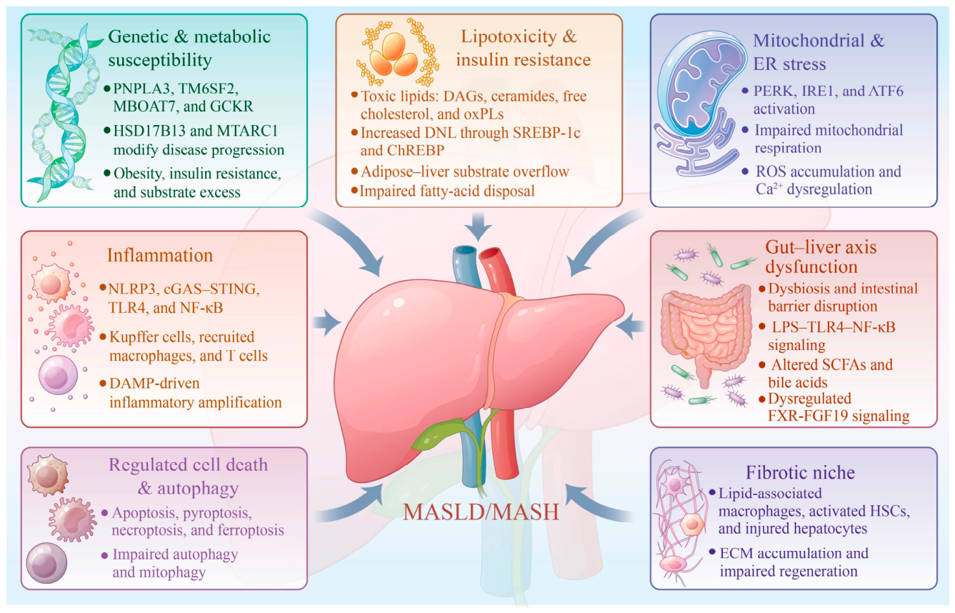
Figure 2.
Principal pharmacological actions of source-plant extracts and isolated bioactive compounds in MASLD/MASH. Plant-derived extracts, defined fractions, and isolated compounds regulate interconnected processes involving hepatic lipid synthesis and oxidation, insulin and bile-acid signaling, mitochondrial homeostasis, inflammatory pathways, gut–liver communication, autophagy, regulated cell death, and fibrogenesis. Recurrent mechanisms include activation of AMPK- and PPARα-associated fatty-acid oxidation, suppression of SREBP-1c-mediated lipogenesis, enhancement of Nrf2- and SIRT1-related stress responses, inhibition of Toll-like receptor 4–NF-κB and NLRP3 signaling, restoration of intestinal barrier function, and attenuation of hepatic stellate-cell activation. The pathways shown represent mechanisms reported across several plant sources and are not shared by every preparation. AAC, acetyl-CoA carboxylase; AMPK, AMP-activated protein kinase; ATP, adenosine triphosphate; CD86, cluster of differentiation 86; CD206, cluster of differentiation 206; CPT1A, carnitine palmitoyltransferase 1A; ECM, extracellular matrix; FGF15/19, fibroblast growth factor 15/19; FXR, farnesoid X receptor; HO-1, heme oxygenase-1; HSCs, hepatic stellate cells; IL-1β, interleukin-1 beta; IL-6, interleukin-6; M1/M2, classically activated/pro-inflammatory and alternatively activated/anti-inflammatory macrophage phenotypes; MASH, metabolic dysfunction-associated steatohepatitis; MASLD, metabolic dysfunction-associated steatotic liver disease; NF-κB, nuclear factor kappa B; NLRP3, NOD-like receptor family pyrin domain-containing 3; Nrf2, nuclear factor erythroid 2-related factor 2; PGC-1α, peroxisome proliferator-activated receptor gamma coactivator 1-alpha; PINK1, PTEN-induced kinase 1; PPARα, peroxisome proliferator-activated receptor alpha; ROS, reactive oxygen species; SCFAs, short-chain fatty acids; SIRT1, sirtuin 1; SIRT3, sirtuin 3; SREBP-1c, sterol regulatory element-binding protein 1c; TLR4, Toll-like receptor 4; TNF-α, tumor necrosis factor alpha.
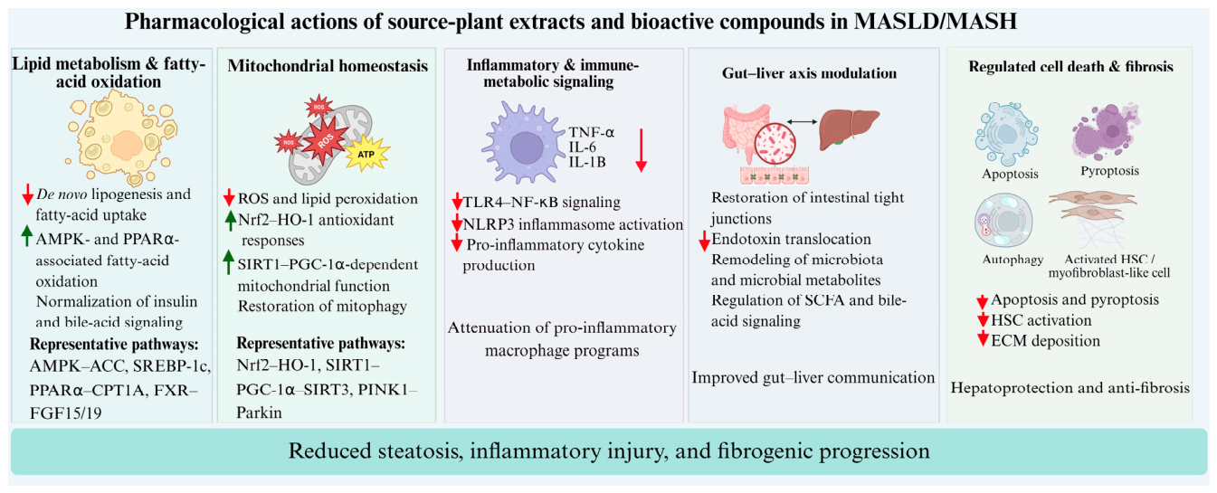
Figure 3.
Common isolation routes and minimum characterization requirements for PELNs evaluated in steatotic liver disease. Plant tissues or juices are processed by homogenization, filtration, and low- to intermediate-speed centrifugation to remove fibers, cells, and large debris. Vesicle-enriched fractions are subsequently obtained using differential ultracentrifugation, polyethylene glycol-assisted precipitation, tangential-flow filtration, size-exclusion chromatography, or combinations of these procedures. These approaches differ in recovery, purity, scalability, and the likelihood of co-isolating soluble proteins, polysaccharides, lipids, and free phytochemicals. Particle size and morphology alone are insufficient to define PELN identity. Minimum characterization should include particle concentration and size distribution, morphology and membrane structure, lipid, protein, and RNA content, surface charge, colloidal and storage stability, contaminant assessment, and batch-to-batch consistency. PBS, phosphate-buffered saline; PEG, polyethylene glycol; PELNs, plant-derived exosome-like nanoparticles; RNA, ribonucleic acid.
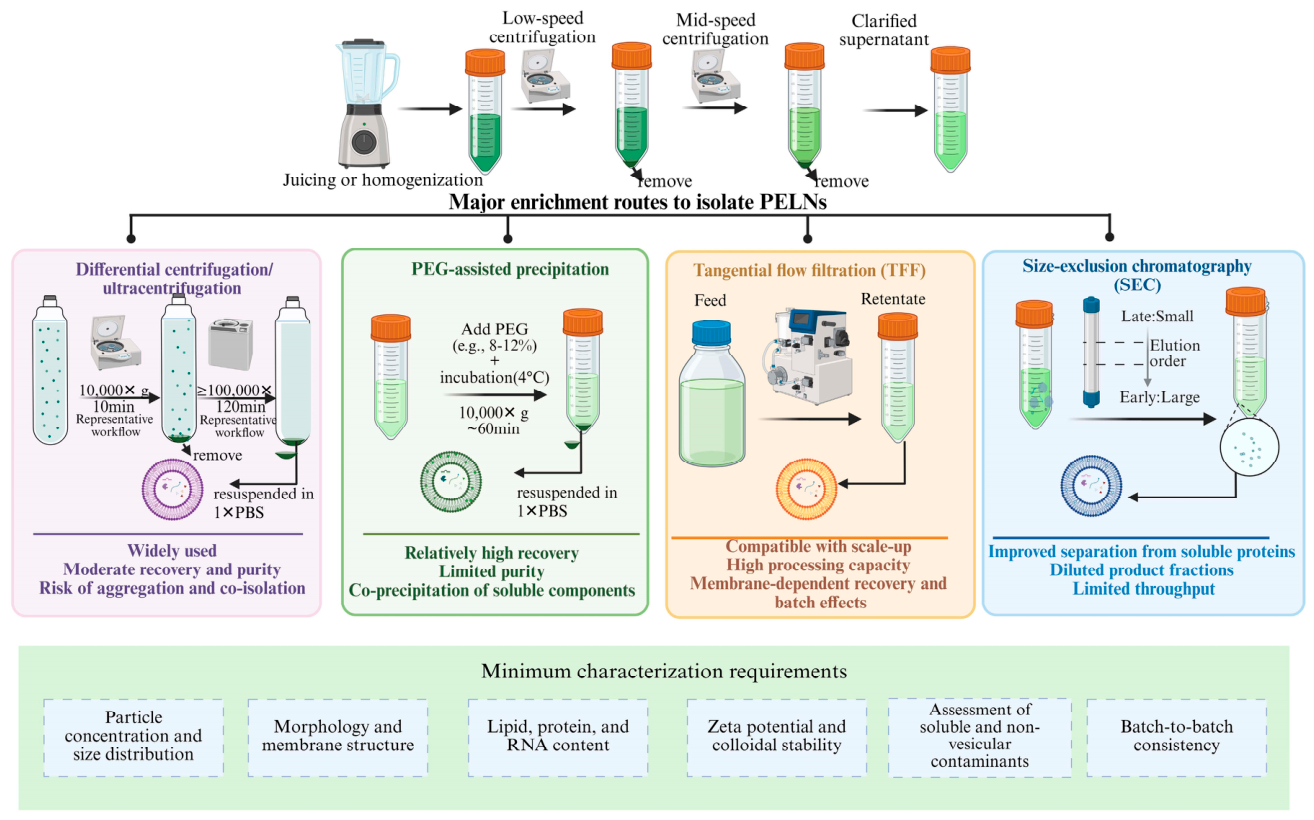
Figure 4.
Reported cargo classes and levels of mechanistic evidence for MASLD/MASH-related PELNs. PELN preparations contain source-dependent combinations of membrane lipids, proteins, small RNAs, phytochemicals, and other metabolites. Their composition is influenced by plant species, tissue, cultivation and harvesting conditions, processing, fermentation, and isolation method. Cargo detection by sequencing, proteomics, lipidomics, or metabolomics establishes composition but not functional relevance. Mechanistic evidence becomes stronger when pathway associations are followed by cargo depletion, inhibition, matched free-cargo controls, or rescue experiments. Garlic-derived miR-396e provides a comparatively well-defined example of cargo-dependent activity through regulation of macrophage PFKFB3, whereas the contribution of most reported cargos remains incompletely validated in in vivo MASLD/MASH models. MASH, metabolic dysfunction-associated steatohepatitis; MASLD, metabolic dysfunction-associated steatotic liver disease; miRNA, microRNA; PFKFB3, 6-phosphofructo-2-kinase/fructose-2,6-bisphosphatase 3.
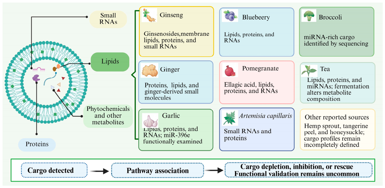
Figure 5.
Proposed oral fate, gut–liver axis effects, cellular uptake, and engineering potential of PELNs in MASLD/MASH. (A) Following oral administration, selected PELNs may protect vesicular cargos during gastrointestinal transit, interact with intestinal epithelial and immune cells, or release cargos within the intestinal lumen. The proportion of intact vesicles that survives digestion and enters the circulation remains uncertain. (B) Local intestinal effects may restore tight-junction integrity, alter microbial composition and metabolites, regulate bile-acid and short-chain-fatty-acid signaling, and reduce endotoxin translocation, thereby modifying hepatic metabolic and inflammatory responses through the gut–liver axis. (C) Uptake by hepatocytes, macrophages, and hepatic stellate cells in liver has been reported in specific experimental systems. Representative effects include regulation of hepatocyte lipid metabolism and oxidative stress, macrophage miR-396e–PFKFB3 signaling and macrophage–hepatocyte communication, and TIMP2–MMP2-associated extracellular-matrix turnover in hepatic stellate cells. These findings do not establish universal cell selectivity or preferential hepatic targeting. (D) Native PELNs may be combined with therapeutic cargos, synthetic lipids, or targeting ligands to generate engineered hybrid nanomedicines. A berberine-loaded, ursodeoxycholic-acid-modified ginger vesicle–liposome hybrid is shown as a representative engineered system. Native and engineered preparations should be evaluated separately because their composition, biodistribution, safety, and regulatory requirements differ. FFAR2/3, free fatty acid receptors 2 and 3; FXR, farnesoid X receptor; GPR109A, G protein-coupled receptor 109A; HSCs, hepatic stellate cells; LPS, lipopolysaccharide; MASH, metabolic dysfunction-associated steatohepatitis; MASLD, metabolic dysfunction-associated steatotic liver disease; miRNA, microRNA; MMP2, matrix metalloproteinase 2; PELNs, plant-derived exosome-like nanoparticles; PFKFB3, 6-phosphofructo-2-kinase/fructose-2,6-bisphosphatase 3; SCFAs, short-chain fatty acids; TIMP2, tissue inhibitor of metalloproteinases 2; TLR4, Toll-like receptor 4.
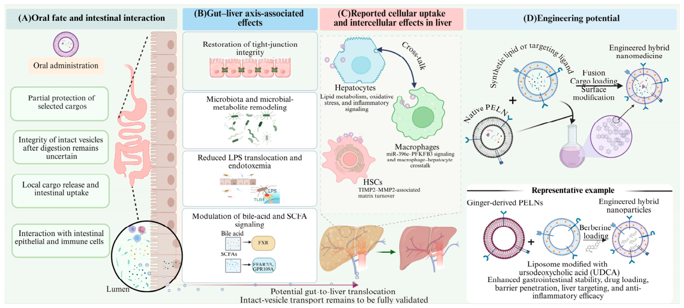

Table 1.
Representative pharmacological evidence for source-plant extracts and bioactive compounds in MASLD/MASH.
Table 1.
Representative pharmacological evidence for source-plant extracts and bioactive compounds in MASLD/MASH.
| Plant source | Representative preparations or compounds | Principal pharmacological modules | Main evidence limitations | Representative references |
|---|---|---|---|---|
| Ginseng | Black or fermented red ginseng; Compound K; ginsenosides Rb2, Rk1, Rd, Rc, Re; PPT; panaxydol | AMPK/PPARα-dependent lipid oxidation; SIRT1/SIRT6; autophagy; NLRP3; gut barrier regulation | Predominantly preclinical; substantial heterogeneity among ginseng preparations | (Chen et al., 2022; Choi et al., 2019; Cui et al., 2023; Hong et al., 2016; Hong et al., 2013; Huang et al., 2017; Kim et al., 2013; Kim et al., 2024b; Lee et al., 2016; Liu et al., 2023b; Lu et al., 2023; Mohammed, 2022; Wei et al., 2022; Wu et al., 2025; Wu et al., 2024; Xiao et al., 2013; Yang et al., 2023b; Zhang et al., 2024) |
| Ginger | Ginger extract and essential oil; zingerone; zerumbone; 6-gingerol | AMPK activation; suppression of SREBP-dependent lipogenesis; mitochondrial protection; TLR4/NLRP3 regulation | Mainly rodent studies; limited quantitative liver-fat or fibrosis assessment in clinical trials | (Liu et al., 2023b; Mohammed, 2022; Panyod et al., 2024; Rafie et al., 2020; Rahimlou et al., 2016) |
| Garlic | Aged black garlic; garlic essential oil; SAC; SAMC; DADS | AMPK/SIRT1 signaling; reduced lipogenesis; restoration of autophagy; inhibition of apoptosis and inflammation | Preparations and doses vary across studies; clinical endpoints mainly metabolic or ultrasound-based | (Hwang et al., 2013; Soleimani et al., 2016; Soleimani et al., 2020; Soleimani et al., 2021; Xiao et al., 2013) |
| Blueberry | Juice, leaf polyphenols, and probiotic combinations; TEC; pterostilbene | SIRT1/PGC-1α/SIRT3; Nrf2; mitochondrial function; autophagy and pyroptosis | Several studies use blueberry–probiotic combinations, limiting attribution to blueberry alone | (Li et al., 2020; Ren et al., 2014; Ren et al., 2017; Ren et al., 2019; Zhu et al., 2022; Zhu et al., 2018) |
| Pomegranate | Juice, peel or fruit extracts, seed oil; punicalagin; punicic acid | Nrf2 and mitochondrial protection; NLRP3 regulation; gut barrier and microbiota remodeling | Marked formulation heterogeneity; clinical trials are generally small and short | (Baghdadi et al., 2025; Chen et al., 2024; Ekhlasi et al., 2015; Goodarzi et al., 2021; Jafarirad et al., 2023; Liu et al., 2021; Ma et al., 2025; Raffaele et al., 2020; Sanchez-Terron et al., 2024; Zou et al., 2014) |
| Honeysuckle | Lonicera extracts and polysaccharides; chlorogenic-acid-rich preparations | AMPK and Nrf2 signaling; lipid metabolism; gut microbiota and barrier regulation | Entirely preclinical as an individual plant intervention; combination studies introduce confounding | (Han et al., 2023; Kim et al., 2018; Liu et al., 2018; Park et al., 2019; Shin et al., 2017; Tzeng et al., 2015) |
| Hemp/cannabis | Hemp seed oil; Δ9-THCA and CBD-related preparations | Inflammatory and fibrotic signaling; oxidative stress; gut microbiota | Interventional evidence is preclinical; human evidence is observational and confounded | (Adejumo et al., 2017; Carmona-Hidalgo et al., 2021; Gong et al., 2023; Kaushal et al., 2020; Vazquez-Bourgon et al., 2019; Zandani et al., 2022) |
| Tangerine peel | Dried tangerine peel polysaccharide; didymin | MD-2/TLR4/NF-κB; SIRT1-associated lipophagy and mitochondrial biogenesis | Limited number of studies; no direct clinical evidence | (Wang et al., 2024; Yang et al., 2023a) |
| Broccoli | Florets, juice, and leaf extracts; sulforaphane | FXR/LXR and FGF21 signaling; gut microbiota; lipid metabolism and barrier function | Evidence remains preclinical and varies by plant tissue and preparation | (Chen et al., 2016; Huang et al., 2025; Lu et al., 2025; Wu et al., 2022; Zandani et al., 2021) |
| Artemisia capillaris | Water and ethanol extracts; scopoletin | AMPK/SIRT1/PPARα; apoptosis regulation; gut microbiota | Clinical evidence relates to a multi-herbal decoction and cannot be assigned to A. capillaris alone | (Liang et al., 2025; Xu et al., 2026; Xu et al., 2024) |
| Tea | Green, yellow, Pu-erh, and fermented dark tea; EGCG | AMPK and Nrf2; FXR–FGF15; lipid metabolism and gut microbiota | Tea composition varies by processing; clinical study numbers and sample sizes are limited | (Huang et al., 2024; Sakata et al., 2013; Xia et al., 2019; Yuan et al., 2025) |
Note: Evidence for the source-plant preparation was evaluated separately from evidence for the corresponding PELNs. Activity of an extract or isolated compound does not establish efficacy of its vesicular fraction. The complete study-level dataset, including experimental model, dose, outcome, pathway, and PMID, is provided in Supplementary Table S1.
Table 2.
Reported isolation and physicochemical characterization of PELNs evaluated in steatotic liver disease.
Table 2.
Reported isolation and physicochemical characterization of PELNs evaluated in steatotic liver disease.
| Source/formulation | Main isolation or preparation method | Reported particle size | Key methodological consideration | Ref. |
|---|---|---|---|---|
| Ginseng GNVs | Differential ultracentrifugation; compared with TFF | 168.7 ± 7.4 nm by dUC; 228.7 ± 4.5 nm by TFF | Product properties differed according to enrichment method | (Kim et al., 2025b) |
| Ginger G-Exos/GR-Exos@B | Native ginger vesicles fused with UDCA-modified liposomes | 118.4 ± 10 nm for G-Exos; 324.5 ± 10 nm for GR-Exos@B | GR-Exos@B is an engineered hybrid, not a native PELN preparation | (Ma et al., 2026) |
| Garlic GDEs | PEG precipitation combined with centrifugation | 160.61 nm | PEG may co-precipitate soluble proteins and phytochemicals | (Liu et al., 2023a) |
| Blueberry BELNs | Differential centrifugation followed by PEG8000 precipitation | 189.62 nm | Vesicle purity relative to co-precipitated components requires assessment | (Zhao et al., 2022) |
| Pomegranate PNVs | Differential centrifugation and 100,000 ×g ultracentrifugation | 152.8 nm | Ultracentrifugation may promote aggregation and co-isolation | (Kim et al., 2025c) |
| Honeysuckle HNVs | Filtration followed by tangential-flow filtration | 152.7 nm | Isolation purity and the contribution of co-isolated metabolites require further assessment | (Li et al., 2025b) |
| Hemp sprout HSNVs | Homogenization, sequential centrifugation, and 100,000 ×g ultracentrifugation | 100–200 nm | Broad size distribution; purity criteria require clarification | (Kim et al., 2024a) |
| Tangerine peel TNVs | Gradient centrifugation followed by 150,000 ×g ultracentrifugation | 172.1 ± 3.7 nm | Plant-matrix contaminants should be quantified | (Zou et al., 2024) |
| Broccoli BDENs | Differential centrifugation, 0.45-μm filtration, and 150,000 ×g ultracentrifugation | Approximately 95 nm | Characterization should extend beyond size and morphology | (Zhang et al., 2026) |
| Artemisia capillaris ACDEs | Differential centrifugation, 120,000 ×g ultracentrifugation, and 0.22-μm filtration | 201.1 nm | RNA and protein cargo require vesicle-versus-soluble-fraction controls | (Xu et al., 2024) |
| Tea TELNs | Differential centrifugation, PEG6000 precipitation, and ultracentrifugation | 274 ± 24.7 nm | Relatively large particle size and PEG-associated contamination require consideration | (Lei et al., 2025) |
| Fermented tea TELNs | Differential centrifugation followed by SEC | 127.00 ± 6.09 nm | Fermentation changes cargo and product identity relative to native tea TELNs | (Guo et al., 2025) |
Note: dUC, differential ultracentrifugation; PEG, polyethylene glycol; PELNs, plant-derived exosome-like nanoparticles; SEC, size-exclusion chromatography; TFF, tangential-flow filtration; UDCA, ursodeoxycholic acid. Reported sizes were obtained using different instruments and analytical procedures and should not be compared as standardized measurements.
Table 3.
Functional evidence and major limitations of PELNs in experimental steatotic liver disease.
Table 3.
Functional evidence and major limitations of PELNs in experimental steatotic liver disease.
| Source/formulation | Experimental model | Main reported effects | Most direct mechanistic evidence | Main limitation | Ref. |
|---|---|---|---|---|---|
| Ginseng GNVs | Western-style diet-induced steatotic liver disease and fibrosis | Reduced liver injury, lipid accumulation, oxidative stress, and fibrosis; improved gut barrier-related outcomes | TIMP2–MMP2-associated matrix turnover in hepatic stellate cells | Active cargo remains undefined; limited independent replication | (Kim et al., 2025b) |
| Ginger GR-Exos@B | OA/PA-treated AML12 cells and diet-induced NASH mice | Reduced steatosis and inflammation and improved insulin-related signaling | AMPK/AKT/IRS-1 and NLRP3-associated effects | Engineered liposome–vesicle hybrid; findings cannot be attributed to native ginger PELNs | (Ma et al., 2026) |
| Garlic GDEs | HFD-fed mice and macrophage–hepatocyte models | Reduced hepatic inflammation and lipid accumulation | Vesicular miR-396e targeting macrophage PFKFB3 | Requires replication and quantitative biodistribution | (Liu et al., 2023a) |
| Blueberry BELNs | HFD-fed mice | Reduced oxidative stress, insulin resistance, lipid accumulation, and apoptosis | Nrf2 nuclear translocation and HO-1-associated signaling | Functional cargo responsible for the effect was not established | (Zhao et al., 2022) |
| Pomegranate PNVs | Western-diet MASLD model | Improved intestinal barrier function and reduced endotoxemia, oxidative injury, and fibrosis markers | Restoration of junctional proteins and gut–liver axis-associated effects | Contribution of ellagic acid versus other vesicular or co-isolated components remains unresolved | (Kim et al., 2025c) |
| Honeysuckle HNVs | HFD-induced steatotic liver disease | Reduced lipid accumulation and collagen deposition; phenotype reportedly transferable by FMT | Microbiota-dependent effect | Single-study evidence; functional vesicular cargo remains undefined | (Li et al., 2025b) |
| Hemp sprout HSNVs | Western-diet and FFC-diet fibrosis models | Reduced fibrosis, oxidative stress, intestinal barrier injury, and endotoxemia | LPS/TLR4-associated signaling and reduced HSC activation | Direct vesicular cargo and proximal molecular target remain unclear | (Kim et al., 2024a) |
| Tangerine peel TNVs | Type 2 diabetes-associated hepatic steatosis | Improved insulin resistance, hepatic lipid deposition, barrier function, and dysbiosis | Coordinated changes in lipid-metabolic and gut microbial pathways | No causal cargo validation; broad pathway associations | (Zou et al., 2024) |
| Broccoli BDENs | HFD-induced MASLD | Reduced hepatic steatosis and inflammation and maintained barrier integrity | Gut–liver axis modulation and restoration of MUC2/claudin-1-associated barrier markers | miRNA functions were largely inferred from sequencing | (Zhang et al., 2026) |
| Artemisia capillaris ACDEs | Hepatocyte and experimental NAFLD models | Reduced lipid accumulation, inflammation, and fibrotic changes | Macropinocytosis/caveolin-associated uptake and small-RNA-related effects | Limited quantitative in vivo biodistribution and cargo-rescue evidence | (Xu et al., 2024) |
| Tea TELNs | OA-treated HepG2 cells | Improved lipid metabolism and oxidative injury | Altered host miRNAs and increased PPARα/CYP7A1/CPT1A-associated signaling | In vitro evidence only | (Lei et al., 2025) |
| Fermented tea TELNs | Experimental NAFLD model | Greater lipid-lowering and anti-inflammatory activity than unfermented TELNs | Fermentation-dependent changes in physicochemical and metabolomic profiles | Processing changes product composition; active component remains undefined | (Guo et al., 2025) |
Note: FFC, fat-, fructose-, and cholesterol-rich diet; FMT, fecal microbiota transplantation; HFD, high-fat diet; HSC, hepatic stellate cell; LPS, lipopolysaccharide; NASH, non-alcoholic steatohepatitis; OA, oleic acid; PA, palmitic acid. “Most direct mechanistic evidence” denotes the strongest experimentally tested mechanism in each study and does not imply complete causal validation.
Table 4.
Clinical evidence for source-plant interventions in NAFLD/MASLD.
| Source and formulation | Study design | Main reported outcomes | Main limitations and relevance to PELNs | Ref. |
|---|---|---|---|---|
| Korean red ginseng | Randomized controlled study in patients with NAFLD | Changes in inflammatory and oxidative-stress measures, adiponectin, liver-related indices, and fatigue | No quantitative liver-fat or histological fibrosis endpoint; extract is not equivalent to GNVs | (Hong et al., 2016) |
| Ginger powder or supplementation | Two randomized clinical trials | Improvements in selected liver enzymes, insulin resistance, lipids, and inflammatory markers | Small, short-term studies; hepatic outcomes mainly biochemical or ultrasound-based | (Rafie et al., 2020; Rahimlou et al., 2016) |
| Garlic powder or supplementation | Randomized clinical trials | Reduced body weight or adiposity, steatosis scores, blood pressure, and hs-CRP | Geographically concentrated cohorts; heterogeneous endpoints and preparations | (Soleimani et al., 2016; Soleimani et al., 2020; Soleimani et al., 2021) |
| Pomegranate juice, extract, or peel | Randomized clinical trials | Improvements in metabolic indices, liver enzymes, inflammatory markers, steatosis, and, in one trial, liver stiffness | The effect of the combined juice intervention cannot be attributed to pomegranate alone; marked formulation heterogeneity; no evidence that purified PNVs produce the same effects | (Baghdadi et al., 2025; Ekhlasi et al., 2015; Goodarzi et al., 2021; Jafarirad et al., 2023) |
| Catechin-rich green tea | Double-blind placebo-controlled study | Improved liver-to-spleen CT attenuation ratio, ALT, body fat, and oxidative-stress markers | Very small sample; no direct assessment of steatohepatitis or fibrosis | (Sakata et al., 2013) |
| Yinchenhao decoction | Randomized double-blind placebo-controlled trial | Reduced MRI-PDFF, liver enzymes, liver stiffness, and BMI | Multi-herbal formulation; effect cannot be attributed specifically to A. capillaris | (Xu et al., 2026) |
| Cannabis exposure | Cross-sectional and longitudinal observational studies | Lower prevalence or slower increase in steatosis-related indices | Non-interventional and highly susceptible to confounding; does not support therapeutic use | (Adejumo et al., 2017; Vazquez-Bourgon et al., 2019) |
Note: ALT, alanine aminotransferase; BMI, body mass index; CT, computed tomography; hs-CRP, high-sensitivity C-reactive protein; MRI-PDFF, magnetic resonance imaging proton-density fat fraction; NAFLD, non-alcoholic fatty liver disease; PNVs, pomegranate-derived nanovesicles. None of the listed studies administered a purified or enriched PELN preparation. Clinical activity of a source-plant product should not be considered clinical validation of the corresponding vesicular fraction.
Disclaimer/Publisher’s Note: The statements, opinions and data contained in all publications are solely those of the individual author(s) and contributor(s) and not of MDPI and/or the editor(s). MDPI and/or the editor(s) disclaim responsibility for any injury to people or property resulting from any ideas, methods, instructions or products referred to in the content. |
© 2026 by the authors. Licensee MDPI, Basel, Switzerland. This article is an open access article distributed under the terms and conditions of the Creative Commons Attribution (CC BY) license (http://creativecommons.org/licenses/by/4.0/).
Copyright: This open access article is published under a Creative Commons CC BY 4.0 license, which permit the free download, distribution, and reuse, provided that the author and preprint are cited in any reuse.