

Submitted:
29 September 2026
Posted:
30 September 2026
You are already at the latest version
Abstract
Tumor hypoxia is a pervasive feature of solid tumors that promotes malignant progression, metastatic dissemination, and resistance to chemotherapy, radiotherapy, and immunotherapy. At the same time, low oxygen tension creates a chemically exploitable vulnerability for selective drug activation. Hypoxia-activated prodrugs (HAPs) and activatable small-molecule theranostics are designed to remain comparatively inactive in oxygenated tissues and to undergo reductase-mediated conversion within hypoxic tumor regions, thereby releasing cytotoxic, targeted, immunomodulatory, or imaging-active species. In this Review, we organize recent advances according to the chemistry of the bioreductive trigger and the mechanism that links reduction to payload release or signal generation. We discuss nitroaromatic and nitroheterocyclic groups, azo linkages, N-oxides, quinones, azides, and redox-active transition-metal complexes, with emphasis on one-electron redox cycling, multielectron reduction, self-immolative fragmentation, and the influence of reduction potential and local oxygen concentration. Representative small-molecule systems are then used to illustrate how hypoxia activation can be integrated with fluorescence and multimodal imaging, phototherapy, immunomodulation, and targeted delivery. Finally, we examine the principal translational barriers including spatial and temporal heterogeneity of tumor oxygenation, reductase dependence, incomplete intratumoral activation, pharmacokinetic limitations, and the need for patient-selection biomarkers and outline design criteria for improving therapeutic index and clinical tractability. Together, these developments position hypoxia-responsive chemistry as a mechanistically tunable platform for precision drug delivery rather than simply a means of targeting oxygen-poor tumor regions.
Keywords:
hypoxia-activated prodrugs
; tumor hypoxia
; bioreductive activation
; small-molecule theranostics
; precision drug delivery
; tumor microenvironment
1. Introduction
1.1. Features of Tumor Hypoxia
Solid tumors remain a major cause of cancer morbidity and mortality, in part because heterogeneous tumor microenvironments (TMEs) promote treatment resistance and disease recurrence [1,2]. Among the defining features of the TME, hypoxia is particularly important because it compromises radiotherapy, alters drug sensitivity, and limits oxygen-dependent photodynamic therapy (PDT) [3,4,5]. Tumor hypoxia arises when oxygen delivery through structurally and functionally abnormal microvessels fails to match the metabolic demand of proliferating tumor and stromal cells [6]. The oxygen effect in radiobiology was established by Gray and co-workers in 1953, providing an early mechanistic link between tissue oxygenation and therapeutic response [7]. Hypoxia is commonly categorized as acute (perfusion-limited), resulting from transient fluctuations in blood flow, or chronic (diffusion-limited), resulting from oxygen diffusion distances that exceed approximately 70-150 micrometers [8]. Importantly, oxygenation exists on a continuum rather than as a binary normoxic/hypoxic state. Whereas many normal tissues exhibit oxygen tensions of approximately 30-52 mmHg, regions of solid tumors can fall below 7.5 mmHg and, in severely hypoxic or perinecrotic areas, approach <0.1 mmHg [8,9]. This spatial and temporal heterogeneity is central to the design of hypoxia-activated therapeutics because the activation threshold of a bioreductive trigger must overlap with clinically relevant oxygen distributions while remaining sufficiently high to permit drug penetration into viable hypoxic tissue.
Hypoxia also drives coordinated metabolic, redox, genomic, and immunological adaptations. Increased glycolytic flux and lactate production contribute to extracellular acidification, while altered mitochondrial electron transport can increase reactive oxygen species (ROS) and redox stress [10,11,12]. Persistent or cycling hypoxia can impair DNA-repair processes and promote genomic instability, and hypoxia-associated signaling can suppress antitumor immune function [13]. A central regulator of these responses is hypoxia-inducible factor 1 (HIF-1). Stabilized HIF-1alpha heterodimerizes with HIF-1beta and binds hypoxia-response elements to regulate genes involved in angiogenesis, metabolic adaptation, epithelial-to-mesenchymal transition, invasion, and metastasis [14,15]. These biological consequences explain why hypoxia is associated with aggressive disease; however, for drug design, the more immediate opportunity is chemical: low oxygen tension changes the fate of reductively generated intermediates and can therefore be used as an oxygen-dependent molecular switch.
1.2. Bio-Reductive Cascades and Activation Pathways of Hypoxia-Responsive Systems
This oxygen-dependent redox switch underpins most hypoxia-responsive small molecules. A useful design framework separates three components: (i) a bioreductive trigger whose reduction kinetics and potential determine oxygen sensitivity; (ii) a linker or self-immolative spacer that converts reduction into bond cleavage or electronic reorganization; and (iii) an effector, such as a cytotoxin, targeted inhibitor, immunomodulator, photosensitizer, or imaging reporter. Common trigger classes include nitroaromatics and nitroheterocycles, N-oxides, quinones, azo groups, aryl azides, and redox-active metal complexes [16,17,18,19,20]. Their activation is mediated by cellular oxidoreductases, but hypoxia selectivity does not arise simply because a reductase is “upregulated” in tumors. Rather, for many one-electron substrates, molecular oxygen competes for the reduced intermediate and rapidly regenerates the parent compound. The balance among reductase activity, oxygen concentration, reduction potential, and subsequent fragmentation therefore determines whether activation is truly hypoxia-selective [21].
One-electron bioreduction and oxygen-dependent redox cycling: Flavin-dependent oxidoreductases, including cytochrome P450 oxidoreductase (POR) and related cellular reductases, can transfer one electron to an electron-deficient trigger to generate a radical anion [22,23]. In oxygenated environments, molecular oxygen efficiently reoxidizes this intermediate, regenerating the parent compound and producing superoxide. This redox cycling suppresses net activation and functions as an oxygen-sensitive checkpoint [24]. As oxygen tension decreases, back-oxidation becomes less competitive, allowing further electron/proton transfer and, for appropriately designed substrates, irreversible fragmentation or self-immolation that releases the active species [25,26]. Accordingly, hypoxia selectivity should be described in terms of oxygen-dependent reaction kinetics rather than by a universal pO2 cutoff, because the activation threshold varies with substrate structure, reductase expression, intracellular reducing equivalents, and assay conditions.
Two-electron bio-reduction cascades: Two-electron reductases such as NAD(P)H: quinone oxidoreductase 1 (NQO1) can reduce selected substrates without forming an oxygen-sensitive radical intermediate [26,27]. For quinones and related electrophilic systems, direct two-electron reduction can generate hydroquinones that undergo elimination, lactonization, or other fragmentation reactions [27,28]. Because these pathways can be less dependent on oxygen-mediated back-oxidation, enzyme abundance may contribute as strongly as oxygen tension to selectivity. This distinction is important: not every “bioreductive” substrate is intrinsically hypoxia-selective. For HAP design, the preferred trigger therefore couples favorable reduction kinetics under tumor-relevant conditions with a fragmentation step that efficiently unmasks the therapeutic payload [29,30].
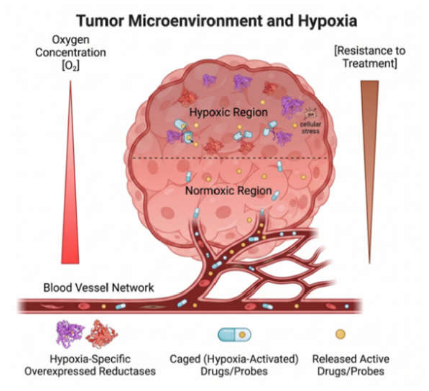
Figure 1.
Schematic illustration of hypoxia-associated tumor microenvironment and the hypoxia-activation mechanism of targeted compounds.
Figure 1.
Schematic illustration of hypoxia-associated tumor microenvironment and the hypoxia-activation mechanism of targeted compounds.
Multi-electron cascades: As illustrated in Figure 2G, several hypoxia-responsive trigger classes most prominently arylazo and nitroaromatic motifs—operate via a stepwise multielectron cascade [31,32,33,34,35,36,37,38]. In these systems, an initially reversible single-electron transfer is followed, under pathological hypoxia (pO2 < 10 mmHg), by consecutive electron- and proton-transfer events (+ne-/nH+) proceeding through cumulative 4e- or 6e- pathways. This irreversible reduction suppresses futile oxygen back-oxidation and drives substantial electronic reorganization, providing the thermodynamic driving force for bond heterolysis or self-immolative disassembly (e.g., 1,6-elimination) to unmask the uncaged active drug or fluorophore. Conceptually, this sequence converts a reversible oxygen-sensing step (redox cycling) into an irreversible chemical output. Because the stoichiometry, kinetics, and sequence of electron-transfer steps differ across functional scaffolds (e.g., nitroaromatics, azos, quinones, and coordination complexes), "multielectron reduction" is best conceptualized as a mechanistic continuum rather than a single universal pathway. Accordingly, Figure 2 depicts a comparative landscape of these representative activation routes rather than an identical, single-step kinetic scheme.
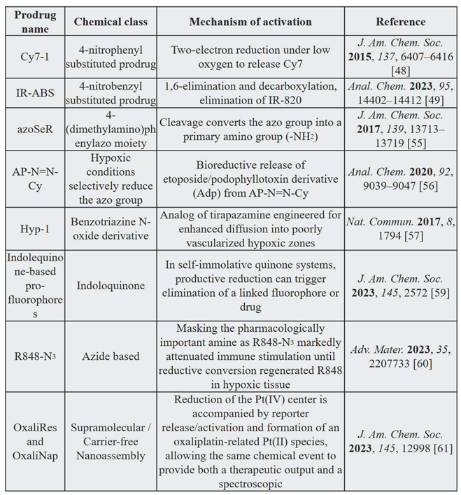
To overcome the challenges associated with tumor microenvironment (TME) heterogeneity, a wide array of hypoxia-activated prodrugs (HAPs) and activatable theranostic platforms have been engineered to exploit low intratumoral oxygen tensions. These systems utilize specific chemical moieties—such as nitroaromatics, aliphatic and heterocyclic N-oxides, quinones, and azo linkages—that undergo selective bioreductive transformation within hypoxic regions while remaining inactive under normoxic conditions. Table 1 summarizes the primary classes of representative HAPs, outlining their chemical classifications, distinct activation pathways, and key literature references.
2. Chemical Design and Bio-Reduction Mechanisms of Hypoxia-Sensitive Trigger Moieties
Across hypoxia-responsive platforms, trigger selection governs both the oxygen threshold for activation and the efficiency with which reduction is translated into payload release or signal generation [39,40]. The following sections compare the principal trigger classes from a structure-mechanism perspective, emphasizing (i) the relevant reductive intermediates, (ii) the role of oxygen-mediated back-oxidation, (iii) the fragmentation or electronic switching event, and (iv) representative applications that illustrate the strengths and limitations of each chemistry.
2.1. Nitroaromatic and Nitro-Heterocyclic Derivatives
Nitroaromatic and nitroheterocyclic groups, including 2-nitroimidazole, 5-nitrofuran, and 4-nitrobenzyl motifs, are among the most extensively studied hypoxia-responsive triggers [41,42,43,44,45,46]. One-electron reduction produces a nitro radical anion that is readily reoxidized by O2 under oxygenated conditions. At lower oxygen tensions, sequential reduction can proceed through nitroso (–NO) and hydroxylamine (–NHOH) intermediates to the corresponding amine. In 4-Nitrobenzyl carbamates and related self-immolative systems, formation of the electron-donating amine can initiate 1,6-elimination and release a caged drug or fluorophore [47]. This chemistry provides a direct connection between oxygen-sensitive reduction and irreversible payload liberation. Li et al. reported the nitrobenzyl-caged cyanine probe Cy7-1 for NTR-responsive NIR imaging [48]. As summarized in Figure 3A-C, the probe showed low fluorescence under oxygenated conditions, increased signal with decreasing oxygen availability in cells, and time-dependent tumor-associated fluorescence in vivo. Yan et al. developed the NIR-activatable cyanine probe IR-ABS for orthotopic lung-tumor imaging by tracheal inhalation (Figure 3D, E) [49]. These examples illustrate the high signal amplification achievable with nitro-triggered fluorogenic systems, although fluorescence activation should not be interpreted as a direct quantitative measurement of pO2 unless probe reduction kinetics and reductase dependence have been independently calibrated.
Beyond single-modality fluorescence, expanding nitro-triggered systems into multimodal imaging platforms has opened new avenues for deep-tissue solid tumor detection. For instance, Karan and co-workers developed NO2-Eu, a hypoxia-responsive molecular platfrom that couples optical signaling with chemical exchange saturation transfer (CEST) MRI [50], illustrating how reductive activation can be linked to complementary imaging readouts. Expanding the utility of hypoxia-directed systems toward combating tumor-associated microenvironmental complications such as the presence of cancer-associated bacteria that promote metastasis and treatment resistance has led to the development of dual-action theranostic prodrugs. In a separate study, a self-immolative nitro-containing theranostic agent (NR-NO2) was designed to respond to reductive environments associated with therapeutic activity [51]. This platform leverages elevated NTR activity in hypoxic and bacterial microenvironments to trigger robust fluorescence "turn-on" signals for real-time tracking while exerting potent cytotoxicity against both tumor cells and Gram-positive/Gram-negative bacteria, thereby offering a promising approach for image-guided cancer and infection therapy. Collectively, these studies demonstrate the modularity of nitro-based triggers, but they also highlight a recurring translational issue: NTR activity, oxygen tension, and tissue distribution jointly determine activation, and each contribution should be resolved experimentally when claiming hypoxia selectively.
2.2. Azo Derivatives
Azo compounds are versatile bioreductive switches because reduction of the N=N bond can couple a change in molecular electronics to irreversible bond cleavage. Enzymatic one-electron transfer generates reduced azo intermediates that can progress through a hydrazo state and ultimately yield the corresponding aniline fragments. In oxygenated environments, competition from molecular oxygen can suppress productive reduction, whereas lower oxygen availability favors continued reduction and cleavage. This behavior has been exploited in both imaging agents and prodrugs: the azo group can quench a fluorophore or photosensitizer while inactive, or can serve as a cleavable bridge between a reporter and a pharmacologically active cargo [52,53,54,55,56] The key design variable is therefore not simply the presence of an azo bond, but now its reduction kinetics, electronic coupling, and post-reduction fragmentation are matched to the intended biological oxygen range.
Piao et al. provided an instructive example azoSeR, an activatable seleno-rosamine photosensitizer designed for photodynamic therapy [55]. Incorporation of the azo unit into the conjugated seleno-rosamine scaffold suppresses the excited-state pathway required for efficient intersystem crossing and singlet-oxygen generation. After reductive cleavage under hypoxic conditions, the photophysically active SeR species are generated, restoring both fluorescence and photosensitizing activity. Importantly, the system was designed to respond not only to near-anoxia but also to mild hypoxia: cultures cells could be selectively ablated after activation at oxygen concentrations as high as approximately 5%, a range relevant to viable hypoxic regions of solid tumors. This study therefore illustrates a useful principle for hypoxia-directed PDT: rather than attempting to generate reactive oxygen species efficiently in every tissue compartment, the photosensitizer itself can be chemically gated so that phototoxicity is preferentially enabled in oxygen-poor tumor regions while remaining attenuated in normoxic cells (Figure 4A, B). At the same time, the approach retains an intrinsic constraint of Type II PDT, because the activated sensitizer still requires sufficient molecular oxygen for singlet-oxygen production; consequently, the most severely anoxic regions may remain less responsive than mildly hypoxic regions.
A different use of azo chemistry was demonstrated by Zhao et al. with AP-N=N-Cy [56]. This construct linked a podophyllotoxin-derived precursor (AP) to a NIR cyanine reporter through a multifunctional azobenzene unit. Azoreductase-mediated cleavage initiates a self-elimination process that simultaneously liberates the active anticancer product AdP and restores cyanine fluorescence. The synchronized therapeutic release and fluorescence turn-on enabled the delivery process to be followed in vitro, ex vivo, and in vivo, while imaging mass spectrometry was additionally used to map the tissue distributions of the released drug and reporter. This distinction is mechanistically important for the present review: AP-N=N-Cy demonstrates the power of an azo linker to integrate drug release with a traceable optical output, but it should be regarded as an azoreductase-responsive delivery paradigm that can inform hypoxia-responsive design, not as direct evidence that azo cleavage is intrinsically tumor-hypoxia selective (Figure 4C-G).
Taken together, these studies show that azo triggers can support two conceptually different theranostic strategies. In azoSeR, reduction restores the function of the photosensitizer itself, whereas in AP-N=N-Cy, reduction breaks a linker and coordinates payload liberation with an imaging signal. For cancer applications, future azo systems should establish oxygen-response curves in the relevant tumor cell type, identify the reductase contribution, and quantify parent-to-product conversion in normoxic tissues as well as tumors. Such experiments are essential because azoreductase activity is also abundant in the gastrointestinal tract and can vary among mammalian tissues and microbial compartments.
2.3. Aliphatic and Heterocyclic N-Oxides
N-Oxide triggers exploit reduction deoxygenation to convert a relatively polar N-oxide into the corresponding amine or heterocycle. Unlike nitroaromatic triggers, which can generate several partially reduced intermediates, N-oxide reduction can provide a comparatively direct two-electron chemical transformation. The reaction can alter electron donation, absorption wavelength, charge distribution, lipophilicity, and biological activity at the same time. Oxygen dependence arises in part because molecular oxygen competes for the heme center of reductive enzymes such as cytochrome P450 systems; under low oxygen, N-oxide binding and reduction become more favorable. This mechanistic architecture can therefore translate tissue oxygenation into a large spectroscopic or pharmacological change [57].
Knox et al. developed HyP-1 as a small molecule N-oxide probe for photoacoustic imaging of hypoxia [57]. The probe is based on an unsymmetrical aza-BODIPY scaffold in which conversion of Hyp-1 to Red-Hyp-1 shifts the absorption maximum from approximately 670 to 760 nm. Because the reduced product absorbs strongly in the near-infrared region where the parent probe contributes little signal, reduction can be monitored by photoacoustic imaging as well as by ratiometric NIR fluorescence. In rat-liver-microsome experiments, hypoxic incubation produced an approximately 105-fold ratiometric fluorescence response after 2 h, compared with a much smaller response under normoxia, and the corresponding photoacoustic signal was about 4-fold greater under hypoxic conditions (Figure 5A-C)). Inhibition by diphenyliodonium and dependence on NADPH supported the involvement of heme-based redox proteins, whereas iron chelation had little effect, helping to separate enzymatic/heme-mediated reduction from nonspecific labile-iron chemistry. The cellular and in vivo experiments further demonstrated why a ratiometric, chemically transformed product can be advantageous. In 4T1 cells, the hypoxic-to-normoxic ratiometric fluorescence difference increased with incubation time, and systemic administration in tumor-bearing mice produced a tumor-selective photoacoustic signal that could be reconstructed in three dimensions. The same probe also detected oxygen deficiency in a hindlimb ischemia model, indicating that its activation reflects hypoxic physiology rather than a tumor-specific enzyme phenotype. This broad responsiveness is valuable for oxygen sensing, but it also means that disease specificity must come from the anatomical context or an additional targeting element rather than from the N-oxide trigger alone.
More generally, N-oxide systems illustrate the distinction between a hypoxia-responsive chemical reaction and a cancer-specific targeting mechanism. Their major strengths include an irreversible chemical conversion, substantial changes in optical properties, and compatibility with deep-tissue photoacoustic readouts. Their limitations include dependence on the local abundance and activity of heme reductases and the possibility of activation in nonmalignant ischemic tissues. Accordingly, quantitative oxygen-response curves, reductase perturbation, and pharmacokinetic analysis should accompany claims of tumor-selective activation.
In 2021, Kang et al. developed a enamine N-oxides based hypoxia responsive prodrugs and imaging agents [58]. They introduced the synthesis and application of hypoxia-dependent reduction by hemeproteins results in the concomitant activation of cage molecules and confocal immunofluorescence co-localization images displaying the spatial distribution of probe relative to established hypoxia and metabolic marker (Figure 5D-F). The dual drug release and in vivo imaging modalities an attractive opportunity for theranostic developments.
2.4. Quinone Derivatives
Quinones provide a tunable platform for oxygen-sensitive reduction because they can accept either one or two electrons. One-electron reduction generates a semiquinone radical that can be rapidly reoxidized by molecular oxygen, whereas two-electron reduction generates a hydroquinone and may proceed through enzymes such as NQO1 with weaker oxygen dependence. In self-immolative quinone systems, productive reduction can trigger elimination of a linked fluorophore or drug. Consequently, the apparent oxygen threshold is governed not only by the redox potential of the quinone but also by the lifetime of the reduced intermediate and the kinetics of the fragmentation step. These features make quinones especially useful for engineering graded rather than binary responses to tumor oxygenation.
Wallabregue et al. systematically exploited these relationships using a series of indolequinone-based pro-fluorophores [59]. DDAO, resorufin, and Me-Tokyo Green reporters were attached to indolequinone triggers, and both the reported pKa and substitution at the benzylic position was varied to tune release kinetics. A lower pKa fluorophore is a better leaving group and can be released from a relatively short-lived reduced species and therefore shifts activation toward more severe oxygen depletion. Enzymatic studies with NADPH-cytochrome P450 reductase confirmed markedly different reduction rates among the constructs, linking molecular design parameters to functional oxygen sensitivity. This tuning translated directly to cellular oxygen thresholds. The resorufin-based probe 2 was activated at approximately 4% O2 and below, whereas the Me-Tokyo Green-based probe 4 responded predominantly at severe hypoxia of approximately 0.5% O2 and below; an intermediate construct showed an intermediate response. In multicellular spheroids, simultaneous use of probes with different thresholds generated spatially distinct signals: probe 2 reported regions comparable to those labeled by pimonidazole, whereas probe 4 identified more severely hypoxic regions in a pattern analogous to EF5. Thus, the study moved beyond the conventional classification of cells as simply normoxic or hypoxic and demonstrated that small-molecule trigger chemistry can be calibrated to resolve oxygen heterogeneity within three-dimensional tumor models (Figure 6).
The broader implication is that quinone probes can function as chemically encoded oxygen-threshold sensors, but interpretation requires careful separation of oxygen-dependent radical chemistry from enzyme-selective two-electron reduction. For therapeutic prodrugs, the same design logic could be used to match the activation threshold to the viable hypoxic compartment most relevant to treatment resistance. However, an oxygen threshold established for one quinone-reporter combination cannot be generalized to another because reduction potential, leaving-group ability, reductase expression, and intracellular localization all contribute to the observed response.
2.5. Azide Based Hypoxia Activatable Probe
Aryl azides can function as reductively removable masking groups when conversion of the azide to an amine restores the electronic or pharmacological properties of the parent molecule. This strategy is particularly attractive when a therapeutically essential amino group can be temporarily blocked without introducing a large linker or carrier. In contrast to fluorogenic systems in which reduction mainly changes optical output, azide masking can directly silence biological activity and thereby widen the therapeutic window of potent immune modulators or other systemically toxic agents.
Sun et al. applied this concept to resiquimod (R848), a Toll-like receptor 7/8 agonist whose systemic use is limited by strong inflammatory toxicity [60]. Masking the pharmacologically important amine as R848-N3 markedly attenuated immune stimulation until reductive conversion regenerated R848 in hypoxic tissue. Chemical conversion was evaluated under defined oxygen conditions, and the biological consequence of unmasking was assessed through dendritic-cell activation and macrophage polarization. In mice, the prodrug reduced systemic pro-inflammatory cytokine production to approximately one-twelfth of that produced by an equivalent dose of R848, demonstrating that chemical masking can materially reduce the systemic pharmacology of the parent agonist rather than merely alter its biodistribution. The investigators further combined R848-N3 with combretastatin A4 nanoparticles (CA4-NPs), using vascular disruption to intensify tumor hypoxia and thereby increase prodrug activation. This combination produced a strong tumor-to-normal-tissue exposure gradient; the concentration of active R848 in tumor was reported to be 21.45-fold higher than that in the heart. When the hypoxia-amplifying strategy was combined with anti-PD-1 therapy, the reported tumor suppression reached 94.1%, with complete tumor regression in 40% of treated animals. These results illustrate an important extension of hypoxia-activated prodrug design: the microenvironmental trigger can be pharmacologically amplified, rather than treated as a fixed property of the tumor, to increase spatially restricted activation. At the same time, this example emphasizes the need for rigorous mechanistic attribution. Changes in dendritic-cell activation, macrophage phenotype, or checkpoint-therapy response are downstream biological outcomes and cannot by themselves quantify oxygen-dependent azide reduction. For future azide-masked immunotherapeutics, direct measurement of parent and active drug across defined oxygen levels, together with tumor and normal-organ pharmacokinetics, is essential. The R848-N3 study is particularly informative because it links chemical activation, reduced systemic inflammatory toxicity, deliberate modulation of tumor hypoxia, and combination immunotherapy within a single development strategy (Figure 7).
2.6. Bioreducible Transition Metal Complexes
Redox-active transition-metal complexes offer a distinct hypoxia-activation principle because reduction can alter metal oxidation state, coordination geometry, ligand-exchange kinetics, and drug release simultaneously. High-valent complexes such as Pt(IV) are generally more substitution-inert than their lower-valent counterparts and can therefore serve as prodrug scaffolds. Reduction to Pt(II) can restore the reactivity associated with platinum chemotherapy while releasing axial ligands or reporters. Unlike purely organic triggers, however, the activation behavior of metal complexes depends on a network of variables that includes oxygen tension, intracellular reductants, ligand field, protein binding, and cellular metal handling.
Boulet et al. developed the Oxali(IV)Fluor series to couple Pt(IV) reduction to a measurable fluorescence response [61]. OxaliRes and OxaliNap are carbamate-functionalized oxaliplatin(IV) derivatives incorporating resorufin- and naphthalimide-related reporters. Reduction of the Pt(IV) center is accompanied by reporter release/activation and formation of an oxaliplatin-related Pt(II) species, allowing the same chemical event to provide both a therapeutic output and a spectroscopic readout. This architecture is particularly useful for mechanistic studies because fluorescence provides a surrogate measure of intracellular Pt(IV) reduction rather than relying only on downstream loss of cell viability.
In cancer cells, both complexes displayed oxygen-dependent activation, with the strongest fluorescence observed under severe hypoxia (<0.1% O2). The oxygen dependence was also reflected in clonogenic survival assays, which showed significant differences in cytotoxicity between severe hypoxia and normoxia (21% O2). These data support the concept that appropriately engineered Pt(IV) complexes can exploit the reducing capacity of hypoxic cells while remaining comparatively less active in oxygenated conditions. The study also highlights a useful design criterion for theranostic prodrugs: when the reporter is released through the same reduction event that generates the active platinum species, fluorescence can provide mechanistically linked information about prodrug activation (Figure 8). Nevertheless, fluorescence turn-on should not be assumed to provide a quantitative measure of local pO2 without calibration, because cellular reducing equivalents and reductase activity also vary. Moreover, extracellular reductants, thiols, ascorbate, albumin binding, and ligand exchange can influence Pt(IV) stability before the compound reaches hypoxic tumor cells. Translation of this class will therefore require parallel measurement of intact Pt(IV) prodrug, reduced Pt species, reporter release, and tissue oxygenation. Such multidimensional analysis is necessary to determine whether hypoxia is the dominant driver of activation in vivo or one component of a broader reductive pharmacology.
3. Translational Design Principles and Current Limitations
The studies summarized above establish that hypoxia-responsive small molecules are chemically diverse but share a common translational problem: selective reduction in a simplified hypoxic assay does not necessarily predict selective activation in a heterogeneous tumor. Four design variables are particularly important. First, the reduction potential and reduction kinetics of the trigger must define an activation window that overlaps viable hypoxic tumor tissue without causing substantial conversion in physiologically oxygenated organs. Second, reductase expression is heterogeneous across tumor types and even within a single lesion; therefore, oxygen dependence and enzyme dependence should be experimentally separated. Third, activation must occur only after sufficient parent compound has penetrated the hypoxic compartment. Highly reactive or rapidly activated compounds may be consumed near perfused tumor regions and fail to reach more distant hypoxic cells, whereas overly stable compounds may leave the tumor before activation. Fourth, the released effector must have appropriate diffusion and retention properties. For cytotoxic HAPs, a controlled bystander effect may be advantageous because the most severely hypoxic cells can be nonproliferative, whereas neighboring viable cells may drive recurrence. For imaging agents, by contrast, diffusion of the activated reporter can reduce spatial fidelity. These trade-offs argue for application-specific optimization rather than a single “ideal” hypoxia trigger.
A second issue is how hypoxia activation should be integrated with other therapeutic modalities. Nanoparticle or ligand-directed delivery can increase tumor exposure but does not by itself guarantee delivery to hypoxic regions; vascular distance and interstitial transport remain limiting. PDT introduces an additional paradox because oxygen consumption during illumination can intensify hypoxia even as conventional Type II photochemistry requires molecular oxygen [62,63,64,65]. Hypoxia-responsive activation may therefore be most useful when it enables oxygen-independent Type I photochemistry, releases an alternative cytotoxin, or provides temporal control over combination therapy. Immunomodulatory HAPs represent another emerging direction because hypoxia contributes to immune suppression, yet immune cells are themselves sensitive to oxygen tension. In this setting, mechanistic studies should distinguish direct payload-mediated immune activation from hypoxia-induced changes in immune-cell state.
For clinical translation, pharmacodynamic evidence of intratumoral activation will be as important as systemic pharmacokinetics. Pairing a therapeutic prodrug with an imaging readout, a hypoxia biomarker, or a measurable released metabolite could enable patient selection and dose optimization. Pimonidazole staining, CAIX/HIF-associated biomarkers, PET hypoxia tracers, and direct oxygen measurements each capture different aspects of tumor oxygen biology and should not be treated as interchangeable. A clinically useful development strategy will therefore require co-development of an activation biomarker that reflects the chemistry of the specific trigger.
4. Future Perspectives and Conclusion
Hypoxia-responsive small molecules have evolved from relatively simple bioreductive cytotoxins into modular platforms that couple oxygen-sensitive chemistry with targeted inhibitors, immunomodulators, photosensitizers, and diagnostic reporters. The central design challenge is no longer merely to identify a reducible functional group; it is to engineer a quantitatively appropriate relationship among reduction potential, oxygen concentration, reductase activity, fragmentation kinetics, tissue penetration, and payload pharmacology. Reported reduction potentials can provide useful guidance, but a universal optimal range should not be assumed across chemically distinct trigger classes. Instead, electrochemical measurements should be integrated with enzymatic kinetics and product-formation studies under controlled oxygen gradients.
Several priorities could accelerate translation. First, activation should be characterized across physiologically relevant oxygen concentrations rather than only at 21% O2 versus near-anoxia. Second, three-dimensional tumor models and spatially resolved in vivo measurements should be used to determine whether parent compounds reach viable hypoxic regions before conversion. Third, the field would benefit from standardized reporting of oxygen conditions, reductase systems, conversion yields, active-metabolite exposure, and normoxic/hypoxic selectivity ratios. Fourth, theranostic designs should be used strategically: an imaging signal is most valuable when it reports the same chemical event that releases the therapeutic species. Finally, patient stratification should incorporate both tumor hypoxia and the enzymatic machinery required for activation. With these principles, hypoxia-activated chemistry can move beyond empirical stimulus-responsive design toward mechanism-based precision drug delivery in which activation thresholds, tissue distribution, and therapeutic response are prospectively matched to tumor biology.
Declaration of Competing Interest
The authors declare no conflicts of interest that could have influenced the work.
Author Contributions
Sanu Karan: writing – original draft. Jong Seung Kim: Supervision, resources, and writing, review & editing. Kwan Soo Hong: Supervision, project administration, funding acquisition, and writing, review & editing.
Acknowledgments
This work was supported by the grant (KSH, C623100) from Korea Basic Science Institute, and by the KEIT grant from the Korea government (MTIS) (KSH, RS-2025-25462597).
References
- Siegel, R. L.; Giaquinto, A. N.; Jemal, A. Cancer statistics, 2024. CA Cancer J. Clin. 2024, 74, 12–49. [Google Scholar] [CrossRef] [PubMed]
- Anderson, K. G.; Stromnes, I. M.; Greenberg, P. D. Obstacles posed by the tumor microenvironment to T cell activity: A case for synergistic therapies. Cancer Cell 2017, 31, 311–325. [Google Scholar] [CrossRef] [PubMed]
- Rohwer, N.; Cramer, T. Hypoxia-mediated drug resistance: Novel insights on the functional interaction of HIFs and cell cycle inhibitors. Drug Resist. Updat. 2011, 14, 191–201. [Google Scholar] [CrossRef] [PubMed]
- Horsman, M. R.; Overgaard, J. The impact of hypoxia and its modification of the outcome of radiotherapy. J. Radiat. Res. 2016, 57, i90–i98. [Google Scholar] [CrossRef] [PubMed]
- Li, M.; Xu, Y.; Peng, X.; Kim, J. S. From low to no O2-dependent hypoxia photodynamic therapy (hPDT): a new perspective. Acc. Chem. Res. 2022, 55, 3253–3264. [Google Scholar] [CrossRef] [PubMed]
- Sharma, A.; Arambula, J. F.; Koo, S.; Kumar, R.; Singh, H.; Sessler, J. L.; Kim, J. S. Hypoxia-targeted drug delivery systems and theranostics in cancer care. Chem. Soc. Rev. 2019, 48, 771–813. [Google Scholar] [PubMed]
- Gray, L. H.; Conger, A. D.; Ebert, M.; Hornsey, S.; Scott, O. C. The concentration of oxygen dissolved in tissues at the time of irradiation as a factor in radiotherapy. Br. J. Radiol. 1953, 26, 638–648. [Google Scholar] [CrossRef] [PubMed]
- Vaupel, P.; Mayer, A. Hypoxia in cancer: Significance and impact on clinical outcome. Cancer Metastasis Rev. 2007, 26, 225–239. [Google Scholar] [CrossRef] [PubMed]
- McKeown, S. R. Defining normoxia, physoxia and hypoxia in tumours implications for treatment response. Br. J. Radiol. 2014, 87, 20130676. [Google Scholar] [CrossRef] [PubMed]
- Liberti, M. V.; Locasale, J. W. The Warburg effect: How does it benefit cancer cells? Trends Biochem. Sci. 2016, 41, 211–218. [Google Scholar] [CrossRef] [PubMed]
- Boedtkjer, E.; Pedersen, S. F. The acidic tumor microenvironment as a cancer. Annu. Rev. Physiol. 2020, 82, 103–126. [Google Scholar] [CrossRef] [PubMed]
- Chandel, N. S.; McClintock, D. S.; Feliciano, C. E.; Wood, T. M.; Melendez, J. A.; Rodriguez, A. M.; Schumacker, P. T. Reactive oxygen species generated at mitochondrial complex III stabilize hypoxia-inducible factor-1 alpha during hypoxia. J. Biol. Chem. 2000, 275, 25130–25138. [Google Scholar] [CrossRef] [PubMed]
- Noman, M. Z.; Hasmim, M.; Messai, Y.; Terry, S.; Kieda, C.; Janji, B.; Chouaib, S. Hypoxia: A key player in antitumor immune response. Front. Immunol. 2015, 6, 29. [Google Scholar]
- Semenza, G. L. Hypoxia-inducible factors in physiology and medicine. Cell 2012, 148, 399–408. [Google Scholar] [CrossRef] [PubMed]
- Schito, L.; Semenza, G. L. Hypoxia-inducible factors: Master regulators of cancer progression. Trends Cancer 2016, 2, 758–770. [Google Scholar] [CrossRef] [PubMed]
- Wilson, W. R.; Hay, M. P. Targeting hypoxia in cancer therapy. Nat. Rev. Cancer 2011, 11, 393–410. [Google Scholar] [CrossRef] [PubMed]
- Brown, J. M.; Wilson, W. R. Exploiting tumour hypoxia in cancer treatment. Nat. Rev. Cancer 2004, 4, 437–447. [Google Scholar] [CrossRef] [PubMed]
- Sharma, A.; Arambula, J. F.; Koo, S.; Kumar, R.; Singh, H.; Sessler, J. L.; Kim, J. S. Hypoxia-targeted drug delivery systems and theranostics in cancer care. Chem. Soc. Rev. 2019, 48, 771–813. [Google Scholar] [PubMed]
- Chen, G.; Wu, K.; Li, H.; Xia, D.; He, T. Role of hypoxia microenvironment and targeted therapy. Front. Oncol. 2022, 12, 961637. [Google Scholar] [CrossRef] [PubMed]
- Zhang, S.; Chen, H.; Wang, L.; Qin, X.; Jiang, B. P.; Ji, S. C.; Shen, X. C.; Liang, H. A general approach to design dual ratiometric fluorescent and photoacoustic probes for quantitatively visualizing tumor hypoxia levels in vivo. Angew. Chem. 2022, 134, e202107076. [Google Scholar] [CrossRef]
- Guise, C. P.; Mowday, A. M.; Theys, J.; Abbattista, M.; Lambin, P.; Patterson, A. V. Bioreductive prodrugs as cancer therapeutics: Targeting tumor hypoxia. Chin. J. Cancer 2014, 33, 80–86. [Google Scholar] [CrossRef] [PubMed]
- Patterson, A. V.; Ferry, D. M.; Edmunds, S. J.; Gu, Y.; Singleton, R. S.; Patel, K.; Pullen, S. M.; Hicks, K. O.; Syddall, S. P.; Atwell, G. J.; Yang, S. Mechanism of action and preclinical antitumor activity of the novel hypoxia-activated DNA cross-linking agent PR-104. Clin. Cancer Res. 2007, 13, 3922–3932. [Google Scholar] [CrossRef] [PubMed]
- Hunter, F. W.; Wouters, B. G.; Wilson, W. R. Hypoxia-activated prodrugs: Paths to application in modern oncology. Future Med. Chem. 2016, 8, 1693–1713. [Google Scholar]
- Wardman, P. Electron transfer and oxidative stress as key factors in the design of drugs selectively active in hypoxia. Curr. Med. Chem. 2001, 8, 739–761. [Google Scholar] [CrossRef] [PubMed]
- Li, Y.; Zhao, L.; Li, X. Targeting hypoxia: Hypoxia-activated prodrugs in cancer therapy. Front. Oncol. 2021, 11, 700407. [Google Scholar] [CrossRef] [PubMed]
- Siegel, D.; Yan, C.; Ross, D. NAD(P)H:quinone oxidoreductase 1 (NQO1) in the sensitivity and resistance to antitumor quinones. Biochem. Pharmacol. 2012, 83, 1033–1040. [Google Scholar] [CrossRef] [PubMed]
- Dinkova-Kostova, A. T.; Talalay, P. NAD(P)H:quinone acceptor oxidoreductase 1 (NQO1), a multifunctional antioxidant enzyme and exceptionally versatile cytoprotector. Arch. Biochem. Biophys. 2010, 501, 116–123. [Google Scholar] [CrossRef] [PubMed]
- Kwon, H. J.; Shin, K.; Soh, M.; Chang, H.; Kim, J.; Lee, J.; Ko, G.; Kim, B. H.; Kim, D.; Hyeon, T. Large-scale synthesis and medical applications of uniform-sized metal oxide nanoparticles. Adv. Mater. 2018, 30, 1704290. [Google Scholar] [CrossRef] [PubMed]
- Phillips, R. M. Targeting the hypoxic tumor microenvironment using hypoxia-activated prodrugs. Curr. Opin. Pharmacol. 2016, 27, 44–52. [Google Scholar]
- Shi, Y.; Yu, Q.; Tan, L.; Wang, Q.; Zhu, W. H. Tumor microenvironment-responsive polymer delivery platforms for cancer therapy. Angew. Chem. 2025, 137, e202503776. [Google Scholar] [CrossRef]
- Zhang, Y.; Zhao, W.; Chen, Y.; et al. Rational construction of a reversible arylazo-based NIR probe for cycling hypoxia imaging in vivo. Nat. Commun. 2021, 12, 2772. [Google Scholar] [CrossRef] [PubMed]
- Tian, X.; Li, Z.; Sun, Y.; Wang, P.; Ma, H. Near-infrared fluorescent probes for hypoxia detection via joint regulated enzymes: design, synthesis, and application in living cells and mice. Anal. Chem. 2018, 90, 13759–13766. [Google Scholar] [CrossRef] [PubMed]
- Kiyose, K.; et al. Hypoxia-sensitive fluorescent probes for in vivo real-time fluorescence. J. Am. Chem. Soc. 2010, 132, 15846–15848. [Google Scholar] [CrossRef] [PubMed]
- Geng, W. C.; Jia, S.; Zheng, Z.; Li, Z.; Ding, D.; Guo, S. D. A noncovalent Fluorescence turn-on strategy for hypoxia imaging. Angew. Chem. Int. Ed. 2019, 131, 2399–2403. [Google Scholar] [CrossRef]
- Xue, T.; Shao, K.; Xiang, J.; Pan, X.; Zhu, Z.; He, Y. In situ construction of self-assembly AIE probe for tumor hypoxia. Nanoscale 2020, 12, 7509–7513. [Google Scholar] [CrossRef] [PubMed]
- Xue, T.; Jia, X.; Wang, J.; Xiang, J.; Wang, W.; Du, J.; He, Y. “Turn-On” activatable AIE dots for tumor hypoxia imaging. Chem. Eur. J. 2019, 25, 9634–9638. [Google Scholar] [CrossRef] [PubMed]
- Piao, W.; Tsuda, S.; Tanka, Y.; Maeda, S.; Liu, F.; Takahashi, S.; Kushida, Y.; Komatsu, T.; Ueno, T.; Terai, T.; Nakazawa, T.; Uchiyama, M.; Morokuma, K.; Nagano, T.; Hanaoka, K. Development of Azo-based fluorescent probes to detect different levels of hypoxia. Angew. Chem. Int. Ed. 2013, 52, 13028–13032. [Google Scholar] [CrossRef] [PubMed]
- Chevalier, A.; Renard, P.-Y.; Romieu, A. Azo-based fluorogenic probes for biosensing and bioimaging: recent advances and upcoming challenges. Chem. Asian J. 2017, 12, 2008–2028. [Google Scholar] [CrossRef] [PubMed]
- Liu, J.-N.; Bu, W.; Shi, J. Chemical design and synthesis of functionalized probes for imaging and treating tumor hypoxia. Chem. Rev. 2017, 117, 6160–6224. [Google Scholar] [CrossRef] [PubMed]
- Sharma, A.; Arambula, J. F.; Koo, S.; Kumar, R.; Singh, H.; Sessler, J. L.; Kim, J. S. Hypoxia-targeted drug delivery. Chem. Soc. Rev. 2019, 48, 771–813. [Google Scholar] [CrossRef] [PubMed]
- Ao, X.; Bright, S. A.; Taylor, N. C.; Elmes, R. B. 2-Nitroimidazole based fluorescent probes for nitroreductase; monitoring reductive stress in cellulo. Org. Biomol. Chem. 2017, 15, 6104–6108. [Google Scholar] [CrossRef] [PubMed]
- Zhao, X.; Rong, X.; Liu, C.; Qu, J.; Zheng, M.; Wang, K.; Zhu, B. A novel fluorescent probe with 2-nitroimidazole as hypoxia marker for long-term tracking and detection of HClO in tumor cells. Sens. Actuators B Chem. 2025, 442, 138083. [Google Scholar] [CrossRef]
- Li, Z.; Li, X.; Gao, X.; Zhang, _Y.; Shi, W.; Ma, H. Nitroreductase detection and hypoxic tumor cell imaging by a designed sensitive and selective fluorescent probe, 7-[(5-nitrofuran-2-yl)methoxy]-3H-phenoxazin-3-one. Anal. Chem. 2013, 85, 3926–3932. [Google Scholar] [CrossRef] [PubMed]
- Elmes, R. B. Bioreductive fluorescent imaging agents: applications to tumour hypoxia. Chem. Commun. 2016, 52, 8935–8956. [Google Scholar] [CrossRef] [PubMed]
- Karan, S.; Cho, M. Y.; Lee, H.; Lee, H.; Park, H. S.; Sundararajan, M.; Hong, K. S. Near-infrared fluorescent probe activated by nitroreductase for in vitro and in vivo hypoxic tumor detection. J. Med. Chem. 2021, 64, 2971–2981. [Google Scholar] [CrossRef] [PubMed]
- Wang, Z.; Lau, J. W.; Liu, S.; Ren, Z.; Gong, Z.; Liu, X.; Xing, B. A nitroreductase-activatable metabolic reporter for covalent labeling of pathological hypoxic cells in tumorigenesis. Angew. Chem. Int. Ed. 2024, 136, e202411636. [Google Scholar] [CrossRef] [PubMed]
- Kumar, R.; Kim, E. J.; Han, J.; Lee, H.; Shin, W. S.; Kim, H. M.; Hong, K. S. Hypoxia-directed and activated theranostic agent: Imaging and treatment of solid tumor. Biomaterials 2016, 104, 119–128. [Google Scholar] [CrossRef] [PubMed]
- Li, Y.; Sun, Y.; Li, J.; Su, Q.; Yuan, W.; Dai, Y.; Li, F. Ultrasensitive near-infrared fluorescence-enhanced probe for in vivo nitroreductase imaging. J. Am. Chem. Soc. 2015, 137, 6407–6416. [Google Scholar] [CrossRef] [PubMed]
- Yan, J.; Wang, K.; Gui, L.; Liu, X.; Ji, Y.; Lin, J.; Yuan, Z. Diagnosing orthotopic lung tumor using a NTR-activatable near-infrared fluorescent probe by tracheal inhalation. Anal. Chem. 2023, 95, 14402–14412. [Google Scholar] [CrossRef] [PubMed]
- Karan, S.; Cho, M. Y.; Lee, H.; Park, H. S.; Han, E. H.; Song, Y.; Lee, Y.; Kim, M.; Cho, J.-H.; Sessler, J. L.; Hong, K. S. Hypoxia-responsive luminescent CEST MRI agent for in vitro and in vivo tumor detection and imaging. J. Med. Chem. 2022, 65, 7106–7117. [Google Scholar] [CrossRef] [PubMed]
- Karan, S.; Cho, M. Y.; Lee, H.; Kim, H. M.; Park, H. S.; Han, E. H.; Sessler, J. L.; Hong, K. S. Hypoxia-directed and self-immolative theranostic agent: Imaging and treatment of cancer and bacterial infections. J. Med. Chem. 2023, 66, 14175–14187. [Google Scholar] [CrossRef] [PubMed]
- Piao, W.; Hanaoka, K.; Fujisawa, T.; Takeuchi, S.; Komatsu, T.; Ueno, T.; Urano, Y. Development of an azo-based photosensitizer activated under mild hypoxia for photodynamic therapy. J. Am. Chem. Soc. 2017, 139, 13713–13719. [Google Scholar] [CrossRef] [PubMed]
- Chen, S.; Chen, M.; Yang, J.; Zeng, X.; Zhou, Y.; Yang, S.; Zheng, J. Design and engineering of hypoxia and acidic pH dual-stimuli-responsive intelligent fluorescent nanoprobe for precise tumor imaging. Small 2021, 17, 2100243. [Google Scholar] [CrossRef] [PubMed]
- Uddin, M. I.; Evans, S. M.; Craft, J. R.; Marnett, L. J.; Uddin, M. J.; Jayagopal, A. Applications of azo-based probes for imaging retinal hypoxia. ACS Med. Chem. Lett. 2015, 6, 445. [Google Scholar] [CrossRef] [PubMed]
- Piao, W.; Tsuda, S.; Tanaka, Y.; Maeda, S.; Liu, F.; Takahashi, S.; Kushida, Y.; et al. Development of azo-based fluorescent probes to detect different levels of hypoxia. Angew. Chem. 2013, 125, 13266–13270. [Google Scholar] [CrossRef]
- Zhao, X. B.; Ha, W.; Gao, K.; Shi, Y. P. Precisely traceable drug delivery of azoreductase-responsive prodrug for colon targeting via multimodal imaging. Anal. Chem. 2020, 92, 9039–9047. [Google Scholar] [CrossRef] [PubMed]
- Knox, H. J.; Hedhli, J.; Kim, T. W.; Khalili, K.; Dobrucki, L. W.; Chan, J. A bioreducible N-oxide-based probe for photoacoustic imaging of hypoxia. Nat. Commun. 2017, 8, 1794. [Google Scholar] [CrossRef] [PubMed]
- Kang, D.; Cheung, S. T.; Wong-Rolle, A.; Kim, J. Enamine N-oxides: synthesis and application to hypoxia-responsive prodrugs and imaging agents. ACS Cent. Sci. 7 631–640. [CrossRef] [PubMed]
- Wallabregue, A. L.; Bolland, H.; Faulkner, S.; Hammond, E. M.; Conway, S. J. Two color imaging of different hypoxia levels in cancer cells. J. Am. Chem. Soc. 2023, 145, 2572. [Google Scholar] [CrossRef] [PubMed]
- Sun, J.; Liu, Z.; Yao, H.; Zhang, H.; Zheng, M.; Shen, N.; Chen, X. Azide-masked resiquimod activated by hypoxia for selective tumor therapy. Adv. Mater. 2023, 35, 2207733. [Google Scholar] [CrossRef] [PubMed]
- Boulet, M. H.; Bolland, H. R.; Hammond, E. M.; Sedgwick, A. C. Oxali(IV) Fluors: fluorescence responsive oxaliplatin(IV) complexes identify a hypoxia-dependent reduction in cancer cells. J. Am. Chem. Soc. 2023, 145, 12998. [Google Scholar] [CrossRef] [PubMed]
- Hong, L.; Li, J.; Luo, Y.; Guo, T.; Zhang, C.; Ou, S.; Long, Y.; Hu, Z. Recent advances in strategies for addressing hypoxia in tumor photodynamic therapy. Biomolecules 2022, 12, 81. [Google Scholar] [CrossRef] [PubMed]
- Karan, S.; Lee, H.; Naskar, R.; Cho, M. Y.; Park, H. S.; Han, E. H.; Hong, K. S. Nanobody-conjugated theranostic prodrug targeting αvβ3 integrin enables precision cancer therapy with real-time imaging. Adv. Health Mater. 2026, 15, e05197. [Google Scholar] [CrossRef] [PubMed]
- Turan, I. S.; Yildiz, D.; Turksoy, A.; Gunaydin, G.; Akkaya, E. U. A bifunctional photosensitizer for enhanced fractional photodynamic therapy: singlet oxygen generation in the presence and absence of light. Angew. Chem. Int. Ed. 2016, 55, 2875–2878. [Google Scholar] [CrossRef] [PubMed]
- Lv, Z.; Wei, H.; Li, Q.; Su, X.; Liu, S.; Zhang, K. Y.; Lv, W.; Zhao, Q.; Li, X.; Huang, W. Achieving efficient photodynamic therapy under both normoxia and hypoxia using cyclo-metalated Ru(II) photosensitizer through type I photochemical process. Chem. Sci. 2018, 9, 502–512. [Google Scholar] [CrossRef] [PubMed]
Figure 2.
Mechanisms of bioreductive activation for various prodrug and molecular probe scaffolds. (A) General schematic illustrating 1-electron (1e-) reduction pathways subject to oxygen (O2) reoxidation (redox cycling) versus 2-electron (2e-) reduction cascades. (B) Azo-group reduction proceeding via a reversible 1e- step followed by an irreversible multi-electron reduction to liberate an active amine species. (C) Stepwise cascade reduction of a nitroaromatic trigger through nitroso (NO) and hydroxylamine (NHOH) intermediates, leading to 1,6-elimination and active species release. (D) Two-electron reduction of N-oxide-based triggers sensitive to oxygen levels. (E) Quinone-based one- and two-electron bioreductive pathways driving ligand release or altering metal-complex coordination. (F) Bioreduction-triggered metal-complex activation and subsequent ligand dissociation. (G) Multi-electron irreversible bioreductive cleavage resulting in the generation of active species and modified core structures.
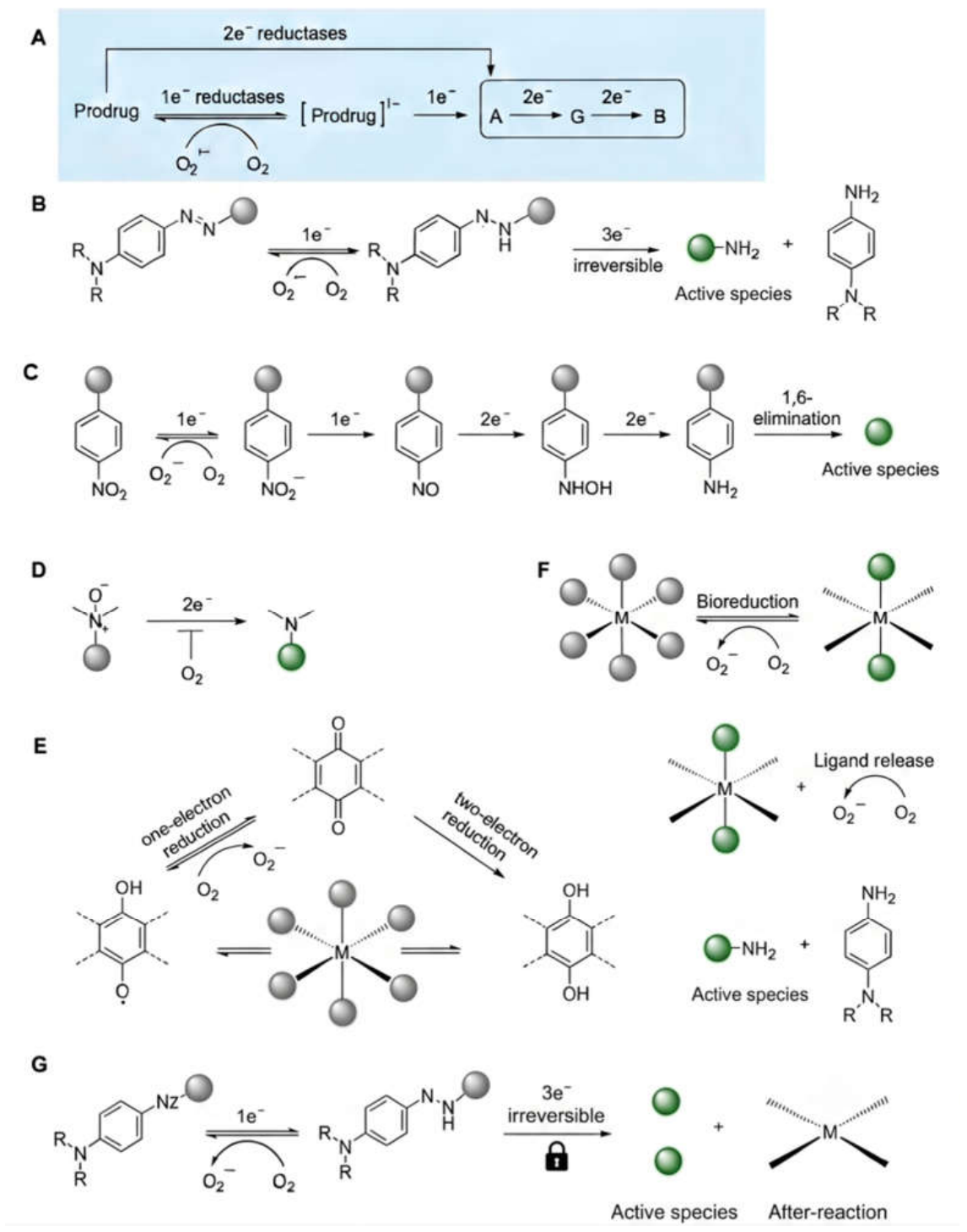
Figure 3.
In vitro and in vivo evaluation of hypoxia-responsive cyanine-based probes and imaging platforms. (A) Chemical structure of the nitrobenzyl-caged cyanine probe (Cy7-1). (B) Confocal fluorescence and bright-field microscopy images showing probe activation under varying hypoxic conditions in cell culture models. (C) In vivo real-time fluorescence imaging of a tumor-bearing mouse model following administration, including time-course tracking images and quantitative fluorescence intensity plots over 1,800 seconds. (D) Structure of a sulfonamide-functionalized cyanine derivative and its corresponding validation in cellular hypoxia imaging, zebrafish models, and targeted in vivo lung imaging. Adapted from Li et al. [48], and Yan et al. [49] with permission of ACS.
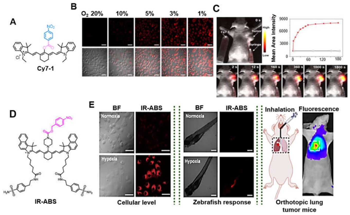
Figure 4.
Evaluation of azo-based hypoxia-activatable photosensitizing and therapeutic prodrug systems. (A) Chemical structure and bioreductive conversion of the inactive photosensitizer azoSeR to the active fluorescent form SeR mediated by reductases under hypoxic conditions. (B) Live/dead cell viability assays under hypoxic (Hyp) and normoxic (Nor) conditions in the absence (-) and presence (+) of light irradiation. (C) Structure and activation mechanism of the hypoxia-responsive prodrug AP-N=N-Cy (3), which undergoes azo reduction to release the active drug. (D) Confocal microscopy images displaying subcellular co-localization of the prodrug with Mito-Tracker in HeLa and HepG2 cells. (E) In vivo real-time fluorescence imaging of mice tracking drug activation and accumulation over a time course from 0 to 36 hours. (F) Corresponding quantitative profile of the fluorescence signal-to-noise ratio over time. (G) Ex vivo fluorescence imaging of excised tissues and organs comparing control and prodrug-treated groups. Adapted from Piao et al. [55], and Zhao et al. [56] with permission of Wiley and ACS, respectively.
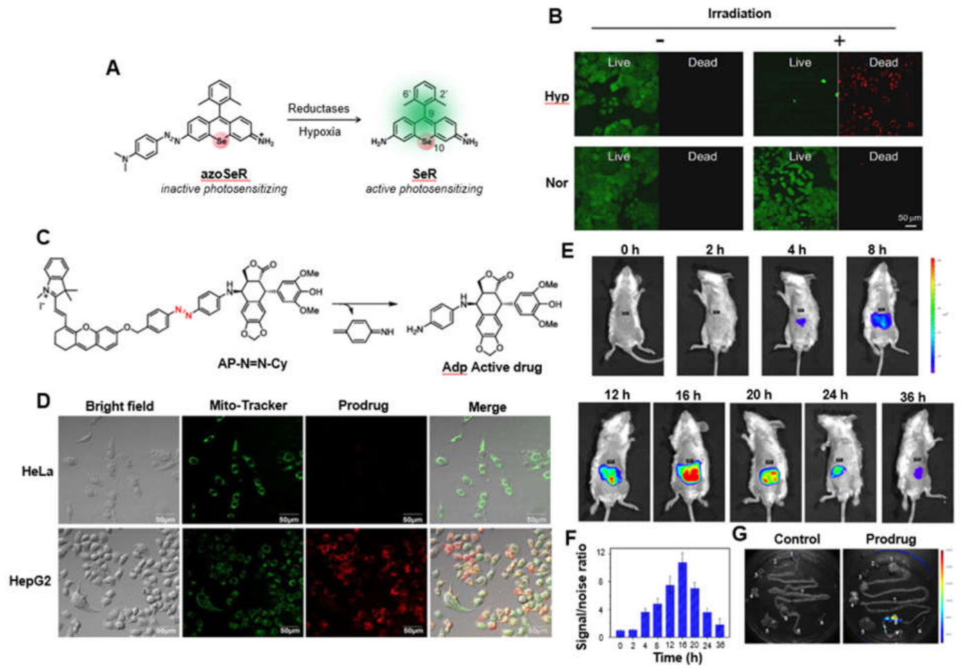
Figure 5.
Design, in vitro validation, and in vivo imaging performance of N-oxide based hypoxia-responsive ratiometric and near-infrared (NIR) fluorescent probes. (A) Chemical structures illustrating the 2-electron bioreductive conversion of the Hyp-1 probe to Red-Hyp-1. (B) Representative fluorescence microscopy images (Cy7 and Cy5 channels) and corresponding ratiometric fluorescence quantification comparing hypoxic versus normoxic cells at 2, 4, and 6 hours. (C) Plate-reader fluorescence imaging and normalized ratiometric fluorescence responses under different treatments (BIPY, DPI) and oxygen levels. (D) Chemical structure of the enamine N-oxide-functionalized NIR fluorescent probe. (E) Confocal immunofluorescence co-localization images displaying the spatial distribution of the probe (red) relative to established hypoxia and metabolic markers GLUT1, HIF1α, CAIX, and Pimonidazole (green) with DAPI nuclear counterstaining (blue) across necrotic (Nec) tissue regions. (F) Real-time in vivo fluorescence imaging of tumor-bearing mice showing probe activation, retention, and radiant efficiency dynamics from prior to injection up to 30 hours post-injection. Adapted from Knox et al. [57], and Kang et al. [58], with permission of Springer and ACS, respectively.
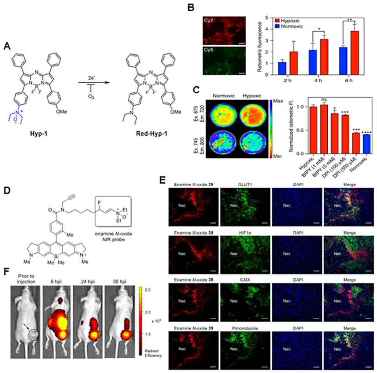
Figure 6.
Multicolor hypoxia-responsive imaging and spatial oxygen gradient mapping using quinone-based modular probes in multicellular spheroids. Schematic illustration of oxygen-level-dependent activation of non-fluorescent probes that release distinct fluorescent dyes (including DDAO, Resorufin, and Me-Tokyo green) across a decreasing oxygen gradient from 6% to 0.5% O2. (B) Confocal fluorescence microscopy images of multicellular spheroids treated with probes 2, 3, 4, EF5, or Pimonidazole, displaying probe fluorescence (red/green channels), DAPI nuclear counterstaining (blue), and merged channels. (C) Whole-spheroid fluorescence images and corresponding quantitative bar graphs of integrated fluorescence intensity across radial bin numbers, illustrating differential activation profiles from the periphery to the hypoxic core. (D) Representative spatial color-mapped distribution profile showing core-localized activation. Adapted from Wallabregue et al. [59] with permission of ACS.
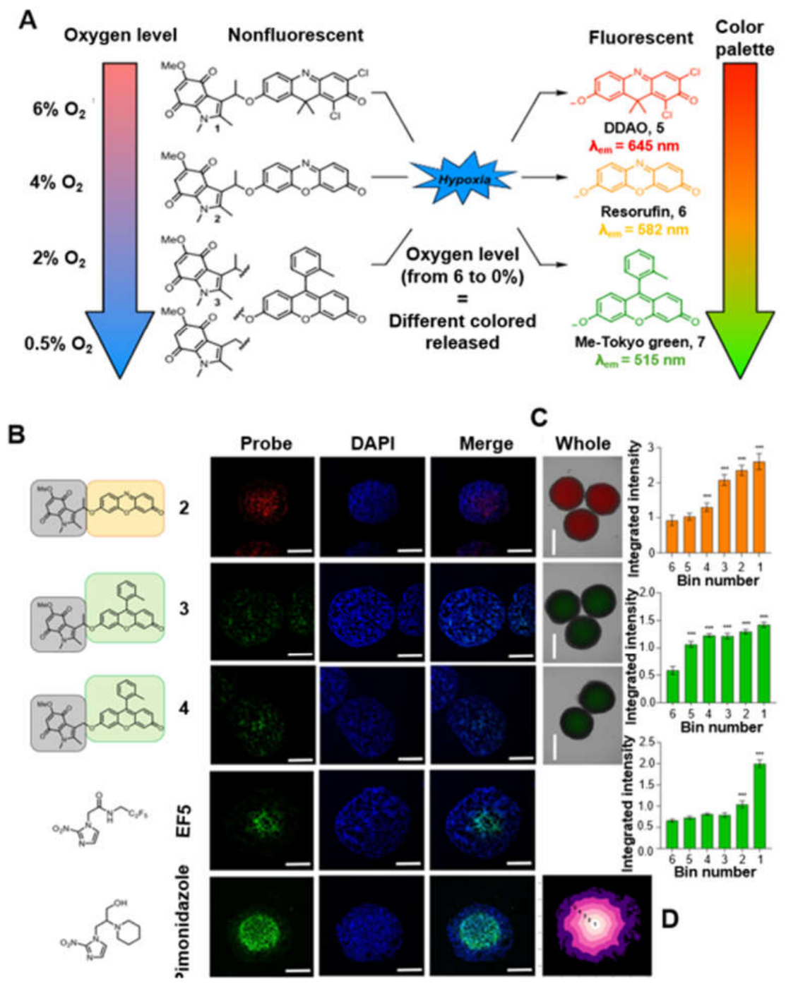
Figure 7.
Bioreductive activation mechanism, chemical stability, and immunological evaluation of the azide-based hypoxia-responsive R848 prodrug R848-N3. (A) Schematic illustration of the hypoxia-dependent, CYP450/NADPH-mediated reduction cascade converting inactive R848-N3 to active R848, which is inhibited under normoxic conditions due to oxygen-mediated redox cycling. (B) High-Performance Liquid Chromatography (HPLC) chromatograms showing the stability and conversion of R848 and $\text{R848-N}_3$ over a time course from 10 minutes to 24 hours under normoxic conditions. (C) Flow cytometric quantification of activated dendritic cells (CD11c+CD80+ and CD11c+MHC II+) following 24-hour co-incubation of bone marrow-derived dendritic cells (BMDCs) with R848 or R848-N3 under normoxic and hypoxic conditions. (D) HPLC chromatograms of R848-N3 stability profiles under hypoxic conditions. (E) Flow cytometric analysis of macrophage polarization markers showing M1 (F4/80+ CD80+) and M2 (F4/80+\CD206+) macrophage populations across PBS, R848, and R848-N3 treatment groups under normoxia and hypoxia (*p < 0.05, p < 0.01, ***p < 0.001, ****p < 0.0001$). Adapted from Sun et al. [60] with permission of Wiley .
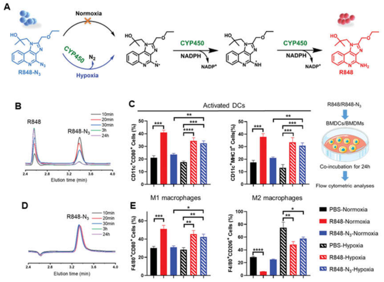
Figure 8.
Chemical design, spectroscopic characterization, fluorescence imaging response, and clonogenic evaluation of metal-based theranostic prodrugs OxaliRes and OxaliNap. Chemical structures of the dual-function platinum(IV) prodrugs OxaliRes (conjugated with a resorufin-derived fluorophore) (A) and OxaliNap (conjugated with a naphthalimide-derived fluorophore) (B). Fluorescence emission spectra of OxaliRes (C) and OxaliNap showing progressive fluorescence enhancement upon activation. Time-dependent fluorescence intensity profiles of OxaliRes (E) and OxaliNap (F) over an 8-hour period. (G-H) Representative fluorescence microscopy images of cells treated with increasing concentrations of sodium ascorbate (NaAsc, from 0 µM to 1–2 mM). Corresponding quantitative analyses of geometric mean fluorescence intensity (MFI) for OxaliRes (I) and OxaliNap (J) treatments across varying ascorbate levels (*p < 0.05, ***p < 0.001). (K) Colony formation assays (clonogenic survival) evaluating the therapeutic efficacy of OxaliNap, OxaliRes, and untreated controls under normoxic (21% O2) and hypoxic (0.1% O2) conditions. Adapted from Boult et al. [61] with permission of ACS.
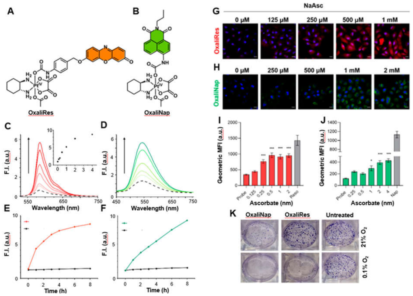

Table 1.
Comprehensive registry of representative hypoxia-responsive prodrugs (HAPs), detailing their chemical classifications, specific mechanisms of bioreductive activation, and primary literature references.
Table 1.
Comprehensive registry of representative hypoxia-responsive prodrugs (HAPs), detailing their chemical classifications, specific mechanisms of bioreductive activation, and primary literature references.
 |
Disclaimer/Publisher’s Note: The statements, opinions and data contained in all publications are solely those of the individual author(s) and contributor(s) and not of MDPI and/or the editor(s). MDPI and/or the editor(s) disclaim responsibility for any injury to people or property resulting from any ideas, methods, instructions or products referred to in the content. |
© 2026 by the authors. Licensee MDPI, Basel, Switzerland. This article is an open access article distributed under the terms and conditions of the Creative Commons Attribution (CC BY) license (http://creativecommons.org/licenses/by/4.0/).
Copyright: This open access article is published under a Creative Commons CC BY 4.0 license, which permit the free download, distribution, and reuse, provided that the author and preprint are cited in any reuse.