

Submitted:
26 September 2026
Posted:
29 September 2026
You are already at the latest version
Abstract

Fruits of Sonneratia apetala (S. apetala) are widely consumed as food and employed in traditional medicine across tropical coastal communities. Nevertheless, their pharmacological potential remains insufficiently explored. Our previous study identified a series of structurally diverse chromone derivatives from the fruits of S. apetala, termed sonnerachromones A–G (SCA–SCG), all of which extended lifespan in Caenorhabditis elegans (C. elegans). In the present study, we further evaluated their anti-aging activities and investigated their underlying molecular mechanisms. Treatment with SCA–SCG significantly enhanced heat stress resistance, reduced lipofuscin accumulation and intracellular reactive oxygen species (ROS) levels. In addition, lipid deposition was significantly decreased by SCA–SCD. Importantly, these beneficial effects were achieved without detectable adverse effects on reproductive capacity. Among the tested compounds, SCA–SCC exhibited the strongst senescence-delaying effect. Mechanistic investigations suggested that these chromone derivatives may potentially bind to telomere-associated proteins POT-1 and POT-2 and thereby influence telomere-related regulatory processes. This was associated with altered expression of telomere-related factors and increased telomerase activity, ultimately correlating with a modest attenuation of telomere shortening. Chromone derivatives from S. apetala, particularly SCA–SCC, exhibit promising anti-aging activity in C. elegans and are associated with coordinated modulation of telomere–telomerase-related processes, including changes in POT-1/POT-2-associated regulation, telomerase activity, and telomere length dynamics. These findings provide new insights into the anti-aging mechanisms of mangrove-derived chromone derivatives and support their potential as lead compounds for the development of natural anti-aging agents.
Keywords:
Sonneratia apetala
; chromone derivatives
; phytochemicals
; anti-aging
; Caenorhabditis elegans
; telomere biology
1. Introduction
Aging is a complex and multifactorial biological process characterized by a progressive decline in physiological functions and the gradual loss of organismal homeostasis [1]. It is widely recognized as a major risk factor for numerous chronic diseases, including metabolic disorders, neurodegeneration, cardiovascular disease, and cancer[2]. At the molecular level, aging is accompanied by the accumulation of cellular damage and dysregulation of interconnected pathways, such as oxidative stress, mitochondrial dysfunction, impaired proteostasis, chronic low-grade inflammation, altered nutrient-sensing signaling, and genomic instability[3,4]. Although several conserved signaling pathways, including insulin/IGF-1 signaling, mTOR signaling, and stress-responsive transcriptional programs, have been shown to influence lifespan and healthspan across species, the mechanisms underlying aging remain incompletely understood. Importantly, aging is not driven by a single causal pathway but rather emerges from coordinated perturbations across multiple cellular systems[5].
Telomeres are specialized nucleoprotein structures located at chromosome ends that play an essential role in maintaining genomic stability [6]. Progressive telomere shortening is widely recognized as a hallmark of cellular aging and organismal senescence [7,8], and accelerated telomere erosion has been associated with premature aging phenotypes in both human disorders and experimental models [9,10]. Accordingly, telomere attrition has been proposed as a potential biomarker of aging and, in some contexts, a predictor of species lifespan [11,12]. In C. elegans, several studies have reported associations between telomere length, longevity, and stress resistance, with worms harboring longer telomeres exhibiting improved survival under heat stress conditions [13,14]. However, other studies indicate that natural variation in telomere length does not necessarily predict lifespan across wild-type strains, suggesting that telomere effects on longevity may be context-dependent and not universally causal [15]. Precisely because this topic remains controversial, it is worthwhile to further explore it within the context of natural product research.
Natural products represent an important source of structurally diverse bioactive compounds and have historically contributed to the discovery of therapeutic agents. Increasing evidence suggests that phytochemicals may exert beneficial effects on aging-related phenotypes by targeting multiple molecular pathways simultaneously, making them particularly attractive candidates for the development of functional foods or nutraceutical interventions aimed at promoting healthy aging. S. apetala is an obligate mangrove species that grows on intertidal mudflats and is native to Bangladesh and Sri Lanka. Its fruits are edible and have traditionally been consumed as pickles, jams, and tonics [16]. In addition to their nutritional value, the fruits have been used in traditional medicine for their analgesic, anthelmintic, and antibacterial properties [17,18]. Since the 1990s, S. apetala has been introduced into coastal regions of Hainan and Guangdong provinces in China [19]. Phytochemical investigations have shown that the fruits are rich in flavonoids, polyphenols, and glycosides, and a growing body of evidence supports their diverse biological activities, including hepatoprotective, antioxidant, anti-aging, and iron-chelating effects [20,21,22].
In our previous study, we isolated a series of structurally diverse chromone derivatives from the fruits of S. apetala, designated sonnerachromones A–G (SCA–SCG), and demonstrated that these compounds significantly extend lifespan and improve healthspan in the model organism Caenorhabditis elegans [23]. Despite these promising bioactivities, the molecular mechanisms underlying their senescence-delaying effects remain largely unknown. Elucidating these mechanisms is essential for evaluating the pharmacological potential of these compounds and for supporting the further development of S. apetala as a functional food resource.
Considering the robust lifespan and healthspan promoting effects of chromone derivatives from S. apetala, we considered it worthwhile to further explore, within the context of natural product intervention, whether these compounds are associated with alterations in telomere-related phenotypes. This exploratory analysis was conducted as part of a broader effort to characterize their potential molecular actions.
In the present study, we show that chromone derivatives enhance stress resistance and reduce the accumulation of lipofuscin, lipids, and reactive oxygen species (ROS) in C. elegans without impairing reproductive fitness. Through activity-guided screening, three compounds (SCA–SCC) characterized by rare C-glycosyl spirocyclic skeletons were identified as exhibiting the most pronounced protective effects against age-related physiological decline. Further analyses suggest that these compounds are associated with increased telomerase activity, together with changes in the expression of telomerase-related genes (trt-1 and TERRA) and potential interactions with telomere-associated proteins POT-1 and POT-2. These molecular alterations were observed alongside a modest attenuation of telomere shortening. Collectively, our results suggest that chromone derivatives influence telomere-associated processes, which may contribute to their anti-aging effects. These processes likely represent one component of the underlying mechanisms, without excluding the contribution of other longevity-regulating pathways. Moreover, these findings support the further exploration of S. apetala fruit as a valuable natural resource for the discovery of bioactive compounds with potential relevance to healthy aging.
2. Results
2.1. Chromone Derivatives Enhance Thermotolerance Without Affecting Reproductive Capacity
To evaluate the effects of SCA–SCG on lifespan, a heat-resistance-based screening assay was performed. Dietary supplementation with chromone derivatives significantly prolonged the lifespan of C. elegans under thermal stress (37 °C, P < 0.05), as demonstrated by standardized survival assays (Figure 1A–E). Among the tested compounds, six significantly increased survival under heat stress, as reflected by a clear rightward shift in survival curves. SCA, SCB, and SCC exhibited lifespan-extending effects comparable to those of the positive control NMN, with a maximum increase in longevity of up to 16 hours. In contrast, SCE did not show a significant effect under these conditions. Overall, with the exception of SCE, the chromone derivatives significantly enhanced thermotolerance in C. elegans by mitigating damage induced by exogenous heat stress, as reflected by reduced mortality during heat challenge.
To determine whether lifespan extension occurred at the expense of physiological fitness, reproductive performance was assessed by quantifying offspring production. No significant differences in average daily egg laying or total brood size between control and compound-treated worms (P > 0.05; Figure 1F–G and Figure S2), indicating that the tested chromone derivatives extend lifespan without altering reproductive capacity.
2.2. Chromone Derivatives Alleviate Age-Associated Physiological Decline in C. elegans
As enhanced stress resistance is often associated with broader health benefits and indicators of slowed aging, we next examined established physiological markers of aging in C. elegans [24]. To evaluate whether the compounds affected age-related deterioration, lipofuscin autofluorescence was quantified. The results showed that treatment with chromone derivatives for 3 and 6 days significantly reduced lipofuscin fluorescence compared with the control group (P < 0.01), indicating reduced age pigment accumulation (Figure 2A and B). However, after 12 days of continuous exposure, SCD and SCE no longer showed significant effects on lipofuscin levels. These results indicate that SCD and SCE exert short-term inhibitory effects on lipofuscin accumulation, whereas prolonged treatment diminishes this effect.
ORO staining revealed that lipid levels were significantly reduced in worms treated with SCA–SCD at day 7 (Figure 2C and D). In contrast, SCE and SCF showed no significant changes (P > 0.05), while SCG exhibited a non-significant trend toward increased lipid accumulation.
2.3. Molecular Docking Predicts Interactions Between SCA–SCC and Telomere-Associated Proteins
To explore potential molecular targets underlying the observed phenotypic effects, we performed in silico molecular docking analysis. Among the tested chromone derivatives, SCA, SCB, and SCC were selected for molecular docking based on their consistent and reproducible bioactivity in the phenotypic assays, thereby allowing a more focused exploration of potential molecular interactions.
Docking simulations were performed to explore the potential interactions between the three most active chromone derivatives (SCA–SCC, Figure 3A) and four telomere-associated proteins (POT-1, POT-2, DTN-1, and DTN-2; Figure 3B). As shown in Figure 3C, all three compounds could be accommodated within the predicted ligand-binding cavities of these proteins, adopting stable poses in a confined pocket region rather than binding nonspecifically on the protein surface. For POT-2, the docking models suggested that SCA–SCC were positioned within a hydrophobic groove surrounded by aromatic residues, allowing close contact with several key amino acids (Figure 3C). The ligand-binding conformations indicated that the chromone core was stabilized through π-related interactions with TRP-225, while additional hydrogen-bonding contacts were predicted with TYR-26 and TYR-145. Notably, the docked poses of SCA and SCC displayed a more compact alignment within the pocket compared with SCB, suggesting subtle stereochemistry-dependent differences in binding orientation.
Consistent with the 3D docking poses, the 2D interaction maps further indicated that the compounds were stabilized by a combination of hydrogen bonding and hydrophobic interactions (Figure 3D). Multiple van der Waals contacts with surrounding residues were observed, together with aromatic interactions that may contribute to binding stabilization. These interaction patterns were conserved across SCA–SCC, supporting the shared chromone scaffold may serve as a key pharmacophore for engaging telomere-associated proteins. Binding affinity analysis showed that all ligand–protein complexes exhibited favorable predicted binding energies (≤ −7.0 kcal/mol; Figure 3E). Among the tested targets, POT-2 consistently showed the strongest predicted binding scores for SCA (−7.9 kcal/mol), SCB (−7.7 kcal/mol), and SCC (−7.8 kcal/mol), whereas slightly weaker but still favorable binding was predicted for POT-1, DTN-1, and DTN-2. Collectively, these in silico results suggest that POT-2 may represent a preferential binding candidate among the evaluated telomere-associated factors. These docking results were used as a hypothesis-generating approach to prioritize potential molecular targets for subsequent experimental validation rather than as direct evidence of physical binding.
2.4. SCA–SCC Are Associated with Altered Expression of Telomere-Associated Proteins
Based on the docking predictions, we next examined whether treatment with SCA, SCB, or SCC was associated with changes in the expression or conformation of telomere-associated proteins in vivo. Western blot analysis revealed that exposure to the compounds reduced the protein levels of POT-1, POT-2, and DTN-1 compared with the control group (Figure 4A–C), while DTN-2 protein levels were not significantly altered (Figure 4D). These results suggest that the compounds might influence the expression of certain telomere-associated proteins. Additionally, molecular docking analysis, which predicts binding interactions between the compounds and these proteins, indicates that the compounds may alter the conformation of the target proteins. The coordinated changes in protein expression and predicted structural alterations support the hypothesis that these compounds may affect telomere-associated regulatory pathways at both the expression and functional levels.
2.5. POT-1 and POT-2 Contribute to Compound-Mediated Lifespan Extension
Based on the docking predictions and protein expression analyses, POT-1 and POT-2 were selected for further functional validation because they are core components of the telomere-binding complex and have been most consistently implicated in telomere regulation in C. elegans.
To examine whether these proteins are functionally involved in the lifespan-extending effects of the compounds, lifespan assays were performed using pot-1 (tm1620) and pot-2 (tm1400) mutant strains. In both mutant backgrounds, the lifespan extension observed in wild-type animals following treatment with SCA, SCB, and SCC was no longer detected (Figure 4E and F). These findings suggest that POT-1 and POT-2 contribute to the lifespan-promoting effects of the compounds and support the hypothesis that telomere-associated regulatory components are functionally linked to the observed phenotype.
2.6. SCA–SCC Are Associated with Changes in Telomerase Activity and Telomere-Related Transcriptional Markers
To further explore whether the compounds influence telomere-associated molecular processes, we assessed telomerase activity using the TRAP assay. Telomere regulation in C. elegans involves telomerase activity and is coordinated by multiple telomere-associated factors, which is generally low or tightly regulated under physiological conditions[25,26]. Treatment with SCA, SCB, and SCC was associated with increased detectable telomerase activity compared with the control group (Figure 5A and B), whereas NMN did not significantly alter telomerase activity under the same experimental conditions.
Consistent with this observation, transcriptional analysis revealed a modest but statistically significant increase in the expression of the telomerase catalytic subunit gene trt-1 following compound treatment (Figure 5C). In addition, expression of telomeric repeat-containing RNA (TERRA), a regulatory component implicated in telomere homeostasis, showed a similar modest but statistically significant increase (Figure 5D).
Taken together, these findings indicate that chromone derivatives are associated with coordinated changes in telomerase activity and telomere-related transcriptional markers, suggesting potential involvement of telomere-associated regulatory pathways in the observed biological effects.
2.7. SCA–SCC Are Associated with Modest Alterations in Telomere Length
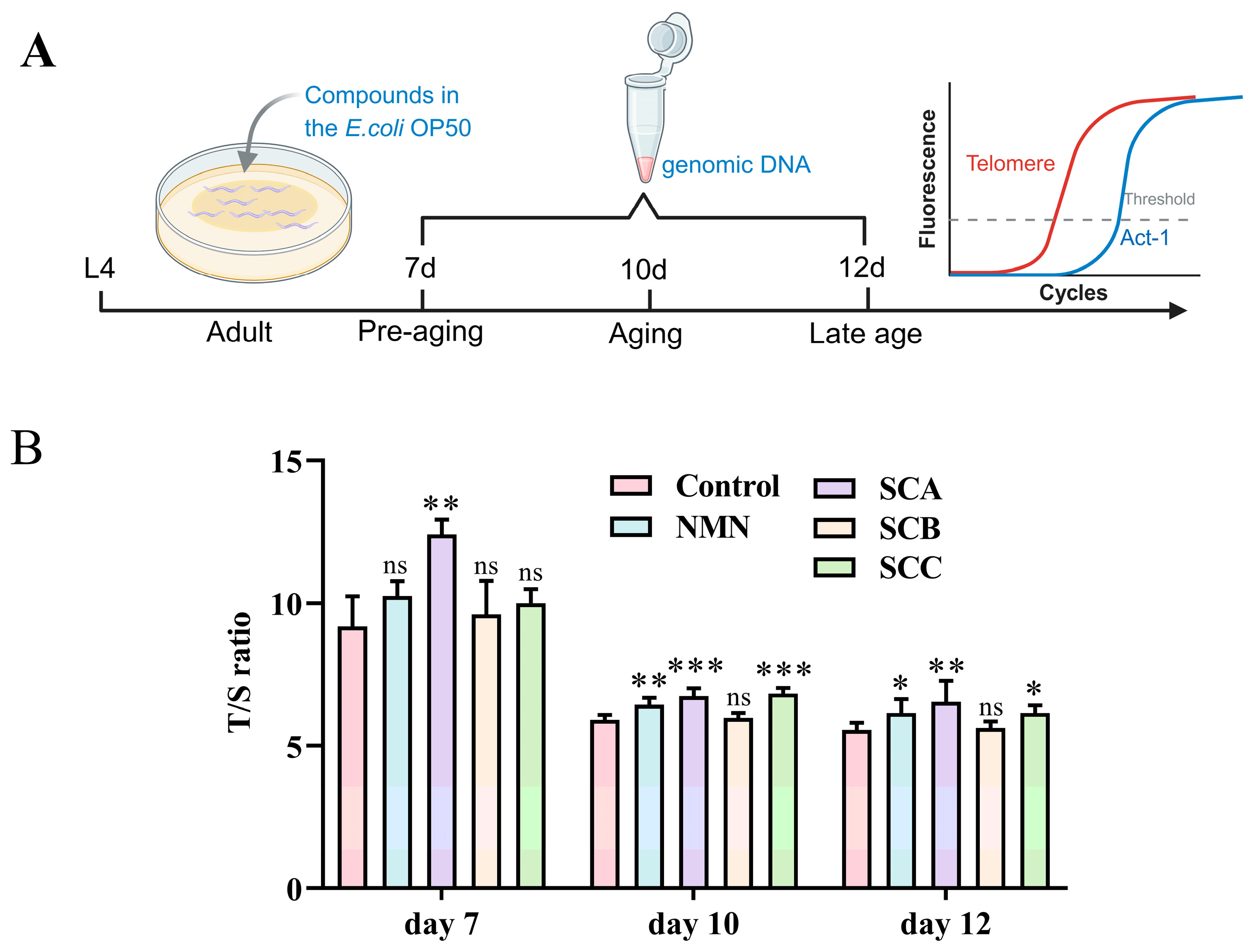
Given the observed effects on telomere-associated proteins and telomerase-related molecular markers, TL was next evaluated as an additional phenotypic endpoint. Quantitative PCR analysis revealed that prolonged treatment with SCA and SCC was associated with a modest but statistically significant attenuation of telomere shortening compared with the control group (Figure 6A and B). In contrast, SCB did not significantly alter telomere length under the same experimental conditions. These findings suggest that, under the experimental conditions tested, some chromone derivatives are associated with detectable changes in telomere dynamics, although the magnitude of these changes is modest.
3. Discussion
The fruits of S. apetala represent an abundant yet underutilized natural resource in China, largely due to limited understanding of their bioactive constituents [27]. In our previous work, we identified a series of chromone derivatives from S. apetala, including three rare C-glycosyl spirocyclic chromones (SCA–SCC), and demonstrated their capacity to extend lifespan and improve healthspan in C. elegans [23]. The present study further expands these findings by providing a systematic evaluation of their physiological and molecular effects associated with aging.
At the organismal level, chromone derivatives consistently enhanced stress resistance and attenuated multiple age-associated phenotypes, including lipofuscin accumulation, lipid deposition, and intracellular ROS levels, without compromising reproductive fitness. Notably, compounds bearing the C-glycosyl spirocyclic scaffold (SCA–SCC) exhibited more pronounced bioactivity than structurally related derivatives lacking this motif, suggesting that this rare structural feature may function as a privileged scaffold contributing to biological efficacy. Given the exceptional scarcity of C-glycosyl spirocyclic chromones among known chromone derivatives. Structurally, among the 754 chromone derivatives reported to date, only 15 possess this scaffold [28]. These findings highlight their potential relevance as lead structures for the development of anti-aging agents derived from natural products.
Beyond phenotypic outcomes, our study explored molecular changes associated with compound treatment. Previous studies suggest that telomere regulation may contribute to aspects of organismal aging and stress adaptation in C. elegans, and that genetic perturbation of telomere-associated factors can influence lifespan and physiological homeostasis [13,14,15]. In C. elegans, POT-1 and POT-2 are canonical telomere-binding proteins that function as negative regulators of telomere length maintenance [29,30]. DTN-1 and DTN-2 have recently been identified as direct interaction partners of POT-1 but exert opposing regulatory effects on telomere dynamics, with DTN-1 promoting telomere elongation and DTN-2 contributing to telomere shortening[31,32,33]. Based on their established roles in telomere homeostasis and their emerging relevance to aging-related phenotypes, these proteins were selected as candidate targets for molecular docking analysis. And the changes were detected at the protein level. We observed coordinated alterations in several telomere-related parameters, including changes in the expression of telomere-binding proteins (POT-1, POT-2, and DTN-1), increased telomerase activity, and a modest attenuation of telomere shortening under the experimental conditions employed. Importantly, these findings do not establish telomere regulation as the primary driver of lifespan extension. Rather, they suggest that chromone derivatives may influence telomere-associated processes as part of a broader biological response linked to improved physiological resilience. Further biochemical and biophysical studies will be required to determine whether direct molecular interactions occur between these compounds and their predicted protein targets.
Comparison with NMN underscores mechanistic distinctions between chromone derivatives and established longevity-promoting compounds. NMN, a well-characterized NAD⁺ precursor [34,35,36]. Extends lifespan primarily through pathways involving SIR-2.1, mitochondrial stress responses, and FOXO (Forkhead box O) signaling [8,37]. NMN administration has been reported to mitigate telomere attrition in mice, whereas comparable effects have not been consistently observed in C. elegans [38]. While NMN modestly attenuated telomere shortening in our study, it did not significantly affect telomerase-related readouts, consistent with previous reports in C. elegans [39,40]. In contrast, chromone derivatives—particularly SCA–SCC—were associated with more pronounced changes in telomere-related molecular features, suggesting partially divergent mechanisms underlying their pro-longevity effects.
Interestingly, subtle stereochemical differences among SCA, SCB, and SCC were associated with distinct molecular outcomes. For example, SCB enhanced telomerase activity without significantly delaying telomere shortening, indicating that telomerase activation alone may be insufficient to confer telomere protection. This observation highlights the complexity of telomere regulation and suggests that multiple regulatory layers collectively shape telomere dynamics and aging-related phenotypes.
Figure 7.
Schematic illustration of the potential mechanisms underlying the lifespan-extending effects of SCA–SCC in C. elegans.
Figure 7.
Schematic illustration of the potential mechanisms underlying the lifespan-extending effects of SCA–SCC in C. elegans.
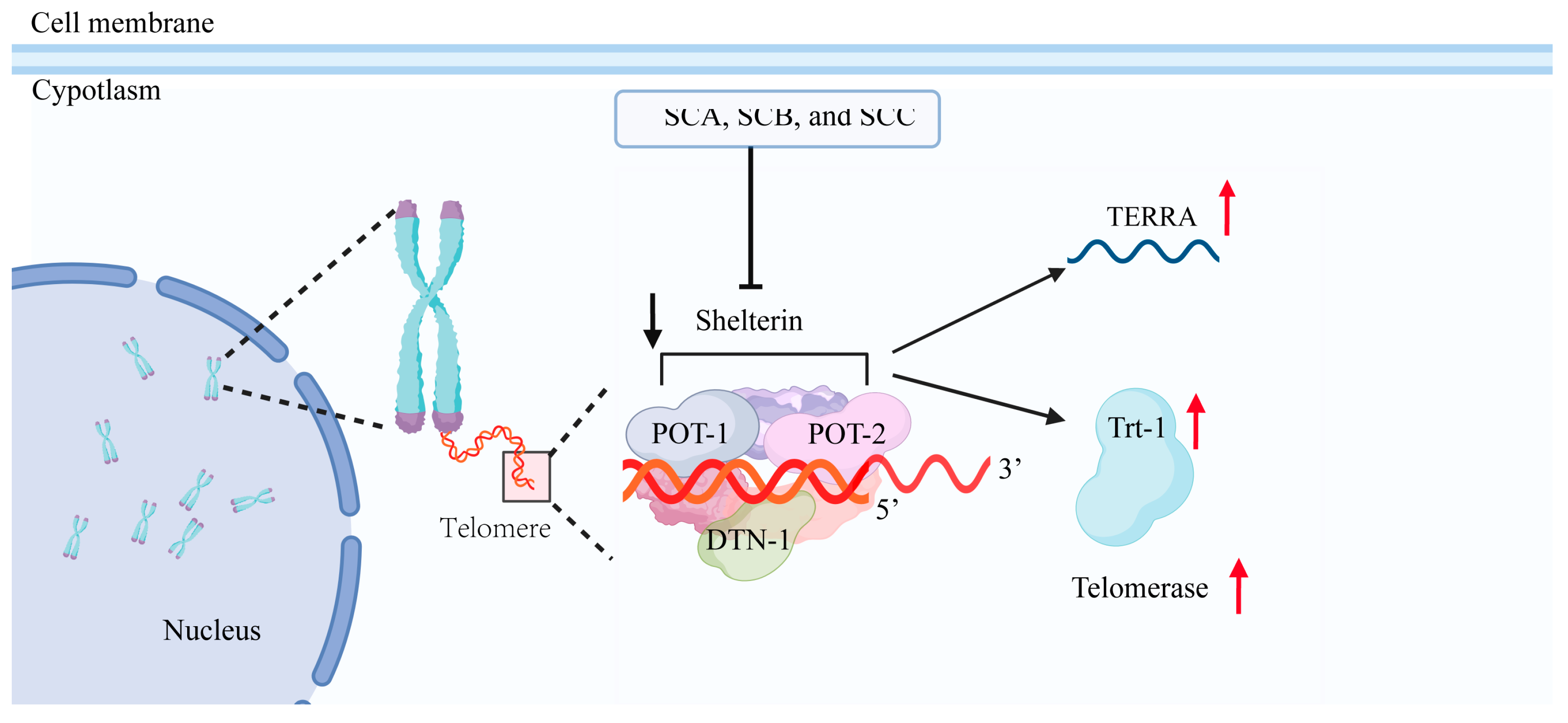
Taken together, our results demonstrate that the chromone derivatives SCA, SCB, and SCC consistently alleviate multiple senescence-associated phenotypes in C. elegans, including reduced accumulation of aging markers and enhanced stress resistance, and are associated with coordinated modulation of telomere-related molecular features. Genetic evidence indicates that POT-1 and POT-2 are required for the longevity-promoting effects of these compounds, as the lifespan extension was abolished in the corresponding mutant backgrounds. However, the observed telomere-related changes, including altered expression of telomere-associated proteins, increased telomerase-associated activity, and modest attenuation of telomere shortening, should not be interpreted as establishing telomere regulation as the primary determinant of lifespan extension. Rather, these findings are consistent with the possibility that modulation of telomere-associated processes represents one component of a broader biological response associated with improved physiological resilience. Although docking analyses raise the possibility that chromone derivatives may physically associate with telomere-related proteins, the present data do not establish direct molecular targeting, and further biochemical studies will be required to clarify the nature of these interactions. In addition, given the well-established roles of canonical longevity-regulating pathways in C. elegans, including insulin/IGF-1 signaling, mitochondrial stress responses, and FOXO-related networks, it is likely that multiple pathways contribute to the observed phenotypes. Overall, this study provides new insights into the biological activities of chromone derivatives from S. apetala and supports the potential of this mangrove species as a valuable source of bioactive compounds for functional foods or nutraceutical applications relevant to healthy aging.
4. Materials and Methods
4.1. Chemical Reagents
Chromone derivatives SCA–SCG were previously isolated from the fruits of the mangrove plant S. apetala, and their chemical structures were characterized and reported in our previous study[23]. The structure of SCA–SCC is shown in Figure 1 A, and the structure of SCD–SCG is shown in the Figure S1. Stock solutions of SCA–SCG were prepared in dimethyl sulfoxide (DMSO) and subsequently diluted with OP50 to obtain working solutions at a final concentration of 100 µM. The final concentration of DMSO was adjusted to 0.1% in all treatment groups. Nicotinamide mononucleotide (NMN) was used as a positive control, while 0.1% DMSO was used as the vehicle control.
β-nicotinamide mononucleotide (NMN; #B02A11T120267) was purchased from Shanghai Yuanye Bio-Technology Co., Ltd. 2’,7’-Dichlorodihydrofluorescein diacetate (H2DCFDA; #L2209540) was obtained from Shanghai Aladdin Biochemical Technology Co., Ltd. Oil Red O (ORO; #1003572129) was purchased from Thermo Scientific. The β-actin antibody (#388168) was purchased from MedChemExpress. Antibodies against POT-1, POT-2, DTN-1, and DTN-2 were kindly provided by Dr. Hiroki Shibuya from the Institute of Physical and Chemical Research (RIKEN), Japan.
4.2. Caenorhabditis Elegans Strains and Maintenance
C. elegans strains were maintained on nematode growth medium (NGM) plates seeded with Escherichia coli OP50 (OP50) at 20 °C under standard laboratory conditions unless otherwise specified. The N2 wild-type strain was obtained from the Caenorhabditis Genetics Center (CGC). The mutant strains pot-1 (tm1620) and pot-2 (tm1400) were obtained from the National Bio Resource Project (NBRP), Japan.
4.3. Grouping and Drug Dietary Intervention
Synchronous eggs were obtained by bleaching gravid adults using a freshly prepared solution of sodium hypochlorite (10%) and sodium hydroxide (5 M). Eggs were allowed to hatch and develop at 20 °C, and L4-stage worms (approximately 48 hours post-hatching) were transferred onto NGM plates supplemented with test compounds. To ensure continuous exposure, worms were transferred daily to fresh plates containing the respective compounds.
4.4. Lifespan Assays in Mutant Strains
Lifespan assays were performed using pot-1 (tm1620) and pot-2 (tm1400) mutant strains. Synchronous L4-stage worms (60 per group) were transferred onto NGM plates with or without compounds. The day of transfer was defined as day 0. Worms were transferred daily to fresh treatment plates. Animals that crawled off the plate, burrowed into the agar, or exhibited internal hatching were excluded. Death was scored when worms failed to respond to gentle mechanical stimulation and showed no pharyngeal pumping. Each assay was independently repeated three times.
4.5. Heat Stress Resistance Assays
Worms were treated with compounds from the L4 stage for four days. Animals were then transferred to fresh NGM plates without OP50 and subjected to heat stress at 37 °C. Survival was monitored every 2 hours until all worms had died. Worms escaping from plates or burrowing were excluded. Death was defined as the absence of spontaneous movement and pharyngeal pumping and lack of response to platinum wire stimulation.
4.6. Lipofuscin Assays
Lipofuscin accumulation was assessed on days 3, 6, and 12 after compound treatment. Worms were washed three times with PBS and anesthetized using 10 mM levamisole before mounting on agarose pads (2%). Autofluorescence was observed using a Leica DMI8 fluorescence microscope under UV excitation (365–390 nm). Exposure time and imaging parameters were kept constant across groups. Fluorescence intensity was quantified using Fiji (ImageJ) software.
4.7. Oil Red O (ORO) Staining
After 7 days of treatment, worms were collected and washed three times with PBS. Samples were fixed in 4% paraformaldehyde for 15 min and frozen at −80 °C for 10 min. After thawing, worms were stained with freshly prepared ORO working solution for 2 hours in the dark. The ORO staining solution was prepared one day prior to the experiment by mixing 40 mL of distilled water with 60 mL of the ORO stocking solution. Then, the sample was agitated on an automatic stirrer for 2 hours and filtered through a 0.2 µm syringe filter. Once the staining procedure was completed, excess dye was removed by washing with PBS. Images were acquired using a Leica DMI8 microscope and lipid content was quantified using Fiji software.
4.8. Reactive Oxygen Species (ROS) Assay
Worms treated for 3 days were collected and incubated with 50 µM H2DCFDA in PBS for 2 hours in the dark following fixation in 4% paraformaldehyde. After incubation in the dark for 2 hours, the worms were washed three times with PBS to remove the H2DCFDA staining solution. Worms were mounted on agarose pads (2%) and imaged under blue excitation (450–490 nm). Fluorescence intensity was quantified using Fiji (ImageJ) software.
4.9. Molecular Docking
Molecular docking simulations were performed using AutoDock Vina (version 4.2.6). Structures of DTN-1 (PDB ID: 8U8M) and DTN-2 (PDB ID: 8U8L) were obtained from the RCSB Protein Data Bank. Amino acid sequences of POT-1 (UniProt: P42001) and POT-2 (UniProt: O45595) were retrieved from UNIPROT and corresponding structures were predicted using AlphaFold. Docking scores were expressed as estimated binding free energy (ΔG, kcal/mol).
4.10. Western Blot Analysis
After 10 days of treatment, worms were harvested and lysed in RIPA buffer containing 1 mM PMSF. After sonication on ice, protein extracts were obtained by centrifugation at 12,000 rpm for 20 min at 4 °C. The supernatant was carefully collected for Western blot analysis. Equal amounts of protein denatured by boiling at 100 ℃ for 10 min and then load onto an 8%~12% proteinaceous gel. Electrophoresis was running at 160 V for 40 ~ 60 min in tris-glycine buffer and transferred to PVDF membranes. Membranes were incubated with primary antibodies followed by appropriate secondary antibodies and visualized using standard chemiluminescence procedures.
4.11. Telomerase Activity Assay (TRAP)
Telomerase activity in C. elegans was evaluated using the telomeric repeat amplification protocol (TRAP) assay. After 10 days of treatment with the test compounds, worms were collected and lysed in 500 µL of lysis buffer consisting of 10 mM Tris-HCl (pH 8.0), 1 mM EGTA, 5 mM MgCl2, 10 mM β-mercaptoethanol, 0.1% PMSF, 0.5% CHAPS, and 10% glycerol. Worm tissues were disrupted using an ultrasonic cell disruptor, followed by incubation on ice for 30 min to ensure complete lysis. The lysates were then centrifuged at 12,000 rpm at 4 °C for 15 min, and the supernatants were collected. Protein concentrations were determined using a BCA protein assay kit (Solarbio). For each reaction, 10 µg of total protein, 2 µL of worm lysate, PCR reaction master mix, and telomerase primers TS (AATCCGTCGAGCAGAGTT), RP (GCGCGGCTTACCCTTACCCTTACCCTAACC), NT (ATCGCTTCTCGGCCTTTT), and TSNT (AATCCGTCGAGCAGAGTTAAAAGGCCGAGAAGCGAT) were combined to prepare the reaction mixture. To inactivate telomerase, samples were incubated at 37 °C for 30 min prior to amplification. PCR amplification was performed under the following conditions: initial denaturation at 94 °C for 5 min; 35 cycles of 94 °C for 30 s, 55 °C for 30 s, and 72 °C for 30 s; followed by a final extension at 72 °C for 5 min. The PCR products were separated on 10% native PAGE gels (acrylamide/bisacrylamide ratio of 29:1) in 1× TBE buffer at 150 V for 40 min. Gels were stained with SYBR Green I, visualized using an ultraviolet gel imaging system, and quantified using Fiji (ImageJ) software.
4.12. Quantitative PCR (q-PCR)
Total RNA was extracted from approximately 800 worms after 10 days of treatment using TRIzol reagent. cDNA was synthesized using a commercial reverse transcription kit. Quantitative PCR was performed to determine the expression levels of trt-1 and TERRA, using act-1 as the internal reference gene. Relative expression was calculated using the 2−ΔΔCT method. Primer sequences are provided in Table S2.
4.13. Telomere Length Analysis
Genomic DNA was extracted from approximately 1000 worms per group at the designated time points. Worms were first collected, washed three times with PBS to remove residual bacteria, and then subjected to genomic DNA extraction using the Universal Genomic DNA Purification Mini Spin Kit according to the manufacturer’s instructions. The integrity of the extracted DNA was assessed by agarose gel electrophoresis, and DNA concentration and purity were determined using a Nano Drop One spectrophotometer (Thermo Scientific). Only samples with an A260/A280 ratio close to 1.80 were used for subsequent analysis.
Relative telomere length (TL) was measured using a quantitative real-time PCR (qPCR) method as previously described [41]. Primers designed for C. elegans telomere analysis by Cook et al. were used [42]. Each qPCR reaction contained 20 ng of genomic DNA, 2 × Q3 SYBR qPCR Master Mix (Universal), and was performed on a Light Cycler 480 II Real-Time PCR system (Roche Applied Science, Switzerland). The act-1 gene was used as the single-copy reference gene. The primer sequences for act-1 were as follows: forward primer, GTCGGTATGGGACAGAAGGA; reverse primer, GCTTCAGTGAGGAGG
ACTGG. The TL was measured by comparing the ratio of T repeat copy number to the S copy number (act-1), which was expressed as the telomere length (T/S) ratio. (T/S) ratio= [2Ct (telomeres) /2Ct (act-1)]-1=2−ΔCt.
4.14. Brood Size Measurement
To obtain synchronized eggs, gravid adult worms were transferred onto NGM plates seeded with OP50 and allowed to lay eggs for 3 hours. The adults were then carefully removed using a platinum wire, and the eggs were incubated at 20 °C until they reached the L4 stage. The L4 larvae were randomly selected and transferred to NGM plates supplemented with different compounds. Worms were transferred to fresh NGM plates containing the test compounds every 24 hours. Plates containing the offspring were incubated at 20 °C for 48 hours, after which the number of hatched individuals was counted using a stereomicroscope.
4.15. Statistical Analysis
All data were analyzed using SPSS 26.0. One-way ANOVA was applied followed by LSD or Games–Howell post hoc tests depending on data distribution and variance homogeneity. Lifespan data were analyzed using Kaplan–Meier survival analysis. Results were expressed as mean ± SEM unless otherwise stated. “ns” indicates no statistical difference. Differences were considered statistically significant at P < 0.05. An asterisk (*) represents P < 0.05, two asterisks (**) signify P < 0.01, and three asterisks (***) indicate P < 0.001.
Supplementary Materials
The following supporting information can be downloaded at website of this paper posted on Preprints.org, Figure S1: Structures of compounds SCA–SCC; Figure S2: total brood size of worms fed with SCA–SCG (n=10); Figure S3: Representative fluorescence-based visualization of ROS levels in nematodes after treatment with SCA–SCG; Figure S4: Detailed docking conformations of SCA, SCB, and SCC with POT-1, DTN-1, and DTN-2. Table S1: Statistical analysis of lifespan assays in Caenorhabditis elegans strains treated with sonnerachromones and NMN; Table S2: Lifespan assay statistics of Caenorhabditis elegans mutant strains treated with SCA–SCC; Table S3: Primer sequences used for quantitative real-time PCR analysis.
Author Contributions
Conceptualization, J.L., C.G. and X.Y.; methodology, J.L.; formal analysis, J.L.; F.Y. and X.C.; investigation, J.L.; F.Y., X.C. and J.W.; resources, J.L.; data curation, J.L.; writing—original draft preparation, J.L.; writing—review and editing, M.Z., C.G. and X.Y.; visualization, L.W. and K.L.; supervision, X.J., C.G. and X.Y.; project administration, C.G. and X.Y.; funding acquisition, C.G. and X.Y. All authors have read and agreed to the published version of the manuscript.
Funding
This research was funded by National Natural Science Foundation of China (42466005), the Guangxi Science and Technology Program (LT2600640033), the Guangxi Key Research and Development Project (AB24010109), the Guangxi Young Qihuang Project (GXQH202412), the Development Program of High-Level Talent Teamunder the Qihuang Project of Guangxi University of Chinese Medicine (202404), and Guangxi Peak Discipline: Traditional Chinese Medicine (Chinese Materia Medica) (Document No. 2 [2026] of the Department of Education of Guangxi for Scientific Research).
Acknowledgments
We thank Dr. Hiroki Shibuya for sharing with us the protein antibodies used in the Western blotting.
Conflicts of Interest
The authors have declared that no competing interest exists.
Abbreviations
The following abbreviations are used in this manuscript:
| SCA–SCG | sonnerachromones A–G |
| SCA–SCC | sonnerachromones A–C |
| S. apetala | Sonneratia apetala |
| C. elegans | Caenorhabditis elegans |
| ROS | reactive oxygen species |
| NGM | nematode growth media |
| OP50 | Escherichia coli OP50 |
| ORO | Oil Red O |
| H2DCFDA | 2’,7’-Dichlorodihydrofluorescein diacetate |
| POT-1 | protection of telomeres 1 |
| POT-2 | protection of telomeres 2 |
| DTN-1 | double-strand telomeric DNA-binding proteins 1 |
| DTN-2 | double-strand telomeric DNA-binding proteins 2 |
| Trt-1 | Telomerase reverse transcriptase |
| TERRA | doublesex and mab-3 related transcription factor 2 |
References
- Kemoun, P.; Ader, I.; Planat-Benard, V.; Dray, C.; Fazilleau, N.; Monsarrat, P.; Cousin, B.; Paupert, J.; Ousset, M.; Lorsignol, A.; et al. A Gerophysiology Perspective on Healthy Ageing. Ageing Res. Rev. 2022, 73, 101537. [Google Scholar] [CrossRef] [PubMed]
- Guo, J.; Huang, X.; Dou, L.; Yan, M.; Shen, T.; Tang, W.; Li, J. Aging and Aging-Related Diseases: From Molecular Mechanisms to Interventions and Treatments. Sig Transduct. Target Ther. 2022, 7, 391. [Google Scholar] [CrossRef] [PubMed]
- Li, Y.; Tian, X.; Luo, J.; Bao, T.; Wang, S.; Wu, X. Molecular Mechanisms of Aging and Anti-Aging Strategies. Cell Commun. Signal 2024, 22, 285. [Google Scholar] [CrossRef] [PubMed]
- Ross, J.M.; Olson, L.; Coppotelli, G. Mitochondrial Dysfunction and Protein Homeostasis in Aging: Insights from a Premature-Aging Mouse Model. Biomolecules 2024, 14, 162. [Google Scholar] [CrossRef] [PubMed]
- López-Otín, C.; Blasco, M.A.; Partridge, L.; Serrano, M.; Kroemer, G. Hallmarks of Aging: An Expanding Universe. Cell 2023, 186, 243–278. [Google Scholar] [CrossRef] [PubMed]
- Jones-Weinert, C.; Mainz, L.; Karlseder, J. Telomere Function and Regulation from Mouse Models to Human Ageing and Disease. Nat. Rev. Mol. Cell Biol. 2025, 26, 297–313. [Google Scholar] [CrossRef] [PubMed]
- Daios, S.; Anogeianaki, A.; Kaiafa, G.; Kontana, A.; Veneti, S.; Gogou, C.; Karlafti, E.; Pilalas, D.; Kanellos, I.; Savopoulos, C. Telomere Length as a Marker of Biological Aging: A Critical Review of Recent Literature. Curr. Med. Chem. 2022, 29, 5478–5495. [Google Scholar] [CrossRef] [PubMed]
- Huang, X.; Huang, L.; Lu, J.; Cheng, L.; Wu, D.; Li, L.; Zhang, S.; Lai, X.; Xu, L. The Relationship between Telomere Length and Aging-Related Diseases. Clin. Exp. Med. 2025, 25, 72. [Google Scholar] [CrossRef] [PubMed]
- Ahmed, M.S.; Ikram, S.; Bibi, N.; Mir, A. Hutchinson-Gilford Progeria Syndrome: A Premature Aging Disease. Mol. Neurobiol. 2018, 55, 4417–4427. [Google Scholar] [CrossRef] [PubMed]
- Wang, B.; Kou, H.; Wang, Y.; Zhang, Q.; Jiang, D.; Wang, J.; Zhao, Z.; Zhou, Y.; Zhang, M.; Sui, L.; et al. LAP2α Orchestrates Alternative Lengthening of Telomeres Suppression through Telomeric Heterochromatin Regulation with HDAC1: Unveiling a Potential Therapeutic Target. Cell Death Dis. 2024, 15, 761. [Google Scholar] [CrossRef] [PubMed]
- Whittemore, K.; Vera, E.; Martínez-Nevado, E.; Sanpera, C.; Blasco, M.A. Telomere Shortening Rate Predicts Species Life Span. Proc. Natl. Acad. Sci. U S A 2019, 116, 15122–15127. [Google Scholar] [CrossRef] [PubMed]
- Eastwood, J.R.; Dupoué, A.; Delhey, K.; Verhulst, S.; Cockburn, A.; Peters, A. When Does Early-Life Telomere Length Predict Survival? A Case Study and Meta-Analysis. Mol. Ecol. 2023, 32, 3000–3013. [Google Scholar] [CrossRef] [PubMed]
- Joeng, K.S.; Song, E.J.; Lee, K.-J.; Lee, J. Long Lifespan in Worms with Long Telomeric DNA. Nat. Genet 2004, 36, 607–611. [Google Scholar] [CrossRef] [PubMed]
- Park, M.C.; Park, D.; Lee, E.-K.; Park, T.; Lee, J. Genomic Analysis of the Telomeric Length Effect on Organismic Lifespan in Caenorhabditis Elegans. Biochem Biophys. Res. Commun. 2010, 396, 382–387. [Google Scholar] [CrossRef] [PubMed]
- Raices, M.; Maruyama, H.; Dillin, A.; Karlseder, J. Uncoupling of Longevity and Telomere Length in C. Elegans. PLoS Genet 2005, 1, e30. [Google Scholar] [CrossRef] [PubMed]
- Mithila, M.; Islam, M.R.; Khatun, M.R.; Gazi, M.S.; Hossain, S.J. Sonneratia Apetala (Buch.-Ham.) Fruit Extracts Ameliorate Iron Overload and Iron-Induced Oxidative Stress in Mice. Prev. Nutr. Food Sci. 2023, 28, 278–284. [Google Scholar] [CrossRef] [PubMed]
- Shefa, A.A.; Baishakhi, F.S. Phytochemical and Pharmacological Evaluation of Fruits of Sonneratia Apetala. Glob. J. Med. Res. 2014, 14, 1–6. [Google Scholar]
- Hossain, S.J.; Islam, M.R.; Pervin, T.; Iftekharuzzaman, M.; Hamdi, O.A.A.; Mubassara, S.; Saifuzzaman, M.; Shilpi, J.A. Antibacterial, Anti-Diarrhoeal, Analgesic, Cytotoxic Activities, and GC-MS Profiling of Sonneratia Apetala (Buch.-Ham.) Seed. Prev. Nutr. Food Sci. 2017, 22, 157–165. [Google Scholar] [CrossRef] [PubMed]
- Jiang, S.; Jiang, C.-P.; Cao, P.; Liu, Y.-H.; Gao, C.-H.; Yi, X.-X. Sonneradon A Extends Lifespan of Caenorhabditis Elegans by Modulating Mitochondrial and IIS Signaling Pathways. Mar. Drugs 2022, 20, 59. [Google Scholar] [CrossRef] [PubMed]
- Liu, J.; Luo, D.; Wu, Y.; Gao, C.; Lin, G.; Chen, J.; Wu, X.; Zhang, Q.; Cai, J.; Su, Z. The Protective Effect of Sonneratia Apetala Fruit Extract on Acetaminophen-Induced Liver Injury in Mice. Evid. Based Complement Altern. Med. 2019, 2019, 6919834. [Google Scholar] [CrossRef] [PubMed]
- Yi, X.; Jiang, S.; Qin, M.; Liu, K.; Cao, P.; Chen, S.; Deng, J.; Gao, C. Compounds from the Fruits of Mangrove Sonneratia Apetala: Isolation, Molecular Docking and Antiaging Effects Using a Caenorhabditis Elegans Model. Bioorg Chem. 2020, 99, 103813. [Google Scholar] [CrossRef] [PubMed]
- Uddin, M.R.; Akhter, F.; Abedin, M.J.; Shaikh, M.A.A.; Al Mansur, M.A.; Saydur Rahman, M.; Molla Jamal, A.S.I.; Akbor, M.A.; Hossain, M.H.; Sharmin, S.; et al. Comprehensive Analysis of Phytochemical Profiling, Cytotoxic and Antioxidant Potentials, and Identification of Bioactive Constituents in Methanoic Extracts of Sonneratia Apetala Fruit. Heliyon 2024, 10, e33507. [Google Scholar] [CrossRef] [PubMed]
- Xia, J.-L.; Liu, J.-Y.; Liu, K.; Li, Y.-Y.; Bai, M.; Gao, C.-H.; Yi, X.-X. Sonnerachromones A-G, Antiaging Chromones from the Fruit of Mangrove Sonneratia Apetala. J. Nat. Prod. 2025, 88, 1980–1987. [Google Scholar] [CrossRef] [PubMed]
- Soo, S.K.; Rudich, Z.D.; Ko, B.; Moldakozhayev, A.; AlOkda, A.; Van Raamsdonk, J.M. Biological Resilience and Aging: Activation of Stress Response Pathways Contributes to Lifespan Extension. Ageing Res. Rev. 2023, 88, 101941. [Google Scholar] [CrossRef] [PubMed]
- Cheung, I.; Schertzer, M.; Rose, A.; Lansdorp, P.M. High Incidence of Rapid Telomere Loss in Telomerase-Deficient Caenorhabditis Elegans. Nucleic Acids Res. 2006, 34, 96–103. [Google Scholar] [CrossRef] [PubMed]
- Roake, C.M.; Artandi, S.E. Regulation of Human Telomerase in Homeostasis and Disease. Nat. Rev. Mol. Cell Biol. 2020, 21, 384–397. [Google Scholar] [CrossRef] [PubMed]
- Yasmine, S.; Proma, R.Z.; Uddin, Md.R.; Rahman, M.S.; Islam, Md.H.; Mansur, M.A.A.; Jamal, A.S.I.M.; Yousuf, A.; Mustak, Md.H.; Kamruzzaman, S.; et al. Exploring the Multifaceted Roles of Sonneratia Apetala and Nipa Fruticans in Coastal Habitat Restoration and Bioactive Properties Discovery. Phytomed. Plus 2025, 5, 100687. [Google Scholar] [CrossRef]
- Zhang, Y.-Q.; Zhang, M.; Wang, Z.-L.; Qiao, X.; Ye, M. Advances in Plant-Derived C-Glycosides: Phytochemistry, Bioactivities, and Biotechnological Production. Biotechnol. Adv. 2022, 60, 108030. [Google Scholar] [CrossRef] [PubMed]
- Shtessel, L.; Lowden, M.R.; Cheng, C.; Simon, M.; Wang, K.; Ahmed, S. Caenorhabditis Elegans POT-1 and POT-2 Repress Telomere Maintenance Pathways. G3 2013, 3, 305–313. [Google Scholar] [CrossRef] [PubMed]
- Yu, X.; Gray, S.; Ferreira, H.C. POT-3 Preferentially Binds the Terminal DNA-Repeat on the Telomeric G-Overhang. Nucleic Acids Res. 2023, 51, 610–618. [Google Scholar] [CrossRef] [PubMed]
- Yamamoto, I.; Zhang, K.; Zhang, J.; Vorontsov, E.; Shibuya, H. Telomeric Double-Strand DNA-Binding Proteins DTN-1 and DTN-2 Ensure Germline Immortality in Caenorhabditis Elegans. eLife 2021, 10, e64104. [Google Scholar] [CrossRef] [PubMed]
- Dietz, S.; Almeida, M.V.; Nischwitz, E.; Schreier, J.; Viceconte, N.; Fradera-Sola, A.; Renz, C.; Ceron-Noriega, A.; Ulrich, H.D.; Kappei, D.; et al. The Double-Stranded DNA-Binding Proteins TEBP-1 and TEBP-2 Form a Telomeric Complex with POT-1. Nat. Commun. 2021, 12, 2668. [Google Scholar] [CrossRef] [PubMed]
- Padmanaban, S.; Lambacher, N.J.; Tesmer, V.M.; Zhang, J.; Shibuya, H.; Nandakumar, J. Caenorhabditis Elegans Telomere-Binding Proteins TEBP-1 and TEBP-2 Adapt the Myb Module to Dimerize and Bind Telomeric DNA. Proc. Natl. Acad. Sci. U S A 2024, 121, e2316651121. [Google Scholar] [CrossRef] [PubMed]
- Xu, C.; Dai, J.; Ai, H.; Du, W.; Ji, H. β-Nicotinamide Mononucleotide Promotes Cell Proliferation and Hair Growth by Reducing Oxidative Stress. Molecules 2024, 29, 798. [Google Scholar] [CrossRef] [PubMed]
- Deng, H.; Ding, D.; Ma, Y.; Zhang, H.; Wang, N.; Zhang, C.; Yang, G. Nicotinamide Mononucleotide: Research Process in Cardiovascular Diseases. Int. J. Mol. Sci. 2024, 25, 9526. [Google Scholar] [CrossRef] [PubMed]
- Yaku, K.; Palikhe, S.; Iqbal, T.; Hayat, F.; Watanabe, Y.; Fujisaka, S.; Izumi, H.; Yoshida, T.; Karim, M.; Uchida, H.; et al. Nicotinamide Riboside and Nicotinamide Mononucleotide Facilitate NAD+ Synthesis via Enterohepatic Circulation. Sci. Adv. 2025, 11, eadr1538. [Google Scholar] [CrossRef] [PubMed]
- Viswanathan, M.; Kim, S.K.; Berdichevsky, A.; Guarente, L. A Role for SIR-2.1 Regulation of ER Stress Response Genes in Determining C. Elegans Life Span. Dev. Cell 2005, 9, 605–615. [Google Scholar] [CrossRef] [PubMed]
- Niu, K.-M.; Bao, T.; Gao, L.; Ru, M.; Li, Y.; Jiang, L.; Ye, C.; Wang, S.; Wu, X. The Impacts of Short-Term NMN Supplementation on Serum Metabolism, Fecal Microbiota, and Telomere Length in Pre-Aging Phase. Front Nutr. 2021, 8, 756243. [Google Scholar] [CrossRef] [PubMed]
- Yang, L.; Shen, J.; Liu, C.; Kuang, Z.; Tang, Y.; Qian, Z.; Guan, M.; Yang, Y.; Zhan, Y.; Li, N.; et al. Nicotine Rebalances NAD+ Homeostasis and Improves Aging-Related Symptoms in Male Mice by Enhancing NAMPT Activity. Nat. Commun. 2023, 14, 900. [Google Scholar] [CrossRef] [PubMed]
- Zhou, D.; Sun, Y.; Dong, C.; Wang, Z.; Zhao, J.; Li, Z.; Huang, G.; Li, W. Folic Acid Alleviated Oxidative Stress-Induced Telomere Attrition and Inhibited Apoptosis of Neurocytes in Old Rats. Eur. J. Nutr. 2024, 63, 291–302. [Google Scholar] [CrossRef] [PubMed]
- Cawthon, R.M. Telomere Measurement by Quantitative PCR. Nucleic Acids Res. 2002, 30, e47. [Google Scholar] [CrossRef] [PubMed]
- Cook, D.E.; Zdraljevic, S.; Tanny, R.E.; Seo, B.; Riccardi, D.D.; Noble, L.M.; Rockman, M.V.; Alkema, M.J.; Braendle, C.; Kammenga, J.E.; et al. The Genetic Basis of Natural Variation in Caenorhabditis Elegans Telomere Length. GENETICS 2016, 204, 371–383. [Google Scholar] [CrossRef] [PubMed]
Figure 1.
SCA–SCG (100 µM) enhance thermotolerance without affecting reproductive capacity in C. elegans. (A) Structures of compounds SCA–SCC. (B, C) Mean survival time of worms treated with SCA–SCG under heat stress (37 °C). (D, E) Survival curves of worms exposed to 37 °C following treatment with SCA–SCG. (F, G) Average daily egg laying of worms fed with SCA–SCG (n=10). Data are presented as mean ± SEM. ns, p > 0.05; * P < 0.05; ** P < 0.01; *** P < 0.001.
Figure 1.
SCA–SCG (100 µM) enhance thermotolerance without affecting reproductive capacity in C. elegans. (A) Structures of compounds SCA–SCC. (B, C) Mean survival time of worms treated with SCA–SCG under heat stress (37 °C). (D, E) Survival curves of worms exposed to 37 °C following treatment with SCA–SCG. (F, G) Average daily egg laying of worms fed with SCA–SCG (n=10). Data are presented as mean ± SEM. ns, p > 0.05; * P < 0.05; ** P < 0.01; *** P < 0.001.
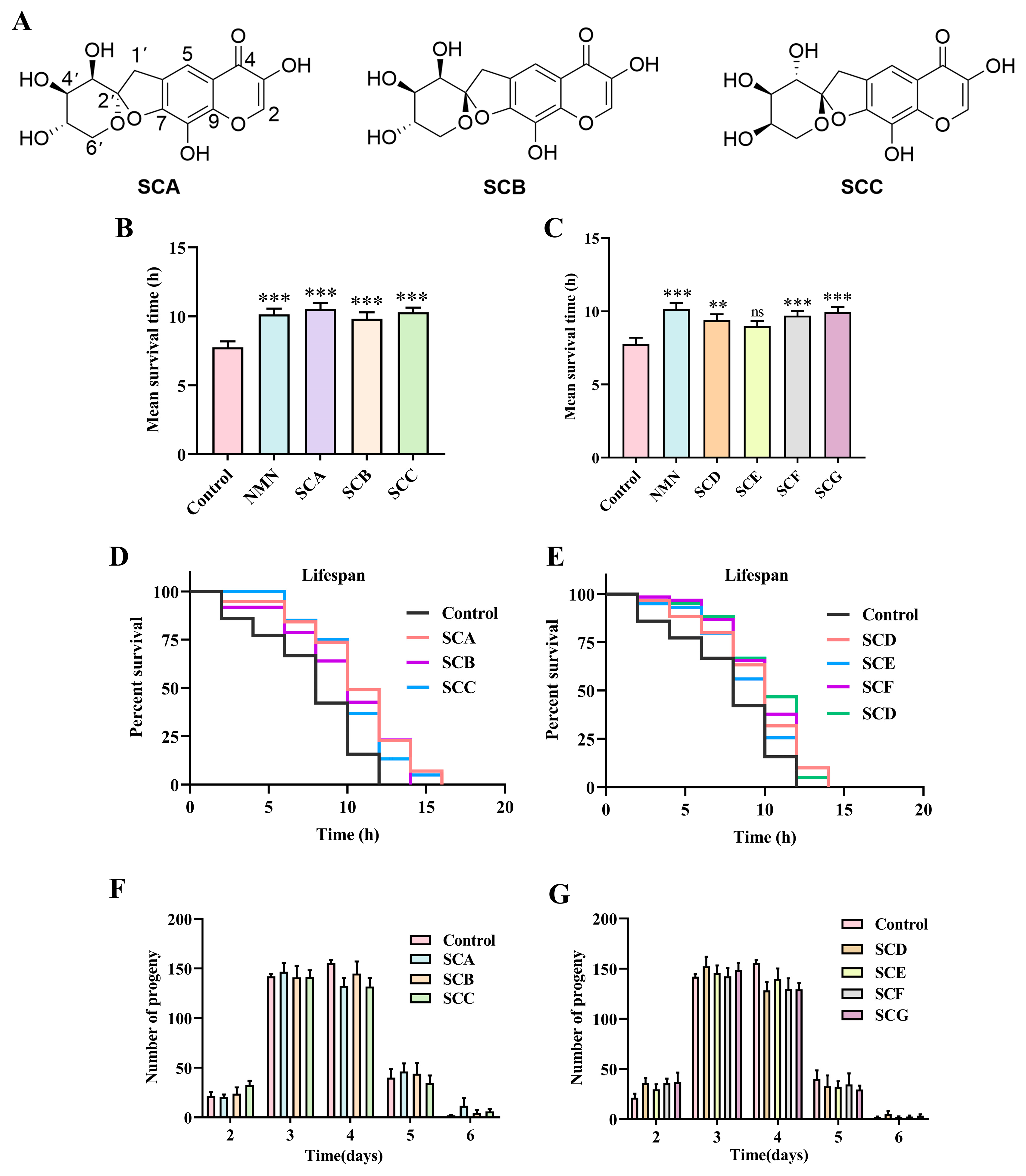
Figure 2.
SCA–SCG (100 µM) attenuate age-related physiological decline in C. elegans. (A) Representative fluorescence images of lipofuscin accumulation in worms treated with the indicated compounds for 12 days. Scale bar = 100 µm. (B) Quantification of lipofuscin fluorescence intensity. Data are presented as mean ± SD (n = 10 worms). Experiments were performed in triplicate. (C) Representative images showing lipid staining in worms treated with different compounds. Scale bar = 75 µm. (D) Quantification of lipid levels based on fluorescence intensity (n = 25 worms). Data are presented as mean ± SD. (E) Quantification of intracellular ROS levels in worms following compound treatment (n = 10 worms). Data are presented as mean ± SEM. ns, P > 0.05; * P < 0.05; ** P < 0.01; *** P < 0.001.
Figure 2.
SCA–SCG (100 µM) attenuate age-related physiological decline in C. elegans. (A) Representative fluorescence images of lipofuscin accumulation in worms treated with the indicated compounds for 12 days. Scale bar = 100 µm. (B) Quantification of lipofuscin fluorescence intensity. Data are presented as mean ± SD (n = 10 worms). Experiments were performed in triplicate. (C) Representative images showing lipid staining in worms treated with different compounds. Scale bar = 75 µm. (D) Quantification of lipid levels based on fluorescence intensity (n = 25 worms). Data are presented as mean ± SD. (E) Quantification of intracellular ROS levels in worms following compound treatment (n = 10 worms). Data are presented as mean ± SEM. ns, P > 0.05; * P < 0.05; ** P < 0.01; *** P < 0.001.
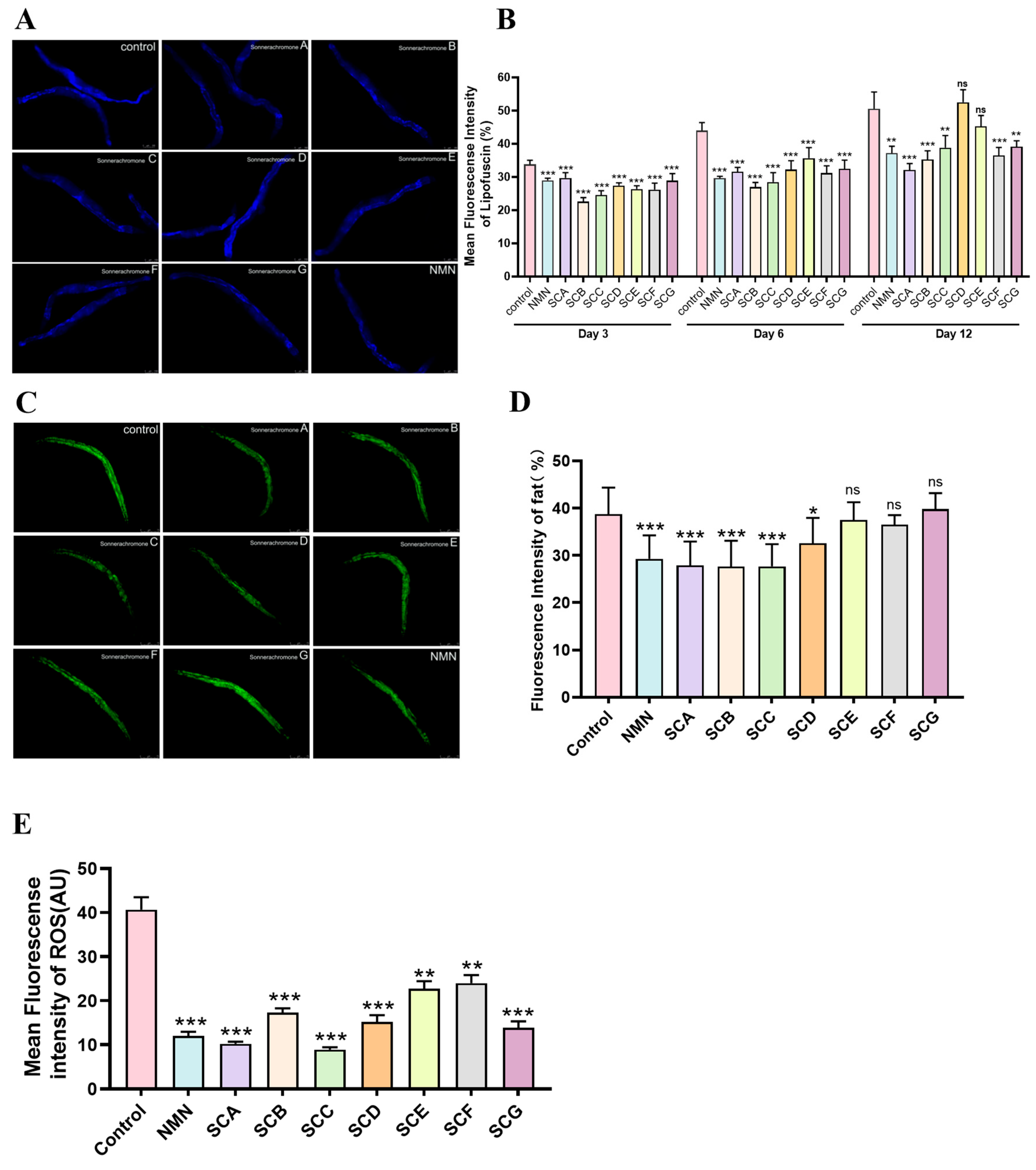
Figure 3.
Molecular docking analysis of chromone derivatives with telomere-associated proteins. (A) Chemical structures of the chromone derivatives SCA (green), SCB (white), and SCC (yellow). (B) Structural models of the target proteins POT-1, POT-2, DTN-1, and DTN-2 used for docking analysis. (C) Representative docking poses illustrating the predicted interactions between the compounds and POT-2. (D) Two-dimensional interaction diagrams showing intermolecular interactions between SCA, SCB, and SCC and POT-2. (E) Docking scores (binding energies) of the compounds against the four telomere-associated proteins.
Figure 3.
Molecular docking analysis of chromone derivatives with telomere-associated proteins. (A) Chemical structures of the chromone derivatives SCA (green), SCB (white), and SCC (yellow). (B) Structural models of the target proteins POT-1, POT-2, DTN-1, and DTN-2 used for docking analysis. (C) Representative docking poses illustrating the predicted interactions between the compounds and POT-2. (D) Two-dimensional interaction diagrams showing intermolecular interactions between SCA, SCB, and SCC and POT-2. (E) Docking scores (binding energies) of the compounds against the four telomere-associated proteins.
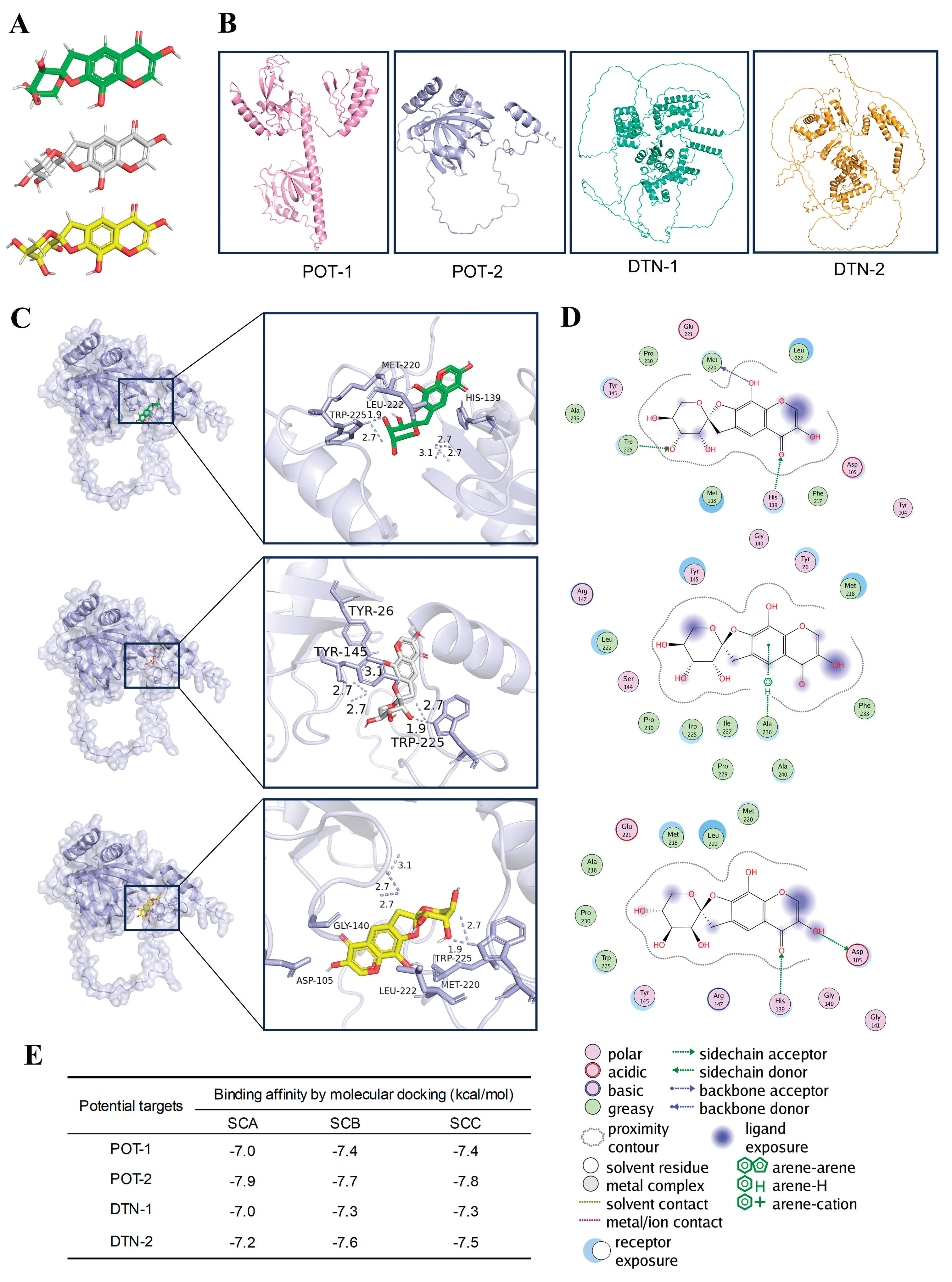
Figure 4.
SCA–SCC are associated with reduced protein levels of telomere-associated factors. (A) Representative Western blot and densitometric analysis of POT-1. (B) Representative Western blot and densitometric analysis of POT-2. (C) Representative Western blot and densitometric analysis of DTN-1. (D) Representative Western blot and densitometric analysis of DTN-2. (E, F) Lifespan curves of pot-1(tm1620) and pot-2(tm1400) mutant worms treated with SCA, SCB, or SCC (100 µM). Under these mutant backgrounds, the lifespan-extending effects of the compounds observed in N2 animals were not detected. Survival data were analyzed using the Kaplan-Meier test. Detailed lifespan statistics are provided in Table S1. Band intensities were quantified by densitometry and normalized to the corresponding β-actin loading control within each independent experiment. Data are presented as mean ± SEM, with each dot representing one independent biological replicate (n = 3). Statistical analysis was performed using one-way ANOVA followed by LSD or Games–Howell post hoc tests. ns, P > 0.05; * P < 0.05; ** P < 0.01; *** P < 0.001.
Figure 4.
SCA–SCC are associated with reduced protein levels of telomere-associated factors. (A) Representative Western blot and densitometric analysis of POT-1. (B) Representative Western blot and densitometric analysis of POT-2. (C) Representative Western blot and densitometric analysis of DTN-1. (D) Representative Western blot and densitometric analysis of DTN-2. (E, F) Lifespan curves of pot-1(tm1620) and pot-2(tm1400) mutant worms treated with SCA, SCB, or SCC (100 µM). Under these mutant backgrounds, the lifespan-extending effects of the compounds observed in N2 animals were not detected. Survival data were analyzed using the Kaplan-Meier test. Detailed lifespan statistics are provided in Table S1. Band intensities were quantified by densitometry and normalized to the corresponding β-actin loading control within each independent experiment. Data are presented as mean ± SEM, with each dot representing one independent biological replicate (n = 3). Statistical analysis was performed using one-way ANOVA followed by LSD or Games–Howell post hoc tests. ns, P > 0.05; * P < 0.05; ** P < 0.01; *** P < 0.001.
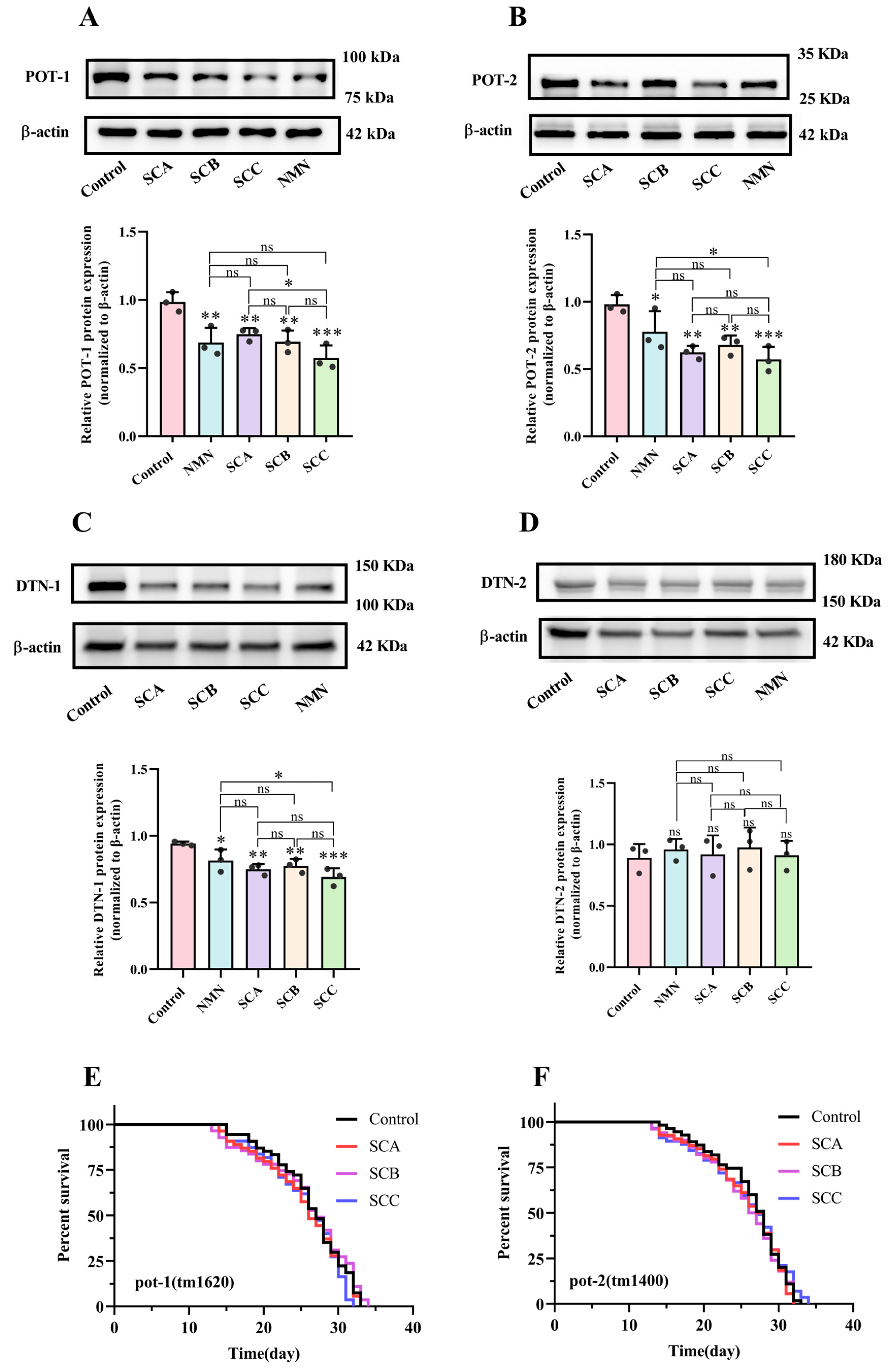
Figure 5.
Effects of SCA–SCC on telomerase activity and telomere-related transcriptional markers in C. elegans. (A) Representative TRAP assay gel showing telomerase activity in worms treated with SCA, SCB, and SCC (100 µM). (B) Quantification of TRAP assay results. (C, D) Relative expression levels of trt-1 and TERRA in worms following compound treatment, as determined by RT-qPCR. Data are presented as mean ± SEM. ns indicates p > 0.05, *P < 0.05, **P < 0.01, ***P < 0.001.
Figure 5.
Effects of SCA–SCC on telomerase activity and telomere-related transcriptional markers in C. elegans. (A) Representative TRAP assay gel showing telomerase activity in worms treated with SCA, SCB, and SCC (100 µM). (B) Quantification of TRAP assay results. (C, D) Relative expression levels of trt-1 and TERRA in worms following compound treatment, as determined by RT-qPCR. Data are presented as mean ± SEM. ns indicates p > 0.05, *P < 0.05, **P < 0.01, ***P < 0.001.
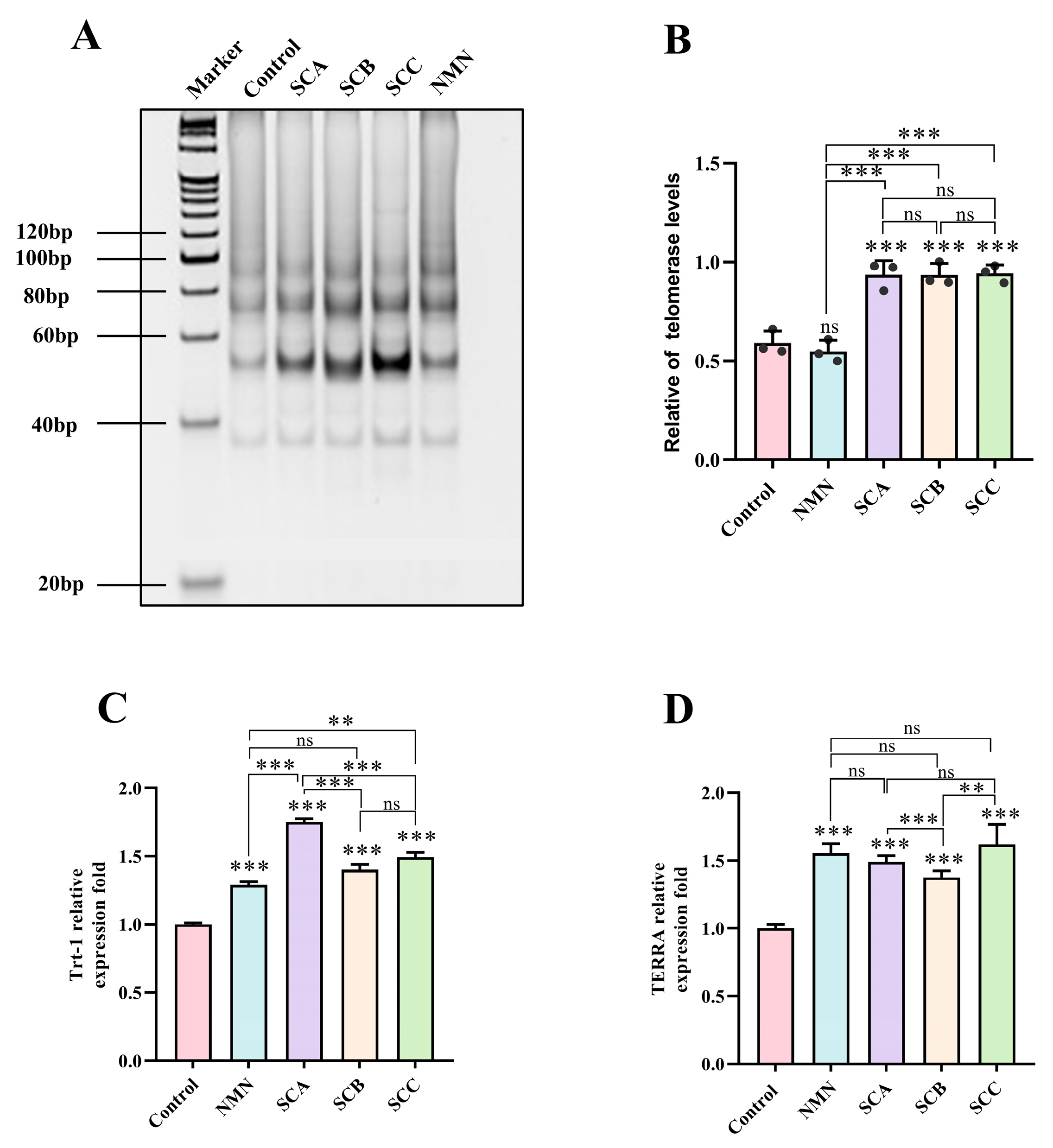
Figure 6.
Assessment of TL in C. elegans following SCA–SCC treatment. (A) Schematic workflow of the telomere length quantification assay. (B) TL expressed as the T/S ratio in worms treated with SCA, SCB, and SCC (100 µM). Data are presented as mean ± SD. ns indicates P > 0.05, *P < 0.05, **P < 0.01, ***P < 0.001.
Figure 6.
Assessment of TL in C. elegans following SCA–SCC treatment. (A) Schematic workflow of the telomere length quantification assay. (B) TL expressed as the T/S ratio in worms treated with SCA, SCB, and SCC (100 µM). Data are presented as mean ± SD. ns indicates P > 0.05, *P < 0.05, **P < 0.01, ***P < 0.001.
Disclaimer/Publisher’s Note: The statements, opinions and data contained in all publications are solely those of the individual author(s) and contributor(s) and not of MDPI and/or the editor(s). MDPI and/or the editor(s) disclaim responsibility for any injury to people or property resulting from any ideas, methods, instructions or products referred to in the content. |
© 2026 by the authors. Licensee MDPI, Basel, Switzerland. This article is an open access article distributed under the terms and conditions of the Creative Commons Attribution (CC BY) license (http://creativecommons.org/licenses/by/4.0/).
Copyright: This open access article is published under a Creative Commons CC BY 4.0 license, which permit the free download, distribution, and reuse, provided that the author and preprint are cited in any reuse.