

Submitted:
25 September 2026
Posted:
29 September 2026
You are already at the latest version
Abstract
The rapid integration of artificial intelligence (AI) and machine learning into medicine has launched a transformative epoch in dermatology, a specialty traditionally anchored in visual pattern recognition. While early machine learning research focused primarily on binary diagnostic classification, contemporary advancements have given rise to the para-digm of Synthetic Dermatology. This paradigm uses deep generative models such as Gen-erative Adversarial Networks (GANs) and Latent Diffusion Models (LDMs) together with personalized digital avatars, clinical large language models (LLMs), and multimodal tensor decompositions to synthesize complex clinical and histopathological images, gen-erate structured patient vignettes, and model real-world clinical trajectories. Herein, we review the state-of-the-art applications of synthetic technologies across three fundamental domains: clinical practice, dermatological research, and medical education, with a focus on personalizing dermatological care. By bridging the current clinical realities of hu-man-AI collaboration with a visionary perspective of the next decades, we outline how synthetic methodologies can mitigate data scarcity, address ethnic disparities in algo-rithmic diagnostics, optimize therapeutic regimens via cutaneous digital twins, and rede-fine student curricula. Finally, we critically appraise the technical, ethical, and regulatory hurdles that must be resolved to ensure the safe, equitable, and responsible translation of synthetic dermatology to patients.
Keywords:
digital twin
; generative artificial intelligence
; generative adversarial network
; latent diffusion model
; medical education
; personalized dermatology
; precision medicine
; synthetic control arm
; synthetic data
1. Introduction
Dermatology is fundamentally a visual specialty, relying on a complex diagnostic pathway that traditionally commences with clinical inspection, progresses through non-invasive dermoscopic evaluation, and, when indicated, culminates in tissue biopsy and histopathological examination [1,2,3]. However, the global demand for dermatological expertise far exceeds availability. An estimated 3 billion people worldwide lack adequate access to specialized dermatological care, a deficit that engenders prolonged waiting times and delayed diagnosis even within highly developed healthcare systems [4,5,6].
Over the past decade, deep learning algorithms, particularly Convolutional Neural Networks (CNNs), have demonstrated remarkable diagnostic capabilities, matching or occasionally exceeding the performance of board-certified dermatologists in classifying cutaneous malignancies under experimental, in silico conditions [1,2,3]. Despite these landmark accomplishments, conventional diagnostic artificial intelligence (AI) models are severely constrained by several systemic bottlenecks. First, they are highly data-hungry, requiring vast, expertly annotated datasets to learn stable, generalizable representations [7,8,9]. Second, existing public repositories, such as those curated by the International Skin Imaging Collaboration (ISIC), suffer from profound class imbalances, with an overrepresentation of common benign lesions and a critical paucity of rare malignant phenotypes or atypical presentations [7,10]. Third, a historical bias exists in these datasets, which are predominantly sourced from fair-skinned populations, rendering trained algorithms highly inaccurate and structurally biased when deployed on dark skin tones (Fitzpatrick phototypes V-VI) [4,6]. Lastly, stringent data privacy regulations, such as the Health Insurance Portability and Accountability Act (HIPAA) in the United States and the General Data Protection Regulation (GDPR) in the European Union, severely restrict the sharing of real patient datasets across institutions [11,12,13].
To reconcile the tension between the clinical promise of dermatological AI and these structural limitations, the field of Synthetic Dermatology has emerged [14,15]. Synthetic dermatology refers to algorithmic generation, simulation, and enhancement of dermatological data, including clinical photographs, dermoscopic images, histopathological slides, and clinical narrative as well as biologic data, that replicate the statistical properties and visual fidelity of real-world clinical encounters without containing any identifiable patient information [16,17,18,19,20]. Rather than viewing data generation as a mere computational practice, this paradigm represents a profound shift in clinical methodology: turning sparse, static datasets into dense, dynamic, and privacy-preserving resources that can be tailored to the individual patient biology (Figure 1).
1.1. Literature Search
This narrative review was informed by a structured literature search last updated on September 1st, 2026. PubMed/MEDLINE was searched using combinations of the terms ‘synthetic dermatology’, ‘synthetic data’, ‘large language model’, ‘generative adversarial network’, ‘latent diffusion model’, ‘variational autoencoder’, ‘convolutional neural network’, ‘artificial intelligence’, ‘virtual reality’, ‘digital twin’, and ‘avatar’ (See Table 1 for explanation of terms). Additional searches combined individual AI-related terms with terms related to dermatology and medicine, including individual diseases. Reference lists of relevant reviews and original studies were also examined.
2. Applications in Clinical Dermatological Practice
The application of synthetic dermatology in clinical practice is transitioning from experimental computer-aided diagnosis support to real-time image standardization, prognostic simulation, and human-AI collaboration [18,20,25].
2.1. Optical Standardization
A challenge in clinical teledermatology and automated image analysis is the high variability of photographic conditions [1]. Unlike standardized dermoscopy, clinical photographs captured by patients or physicians using smartphones exhibit significant noise, including inconsistent zoom, variable angles, hair occlusion, and heterogeneous light sources [1,26]. Such optical discrepancies degrade the performance of deep learning diagnostic classifiers and confound visual tracking by human experts [2,18]. To address lighting and device-induced variability, generative models are deployed to achieve color constancy, the standardization of an image’s illumination source to make it appear as if it were acquired under canonical white light [18,27]. Salvi and colleagues developed the Dermatological Color Constancy Generative Adversarial Network (DermoCC-GAN) [18]. Trained on image pairs standardized by custom heuristic algorithms, DermoCC-GAN learns higher-level features to standardize illumination and enhance local contrast in dermoscopic and clinical images [18,27]. When integrated as a preprocessing step, DermoCC-GAN significantly improves the performance of downstream diagnostic classifiers and segmentation networks, achieving a classification accuracy of 79% and a lesion segmentation Dice score of 91% [18]. Similarly, generative architectures are used for color normalization in digital pathology, aligning Hematoxylin & Eosin (H&E) stain variations across different scanner brands and clinical centers [20,28] (Figure 2).
Beyond color normalization, clinical image analysis must contend with physical occlusions and low image resolutions. Hair occlusion is one of the most common pre-diagnostic artifacts in dermoscopy [25,29]. Attia et al. proposed a photo-realistic hair simulator based on context-aware image-to-image translation via conditional generative adversarial networks (cGANs) [25]. By mapping pixels exclusively to predefined dilated hair masks and leveraging a structural similarity index loss function, the simulator can generate realistic hair fibers that seamlessly blend with underlying skin textures and lesion morphology [25,29]. This allows generation of controlled benchmarking-datasets to evaluate digital hair removal algorithms, such as DullRazor, without compromising lesion integrity [25]. To enhance image resolution and clarify fine structural details, super-resolution generative models, such as MELIIGAN [30] and CESR-GAN [28], have been introduced to reconstruct high-frequency details from low-resolution dermoscopic inputs, facilitating early cancer detection.
Similar generative architectures have been used to enhance high-resolution skin imaging. The Vascular Visualization Enhancement Generative Adversarial Network (VVE-GAN) has been engineered to optimize Optical Coherence Tomography Angiography (OCTA) images of skin tissue [31]. VVE-GAN suppressed background scattering noise and resolved fine-grained microvascular morphology in deeper cutaneous layers, aiding the clinical evaluation of highly vascularized conditions such as basal cell carcinoma, nevus flammeus, and psoriasis.
2.2. Clinical Simulation of Disease Progression and Treatment Response
Beyond static diagnostic assistance, generative modeling enables clinicians to visually simulate disease progression and predict personalized treatment trajectories [20,25]. In an era of non-validated, recreational facial-aging applications on social media, scientific transparency and clinical validation are paramount. Raynaud and co-workers developed a validated facial aging simulator utilizing a dual-tool framework: the Elicitation Based Aging Simulator (EBAS) and AgingMapGAN (AMGAN) [14]. EBAS uses the collective clinical knowledge of 28 international dermatologists via a structured Delphi process and causal Bayesian belief networks to model skin trajectories based on lifestyle and exposome factors (such as UV exposure, tobacco consumption, and chronological age). AMGAN, a conditional spatially aware GAN, translates these probabilistic trajectories into high-resolution visual representations, specifically focusing on the density of pigmentary spots on the cheeks. In clinical benchmarking, the model demonstrated an exceptional correlation coefficient of 0.96 with expert clinical grading. In a practical scenario of a 38-year-old female of Chinese descent, the model projected that the 15-year probability of a two-grade progression in pigmentary spot density was 71% in the absence of photoprotection, which decreased to 39% with regular SPF 50+ sunscreen application, and to 11% under a combined chemical and physical photoprotective regimen. This framework provided an objective, scientifically validated educational and preventive tool to enhance patient compliance and visually support informed consent.
In parallel, UV-photo Net, a conditional GAN-based network, converts standard RGB (Red, Green, Blue) visible light photographs into synthetic UV photo images [32]. UV photography is historically utilized in specialized clinics to evaluate sub-clinical epidermal pigmentation and cumulative actinic damage, but the necessary hardware is rarely available in general primary care clinics. Evaluated on standard digital and smartphone-captured images, UV-photo Net reproduces pigmentary spots with high fidelity, attaining a correlation of 0.92 in spot area detection. This tool successfully revealed clinically silent hyperpigmentation, prompting early patient selfcare and facilitating the screening of early pre-malignant lesions.
2.3. Human-AI Clinical Collaboration
As generative AI systems are increasingly integrated into clinical work, such as drafting clinical documentation, generating follow up instructions, and facilitating pre-consultation triage, understanding their impact on the therapeutic alliance is critical. Recent clinical evidence suggests that generative AI is a double-edged sword within dermatological communication [33]. In a multi-component cross-sectional clinical study, Wei et al. evaluated the impact of utilizing an advanced large language model (LLM) (DeepSeek-R1) during a 5-minute pre-consultation window before 10-minute standardized patient encounters. Standardized case performance, evaluated by blinded independent assessors, demonstrated that AI-assisted preparation was associated with significantly higher estimated marginal scores for information gathering, information giving, structural efficiency, and overall communication scores. Furthermore, frequent clinical utilization of generative AI by dermatologists was strongly correlated with a reduction in emotional exhaustion and an increase in communication self-efficacy. Patients whose clinicians utilized generative AI reported higher overall communication satisfaction. However, sensitivity analysis revealed that humanistic care and empathy scores were consistently lower under the AI-assisted preparation condition. Clinicians who relied heavily on structured, model-generated notes and templates appeared to deliver highly organized, complete clinical information at the cost of spontaneity, emotional attunement, and relational warmth. Because dermatological consultations frequently involve visible skin lesions that carry significant psychosocial stigma, anxiety, and chronic recurrence, clinical communication cannot be reduced to administrative efficiency [33,34]. Generative AI must therefore be positioned as a supervised supportive tool rather than a replacement for empathetic relational care [33].
This empowerment focused perspective is strongly echoed by patients. In a cross-sectional study of 349 outpatients, Digis and colleagues found that 61% of patients had actively consulted an AI tool (predominantly ChatGPT, 77%) regarding their skin complaint prior to their clinic visit [6]. Interestingly, the pre-visit AI consultation was not associated with any detectable increase in patient health anxiety. For 74% of patients, the AI response simply confirmed their existing decision to seek professional care, and patients strongly rejected the notion of AI as a standalone physician substitute (mean score 1.66 out of 5) while endorsing it as a tool to help them ask informed questions during their clinical consultation (mean score 2.89).
3. Applications in Dermatological Research
Dermatological AI research has historically been halted by the paucity of high quality, diverse training data [1,8]. Deep learning models are notoriously prone to overfitting when trained on small datasets, and they exhibit poor generalizability when deployed across different clinical devices and patient populations [4,6]. GANs and diffusion models have emerged as pivotal technologies to generate high fidelity, privacy-preserving synthetic data for training, benchmarking, and data sharing [16,17,19,35].
3.1. Addressing Class Imbalance and Data Scarcity
In standard dermatological databases, orphan disease, rare malignancies and atypical presentations are significantly underrepresented [7,8,9]. For example, melanoma accounts for approximately 75% of skin-cancer-related deaths, making early algorithmic detection a high-stakes clinical task [1]. Yet, automated classifiers trained on class-imbalanced datasets develop biased decision boundaries, preferring the majority class (e.g., benign nevi) and yielding unacceptably high rates of false negatives for lethal malignancies [7,10]. To mitigate this, generative data augmentation can be used, wherein generative models are trained on minority classes to synthesize novel, high-resolution training examples [19,35,36]. Lin and co-workers presented a systematic cross-architecture benchmarking study comparing four GAN designs; Deep Convolutional GAN (DCGAN), StyleGAN2, StyleGAN3-T, and StyleGAN3-R, for high-resolution (512 × 512 pixels) melanoma-specific synthesis [35]. Their results demonstrated that while DCGAN lacked the capacity to generate fine-grained dermoscopic cues and StyleGAN3-R introduced rotation-equivariance grid artifacts, StyleGAN2 achieved the optimal balance of quantitative performance (attaining a Fréchet Inception Distance, FID) of 7.96) and visual realism [35]. Importantly, a frozen, external EfficientNet-based melanoma detector recognized 83% of the StyleGAN2-generated images as melanoma. In a controlled downstream experiment, augmenting a class-imbalanced training set with these synthetic melanoma images improved the classifier’s melanoma detection Area Under the Curve (AUC) from 0.925 to 0.945. Similarly, Cho et al. developed the GAN5000 dataset of synthetic melanocytic lesions using a StyleGAN2-ADA architecture trained on clinical photographs harvested from the internet (the CAN2000 dataset) and preprocessed using large mask inpainting to remove rulers, markers, and photographic artifacts [16]. Discriminative CNN models (EfficientNet-Lite0) trained on public datasets supplemented with GAN-generated clinical photographs exhibited superior diagnostic performance compared to models trained on real datasets alone, demonstrating that synthetic data can serve as a robust, privacy-preserving alternative to clinical images when original patient data cannot be legally or ethically shared.
In the histopathological domain, Rodriguez Alonso and colleagues investigated the generation of high-resolution (1024 × 1024 pixels) H&E-stained skin biopsy slides using GANs to distinguish melanoma from benign melanocytic nevi [19]. Mixed training datasets containing both real and synthetic histopathology images achieved an overall accuracy of 96% and a specificity of 97% on a real validation set. Notably, the integration of synthetic images led to a measurable reduction in false negatives (from 80 to 75 cases), minimizing the risk of a missed melanoma diagnosis.
On a technical level, Behara et al. proposed an improved DCGAN classifier for skin lesion synthesis, showing that incorporating global average pooling with fractional-stride backpropagation and a high learning rate significantly stabilized training, achieving an outstanding binary classification accuracy of 99.38% on the ISIC 2017 benchmark [9]. In parallel, Li et al. proposed a fuzzy rank-based ensemble of CNNs (fusing InceptionV3, Xception, and DenseNet121) combined with dermoscopy dataset augmentation GANs and efficient channel integrating spatial attention [29]. By mapping decision scores to nonlinear concavities, this fuzzy ensemble reduced the intraclass sample distance and concentrates learning on error-prone samples. To maximize the morphological variety of generated images, the TED-GAN framework integrated a variational autoencoder (VAE) with a GAN and an auxiliary classifier [37]. Notably, TED-GAN sampled the latent noise vector from a heavy-tailed student t-distribution rather than a standard Gaussian, which avoided mode collapse and increased the average classification accuracy from 66% to 93% [37].
3.2. From GANs to Latent Diffusion Models
While GANs have historically dominated medical image synthesis, Latent Diffusion Models (LDMs) are rapidly becoming the state-of-the-art approach due to their superior training stability, reduced mode collapse, and highly detailed textural preservation [17,27]. Kim and colleagues proposed an enhanced LDM based on the stable diffusion architecture, specifically engineered to generate synthetic skin images across seven diagnostic classes using the HAM10000 dataset [17]. To prevent the loss of fine-grained, clinically diagnostic textures (such as pigment networks, vascular patterns, and blue-gray veils) during latent space compression, they implemented an 8-channel VAE, which doubled the representational capacity of standard 4-channel VAEs. Furthermore, they integrated multi-level CLIP image embeddings extracted across five distinct scales to capture both low-level disease patterns and high-level morphological structures. To enrich training data diversity further, they utilized pre-trained segmentation and inpainting models to isolate lesion areas and generate normal, healthy skin contexts, allowing for latent space interpolation between normal skin and lesion states. By adjusting an interpolation coefficient, the model synthesized a visual continuum of transitions. Although this visual progression is heuristic and does not strictly correspond to clinically validated severity metrics, such as PASI score for psoriasis, it dramatically enhanced semantic and morphological diversity within the training set (Figure 3).
Downstream classification models trained on this synthetically enriched dataset demonstrated a significant performance boost, with average classification accuracy increasing from 80.92% (trained on original data alone) to 84.15% (trained on the mixed dataset) across nine distinct deep learning architectures. Other diffusion-based approaches have emerged to address algorithmic fairness. For instance, Ktena et al. utilized metadata-conditioned diffusion models to synthesize dermoscopic images that explicitly controlled for underrepresented patient characteristics, thereby improving out-of-distribution accuracy and fairness across diverse cohorts [27,38].
3.3. Multi-Modal Research Integration
The frontier of dermatological research lies in multi-modal integration, combining morphological image data with biophysical, biochemical, and genomic signatures [39]. Raman spectroscopy is a non-invasive optical technique that measures the vibrational modes of biomolecules within skin tissue in vivo, yielding biochemically specific spectral fingerprints associated with cellular malignancies. In a clinical study of 731 skin lesions, Zhao et al. utilized a one-dimensional convolutional neural network trained on Raman spectra to differentiate malignant and premalignant lesions (melanoma, basal cell carcinoma, squamous cell carcinoma, and actinic keratosis) from benign lesions [39]. To overcome the classic bottleneck of spectral acquisition variability and low signal-to-noise ratios, the authors utilized generative adversarial data augmentation. By synthesizing artificial training spectra to simulate varied optical conditions and baseline drift, they improved the diagnostic ROC-AUC with 2-4% across their convolutional network and several conventional machine learning models.
Imminently, combining synthetic image generation, such as LDMs, with synthetic spectral datasets, such as Raman spectroscopy or hyperspectral imaging, will allow construction of multi-modal, high-dimensional synthetic cohorts [20]. Such integrated systems will enable the simultaneous modeling of morphological structures and biochemical tissue composition, paving the way for ultra-precise, non-invasive diagnostic platforms.
3.4. Unstructured Clinical Text and Tabular Document Synthesis
While clinical images dominate dermatological discussions, unstructured narrative data, such as discharge summaries and referral notes, and structured electronic health records constitute the backbone of health system processes [20,40]. To automate the extraction of structured clinical information from unstructured text, Eryilmaz and colleagues developed PIGEON, a scalable, privacy-preserving framework [30]. PIGEON utilizes a random sample generator powered by Qwen3-235B to synthesize 75,000 realistic clinical discharge letters by sampling and aggregating structured FHIR (Fast Healthcare Interoperability Resources) data from over 41,000 oncology and dermatology patients [20]. These synthetic letters were paired with their originating FHIR JSON blocks and used to fine-tune MedGemma 27B. In clinical evaluations, the fine-tuned model achieved an F1-score of 0.84 for full ICD diagnosis codes and 0.99 for tumor-related specifications. To suppress hallucinated codes, they implemented a postprocessing retrieval-augmented generation module that embeds BfArM catalogs into a similarity search index, picking the single most plausible clinical code.
In parallel, generating tabular synthetic electronic health records data has seen major methodological advances. Standard deep learning generators like CTGAN and RealTabFormer are often trained on de-identified databases to produce high-fidelity clinical tables [31,40]. Karimian Sichani et al. proposed a generative model relying on generalized canonical polyadic tensor decomposition [41]. By modeling patient factor matrices using sequential decision trees, Gaussian copulas, and Hamiltonian Monte Carlo sampling, their framework synthesized longitudinal laboratory measurements and admissions data from MIMIC-III without establishing a one-to-one link to real patient identities. For simpler tabular generation tasks, Barr et al. demonstrated that zero-shot prompting with GPT-4 can generate highly realistic perioperative tabular data (matching 12 of 13 statistical parameters of the real-world VitalDB dataset) simply by using descriptive statistics and mathematical constraints in the prompt [11].
4. Applications in Medical Education
Medical training in dermatology is heavily reliant on exposure to clinical diversity [42]. However, traditional educational materials, such as paper-based clinical vignettes and atlas photographs, require immense expert effort to compile, lack case-level customizability, and fail to capture the interactive, dynamic nature of clinical communication [10]. Synthetic dermatology addresses these limitations by providing scalable, customizable, and highly interactive educational assets [5,16,42].
4.1. Synthetic Education via LLM-Generated Clinical Vignettes and Dialogues
Standardized examinations, such as the United States Medical Licensing Examination, rely extensively on text-based clinical vignettes to evaluate diagnostic reasoning [5]. To scale the generation of these educational resources, Rao and co-workers evaluated the feasibility of utilizing GPT-4 to generate highly detailed clinical vignettes for 20 skin and soft tissue diseases [5]. A panel of medical experts evaluated the synthetically generated vignettes across multiple dimensions using Likert scales. The GPT-4-generated vignettes achieved exceptionally high scores for: scientific accuracy (4.45 out of 5), comprehensiveness (4.30), and overall quality (4.28). Notably, the vignettes received very low scores for potential clinical harm (1.60) and demographic bias (1.52), establishing that off-the-shelf foundation models are highly accurate and efficient engines for synthetic medical education. However, the study noted that vignettes often lacked spontaneous demographic diversity, underscoring the need for targeted, multi-ethnic prompting to represent diverse patient populations.
Beyond static clinical vignettes, generative AI can produce physician-patient dialogues to train students in medical interview and communication skills [10]. Yanagita et al. evaluated an LLM based framework to generate interactive patient-physician dialogues for clinical training [10]. Evaluated against the Mini-Clinical Evaluation Exercise (Mini-CEX) domains, the synthetic dialogues demonstrated high coherence, medical accuracy, and context-rich scenarios. This allows medical students to practice active information gathering, diagnostic reasoning, and counseling in a safe environment that replicates real-world clinical ambiguity and conversational flow. To support Continuing Medical Education (CME) on topics like acne vulgaris, Naldi et al. prompted GPT-4 to generate educational guidelines [42]. Blinded evaluations by five expert clinicians revealed high ratings for readability (94.8%) and thoroughness (85.2%), although occasional inaccuracies (24.3%) emphasized the ongoing necessity for expert-in-the-loop review.
4.2. Interactive 3D Simulators and Virtual Reality Environments
In a visual specialty, text-based learning must be paired with realistic spatial representation [42]. This has motivated the development of interactive, virtual reality (VR)-based training platforms populated with synthetic pathology [43]. Pielage and colleagues developed an interactive 3D virtual patient simulator powered by generative diffusion models [43]. Diffusion models were trained to generate photorealistic lesions of melanoma and benign nevi, which were then dynamically embedded onto a virtual patient avatar within a VR environment. Medical students could navigate the VR space, inspect the lesions under varied illumination and magnification, and perform diagnostic screening tasks. In a pilot user study, students exposed to the AI-generated synthetic training cases achieved a higher diagnostic accuracy on a validation set of real clinical images compared to a control group trained without synthetic visualization (64% vs. 56% accuracy), while reporting a significant increase in their subjective clinical competence, which rose from 2.28 to 3.12 on a 5-point scale.
On a more specialized level, dermatopathology trainees can utilize intelligent tutoring systems like Slide Tutor [42]. Slide Tutor employs case-specific diagnostic algorithms to analyze a student’s diagnostic path, providing real-time feedback, error analysis, and context-specific hints. Studies evaluating Slide Tutor demonstrated a remarkable reduction in diagnostic errors (from 50% to 20%) and a corresponding increase in optimal solution-path actions over sequential training sessions.
4.3. Standardizing Educational Validation
As the integration of AI-generated educational materials expands, medical institutions require objective frameworks to validate their quality, safety, and ethical compliance [42]. Yap and Lim advocate for the adoption of the CUC-FATE framework to evaluate dermatological educational tools [42]. The CUC-FATE framework evaluates seven core dimensions:
- Cost: Economic viability, computational efficiency, and institutional accessibility.
- Usability: Technical interface design, user engagement, and ease of deployment.
- Credibility: Alignment with established scientific consensus, guidelines, and clinical accuracy.
- Fairness: Equitability of representation across diverse skin phototypes and the active mitigation of demographic bias.
- Accountability: Clear establishment of legal and institutional liability for clinical information accuracy.
- Transparency: Open-source disclosure of model weights, training parameters, and training data provenance.
- Explainability: The capability of generative models to provide traceable, step-by-step clinical reasoning for their outputs.
By systematically applying CUC-FATE, alongside COSMIN (COnsensus-based Standards for the selection of health Measurement INstruments)-informed content validity checklists, medical educators can audit synthetic materials, suppress hallucinated references, and integrate these advanced tools safely into formal medical school and residency curricula.
5. Personalizing Dermatology
The overarching clinical value of synthetic dermatology lies in its power to personalize dermatological care, transitioning the specialty from population-level clinical guidelines to patient-specific precision medicine [21,23,24].
5.1. Mitigating Disparities in Skin Phototypes
A historical disparity exists in dermatological AI performance across different skin tones [4]. Daneshjou and co-workers developed the Diverse Dermatology Images (DDI) dataset, the first publicly available, expertly curated, and pathologically confirmed skin lesion dataset with diverse skin tones, matched for age, sex, and diagnosis [4]. When evaluating three state-of-the-art diagnostic algorithms on the DDI dataset, the authors observed a profound performance drop-off on dark skin tones (Fitzpatrick skin types V-VI) compared to light skin tones (types I-II). For instance, HAM10000 exhibited a ROC-AUC of 0.72 on light skin but only 0.57 on dark skin. More critically, the sensitivity for detecting melanoma in dark skin dropped substantially, indicating a high, clinically unacceptable risk of missed diagnoses in patients with dark skin phototypes. Importantly, this algorithmic performance gap could be closed by fine-tuning the model weights on diverse datasets containing dark skin tones. After fine-tuning the DeepDerm and HAM10000 algorithms, the performance gap between skin types I-II and V-VI was eliminated, and the fine-tuned models significantly outperformed human dermatologists in diagnosing malignancies on dark skin tones. To generate these vital datasets widely, conditional generative models can be used to synthesize diverse, pathologically accurate skin lesions across all Fitzpatrick phototypes [16,17,20]. By conditioning the generation process on both disease labels and skin tone parameters, models like StyleGAN2 or custom diffusion networks can synthesize rare pathological phenotypes directly on underrepresented dark-skinned backgrounds, providing a scalable path to balance clinical image banks and ensure algorithmic fairness.
5.2. Cutaneous Digital Twins
At the forefront of personalized dermatology stands the concept of cutaneous digital twins (CDT), an intricate, dynamic virtual representation of a patient’s skin health, behaviors, and environmental exposures [21,23,24]. Initially conceptualized in aerospace engineering to predict system behaviors, the application of digital twin technology in dermatology represents a profound paradigm shift towards proactive, patient-specific care [21]. A CDT is constructed by integrating multi-source, real-time clinical and patient-collected data, collectively referred to as the digitosome [23] (Table 2).
AI serves as the analytical engine of the digital twin, utilizing these high-dimensional datasets to simulate real-world conditions and predict individual health outcomes exemplified by personalized acne and psoriasis therapeutics [21,24]. While established guidelines exist for managing chronic inflammatory diseases like acne and psoriasis, individual clinical response exhibits substantial heterogeneity, necessitating a trial-and-error approach to systemic and biologic therapies. A patient’s digital twin serves as a virtual testing environment. For a patient with severe psoriasis, the digital twin can simulate how their unique immune system, genetic polymorphisms, and cutaneous microbiome will interact with various systemic biologics (e.g., TNF, IL-17, and IL-23 inhibitors) or phototherapy. By predicting therapeutic efficacy and forecasting potential adverse reactions, such as psoriatic arthritis, steatotic liver disease, hypertension, and insulin resistance, before initiating treatment, the CDT minimizes clinical risk, optimizes drug selection, and dramatically shortens the time required to achieve disease clearance.
Another example is next generation personalized photoprotection. Notably, conventional sunscreens operate on a one-size-fits-all model, with efficacy measured primarily by static SPF and UVA ratings [44]. However, these metrics fail to account for individual genetic variations in UV susceptibility (such as MC1R polymorphisms), skin barrier function, and real-time environmental exposome exposure. In a near-future clinical scenario, a CDT can revolutionize photoprotection. During a routine clinical visit, a non-invasive scan can capture the patient’s epidermal hydration and molecular biomarkers via tape-stripping, while their smartwatch uploads a 3-month exposome record of UV and urban pollution exposure. The AI engine will integrate these data with the patient’s genomic profile, highlighting a polymorphism that impairs DNA repair. The digital twin then simulates their 10-year cumulative photoaging trajectory, automatically generating a prescription for a highly personalized, adaptive sunscreen formulation. This custom formulation is co-formulated with specific UV filters, on-demand smart photostabilizers, topical antioxidants, and DNA-repair enzymes tailored to their biophysical profile. Real-time feedback from wearable sensors and mobile applications dynamically guides the patient on exact application doses and reapplication intervals based on real-time environmental exposure, transforming photoprotection from passive shielding into an interactive, preventive health intervention in which the patient is personally engaged (Figure 4).
5.3. Precision Clinical Trials
Randomized controlled trials (RCTs) remain, so far, the gold standard for therapeutic evaluation [22,45]. However, in rare highly debilitating, orphan skin diseases, such as dystrophic epidermolysis bullosa (EB) and mucous membrane pemphigoid (MMP), traditional placebo-controlled trials raise profound ethical and logistical dilemmas [21,45,46]. For patients with recessive dystrophic EB, randomizing to a placebo arm can expose fragile, blistered skin to chronic wounding and predispose them to aggressive squamous cell carcinoma [21,46]. Furthermore, recruiting a statistically sufficient cohort is often impossible due to extreme patient rarity [45,46]. Synthetic dermatology offers a revolutionary solution through the implementation of Synthetic External Control Arms (SECAs) and Hybrid Control Arms (HCAs) [31,45,47,48]. Rather than placing real patients on placebo, virtual control groups comprised of CDTs can be constructed [21,22]. To build these cohorts with rigorous statistical validity, developers utilize the FRESCA (Framework for Research in Equitable Synthetic Control Arms) architecture [47]. FRESCA integrates patient-level data from historical clinical trials, national disease registries, and real-world electronic health records applying propensity and equity adjustments [47,49]. By using iterative proportional fitting and propensity matching, FRESCA ensures that the virtual cohort matches the treatment arm across key demographic (age, sex, ethnicity) and clinical baseline parameters, establishing complete representation. During the trial, the real-world treatment arm is directly compared to the SECA [21,22]. This virtual control arm undergoes simulated disease progression under standard-of-care or placebo conditions, eliminating the need to withhold active, life-saving therapies from real patients. As the trial progresses, real-time clinical data from the treatment group can be fed back into the generative models, enabling continuous Bayesian refinement of the digital twins and accelerating regulatory drug approval while strictly safeguarding ethical standards and patient safety [21,22,45].
6. Ethical, Legal, and Regulatory Landscapes
While the transformative potential of synthetic dermatology is immense, its translation into clinical practice is constrained by significant scientific, ethical, and regulatory bottlenecks [20,45]. Table 3 outlines key AI reliability, validation, and security frameworks addressing these challenges.
6.1. Technical Challenges
Despite the high visual quality of images generated by advanced generative models like StyleGAN2, critical technical limitations persist [19,35]. First, the morphological fidelity of synthetic images remains statistically inferior to real histopathological and clinical pictures [19]. For example, synthetic histopathological images achieved a FID score of 139.56, indicating a substantial distribution gap between real and synthetic tissue structures [19]. Second, generative models are prone to mode collapse, the phenomenon whereby the generator learns to produce only a limited variety of realistic samples, failing to capture the full spectrum of intra-class morphological variability and atypical presentations [35]. Third, synthetic images are highly homogeneous and lack technical noise, such as differences in scanner brands, staining protocols, lighting configurations, and image formats that characterize real-world datasets, which can lead to a collapse in model robustness when classifiers trained on synthetic data are deployed on external clinical datasets [18,20]. Furthermore, standard quantitative metrics used to evaluate generative quality (such as FID, Inception Score, or structural similarity) are purely mathematical feature-space calculations that do not directly correlate with clinical diagnostic utility or interpretability [27]. To date, robust clinical validation of synthetic systems is scarce, and prospective clinical trials evaluating patient-centered outcomes or direct diagnostic accuracy in real-world settings remain practically non-existent [20].
To overcome this, recent research has introduced unified data-auditing frameworks [50,51]. Santangelo et al. proposed SynthRO, an accessible GUI and dashboard-style interface that allows clinicians and developers to benchmark synthetic datasets across three critical axes: statistical resemblance, diagnostic utility, and privacy-risk profiling [50]. Applying such multi-dimensional benchmarks ensures that synthetic data maintain logical clinical relationships before deployment.
6.2. Ethical and Privacy Risks
A primary motivation for synthetic data generation is patient privacy, as synthetic images do not possess a one-to-one relationship with real individuals [19,20]. However, synthetic data alone does not entirely eliminate privacy risks [20,31]. Generative models, especially deep neural networks, are vulnerable to membership inference attacks and model inversion [31,52]. In these scenarios, malicious actors can exploit the trained model parameters to reconstruct training samples or determine whether a specific patient’s data was used to train the generative model. This is particularly dangerous in longitudinal datasets or rare disease registries, where individual clinical trajectories are highly unique and more susceptible to re-identification [31,51].
To enforce mathematically verifiable privacy, developers have integrated differential privacy during model optimization, such as DP-GAN-HD [52], DP-CGAN, and PrivBayes [31]. DP-GAN-HD adds calibrated Gaussian or Laplacian noise to the discriminator’s gradients during backpropagation, establishing a strict privacy budget that limits the risk of membership leakage, even when subjected to adversarial red-teaming [31,52]. Furthermore, facial photographs are considered direct patient identifiers under HIPAA [12]. For data at scale where obtaining explicit patient consent is infeasible, adequate facial deidentification must be applied before clinical sharing. Traditional feature concealment (such as covering the eyes) is largely insufficient. To address this, cGANs like CIAGAN (Conditional Identity Anonymization GAN) and disentanglement-coupled architectures can be used. These models segment and disentangle identifying facial landmarks (eyes, nose, lips) from underlying skin attributes, replacing the patient’s identity with a synthetic avatar while preserving pixel-level dermatological features of interest, such as erythema, texture, and pigmentation, with high clinical utility.
Finally, the democratization of high-fidelity image-generation tools carries significant ethical hazards, such as the malicious generation of medical deepfakes [20]. Deepfakes can propagate misinformation, fool automated triage software, and facilitate clinical insurance fraud through the generation of fake pathological clinical or histopathological images [20]. Developing robust, real-time forensic detection methods, such as vision transformers trained to identify artificially synthesized dermoscopic lesions, is crucial to safeguard clinical data integrity [53].
6.3. Navigating the Regulatory Coordinate Space
The clinical deployment of synthetic dermatology applications requires strict compliance with evolving regulatory frameworks [20]. The regulatory coordinate space exhibits a significant divergence between the European Union and the United States (Table 3).

Table 4.
Regulatory Paradigms.
| European Union (EU) | United States (US) |
| AI Act | FDA SaMD framework |
| High-risk medical classification | Adaptive product lifecycles |
| Prescriptive and precautionary | Adaptive oversight |
| Strict human oversight and auditing | GMLP guidelines |
Under the European Union’s Artificial Intelligence Act (2024), medical AI is explicitly classified as high risk, mandating strict, prescriptive requirements for risk management, technical documentation, transparency, and human oversight before CE mark certification can be granted under the Medical Device Regulation [20]. Conversely, the United States Food and Drug Administration (FDA) regulates AI under its Software as a Medical Device (SaMD) framework [20]. The FDA emphasizes Good Machine Learning Practice (GMLP) and promotes adaptive, continuous-oversight programs, allowing for iterative, pre-certified software modifications without requiring a formal premarket approval submission for every algorithmic update, provided that clinical safety, diagnostic efficacy, and source transparency are maintained. For both developers and practicing dermatologists, early engagement with regulatory bodies and adherence to “explainable AI” and standard data curation guidelines (such as the CLEAR Derm consensus checklist) are essential to ensure the safe, responsible, and legal translation of synthetic systems into clinical practice [16,20].
To further safeguard clinical data against tampering in multi-center clinical trials, permissioned private Ethereum blockchains can be integrated [48]. By logging every electronic case report form (eCRF) transaction onto an immutable, distributed ledger, blockchain technology eliminates a single point of failure, prevents retrospective data manipulation by sponsors or intruders, and provides regulatory agencies with an unalterable audit trail [48,54]. To reconcile the immutability of blockchain with the patient’s legal right to be forgotten under GDPR, developers utilize hybrid data models where personal identifiable information is stored off-chain behind revocable, cryptographic encryption keys, ensuring both data integrity and regulatory compliance [48,54].
7. Perspectives and Future Directions
Over the next decades, the convergence of high-capacity generative models, multi-modal molecular biosensors, and ubiquitous edge computing will completely reorganize the clinical, research, and educational landscapes of dermatology [21,23,24].
When a patient enters a dermatology clinic a few decades from now, the static, visually isolated, clinical consultation will have been entirely replaced by a dynamic, predictive cyber-physical experience [21]. The patient’s CDT will be initialized at birth, continuously populated by a vast, passive, multi-modal stream of digitosome data [21,23]:
- Continuous molecular monitoring: Wireless epidermal patches, smart clothing textiles, and sweat-analyzing biosensors will continuously monitor skin pH, hydration, drug metabolite levels, and inflammatory biomarkers, such as C-reactive protein, lactate, and essential amino acids.
- Passive imaging surveillance: High-resolution smart mirrors embedded in the patient’s home will passively capture 3D total-body skin topography during their daily routines, utilizing edge-AI algorithms to instantly detect and flag subtle morphological alterations in atypical lesions.
- In silico clinical trials: Clinical trials for rare skin diseases (such as EB or MMP) will be rapidly accelerated by replacing real placebo cohorts with virtual synthetic control groups comprised of digital twins, maximizing statistical power, reducing trial duration, and resolving the profound ethical dilemmas of depriving critically ill patients of novel therapies.
Medical education will have undergone an identical transformation [42]. Medical students and junior doctors will no longer be limited by the random clinical presentations encountered during their rotations. Instead, they will train on dynamic, dialogic clinical simulators [10]. A student can instruct an LLM-based virtual patient narrator to simulate a highly specific clinical case, for example, an atypical, amelanotic melanoma appearing on an elderly patient with dark skin and multiple comorbidities [10,42]. The simulator will dynamically synthesize the patient’s complete history, generate interactive spoken dialogue, and visually project the lesion’s micro-morphology onto a 3D VR holographic avatar, allowing the student to practice diagnostic interviewing, visual inspection, and clinical reasoning in a safe, infinitely customizable environment [10,42,43].
Standardized examinations will be continuously generated and graded by AI-based systems evaluating clinical reasoning, empathy, and technical knowledge, while faculty development programs will focus on equipping educators with digital literacy and CUC-FATE-aligned auditing skills to supervise human-AI clinical teams [42].
8. Conclusions
Synthetic dermatology represents a historic, paradigm-shifting advancement in digital medicine, offering unprecedented opportunities to enhance clinical care, accelerate research, and democratize medical education. By using the computational power of generative adversarial networks, latent diffusion models, and cutaneous digital twins, the specialty can overcome long-standing constraints of medical data scarcity, class imbalance, and privacy regulations. Notably, synthetic dermatology provides a scientifically validated pathway to personalize care, address racial disparities in diagnostic algorithms, and deliver proactive, predictive, and patient-centric precision medicine.
Nonetheless, the realization of this virtual-physical future is not guaranteed. It is contingent upon overcoming significant technical limitations regarding morphological fidelity and model generalization, securing robust data-privacy safeguards against adversarial attacks, and navigating complex regulatory landscapes across global jurisdictions. Achieving this requires the systematic formation of interdisciplinary partnerships uniting dermatologists, computer engineers, statisticians, ethicists, and patients. By advocating transparency, clinical validation, and ethically-grounded CUC-FATE design principles, the dermatological community can ensure that these powerful synthetic technologies are deployed responsibly, ultimately optimizing clinical outcomes and enhancing the human element of relational care for all patients.
Author Contributions
Conceptualization, S.F.T. and E.B.; methodology, S.F.T. and E.B.; software, S.F.T. and E.B.; validation, S.F.T., M.L.N., Z.A. and E.B.; investigation, S.F.T.; resources, S.F.T; writing—original draft preparation, S.F.T.; writing—review and editing, M.L.N, Z.A and E.B.; supervision, S.F.T.; project administration, S.F.T.; All authors have read and agreed to the published version of the manuscript.
Funding
This research received no external funding.
Institutional Review Board Statement
Not applicable.
Informed Consent Statement
Not applicable.
Data Availability Statement
No new data were created or analyzed in this study. Data sharing is not applicable to this article.
Acknowledgments
During the preparation of this manuscript, the authors used [Google Gemini and Gemini Notebook, version 3.6 flash] for the purpose of suggesting an outline and increasing the readability of the manuscript. The authors have reviewed and edited the output and take full responsibility for the content of this publication.
Conflicts of Interest
The authors declare no conflicts of interest in relation to this manuscript.
Abbreviations
The following abbreviations are used in this manuscript:
| AI AUC CDT CNN FID GAN GDPR |
Artificial Intelligence Area Under the Curve Cutaneous Digital Twin Convolutional Neural Network Fréchet Inception Distance Generative Adversarial Networks General Data Protection Regulation |
| GMLP HCA HIPAA |
Good Machine Learning Practice Hybrid Control Arm Health Insurance Portability and Accountability Act |
| ISIC LDM |
International Skin Imaging Collaboration Latent Diffusion Model |
| LLM SCA SECA VAE VR |
Large Language Model Synthetic Control Arm Synthetic External Control Arm Variational Autoencoder Virtual Reality |
References
- Esteva, A.; Kuprel, B.; Novoa, R.A.; Ko, J.; Swetter, S.M.; Blau, H.M.; Thrun, S. Dermatologist-level classification of skin cancer with deep neural networks. Nature 2017, 542, 115–118. [Google Scholar] [CrossRef]
- Haenssle, H.A.; et al. Man against machine: Diagnostic performance of a deep learning CNN for dermoscopic melanoma recognition in comparison to 58 dermatologists. Ann. Oncol. 2018, 29, 1836–1842. [Google Scholar] [CrossRef]
- Tschandl, P.; et al. Comparison of the accuracy of human readers versus machine-learning algorithms for pigmented skin lesion classification. Lancet Oncol. 2019, 20, 938–947. [Google Scholar] [CrossRef]
- Daneshjou, R.; et al. Disparities in dermatology AI performance on a diverse, curated clinical image set. Sci. Adv. 2022, 8, eabq6147. [Google Scholar] [CrossRef]
- Rao, A.S.; et al. Synthetic medical education in dermatology leveraging generative artificial intelligence. npj Digit. Med. 2025, 8, 247. [Google Scholar] [CrossRef]
- Dıgış, M.; Kaya, K.; Demir, B.; Çiçek, İ.N.; Akdağ, D.; Akdağ, Ş. Frequency of use and attitudes toward AI-based health counseling among dermatology outpatients. Cutan. Ocul. Toxicol. 2026, 45, 2711. [Google Scholar] [CrossRef]
- Abbas, A.; Rehman, M.S.; Rehman, S.S. Comparing the performance of popular large language models on the National Board of Medical Examiners sample questions. Cureus 2024, 16, e55991. [Google Scholar] [CrossRef]
- Chen, R.J.; Lu, M.Y.; Chen, T.Y.; Williamson, D.F.K.; Mahmood, F. Synthetic data in machine learning for medicine and healthcare. Nat. Biomed. Eng. 2021, 5, 493–497. [Google Scholar] [CrossRef]
- Behara, K.; Bhero, E.; Agee, J.T. Skin Lesion Synthesis and Classification Using an Improved DCGAN Classifier. Diagnostics 2023, 13, 2635. [Google Scholar] [CrossRef]
- Yanagita, Y.; Yokokawa, D.; Ihara, S.; Yoshida, R.; Okano, Y.; Uehara, T. Quality assessment of large language model–generated medical dialogue for clinical vignettes: Evaluation study. JMIR Form. Res. 2025, 9, e80752. [Google Scholar] [CrossRef]
- Barr, A.A.; Quan, J.; Guo, E.; Sezgin, E. Large language models generating synthetic clinical datasets: a feasibility and comparative analysis with real-world perioperative data. Front. Artif. Intell. 2026, 8, 1533508. [Google Scholar] [CrossRef]
- Park, C.; Jeong, H.K.; Henao, R.; Kheterpal, M. Current landscape of Generative Adversarial. [CrossRef]
- Networks for facial deidentification in dermatology. JMIR Dermatol. 2022, 5, e35497. [CrossRef]
- Goceri, E. Automated skin cancer detection: where we are and the way to the future. TSP 2021, 48–51. [Google Scholar] [CrossRef]
- Raynaud, E.; Bertrand, L.; Flament, F.; Bourland, J.; Tancrède-Bohin, E.; Li, T.; Jouni, H. Predicting age-related facial hyperpigmentation via dermatologist knowledge elicitation and generative modeling. Dermatol. Ther. (Heidelb.) 2026, 16, 4181–4189. [Google Scholar] [CrossRef]
- Naved, N.; Umer, F. A Scoping Review on Synthetic Datasets: Opportunities, Challenges and Future Directions for Endodontics. Int. Endod. J. 2026, 59, 124–131. [Google Scholar] [CrossRef]
- Cho, S.I.; Navarrete-Dechent, C.; Daneshjou, R.; Cho, H.S.; Chang, S.E.; Kim, S.H.; Na, J.-I.; Han, S.S. Generation of a melanoma and nevus dataset from unstandardized clinical photographs on the internet. JAMA Dermatol. 2023, 159, 1223–1231. [Google Scholar] [CrossRef]
- Kim, M.; Yoo, J.; Kwon, S.; Kim, B.J.; Pak, C.J.; Won, C.H.; Moon, S.-H.; Song, W.J.; Cha, H.G.; Park, K.H. Diffusion-based skin disease data augmentation with fine-grained detail preservation and interpolation for data diversity. PLoS ONE 2025, 20, e0331404. [Google Scholar] [CrossRef]
- Salvi, M.; Branciforti, F.; Veronese, F.; Zavattaro, E.; Tarantino, V.; Savoia, P.; Meiburger, K.M. DermoCC-GAN: A new approach for standardizing dermatological images using Generative Adversarial Networks. Comput. Methods Programs Biomed. 2022, 225, 107040. [Google Scholar] [CrossRef]
- Rodriguez Alonso, A.; Sanchez Diez, A.; Cancho Galán, G.; Ibarrola Altuna, R.; Irigoyen Miró, G.; Penas Lago, C.; Boyano López, M.D.; Izu Belloso, R. Enhancing melanoma diagnosis in histopathology with deep learning and synthetic data augmentation. Bioengineering 2025, 12, 1001. [Google Scholar] [CrossRef]
- Eryılmaz, B.; et al. A scalable, privacy-preserving framework using synthetic data from structured FHIR to fine-tune open-source LLMs. J. Med. Internet Res. 2026, 28, e92413. [Google Scholar]
- Akbarialiabad, H.; Pasdar, A.; Murrell, D.F. Digital twins in dermatology, current status, and the road ahead. npj Digit. Med. 2024, 7, 228. [Google Scholar] [CrossRef]
- Akbarialiabad, H.; Pasdar, A.; Murrell, D.F.; et al. Enhancing randomized clinical trials with digital twins. npj Syst. Biol. Appl. 2025, 11, e00592. [Google Scholar] [CrossRef]
- Primiero, C.A.; Janda, M.; Soyer, H.P. Skin 2.0: How cutaneous digital twins could reshape dermatology. J. Invest. Dermatol. 2025, 145, 18–21. [Google Scholar] [CrossRef]
- Haykal, D. Digital twins in dermatology: a new era of personalized skin care. Front. Digit. Health 2025, 7, 1534859. [Google Scholar] [CrossRef]
- Attia, M.; Hossny, M.; Zhou, H.; Nahavandi, S.; Asadi, H.; Yazdabadi, A. Realistic hair simulator for skin lesion images: A novel benchmarking tool. Artif. Intell. Med. 2020, 108, 101933. [Google Scholar] [CrossRef]
- Coates, S.J.; Kvedar, J.; Granstein, R.D. Teledermatology: from historical perspective to emerging techniques of the modern era. J. Am. Acad. Dermatol. 2015, 72, 563–574. [Google Scholar] [CrossRef]
- Liu, Y.; et al. A systematic review of generative artificial intelligence techniques for synthetic medical image datasets: Quality, models, public availability and applications. Comput. Methods Programs Biomed. 2025, 260, 108571. [Google Scholar] [CrossRef]
- Veeramani, N.; Jayaraman, P. A promising AI based super resolution image reconstruction technique for early diagnosis of skin cancer. Sci. Rep. 2025, 15, 5084. [Google Scholar] [CrossRef]
- Li, H.; et al. Dermoscopy lesion classification based on GANs and a fuzzy rank-based ensemble of CNN models. Phys. Med. Biol. 2022, 67, 185005. [Google Scholar] [CrossRef]
- Branciforti, F.; Meiburger, K.M.; Zavattaro, E.; Savoia, P.; Salvi, M. Leveraging synthetic degradation for effective training of super-resolution models in dermatological images. Electronics 2025, 14, 3138. [Google Scholar] [CrossRef]
- Elvatun, S.; Knoors, D.; Brant, S.; Jonasson, C.; Nygård, J.F. Synthetic data as external control armsin scarce single-arm clinical trials. PLoS Digit. Health 2026, 5*, e0000581. [Google Scholar]
- Kojima, K.; et al. Facial UV photo imaging for skin pigmentation assessment using conditional Generative Adversarial Networks. Sci. Rep. 2025, 15, 5084. [Google Scholar]
- Wei, Y.; Xu, H.; Hu, Y. The double-edged sword of generative AI in dermatology: A multi-component cross-sectional study on physician burnout, patient satisfaction, and communication quality. Front. Med. 2026, 13, 1875075. [Google Scholar] [CrossRef]
- Andrén, J.; Najeeb, M.; Ludwig, R.J.; Curman, P. Increased long-term risks of depression, anxiety, stress, and suicidality in rosacea. Cutan. Ocul. Toxicol. 2026, 45, 124–131. [Google Scholar]
- Lin, P.-Y.; et al. Synthetic melanoma image generation and evaluation using Generative Adversarial Networks. Bioengineering 2026, 12, 1001. [Google Scholar] [CrossRef]
- Al Zegair, F.; et al. Naevus distribution manifold and suspicious lesion classification using VAEACGAN. Phys. Eng. Sci. Med. 2025, 48, 1967–1978. [Google Scholar]
- Ahmad, B.; Jun, S.; Palade, V.; You, Q.; Mao, L.; Zhongjie, M. Improving skin cancer classification using heavy-tailed student t-distribution in generative adversarial networks (TED-GAN). Diagnostics 2021, 11, 2147. [Google Scholar] [CrossRef]
- Ktena, A.; et al. Metadata-conditioned diffusion models for dermoscopic images. arXiv 2024, arXiv:2403.11029. [Google Scholar]
- Zhao, J.; Lui, H.; Kalia, S.; Lee, T.; Zeng, H. Improving skin cancer detection by Raman spectroscopy using convolutional neural networks and data augmentation. Front. Oncol. 2024, 14, 1320220. [Google Scholar] [CrossRef]
- Miletic, M.; Sariyar, M. Systematic review of synthetic data generation methods in healthcare-related temporal data. BMC Med. Inform. Decis. Mak. 2026, 26, 30. [Google Scholar] [CrossRef]
- Karimian Sichani, E.; Smith, A.; El Emam, K.; Mosquera, L. Creating High-Quality Synthetic Health Data: Framework for Model Development and Validation. JMIR Form. Res. 2024, 8, e53241. [Google Scholar] [CrossRef]
- Yap, C.H.J.; Lim, Z.V. Artificial intelligence tools in dermatology education: A scoping review on their application, efficacy, and limitations. Ann. Acad. Med. Singap. 2026, 55, 370–380. [Google Scholar] [CrossRef]
- Pielage, L.; Schmidle, P.; Marschall, B.; et al. Interactive high-quality skin lesion generation using diffusion models for VR-based dermatological education. Proc. 30th Int. Conf. Intell. User Interfaces (IUI 2025) 2025, 878–897. [Google Scholar] [CrossRef]
- Haykal, D.; Lim, H.W.; Dréno, B.; Schalka, S.; Berardesca, E.; Cartier, H.; Passeron, T. Beyond Sun Protection Factor: The future of personalized photoprotection through biomarkers, artificial intelligence, and digital twins. J. Invest. Dermatol. 2026, 146, 21–25. [Google Scholar] [CrossRef]
- Thorlund, K.; Dron, L.; Park, J.J.H.; Mills, E.J. Synthetic and external controls in clinical trials – a primer for researchers. Clin. Epidemiol. 2020, 12, 457–467. [Google Scholar] [CrossRef]
- Akbarialiabad, H.; Murrell, D.F. A new dawn for orphan diseases in dermatology: The transformative potential of digital twins. J. Eur. Acad. Dermatol. Venereol. 2024, 38, 2309–2310. [Google Scholar] [CrossRef]
- Neehal, N.; Anand, V.; Bennett, K.P. Framework for Research in Equitable Synthetic Control Arms (FRESCA). AMIA Annual Symposium Proceedings, 2026. [Google Scholar]
- Dron, L.; Golchi, S.; Hsu, G.; Thorlund, K. Minimizing control group allocation in randomized trials using dynamic borrowing of external control data – an application to second line therapy for nonsmall cell lung cancer. Contemp. Clin. Trials Commun. 2019, 16, 100446. [Google Scholar] [CrossRef]
- Agal, S. SynEdu-HEDL: A synthetic dataset for early warning prediction of student success. Zenodo 2026. [Google Scholar] [CrossRef]
- Santangelo, G.; Nicora, G.; Bellazzi, R.; Dagliati, A. Quality assurance of synthetic tabular data for healthcare. BMC Med. Inform. Decis. Mak. 2025, 25, 89. [Google Scholar]
- Boostani, M.; Bánvölgyi, A.; Goldust, M.; Cantisani, C.; Pietkiewicz, P.; Lőrincz, K.; Holló, P.; Wikonkál, N.M.; Paragh, G.; Kiss, N. Diagnostic Performance of GPT-4o and Gemini Flash 2.0 in Acne and Rosacea. Int. J. Dermatol. 2025, 64, 1881–1882. [Google Scholar] [CrossRef]
- Sun, C.; van Soest, J.; Dumontier, M. Generating synthetic personal health data using conditional generative adversarial networks combining with differential privacy. J. Biomed. Inform. 2023, 143, 104404. [Google Scholar] [CrossRef]
- Sharafudeen, M.; Andrew, J.; Chandra, S.S.V. Leveraging Vision Attention Transformers for Detection of Artificially Synthesized Dermoscopic Lesion Deepfakes Using Derm-CGAN. Diagnostics 2023, 13, 825. [Google Scholar] [CrossRef]
- Hirano, T.; et al. Data validation and verification using blockchain in a clinical trial for breast cancer: regulatory sandbox. J. Med. Internet Res. 2020, 22, e18938. [Google Scholar] [CrossRef]
Figure 1.
Improving dermatological use of artificial intelligence with synthetic data. Top: Overview of synthetic data generation used to rectify severe class imbalance and underrepresentation in clinical datasets. Missing cohorts, rare pathologies, and underrepresented darker skin phototypes (Fitzpatrick V–VI; dashed boxes, left) are supplemented via generative architectures to create a comprehensive, balanced training distribution (right). Bottom: Side-by-side comparison of conventional diagnostic AI bottlenecks (including data scarcity, privacy constraints, overfitting on small cohorts, and false negatives in lethal malignancies) against the clinical and computational benefits enabled by generative data synthesis.
Figure 1.
Improving dermatological use of artificial intelligence with synthetic data. Top: Overview of synthetic data generation used to rectify severe class imbalance and underrepresentation in clinical datasets. Missing cohorts, rare pathologies, and underrepresented darker skin phototypes (Fitzpatrick V–VI; dashed boxes, left) are supplemented via generative architectures to create a comprehensive, balanced training distribution (right). Bottom: Side-by-side comparison of conventional diagnostic AI bottlenecks (including data scarcity, privacy constraints, overfitting on small cohorts, and false negatives in lethal malignancies) against the clinical and computational benefits enabled by generative data synthesis.
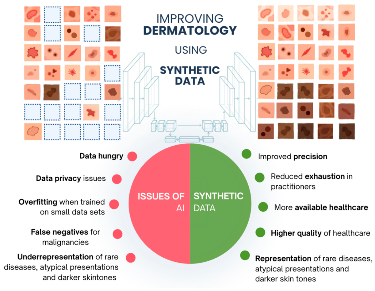
Figure 2.
Generative Adversarial Network architecture for standardizing heterogeneous dermatological data.Heterogeneous real-world clinical and histopathological inputs, exhibiting physical occlusions (e.g., hair), inconsistent illumination, variable photographic angles, and staining artifacts, are processed by a U-Net-based generator featuring deep convolutional layers and skip connections (Steps 1–2). The synthesized outputs are evaluated against high-quality reference samples by a discriminator network (Step 3). By computing a combined loss function (adversarial loss paired with structural reconstruction losses; Step 4), the generator is iteratively optimized via backpropagation until the discriminator can no longer distinguish between synthesized images and authentic clinical photographs (Step 5).
Figure 2.
Generative Adversarial Network architecture for standardizing heterogeneous dermatological data.Heterogeneous real-world clinical and histopathological inputs, exhibiting physical occlusions (e.g., hair), inconsistent illumination, variable photographic angles, and staining artifacts, are processed by a U-Net-based generator featuring deep convolutional layers and skip connections (Steps 1–2). The synthesized outputs are evaluated against high-quality reference samples by a discriminator network (Step 3). By computing a combined loss function (adversarial loss paired with structural reconstruction losses; Step 4), the generator is iteratively optimized via backpropagation until the discriminator can no longer distinguish between synthesized images and authentic clinical photographs (Step 5).
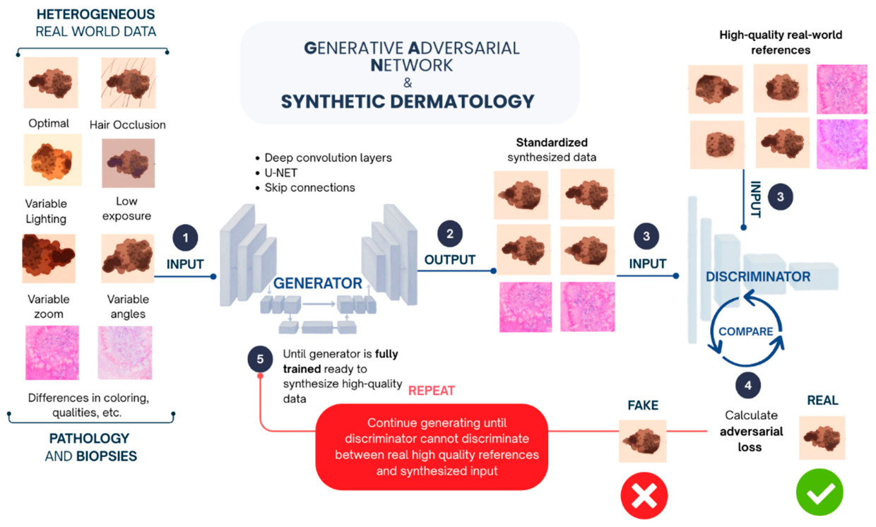
Figure 3.
Latent Diffusion Model pipeline and latent space interpolation for visual lesion progression.High-resolution clinical images are mapped into a compressed, low-dimensional latent space using a pre-trained 8-channel VAE encoder to extract core semantic features while preserving fine-grained microstructures (Step 1). Within the latent space, a forward diffusion process iteratively adds Gaussian noise across discrete timesteps, followed by conditional denoising driven by a cross-attention U-Net (Steps 2–3). Following latent decoding, lesion boundaries are extracted via automated segmentation, enabling inpainting to decouple diseased tissue from healthy baseline skin (Steps 4–6). Continuous linear latent space interpolation synthesizes intermediate pathological transition stages, which are decoded and pooled with authentic photographs for downstream classifier training (Steps 7–8).
Figure 3.
Latent Diffusion Model pipeline and latent space interpolation for visual lesion progression.High-resolution clinical images are mapped into a compressed, low-dimensional latent space using a pre-trained 8-channel VAE encoder to extract core semantic features while preserving fine-grained microstructures (Step 1). Within the latent space, a forward diffusion process iteratively adds Gaussian noise across discrete timesteps, followed by conditional denoising driven by a cross-attention U-Net (Steps 2–3). Following latent decoding, lesion boundaries are extracted via automated segmentation, enabling inpainting to decouple diseased tissue from healthy baseline skin (Steps 4–6). Continuous linear latent space interpolation synthesizes intermediate pathological transition stages, which are decoded and pooled with authentic photographs for downstream classifier training (Steps 7–8).
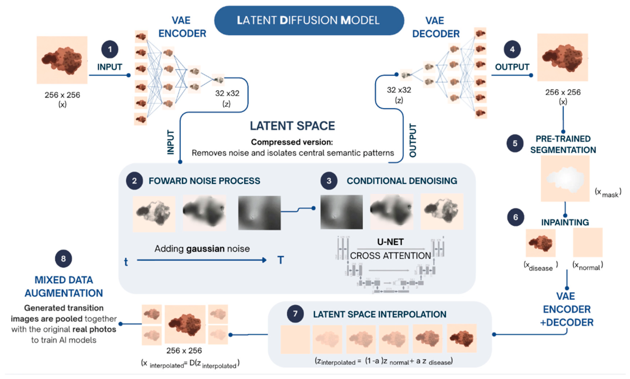
Figure 4.
Cutaneous Digital Twin Multimodal clinical data streams (left); comprising real-time exposome sensors, genomic profiles, electronic health records (EHR), and high-resolution optical scans, are continuously mapped to a patient-specific virtual avatar. The dynamic central engine performs multimodal data fusion, causal predictive modeling (e.g., via Bayesian Belief Networks), and in silico treatment simulation. This architecture yields actionable clinical applications (right), including precision therapeutics (biologic selection and regimen optimization), personalized photoprotection, continuous surveillance for morphological changes and dysplasia, and synthetic control cohorts for rare dermatological disease trials. A closed-loop feedback telemetry system continuously updates the AI engine with real-time patient data to maintain dynamic predictive accuracy.
Figure 4.
Cutaneous Digital Twin Multimodal clinical data streams (left); comprising real-time exposome sensors, genomic profiles, electronic health records (EHR), and high-resolution optical scans, are continuously mapped to a patient-specific virtual avatar. The dynamic central engine performs multimodal data fusion, causal predictive modeling (e.g., via Bayesian Belief Networks), and in silico treatment simulation. This architecture yields actionable clinical applications (right), including precision therapeutics (biologic selection and regimen optimization), personalized photoprotection, continuous surveillance for morphological changes and dysplasia, and synthetic control cohorts for rare dermatological disease trials. A closed-loop feedback telemetry system continuously updates the AI engine with real-time patient data to maintain dynamic predictive accuracy.
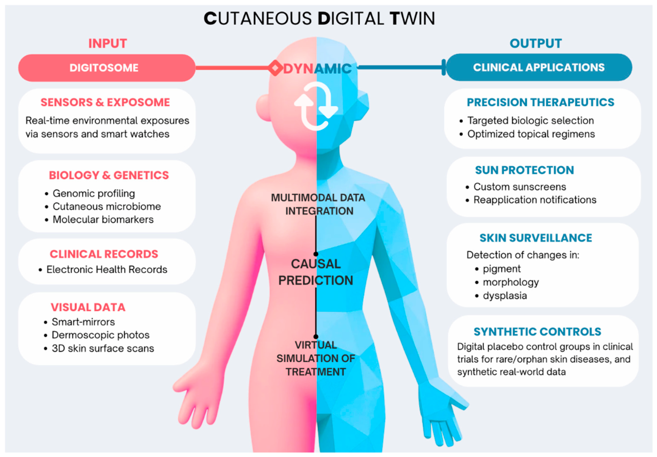
Table 1.
Key models and concepts in synthetic dermatology.
| Term | Category | Explanation | Dermatological Application |
|
AI Artificial Intelligence |
Foundational technology |
Computational frameworks for complex data analysis, pattern recognition, and predictive reasoning |
Triage, diagnostic decision support, and clinical workflow automation |
|
GAN Generative Adversarial Network |
Generative model |
Adversarial training of generator and discriminator networks |
Data augmentation and standardization for rare diseases and skin tone diversity |
|
LDM Latent Diffusion Model |
Generative model |
Diffusion-based denoising in a compressed latent space |
High-resolution skin texture synthesis and visual disease transitions |
|
VAE Variational Autoencoder |
Generative model |
Compresses high-dimensional data into low dimensional data |
Latent feature mapping enables identification of pathological abnormalities without requiring extensive training data |
|
LLM Large Language Model |
Language model | Human-like language processing and generating | Clinical documentation, patient education, and synthetic exam generation |
|
CDT Cutaneous Digital Twin |
Personalized AI |
A dynamic, virtual representation of patient skin |
Personalized therapeutics, adaptive photoprotection, and early continuous cancer surveillance |
|
SECA Synthetic External Control Arm |
Clinical trial |
Synthetic patient profiles or digital twins for placebo arm |
Ethical optimization of trials for rare/orphan skin diseases |
|
HCA Hybrid Control Arm |
Clinical trial | Hybrid patient groups combining real and synthetic patient data in control arms | Increasing statistical power while reducing placebo-group exposure |
|
BBN Bayesian Belief Network |
Causal modeling | Probabilistic models representing causal relationships | Modeling skin trajectories (e.g., photoaging) based on lifestyle factors |
|
Heuristic models |
Data processing |
Rule-based algorithms to simplify or standardize complex data |
Standardization of image illumination and contrast before generative training |
| Inpainting | Image processing |
Generates paired disease/pseudo-normal data samples and continuous skin lesion transitions |
Removing artifacts (hair/markers) to create synthetic healthy skin contexts and enhanced data diversity |
| Gaussian/Laplacian noise | Technical Guard |
Statistical noise distributions used for diffusion |
Calibrated mathematical safeguards to prevent re-identification and generation of fine-grained diagnostic textures |
| Digitosome | Data concept | Multi-source, real-time data from patients | Integration of wearables/sensors into the Cutaneous Digital Twin |
Table 2.
Cutaneous Digital Twin.
| Data dimension | Collection modality and technologies | Clinical utility within the cutaneous digital twin |
| Intrinsic biological data | Deep phenotyping Genomic sequencing Microfluidic patch sampling Epigenetic clocks Microbiome analysis |
Captures genetic predispositions, DNA repair capacity, and inflammatory biomarkers (MMPs, cyclobutane pyrimidine dimers) |
| Morphological imaging | Total body 3D skin mapping Sequential teledermoscopy Smart mirrors |
Passive, continuous tracking of atypical lesions; early alert for subtle color, shape, or border variegations |
| Extrinsic exposome data | Digital phenotyping Medical-purpose IoT devices Wearable UV/pollution sensors |
Real-time tracking of cumulative UV index, air quality, humidity, diet, sleep, and physical activity |
Table 3.
AI Reliability, Validation, and Security Frameworks.
| Term | Category | Explanation | Dermatological Relevance |
| Mode Collapse | Technical Barrier | Limited, non-diverse sample generation | Generates output with visually homogeneous lesions, failing to capture rare malignant phenotypes |
| Hallucination | System Error | AI-generated outputs that appear plausible but are factually incorrect | Creation of non-existent medical literature references, incorrect diagnostic details, or invalid ICD-10 and ATC medication codes |
|
RAG Retrieval-Augmented Generation |
Architecture | Grounding LLM outputs in verified external data | Suppressing clinical hallucinations by linking LLMs to validated catalogs |
|
FID Fréchet Inception Distance |
Quality Metric | Statistical distance between real and synthetic image distributions | Quantitative benchmark to evaluate the visual fidelity and distribution of generated synthetic skin lesions |
|
FMD Fréchet Medoid Distance |
Diversity Metric | Sample-level comparison to real data points to detect partial mode collapse | Prevent mode collapse and verify that synthetic images preserve core diagnostic structures |
|
FRESCA Framework for Research in Equitable Synthetic Control Arms |
Statistical Framework | Construct and evaluate synthetic cohorts with real-world trial arms | Propensity-matching and equity adjustments virtual cohort construction in clinical trials, enabling ethically optimized clinical trials for rare skin diseases without exposing real patients to physical placebos |
|
CUC-FATE Cost, Usability, Credibility, Fairness, Accountability, Transparency, Explainability |
Auditing Framework | Multidimensional governance framework for AI tools | Evaluate dermatological AI tools and LLM simulators for scientific credibility, skin phototype fairness, transparency, and clinical safety |
| Differential Privacy | Privacy Guard | Adding mathematical noise to prevent individual re-identification | Enable synthesizing privacy-preserving clinical images and health records for cross-institutional research sharing |
| Forensic Detection | Security | Detection training to identify synthetic deepfakes | Used to safeguard clinical data integrity, prevent fraudulent insurance claims involving fake lesions, and verify the authenticity |
| Membership Inference | Security Risk | Adversarial attack determining if a patient was in the training set | Security auditing tool to test the privacy vulnerability of generative models trained on sensitive data |
| Model Inversion | Security Risk | Attack method reconstructing original training data from model or output |
Verify that high-capacity generative skin models do not memorize and reconstruct facial architecture or identifiable patient skin features from clinical training sets |
Disclaimer/Publisher’s Note: The statements, opinions and data contained in all publications are solely those of the individual author(s) and contributor(s) and not of MDPI and/or the editor(s). MDPI and/or the editor(s) disclaim responsibility for any injury to people or property resulting from any ideas, methods, instructions or products referred to in the content. |
© 2026 by the authors. Licensee MDPI, Basel, Switzerland. This article is an open access article distributed under the terms and conditions of the Creative Commons Attribution (CC BY) license.
Copyright: This open access article is published under a Creative Commons CC BY 4.0 license, which permit the free download, distribution, and reuse, provided that the author and preprint are cited in any reuse.