

Submitted:
24 September 2026
Posted:
28 September 2026
You are already at the latest version
Abstract
Blunt cardiac trauma is rare but potentially fatal, especially when associated with cardiac chamber rupture. We report a case of a 53-year-old female who sustained right atrial rupture and pericardial tear following a motor vehicle collision. The patient presented with clinical signs of tamponade and was transferred urgently for surgical intervention. Emergency sternotomy, initiation of cardiopulmonary bypass, and right atrial repair led to full recovery. This case highlights the critical importance of rapid diagnosis and prompt surgical management in cases of cardiac trauma.
Keywords:
blunt chest trauma
; right atrial rupture
; fractured rib
Cardiac chamber rupture secondary to blunt chest trauma is a rare event, occurring in less than 0.5% of motor vehicle accidents but it is associated with high mortality [1]. The most common mechanisms of blunt cardiac trauma include: motor vehicle collisions (MVC) (50%), pedestrians struck by motor vehicles (35%), motorcycle crashes (9%), and falls from significant heights [2]. The right atrium is particularly vulnerable due to its anterior location and thinner wall. The mechanisms of rupture include the hydraulic effect which produces high pressures inside the heart or the deceleration where the shearing forces surpass the cardiac elasticity. We believe that in our case penetration of the right atrium by a fractured rib was the real mechanism of cardiac rupture. Sternal and rib fractures rarely cause secondary cardiac rupture [3].
A 53-year-old female passenger was involved in a high-speed frontal vehicle collision without using the safety belt. At the district emergency unit, she was conscious but disoriented, hypotensive (BP 65/35 mmHg), and with signs of respiratory distress. She was intubated immediately and transferred to the national trauma center.
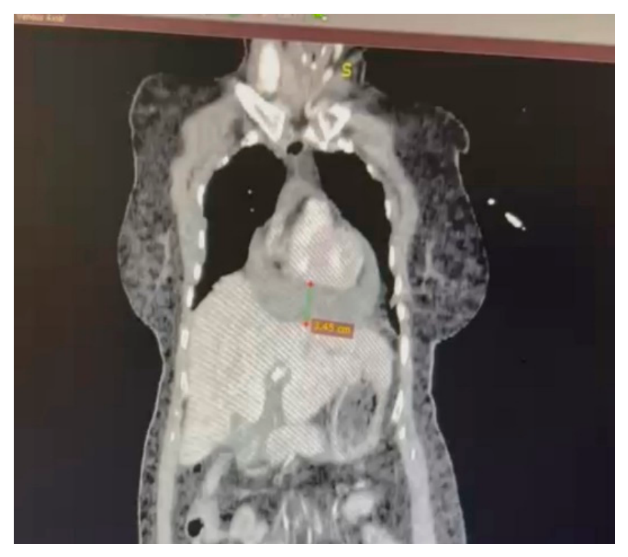
On arrival, physical examination revealed distended jugular veins and upper chest ecchymosis. Imaging (CT scan), showed multiple right rib fractures (ribs 4, 5, and 7), pericardial effusion with high-density contents, and pleural effusion (Figure 1). The patient deteriorated hemodynamically with signs of tamponade.
Ultrasound-guided pericardiocentesis was attempted but failed due to intrapericardial blood clotting within the catheter. The patient was transferred immediately to the cardiothoracic surgery department.
Emergency sternotomy was performed under general anesthesia. Upon pericardial incision, a large volume of blood and clots were released. A clear pericardial tear was visualized near the diaphragm. Active bleeding from the right atrium was identified.
Figure 2.
Rib fracture seen from inside the thoracic cavity.
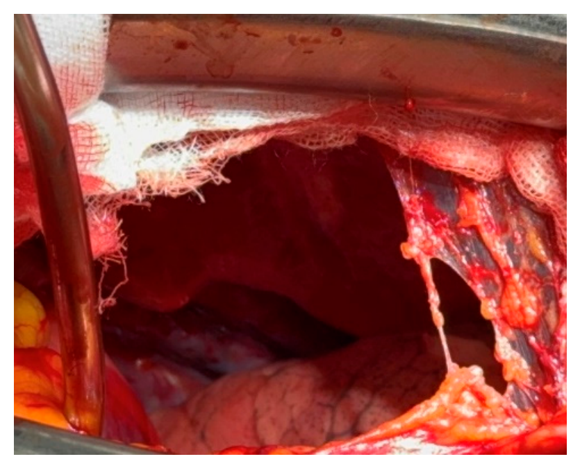
The rupture nearly 8 cm in length was near the atrioventricular sulcus and repair was difficult as long as the right coronary artery couldn’t be visualized.
Figure 3.
a. Clear view of pericardial laceration adjacent to diaphragm and pleural cavity. b. Intra-operative view the right atrial tear with the tip of the aspirator inside it.
Figure 3.
a. Clear view of pericardial laceration adjacent to diaphragm and pleural cavity. b. Intra-operative view the right atrial tear with the tip of the aspirator inside it.
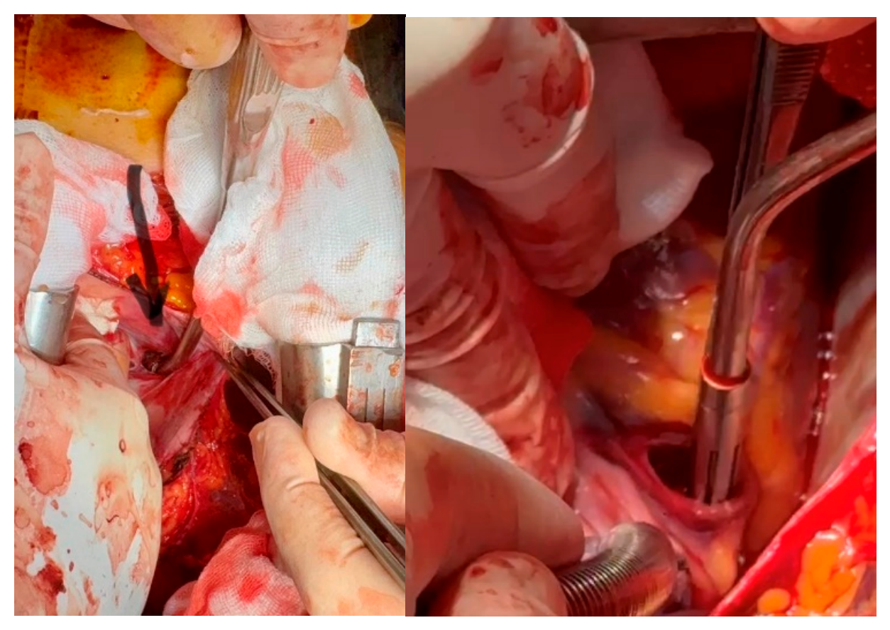
Heparin was administered, and the patient was connected to cardiopulmonary bypass (CPB). The right atrial rupture was sutured using polypropylene with pledgets without arresting the heart. After thorough exploration and hemostasis, the patient was weaned from CPB and transferred to the ICU.
The patient was extubated on postoperative day 2, and transferred to the surgical ward on day 3. Postoperative echocardiography showed no pericardial effusion and good right atrial function. The patient was transferred again to the trauma center to deal with a fracture of the talus.
Right atrial rupture after blunt trauma is usually fatal unless immediate surgical repair is possible. Desforges and coauthors reported the first repair of a blunt myocardial rupture in 1955, successfully closing a right atrial perforation secondary to a motor vehicle collision [4].
Cardiac injury should be suspected with the presence of pericardial hematoma, anterior rib fracture near the heart, massive hemothorax, signs of elevated venous pressure and progressive metabolic acidosis.
Echocardiography and computed tomography usually provide sufficient information for the diagnosis but sometimes it is not clear if the patient has to enter the operating room or not. Even after transiently stabilizing the hemodynamic state by fluid resuscitation, signs of shock or cardiac tamponade may recur, suggesting cardiac rupture. For sure, unresponsive patients to blood transfusions and other medical support, have to be explored for cardiac chamber rupture. Failed pericardiocentesis should raise suspicion for clotted hemopericardium or cardiac rupture.
Sternal and rib fractures rarely cause secondary cardiac rupture [3,5]. We believe that the inward movement of the rib during the collision is the exact mechanism of rupture initially of the pericardium and at the end also of the right atrium.The pressure in the right atrium is low and clots formation can suppress bleeding and temporarily reduce blood loss. The hemodynamic state is more stable, the patient can reach the hospital and prognosis is better with rupture of the right atrium than in other chambers of the heart [6].
The traditional approach for a left-sided hemothorax is a left anterolateral thoracotomy. To improve access to the heart, the incision can be extended with to a median sternotomy or a right anterolateral thoracotomy incision. In situations where primarily a cardiac injury is suspected, it would be appropriate to start with a median sternotomy. A left thoracotomy often does not provide adequate exposure to the heart or ascending aorta.
Cardiopulmonary bypass facilitates definitive repair in these scenarios, allowing exploration and control of bleeding. It is also effective in preventing damage of the vital structures like the coronary arteries which lie often near the rupture as in our case.
In blunt chest trauma, consider cardiac rupture when pericardial effusion and hypotension coexist. Failure of pericardiocentesis to aspirate fluid should raise suspicion of clotted hemopericardium. Early surgical intervention and cardiopulmonary bypass can be life-saving in atrial rupture. Our case reinforces the importance of fast imaging, surgical readiness, and interdepartmental coordination.
Author Contributions
Conceptualization, Marsela Goga; investigation Fjorba Mana and validation Ermal Likaj.
Funding
This research received no external funding.
Institutional Review Board Statement
Not applicable.
Informed Consent Statement
Not applicable.
Acknowledgments
The authors have reviewed and edited the output and take full responsibility for the content of this publication.
Conflicts of Interest
Declare conflicts of interest or state “The authors declare no conflicts of interest.
Abbreviations
The following abbreviations are used in this manuscript:
| MVC | Motor Vehicle Collisions |
| CT | Computed Tomography |
| ICU | Intensive Care Unit |
| CBP | Cardiopulmonary Bypass |
References
- Teixeira, P.G.; Inaba, K.; et al. Blunt cardiac trauma: lessons from the medical examiner. J. Trauma. 2009, 67(6), 1259–1264. [Google Scholar] [CrossRef] [PubMed]
- Yousef, R.; Carr, J.A. Blunt cardiac trauma: a review of the current knowledge and management. Ann. Thorac. Surg. 2014, 98(3), 1134–40. [Google Scholar] [CrossRef] [PubMed]
- Tokur, M.; Ergin, M.; Okumus, M.; Kurkcuoglu, C. Penetrating heart injuries due to puncture by fractured sternum or ribs following blunt trauma. J. Curr. Surg. 2011, 1, 38–40. [Google Scholar] [CrossRef]
- Desforges, G.; Ridder, W.P.; Lenoci, R.J. Successful suture of ruptured myocardium after nonpenetrating injury. N Engl. J. Med. 1955, 252, 567–9. [Google Scholar] [CrossRef] [PubMed]
- Hattori, M.; Akita, M.; Mori, K.; Ichikawa, M.; Takeshige, K.; Kamijo, H.; et al. Cardiac rupture caused by penetration of the heart by fractured costal cartilage following blunt chest trauma: A case report. Acute Med. Surg. 2024, 11, e70024. [Google Scholar] [CrossRef] [PubMed]
- William, C.P.; Anthony, O.U.; Andrew, B.P. Blunt rupture of the myocardium. Ann. Thorac. Surg. 1989, 48, 139–42. [Google Scholar] [CrossRef] [PubMed]
Figure 1.
Frontal plane CT scan showing pericardial effusion.
Disclaimer/Publisher’s Note: The statements, opinions and data contained in all publications are solely those of the individual author(s) and contributor(s) and not of MDPI and/or the editor(s). MDPI and/or the editor(s) disclaim responsibility for any injury to people or property resulting from any ideas, methods, instructions or products referred to in the content. |
© 2026 by the authors. Licensee MDPI, Basel, Switzerland. This article is an open access article distributed under the terms and conditions of the Creative Commons Attribution (CC BY) license (http://creativecommons.org/licenses/by/4.0/).
Copyright: This open access article is published under a Creative Commons CC BY 4.0 license, which permit the free download, distribution, and reuse, provided that the author and preprint are cited in any reuse.