

Submitted:
27 September 2026
Posted:
28 September 2026
You are already at the latest version
Abstract
Vascularized fibular grafting is one of the most widely used hip preservation procedures for young patients with osteonecrosis of the femoral head. However, long-term follow-up data indicate that a substantial proportion of patients ultimately convert to total hip arthroplasty (THA). We hypothesize that recovery of graft or pedicle perfusion may precede restoration of functional sensory-neural signaling within the broader repair region, creating a putative temporal mismatch. Drawing on neurovascular-bone coupling theory, we propose a testable candidate design principle: emerging evidence suggests that neural reinnervation may be a potentially limiting factor in bone repair, and active local reinnervation should be prioritized for empirical testing in hip preservation surgery. We synthesize the potential role of the sensory nerve–calcitonin gene-related peptide (CGRP) axis as a candidate mediator in osteogenesis, vascular maturation, and regulation of the local immune microenvironment.We further propose a tiered neurotization strategy encompassing autologous nerve grafting, bioactive nerve conduits, and stem cell–biomaterial composite systems. Critical issues—including neural source selection, ischemic microenvironmental constraints, risk–benefit trade-offs, and individualized patient eligibility—are also discussed. This article aims to stimulate discussion on a paradigm shift in hip preservation surgery while explicitly framing its central thesis as a hypothesis awaiting empirical validation.
Keywords:
osteonecrosis of the femoral head
; hip preservation surgery
; neuro-vascular-bone coupling
; active neurotization
; neural signaling deprivation window
Introduction
Vascularized fibular grafting, one of the most widely used hip preservation procedures, has long been regarded as a promising option; however, its long-term efficacy remains suboptimal. In one long-term follow-up study of patients with post-traumatic osteonecrosis, 36% converted to total hip arthroplasty at approximately 10.9 years after vascularized fibular grafting[1]. In a separate systematic review of patients with osteonecrosis of heterogeneous etiology, non-vascularized bone grafting was associated with a mid-term failure rate of 21%[2].Their figures are not directly comparable, but these findings suggest that the current revascularization-centered paradigm may harbor theoretical blind spots: although it emphasizes restoration of baseline blood supply, it does not comprehensively coordinate the multifaceted interplay among neural regulation, angiogenesis, and the immune microenvironment during bone repair.Neuro-vascular-bone coupling theory posits that bone is a metabolically active tissue under precise neural control. Sensory nerve endings release neuropeptides such as CGRP, which directly modulate osteoblastic and osteoclastic activity and promote vascular stability[3]. Denervation experiments provide evidence of the converse: denervation leads to severely impaired bone healing and marked deterioration of biomechanical performance[4]. Other epidemiological data show that patients with diabetes mellitus and concomitant peripheral neuropathy have a significantly elevated fracture risk[5]. These observations suggest that timely neural reinnervation may contribute to efficient bone repair.
Yet current outcome assessment of hip preservation surgery focuses almost exclusively on vascularity. Neural innervation is almost entirely absent from this evaluation framework. Analysis of failed cases reveals a paradox: although the transplanted bone exhibits robust graft vascularity, viability, and osseous integration, the surrounding necrotic region may remain unoccupied by newly formed trabecular bone and instead be infiltrated by soft tissue[6]. This repair failure may result from delayed neural recovery or persistent microvascular insufficiency. Moreover, the local microenvironment within the necrotic area is dysregulated and contains angiogenesis inhibitors such as S100A9[7]. This finding is compatible with altered neuro-immune regulation, but it also preserves persistent local ischemia as a competing explanation.This indicates that merely reconstructing the vascular conduit without actively restoring neural regulation may fail to initiate the appropriate repair program.Based on the above analysis, this article proposes a perspective: in hip preservation surgery, functional coupling along the neuro-vascular-bone axis should be actively reestablished in addition to vascularized bone grafting. We posit that restoration of critical neural signaling inputs may systematically optimize the bone repair microenvironment, thereby promoting higher-quality regenerative bone formation.
Biological Basis
Anatomical studies demonstrate that the periosteum is one of the most densely innervated regions and serves as a critical interface for bidirectional communication between bone tissue and the nervous system[8]. Among these nerves, sensory nerve fibers predominate both within bone tissue and in the periosteum; their terminal endings release neuropeptides that play a central role in regulating bone metabolism[8]. Of all neuropeptides, CGRP has been most extensively studied and represents one of the candidate pathways linking neural signaling to bone repair.
The sensory nerve-CGRP axis has three functional roles. At the osteogenic level, CGRP directly stimulates osteoblast proliferation and differentiation while inhibiting osteoclast activity, thereby coordinating the balance between bone formation and bone resorption[9]. At the vascular level, CGRP exerts potent vasodilatory effects and promotes nitric oxide (NO) production by modulating endothelial cell function, thus mediating neurovascular crosstalk at the cellular level[10]. At the immune level, CGRP release helps maintain local homeostasis and prevents a shift toward a proinflammatory state within the repair site. The sympathetic nervous system also contributes to bone metabolism regulation, primarily via β-adrenergic receptors that influence osteoblast function[11]; however, with respect to promoting bone repair, the sensory nerve–CGRP axis appears to play the more central role among currently studied pathways, although other neurotrophic pathways such as NGF-TrkA and Schwann cell paracrine signaling may also contribute.
Neural Signaling Deprivation Window
Vascularized fibular grafting restores blood supply via microvascular anastomosis[12], creating a well-perfused “sentinel site” for bone regeneration[13]. In the early phase of recovery, the effects of revascularization alone and revascularization combined with active neurotization may be similar; however, reinnervation depends on slow host nerve ingrowth, so long-term recovery may favor the latter. This temporal mismatch provides a new perspective for understanding why some grafts fail despite successful revascularization. We propose that this mismatch creates a window of neural signal deprivation during the mid-to-late recovery phase. During this window, insufficient CGRP and other neuropeptides may impair osteogenesis, vascular maturation, and immune homeostasis. From this perspective, active neurotization is not merely an adjunct to vascular reconstruction but an attempt to shorten this window and restore functional coupling. This window concept reframes graft failure around neural timing rather than vascular adequacy alone.
Operational Definition of Active Neurotization
Before elaborating specific strategies, it is essential to clarify the operational definition of active neurotization. In this article, active neurotization is defined as the surgical introduction or induction of functional neural innervation into the recipient site concurrently with or shortly after vascularized bone transplantation, with the aim of shortening the neural signaling deprivation window and restoring the neuro-vascular-bone coupling axis. This definition comprises three core elements: delivery modality, timing window, and validation endpoints.
Regarding delivery modality, active neurotization encompasses three hierarchical approaches: autologous nerve grafting, in which the patient’s own sensory nerve is surgically transferred to the subperiosteal space of the recipient site; bioactive nerve conduits, in which biodegradable conduits loaded with and gradually releasing neurotrophic factors guide directional axonal growth; and stem cell–biomaterial composite systems, in which supportive cells and biomimetic scaffolds are co-engineered to actively construct de novo neural networks. These modalities are not mutually exclusive and may be combined according to the severity of tissue injury.
Regarding timing, active neurotization may be implemented at three distinct time points: concomitant, performed during the same surgical procedure as vascularized bone transplantation; early intervention, involving secondary neural reinnervation performed within days to weeks postoperatively; and delayed intervention, referring to neural reconstruction during secondary revision surgery for cases with prior surgical failure or severe nerve injury. Concomitant intervention may most effectively shorten the neural signaling deprivation window but carries the highest technical complexity, whereas early and delayed strategies are suited to distinct clinical scenarios.
For validation endpoints, active neurotization must be assessed across at least four dimensions: neural regeneration metrics—including CGRP-positive fiber density and PGP9.5-positive nerve fiber density; bone quality metrics—such as bone volume fraction, trabecular thickness, and connectivity density; biomechanical metrics—including maximum compressive load, stiffness, and toughness; and vascular maturation metrics—such as vessel density and pericyte coverage. These metrics must be measured concurrently within the same experimental system to establish the association between improved neural innervation and enhanced bone quality.
In comparative design, the benefits of active neurotization must be explicitly contrasted against those of isolated revascularization, and a control group representing increased surgical complexity alone must be included. Without this control, it is impossible to distinguish the specific therapeutic gains attributable to active neurotization from nonspecific effects arising solely from additional surgical manipulation.
Active Neurotization Strategies
In light of the above analysis, we propose that actively reconstructing neural innervation concurrently with vascularized bone transplantation represents a promising strategy for enhancing hip preservation. Based on current technical maturity and clinical feasibility, these strategies can be stratified into three tiers according to their indications, technical challenges, and short-term clinical readiness.
The most straightforward approach is bridging autologous sensory nerves to the recipient site. The sural nerve cutaneous branch is one of the commonly used and safe donor nerves in peripheral nerve repair, with minimal functional impact on the donor site following harvest [14]. During surgery, this nerve can be coapted to an appropriate sensory nerve branch at the recipient site. The ideal target for implantation is the subperiosteal space—a region not only rich in vascularity and endowed with abundant osteogenic stem or progenitor cells but also the primary anatomical site of physiological neural innervation[8]. This strategy establishes a direct anatomical conduit to rapidly restore neural innervation at the periosteal interface. Its feasibility is well supported by extensive literature in peripheral nerve repair; however, its specific effects within the bone repair microenvironment warrant further investigation. This approach is most suitable for patients with partial nerve injury who require rapid reinnervation. Key limitations include limited donor nerve availability and uncertainty regarding regenerative efficiency in ischemic regions. Nevertheless, its near-term clinical feasibility is relatively high, with key endpoints including CGRP-positive fiber density and bone volume fraction.
In cases where autologous nerve donors are limited or the regenerative microenvironment is suboptimal, bioactive nerve conduits represent a promising alternative or adjunctive strategy. Biodegradable nerve conduits—fabricated from materials such as polycaprolactone or poly(lactic-co-glycolic acid)—can be functionalized via surface drug-loading techniques to deliver and sustainably release neurotrophic factors, including nerve growth factor (NGF) and brain-derived neurotrophic factor (BDNF), thereby providing both physical guidance and chemotactic cues[15,16,17]. In vitro models have confirmed that such conduits actively recruit and accelerate directional axonal growth along their length[16,17]. Translating this technology—already demonstrating promise in peripheral nerve injury repair—to the context of bone regeneration may offer additional options for constructing neuromodulatory units, particularly in pathological microenvironments unfavorable to neural regeneration, such as ischemia and inflammation[18]. This approach is especially suited for complex cases involving ischemia, inflammation, or fibrotic scarring. The primary challenges lie in the yet-to-be-validated long-term safety and drug-loading stability. Its near-term feasibility currently ranges from preclinical to early clinical stages, with key endpoints including axonal regeneration, vascular maturation, and bone mechanical performance.
Looking further ahead, advances in regenerative medicine are enabling more forward-looking strategies for neural reconstruction. Encapsulating supportive cells—such as Schwann cells—within biomimetic hydrogels and implanting them into nerve guidance conduits significantly enhances both neural regeneration and vascularization[19]. Hyaluronic acid–collagen composite hydrogels, which closely mimic the natural neural extracellular matrix, are regarded as promising scaffold candidates[20]. Systems integrating induced neural stem cells with hydrogel scaffolds and neurotrophic factors have demonstrated potential for reconstructing damaged neural circuits in spinal cord injury models[21]. Although this strategy remains in the preclinical exploratory phase, its conceptual framework—actively constructing de novo neural networks—represents the cutting edge of next-generation neural repair approaches[22]. This tier represents a long-term direction for complex neural defects; however, major barriers include ethical considerations, high costs, and technical complexity, resulting in low near-term feasibility. Key endpoints at this level are neural network integration and functional recovery.Figure 1 illustrates the indication stratification logic of the three-tiered strategy in the form of a decision pathway driven by preoperative assessment.
Discussion
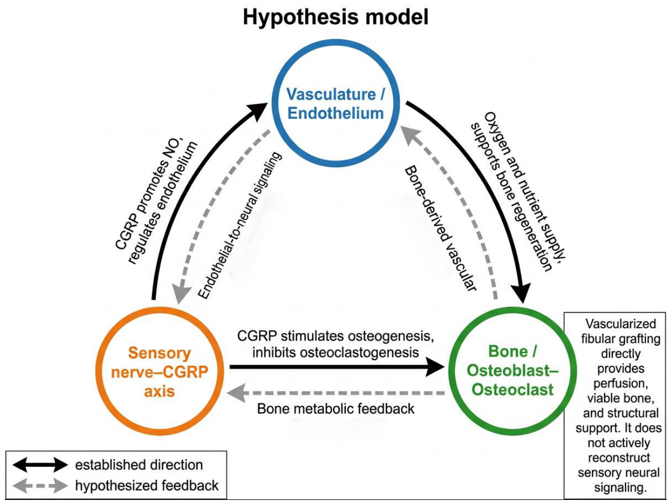
Several key questions merit discussion in relation to the above strategies.First is the selection of neural sources. Bone, particularly the periosteum and bone matrix, is predominantly innervated by sensory nerve fibers whose terminals secrete neuropeptides such as CGRP and substance P[8]. Among these, CGRP has been shown in several experimental models to possess dual osteogenic and pro-angiogenic functions, and it is proposed here as a candidate mediator coordinating the neuro-vascular-bone coupling axis.[9]. Sensory neurons directly modulate endothelial cell function via CGRP release, thereby mediating neurovascular crosstalk[10]. Therefore, considering both its central anatomical distribution and well-established regenerative functions, sensory nerves represent the most biologically appropriate source for reconstructing the neuro-vascular-bone coupling axis, rather than motor nerves. Figure 2 depicts the reciprocal regulatory relationships among the sensory nerve–CGRP axis, vasculature, and bone.
Second is the challenge of regeneration within the ischemic microenvironment. Inflammation, hypoxia, and fibrosis in necrotic bone may impede tissue regeneration[18], and this may also apply to neural regeneration. This underscores the necessity of bioactive conduit strategies—designed to provide physical protection and sustained localized delivery of neurotrophic factors to actively intervene in and improve the local microenvironment. In animal models, analogous bioactive conduits have demonstrated efficacy in promoting axonal regeneration, myelination, and functional neural recovery[23]. Thus, through integrated protection and induction, it may be possible to enhance neuronal survival and regenerative capacity within the ischemic region.
Third is surgical complexity and the risk–benefit ratio. Incorporating additional steps inevitably prolongs operative time and increases technical difficulty. However, decision-making should be grounded in long-term risk–benefit analysis. For patients under 30 years of age with hip pathology, total hip arthroplasty offers reliable mid-term outcomes, but its immediate benefits must be weighed against the uncertainty of potential revision surgery decades later[24]. Therefore, for young, highly active patients at elevated risk of failure with conventional joint-preserving procedures, prioritizing a moderately more complex, enhanced joint-preserving surgery—aimed at improving long-term native joint retention—deserves rigorous evaluation within a well-designed clinical research framework[25]. This approach seeks to avoid prematurely placing such patients on a treatment trajectory likely to necessitate multiple revisions. The added technical complexity can be mitigated through standardized training and optimized instrumentation. Future studies must include a control group undergoing surgery with increased procedural complexity alone, to distinguish the specific benefits attributable to active neurotization from nonspecific effects arising solely from additional surgical manipulation.
Finally, individualized patient selection is essential. How should optimal candidates for active neurotization be identified? A tiered clinical decision-making framework is required, centered on precise preoperative assessment[26]. High-resolution imaging, such as magnetic resonance neurography, can evaluate neural continuity and pathological status within the injured region[27], thereby facilitating stratification of intervention needs across patient subgroups. Figure 1 presents an exploratory research framework rather than a validated clinical algorithm. Preoperative MR neurography may characterize the morphology of candidate peripheral nerves, but macroscopic nerve continuity should not be assumed to represent the density or functional status of periosteal and intraosseous sensory endings within the graft bed. Whether preoperative imaging can predict differential benefit from active neurotization remains to be tested; in cases of severe nerve injury, scarring, or prior surgical failure, bioactive nerve conduits or combined strategies may be considered; and for complex defects, stem cell–biomaterial composite systems represent a more innovative and promising long-term solution. Individualized decision-making based on preoperative assessment is critical to maximizing the clinical value of this approach.
Future Research Directions
The feasibility of the perspectives outlined above must ultimately be evaluated through systematic investigation. Future studies should focus on several testable hypotheses.The first hypothesis concerns timing and specificity: Does early active reinnervation following vascularized fibular transplantation shorten the neural signaling deprivation window and significantly improve bone volume fraction and maximum compressive load? Experimental designs must include control groups undergoing procedures of comparable surgical complexity—without neural intervention—to isolate the specific benefits attributable to reinnervation. In animal models, comparisons should be made between conventional vascular reconstruction alone and combined vascular–neural reconstruction, specifically assessing correlations between nerve fiber density and bone quality within the repair site to determine whether enhanced innervation drives improvements in bone quality[4,6,7].
The second hypothesis addresses strategy comparison: In ischemic microenvironments, do bioactive nerve conduits outperform autologous nerve grafts alone in promoting both nerve regeneration and bone repair? Comparative evaluations of different strategies—across metrics of neural reinnervation and bone regeneration—are needed to inform optimal clinical decision-making[15,16,17,18].
The third hypothesis explores the underlying mechanistic network: Does active reinnervation enhance bone healing via a CGRP-mediated osteogenic–angiogenic–immune network, altering the spatiotemporal distribution of local cellular subpopulations to enhance the quality of regenerated bone? Emerging technologies such as spatial transcriptomics and multiplex immunofluorescence staining can be leveraged to systematically dissect the spatiotemporal dynamics of key pathways—including osteogenesis, angiogenesis, and immune regulation—and associated cellular populations within the local microenvironment following active neurotization interventions[3,9,10].
The fourth proposition concerns individualization: Can the degree of neural injury observed on preoperative magnetic resonance neuroimaging predict the magnitude of clinical benefit a patient derives from active neurotization strategies? Building upon preclinical validation, surgical techniques should be progressively standardized, and well-designed human clinical trials should be conducted to ultimately evaluate the efficacy and safety of this strategy in real-world clinical settings[26].
Conclusions
Introducing neuro-vascular-bone coupling theory into hip preservation surgery represents an exploratory shift from structural to functional reconstruction. We propose that restoring neural regulation—beyond blood supply—may improve outcomes. The central question is whether both perfusion and neural signaling are restored. We hypothesize that delayed sensory-neural restoration may be a limiting factor, but this remains to be tested. Active neurotization is proposed as an experimental strategy to test whether earlier restoration improves repair. Its value lies not in replacing vascular reconstruction but in addressing the dimension of neural instruction that vascular reconstruction alone does not cover. If confirmed, benefits may extend beyond osteonecrosis of the femoral head to traumatic nonunion and large-segment bone defects. Validation of this perspective will require systematic preclinical studies and standardized techniques. We hope this article stimulates further investigation and interdisciplinary collaboration.
References
- Richard, M.J.; DiPrinzio, E.V.; Lorenzana, D.J.; Whitlock, K.G.; Hein, R.E.; Urbaniak, J.R. Outcomes of free vascularized fibular graft for post-traumatic osteonecrosis of the femoral head. Injury 2021, 52(12), 3653–3659. [Google Scholar] [CrossRef]
- Li, J.; Mo, L.; Bai, G.; Wang, Z.; Zhang, H.; Li, J. One in five patients require conversion to arthroplasty after non-vascularized bone grafts in patients with osteonecrosis of the femoral head: a systematic review. J. Orthop. Surg. Res. 2023, 18(1), 82. [Google Scholar] [CrossRef]
- Abeynayake, N.; Arthur, A.; Gronthos, S. Crosstalk between skeletal and neural tissues is critical for skeletal health. Bone 2021, 142, 115645. [Google Scholar] [CrossRef]
- Pan, K.; Cheng, X.; Wu, Y.; Orozco, E.; Shin, S.H.; Du, J.; et al. Denervation induces rapid bone degeneration concurrent to neurovascular and mechanical changes. Bone 2026, 117863. [Google Scholar] [CrossRef]
- Graversen, J.L.; Kvist, A.V.; Rasmussen, N.H.; Vestergaard, P.; Røikjer, J. Diabetic neuropathy and fracture risk – a nation-wide case/control study. Bone 2026, 207, 117860. [Google Scholar] [CrossRef]
- Lim, T.; Tang, Q.; Wang, Q.; Zhu, Z.; Wei, X.; Feng, Y.; et al. Histopathological findings of failed free vascularized fibular grafting for osteonecrosis of the femoral head after long-term follow-up. BioMed Res. Int. 2020, 2020, 6493585. [Google Scholar] [CrossRef]
- Wu, R.W.; Lian, W.S.; Kuo, C.W.; Chen, Y.S.; Ko, J.Y.; Wang, F.S. S100 calcium binding protein A9 represses angiogenic activity and aggravates osteonecrosis of the femoral head. Int. J. Mol. Sci. 2019, 20(22), 5786. [Google Scholar] [CrossRef]
- Khodorovskaya, A.M.; Agranovich, O.E.; Savina, M.V.; Garkavenko, Y.E.; Melchenko, E.V.; Filin, Y.A.; et al. Innervation of bones. Sensory innervation. Part I: a literature review. Pediatr. Traum Orthop. Reconstr. Surg. 2024, 12(4), 511–522. [Google Scholar] [CrossRef]
- Wang, Q.; Qin, H.; Deng, J.; Xu, H.; Liu, S.; Weng, J.; et al. Research progress in calcitonin gene-related peptide and bone repair. Biomolecules 2023, 13(5), 838. [Google Scholar] [CrossRef]
- Leroux, A.; Roque, M.; Casas, E.; Leng, J.; Guibert, C.; L’Azou, B.; et al. The effect of CGRP and SP and the cell signaling dialogue between sensory neurons and endothelial cells. Biol. Res. 2024, 57(1), 60. [Google Scholar] [CrossRef]
- Liu, M.; Liu, Y.; Yu, J.; Gong, J.; Zhao, C.; Liu, Z. Molecular mechanisms and therapeutic implications of the sympathetic nervous system in bone-related disorders: a brain-bone axis perspective. Bone Res. 2025, 13(1), 51. [Google Scholar] [CrossRef]
- Wang, M.; Guo, H.; Zhang, G.; Ruan, P.; Zhi, K. Development and innovation of modern microvascular anastomoses. JBM 2024, 12(10), 105–118. [Google Scholar]
- Meregawa, P.F.; Pratama, R.R. Free vascularized fibular graft. Int. J. Sci. Healthc. Res. 2021, 6(3), 179–188. [Google Scholar] [CrossRef]
- Serrano, E.B.; Serrano, B.R.; Leyva, A.J.; Vaca, J.C.A. Neurorrhaphy of the sciatic nerve with autologous sural nerve graft in a gunshot wound: a case report. Int. J. Res. Med. Sci. 2024, 12(10), 3851–3853. [Google Scholar] [CrossRef]
- Kakinoki, R.; Hara, Y.; Yoshimoto, K.; Kaizawa, Y.; Hashimoto, K.; Tanaka, H.; et al. Fabrication of artificial nerve conduits used in a long nerve gap: current reviews and future studies. Bioengineering 2024, 11(4), 409. [Google Scholar] [CrossRef]
- Sandoval-Castellanos, A.M.; Claeyssens, F.; Haycock, J.W. Bioactive 3D scaffolds for the delivery of NGF and BDNF to improve nerve regeneration. Front Mater. 2021, 8, 734683. [Google Scholar] [CrossRef]
- Zhu, L.; Jia, S.; Liu, T.; Yan, L.; Huang, D.; Wang, Z.; et al. Aligned PCL fiber conduits immobilized with nerve growth factor gradients enhance and direct sciatic nerve regeneration. Adv. Funct. Mater. 2020, 30(39), 2002610. [Google Scholar] [CrossRef]
- Wang, H.; Zhang, J. Research progress on circulating inflammatory proteins and osteonecrosis of the femoral head. JCMP 2025, 7(5), 125–129. [Google Scholar] [CrossRef]
- Huang, Y.; Ye, K.; He, A.; Wan, S.; Wu, M.; Hu, D.; et al. Dual-layer conduit containing VEGF-A-transfected Schwann cells promotes peripheral nerve regeneration via angiogenesis. Acta Biomater. 2024, 180, 323–336. [Google Scholar] [CrossRef]
- Ranjan, S.; Choudhary, P.; Shivalkar, S.; Dwivedi, S.; Singh, S. Potential of hyaluronic acid and collagen-based scaffolds in promoting stem cell neuronal differentiation for neuroregenerative therapies: a review. Int. J. Biol. Macromol. 2025, 309, 142981. [Google Scholar] [CrossRef]
- Son, D.; Zheng, J.; Kim, I.Y.; Kang, P.J.; Park, K.; Priscilla, L.; et al. Human induced neural stem cells support functional recovery in spinal cord injury models. Exp. Mol. Med. 2023, 55(6), 1182–1192. [Google Scholar] [CrossRef]
- Stocco, E.; Barbon, S.; Emmi, A.; Tiengo, C.; Macchi, V.; De Caro, R.; et al. Bridging gaps in peripheral nerves: from current strategies to future perspectives in conduit design. Int. J. Mol. Sci. 2023, 24(11), 9170. [Google Scholar] [CrossRef]
- Cai, Y.; Chen, Y.; Zhang, G.; Lin, Y.; Zhang, J.; Liang, J.; et al. The GDNF-gel/HA-Mg conduit promotes the repair of peripheral nerve defects by regulating PPAR-γ/RhoA/ROCK signaling pathway. iScience 2024, 27(2), 108969. [Google Scholar] [CrossRef]
- Katzman, J.L.; Manjunath, A.; Brandstetter, A.S.; Ben-Ari, E.; Schwarzkopf, R.; Snir, N. Mid-term outcomes of total hip arthroplasty in patients younger than 30 years. Arch. Orthop. Trauma Surg. 2024, 144(8), 3775–3786. [Google Scholar] [CrossRef]
- Parilla, F.W.; Anthony, C.A.; Bartosiak, K.A.; Pashos, G.E.; Thapa, S.; Clohisy, J.C. Ten year outcomes of contemporary total hip arthroplasty in adolescent and young adult patients are favorable. J. Arthroplast. 2024, 39(3), 754–759. [Google Scholar] [CrossRef]
- Bateman, E.A.; Larocerie-Salgado, J.; Ross, D.C.; Miller, T.A.; Pripotnev, S. Assessment, patient selection, and rehabilitation of nerve transfers. Front Rehabil. Sci. 2023, 4, 1267433. [Google Scholar] [CrossRef]
- Goyal, A.; Wadgera, N.; Srivastava, D.N.; Ansari, M.T.; Dawar, R. Imaging of traumatic peripheral nerve injuries. J. Clin. Orthop. Trauma 2021, 21, 101510. [Google Scholar] [CrossRef]
Figure 1.
Exploratory research framework for preoperative imaging stratification and active neurotization. Preoperative MR neurography assesses the morphology of candidate peripheral nerves; it does not directly represent periosteal or intraosseous sensory terminal integrity. Technology tiers are exploratory categories, not clinical grades.
Figure 1.
Exploratory research framework for preoperative imaging stratification and active neurotization. Preoperative MR neurography assesses the morphology of candidate peripheral nerves; it does not directly represent periosteal or intraosseous sensory terminal integrity. Technology tiers are exploratory categories, not clinical grades.
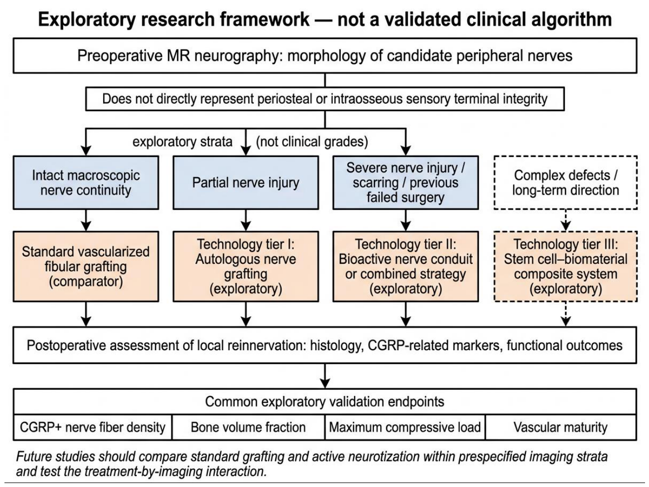
Figure 2.
Evidence integration and hypothesis model of sensory nerve–vascular–bone interactions. Solid arrows indicate directions supported by direct evidence; dashed arrows indicate hypothesized feedback relationships that remain to be validated. The sensory nerve–CGRP axis, vasculature/endothelium, and bone/osteoblast–osteoclast form a mutually regulatory network, but the evidence strength across different directions is not equivalent. In particular, the mediators, temporal sequence, and causal direction of endothelial-to-neural and bone-to-neural feedback remain to be established.
Figure 2.
Evidence integration and hypothesis model of sensory nerve–vascular–bone interactions. Solid arrows indicate directions supported by direct evidence; dashed arrows indicate hypothesized feedback relationships that remain to be validated. The sensory nerve–CGRP axis, vasculature/endothelium, and bone/osteoblast–osteoclast form a mutually regulatory network, but the evidence strength across different directions is not equivalent. In particular, the mediators, temporal sequence, and causal direction of endothelial-to-neural and bone-to-neural feedback remain to be established.
Disclaimer/Publisher’s Note: The statements, opinions and data contained in all publications are solely those of the individual author(s) and contributor(s) and not of MDPI and/or the editor(s). MDPI and/or the editor(s) disclaim responsibility for any injury to people or property resulting from any ideas, methods, instructions or products referred to in the content. |
© 2026 by the authors. Licensee MDPI, Basel, Switzerland. This article is an open access article distributed under the terms and conditions of the Creative Commons Attribution (CC BY) license (http://creativecommons.org/licenses/by/4.0/).
Copyright: This open access article is published under a Creative Commons CC BY 4.0 license, which permit the free download, distribution, and reuse, provided that the author and preprint are cited in any reuse.