

Submitted:
24 September 2026
Posted:
28 September 2026
You are already at the latest version
Abstract
Background: Cable wood-chop exercises are commonly progressed by changing body position and prescribing loads relative to the maximal capacity achieved in each posture. However, equivalent relative loads may not produce equivalent mechanical responses across postural configurations. Objective: To compare smartphone-derived thoracic acceleration across five cable wood-chop positions prescribed using posture-specific relative loads and to determine the intra- and inter-session reliability of this measure. Methods: Twenty-two physically active men were enrolled in a two-session repeated-measures study. Maximal break force was assessed in seated, kneeling, half-kneeling, parallel-stance and staggered-stance positions with pulley height standardized to shoulder level in each posture. Participants then performed 12-s isometric (70% maximal break force) and dynamic (40% maximal break force) wood-chop trials while attempting to maintain a neutral spinal posture. Thoracic acceleration was recorded using a smartphone secured at T3. Reliability was evaluated using intraclass correlation coefficients, typical error, and minimal detectable change. Differences between positions were examined using linear mixed-effects models, with complementary models accounting for posture-specific maximal break force. Results: Smartphone-derived thoracic acceleration showed good-to-excellent intra-session reliability, whereas inter-session reliability was posture dependent and lower in standing positions. Half-kneeling elicited greater unadjusted thoracic acceleration than all other positions during both isometric and dynamic conditions (all Holm-adjusted p < 0.001; |dz| = 1.18–1.57). Maximal break force was positively associated with thoracic acceleration in both conditions (p < 0.001). In the force-adjusted models, half-kneeling remained higher than all other positions during the isometric condition (all unadjusted p ≤ 0.015), whereas differences were substantially attenuated during the dynamic condition. The relative position-related pattern was consistent across testing sessions. Conclusions: Prescribing the cable wood-chop using posture-specific relative loads does not necessarily result in equivalent thoracic acceleration responses across postural configurations. Body position should therefore be considered an additional programming variable when prescribing the exercise. Smartphone accelerometry may provide a practical field-based method for quantifying thoracic acceleration responses during rotational resistance exercise.
Keywords:
core exercise
; stabilization training
; resistance exercise
; smartphone accelerometry
; postural configuration
; exercise prescription
1. Introduction
Rotational and anti-rotational trunk exercises are widely incorporated into strength and conditioning and rehabilitation programs to enhance the ability to generate, transfer, and resist rotational forces across the kinetic chain [1,2,3]. Among these exercises, the cable wood-chop is widely utilized due to its multiplanar nature and the ability to progressively scale external resistance, enabling practitioners to target trunk function under a wide range of loading conditions [4,5]. Consequently, it is commonly prescribed for athletes involved in throwing, striking, and racquet sports, as well as in clinical rehabilitation requiring progressive rotational or anti-rotational trunk loading [6,7].
Exercise intensity during resistance training is commonly prescribed relative to an individual’s maximal capacity. This approach facilitates individualization of external resistance but may implicitly assume that equivalent relative intensities across exercise variations impose comparable mechanical demands. However, slight changes in body position or exercise technique can substantially alter joint moments, muscle recruitment patterns, and force transmission throughout the kinetic chain [8,9]. In anti-rotational exercises such as the cable wood-chop, changing posture modifies the base of support, lower-limb contribution, and the available postural stabilization strategies. Consequently, posture-specific relative loading may standardize resistance relative to the maximal force capacity achieved within each posture without necessarily standardizing the resulting thoracic response [10,11]. Understanding whether body position contributes to this response independently of posture-specific force capacity has important practical implications, as strength coaches and clinicians frequently manipulate body position when progressing rotational and anti-rotational exercises.
Smartphone accelerometers provide an inexpensive and accessible method for quantifying segmental acceleration during exercise outside laboratory environments. Previous research has demonstrated that smartphone-derived acceleration can reliably quantify responses across various core stability exercises and progressions, while offering a practical alternative to laboratory-based biomechanical assessment [12,13,14,15]. However, no previous study has investigated whether smartphone-derived thoracic acceleration can characterize differences among cable wood-chop configurations when resistance is prescribed relative to posture-specific maximal force capacity.
Therefore, the primary aim of this study was to determine whether prescribing cable wood-chop exercises using posture-specific relative loads results in equivalent thoracic acceleration responses across seated, kneeling, half-kneeling, parallel stance, and staggered stance positions. A secondary aim was to determine the intra- and inter-session reliability of smartphone-derived thoracic acceleration during isometric and dynamic cable wood-chop exercises. We hypothesized that smartphone-derived thoracic acceleration would demonstrate good-to-excellent intra-session reliability, although between-session reproducibility would be lower in standing postures due to greater available movement degrees of freedom. Furthermore, we expected that posture-specific relative loads would not produce equivalent thoracic acceleration responses across postural configurations and that some of these differences would persist after accounting for posture-specific maximal force capacity.
2. Methods
2.1. Participants
Twenty-two healthy, physically active men (age: 30.5 ± 11.0 years; height: 173.7 ± 7.3 cm; body mass: 77.0 ± 6.7 kg) voluntarily participated in this study. Participants reported engaging in at least two sessions of moderate-to-vigorous physical activity per week (120–300 min·week⁻¹) and were familiar with resistance training involving cable-pulley exercises. Exclusion criteria included: (i) musculoskeletal disorders or medical conditions contraindicating physical activity; (ii) trunk or upper-/lower-limb injuries during the previous 12 months; (iii) previous spinal or abdominal surgery; and (iv) participation in a structured trunk-training program within the previous six months. Before participation, all volunteers received written and verbal information regarding the study procedures and provided written informed consent. The study was conducted in accordance with the Declaration of Helsinki and approved by the Committee of Ethics and Integrity in Research (CEII) of Miguel Hernandez University of Elche (protocol code DCD.FBM.01.23; 7 November 2023).
An a priori sample-size calculation was performed using G*Power 3.1 for a repeated-measures ANOVA design with one group and five within-subject conditions. Based on the findings of De Los Ríos-Calonge et al. [16], the calculation assumed a Cohen’s f of 0.30, an alpha level of 0.05, statistical power of 80%, a correlation of 0.50 among repeated measurements, and a nonsphericity correction of 0.75. The analysis indicated that a minimum of 18 participants were required. The final sample of 22 participants exceeded this threshold.
2.2. Experimental Design
A repeated-measures design was used to examine thoracic acceleration during cable wood-chop exercises performed in five postural configurations: seated, kneeling, half-kneeling, staggered stance, and parallel stance (Figure 1). Participants attended two identical testing sessions separated by one week. During each session, maximal break force was first determined for each posture and subsequently used to prescribe posture-specific individualized loads for the isometric (70% of break force) and dynamic (40% of break force) wood-chop exercises.
2.3. Instrumentation
External force was recorded using a Tindeq Progressor 300 load cell (Tindeq AS, Trondheim, Norway) connected in series with the cable system. The load cell was positioned between the cable attachment and the exercise bar and was used to record maximal break force for each posture. The exercises were performed using an adjustable cable-pulley machine (Technogym S.p.A., Cesena, Italy), with the cable set approximately at shoulder height.
Thoracic acceleration was recorded using a smartphone (Apple iPhone SE, model MHGQ3QL/A, Apple Inc., Cupertino, CA, USA) secured over the upper thoracic spine at T3 level using an adjustable elastic strap. Acceleration signals were acquired at 100 Hz using the CoreMaker application [15]. To ensure consistency across testing sessions, the same investigator performed smartphone placement for all trials.
2.4. Experimental Protocol
Following a standardized warm-up consisting of full-body joint mobility exercises (from the shoulders to the ankles) and a 20-s dynamic set of the wood-chop exercise to activate the trunk musculature, participants received standardized instructions and performed ten submaximal practice repetitions in each postural configuration until they were familiar with the execution of the cable wood-chop exercise and the testing procedures. Subsequently, participants performed the testing protocol in the five postural configurations shown in Figure 1. The postural configurations were performed in a fixed ascending order of biomechanical difficulty to standardize task sequencing across participants. For all trials, external cable resistance pulled laterally toward the participant’s right side. In asymmetric postures (half-kneeling and staggered stance), the leg ipsilateral to the direction of cable pull was positioned forward.
2.5. Break-Test Assessment
Maximal break force was determined using a standardized break-test protocol previously described by De Los Ríos-Calonge et al. [16]. Briefly, participants resisted a progressively increasing externally applied force while maintaining each of the prescribed wood-chop positions. Force was applied continuously until the participant could no longer maintain the required posture (break point). Trials were considered valid only when force increased progressively throughout the test and maximal force was reached within approximately 7–12 s. The highest force achieved in each posture was used to individualize the exercise loads.
2.6. Wood-Chop Exercise Trials
Following a 3-min recovery period after the break-test assessment, participants performed the isometric condition followed by the dynamic condition across all postures. During the isometric condition, participants maintained the wood-chop position for 12 s against a load corresponding to 70% of the posture-specific maximal break force while minimizing trunk movement. For the dynamic condition, participants performed continuous concentric and eccentric movements for 12 s at 40% of maximal break force, using a standardized cadence controlled by a metronome set at 45 beats·min⁻¹. Each metronome beat indicated the transition between the concentric and eccentric phases, resulting in four complete movement cycles and one additional half-cycle during each 12-s trial. Two valid trials were recorded for each condition and posture, with 30-s recovery intervals between trials and 1-min recovery intervals between conditions and postures.
2.7. Data Processing
The CoreMaker application provided gravity-corrected linear acceleration signals, and no additional digital filtering was applied. The resultant thoracic acceleration was calculated as the vector magnitude of the three orthogonal acceleration axes. To eliminate initial and final movement artifacts, the first and final 1 s of each 12-s trial were excluded, and the mean resultant acceleration was calculated from the remaining 10-s analysis window. For the dynamic condition, mean resultant acceleration was calculated across all movement cycles completed within the 10-s analysis window. The mean of the two valid trials was used for statistical analysis. The same investigator positioned the smartphone and processed all recordings to ensure consistency across testing sessions.
2.8. Statistical Analysis
Descriptive statistics are presented as mean ± standard deviation. Intra- and inter-session reliability were assessed using two-way mixed-effects intraclass correlation coefficients [ICC(3,k)], together with the typical error (TE), minimal detectable change at the 80% confidence level (MDC80), and mean differences with 95% confidence intervals. MDC80 was calculated as TE × 1.28 × √2 [17,18]. ICC(3,k) values were interpreted according to the criteria proposed by Koo and Li [19]: values below 0.50 were considered poor, 0.50–0.75 moderate, 0.75–0.90 good, and above 0.90 excellent.
Analyses were performed using all available valid data. The primary between-posture analyses were based on Session 2 and included 21 participants. Session 2 was selected to minimize potential familiarization effects and because it provided the most complete valid dataset. Session 1 recordings were unavailable or invalid for five participants due to technical issues during data acquisition; consequently, inter-session reliability analyses and sensitivity models including both sessions were conducted using the 16 participants with valid data from both testing sessions.
Unadjusted differences in thoracic acceleration between postures were first examined separately for the isometric and dynamic conditions using linear mixed-effects models, with posture entered as a fixed effect and participant included as a random intercept. These models represented the primary practical comparison because they preserved the actual posture-specific loads prescribed during the experiment. Cohen’s dz was calculated as the mean paired difference divided by the standard deviation of the paired differences.
Adjusted differences were subsequently examined using linear mixed-effects models including posture as a fixed effect, posture-specific maximal break force as a covariate, and participant as a random intercept. Because maximal break force determined the absolute resistance prescribed within each posture, these models were interpreted as complementary analyses examining whether posture-related differences in thoracic acceleration persisted after accounting for posture-specific force capacity, rather than as conventional confounder-adjusted estimates. Maximal break force was mean-centered and entered into the models in kilogram-force (kgf). Therefore, the regression coefficient represents the expected change in thoracic acceleration associated with a 1-kgf increase in maximal break force after accounting for posture. Post-hoc pairwise comparisons for the unadjusted models were obtained from estimated marginal means and corrected for multiple comparisons using the Holm method. For the force-adjusted models, fixed-effect coefficients, standard errors, 95% confidence intervals, and corresponding unadjusted p-values were reported, with half-kneeling specified as the reference posture. Estimated marginal means from the force-adjusted models were additionally calculated for graphical presentation. The sensitivity analysis included data from both testing sessions and retained posture and maximal break force as fixed effects and participant as a random intercept, while adding Session and the Position × Session interaction as fixed effects. Statistical significance was set at p < 0.05. All statistical analyses were performed using R (R Foundation for Statistical Computing, Vienna, Austria). Linear mixed-effects models were fitted using the lme4 and lmerTest packages. Estimated marginal means and post-hoc pairwise comparisons for the unadjusted models were obtained using the emmeans package, with multiplicity adjustment performed using the Holm method. Intraclass correlation coefficients were calculated using the psych package.
3. Results
3.1. Reliability of Thoracic Acceleration
Valid Session 2 data were available for 21 participants, whereas 16 participants had valid recordings from both sessions. Descriptive statistics and reliability outcomes are presented in Supplementary Tables S1 and S2. For the isometric cable wood-chop, intra-session ICC(3,k) values ranged from 0.91 to 0.98, with typical errors ranging from 0.03 to 0.06 m·s⁻² and MDC80 values ranging from 0.06 to 0.12 m·s⁻². For the dynamic cable wood-chop, intra-session ICC(3,k) values ranged from 0.87 to 0.93, with typical errors ranging from 0.03 to 0.05 m·s⁻² and MDC80 values ranging from 0.05 to 0.08 m·s⁻².
Inter-session reliability showed greater variation across postures. For the isometric condition, ICC(3,k) estimates were ≥0.72 in half-kneeling, seated, and kneeling, but ≤0.35 in parallel and staggered stance. For the dynamic condition, inter-session ICC(3,k) values ranged from 0.49 to 0.92, with half-kneeling showing the highest reproducibility. Mean changes and their 95% confidence intervals are reported in the Supplementary Tables.
3.2. Thoracic Acceleration Across Postures
Figure 2 shows the distribution of Session 2 thoracic acceleration across postures. In both exercise modes, half-kneeling produced the highest acceleration values, whereas seated, kneeling, parallel stance, and staggered stance showed more similar distributions.
During the isometric cable wood-chop, thoracic acceleration was greater in half-kneeling than in seated, kneeling, parallel stance, and staggered stance (all Holm-adjusted p< 0.001; |dz|= 1.45 − 1.57). No other pairwise comparison remained significant after Holm correction. The same pattern was observed during the dynamic cable wood-chop, with greater thoracic acceleration in half-kneeling than in each of the other four positions (all Holm-adjusted p< 0.001; |dz|= 1.18 − 1.34), whereas the remaining comparisons were not significant (Table 1).
3.3. Maximal Break Force and Thoracic Acceleration
Maximal break force was positively associated with thoracic acceleration in the isometric model (β= 0.033, 95% CI 0.021 to 0.045, p< 0.001) and in the dynamic model (β= 0.021, 95% CI 0.014 to 0.029, p< 0.001). Thus, greater posture-specific force capacity was associated with higher thoracic acceleration.
In the force-adjusted models, fixed-effect coefficients indicated lower isometric thoracic acceleration for seated, kneeling, parallel stance, and staggered stance relative to the half-kneeling reference posture (all unadjusted p ≤ 0.015). In the dynamic model, only kneeling differed from the half-kneeling reference posture (unadjusted p = 0.048).
Adding the Position × Break Force interaction did not significantly improve model fit for the isometric (likelihood-ratio χ²(4)= 7.82, p= 0.099) or dynamic condition (χ²(4)= 3.80, p= 0.433). No statistically significant evidence was found that the force–acceleration association differed among postures (Table 2).
β, regression coefficient; SE, standard error; CI, confidence interval. For maximal break force, β represents the change in thoracic acceleration (m·s⁻²) associated with a 1-kgf increase in force. Reported coefficient p-values are unadjusted.
3.4. Force-Adjusted Estimated Marginal Means
Force-adjusted estimated marginal means are displayed in Figure 3. The adjusted isometric mean for half-kneeling was 0.478 m·s⁻², compared with 0.355-0.392 m·s⁻² for the other postures. In the dynamic condition, adjusted means were more closely clustered, indicating substantial attenuation of the raw between-posture differences after accounting for maximal break force.
3.5. Sensitivity Analysis
To evaluate the robustness of the main findings, an additional linear mixed-effects model including both testing sessions was performed. The model included position, session, and posture-specific maximal break force as fixed effects, participant as a random intercept, and the Position × Session interaction. A significant overall Session effect was observed for both the isometric (χ² = 9.20, p = 0.002) and dynamic conditions (χ² = 11.04, p < 0.001), indicating a systematic change in thoracic acceleration between sessions. However, the Position × Session interaction was not significant in either condition (p = 0.413 and p = 0.542, respectively), indicating that the relative posture-related pattern was consistent across sessions. Detailed results are presented in Supplementary Table S3.
4. Discussion
The present study investigated whether prescribing cable wood-chop exercises using posture-specific relative loads produced comparable thoracic acceleration responses across different postural configurations. Four main findings emerged. First, smartphone-derived thoracic acceleration showed good-to-excellent intra-session reliability across all postures, whereas between-session reproducibility was posture-dependent, being higher in seated, kneeling, and half-kneeling than in the standing configurations. Second, matching relative load did not standardize the thoracic acceleration response, as the half-kneeling posture consistently elicited greater thoracic acceleration than all remaining postures in the unadjusted models. Third, posture-specific maximal break force was positively associated with thoracic acceleration and explained part of the observed between-posture differences when maximal break force was included in the model. Finally, although thoracic acceleration showed an overall change between sessions, the absence of Position × Session interactions indicated that the relative pattern across postures was preserved.
The lower between-session reproducibility observed in parallel and staggered stance should not be interpreted as indicating that standing postures are less valid or useful exercise options. Rather, the thoracic acceleration response was more variable in the present protocol when participants performed rotational cable exercises while standing. According to the constraints-led perspective and the concept of motor abundance, the same task objective can be achieved through a wider spectrum of coordination strategies when movement solutions are abundant [10,11]. Standing postures may provide greater freedom to alter lower-limb, pelvic, and trunk contributions across testing sessions, whereas seated and kneeling configurations mechanically constrain body position and restrict available movement strategies. Therefore, more constrained postures (seated, kneeling, and half-kneeling) showed greater reproducibility under the present testing conditions, whereas standing configurations may require stricter methodological standardization (e.g., standardized stance width, pelvic orientation, and trunk alignment) when thoracic acceleration is monitored over time.
The most prominent finding was the greater thoracic acceleration elicited by the half-kneeling posture during both isometric and dynamic conditions. This pattern occurred despite prescribing exercise loads relative to the maximal break force individually obtained within each posture. Thus, matching relative intensity (70% and 40% of posture-specific maximal break force) did not normalize the thoracic acceleration response across postures. Biomechanically, half-kneeling presents an asymmetric base of support that combines lower-extremity stabilization with asymmetric hip joint constraints (flexion of the forward leg versus extension of the trailing leg). Because the cable pulled laterally toward the participant’s right side with the ipsilateral leg positioned forward, this configuration may impose distinct anti-rotational and lateral-bending requirements and may require different coordination strategies compared with symmetric postures [20,21]. Accordingly, half-kneeling should be considered a mechanically distinct task rather than a minor postural variation of the same exercise.
An important component of the greater thoracic acceleration observed in half-kneeling is likely related to the absolute resistance imposed during the task. Half-kneeling yielded the highest maximal break force among all tested configurations. Consequently, prescribing 70% and 40% of the posture-specific maximum translated into greater absolute loads in this posture during the isometric and dynamic conditions, respectively. Mechanical output during cable wood-chop exercises is load-dependent, and substantial interindividual variability has been reported across loading conditions [22]. Higher external resistance would be expected to increase the perturbation transmitted through the kinetic chain and may therefore contribute to greater thoracic acceleration.
When maximal break force was included in the model, half-kneeling retained significantly higher isometric thoracic acceleration than all other postures (all unadjusted p ≤ 0.015). In contrast, during the dynamic condition, force adjustment attenuated the differences between half-kneeling and the standing and seated postures, leaving only the contrast between kneeling and half-kneeling statistically significant (unadjusted p = 0.048). The sensitivity analysis incorporating both testing sessions showed no Position × Session interaction, indicating that the relative posture-related pattern was preserved across sessions. Together, these findings suggest that posture-specific force capacity contributes importantly to dynamic thoracic acceleration, whereas posture-related factors may retain an additional influence during the isometric condition.
The influence of posture was more pronounced during the isometric condition than during the dynamic condition. In isometric trials, participants resisted a high relative load (70% of maximal break force) while actively attempting to minimize trunk movement over 12 s. Under these static conditions, thoracic acceleration likely reflected a combination of residual segmental motion, mechanical vibration, and the response to sustained external loading. Conversely, dynamic execution at 40% of maximal break force—controlled at a cadence of 45 beats·min⁻¹ over four complete movement cycles—incorporated concentric and eccentric upper-limb movements while maintaining a neutral spine to actively resist rotation, alongside continuous fluctuations in cable tension. These dynamic components may have increased within-task signal variability and reduced the relative contribution of posture-specific differences once maximal break force was accounted for.
More broadly, these findings demonstrate that matching relative intensity does not necessarily standardize segmental mechanical responses across exercise configurations. Relative loading expresses resistance as a percentage of maximal posture-specific strength, but altering body posture changes joint leverage, force-transmission vectors, and segmental mechanical responses. Similar phenomena have been documented in lower-limb multi-joint exercises, where subtle positional modifications alter internal moment arms and muscular force distribution despite identical relative intensities [9]. Consequently, progressing from standing or kneeling to half-kneeling wood-chops should not be viewed merely as adjusting task difficulty, but as producing a different thoracic acceleration response under the tested conditions.
An essential methodological consideration is the physical interpretation of smartphone-derived thoracic acceleration. The triaxial signal recorded at the T3 level must not be misinterpreted as a direct proxy for trunk stability or postural control. Rather, it represents an integrative biomechanical response reflecting force transmission, muscle-generated stiffness, scapular girdle dynamics, and mechanical vibrations transmitted through the cable system. During the isometric condition, part of the recorded acceleration may also reflect small mechanical oscillations associated with sustained muscle force production [23]; however, the present design does not allow these components to be isolated from other sources of segmental acceleration. Therefore, higher thoracic acceleration should not automatically be viewed as a negative outcome or an indicator of reduced stability. Instead, it indicates that the thoracic segment experienced greater acceleration under the specific loading and postural constraints of that exercise. This interpretation is consistent with a constraints-based perspective of motor behavior, in which postural behavior emerges from the interaction between task constraints, environmental demands, and individual coordination capabilities [10,11]. Accordingly, thoracic acceleration should be interpreted as an integrative biomechanical outcome rather than a surrogate measure of trunk stability.
From a practical perspective, smartphone-derived thoracic acceleration may complement conventional external-load prescription when practitioners wish to quantify how different exercise configurations influence the movement response of the thorax. The half-kneeling position warrants particular attention in future research as a potential field-based configuration for assessing anti-rotational trunk function, given that standardized field methods for assessing anti-rotational force capacity remain limited [16] and this posture elicited the highest maximal break force, the greatest thoracic acceleration response, and the highest inter-session ICC estimates (ICC(3,k) = 0.77–0.92). However, further research is needed to establish criterion and construct validity, responsiveness, associations with performance, and the ability to discriminate between populations. Given the accessibility and low cost of smartphones, accelerometry-based monitoring may be particularly useful in applied settings where external resistance is nonlinear or difficult to quantify in absolute units, such as during elastic-band exercises.
Several limitations should be considered. First, using a single smartphone positioned at T3 did not allow differentiation between thoracic and lumbopelvic motion. Second, linear acceleration could not be fully decoupled from rotational motion, scapular dynamics, cable-transmitted vibrations, soft-tissue artifacts, or potential device displacement. Third, the lack of electromyography prevented direct assessment of muscle activation dynamics accompanying the observed accelerations. Fourth, postural configurations were performed in a fixed rather than randomized or counterbalanced order; consequently, posture was confounded with exercise sequence, and potential effects of fatigue, learning, or potentiation cannot be completely excluded. Fifth, Session 1 data were unavailable or invalid for five participants, reducing the sample available for inter-session reliability analyses and widening the uncertainty around some ICC estimates. Sixth, only one cable-pull direction and one configuration of the forward limb in asymmetric postures were examined, preventing conclusions regarding side-dependent effects. Finally, the sample comprised healthy, physically active men, limiting immediate generalization to women, older adults, or clinical populations. Future research should combine multi-sensor accelerometry (placed at thoracic, lumbar, and pelvic levels) with electromyography and 3D motion analysis to capture pelvic-thoracic dissociation and segmental coordination during rotational exercises. This approach could allow a more detailed characterization of segmental rotation and relative motion between trunk regions, which is particularly relevant in exercises aimed at maintaining a neutral spine position while the limbs generate or resist rotational forces.
5. Conclusions
Smartphone-derived thoracic acceleration showed good-to-excellent intra-session reliability, whereas between-session reproducibility was posture-dependent, being lower in the standing configurations. Despite matching posture-specific relative loads, half-kneeling elicited the greatest thoracic acceleration response, particularly during the isometric condition, in which differences persisted after accounting for maximal break force. In contrast, most dynamic between-posture differences were attenuated when maximal break force was included in the model, indicating an important contribution of posture-specific force capacity to the observed acceleration response. These findings indicate that matching relative loading does not necessarily equalize thoracic acceleration responses across postural configurations. Half-kneeling may be a promising configuration for further investigation as a field-based assessment of anti-rotational trunk function. Smartphone accelerometry may provide a portable method for quantifying these responses in applied settings, although further validation is required before thoracic acceleration can be interpreted as a direct measure of trunk stability, mechanical demand, or anti-rotational capacity.
Supplementary Materials
The following supporting information can be downloaded at the website of this paper posted on Preprints.org, Table S1, Intra- and inter-session reliability of thoracic acceleration during the isometric cable wood-chop; Table S2, Intra- and inter-session reliability of thoracic acceleration during the dynamic cable wood-chop; Table S3, Sensitivity analysis using linear mixed-effects models including both testing sessions.
Author Contributions
Conceptualization, J.D.L.R.-C., C.J.-R., A.M.-I., D.B. and F.J.V.-G.; methodology, J.D.L.R.-C., C.J.-R. and A.M.-I.; software, A.M.-I.; formal analysis, J.D.L.R.-C. and C.J.-R.; investigation, J.D.L.R.-C., C.J.-R., A.M.-I., D.B. and F.J.V.-G.; data curation, J.D.L.R.-C., C.J.-R. and A.M.-I.; writing—original draft preparation, C.J.-R. and F.J.V.-G.; writing—review and editing, J.D.L.R.-C., A.M.-I., D.B. and F.J.V.-G.; supervision, D.B. and F.J.V.-G.; project administration, D.B. and F.J.V.-G.; funding acquisition, D.B. and F.J.V.-G. All authors have read and agreed to the published version of the manuscript.
Funding
This study was supported by the Generalitat Valenciana Emerging Research Groups Programme (CIGE/2024/070 and CIGE/2022/22). The funders had no role in the design of the study; in the collection, analyses, or interpretation of data; in the writing of the manuscript; or in the decision to publish the results.
Institutional Review Board Statement
The study was conducted in accordance with the Declaration of Helsinki and approved by the Committee of Ethics and Integrity in Research (CEII) of Miguel Hernandez University of Elche (protocol code DCD.FBM.01.23; approved on 7 November 2023).:.
Informed Consent Statement
Informed consent was obtained from all subjects involved in the study.
Data Availability Statement
The data supporting the findings of this study are available from the corresponding author upon reasonable request.
Acknowledgments
The authors thank all participants for their involvement. During the preparation of this manuscript, the authors used ChatGPT (OpenAI; accessed September 2026) for language editing and manuscript-formatting assistance. The authors reviewed and edited the output and take full responsibility for the content of this publication.
Conflicts of Interest
The authors declare no conflicts of interest.
References
- Behm, D.G.; Drinkwater, E.J.; Willardson, J.M.; Cowley, P.M. Canadian Society for Exercise Physiology position stand: The use of instability to train the core in athletic and nonathletic conditioning. Appl. Physiol. Nutr. Metab. 2010, 35, 109–112. [Google Scholar] [CrossRef]
- Palmer, T.; Uhl, T.L.; Howell, D.; Hewett, T.E.; Viele, K.; Mattacola, C.G. Sport-specific training targeting the proximal segments and throwing velocity in collegiate throwing athletes. J. Athl. Train. 2015, 50, 567–577. [Google Scholar] [CrossRef]
- Willardson, J.M. Core stability training: Applications to sports conditioning programs. J. Strength Cond. Res. 2007, 21, 979–985. [Google Scholar] [CrossRef]
- Rodríguez-Perea, A.; Morenas Aguilar, M.D.; Escobar-Molina, R.; Martínez-García, D.; Chirosa Ríos, I.; Jerez-Mayorga, D.; Chirosa Ríos, L.; Janicijevic, D.; Reyes-Ferrada, W. Strength assessment of trunk rotator muscles: A multicenter reliability study. Healthcare 2023, 11, 2331. [Google Scholar] [CrossRef]
- Zemková, E.; Cepková, A.; Uvaček, M.; Šooš, L. A novel method for assessing muscle power during the standing cable wood chop exercise. J. Strength Cond. Res. 2017, 31, 2246–2254. [Google Scholar] [CrossRef]
- Vasudevan, J.M.; Logan, A.; Shultz, R.; Koval, J.J.; Roh, E.Y.; Fredericson, M. Comparison of muscle onset activation sequences between a golf or tennis swing and common training exercises using surface electromyography: A pilot study. J. Sports Med. 2016, 2016, 3987486. [Google Scholar] [CrossRef]
- Voight, M.L.; Hoogenboom, B.J.; Cook, G. The chop and lift reconsidered: Integrating neuromuscular principles into orthopedic and sports rehabilitation. N. Am. J. Sports Phys. Ther. 2008, 3, 151–159. [Google Scholar]
- Schoenfeld, B.J. Squatting kinematics and kinetics and their application to exercise performance. J. Strength Cond. Res. 2010, 24, 3497–3506. [Google Scholar] [CrossRef]
- Swinton, P.A.; Stewart, A.D.; Lloyd, R.; Keogh, J.W.L.; Agouris, I. A biomechanical comparison of the traditional squat, powerlifting squat, and box squat. J. Strength Cond. Res. 2012, 26, 1805–1816. [Google Scholar] [CrossRef]
- Latash, M.L. The bliss (not the problem) of motor abundance (not redundancy). Exp. Brain Res. 2012, 217, 1–5. [Google Scholar] [CrossRef]
- Newell, K.M. Constraints on the development of coordination. In Motor Development in Children: Aspects of Coordination and Control; Wade, M.G., Whiting, H.T.A., Eds.; Martinus Nijhoff: Dordrecht, The Netherlands, 1986; pp. 341–360. [Google Scholar] [CrossRef]
- Barbado, D.; Irles-Vidal, B.; Prat-Luri, A.; García-Vaquero, M.P.; Vera-Garcia, F.J. Training intensity quantification of core stability exercises based on a smartphone accelerometer. PLoS ONE 2018, 13, e0208262. [Google Scholar] [CrossRef]
- Heredia-Elvar, J.R.; Juan-Recio, C.; Prat-Luri, A.; Barbado, D.; Vera-Garcia, F.J. Observational screening guidelines and smartphone accelerometer thresholds to establish the intensity of some of the most popular core stability exercises. Front. Physiol. 2021, 12, 751569. [Google Scholar] [CrossRef]
- Heredia-Elvar, J.R.; Juan-Recio, C.; Prat-Luri, A.; Barbado, D.; de Los Ríos-Calonge, J.; Vera-Garcia, F.J. Exercise intensity progressions and criteria to prescribe core stability exercises in young physically active men: A smartphone accelerometer-based study. J. Strength Cond. Res. 2024, 38, 266–273. [Google Scholar] [CrossRef]
- De Los Ríos-Calonge, J.; Juan-Recio, C.; Prat-Luri, A.; Miralles-Iborra, A.; Elvira, J.L.; Barbado, D.; Vera-Garcia, F.J. Smartphone-Based Field Assessment of Trunk Stability and Its Relationship With Whole-Body Balance in Older Adults: Cross-Sectional Study. JMIR Aging 2026, 9, e83546. [Google Scholar] [CrossRef]
- De Los Ríos-Calonge, J.; Juan-Recio, C.; Miralles-Iborra, A.; Prat-Luri, A.; Barbado, D.; Vera-Garcia, F.J. A load-cell-based approach to assess maximal break force during the Pallof press: Test–retest reliability and postural differences. Preprints 2026. [Google Scholar] [CrossRef]
- Hopkins, W.G. Measures of reliability in sports medicine and science. Sports Med. 2000, 30, 1–15. [Google Scholar] [CrossRef]
- Weir, J.P. Quantifying test-retest reliability using the intraclass correlation coefficient and the SEM. J. Strength Cond. Res. 2005, 19, 231–240. [Google Scholar] [CrossRef]
- Koo, T.K.; Li, M.Y. A guideline of selecting and reporting intraclass correlation coefficients for reliability research. J. Chiropr. Med. 2016, 15, 155–163. [Google Scholar] [CrossRef]
- Jeong, T.; Chung, Y. The effect of squats on muscle activity in standing, kneeling, and half-kneeling positions: A cross-sectional study. Medicine 2024, 103, e39902. [Google Scholar] [CrossRef]
- Nakai, Y.; Kawada, M.; Miyazaki, T.; Araki, S.; Takeshita, Y.; Kiyama, R. A self-oblique exercise that activates the coordinated activity of abdominal and hip muscles—A pilot study. PLoS ONE 2021, 16, e0255035. [Google Scholar] [CrossRef]
- Zemková, E. Strength and power-related measures in assessing core muscle performance in sport and rehabilitation. Front. Physiol. 2022, 13, 861582. [Google Scholar] [CrossRef]
- Schaefer, L.V.; Bittmann, F.N. Parkinson patients without tremor show changed patterns of mechanical muscle oscillations during a specific bilateral motor task compared to controls. Sci. Rep. 2020, 10, 1168. [Google Scholar] [CrossRef]
Figure 1.
Cable wood-chop positions: (A) seated; (B) kneeling; (C) half-kneeling; (D) staggered stance; (E) parallel stance; and (F) initial and final positions of the dynamic cable wood-chop.
Figure 1.
Cable wood-chop positions: (A) seated; (B) kneeling; (C) half-kneeling; (D) staggered stance; (E) parallel stance; and (F) initial and final positions of the dynamic cable wood-chop.
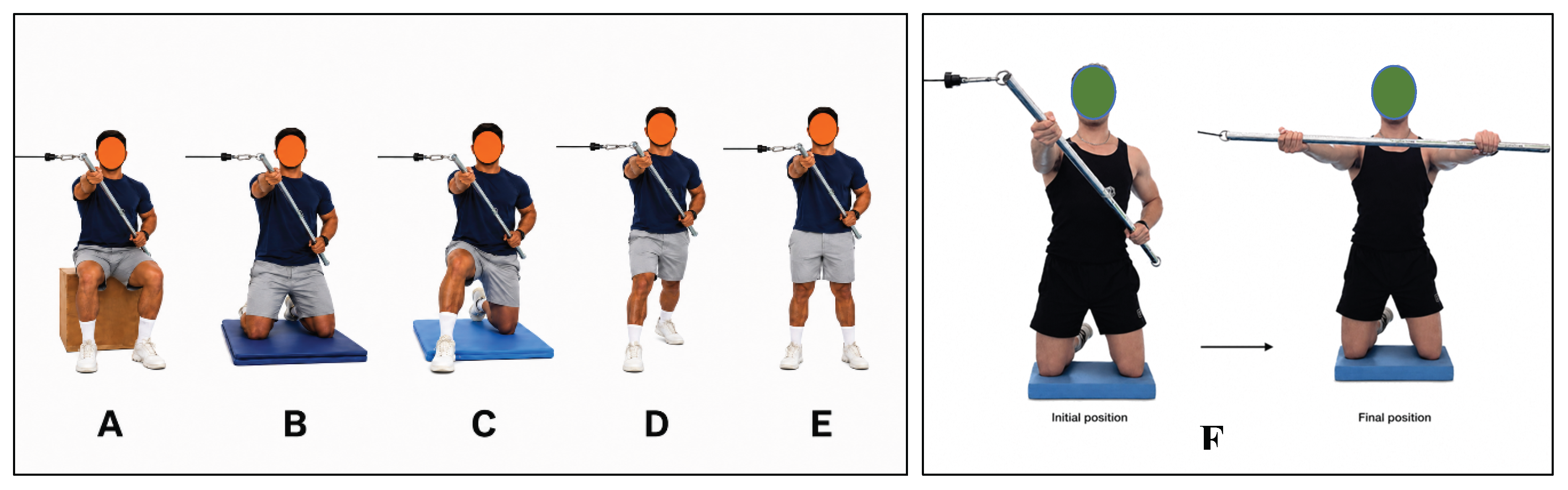
Figure 2.
Distribution of thoracic acceleration during Session 2 for the isometric (A) and dynamic (B) cable wood-chop. Boxes represent the interquartile range, horizontal lines represent the median, whiskers extend to 1.5 times the interquartile range, and dots represent individual participants. Different letters indicate significant pairwise differences after Holm correction.
Figure 2.
Distribution of thoracic acceleration during Session 2 for the isometric (A) and dynamic (B) cable wood-chop. Boxes represent the interquartile range, horizontal lines represent the median, whiskers extend to 1.5 times the interquartile range, and dots represent individual participants. Different letters indicate significant pairwise differences after Holm correction.
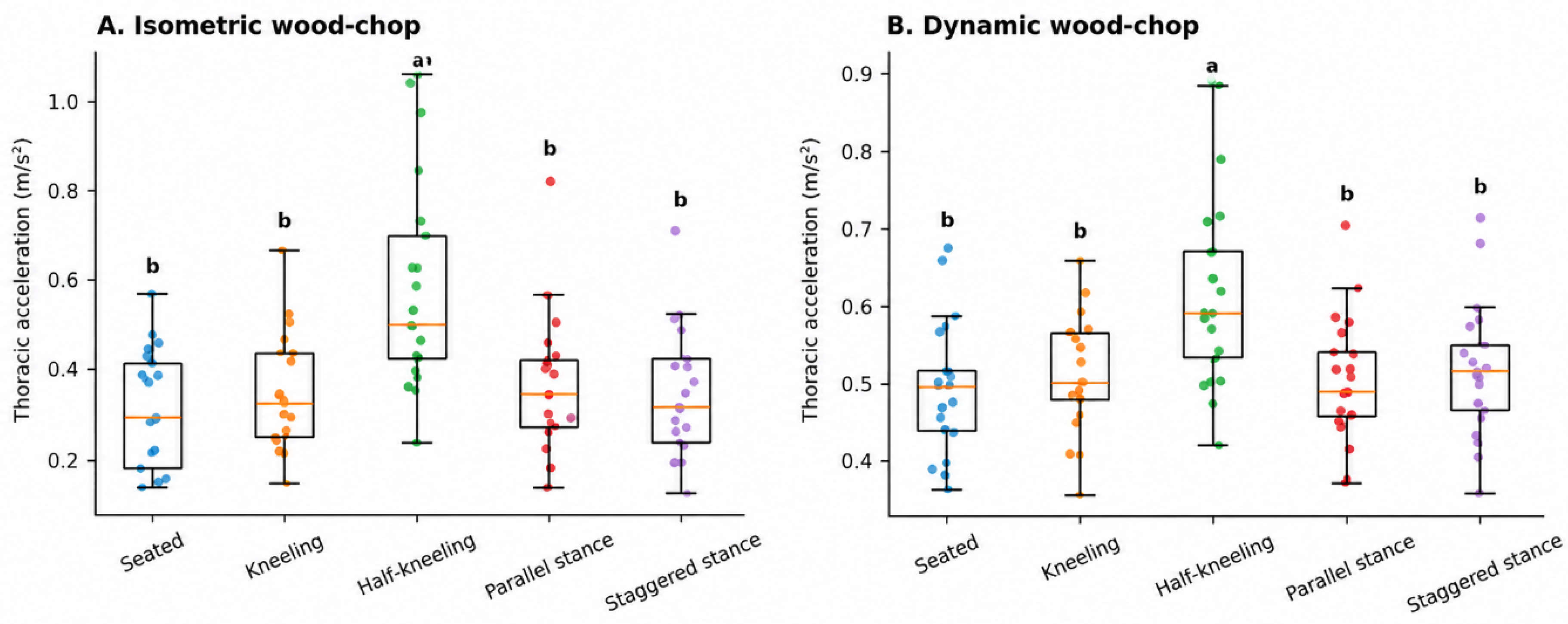
Figure 3.
Estimated marginal means and 95% confidence intervals for thoracic acceleration adjusted for maximal break force during the isometric (A) and dynamic (B) cable wood-chop.
Figure 3.
Estimated marginal means and 95% confidence intervals for thoracic acceleration adjusted for maximal break force during the isometric (A) and dynamic (B) cable wood-chop.
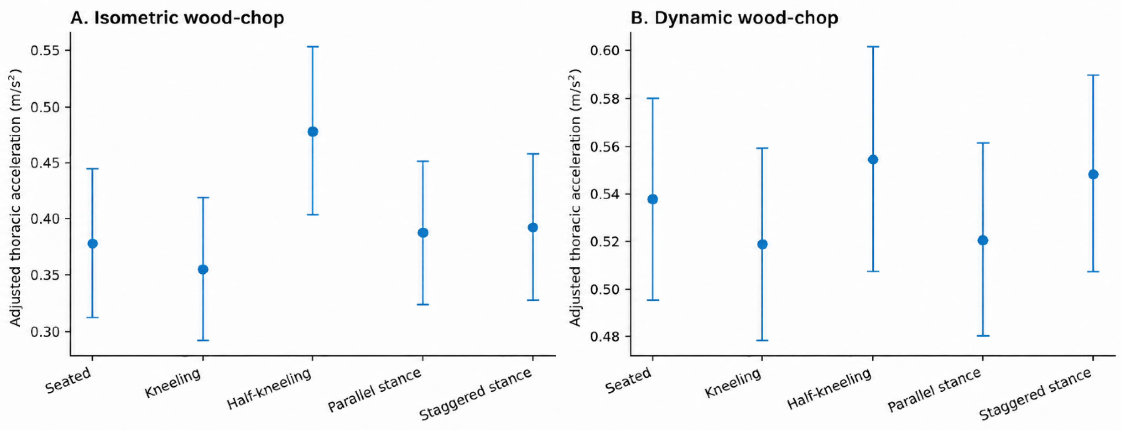

Table 1.
Pairwise comparisons of Session 2 thoracic acceleration across postures during the cable wood-chop.
Table 1.
Pairwise comparisons of Session 2 thoracic acceleration across postures during the cable wood-chop.
| Condition | Comparison (n=21) | Mean difference (95% CI) |
p (Holm) | Cohen’s dz |
|---|---|---|---|---|
|
Isometric |
Seated vs Kneeling | -0.031 (-0.056 to -0.005) | 0.105 | -0.55 |
| Seated vs Half-kneeling | -0.264 (-0.341 to -0.188) | <0.001 | -1.57 | |
| Seated vs Parallel stance | -0.050 (-0.087 to -0.014) | 0.058 | -0.62 | |
| Seated vs Staggered stance | -0.032 (-0.073 to 0.010) | 0.502 | -0.35 | |
| Kneeling vs Half-kneeling | -0.234 (-0.305 to -0.162) | <0.001 | -1.49 | |
| Kneeling vs Parallel stance | -0.020 (-0.055 to 0.016) | 0.784 | -0.25 | |
| Kneeling vs Staggered stance | -0.001 (-0.048 to 0.046) | 0.959 | -0.01 | |
| Half-kneeling vs Parallel stance | 0.214 (0.147 to 0.282) | <0.001 | 1.45 | |
| Half-kneeling vs Staggered stance | 0.233 (0.161 to 0.304) | <0.001 | 1.48 | |
| Parallel stance vs Staggered stance | 0.018 (-0.034 to 0.071) | 0.945 | 0.16 | |
| Dynamic | Seated vs Kneeling | -0.016 (-0.041 to 0.009) | 0.988 | -0.29 |
| Seated vs Half-kneeling | -0.125 (-0.167 to -0.082) | <0.001 | -1.34 | |
| Seated vs Parallel stance | -0.010 (-0.037 to 0.018) | 1.000 | -0.16 | |
| Seated vs Staggered stance | -0.022 (-0.053 to 0.010) | 0.972 | -0.32 | |
| Kneeling vs Half-kneeling | -0.108 (-0.147 to -0.069) | <0.001 | -1.27 | |
| Kneeling vs Parallel stance | 0.007 (-0.017 to 0.030) | 1.000 | 0.13 | |
| Kneeling vs Staggered stance | -0.006 (-0.033 to 0.021) | 1.000 | -0.09 | |
| Half-kneeling vs Parallel stance | 0.115 (0.071 to 0.159) | <0.001 | 1.18 | |
| Half-kneeling vs Staggered stance | 0.103 (0.066 to 0.140) | <0.001 | 1.26 | |
| Parallel stance vs Staggered stance | -0.012 (-0.049 to 0.024) | 1.000 | -0.15 | |
| CI: Confidence interval | ||||
Table 2.
Fixed effects from the linear mixed-effects models adjusted for maximal break force.
| Condition | Predictor | β | SE | 95% CI | p |
|---|---|---|---|---|---|
| Isometric | Intercept (half-kneeling) | 0.478 | 0.038 | 0.403 to 0.553 | <0.001 |
| Seated vs half-kneeling | -0.100 | 0.038 | -0.175 to -0.026 | 0.008 | |
| Kneeling vs half-kneeling | -0.123 | 0.030 | -0.183 to -0.064 | <0.001 | |
| Parallel stance vs half-kneeling | -0.091 | 0.032 | -0.154 to -0.028 | 0.005 | |
| Staggered stance vs half-kneeling | -0.086 | 0.035 | -0.155 to -0.016 | 0.015 | |
| Maximal break force (centered) | 0.033 | 0.006 | 0.021 to 0.045 | <0.001 | |
| Dynamic | Intercept (half-kneeling) | 0.554 | 0.024 | 0.507 to 0.602 | <0.001 |
| Seated vs half-kneeling | -0.017 | 0.023 | -0.062 to 0.028 | 0.459 | |
| Kneeling vs half-kneeling | -0.036 | 0.018 | -0.072 to 0.000 | 0.048 | |
| Parallel stance vs half-kneeling | -0.034 | 0.019 | -0.072 to 0.004 | 0.079 | |
| Staggered stance vs half-kneeling | -0.006 | 0.021 | -0.048 to 0.035 | 0.767 | |
| Maximal break force (centered) | 0.021 | 0.004 | 0.014 to 0.029 | <0.001 |
Disclaimer/Publisher’s Note: The statements, opinions and data contained in all publications are solely those of the individual author(s) and contributor(s) and not of MDPI and/or the editor(s). MDPI and/or the editor(s) disclaim responsibility for any injury to people or property resulting from any ideas, methods, instructions or products referred to in the content. |
© 2026 by the authors. Licensee MDPI, Basel, Switzerland. This article is an open access article distributed under the terms and conditions of the Creative Commons Attribution (CC BY) license (http://creativecommons.org/licenses/by/4.0/).
Copyright: This open access article is published under a Creative Commons CC BY 4.0 license, which permit the free download, distribution, and reuse, provided that the author and preprint are cited in any reuse.