

Submitted:
25 September 2026
Posted:
28 September 2026
You are already at the latest version
Abstract
Abstract
Background/Objectives
Severe traumatic injuries of the upper extremity present a major therapeutic challenge because of extensive tissue damage, severe pain, loss of function, and substantial psychosocial consequences. Continuous multidisciplinary re-habilitation that adapts to the patient's evolving clinical condition may optimize functional outcomes across the entire continuum of care.
Methods
A 42-year-old man with no prior comorbidities sustained a severe work-related traumatic injury to his left hand. Despite reconstructive surgery and prolonged multidisciplinary treatment, he developed CRPS with persistent neuropathic pain, progressive functional deterioration, and psychological distress. Fourteen months after injury, the affected limb became completely nonfunctional, and elective transradial amputation was recommended after multidisciplinary evaluation.
Results
Rehabilitation was delivered as a continuous, phase-adapted multidisciplinary program. Before amputation, treatment focused on pain control, reduction of edema and inflam-mation, preservation of joint mobility and muscle function, and management of CRPS using multimodal physical therapy combined with pharmacological treatment and psychological support. As amputation became inevitable, rehabilitation goals shifted toward pre-amputation preparation, promotion of adaptive neuroplasticity, and preven-tion of phantom limb pain.
After transradial amputation, pain intensity markedly decreased, functional adaptation improved, and the patient successfully completed prosthetic rehabilitation.
Conclusions
This case highlights the importance of a continuous, individualized multidisciplinary rehabilitation strategy extending from limb salvage through post-amputation recovery. Integrating multimodal physical therapy, neuroplasticity based interventions, psycho-logical support, and prosthetic rehabilitation may improve pain control, facilitate func-tional recovery, and enhance long-term adaptation after severe traumatic upper extremity injury.
Keywords:
complex regional pain syndrome
; traumatic hand injury
; amputation
; multidisciplinary rehabilitation
; prosthetic rehabilitation
1. Introduction
1.1. Traumatic Upper Limb Amputation and the Role of Multidisciplinary Rehabilitation
Traumatic upper limb amputation represents a relatively small proportion of all amputations but leads to disproportionately greater functional impairment. This is due to the complex motor and sensory functions of the hand and upper limb, altered body image, reduced independence in activities of daily living, and a substantial psychosocial burden [1]. In addition to the immediate loss of limb function, patients may experience residual limb pain, phantom limb pain (PLP), anxiety, depression, reduced acceptance of the prosthesis, and difficulty returning to work, underscoring the need for comprehensive multidisciplinary rehabilitation from the earliest stages of treatment [2].
Rehabilitation after traumatic injury of the upper extremity should not be viewed as a process that starts only after amputation. Whenever limb salvage becomes uncertain, pre-amputation rehabilitation can help preserve joint mobility, maintain muscle strength, reduce pain and edema, plan for secondary complications, and prepare patients physically and mentally for surgery and subsequent prosthesis fitting [3]. Early rehabilitation is also a key factor in successful prosthesis adaptation, long-term prosthesis acceptance, and the restoration of functional independence [1]. After amputation, the goals of rehabilitation are to optimize wound healing, shape the residual limb, prevent contractures, facilitate prosthetic training, and maximize functional recovery [4]. Emerging evidence further suggests that early rehabilitation may attenuate maladaptive neuroplastic changes associated with chronic pain syndromes, particularly phantom limb pain [5].
1.2. Multimodal Physical Therapy and Neuroplasticity in Post-Amputation Rehabilitation
Physical therapy modalities (transcutaneous electrical stimulation, therapeutic ultrasound, low-level laser therapy, pulsed electromagnetic field therapy) are often included in multimodal rehabilitation programs because they provide nonpharmacological pain management and support tissue healing and functional recovery. Although each modality has distinct biological mechanisms, their combined use can achieve a synergistic effect by simultaneously addressing pain, inflammation, tissue repair, and restoration of function [6]. Among these modalities, transcutaneous electrical nerve stimulation (TENS) is used to treat acute and chronic musculoskeletal pain [7]. Its analgesic effects are attributed to the activation of large-diameter Aβ afferent fibers, which inhibit nociceptive transmission within the dorsal horn of the spinal cord according to the gate control theory, as well as the activation of descending inhibitory pathways and increased release of endogenous opioids, serotonin, and γ-aminobutyric acid (GABA), which modulate pain processing in both spinal and supraspinal regions [8]. In a 2022 Meta TENS study, Jonson MI et al. supported the use of TENS as a safe method of analgesia within multimodal rehabilitation [7]. Because of its favorable safety profile, ease of use, and minimal side effects, TENS is an attractive adjunct during preoperative and postoperative rehabilitation [9].
Therapeutic ultrasound is used in musculoskeletal rehabilitation to achieve thermal or nonthermal biological effects, depending on the treatment parameters. Mechanical acoustic energy improves cell membrane permeability, stimulates fibroblast proliferation, promotes collagen synthesis, enhances angiogenesis, and accelerates tissue repair, while thermal effects increase local blood flow, reduce muscle spasm, and improve connective tissue extensibility, facilitating pain reduction and the restoration of joint mobility [10]. Its clinical benefit depends on the pathology and treatment protocol, and it is incorporated into rehabilitation programs to optimize soft tissue healing after complex orthopedic trauma [11].
Photobiomodulation therapy (PBM), or low-level laser therapy (LLLT), is increasingly used in multimodal programs as an adjunct treatment for musculoskeletal injuries [12]. Its biological effects are mediated by the absorption of light energy by mitochondrial cytochrome c oxidase, resulting in increased production of adenosine triphosphate (ATP), modulation of reactive oxygen species, and activation of transcription factors involved in tissue repair [13]. These mechanisms contribute to anti-inflammatory, analgesic, and regenerative effects, including enhanced collagen deposition, accelerated wound healing, reduced edema, and improved peripheral nerve regeneration [14].
Pulsed electromagnetic field therapy (PEMF) is increasingly being introduced as a complementary method in standardized rehabilitation protocols after orthopedic trauma because of its noninvasive nature and potential biological benefits [15]. Experimental and clinical studies have shown that low-frequency electromagnetic fields affect transmembrane ion transport, intracellular calcium signaling, nitric oxide synthesis, mitochondrial activity, and cellular metabolism, and thus can improve microcirculation, reduce inflammation, stimulate angiogenesis, promote tissue healing, and reduce pain [16].
Together, these physical therapy modalities, when integrated into individualized rehabilitation programs, may contribute to improved long-term functional outcomes [17].
Functional tissue recovery after injury also depends on adaptive changes within the central nervous system, particularly the continuous interaction between peripheral sensory information and cortical processing that shapes motor control, body representation, and pain perception [18]. Consequently, rehabilitation should target peripheral tissues as well as therapeutic strategies capable of promoting beneficial neuroplastic changes and preventing maladaptive cortical reorganization associated with limb loss, especially in patients at risk of developing phantom limb pain [19].
Successful rehabilitation after limb amputation largely depends on adaptive changes within the central nervous system [20]. Following peripheral tissue injury, the brain engages in neuroplasticity, allowing the formation of new synaptic connections, the strengthening of existing pathways, and the reorganization of cortical representations, thereby supporting functional recovery after neurological and musculoskeletal injuries [21].
However, if normal afferent input is permanently disrupted, as occurs after limb amputation, maladaptive processes can develop that induce functional and structural changes throughout the sensorimotor network, affecting cortical excitability, body representation, motor planning, and pain processing [22].
One of the most extensively studied manifestations of post-amputation neuroplasticity is cortical remapping, also known as cortical reorganization [23]. Functional magnetic resonance imaging and electrophysiological studies have shown that stimulation or voluntary movement of an intact limb, face, or proximal upper limb can activate cortical areas that previously represented the amputated limb, illustrating the remarkable plasticity of the adult brain [5].
Although cortical remapping is considered a physiologic adaptive response, excessive or maladaptive reorganization is associated with disturbed body representation, sensorimotor mismatch, and persistent neuropathic pain [24]. Current evidence therefore suggests that rehabilitation should aim not only to restore physical function but also to promote adaptive cortical reorganization while minimizing maladaptive neuroplastic changes [25].
1.3. Phantom Limb Pain, Mirror Therapy, and Rationale for the Present Case
Phantom limb pain (PLP) is one of the most common long-term complications after limb amputation, affecting approximately 60–85% of amputees [26]. PLP is defined as painful sensations perceived in the absent limb and should be distinguished from painless phantom limb sensations and residual limb pain localized to the stump [24]. The pathophysiology of PLP is multifactorial and includes peripheral nerve injury, ectopic discharges within neuromas, spinal cord sensitization, altered thalamocortical processing, maladaptive cortical reorganization, and immunologic and psychological mechanisms [27]. Growing evidence suggests that persistent nociceptive input before amputation, together with postoperative pain, enhances these neuroplastic changes and contributes to the development of chronic phantom limb pain [28].
Among nonpharmacologic interventions, mirror therapy (MT) has emerged as one of the most extensively investigated neurorehabilitation techniques for the management of phantom limb pain [29]. Mirror therapy, originally introduced by Ramachandran and Rogers-Ramachandran, provides visual feedback by reflecting the movements of the intact limb, thereby creating the illusion that the amputated limb is present and moving normally [23,30]. This visual illusion is believed to reduce the mismatch between motor intention and the absent sensory feedback, facilitating restoration of the body schema and normalization of sensorimotor cortical networks [25].
Functional neuroimaging studies further show that successful mirror therapy is accompanied by measurable changes in cortical activation patterns, supporting the hypothesis that visual feedback can partially reverse the maladaptive cortical reorganization associated with phantom limb pain [31]. More recently, mirror therapy has been considered part of a broader spectrum of neurorehabilitation interventions that includes motor imagery, graded motor imagery, sensory discrimination training, and virtual reality-based rehabilitation, all of which aim to restore coherent sensorimotor integration and normalize cortical representation [19,20].
Recent systematic reviews, randomized controlled trials, and expert recommendations consistently support including mirror therapy in comprehensive rehabilitation programs for patients with phantom limb pain [29]. Although treatment protocols differ in session duration and therapy frequency during the intervention period, regular mirror therapy is associated with reduced pain intensity, enhanced motor imagery, improved cortical reorganization, and better functional adaptation to limb loss [18,29]. Furthermore, combining mirror therapy with conventional physical rehabilitation, early prosthetic fitting, and individualized multidisciplinary treatment may provide synergistic benefits by simultaneously addressing peripheral tissue recovery and central neuroplastic mechanisms [17,25].
Taken together, current evidence supports a multimodal rehabilitation approach that integrates conventional physical therapy modalities with interventions specifically targeting cortical reorganization to optimize outcomes after traumatic upper limb amputation [26].
Despite significant advances in surgical techniques, prosthetic technologies, and rehabilitation strategies, the optimal integration and timing of different physical therapy modalities across the continuum of care after traumatic upper limb amputation remain underexplored [17]. Most published studies have evaluated individual rehabilitation interventions, such as mirror therapy, prosthetic training, or pharmacological treatment of phantom limb pain, whereas relatively few reports have described a comprehensive rehabilitation program that begins before amputation, continues throughout the postoperative period, and integrates conventional physical therapy modalities with interventions specifically targeting neuroplasticity [32]. Consequently, clinical evidence on the potential synergistic effects of combining peripheral tissue-directed therapies with central nervous system-directed rehabilitation in patients undergoing traumatic upper limb amputation remains limited [17,25].
This case illustrates a multidisciplinary rehabilitation strategy applied to a patient with a severe traumatic upper limb injury that ultimately required amputation after unsuccessful limb salvage [17,32]. During the pre-amputation period, rehabilitation focused on pain control, inflammation reduction, preservation of joint mobility, and optimization of soft tissue healing through a multimodal therapeutic program [6,15]. After amputation, rehabilitation priorities shifted toward preventing secondary complications of the stump, facilitating adaptive cortical reorganization, and reducing phantom limb pain with mirror therapy [18,33].
This multimodal rehabilitation strategy reflects the contemporary understanding that successful recovery from limb amputation depends on addressing both peripheral biological processes and central neuroplastic mechanisms [33]. Conventional physical therapy methods can optimize tissue healing, reduce nociceptive input, and create favorable conditions for functional recovery during the acute and subacute phases of rehabilitation [6]. In contrast, mirror therapy primarily targets maladaptive cortical reorganization by restoring congruence between motor intention and visual feedback, thereby facilitating normalization of body representation and sensorimotor cortical networks [18]. Together with individualized prosthetic rehabilitation, these complementary interventions can promote functional adaptation, improve prosthesis acceptance, reduce phantom limb pain, and enhance overall quality of life [3,34]. The aim of this case report is to describe the continuity of multidisciplinary rehabilitation from limb salvage through transradial amputation to prosthetic rehabilitation in a patient with CRPS following severe hand trauma.
2. Case Presentation
This case report describes the clinical course and multidisciplinary rehabilitation of a patient with a severe traumatic upper limb injury that ultimately required amputation. Rehabilitation was provided as a phase-adapted, stepwise program that included multimodal physical rehabilitation before amputation, mirror therapy during the postoperative period, and structured prosthetic training during functional recovery. The report highlights the value of a continuous, phase-adapted multidisciplinary rehabilitation strategy that bridges limb-salvage management and post-amputation rehabilitation while integrating tissue-directed interventions with neuroplasticity-based therapies to support functional recovery.
A 42-year-old man with no prior history of peripheral neuropathy, chronic pain disorders, or psychiatric illness sustained a severe injury to his left hand while working on a woodworking machine at work in September 2021. The injury to fingers II to V of the left hand resulted in extensive soft tissue damage, skin defects, and tendon injuries to the flexor digitorum profundus of the second and fourth fingers. On the day of the injury, the patient underwent emergency reconstructive surgery under general anesthesia, including a cross-finger flap, plastica defecti cutis sec. Thiersch, and immobilization. The early postoperative course was uneventful, and he was discharged on the fifth day with antibiotic therapy and routine wound care. Three weeks later, the flap was separated from the donor finger, and a secondary wound revision was performed under local anesthesia (Figure 1).
Despite satisfactory wound healing, functional recovery was slow. During follow-up examinations over the next months, the patient progressively developed severe neuropathic pain, hypersensitivity, persistent dysesthesia, paresthesia, burning sensations, cold intolerance, muscle cramps, progressive finger contractures, significant functional impairment, and hyperpigmentation of the affected hand. The pain was present both during activity and at rest, and its intensity gradually increased.
Based on the clinical findings, a diagnosis of complex regional pain syndrome (CRPS 1) was established according to the Budapest clinical diagnostic criteria [35]. The patient reported ongoing pain disproportionate to the initial injury and met the required symptom and sign categories, including sensory abnormalities (mechanical allodynia and hyperesthesia), vasomotor changes (temperature sensitivity), sudomotor/trophic changes (persistent edema and skin discoloration), and motor dysfunction (reduced range of motion, weakness, and progressive finger contractures). No alternative diagnosis better accounted for the clinical presentation.
Six months after the injury, the initial pain assessment showed continuous neuropathic pain rated on a numerical rating scale (NRS) between 7 and 10 (Figure 2).
At the plastic surgeon’s initiative, the patient was referred to the pain clinic in March 2022. Physical examination at that time documented pain rated 9 on the NRS, confirmed pronounced mechanical allodynia affecting the palm and fingers of the left hand, and led to the immediate initiation of multimodal physical treatment, including transcutaneous electrical nerve stimulation (TENS), magnetotherapy, low-level laser therapy (LLLT), ultrasound therapy, acupuncture, and pharmacological treatment combining pregabalin, gabapentin, nonsteroidal anti-inflammatory drugs, tramadol, duloxetine, and later tapentadol, depending on the progression of symptoms and pain (Table 1).
From March 2022 to March 2023, four cycles of multimodal physical treatment were administered, which achieved temporary improvement by reducing pain to 7 on the NRS, but the pain consistently recurred and functional deterioration continued (Figure 3).
Psychiatric evaluation revealed the development of an adjustment disorder with subsequent posttraumatic stress disorder (PTSD) associated with the work-related injury. Symptoms included intrusive memories, sleep disturbance, anxiety, depressed mood, irritability, emotional withdrawal, reduced frustration tolerance, and significant deterioration in quality of life. From May 2022 to February 2024, he underwent cyclical psychiatric treatment in a day hospital setting, along with structured psychotherapy and antidepressant and anxiolytic medication (Figure 4).
Psychiatric and psychological management consisted of continuous day-hospital treatment cycles starting in May 2022. The protocol included standardized neuropsychological and psychodiagnostic assessments using the Nonverbal Cognitive Intelligence Test (KNT), Montreal Cognitive Assessment (MoCA), Bender–Gestalt II Test of Visual Perception and Visuomotor Integration, Crown–Crisp Experiential Index (CCEI), Trauma Symptom Inventory (TSI), Minnesota Multiphasic Personality Inventory-201 (MMPI-201), and the Wartegg Drawing Completion Test, followed by clinical interviews, psychological counseling, and ongoing psychological support throughout the rehabilitation program.
Follow-up examinations by a plastic surgeon documented progressive extension contracture of the proximal and distal interphalangeal joints, persistent weakness of active finger movements, sensory hypersensitivity, and loss of hand function. Hyperbaric oxygen therapy was recommended for 10 sessions, which did not significantly improve symptoms. Clinical evaluation by a vascular surgeon noted preserved radial and ulnar arterial circulation with no indication for vascular reconstruction, despite reduced capillary refill in the affected fingers. Neurological evaluation revealed severe neuropathic pain associated with hypersensitivity and functional impairment; however, somatosensory evoked potentials of both median nerves remained within normal limits. During follow-up examinations by a plastic surgeon, an algologist, and a neurologist, the patient continued to report severe burning pain, temperature hypersensitivity, numbness, and progressive inability to use his left hand. By October 2023, despite long-term multidisciplinary treatment that included reconstructive surgery, physical rehabilitation, pain management, neurology, vascular surgery, clinical psychology, and psychiatry, the patient continued to experience unbearable pain with NRS 9 intensity, associated with complete functional loss of the affected limb and progression of PTSD. Evaluation at the Center for Plastic Surgery and Hand Reconstruction concluded that further reconstructive procedures were not feasible. After multidisciplinary discussion and detailed counseling, the diagnosis was made: Sudeck antebrachii lat. sin. syndrome. Elective transradial amputation was recommended due to therapy-resistant pain and a completely nonfunctional hand. From October 2023 to January 2024, three additional cycles of multimodal physical therapy, each lasting 15 days, were conducted to reduce the risk of developing phantom pain and to facilitate subsequent prosthetic rehabilitation. In February 2024, to reduce the risk of symptomatic neuroma and recurrent neuropathic pain, the patient underwent left transradial amputation with intraoperative targeted nerve reconstruction consisting of amputatio antebrachii sin. with anastomosis of the n. medianus and n. ulnaris antebrachii of the left hand, and anastomosis of the muscularis muscle of the flexor digitorum superf. cum n. radialis r. superficialis.
At the first postoperative follow-up, 3 weeks after transradial amputation of the left forearm, the patient reported a significant decrease in pain intensity (numerical rating scale NRS 3) compared with the preoperative state. Residual symptoms consisted of occasional stabbing pain and intermittent paresthesia within the patella. Pharmacological treatment was gradually tapered over the next 6 months, with reduction in the doses of pregabalin, duloxetine, and opioid analgesics until complete discontinuation of pregabalin and duloxetine 2 months after amputation. During the first 2 months after amputation, the patient followed a structured home rehabilitation program focused on mirror therapy, stump desensitization, shoulder and elbow range-of-motion exercises, scar mobilization, and gradual functional adaptation guided by a physiotherapist (Figure 5).
Therapy was administered twice daily for 15 minutes. The mirror was positioned in the sagittal plane so that only the reflection of the intact hand was visible. The patient was instructed to focus on the mirror image while imagining synchronous movement of the phantom limb. Exercises progressed from simple hand and wrist movements to object manipulation and fine motor tasks. All movements were performed slowly and in a controlled manner.
Mirror therapy was performed daily according to the rehabilitation team’s instructions and continued until the start of formal prosthetic rehabilitation. Prosthetic rehabilitation was initiated in June 2024. In August 2024, the patient completed a one-week inpatient rehabilitation program at a specialized prosthetic rehabilitation institute, where he received structured training and education in the use and functional application of a forearm prosthesis with a myoelectric hand (Figure 6).
The patient continued to be followed by an algologist through control examinations every 3 months, reporting stable residual stump pain related to the weight of the prosthesis, with occasional phantom limb pain (NRS 3–5) compared with the pain before amputation (NRS 7–9). Intermittent multimodal physical therapy was continued every 6 months for symptom control and management of pain in the phantom hand of the left upper limb. The patient also continues to participate in a day hospital program with monitoring by a psychologist and psychiatrist (Table 2).
3. Discussion
This case report describes a complex, long-term, multidisciplinary rehabilitation process in a patient with a severe traumatic upper extremity injury that, despite treatment and rehabilitation, ultimately required transradial amputation. The uniqueness of this case lies in the continuous rehabilitation approach that encompassed all phases of care from acute injury management, through the period before amputation and early postoperative recovery, to prosthetic rehabilitation and return to functional activities. In the pre-amputation period, rehabilitation goals focused on preserving limb function, reducing pain and inflammatory response, controlling edema, maintaining joint mobility, preserving muscle activity, and optimizing soft tissue healing. To achieve these goals, a multimodal rehabilitation program was implemented, including transcutaneous electrical nerve stimulation (TENS), therapeutic ultrasound, magnetotherapy, low-intensity laser therapy, and acupuncture, combined with continuous psychological and psychiatric support through a day hospital program [36]. The use of different physical modalities in this phase was intended to modulate pain, improve local biological conditions for healing, and preserve the limb’s functional potential [32].
Deciding to amputate is among the most challenging aspects of treating patients with complex traumatic limb injuries and requires an individualized assessment by a multidisciplinary team [26]. In this case, the decision to perform a transradial amputation was made after determining that this approach could provide better overall function, lessen the long-term physical and psychological burden associated with the injured limb, and increase the likelihood of returning to independent living and work [37].
After amputation, rehabilitation priorities shifted toward preserving residual limb function, preventing secondary complications, promoting adaptive neuroplasticity, managing phantom limb pain, and preparing for prosthetic use [38]. Mirror therapy was incorporated into the rehabilitation program to facilitate sensorimotor adaptation and address pain mechanisms [39]. It aimed to stimulate sensorimotor reorganization in the central nervous system by reestablishing the connection between motor intention and visual sensory feedback [21]. Today, mirror therapy is one of the most frequently used non-pharmacological methods for treating phantom limb pain and for sensorimotor rehabilitation after amputation. Proposed mechanisms of action include modulation of maladaptive cortical reorganization, enhancement of sensorimotor integration, and reduction of abnormal neural activity patterns associated with phantom limb pain [30]. In this case, mirror therapy was introduced after the surgical wound had healed and was performed during the period before prosthetic rehabilitation, which is consistent with the concept of early implementation of interventions that target neuroplasticity after amputation [18].
Successful prosthetic rehabilitation is not just a matter of fitting a prosthesis, but a complex process that includes conditioning the residual limb, developing muscle control, learning new motor strategies, achieving functional performance in activities of daily living, and psychosocial adaptation to changed physical abilities. In this case, a structured prosthetic rehabilitation program was implemented with ongoing pain management, physical therapy, and psychological support, allowing for a gradual increase in the patient’s functional independence. The relatively favorable functional outcome in this case is likely the result of the timely initiation and continuity of a multidisciplinary rehabilitation approach that encompassed different phases of recovery. The combination of peripheral tissue-focused interventions, neuroplasticity-based methods, and individualized prosthetic rehabilitation may contribute to better functional adaptation, greater prosthesis acceptance, reduced phantom and nociceptive pain, and improved quality of life after traumatic upper-extremity amputation.
In the present case, twenty months after amputation and completion of prosthetic rehabilitation, the patient returned to a work environment in a position adapted to his functional capabilities. However, despite satisfactory physical recovery, the psychological consequences of the traumatic event, including emotional reactions to the moment of limb loss and challenges associated with the social stigmatization of disability, remain a significant part of the recovery process. Therefore, long-term rehabilitation after amputation should not be limited to physical function alone, but should include ongoing psychological support, monitoring of anxiety symptoms, and adjustment to changing life roles [40]. Combining psychological interventions, such as structured therapeutic interviews and sensory feedback methods, with physical therapy and appropriate pharmacological support may offer additional benefits for the long-term control of psychological symptoms and preservation of function.
4. Clinical Implications for Hand Therapy
This case underscores the importance of maintaining active involvement of hand therapists across the entire continuum of care following severe traumatic upper extremity injury, including situations in which limb salvage ultimately proves unsuccessful. Rehabilitation should not be viewed as separate pre- and post-amputation interventions but as a continuous, individualized process that evolves with the patient’s changing clinical needs. During limb salvage, hand therapy should prioritize pain management, edema control, preservation of joint mobility, maintenance of muscle function, sensory reeducation, and patient education. When amputation becomes inevitable, rehabilitation goals should gradually shift toward pre-amputation preparation, psychological support, prevention of secondary complications, promotion of adaptive neuroplasticity, and planning for prosthetic rehabilitation. After amputation, mirror therapy, residual limb desensitization, structured prosthetic training, and continued multidisciplinary follow-up may facilitate functional adaptation, reduce phantom limb pain, improve prosthesis acceptance, and support return to meaningful daily activities and employment.
5. Limitations
This case report has several limitations. Because multiple surgical, pharmacological, rehabilitation, and psychological interventions were used during treatment, it is not possible to precisely determine the specific contribution of each method to the overall functional outcome. In addition, the conclusions are limited by the fact that this is a single-patient report and cannot be directly generalized to a broader population. Furthermore, validated upper-extremity functional outcome measures (e.g., QuickDASH or PRWE) were not prospectively collected, limiting the objective assessment of functional improvement over time.
6. Patient’s Perspective
“After my arm was amputated, my life changed completely. Although deciding to undergo the amputation was very difficult for me, over time I realized it was the only way to relieve the unbearable pain and begin living a more normal life again. Today, the pain is much less intense than before the amputation, but it still occasionally appears as phantom or stump pain of varying intensity and duration. I have learned to live with these symptoms, although they remain part of my everyday life.
I have returned to a job that is adapted to my functional abilities and disability. This means a lot to me because it allows me to be active again and feel useful. However, daily work also brings new challenges. I feel that some of my colleagues have difficulty understanding the limitations I live with and sometimes expect me to perform the same tasks at the same pace as before the injury or as other employees. Such situations can cause frustration and remind me that the adaptation process is a challenge not only for me but also for the people in my work environment.
The psychological consequences of the injury and amputation are still present. That is why I regularly participate in a day hospital program where, with the help of a clinical psychologist, I undergo behavioral therapy and other forms of psychological support that help me cope with the trauma, anxiety, and changes the amputation has brought into my life.
I am aware that no one can predict with certainty how my symptoms will develop in the future. The pain still occasionally occurs with varying intensity, but I have learned to recognize its patterns and cope with them. I believe that ongoing physical therapy, regular checkups, and continuous psychological support will help me preserve my physical and mental health for as long as possible, maintain my functional independence and quality of life, and continue to live as actively as possible despite my permanent disability.”
7. Conclusions
This case report underscores the importance of a continuous, individualized, multidisciplinary rehabilitation approach after severe traumatic upper limb injury. Integrating procedures that promote peripheral tissue healing, neuroplasticity-based interventions, and structured prosthetic rehabilitation may provide a valuable framework for achieving optimal functional recovery and improving quality of life for patients after amputation. This case supports viewing rehabilitation as a continuous therapeutic process rather than separate pre- and post-amputation interventions.
Author Contributions
Ozana Katarina Tot: Conceptualization, Methodology, Formal analysis, Data curation, Writing—original draft, Project administration. Anja Petričević: Investigation, Resources, Writing—review & editing. Dijana Hnatešen: Methodology, Formal analysis, Visualization, Writing—review & editing. Ana Cicvarić: Conceptualization, Methodology, Investigation, Supervision, Writing—review & editing. Iva Dimitrijević: Investigation, Resources, Data curation, Writing—review & editing. Vanja Matković: Investigation, Resources, Writing—review & editing. All authors have read and agreed to the published version of the manuscript.
Funding
This research received no external funding.
Institutional Review Board Statement
This case report was conducted in accordance with the Declaration of Helsinki. According to institutional policy, formal approval by the Ethics Committee was not required for a single-patient case report.
Informed Consent Statement
Patient consent: Written informed consent was obtained from the patient for publication of this case report and all accompanying clinical images.
Data Availability Statement
The data supporting the findings of this case report are available from the corresponding author upon reasonable request. No publicly available datasets were generated or analyzed.
Acknowledgments
Declaration of Generative AI and AI-assisted technologies in the writing process: During the preparation of this manuscript, the authors used Insta Text to improve the language and readability of the manuscript. The authors used ChatGPT (OpenAI) to assist in organizing the conceptual structure and wording of selected figures. The scientific concepts, figure content, interpretation, and final design were developed, critically reviewed, verified, and approved by the authors, who take full responsibility for the accuracy and integrity of the published work.
Conflicts of Interest
The authors declare that they have no known competing financial interests or personal relationships that could have appeared to influence the work reported in this paper.
Abbreviations
The following abbreviations are used in this manuscript:
| ATP | Adenosine triphosphate |
| CCEI | Crown–Crisp Experiential Index |
| CRPS | Complex Regional Pain Syndrome |
| FDS | Flexor digitorum superficialis |
| GABA | Gamma-aminobutyric acid |
| KNT | Nonverbal Cognitive Intelligence Test |
| LLLT | Low-level laser therapy |
| MMPI-201 | Minnesota Multiphasic Personality Inventory-201 |
| MoCA | Montreal Cognitive Assessment |
| MT | Mirror therapy |
| NRS | Numerical Rating Scale |
| PBM | Photobiomodulation |
| PEMF | Pulsed electromagnetic field therapy |
| PLP | Phantom limb pain |
| PRWE | Patient-Rated Wrist Evaluation |
| PTSD | Posttraumatic stress disorder |
| Quick DASH | Quick Disabilities of the Arm, Shoulder and Hand |
| TENS | Transcutaneous electrical nerve stimulation |
| TSI | Trauma Symptom Inventory |
| VAS | Visual Analog Scale |
References
- Resnik, L.J.; Ni, P.; Borgia, M.L.; Clark, M.A. A Psychosocial Adjustment Measure for Persons With Upper Limb Amputation. Can. Prosthet. Orthot. J. 2022, 5, 37873. [Google Scholar] [CrossRef]
- Yamamoto, M.; Chung, K.C.; Sterbenz, J.; Shauver, M.J.; Tanaka, H.; Nakamura, T.; et al. Cross-sectional International Multicenter Study on Quality of Life and Reasons for Abandonment of Upper Limb Prostheses. Plast. Reconstr. Surg. Glob. Open 2019, 7, e2205. [Google Scholar] [CrossRef] [PubMed]
- Engdahl, S.M.; Gonzalez, M.A.; Lee, C.; Gates, D.H. Perspectives on the comparative benefits of body-powered and myoelectric upper limb prostheses. J. Neuroeng. Rehabil. 2024, 21, 138. [Google Scholar] [CrossRef] [PubMed]
- Alterman, B.L.; Ali, S.; Keeton, E.; Binkley, K.; Hendrix, W.; Lee, P.J.; et al. Interlimb training improves motor function in partial-hand but not necessarily transradial simulated prosthesis use. Sci. Rep. 2025, 15, 38761. [Google Scholar] [CrossRef] [PubMed]
- Tucciarelli, R.; Ejaz, N.; Wesselink, D.B.; Kolli, V.; Hodgetts, C.J.; Diedrichsen, J.; et al. Does Ipsilateral Remapping Following Hand Loss Impact Motor Control of the Intact Hand? J. Neurosci. 2024, 44, e0948232023. [Google Scholar] [CrossRef] [PubMed]
- He, P.; Fu, W.; Shao, H.; Zhang, M.; Xie, Z.; Xiao, J.; et al. The effect of therapeutic physical modalities on pain, function, and quality of life in patients with myofascial pain syndrome: A systematic review. BMC Musculoskelet. Disord. 2023, 24, 376. [Google Scholar] [CrossRef] [PubMed]
- Johnson, M.I.; Paley, C.A.; Jones, G.; Mulvey, M.R.; Wittkopf, P.G. Efficacy and safety of transcutaneous electrical nerve stimulation (TENS) for acute and chronic pain in adults: A systematic review and meta-analysis of 381 studies (the meta-TENS study). BMJ Open 2022, 12, e051073. [Google Scholar] [CrossRef] [PubMed]
- Vance, C.G.; Dailey, D.L.; Rakel, B.A.; Sluka, K.A. Using TENS for pain control: The state of the evidence. Pain Manag. 2014, 4, 197–209. [Google Scholar] [CrossRef] [PubMed]
- Viderman, D.; Nabidollayeva, F.; Aubakirova, M.; Sadir, N.; Tapinova, K.; Tankacheyev, R.; et al. The Impact of Transcutaneous Electrical Nerve Stimulation (TENS) on Acute Pain and Other Postoperative Outcomes: A Systematic Review with Meta-Analysis. J. Clin. Med. 2024, 13, 427. [Google Scholar] [CrossRef] [PubMed]
- Zhou, J.; Ning, E.; Lu, L.; Zhang, H.; Yang, X.; Hao, Y. Effectiveness of low-intensity pulsed ultrasound on osteoarthritis: Molecular mechanism and tissue engineering. Front. Med. 2024, 11, 1292473. [Google Scholar] [CrossRef] [PubMed]
- Gomez, R.W.; Walsh, A.; Valdes, K.G.; Matullo, K.S. Enhancing Healing with Noncontact Low-Frequency Ultrasound in Fingertip Amputation Treatment: A Comparative Pilot Study. J. Hand Surg. Glob. Online 2025, 8, 100843. [Google Scholar] [CrossRef] [PubMed]
- Cronshaw, M.; Parker, S.; Grootveld, M.; Lynch, E. Photothermal Effects of High-Energy Photobiomodulation Therapies: An In Vitro Investigation. Biomedicines 2023, 11, 1634. [Google Scholar] [CrossRef] [PubMed]
- Vieira, W.F.; Kenzo-Kagawa, B.; Cogo, J.C.; Baranauskas, V.; da Cruz-Höfling, M.A. Low-Level Laser Therapy (904 nm) Counteracts Motor Deficit of Mice Hind Limb following Skeletal Muscle Injury Caused by Snakebite-Mimicking Intramuscular Venom Injection. PLoS ONE 2016, 11, e0158980. [Google Scholar] [CrossRef] [PubMed]
- Piotrzkowska, D.; Siwak, M.; Adamkiewicz, J.; Dziki, L.; Majsterek, I. The Therapeutic Potential of Pulsed Electromagnetic Fields (PEMF) and Low-Intensity Pulsed Ultrasound (LIPUS) in Peripheral Nerve Regeneration: A Comprehensive Review. Int. J. Mol. Sci. 2025, 26, 9311. [Google Scholar] [CrossRef] [PubMed]
- Hu, H.; Yang, W.; Zeng, Q.; Chen, W.; Zhu, Y.; Liu, W.; et al. Promising application of Pulsed Electromagnetic Fields (PEMFs) in musculoskeletal disorders. Biomed. Pharmacother. 2020, 131, 110767. [Google Scholar] [CrossRef] [PubMed]
- Zadeh-Haghighi, H.; Simon, C. Magnetic field effects in biology from the perspective of the radical pair mechanism. J. R. Soc. Interface 2022, 19, 20220325. [Google Scholar] [CrossRef] [PubMed]
- Grover, P.; Geng, E.; Tabak, R.G. The Multilevel Limb-loss and Preservation Rehabilitation Continuum model (MLPRC): An evidence-based implementation model integrating multiple perspectives to improve outcomes for people facing limb loss. PM R 2025, 17 (Suppl. 2), S81–S92. [Google Scholar] [CrossRef] [PubMed]
- Makin, T.R.; Flor, H. Brain (re)organisation following amputation: Implications for phantom limb pain. Neuroimage 2020, 218, 116943. [Google Scholar] [CrossRef] [PubMed]
- Mrachacz-Kersting, N.; Pasluosta, C.; Meyer, B.; do Nascimento, O.F.; Stieglitz, T.; Farina, D. Cortical Activity Associated With Phantom Leg Movements. IEEE Trans. Neural Syst. Rehabil. Eng. 2026, 34, 2994–3003. [Google Scholar] [CrossRef] [PubMed]
- El-Gabalawy, R.; Crooks, M.; Smith, M.S.D.; Hammond, E.; Gross, P.; Roznik, M.; et al. Treating Lower Phantom Limb Pain in the Postoperative Acute Care Setting Using Virtual Reality: Protocol for a 4-Phase Development and Feasibility Trial. JMIR Res. Protoc. 2025, 14, e68008. [Google Scholar] [CrossRef] [PubMed]
- Mercier, C.; Léonard, G. Interactions between Pain and the Motor Cortex: Insights from Research on Phantom Limb Pain and Complex Regional Pain Syndrome. Physiother. Can. 2011, 63, 305–314. [Google Scholar] [CrossRef] [PubMed]
- Makin, T.R.; Cramer, A.O.; Scholz, J.; Hahamy, A.; Henderson Slater, D.; Tracey, I.; et al. Deprivation-related and use-dependent plasticity go hand in hand. eLife 2013, 2, e01273. [Google Scholar] [CrossRef] [PubMed]
- Ramachandran, V.S. Behavioral and magnetoencephalographic correlates of plasticity in the adult human brain. Proc. Natl. Acad. Sci. USA 1993, 90, 10413–10420. [Google Scholar] [CrossRef] [PubMed]
- Erlenwein, J.; Diers, M.; Ernst, J.; Schulz, F.; Petzke, F. Clinical updates on phantom limb pain. Pain Rep. 2021, 6, e888. [Google Scholar] [CrossRef] [PubMed]
- Raffin, E. The various forms of sensorimotor plasticity following limb amputation and their link with rehabilitation strategies. Rev. Neurol. 2021, 177, 1112–1120. [Google Scholar] [CrossRef] [PubMed]
- Limakatso, K.; Ndhlovu, F.; Usenbo, A.; Rayamajhi, S.; Kloppers, C.; Parker, R. The prevalence and risk factors for phantom limb pain: A cross-sectional survey. BMC Neurol. 2024, 24, 57. [Google Scholar] [CrossRef] [PubMed]
- Shi, W.; Prabhakaran, B.; Annaswamy, T. Editorial: Phantom pain: Mechanisms and updates in management. Front. Pain Res. 2025, 6, 1567956. [Google Scholar] [CrossRef] [PubMed]
- Sugawara, A.T.; De Pretto, L.R.; Simis, M.; Fregni, F.; Battistella, L.R. Primary Motor Area Activity in Phantom Limb Imagery of Traumatic Unilateral Lower Limb Amputees With Phantom Limb Pain. Adv. Rehabil. Sci. Pract. 2024, 13, 27536351241261023. [Google Scholar] [CrossRef] [PubMed]
- Guémann, M.; Olié, E.; Raquin, L.; Courtet, P.; Risch, N. Effect of mirror therapy in the treatment of phantom limb pain in amputees: A systematic review of randomized placebo-controlled trials does not find any evidence of efficacy. Eur. J. Pain 2023, 27, 3–13. [Google Scholar] [CrossRef] [PubMed]
- Ramachandran, V.S.; Rogers-Ramachandran, D. Synaesthesia in phantom limbs induced with mirrors. Proc. Biol. Sci. 1996, 263, 377–386. [Google Scholar] [CrossRef] [PubMed]
- Zhang, K.; Ding, L.; Wang, X.; Zhuang, J.; Tong, S.; Jia, J.; et al. Evidence of mirror therapy for recruitment of ipsilateral motor pathways in stroke recovery: A resting fMRI study. Neurotherapeutics 2024, 21, e00320. [Google Scholar] [CrossRef] [PubMed]
- Parvizi, D.; Kandikattu, S.; Sahafi, R.; Allahverdian, A.; Han, B.; Fraix, M.P.; et al. Multimodal Approaches for Pain Management and Improving Functional Outcomes Following Amputation. J. Orthop. Sports Med. 2025, 7, 449–463. [Google Scholar] [CrossRef] [PubMed]
- Limakatso, K.; Parker, R. Treatment Recommendations for Phantom Limb Pain in People with Amputations: An Expert Consensus Delphi Study. PM R 2021, 13, 1216–1226. [Google Scholar] [CrossRef] [PubMed]
- Sturma, A.; Stamm, T.; Hruby, L.A.; Bischof, B.; Salminger, S.; Gstoettner, C.; et al. Rehabilitation of high upper limb amputees after Targeted Muscle Reinnervation. J. Hand Ther. 2022, 35, 58–66. [Google Scholar] [CrossRef] [PubMed]
- Harden, R.N.; Bruehl, S.; Perez, R.S.G.M.; Birklein, F.; Marinus, J.; Maihofner, C.; et al. Validation of proposed diagnostic criteria (the “Budapest Criteria”) for Complex Regional Pain Syndrome. Pain 2010, 150, 268–274. [Google Scholar] [CrossRef] [PubMed]
- Sturma, A.; Boesendorfer, A.; Gstoettner, C.; Baumgartner, B.; Salminger, S.; Farina, D.; et al. Long-Term Functional And Clinical Outcome Of Combined Targeted Muscle Reinnervation And Osseointegration For Functional Bionic Reconstruction In Transhumeral Amputees: A Case Series. J. Rehabil. Med. 2024, 56, 34141. [Google Scholar] [CrossRef] [PubMed]
- Mioton, L.M.; Dumanian, G.A.; Shah, N.; Qiu, C.S.; Ertl, W.J.; Potter, B.K.; et al. Targeted Muscle Reinnervation Improves Residual Limb Pain, Phantom Limb Pain, and Limb Function: A Prospective Study of 33 Major Limb Amputees. Clin. Orthop. Relat. Res. 2020, 478, 2161–2167. [Google Scholar] [CrossRef] [PubMed]
- Subedi, B.; Grossberg, G.T. Phantom Limb Pain: Mechanisms and Treatment Approaches. Pain Res. Treat. 2011, 2011, 864605. [Google Scholar] [CrossRef] [PubMed]
- Donati, D.; Boccolari, P.; Giorgi, F.; Berti, L.; Platano, D.; Tedeschi, R. Breaking the Cycle of Pain: The Role of Graded Motor Imagery and Mirror Therapy in Complex Regional Pain Syndrome. Biomedicines 2024, 12, 2140. [Google Scholar] [CrossRef] [PubMed]
- Escamilla-Nunez, R.; Michelini, A.; Andrysek, J. Biofeedback Systems for Gait Rehabilitation of Individuals with Lower-Limb Amputation: A Systematic Review. Sensors 2020, 20, 1628. [Google Scholar] [CrossRef] [PubMed]
Figure 1.
Clinical appearance of the left hand 4 weeks (a, b) and 2 months (c) after surgery, demonstrating the postoperative recovery process.
Figure 1.
Clinical appearance of the left hand 4 weeks (a, b) and 2 months (c) after surgery, demonstrating the postoperative recovery process.
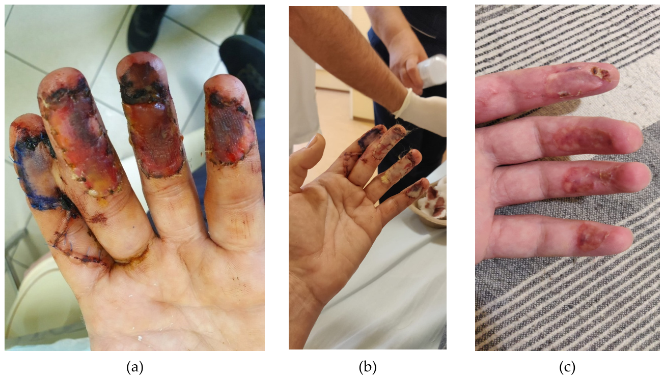
Figure 2.
Clinical appearance of the left hand 6 months after surgery, demonstrating the functional and morphological outcome of rehabilitation (a,b).
Figure 2.
Clinical appearance of the left hand 6 months after surgery, demonstrating the functional and morphological outcome of rehabilitation (a,b).
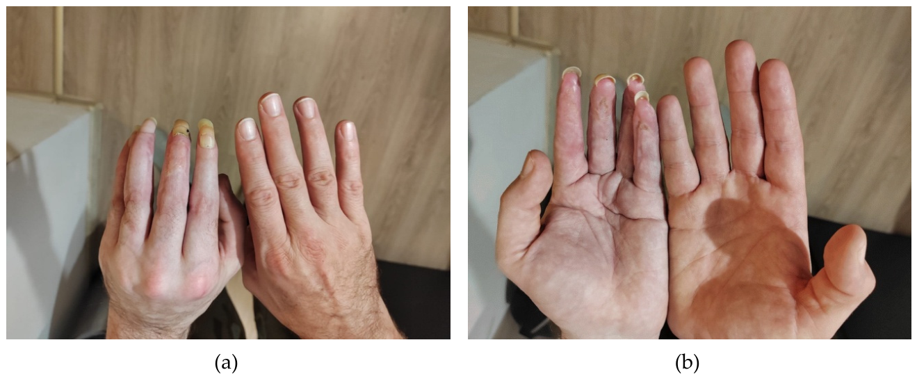
Figure 3.
Timeline of the Patient’s Multidisciplinary Treatment and Rehabilitation Pathway from Injury to Long-term Follow-up.
Figure 3.
Timeline of the Patient’s Multidisciplinary Treatment and Rehabilitation Pathway from Injury to Long-term Follow-up.
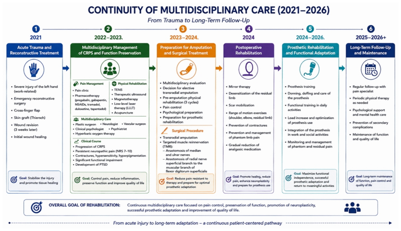
Figure 4.
Psychiatry/Psychology intervention protocol.
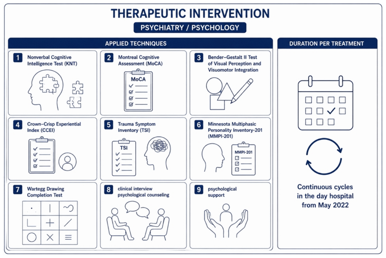
Figure 5.
Mirror therapy protocol used during the first two months following transradial amputation.
Figure 5.
Mirror therapy protocol used during the first two months following transradial amputation.
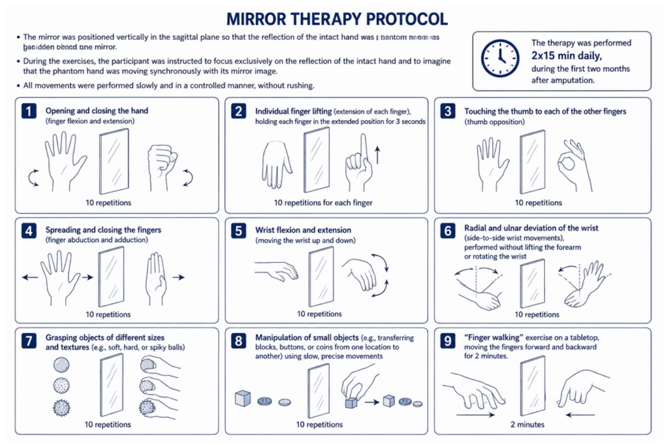
Figure 6.
Functional prosthetic rehabilitation after transradial amputation of the left forearm. (a) Functional use of a forearm prosthesis with a myoelectric hand. (b) Residual limb of the left forearm prepared for prosthetic fitting.
Figure 6.
Functional prosthetic rehabilitation after transradial amputation of the left forearm. (a) Functional use of a forearm prosthesis with a myoelectric hand. (b) Residual limb of the left forearm prepared for prosthetic fitting.
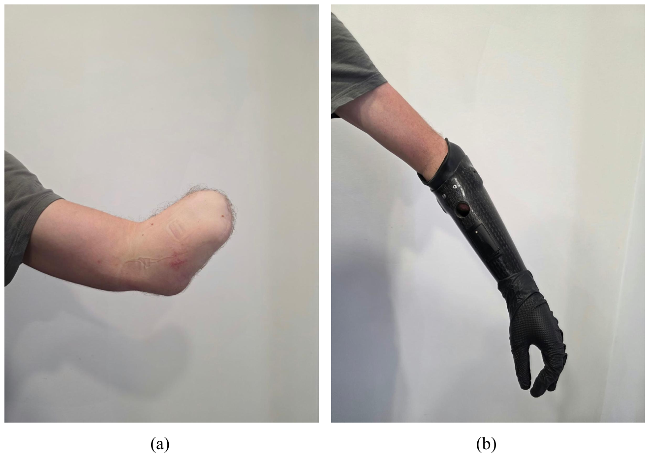

Table 1.
Physical therapy interventions included in the multidisciplinary rehabilitation program. The rehabilitation protocol comprised low-level laser therapy (LLLT), therapeutic ultrasound, transcutaneous electrical nerve stimulation (TENS), pulsed magnetotherapy, and acupuncture. The figure summarizes the treatment parameters, device settings, and duration of each intervention. All electrotherapy modalities were administered using the BTL-4000 SMART device according to standardized protocols. Acupuncture was performed on the right hand and left forearm using predefined auricular and body acupoints. Session duration was 5 minutes for LLLT, ultrasound, and magnetotherapy, 10 minutes for TENS, and 20 minutes for acupuncture.
| Therapeutic intervention | Applied techniques | duration per treatment |
| Low-level laser therapy (LLLT) | Device: BTL-4000 SMART Shape: Continuous Wavelength: 830 nm infrared Frequency: 5000 Hz Energy dose: 4-12 J/cm2 Laser class: 3B |
5 min |
| Therapeutic ultrasound | Device: BTL-4000 SMART Shape: Pulsed Frequency: 1 MHz Power: 3.2 W Intensity: 0.2–0.5 W/cm2 |
5 min |
| Transcutaneous electrical nerve stimulation (TENS) | Device: BTL-4000 SMART Shape: standard Frequency: 100 Hz Pulse width: 100 µs |
10 min |
| Magnetotherapy | Device: BTL-4000 SMART Magnetic field mode: pulse train Magnetic pulse shape: extended rectangular Pulse frequency: 0 to 166 Hz ± 5% Intensity: 54 ᵐᵀ/10 |
5 min |
| Acupuncture |
Acupuncture right hand and left forearm Acupuncture points: ear: Shen Men; thalamus (point 27); PC 7; TE 5; LI 3; SI 11; SI 4; puncta externa 28 |
20 min |
Table 2.
Long-term Clinical and Rehabilitation Outcomes.
| Domain | Outcome | Status |
| Pain intensity | NRS 3–5 (pre-amputation NRS 7–9) | ✓ Improved |
| Phantom limb pain | Occasional phantom limb pain of mild to moderate intensity | ✓ Controlled |
| Residual limb pain | Stable residual limb pain primarily associated with prosthesis weight and use | ✓ Stable |
| Pharmacological treatment | Gradual tapering of analgesic medication; pregabalin and duloxetine discontinued 2 months after amputation, with progressive reduction of opioid analgesics | ✓ Reduced |
| Functional rehabilitation | Successful postoperative rehabilitation including mirror therapy, residual limb desensitization, scar mobilization, and range-of-motion exercises | ✓ Completed |
| Prosthetic rehabilitation | Ongoing functional adaptation with prosthesis use in daily activities | ✓ Improved |
| Return to work | Return to an adapted workplace compatible with the patient’s functional abilities and disability following prosthetic rehabilitation | ✓ Achieved |
| Psychological rehabilitation | Continued follow-up by psychologist and psychiatrist for PTSD and psychosocial adaptation | ↺ Ongoing |
| Multidisciplinary follow-up | Regular follow-up by the pain specialist with periodic physical therapy and continuous multidisciplinary rehabilitation | ↺ Ongoing |
Disclaimer/Publisher’s Note: The statements, opinions and data contained in all publications are solely those of the individual author(s) and contributor(s) and not of MDPI and/or the editor(s). MDPI and/or the editor(s) disclaim responsibility for any injury to people or property resulting from any ideas, methods, instructions or products referred to in the content. |
© 2026 by the authors. Licensee MDPI, Basel, Switzerland. This article is an open access article distributed under the terms and conditions of the Creative Commons Attribution (CC BY) license.
Copyright: This open access article is published under a Creative Commons CC BY 4.0 license, which permit the free download, distribution, and reuse, provided that the author and preprint are cited in any reuse.