

Submitted:
24 September 2026
Posted:
28 September 2026
You are already at the latest version
Abstract
Bone health is a significant contributor to functional ability and well-being. Bone strength and homeostasis are maintained by a remodeling process involving bone resorption and bone formation. An imbalance of these processes increases bone turnover and triggers loss of bone structures. Excessive loss of bone mineral density and deterioration of bone microarchitecture with an augmented risk of fragility fractures are the hallmarks of osteoporosis. Osteoporosis is highly prevalent in the aging population with postmenopausal women being at particular risk. Clinical studies have reported a positive impact of polyphenol-rich nutrition on bone health. Besides phenolic compounds genuinely present in food sources, additional and potentially bioactive molecules are generated by gut microbiota from polyphenol precursors. These metabolites might contribute significantly to the health effects of dietary polyphenols. Some, such as urolithins or equol, are exclusively produced by gut bacteria and are not present in any food source (obligatory microbial metabolites). Others, such as caffeic or vanillic acid, may originate from gut microbial biotransformation of precursors or occur naturally in food sources (facultative microbial metabolites). Evidence for bioactivity of these metabolites on bone-relevant cellular pathways and metabolic bone markers is summarized stratified by study type. It seems likely that individual polyphenol-derived microbial metabolites contribute to bone-protective effects in humans. Strongest evidence is available for S-(−)-equol and consistent promising results exist for urolithin A. Notably, structurally unrelated metabolites often target the same signaling pathways. Supplementation with polyphenols appears reasonable as primary prevention for people at risk of osteoporosis or in the pre-therapeutic stage.
Keywords:
polyphenols
; gut microbiota
; osteoporosis
; bone metabolism
; urolithin
; equol
; phenolic acids
1. Introduction
Bone health is a pillar of functional ability and well-being. It is essential for mobility, independence as well as for participation in social interactions and activities [1]. Bone strength and bone mineral homeostasis is maintained by a constant bone remodeling process involving bone resorption and bone formation activities. Thereby, osteoclasts, originating from haematopoietic stem cells of the mononuclear/phagocytic lineage, degrade bone textures, while osteoblasts, derived from mesenchymal stromal cells (MSC), rebuild bone structures. Throughout adulthood, it is estimated that roughly 5-10% of the existing bone are remodeled within a year to sustain the mechanical integrity of the skeleton [2].
An imbalance in number and activity of osteoclasts and osteoblasts increases rates of bone turnover and trigger loss of bone tissue. Decrease of bone mineral density (BMD) is paralleled by a deterioration of bone microarchitecture and increased risk of fragility fractures. Accelerated and above-average decline in BMD is the hallmark of osteoporosis and its pre-therapeutic stage termed osteopenia. Osteoporosis is an asymptomatic progressive skeletal disease and persons concerned often experience a sudden manifestation when suffering a fracture upon low mechanical impact. Therefore, public awareness campaigns coined the metaphor “silent thief” for this medical condition [3]. Typically, most osteoporotic fractures are hip, vertebral and distal forearm fractures which significantly contribute to the high burden of disease and its clinical and social implications [4]. Osteoporosis is highly prevalent in the aging population with postmenopausal women being at particular risk. Postmenopausal women have a higher burden of years lived with disability and of disease mortality [5].
Osteoporosis is not an isolated skeletal disease; comorbid conditions such as cardiovascular diseases are frequent and fuel a complex interplay of reciprocal increase of bone loss and fractures risks [4,6]. Furthermore, osteoporosis is often associated with sarcopenia, adding loss of skeletal muscle mass to bone atrophy [7,8]. Recently, the biochemical crosstalk of bone, muscles and gut microbiota has attracted the interest of various researchers [9,10,11].
2. Earlier and Later Measures to Osteoporosis
Due to the high prevalence and disease burden of osteoporosis, prevention and treatment of individuals at risk, respectively osteoporosis patients, are of social and economic significance. Primary prevention strategies cover non-pharmacotherapeutic measures such as diet and exercise to maintain BMD and prevent fractures (Table 1). Secondary prevention approaches with antiresorptive or osteoanabolic pharmacological treatments aim at osteoporosis management to prevent (additional) fractures [2].
3. The Role of Nutrition-Derived Polyphenols in Bone Health
Besides the established role of a sufficient intake of calcium, vitamin D and high-quality proteins (Table 1), effects of nutritive polyphenolic compounds for improvement of bone health have been investigated and evidence for a positive impact on bone health in humans has been uncovered in clinical studies [20,21,22]. Beyond clinical observations which typically focus on BMD measurements, fracture rates, bone metabolic markers and occasionally quality of life, the bioactivity and potential cellular or molecular effect pathways have been determined for multiple polyphenolic compounds in cell culture or animal experimental settings [23,24,25,26,27]. Recently, polyphenols have been also suggested as promising option in fracture healing based on enhanced osteogenic differentiation [28].
Besides phenolic compounds which are genuinely present in food sources, such as resveratrol or epigallocatechin gallate [24,29], numerous additional and potentially bioactive molecules are generated by gut microbiota from polyphenol precursor molecules [30,31,32,33]. These microbial metabolites are generally bioavailable and might contribute significantly to the health effects of food-derived polyphenols. To the best of our knowledge, these microbial metabolites have not yet been summarized in a review with particular focus on bone-protective effects.
4. Dysbalanced Cellular Pathways as Options for Interventions
Increased rates of bone turnover emerge from dysbalanced activities of bone-resorbing osteoclast and bone-forming osteoblast. Various local and systemic factors are involved in the regulation of osteoclast / osteoblast formation and function. Osteoclast development is controlled by various hormones such as estrogen, parathyroid hormone (PTH), or vitamin D, as well as, e.g., tumor necrosis factors (TNF) and interleukins (IL) IL-1 and IL-6. Hormones and cytokines also act on osteoblastic lineage cells, which express the relevant cell surface molecule receptor activator of nuclear factor κB (RANK) ligand (RANKL), and a cell surface cytokine receptor, osteoprotegerin (OPG) [2].
Molecular mechanisms behind bone-protective effects of dietary supplementation with polyphenols have been comprehensively reviewed earlier [26,27]. Therefore, the present condensed overview is focused on cellular pathways that have been addressed with polyphenol-derived microbial metabolites.
Osteoblasts and osteoclasts arise from two separate precursor cell pools (Figure 1). Osteoblasts originate from mesenchymal stromal cells (MSCs), which are also precursors of adipocytes. Adipocytes play an active part in this process rather than merely competing for precursors. Factors secreted by marrow-derived adipocytes inhibit osteoblast differentiation of mesenchymal cells [34]. As a source of RANKL and TNF-α, these cells also support osteoclastogenesis, though stimulatory effects have likewise been reported [35]. Bone morphogenetic protein (BMP) and Wnt signaling directs MSC differentiation towards the osteoblastic lineage, whereas peroxisome proliferator-activated receptor γ (PPARγ) together with CCAAT/enhancer-binding protein α (C/EBPα) drives adipogenesis. Thereby, osteoclastogenesis and adipogenesis suppress each other [36,37]. Osteogenesis and adipogenesis of mesenchymal stromal cells are typically in a dynamic equilibrium. Its disturbance under pathological conditions, with reduced osteogenic and enhanced adipogenic differentiation, contributes to bone disorders including osteoporosis [38]. Osteoblast differentiation is reflected in markers such as alkaline phosphatase (ALP), type I collagen (Col I) and osteocalcin (OC) [39], adipogenic differentiation in lipoprotein lipase (LPL), fatty acid-binding protein 4 (FABP4) and adiponectin [40,41].
Osteoclasts arise from bone marrow macrophages (BMMs) of hematopoietic origin (Figure 1), for which macrophage colony-stimulating factor (M-CSF) and RANKL are two essential signals [42,43]. Mature osteoclasts express markers such as tartrate-resistant acid phosphatase (TRAP), cathepsin K (CTSK) and matrix metalloproteinase-9 (MMP-9) [44].
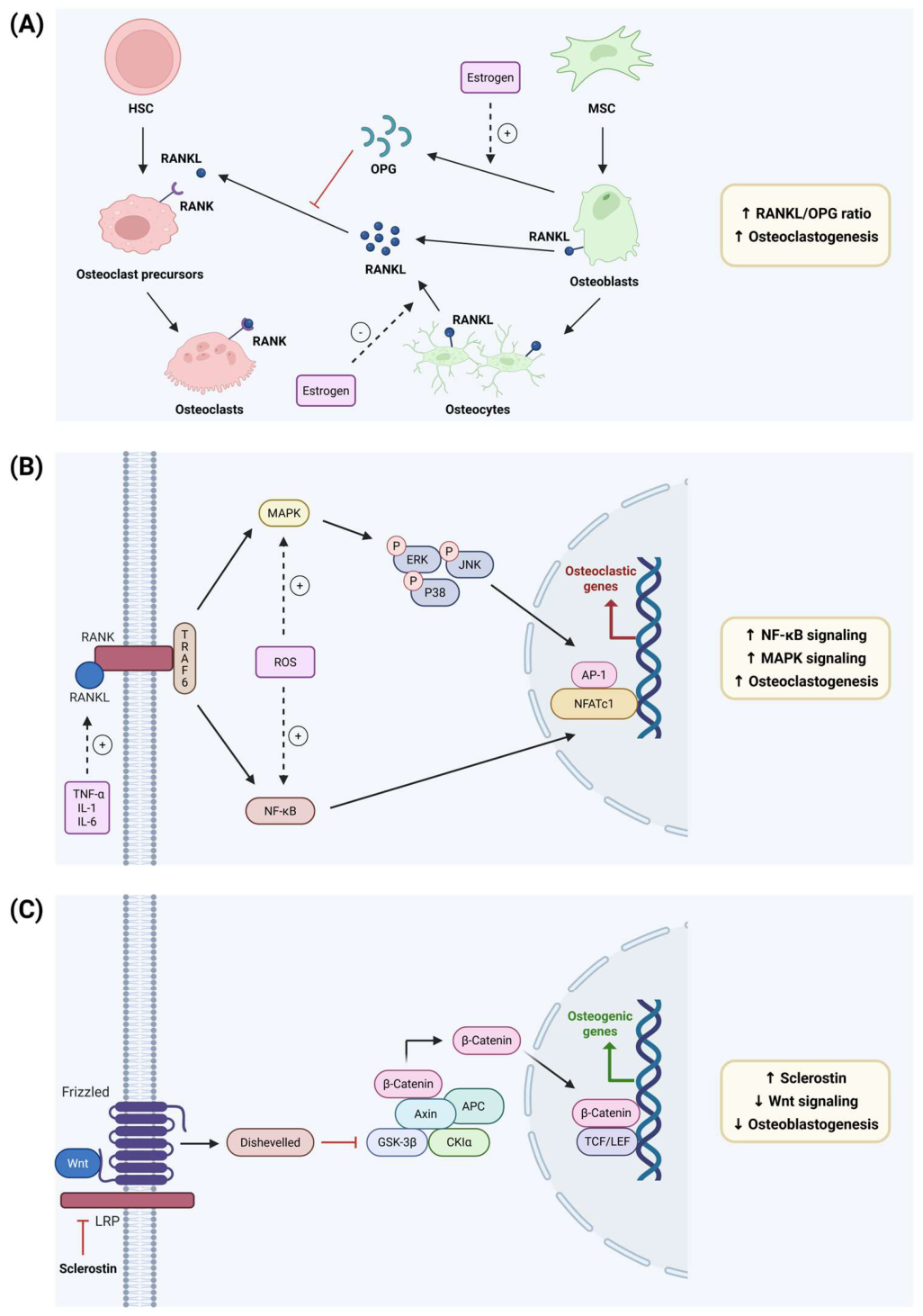
Osteoclastogenesis is largely governed by the RANKL/RANK/OPG axis (Figure 2A). Cells of the osteoblast lineage, arising from mesenchymal stromal cells, express RANKL, which binds RANK on osteoclast precursors and drives their differentiation into multinucleated, bone-resorbing osteoclasts [42]. Osteocytes, terminally differentiated osteoblasts embedded in the mineralized matrix, contribute most of the RANKL that drives osteoclast formation in trabecular bone [45,46]. The opposing signal comes mainly from osteoblasts, which secrete OPG [47], a soluble decoy receptor that binds RANKL and prevents its interaction with RANK [48]. The number of osteoclasts formed at a given site thus depends on the relative availability of both proteins. A rising RANKL/OPG ratio shifts remodeling towards resorption [49]. Estrogen usually keeps the ratio low by suppressing RANKL and inducing OPG, so that its absence after menopause accelerates bone loss [42].
Downstream of RANK, several pathways drive osteoclast formation, of which those addressed by the metabolites reviewed here are shown in Figure 2B. Binding of RANKL recruits TNF receptor -associated factor 6 (TRAF6) to the cytoplasmatic tail of RANK, which activates nuclear factor κB (NF-κB) and the mitogen-activated kinases (MAPKs) c-Jun N-terminal kinase (JNK), extracellular signal-regulated kinase (ERK) and p38 [43,50]. The MAPKs contribute to the transcriptional response through activator protein-1 (AP-1), a dimer of Fos and Jun proteins in which ERK targets c-Fos and JNK targets c-Jun [44,51]. Together with NF-κB, AP-1 acts on the promotor of nuclear factor of activated T cells (NFATc1) and initiates its induction [52]. NFATc1 acts as the central transcription factor of terminal osteoclast differentiation and drives expression of genes that confer resorptive activity, among them CTSK, MMP-9 and TRAP [44,52]. Reactive oxygen species (ROS) amplify both the MAPK and the NF-κB pathway and thus act as second messengers [53,54]. The pro-inflammatory cytokines TNF-α, IL-1 and IL-6 play a role in the same signal cascade mainly by raising RANKL production in osteoblastic cells [42]. TNF-α and IL-1 also activate NF-κB and MAPK signaling in osteoclast precursors through their own receptors [49]. Inflammation and RANKL signaling thereby reinforce each other.
Canonical Wnt signaling is a central regulator of osteoblast differentiation (Figure 2C). In the absence of Wnt ligands, cytoplasmic β-catenin is quenched by a destruction complex comprising Axin, adenomatous polyposis coli (APC), glycogen synthase kinase 3β (GSK-3β) and casein kinase Iα (CKIα), which initiates its proteasomal degradation after phosphorylation. Binding of Wnt to Frizzled and the co-receptor low-density lipoprotein receptor-related protein 5 or 6 (LRP5/6) recruits Dishevelled, which disables this complex, so that β-catenin accumulates and enters the nucleus. There it partners with T-cell factor/lymphoid enhancer factor (TCF/LEF) to activate transcription [55,56]. β-catenin cooperates with the osteogenic transcription factors Runt-related transcription factor 2 (Runx2) and osterix (Osx), and its activation is accompanied by increased expression of osteoblast markers including ALP and type I collagen [57]. The pathway also suppresses adipogenic differentiation of mesenchymal progenitors [56]. In differentiated osteoblasts, the β-catenin-TCF complex controls OPG expression, which links the pathway to the RANKL/OPG axis described above (Figure 2A) [58]. The main endogenous antagonist of the pathway is sclerostin, an osteocyte-derived protein that binds LRP5/6 and blocks signal transduction [55].
Figure 1.
Bone cell differentiation and points of action of nutrition-derived bioactive polyphenolic metabolites produced by the gut microbiota. Polyphenols reaching the colon are converted by the gut microbiota into low-molecular-weight metabolites that enter the circulation. Mesenchymal stromal cells (MSC) give rise to osteoblasts under bone morphogenetic protein (BMP) and Wnt/β-catenin signaling or to adipocytes under peroxisome proliferator-activated receptor γ (PPARγ) and CCAAT/enhancer-binding protein α (C/EBPα). Bone marrow macrophages (BMM) of heamatopoetic origin differentiate into osteoclasts in response to macrophage colony-stimulating factor (M-CSF) and receptor activator of nuclear factor κB ligand (RANKL). Markers of the differentiated phenotypes are listed below each cell type. Arrows indicate the direction in which the metabolites discussed here have been reported to act, favoring osteogenic differentiation while restraining adipogenesis and osteoclastogenesis. Created in BioRender. Högger, P. (2026) https://core.local.biorender.dev/api/short-link/xjf5caw.
Figure 1.
Bone cell differentiation and points of action of nutrition-derived bioactive polyphenolic metabolites produced by the gut microbiota. Polyphenols reaching the colon are converted by the gut microbiota into low-molecular-weight metabolites that enter the circulation. Mesenchymal stromal cells (MSC) give rise to osteoblasts under bone morphogenetic protein (BMP) and Wnt/β-catenin signaling or to adipocytes under peroxisome proliferator-activated receptor γ (PPARγ) and CCAAT/enhancer-binding protein α (C/EBPα). Bone marrow macrophages (BMM) of heamatopoetic origin differentiate into osteoclasts in response to macrophage colony-stimulating factor (M-CSF) and receptor activator of nuclear factor κB ligand (RANKL). Markers of the differentiated phenotypes are listed below each cell type. Arrows indicate the direction in which the metabolites discussed here have been reported to act, favoring osteogenic differentiation while restraining adipogenesis and osteoclastogenesis. Created in BioRender. Högger, P. (2026) https://core.local.biorender.dev/api/short-link/xjf5caw.
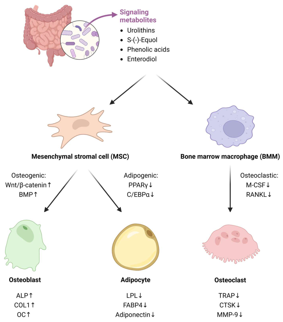
Figure 2.
Signaling pathways governing bone remodeling, shown in the direction that favors bone loss. (A) RANKL/OPG axis. Osteoblasts and osteocytes derived from them express receptor activator of nuclear factor κB ligand (RANKL), which binds RANK on osteoclast precursors and drives their differentiation into bone-resorbing osteoclasts. Osteoprotegerin (OPG), secreted by osteoclasts, acts as a decoy receptor for RANKL and prevents its interaction with RANK, so that the balance of the two determines osteoclast formation. Estrogen promotes OPG and suppresses RANKL production. Adapted from Kim et al. [59], published under a CC BY 4.0 license (https://creativecommons.org/licenses/by/4.0/) (B) NF-κB and MAPK signaling driving osteoclastogenesis. TNF receptor-associated factor 6 (TRAF-6) is recruited to RANK and activates nuclear factor κB (NF-κB) and the mitogen-activated protein kinases (MAPKs) ERK, JNK and p38. Boths arms converge in the nucleus on nuclear factor of activated T-cells (NFATc1), which acts with activator protein-1 (AP-1) to drive expression of osteoclastic genes. Reactive oxygen species (ROS) amplify both arms, and pro-inflammatory cytokines raise RANKL availability. (C) Canonical Wnt/β-catenin signaling, a central pathway of osteoblast differentiation. Without Wnt, β-catenin is degraded by a destruction complex of axin, adenomatous polyposis coli (APC), glycogen synthase kinase 3β (GSK-3β) and casein kinase Iα (CKIα). Wnt binding to Frizzled and LRP recruits Dishevelled, which disables the complex, allowing β-catenin to enter the nucleus and activate osteogenic gene expression with T-cell factor/lymphoid enhancer factor (TCF/LEF). Sclerostin blocks the pathway at low-density lipoprotein receptor-related protein (LRP). Created in BioRender. Högger, P. (2026) https://core.local.biorender.dev/api/short-link/9pujdo.
Figure 2.
Signaling pathways governing bone remodeling, shown in the direction that favors bone loss. (A) RANKL/OPG axis. Osteoblasts and osteocytes derived from them express receptor activator of nuclear factor κB ligand (RANKL), which binds RANK on osteoclast precursors and drives their differentiation into bone-resorbing osteoclasts. Osteoprotegerin (OPG), secreted by osteoclasts, acts as a decoy receptor for RANKL and prevents its interaction with RANK, so that the balance of the two determines osteoclast formation. Estrogen promotes OPG and suppresses RANKL production. Adapted from Kim et al. [59], published under a CC BY 4.0 license (https://creativecommons.org/licenses/by/4.0/) (B) NF-κB and MAPK signaling driving osteoclastogenesis. TNF receptor-associated factor 6 (TRAF-6) is recruited to RANK and activates nuclear factor κB (NF-κB) and the mitogen-activated protein kinases (MAPKs) ERK, JNK and p38. Boths arms converge in the nucleus on nuclear factor of activated T-cells (NFATc1), which acts with activator protein-1 (AP-1) to drive expression of osteoclastic genes. Reactive oxygen species (ROS) amplify both arms, and pro-inflammatory cytokines raise RANKL availability. (C) Canonical Wnt/β-catenin signaling, a central pathway of osteoblast differentiation. Without Wnt, β-catenin is degraded by a destruction complex of axin, adenomatous polyposis coli (APC), glycogen synthase kinase 3β (GSK-3β) and casein kinase Iα (CKIα). Wnt binding to Frizzled and LRP recruits Dishevelled, which disables the complex, allowing β-catenin to enter the nucleus and activate osteogenic gene expression with T-cell factor/lymphoid enhancer factor (TCF/LEF). Sclerostin blocks the pathway at low-density lipoprotein receptor-related protein (LRP). Created in BioRender. Högger, P. (2026) https://core.local.biorender.dev/api/short-link/9pujdo.
5. Polyphenol-Derived Bioactive Compounds with Potential Relevance for Bone Health
To review current evidence on polyphenol-derived gut microbial metabolites with potential implications for bone health, a literature search was performed using the databases PubMed and Web of Science, including publications up to March 2026. The primary focus were human studies, particularly randomized controlled trials. However, given the scarcity of available data, relevant in vitro studies, preferably employing human or humanized cell models, as well as in vivo studies with rodents were additionally included. A Boolean search strategy was applied, incorporating keywords covering three main topics: nutrition (e.g., polyphenols, flavonoids, plant-derived, procyanidins, diet), microbiota (e.g., microbial, metabolites, gut, microbiota) and bone health outcomes (e.g., bone, osteoporosis, osteopenia, bone turnover, osteoblasts, osteoclasts). Studies were excluded when they involved non-human, non-rodent models, and those restricted to children, pregnant or lactating women. The scope was restricted to microbial metabolites derived from dietary polyphenols. Several other major metabolite classes relevant to the gut-bone axis, such as short-chain fatty acids, amino-acid-derived metabolites, bile acids, and vitamins were not covered here, as they have recently been reviewed in detail [60].
Two groups of polyphenols-derived gut microbial metabolites have been identified (Table 2 and Table 3). Some of the metabolites, such as urolithins or equol, are exclusively produced by gut bacteria and are not present in any food source (obligatory microbial metabolites). Other metabolites, such as caffeic or vanillic acid, may originate from gut microbial biotransformation of precursor compounds or may be naturally occurring compounds in food sources (facultative microbial metabolites).
In human clinical studies, microbial metabolites are only occasionally administered as pure compounds. This has been reported for, e.g., urolithin A, but no bone-related effects have been addressed in that study [61]. Typically, in studies polyphenols are ingested from food sources or dietary supplements [20,21,22] yielding microbial metabolites that are detectable in blood samples. Ranges of plasma or serum concentrations determined in human or animal study settings have been summarized to give an impression how the magnitude of reported in vivo levels relate to compound concentrations employed in cell culture settings (Table 2). The in vitro approach allows for detailed investigation of cellular and molecular pathways that are modified by individual microbial polyphenol metabolites.

Table 2.
Reported maximal plasma concentrations of polyphenol-derived gut microbial metabolites in explorative studies versus investigated concentrations in bone cell models. Concentrations are derived from [62] unless otherwise indicated. For split rows, the upper value refers to the free (aglycone) form and the lower value to the predominant phase II metabolite (sulfate or glucuronide). Metabolites highlighted in grey are obligately microbial in origin.
Table 2.
Reported maximal plasma concentrations of polyphenol-derived gut microbial metabolites in explorative studies versus investigated concentrations in bone cell models. Concentrations are derived from [62] unless otherwise indicated. For split rows, the upper value refers to the free (aglycone) form and the lower value to the predominant phase II metabolite (sulfate or glucuronide). Metabolites highlighted in grey are obligately microbial in origin.
| Reported maximal plasma concentrations in explorative studies | Bone cell models | ||
| Microbiota metabolite | In vivo human [µM] | In vivo animal [µM] | In vitro [µM] |
| 5-(3’,4’-Dihydroxyphenyl)-γ-valerolactone (M1, DHPV) | - | 0.014 | 5-15 |
| 1.4 | 0.189-0.683 | ||
| 4-Hydroxyphenylacetic acid (4-HPA) |
0.36-0.45 | 0.151-7 | 25-50 |
| Caffeic acid (CA) | Trace-7.40 | Trace-0.032 | 1-55 |
| 0.416 | Trace-2.8 | ||
| Ferulic acid (FA) | 1.382 | Trace-6.50 | 0.5-100 |
| 0.012-11 | 0.002-4.87 | ||
| Protocatechuic acid (PCA) | 0.52 | 0.008-11.2 | 8-100 |
| 0.125 | 0.015 | ||
| Vanillic acid (VA) | 0.101 | 0.092-3.21 | 10-6-100 |
| 0.463 | 0.814-1.58 | ||
| Gallic acid (GA) | 0.019 | 0.694-11.0 | 5-40 |
| 0.723 | 0.018-0.472 | ||
| Syringic acid (SA) | 5.21 | Trace-0.023 | 1-300 |
| p-Coumaric acid (pCA) | 0.114 | 0.002-0.364 | 0.001-10 |
| 2.205 | 0.015 | ||
| Hydrocinnamic acid | 0.54-1.06 | 0.019 | - |
| 3-(3-Hydroxyphenyl) propionic acid (3-HPPA) | 0.034-0.086 | 0.006-2.52 | 0.06-60 |
| Hippuric acid (HA) | 9-387 | 4.6-90.6 | 0.03-33.5 |
| Urolithin A (UA) | 0.004-0.007 [63] | Trace-0.015 [64] | 1-220 |
| 0.024-35 [63] | 0.121-0.394 [64] | ||
| Urolithin B (UB) | - | - | 1-100 |
| 7.3 [65] | 0.065-5.858 [64] | ||
| Enterolactone (EL) | 0.056 (total) [66] | - | 0.001-30,000 |
| Enterodiol (ED) | 0.073 (total) [66] | 0.093 (total) [67] | 10-6-30,000 |
| S-(−)-Equol | 4.95 (total) [68] | 3.48-7.19 (total) [69] | 10-5-30 |
| 8-Prenylnaringenin (8-PN) | 2.83 (total) [70] | 22.8 (total) [71] | 1-10 |
Notably, analysis of the compounds of interest in the blood compartment allows for no conclusive deduction of concentrations present at the site of action. It has been shown that individual metabolites distribute differentially into particular cells or body compartments and might be enriched at certain anatomical sites [72]. In that context, bone tissue has been rarely investigated as distribution site of polyphenol-derived microbial metabolites. However, accumulation of urolithins A and B as well as of p-coumaric acid in pig bones has been previously established and it was suggested that these compounds bind to the mineral bone component hydroxyapatite [73].
Another aspect that needs attention when serum or plasma concentrations are examined is that microbial metabolites are typically subjected to phase-II-biotransformation in enterocytes and liver [74]. Thus, circulating compounds are usually to certain degree conjugated, e.g., with sulfate or glucuronic acid [62,72,75]. The conjugated metabolites are typically more hydrophilic which hampers cell entrance by diffusion [33,76,77]. Interestingly, the polyphenol-derived microbial metabolites detected in pig bones were not conjugated [73] which would be consistent with the observation that phase-II conjugates can undergo local deconjugation [78], particularly under inflammatory conditions [79,80,81].
Polyphenol-derived gut microbial metabolites that have been investigated in the context of bone health were summarized with typical nutritional sources, precursor molecules, time of maximal concentrations recorded in blood (tmax) and bone health-related effects, categorized as “C” for cell culture data, “B” for in vivo animal data, and “A” for human study data (Table 3).
Table 3.
Polyphenol-derived gut microbial metabolites with potential impact on bone health parameters. Evidence is stratified by study type: A (human studies), B (in vivo animal studies), C (in vitro studies). Metabolites highlighted in grey are obligately microbial in origin
Table 3.
Polyphenol-derived gut microbial metabolites with potential impact on bone health parameters. Evidence is stratified by study type: A (human studies), B (in vivo animal studies), C (in vitro studies). Metabolites highlighted in grey are obligately microbial in origin
| Microbiota metabolite | Nutritionalsource | Precursor | Human plasma / serum tmax | Type | Impact on bone health parameters | |
| Metabolites of Flavan-3-ols | ||||||
5-(3’,4’-Dihydroxyphenyl)-γ-valerolactone (M1, DHPV) 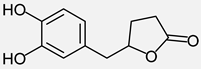
|
Cocoa, tea (green/black) red wine, berries, Pycnogenol® | Flavan-3-ols, catechins | tmax = 10 h [82] tmax = 5-12 h [83] |
C | M1 (5 and 15 µM), H2O2-induced inhibition of osteoblast differentiation ⇩, ALP activity and mineralization capacity ⇧ in MC3T3-E1 cells (murine), ROS and MDA levels ⇩ through SIRT1-mediated autophagy [84] M1 (10 µM, in cocktail), osteogenic differentiation of human MSC ⇧ [85] |
|
| A | Inverse association between serum M1 levels and the bone resorption markers CTX-1 and NTX-1 [85] | |||||
4-Hydroxyphenylacetic acid (4-HPA)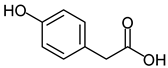
|
Cocoa, tea, red wine, berries | Flavan-3-ols (and other sources) | - | C / B | 4-HPA (25 and 50 µM), osteoclast differentiation in BMM ⇩ (mice), osteoclast-specific genes ⇩, ROS ⇩ by upregulating Nrf2 expression, NF-κB and MAPK pathway ⇩, OVX mice (4-HPA 25 mg/kg every 2 days for 8 weeks) bone loss ⇩, BV/TV, BS/TV, Tb. N, and Tb.Sp ⇧ [86] | |
| Metabolites of Chlorogenic acids | ||||||
Caffeic acid (CA)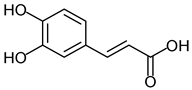
|
Coffee, tea, berries, wine | Chlorogenic acid | tmax = 1h (after instant coffee and green tea) [87] tmax = 1.4h [88] |
C | CA, osteoclastogenesis and bone resorption ⇩, expression of osteoblast markers ⇧ [89] CA (1 µM), MG63 (human osteoblast cell line), osteoblast-related marker gene expression ⇧, (RUNX2, ALP, COL I, OSX, OC), [90] CA (1 and 10 µg/mL), BMM (murine), osteoclastogenesis ⇩, RAW-D, osteoclastogenesis and NF-κB activity ⇩ [91] |
|
| B | CA (50 mg/kg p.o. for 4 weeks), OVX Wistar rats, tibia mineralization and cortical bone mechanics ⇧ [92] | |||||
Ferulic acid (FA)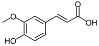
|
Coffee, whole grains, cereals, fruits | Chlorogenic acids, caffeic acid | tmax = 8.2h (after 13C-anthocyanins) [93] tmax = 2.0h [88] |
C | FA (10 µM), hMSC, β-catenin expression ⇧, osteogenic differentiation ⇧ [94] FA (0.5-3 µM), MC3T3-E1 cell differentiation and mineralization ⇧, ALP mRNA, osteogenic marker expression ⇧, β-Catenin, Lrp-5 and GSK-3β protein expression ⇧, OPG/RANKL mRNA expression ratio ⇧ [95] FA, human CD14+ peripheral blood monocytes, ex vivo IC50 of 39 µM, actin ring formation and bone resorption activity ⇩, RANKL-induced expression of DC-STAMP ⇩, MMP-9 and cathepsin K ⇩, mature osteoclast numbers ⇩, caspase-3 activity ⇧ [96] FA (25 – 100 µM), RAW264.7, RANKL-induced osteoclast differentiation (TRAP staining) ⇩, bone resorption activity ⇩, NF-kB activation and downstream factors (NFATc1, c-Fos, TRAP, cathepsin K, MMP-9) ⇩ [97] FA and CA (10 and 11 µM), osteogenic differentiation of human MSC ⇧ [85] |
|
| B | FA (50 and 100 mg/kg p.o. daily for 8 weeks), GIO rats, restoring of bone density and microarchitecture [95] FA (50 mg/kg b.w. daily for 37 days), mice, TBI-mediated mortality, stem cell senescence, and bone mass loss ⇩, antioxidant defense systems ⇧, osteogenic marker expression ⇧, adipogenic differentiation in BM ⇩ [98] |
|||||
| Metabolites of Anthocyanidins | ||||||
Protocatechuic acid (PCA) 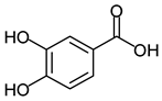
|
Berries, grapes, whole grains, coffee, tea | Anthocyanins (cyanidin), Flavonols, chlorogenic acids | tmax = 1h (after chicory) [99] tmax = 3.3h (after 13C anthocyanins) [93] |
C | PCA (50 and 100 µM), primary human osteoblasts, osteogenic gene expression (OPN, IBSP, ALPL, BMP6) ⇧, IL-1β ⇧, TNF and MMP-1 ⇩ [100] PCA (40 and 80 µM), murine MSCs, mineralization and ALP activity ⇧, osteogenic gene expression (RUNX2, ALP, OSX) ⇧ Wnt/β-catenin pathway ⇧, 3T3-L1 preadipocyte cells, adipogenesis and lipid accumulation ⇩, PPARγ expression ⇩, [101] PCA (12.5 – 50 µM), MC3T3-E1 cell proliferation and ALP activity ⇧, ROS and MDA level ⇩ SOD, GSH and NO level ⇧ [102] PCA (8 µM), RAW264.7 murine macrophage cells, RANKL-induced TRAP activity and osteoclast formation ⇩, ROS ⇩, osteoclast marker expression (MMP, TRAP, Cathepsin, TRAF-6) ⇩, NFATc1 expression ⇩, NF-kB and COX-2 expression ⇩ [103] PCA (25 µM), mouse bone marrow macrophages (BMM), RANKL-induced osteoclast differentiation and bone resorption ⇩, phosphorylation of JNK ⇩, expression of c-Fos and NFATc1 ⇩ [104] |
|
| B | PCA (20 mg/kg/day for 12 weeks), OVX mice, osteoclast activity ⇩, serum RANKL, OC, bALP ⇩, serum OPG ⇧, loss of trabecular bone ⇩, BV/TV, Tb.Th, Tb.N, BS/BV, and BMD ⇧ [105] PCA (25 mg/kg daily for 9 days), Mice, LPS-induced bone loss ⇩, BV/TV, Tb.Sp, and Tb.N ⇧ [104] PCA (50 mg/kg), Alcohol-induced osteopenic mice, tibial trabecular BMD ⇧, BV/TV, Tb.N [106] |
|||||
Vanillic acid (VA)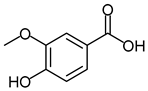
|
Berries, whole grains, coffee, tea | Anthocyanins (peonidin), ferulic acid | tmax = 12.5h (after 13C anthocyanins) [93] | C | VA (1 pM-10 nM), rat osteoblast-like UMR 106 cells, proliferation and ALP activity ⇧, osteoblastic genes expression ⇧, phosphorylation of MEK1/2, ERK1/2 and ERα [107] VA (1-100 µM), RAW264.7, RANKL-activated TRAP activity and differentiation in multinuclear TRAP-positive cells ⇩ [108] |
|
| B | VA (50 or 100 mg/kg b.w. for 12 weeks), OVX rats, BMD and BMC ⇧, Ca, P, OC, ALP, DPD and inflammatory markers (IL-1β, IL-6, and TNF-α) ⇩ [109] VA (100 mg/kg b.w. for 4 weeks), OVX mice, bone resorption ⇩ [108] |
|||||
Gallic acid (GA)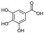
|
Tea (green/black), red wine, grapes | Anthocyanins (delphinidin), Flavan-3-ol gallate esters | tmax = 2.2h (after consumption of mango pulp) [110] | C | GA-g-chitosan (GAC), 111 mg GA/g GAC, BMMSC (murine), ALP activity and mineralization ⇧, Wnt1 and Wnt3a mRNA and protein expression ⇧, expression of ß-catenin targeted genes (Runx2, OSX, type I collagen) ⇧ [111] GA (10 – 40 µM), hPDL cells, LPS-induced inflammation cytokines (IL-6, IL-1ß) ⇩, ALP activity and mineralization in hPDL cells ⇧, GPR35/GSK3ß/ß-catenin signaling pathway ⇧ [112] GA (5 and 10 µM), RANKL-induced osteoclast formation and function in BMM (mice) and RAW264.7 cells ⇩, Akt, ERK and JNK pathways ⇩, osteoclastogenesis-related marker expression (NFATc1, c-Fos and cathepsin K) ⇩ [113] |
|
| B | GA (10 mg/kg every 2 days for 8 weeks), OVX mice, bone loss ⇩, BMD, BV/TV, Tb/Th and Tb.N ⇧ [113] | |||||
Syringic acid (SA)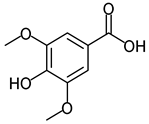
|
Berries, red wine, whole grains | Anthocyanidins (malvidin), sinapic acid | tmax = 3.9h (after ingestion of cranberry juice) [114] | C | SA (100 – 300 µM), MSC (mouse), ALP activity and calcium deposition ⇧, expression of Runx2, ALP, type I collagen and osteocalcin ⇧ [115] SA (1-100 µM), RAW264.7, RANKL-activated TRAP activity and differentiation in multinuclear TRAP-positive cells ⇩ [108] |
|
| B | SA (100 mg/kg body weight/day for 10 weeks), OVX mice, femoral bone mineral density ⇧, number of osteoclasts ⇩ and osteoblasts ⇧ [116] SA (100 mg/kg b.w. for 4 weeks), OVX mice, bone resorption ⇩ [108] |
|||||
| Shared microbial polyphenol-derived metabolites | ||||||
| p-Coumaric acid (pCA) 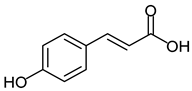
|
Fruits, red wine, tea, whole grains | Flavanols, chlorogenic acids | Plasma 64 nmol/L 0.7h after 350 mL concord grape juice [117] | C | pCA (1-1000 nM), mouse BMC, osteoblastogenesis and mineralization ⇧, adipogenesis ⇩, pCA (0.1-100 nM), adipogenesis in 3T3-L1 preadipocytes ⇩, lipogenesis in mature 3T3-L1 ⇩ [118] pCA (0.01-10 µM), MC3T3, osteogenic differentiation and mineralization ⇧, RAW264.7, osteoclast formation ⇩, NF-kB activation ⇩ [119] |
|
| B | pCA (250 or 500 µg/100g b.w. for 30 days), OVX rats, mineral content, mineral density and polar strength strain index in femoral-metaphyseal tissue ⇧, ALP activity ⇧[120] | |||||
3-Phenylpropanoic acid (hydrocinnamic acid)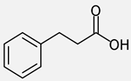
|
Fruits, cocoa, tea, whole grains | Flavanols, chlorogenic acids | / | A | Serum concentrations associated with BMD [121] |
|
Hippuric acid (HA)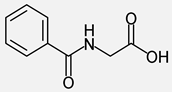 3-(3-Hydroxyphenyl) propionic acid (3-HPPA) 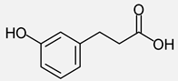
|
Fruits, vegetables, coffee, tea, whole grains | Flavanols, procyanidins, chlorogenic acids | tmax = 15.7h (after 13C anthocyanins) [93] 3-3-PPA: Plasma 355 nmol/L, 5.8h after 350 mL concord grape juice [117] |
C | HA (0.03-33.5 µM) and 3-HPPA, (0.06-60 µM), RAW264.7 and mouse BMC, osteoclastic cell differentiation and osteoclast resorptive activity ⇩, RANKL-stimulated increase of osteoclastic markers (NF-κB, NFATc1, MMP-9) ⇩ [122] | |
| B | HA (0.1-5 mg/kg/day for 2 weeks), prepubertal female mice, bone-forming gene expression ⇧, PPARγ expression ⇩, bone mass ⇧ [123] 3-HPPA (0.1-5 mg/kg/day for 30 days), 1-month old female mice, bone volume and trabecular thickness ⇧, osteoblast number ⇧, osteoclast number ⇩ β-galactosidase activity and PPARγ expression ⇩ [124] HA and 3-HPPA (5 mg/kg/day for 8 weeks), OVX mice, bone loss ⇩, bone marrow adipocyte-like cell formation ⇩, adipogenic gene expression PPARγ and Lpl ⇩ [125] |
|||||
| Metabolites of Ellagitannins | ||||||
Urolithin A (UA)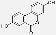 Urolithin B (UB) 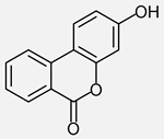 |
Pomegranate, nuts, berries | Ellagic acid | Uro A tmax = 6h [126,127] | C | UA (5 and 10 µM) RANKL-induced osteoclastogenesis in BMM (murine) ⇩ bone resorption and expression of osteoclast-related genes (MMP9, cathepsin K and NFATc1) ⇩, autophagy capacity of BMM ⇧, MAPK signaling pathway ⇩ [128] UA (5 – 20 µM), RANKL-triggered BMM (mice): osteoclast formation, F-actin rings and TRAP activity ⇩, LPS-challenged RAW264.7: phosphorylation of NF-kB p65, JNK and ERK1/2 ⇩ activation of Nrf2 signaling [129] UA (25 µM), RANKL-induced osteoclastogenesis in RAW264,7 cells ⇩, ERK1/2, p38 and Akt signaling ⇩, BMP2 target for UA [130] UA (5 and 10 µM), BMM (murine), osteoclast-related gene expression and bone resorption ⇩, PI3K/AKT/mTOR signaling pathway phosphorylation and NFATc1 transcription/translation ⇩ [131] UA (10, 25 and 50 µg/mL), mineralization of MSC (mice) ⇧, osteogenic marker (Sox9, Col2a1, Runx2) ⇧ [132] UA (1-10 µM), no direct effect on osteogenic differentiation of BMSC (mouse), TNF-α induced activation of NF-κB signaling ⇩ [133] UB (10 – 100 µM), RANKL- induced osteoclastogenesis in RAW264.7 ⇩, F-actin belts formation, osteoclast-specific gene expressions and ROS activity ⇩, Nrf2 and ROS scavenging enzymes ⇧, RANKL-induced NF-kB, MAPK and Akt signaling pathways ⇩, expression of c-Fos and NFATc1 ⇩ [134] UB (1-25 µM), BMM (mice) and RAW.264.7, RANKL-induced osteoclast differentiation ⇩ expression of osteoclast-related genes (MMP-9, cathepsin K, NFATc1) ⇩, ERK/NF-κB pathway ⇩ [135] |
|
| B | UA (10 or 20 mg/kg daily for 8 weeks), OVX mice, trabecular bone loss in distal femur ⇩, BMD, Tb.N, and N.Ob/BS ⇧, OPG/RANKL ratio ⇧ , TNF-α, IL-1β, Il-6 and CTX-1 ⇩ [136] UA (15 or 30 mg/kg daily for 6 weeks), OVX mice, bone loss ⇩ , BV/TV, BS/TV, and Tb.N ⇧ [131] UA (10 or 20 mg/kg four times a week for 20 weeks), mouse model, bone loss ⇩ BMD, BV, Tb.N and Tb.Sp ⇧ [128] UA (25 mg/kg twice daily for 8 days), mice, LPS-induced bone loss ⇩, Tb.N, BV/TV, BS/BV, and Tb.Sp ⇧ [129] UB (10 or 50 mg/kg every two days for 8 weeks), OVX mice, bone loss ⇩, BMD, BV, BV/TV, BS/TV, Tb.Sp, Tb.N ⇧, serum CTX-1 ⇩ [135] |
|||||
| Metabolites of Lignans | ||||||
Enterolactone (EL)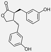 Enterodiol (ED) 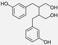 |
Sesame seeds, Flaxseed | Secoisolariciresinol-diglucoside (SDG) | EL tmax = 20h ED tmax = 15h [66] |
C | EL and ED (0.01-10 mg/mL), MG-63 cells (human osteoblast-like cell line): ALP activity ⇧, mRNA levels of OC, ALP and OPN ⇧; col I and ON ⇧ (low conc.) [137] EL (1 nM), MC3T3-E1 cells, ALP activity ⇧, MG-63 cells, gene expression of OPG ⇧ (1 pM-1 nM) and RANKL ⇩ (10 and 100 pM), ED (1 pM), MG-63 cells RANKL gene expression ⇩, OPG/RANKL ratio ⇧ [138] |
|
| B | EL or ED (10 mg/kg b.w. for 22 weeks), OVX mice with established MCF-7 tumors, no effect on femoral BMD or strength [139] | |||||
| A | Positive correlation between urinary EL and BMD of L2-L4, and BMD of the femoral neck and ward’s triangle, in OP patients EL lower than in controls [140] Total lignans, highest quartile associated with reduced hip fracture risk compared with lowest quartile, similar finding for estimated EL level [141] Blackcurrant (BC) anthocyanins, ED positively correlated with plasma bALP, EL positively correlated with plasma OC and negatively with sclerostin [142] |
|||||
| Metabolites of Isoflavones | ||||||
S-(−)-Equol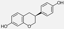
|
Soybean, soy food (e.g. tofu, tempeh, miso, soy milk) | Daidzein, isoflavones |
tmax = 36 h (after soy milk) [143] tmax = 1,5 – 3 h (after S-Equol) [144] |
C | S-Equol (10 and 30 µM), osteoclast differentiation in BMC (mice) and RAW264.7 ⇩, apoptosis of mature osteoclasts by inducing caspase-3/7 activity, RANKL-induced activation of JNK, NF-κB and NFATc1 ⇩, osteoblast differentiation in MC3T3-E1 cells ⇧ [145] Equol (10 nM-1 µM), ROS17/2.8 (rat osteoblast-like cells) proliferation ⇧ and apoptosis ⇩, OPG/RANKL ratio ⇧, mainly mediated through ER-β [146] S-Equol (10 pM-1 nM), ROS17/2.8, OPG/RANKL ratio ⇧ , ER-β expression and AKT phosphorylation ⇧, activation of PI3K/AKT via ER-β [147] |
|
| B | S-Equol (0.5 mg/d for 4 weeks), OVX mice, BMD in the whole, proximal, middle, and distal femora ⇧ [145] Equol (0.5 mg/d for 4 weeks), OVX mice, BMD of whole body, femur and lumbar spine ⇧, N.Oc ⇩, BMD of proximal femur same as sham group [148] Equol (0.5 mg/d for 3 weeks), OVX mice, BMD of the whole, proximal, and distal femur ⇧, whole body fat mass and plasma lipids ⇩ [149] S-Equol (0.06% diet for 2 weeks), OVX mice, bone volume of the femoral distal metaphysis ⇧, expression of inflammatory (Oct-2, IL-7), osteoclastogenesis (RANKL, NFATc1, CTSK) and adipogenesis-related genes (PPARγ, FAS) in bone marrow cells of OVX mice ⇩ [150] Equol (10 μg/g b.w./day for 3 months), OVX rats, femoral BMD and bone strength ⇧ [151] Equol (0.25 and 0.5 mg/kg/day), OVX rats, femoral BMD and trabecular microarchitecture ⇧, mRNA and protein expression of OPG and OPG/RANKL ratio ⇧ [146] S-Equol (20-80 mg/kg/day for 12 weeks), streptozotocin-treated rats, femoral BMD and trabecular microarchitecture ⇧, OPG/RANKL ratio ⇧, [147] Equol (400 mg/kg for 6 weeks), OVX rats, lumbar spine BMD ⇧, no effect at proximal tibia, mild uterotropic potential [152] |
|||||
| A | S-Equol (10 mg/day), postmenopausal women, urinary DPD ⇩, Whole-body BMD decrease after 1 year ⇩ [153] Soy isoflavone (75 mg conjugates/day) postmenopausal women, equol producers: loss of total hip BMD vs placebo after 1 year ⇩ [154] 200 mg fermented soy (10 mg equol and 25 mg resveratrol), postmenopausal women, OC and bALP ⇧, DPD ⇩, Whole-body BMP ⇧ [155] Supplemental soy foods, postmenopausal women, no difference in total and site-specific BMD between equol producers and non-producers [156] Serum S-equol lower in OP patients than in controls, positive correlation between S-equol levels and beneficial gut bacteria and amino acid-related metabolites [157] |
|||||
| Metabolites of Prenylated Flavonoids | ||||||
8-Prenylnaringenin (8-PN) 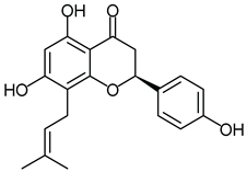
|
Hop-derived dietary products, beer | Isoxanthohumol | tmax = 1.2h, 7.7h (after single oral dose of 8-PN) [158] tmax = 1-3h (after xanthohumol) [159] |
C | 8-PN (10 µM), MC3T3-E1 and RAW264.7, ALP activity, OC and mineralized nodules ⇧, osteoclast differentiation and bone resorption ⇩, protein/mRNA expression of RANKL ⇩ and OPG ⇧, effects mediated by ER-α [160] 8-PN (1 µM), rat calvarial osteoblasts, ALP activity and staining ⇧, mineralization ⇧, osteogenic gene expression ⇧, OPG/RANKL ratio ⇧, BMSC (rabbit), osteoclast formation and activity ⇩ osteoclast gene expression ⇩ [161] 8-PN (1 µM), BMSC (rat), osteogenic differentiation and mineralization ⇧, OPG expression ⇧,BMP-2/p38MAPK/Runx2/Osterix signal pathway ⇧ [162] |
|
| B | 8-PN (0.67, 1.77 and 18 mg/kg/d for 28 days), OVX rats, trabecular bone loss ⇩, minimal trophic effects on uterus [163] 8-PN (6.8 and 68.4 mg/kg b.w. for 12 weeks), OVX rats, BMD on the upper tibial metaphysis ⇧, bone biochemical properties ⇧ [164] 8-PN (1.77 mg/kg daily for 10 weeks), OVX rats, no fundamental improvements in osteoporotic bones, slight increase in uterine wet weight [165] |
|||||
| A | Hop extract standardized in 8-PN and Ca/vit d3, total body BMD ⇧, higher proportion of women experiencing an increase ≥ 1% compared to placebo, SF-36 physical functioning score ⇧ [166] | |||||
Time of maximal concentrations recorded in plasma or serum (tmax) are typically around 8-10 hours when the ingested precursor molecules are of high molecular weight, as in the case of procyanidin biopolymers or ellagitannins. With smaller precursor molecules, the gut microbiota produce the respective metabolites significantly faster, and they are detectable in plasma / serum roughly after 2-3 hours.
The majority of studies with microbial metabolites have investigated effects on the differentiation of osteoblasts or osteoclasts. The influence of microbial metabolites on cellular signal transduction pathways was most often explored for the proinflammatory NF-kB / MAPK pathway, followed by the RANKL / OPG axis. Only few studies addressed the Wnt / β-catenin signal network.
5.1. Metabolites of Flavan-3-Ols
The plant-based bioactive group of flavan-3-ols or flavanols consists of monomeric and polymeric compounds such as procyanidins [167]. Among microbial flavan-3-ol metabolites, evidence regarding effects related to bone health is confined to 5-(3’,4’-dihydroxyphenyl)-γ-valerolactone (M1, DHPV) and 4-hydroxyphenylacetic acid (4-HPA), and the available studies employed different cell types. For M1, osteoblasts have been the focus. In MC3T3-E1 cells, it raised ALP activity and mineralization (Figure 1) and restored differentiation impaired by oxidative stress, with lower ROS and malondialdehyde levels, an effect abolished by silencing SIRT1 [84]. In a study investigating the differentiation of MSC to osteoblasts, M1 significantly increased the osteoblast formation in a mixture with trans para-coumaric acid, protocatechuic acid, caffeic acid, ferulic acid, gallic acid and taxifolin (also see 5.2). Tested as single compound, it stimulated the differentiation but did to reach statistical significance in the concentration tested [85]. ROS also amplify the RANKL-induced osteoclastogenesis (Figure 2B), suggesting effects of M1, based on its radical scavenging activity [168], on osteoclast differentiation as well, though this has not yet been tested. Notably, M1 is one of the few metabolites in this review for which human data exists, with serum levels inversely related to the bone resorption markers CTX-1 and NTX-1 in a trial with osteopenic women [85].
4-HPA, a shorter-chain phenylacetic acid formed further along the same catabolic cascade [169], has been examined with focus on osteoclast effects. It suppressed RANKL-induced osteoclast differentiation in murine bone-marrow macrophages (Figure 1), lowering ROS through Nrf2 activation and attenuating NF-κB and MAPK signaling (Figure 2B). Moreover, 4-HPA preserved trabecular bone in ovariectomized mice [86].
Both metabolites thus converge on redox control, though the reported mechanisms differ, SIRT1-dependent autophagy for M1 and Nrf2 induction for 4-HPA. Since the two have not been compared in the same system, it remains open whether this reflects a genuine difference or the design of the individual studies. Both metabolites also affect the proinflammatory axis, as M1 also suppressed TNF-α induced NF-κB activation in endothelial cells [170] and 4-HPA inhibited NF-κB and MAPK signaling directly. Evidence in the context of osteoporosis remains sparse, with no in vivo animal data for M1 and no human data for 4-HPA.
5.2. Metabolites of Chlorogenic acids
Within this group, bone-related effects have been reported for caffeic acid (CA) and its methylated counterpart ferulic acid (FA). CA affects both bone remodeling cell types in vitro. It upregulated osteogenic genes in human MG-63 osteoblasts [90] and suppressed RANKL/TNF-α-induced osteoclast differentiation (Figure 1) in murine precursor cells, primarily by inhibiting NF-κB signaling (Figure 2B) [91]. The in vivo data is less consistent, as only the higher of two oral doses improved tibial mineralization and cortical mechanics in ovariectomized rats, while the lower dose was ineffective or even detrimental [92]. An effect requiring high doses is relevant for dietary exposure, since circulating CA phase II conjugates were not reported to exceed low micromolar levels (Table 1).
For FA, osteogenic differentiation was promoted in both human mesenchymal stromal cells and murine MC3T3-E1 cells, accompanied by upregulation of β-catenin (Figure 2C) [94] and by an increased OPG/RANKL ratio (Figure 2A) [95]. On the osteoclast side, FA inhibited RANKL-induced osteoclast differentiation in human CD14+ precursors (Figure 1) [96] and reduced NF-κB and its downstream targets in RAW264.7 macrophages (Figure 2B) [97], similarly to the mechanism addressed by CA. In vivo, FA preserved bone mass in dexamethasone-treated rats [95] and limited irradiation-induced bone loss in mice, shifting mesenchymal differentiation from the adipogenic towards the osteogenic lineage (Figure 1) and raising antioxidant defense systems [98]. Neither model reflected estrogen deficiency, the standard setting for postmenopausal osteoporosis.
In human mesenchymal stromal cells, a mixture of the phenolic acids CA and FA, together with protocatechuic and p-coumaric acid significantly enhanced mineralization (Figure 1), as did the mixture of their corresponding phase-II-conjugates [85]. Notably, the conjugates were as effective as their parent compounds, possibly through local deconjugation by cell-associated enzymes such as sulfatases or glucuronidases [79,80,171].
Methylation of the catechol group to a guaiacyl group therefore does not abolish activity, and the evidence for FA is arguably the stronger compared to CA, covering more pathways and including human primary cells. Whether this reflects a difference in potency is open, since the two acids have not been tested in direct comparison. Their research gaps are complementary, with CA examined in the estrogen-deficient model but with an inconsistent dose response, and FA investigated in human cells yet only in secondary models of bone loss. Clinical data are absent for both.
5.3. Metabolites of Anthocyanidins
Bone health-related information in this group is available for protocatechuic acid (PCA) and its methylated counterpart vanillic acid (VA), and for gallic acid (GA) and its dimethylated derivative syringic acid (SA).
Most data are published for PCA which acts on both bone remodeling cell types. It promoted osteogenic differentiation and mineralization of murine mesenchymal stem cells (Figure 1) while activating Wnt/β-catenin signaling (Figure 2C) and suppressed adipogenesis in 3T3-L1 praeadipocytes through downregulation of PPARγ (Figure 1) [101]. Comparable osteogenic effects were seen in MC3T3-E1 cells, there accompanied by reduced ROS and MDA levels [102]. In primary human osteoblasts, it upregulated osteogenic genes [100]. The anti-osteoclastic mechanism is congruent across two models: PCA inhibited RANKL-induced differentiation in RAW264.7 cells with suppression of NF-κB and NFATc1 (Figure 1, Figure 2B) [103] and in mouse bone marrow macrophages through JNK and c-Fos/NFATc1 (Figure 1, Figure 2B) [104]. In vivo, PCA attenuated bone loss across three distinct systems, in ovariectomized mice, after lipopolysaccharide challenge and in alcohol-fed mice [104,105,106]. In the OVX model this was accompanied by a beneficially raised OPG/RANKL ratio (Figure 2A) [105].
VA has been examined mainly in osteoblasts, stimulating proliferation and osteoblastic genes expression in rat osteoblast-like UMR-106 cells at picomolar to nanomolar concentrations through MAPK-mediated estrogen receptor signaling (Figure 1) [107], and improving bone mineral density in ovariectomized rats alongside lower pro-inflammatory IL-1β, IL-6 and TNF-α concentrations (Figure 2B) [109]. SA likewise promoted osteogenic differentiation of murine mesenchymal stem cells (Figure 1) [115] and raised femoral bone mineral density without uterine effects [116]. Both acids inhibited RANKL-induced osteoclast differentiation in RAW264.7 cells (Figure 1) and reduced trabecular bone loss in ovariectomized mice, more markedly for SA [108].
For GA, osteogenic differentiation of murine mesenchymal stem cells was promoted with increased Wnt1 and Wnt3a expression and nuclear translocation of β-catenin (Figure 1, Figure 2C) [111], although a chitosan conjugate rather than free GA was used. Consistent with this, GA raised mineralization via GSK3β/β-catenin signaling pathway in human periodontal ligament cells (Figure 1, Figure 2C) while lowering lipopolysaccharide-induced IL-6 and IL-1β (Figure 2B) [112]. In osteoclast precursors, GA suppressed RANKL-induced differentiation in both bone marrow macrophages and RAW264.7 cells through ERK and JNK inhibition (Figure 1, Figure 2B) and limited ovariectomy-induced bone loss [113].
Two observations emerge. Reported effects of the four acids converge on a shared pattern, with Wnt/β-catenin (Figure 2C) engaged during osteoblast differentiation by PCA and GA, and the NF-κB and MAPK pathway (Figure 2B) targeted in osteoclastogenesis by PCA and GA. For VA and SA, the anti-osteoclastic effect is documented but not mechanistically resolved. Notably, O-methylation did not diminish activity. In the only head-to-head comparison, the dimethylated SA was more effective in vivo than the monomethylated VA [108], and VA was active at picomolar concentrations, whereas PCA has been tested only in the micromolar range. Since methylation reduces the hydrogen-donating capacity of the phenolic ring, the retained activity of VA and SA points to receptor- and kinase-directed actions rather than to direct antioxidant effects. Most experimental evidence has been compiled for PCA, spanning both bone remodeling cell types, three in vivo models and primary human cells, though the absence of direct comparisons leaves the relative potency of the four metabolites open. Clinical data are lacking for all of them.
5.4. Shared Microbial Polyphenol-Derived Metabolites
These metabolites arise from several polyphenol classes rather than from a single precursor group. The group comprises the hydroxycinnamic acid p-coumaric acid (pCA) on the one hand, and the phenylpropanoic acids on the other, namely 3-(3-hydroxyphenyl) propionic acid (3-HPPA) together with its non-hydroxylated analogue hydrocinnamic acid and the downstream product hippuric acid (HA) [172].
Evidence for bone-related pCA effects derives from the Yamaguchi group. Osteogenic differentiation in mouse bone marrow cells was promoted while adipogenic differentiation in 3T3-L1 praeadipocytes was suppressed at low nanomolar concentrations (Figure 1) [118]. At higher concentrations, pCA also enhanced osteogenic differentiation of MC3T3-E1 cells and inhibited RANKL-induced osteoclastogenesis in RAW264.7 macrophages via NF-κB (Figure 1, Figure 2B) [119]. In ovariectomized mice, it raised femoral mineral content, mineral density and strength [120].
For hydrocinnamic acid, only correlation data from human studies has been published. In 499 peri- and early postmenopausal women, higher serum levels of hydrocinnamic acid were positively associated with bone mineral density after adjustment for age, BMI, physical activity and hormonal status (β = 0.1, p = 0.022) [121]. Given how rarely human data are available for the metabolites reviewed here, this finding is notable despite its exploratory nature.
3-HPPA and HA have been examined simultaneously. They suppressed RANKL-induced osteoclast differentiation in RAW264.7 macrophages and murine bone marrow cells through NF-κB and NFATc1 (Figure 1, Figure 2B) [122], paralleling the pCA finding. In vivo, 3-HPPA increased trabecular volume with higher osteoblast and lower osteoclast numbers, alongside with a reduced PPARγ expression [124]. HA increased bone mass in prepubertal mice with a comparable decline in PPARγ [123]. Tested separately, each attenuated ovariectomy-induced bone loss and reduced bone marrow adiposity, with PPARγ and Lpl being downregulated (Figure 1) [125].
The most consistent feature of this metabolite group is the suppression of adipogenic differentiation, as seen in vitro for pCA and, across three in vivo studies, for 3-HPPA and HA. This relates the present group to the effects reported for PCA and to S-(−)-equol, discussed below, and points to the mesenchymal lineage decision as a recurring target of structurally unrelated microbial metabolites. Two limitations apply. The pCA data so far originates from a single research site, and two of the in vivo studies utilized growing or prepubertal animals rather than an established model of bone loss. The nanomolar activity reported for pCA would attribute a very high effect potential and such concentrations can be observed after dietary exposure (Table 2). However, effects remain to be confirmed in human studies.
5.5. Metabolites of Ellagitannins
Bone health related data for the ellagitannin metabolites have been reported for urolithin A (UA) and urolithin B (UB).
UA has been investigated predominantly with regard to osteoclast differentiation, and four studies converge on the same signaling pathways (Figure 2B). In murine bone marrow macrophages, UA suppressed RANKL-induced osteoclastogenesis and resorptive activity (Figure 1) with reduced NFATc1 expression and attenuated MAPK signaling (Figure 2B) [128]. Likewise, Wei et al. reported inhibition of RANKL-induced osteoclast formation in mouse bone marrow macrophages (Figure 1), together with lower phosphorylation of NF-κB p65, JNK, ERK1/2, and p38 in lipopolysaccharide-stimulated RAW264.7 cells (Figure 2B) [129]. In the same cell line, UA also suppressed osteoclast differentiation and reduced ERK1/2 and p38 activation (Figure 1, Figure 2B) along with a decreased Akt signaling [130]. Moreover, UA downregulated osteoclast-related gene expressions as well as NFATc1 transcription and translation (Figure 2B) in murine bone marrow macrophages. This was accompanied by a decreased PI3K/Akt/mTOR signaling [131].
These findings were reproduced in vivo. UA attenuated trabecular bone loss at the distal femur of ovariectomized mice, with a raised OPG/RANKL ratio (Figure 2A) and lower circulating CTX-1, TNF-α, IL-1β and IL-6 concentrations (Figure 2B) [136]. Bone loss was likewise reduced in a second ovariectomy model, together with improved trabecular parameters [131]. Supporting evidence comes from lipopolysaccharide- induced bone loss [129] and from aged mice [128].
For osteogenic differentiation, the data is less abundant und less consistent. UA enhanced mineralization and osteogenic marker expression in murine mesenchymal stem cells (Figure 1) [132]. In a second study, however, a direct effect on osteogenic differentiation of these cells was seen only after TNF-α exposure, via inhibition of NF-κB signaling (Figure 2B) [133]. Whether UA promotes bone formation directly or mainly counteracts inflammatory suppression of differentiation therefore remains open.
UB has been examined in a similar way, though the data basis is smaller. In RAW264.7 cells it suppressed RANKL-induced osteoclastogenesis (Figure 1), with Akt, MAPK and NF-κB signaling and NFATc1 expression being reduced (Figure 2B). ROS level decreased in parallel, accompanied by induction of Nrf2 and ROS-scavenging enzymes [134]. Comparable inhibition was reported in murine bone marrow macrophages and RAW264.7 cells (Figure 1), in that case with NF-κB and the MAPK pathway ERK being attenuated, alongside lower NFATc1 expression (Figure 2B) [135]. The same study reported declined bone loss and improved trabecular parameters in ovariectomized mice [135].
Taken together, investigations with ellagitannin metabolites, particularly UA, provide the most consistent evidence in this review for inhibition of osteoclast differentiation and activity. Results were reproduced across several studies and converged on NF-κB and MAPK signaling (Figure 2B). Comparable effects were seen for earlier discussed metabolites (e.g. PCA, GA, CA and FA), though the data basis is less broad, so that convergence on these cellular signaling pathways is not restricted to a single metabolite class. The picture is far less complete for osteoblasts. Only two studies employed UA, with conflicting results, and no data exists for UB. In vivo the evidence is stronger again, with bone loss attenuated in ovariectomized, inflammation-challenged and aged animals. Human data is not available for both metabolites.
5.6. Metabolites of Lignans
Among the lignan metabolites, bone-related data exists for enterolactone (EL) and enterodiol (ED), which have mostly been examined together.
In human osteoblast-like MG-63 cells, both El and ED raised ALP activity and the expression of osteogenic markers (Figure 1), though only at lower concentrations. For ALP activity, collagen I and osteonectin, the response was biphasic, declining at higher concentrations [137]. A later study utilized a far lower concentration range. EL increased ALP activity in MC3T3-E1 cells starting at 1 nM (Figure 1). In MG-63 cells, EL enhanced OPG expression and lowered RANKL (Figure 2A), in both cases at already picomolar concentrations, while ED reduced RANKL in a similar concentration range (Figure 2A). Again, the response wore off at higher concentrations [138].
These in vitro findings did not translate into in vivo observations in animals. In ovariectomized mice, EL and ED did not affect femoral bone mineral density or strength, whereas the isoflavone genistein, tested in parallel, acted estrogenically on the bone [139].
Human evidence is observational and not completely consistent. In a cross-sectional study of 75 Korean postmenopausal women, urinary EL correlated positively with bone mineral density at the lumbar spine and femoral neck and was lower in osteoporotic than in non-osteoporotic participants. In multivariate analysis, however, age, BMI, and urinary apigenin concentrations were the only relevant variables associated with bone mineral density [140]. A case-control study of 1070 matched pairs of Chinese older adults found a 76% lower hip fracture risk in the highest quartile of dietary lignan intake compared with the lowest, with a comparable association for EL levels calculated from intake [141]. Following six months of blackcurrant anthocyanin supplementation in 37 peri- and early postmenopausal women, fecal ED concentrations were positively associated with bone-specific alkaline phosphatase, whereas EL showed a positive association with osteocalcin and an inverse association with sclerostin (Figure 2C) [142].
Two features stand out for the enterolignans. Effects were observed at picomolar to nanomolar concentrations, the lowest reported for any metabolite in this review and well within the range determined in human plasma (Table 1). This distinguishes the enterolignans from other metabolites that showed effects only at micromolar concentrations. At the same time the concentration-response was consistently non-monotonic, a pattern characteristic for phytoestrogens. Low-dose stimulation is generally attributed to estrogen receptor transactivation and reversal occurs some five- to tenfold higher [173]. Since receptor involvement was not tested directly in any of these studies, this remains an inference.
The evidence base is nonetheless narrow. In vitro work is confined to osteoblast models with little mechanistic resolution, osteoclast models were not reported, and the single animal study found no effect on bone density. Human data is available, which is uncommon among the metabolites reviewed here, but studies are observational throughout. One association was not confirmed after adjustment, and the only study with fracture as an endpoint relied on EL levels calculated rather than measured.
5.7. Metabolites of Isoflavones
S-(−)-Equol is the only isoflavone metabolite for which bone-related data is available.
Tanaka et al. examined effects on both bone remodeling cell types. In murine bone marrow cells and RAW264.7 macrophages, S-equol suppressed osteoclast differentiation (Figure 1). In the macrophages it also attenuated RANKL-induced activation of JNK, NF-κB and NFATc1 (Figure 2B), while promoting apoptosis of mature osteoclasts through caspase 3/7 induction. Osteoblast differentiation was enhanced in MC3T3-E1 cells (Figure 1) [145]. Two further studies used rat osteoblast-like ROS17/2.8 cells and converge on a raised OPG/RANKL ratio (Figure 2A) mediated primarily through the estrogen receptor ER-β. S-Equol promoted proliferation and inhibited apoptosis of these cells (Figure 1), an effect attenuated by ER-β silencing [146]. Under high-glucose and high-fat conditions, S-equol restored OPG and ER-β expression along with Akt phosphorylation. Silencing ER-β or inhibiting PI3K abolished these effects, indicating that S-equol acts through ER-β-mediated PI3K/AKT signaling [147].
In ovariectomized mice, equol preserved bone mineral density at the femur [145,148,149] and lumbar spine [148] without counteracting uterine atrophy, suggesting some degree of tissue selectivity. Dietary S-equol likewise maintained trabecular bone volume in ovariectomized mice while suppressing inflammatory, osteoclastogenic and adipogenic genes in their bone marrow cells (Figure 1) [150]. In ovariectomized rats, equol raised total femoral bone mineral density and, unlike daidzein or genistein given alone, also enhanced bone strength [151]. A second rat study found femoral bone mineral density and trabecular microarchitecture improved alongside a raised OPG/RANKL ratio (Figure 2A) [146], as did a model of diabetic osteoporosis in streptozotocin-treated rats [147]. However, findings are not uniform: in one rat study equol increased trabecular density at the lumbar spine but had no effect at the proximal tibia, and showed mild uterotropic activity [152].
Human data are more substantial for S-equol compared to other metabolites, also because S-equol is the only compound having been directly administered to humans. In a one-year double-blind, randomized, placebo-controlled trial with 93 non- S-equol-producing postmenopausal Japanese women, the highest of three doses lowered the bone resorption marker deoxypyridinoline in urine by 24% versus 3% under placebo. It also attenuated whole-body bone loss, while site-specific bone mineral density and other turnover markers were unaffected [153].
The remaining studies tested isoflavones rather than the metabolite. Over one year, soy isoflavone supplementation slowed the annualized loss of total hip bone mineral density in equol producers compared with non-producers (-0.46% vs -2.28%), a difference not seen under placebo. However, the trial showed no overall effect on bone mineral density and the comparison between producers and non-producers was made post hoc [154]. A combination of equol with resveratrol improved bone turnover markers and whole-body mineral density, though the contribution of either compound cannot be separated [155].
Observational study data is inconsistent. A cross-sectional study of 92 postmenopausal women found no difference in bone mineral density between equol producers and non-producers, whereas producers of O-desmethylangolensin, the alternative daidzein metabolite, had higher whole-body and leg density than non-producers [156]. In contrast, in a cross-sectional pilot study serum S-equol was lower in older adults with osteoporosis than in controls and correlated with several beneficial gut bacteria and their amino acid-derived metabolites [157].
Taken together, the evidence for S-equol converges on influencing ER-β and the OPG/RANKL axis (Figure 2A), with silencing and inhibitor experiments in two studies supporting a causal role. This axis was also addressed for the enterolignans but there without mechanistic follow-up. In vitro the data basis is weak, relying on three studies of which only one addressed osteoclasts. In vivo, the evidence is stronger, with bone-protective effects reported in several rodent ovariectomy models, and much more abundant than for any other metabolite reviewed here.
Several limitations also apply. Dosing regimens differ widely, and not all studies specified or used the active S-enantiomer, which would tend to underestimate rather than inflate the reported effects. Among the animal studies, the highest dose tested also produced the weakest response [152]. Whether S-equol does not affect reproductive tissue, its main proposed advantage over estrogen, remains unresolved [148,152]. In humans the effects are modest, but S-equol is the only metabolite in this review having been tested directly in a randomized, double-blind, placebo-controlled trial [153].
5.8. Metabolites of Prenylated Flavonoids
8-Prenylnarigenin (8-PN) is the only prenylflavonoid metabolite for which bone-related data is available.
Three in vitro studies unfold a consistent picture. 8-PN enhanced mineralization and ALP activity in MC3T3-E1 cells and in rat calvarial osteoblasts, in both cases with the OPG/RANKL ratio being raised (Figure 1, Figure 2A). Also, osteoclast differentiation and resorption were inhibited in RAW264.7 macrophages and in rabbit bone marrow cells (Figure 1) [160,161]. In the former study these effects were mediated through ER-α [160]. Osteogenic differentiation and OPG expression were likewise enhanced in rat bone marrow stromal cells (Figure 1, Figure 2A), in that case via BMP-2/p38MAPK/Runx2/Osterix signaling [162].
In ovariectomized rats, the only model tested, 8-PN reduced trabecular bone loss, though with animal, dose-independent trophic effects on uterus and endometrium [163]. Bone mineral density at the upper tibial metaphysis and biochemical properties improved under treatment [164]. A later study from the same group, this time using a cancer-safe dose, found no fundamental improvements in osteoporotic bone and a slight increase in uterine wet weight, concluding that higher concentrations of 8-PN are not advisable for safety reasons [165].
One clinical trial has been reported. In this randomized, double-blind, placebo-controlled study, 100 postmenopausal, osteopenic women received either a hop extract standardized regarding 8-PN content, or placebo for 48 weeks, both arms additionally supplemented with calcium and vitamin D3. At the end of the study, the total body bone mineral density was higher in the hop extract group, with a greater proportion of women achieving an increase of at least 1% compared to placebo. The physical functioning score of the SF-36 quality-of-life questionnaire also improved relative to placebo [166].
Across three in vitro studies, 8-PN consistently promoted osteoblast differentiation and shifted the OPG/RANKL ratio in favor of bone formation (Figure 2A). This was similar to the results reported for S-equol and the enterolignans, which act on the same axis. Inhibition of osteoclast differentiation is documented in two studies, though without mechanistic resolution.
Interaction with estrogen receptors explains both the interest in 8-PN and its limits. Phytoestrogens are plant-derived compounds that exert estrogen-like actions through ER-α or ER-β [164]. Genistein, daidzein and equol preferentially bind to ER-β [174], whereas 8-PN exhibits the highest affinity for ER-α among all investigated phytoestrogen [175], consistent with ER-α-dependent effects reported in vitro [160]. Since ER-α is central to bone formation and the maintenance of bone mineral density [176], it makes 8-PN a compound of particular interest in osteoporosis research.
However, there are limitations associated with receptor interactions. Osteoprotective effects were seen at high doses [164], but such exposure and ER-α activation raise the risk of endometrial cancer [177,178]. At a cancer-safe dose no benefit was preserved, while the uterotropic signal persisted [163,165]. The clinical trial is encouraging but leaves the key question open. A hop extract standardized in 8-PN also contains xanthohumol and isoxanthohumol, so the effect cannot be attributed to 8-PN alone, and site-specific bone mineral density did not differ significantly. The improvement in physical functioning is nonetheless notable, as it is a patient-relevant endpoint in a condition that markedly affects quality of life [166].
6. Limitations and Future Research Needs
This review summarizes reported potential bone-protecting effects of polyphenol-derived gut microbial metabolites. The aspect of a positive modulation of gut microbiota populations by dietary interventions with polyphenols was not addressed here, as it has recently been discussed elsewhere [60,179,180].
Polyphenol-derived gut microbial metabolites beneficially impact various pathways or mechanisms related to bone remodeling activities and preservation of bone mass. Awareness of the results’ limitations is warranted. Detailed analysis of molecular and cellular signaling cascades is conveniently investigated in cell culture settings. However, in most cases concentrations employed in in vitro experiments exceed those observed in vivo (Table 2) questioning the physiological relevance of the conclusions. On the other hand, it is not clear which concentrations actually reach the potential molecular targets in vivo, such as bone stem cells. Enrichment of compounds in certain compartments is an apparent possibility [72,73] that has not been sufficiently addressed in research. Although bioavailable microbial polyphenol-derived metabolites predominantly circulate as phase II conjugates in blood (Table 2), unconjugated molecules are almost exclusively applied in cell culture experiments. This can be partially accounted to the lack of commercially available sulfated or glucuronidated metabolites which present a high variability in terms of type and position of the conjugation [62]. An experimental setting that incorporates physiologic concentrations of all conjugated metabolites employs human serum samples obtained before and after intake of a dietary intervention or supplement. These samples can be used in cell cultures revealing close approximation to in vivo conditions [72]. Ultimately, interventional clinical studies should confirm the presence of microbial metabolites preferentially in tissues but at least in serum or plasma and correlate the observed effects with compound concentrations.
Another yet unanswered question is whether further derivatives are generated from microbial metabolites in cells or at molecular targets and whether effects are related to single compounds or to combinations thereof. Polyphenols and their metabolites have simple and highly similar chemical structures, yet their effects are sometimes surprisingly different between closely related compounds, e.g., urolithin A and B [181,182,183]. It is conceivable that intracellularly formed additional adducts with, e.g. amino acids [62,184,185], constitute an active principle at the cellular target structure. This might be subject of future investigations using sensitive metabolomic methodology.
7. Conclusion
It seems likely that individual polyphenol-derived microbial metabolites contribute to bone-protective effects in humans. The evidence was strongest for S-(−)-equol, with protection against bone loss reproduced across several ovariectomy models, consistent effects on the OPG/RANKL axis, and the only randomized, double-blind, placebo-controlled trial in which a metabolite was administered directly to humans. For urolithin A, inhibition of osteoclast differentiation was consistent across several studies converging on NF-κB and MAPK signaling. Notably, structurally unrelated metabolites target the same signaling nodes, suggesting that these pathways can be modulated by different microbial phenolic compounds. Apparently, the universe of nutrition-derived potentially bone-density preserving compounds is still expanding. Supplementation with polyphenols appears to be most reasonable as primary prevention for people at risk or in the pre-therapeutic stage of incipient fracture risk due to osteoporosis. Nutrition-derived microbial metabolites may alter the trajectory of BMD decline by positively influencing the remaining bone remodeling capacity.
Author Contributions
Conceptualization, C.S. and P.H.; investigation, C.S.; visualization, C.S.; writing – original draft preparation, C.S. and P.H.; writing – review and editing, C.S. and P.H.; supervision, P.H. All authors have read and agreed to the published version of the manuscript.
Funding
This research received no external funding.
Institutional Review Board Statement
Not applicable.
Informed Consent Statement
Not applicable.
Data Availability Statement
No new data were created or analyzed in this review. Data sharing is not applicable to this article.
Acknowledgments
During the preparation of this manuscript, the author C.S. used Claude Opus 4.8 (Anthropic, PBC, San Francisco, CA, USA) for the purposes of language editing and for assistance in drafting and refining individual passages of author-conceived text. No data, references, or scientific interpretations were generated by the tool. The authors have reviewed and edited the output and take full responsibility for the content of this publication.
Conflicts of Interest
The authors declare no conflicts of interest.
Abbreviations
The following abbreviations are used in this manuscript:
| 3-HPPA | 3-(3-Hydroxyphenyl) propionic acid |
| 4-HPA | 4-Hydroxyphenylactic acid |
| 8-PN | 8-prenylnaringenin |
| Akt | protein kinase B |
| ALP | alkaline phosphatase |
| AP-1 | activator protein-1 |
| APC | adenomatous polyposis coli |
| bALP | bone-specific alkaline phosphatase |
| BM | bone marrow |
| BMC | bone mineral content |
| BMD | bone mineral density |
| BMM | bone marrow macrophages |
| BMP | bone morphogenetic protein |
| BMSC | bone marrow stromal cells |
| BS/BV | bone surface per bone volume |
| BSP | bone sialoprotein |
| BS/TV | bone surface per tissue volume |
| BV/TV | bone volume per tissue volume |
| CA | caffeic acid |
| C/EBPα | CCAAT/enhancer-binding protein α |
| CKIα | casein kinase Iα |
| Col I | type I collagen |
| COX-2 | Cyclooxygenase-2 |
| CTX-1 | C-terminal telopeptide of type I collagen |
| DC-STAMP | dendritic cell-specific transmembrane protein |
| DHPV | 5-(3’, 4’-Dihydroxyphenyl)-γ-valerolactone |
| DPD | deoxypyridinoline |
| ED | enterodiol |
| EL | enterolactone |
| ER | estrogen receptor |
| ERK | extracellular signal-regulated cascade |
| FA | ferulic acid |
| FABP4 | fatty acid-binding protein |
| FAS | fatty acid synthase |
| GA | gallic acid |
| GSK-3β | glycogen synthase kinase 3 β |
| GPR35 | G protein-coupled receptor 35 |
| GIO | glucocorticoid-induced osteoporosis |
| GSH | glutathione |
| HA | hippuric acid |
| HSC | heamatopoetic stem cells |
| IBSP | integrin-binding sialoprotein |
| IL | interleukin |
| JNK | c-Jun N-terminal kinase |
| L2-L4 | second to fourth lumbar vertebra |
| LPL | lipoprotein lipase |
| LPS | lipopolysaccharide |
| LRP5/6 | low-density lipoprotein receptor-related protein 5/6 |
| M1 | 5-(3’, 4’-Dihydroxyphenyl)-γ-valerolactone |
| MAPK | mitogen-activated kinase |
| M-CSF | macrophage colony-stimulating factor |
| MDA | malondialdehyde |
| MEK | mitogen-activated protein kinase |
| MMP | matrix metalloproteinase |
| MSC | mesenchymal stromal cells |
| NFATc1 | nuclear factor of activated T cell c1 |
| NF-κB | nuclear factor κB |
| NO | nitric oxide |
| N.Oc | osteoclast number |
| N.Ob/BS | osteoblast number per bone surface |
| Nrf2 | nuclear factor erythroid 2-related factor 2 |
| NTX-1 | N-terminal telopeptide of type I collagen |
| OC | osteocalcin |
| O-DMA | O-desmethylangolensin |
| ON | osteonectin |
| OPG | osteoprotegerin |
| OPN | osteopontin |
| Osx | osterix |
| OVX | ovariectomized |
| PCA | protocatechuic acid |
| pCA | p-Coumaric acid |
| PDLC | periodontal ligament cells |
| PI3K | phosphoinositide 3-kinase |
| PPARγ | peroxisome proliferator-activated receptor γ |
| PTH | parathyroid hormone |
| RANK | receptor activator of nuclear factor κB |
| RANKL | receptor activator of nuclear factor κB ligand |
| ROS | reactive oxygen species |
| Runx2 | Runt-related transcription factor 2 |
| SA | syringic acid |
| SIRT1 | sirtuin 1 |
| SOD | superoxide dismutase |
| Tb.N | trabecular number |
| Tb.Sp | trabecular separation |
| Tb.Th | trabecular thickness |
| TBI | total body irradiation |
| TCF/LEF | T-cell factor/lymphoid enhancer factor |
| TNF-α | tumor necrosis factor α |
| TRAF6 | TNF receptor-associated factor 6 |
| TRAP | tartrate-resistant acid phosphatase |
| UA | urolithin A |
| UB | urolithin B |
| VA | vanillic acid |
References
- Bickenbach, J.; Rubinelli, S.; Baffone, C.; Stucki, G. The human functioning revolution: implications for health systems and sciences. Front Sci 2023, 1. [CrossRef]
- World Health Organization. Prevention and management of osteoporosis: report of a WHO scientific group. World Health Organ. Tech. Rep. Ser. 2003, 921, 1-164.
- Palmason, D. Osteoporosis: Catching the silent thief. Can. Nurse 1985, 81, 42-44.
- Clynes, M.A.; Harvey, N.C.; Curtis, E.M.; Fuggle, N.R.; Dennison, E.M.; Cooper, C. The epidemiology of osteoporosis. Br. Med. Bull. 2020, 133, 105-117. [CrossRef]
- Liang, H.; Chen, S.; Shi, M.; Xu, J.; Zhao, C.; Yang, B.; Zheng, S.; Tan, J. Global epidemiology and burden of osteoporosis among postmenopausal women: insights from the Global Burden of Disease Study 2021. npj Aging 2025, 11, 78. [CrossRef]
- Castagna, A.; Pujia, C.; Mazza, E.; Maurotti, S.; Ferro, Y.; Rizzo, V.; Formica, M.; Conforto, R.; Mercuri, C.; Sciacqua, A.; et al. Underlying Mechanisms of Osteoporosis in the Context of Multimorbidity: Clinical Challenges and Management Strategies. Nutrients 2026, 18, 262. [CrossRef]
- Taj, J.; Zafar, R.; Ekouo, J.; Junaid, A.; Makeda, D.; Latif, K.; Yokolo, H. The Rising Burden of Osteosarcopenia in Older Adults: A Narrative Review of Pathophysiology, Diagnostic Challenges, and Therapeutic Perspectives. Health Sci Rep. 2026, 9, e72659. [CrossRef]
- Yu, K.-Q.; Ye, T.-T.; Zhao, W.-Q. Prevalence of osteosarcopenia in postmenopausal women: a systematic review and meta-analysis. J. Endocrinol. Invest. 2026. [CrossRef]
- Azzolino, D.; Carnevale-Schianca, M.; Bottalico, L.; Colella, M.; Felicetti, A.; Perna, S.; Terranova, L.; Garcia-Godoy, F.; Rondanelli, M.; Passarelli, P.C.; Lucchi, T. The Oral–Gut Microbiota Axis as a Mediator of Frailty and Sarcopenia. Nutrients 2025, 17, 2408, doi:doi:10.3390/nu17152408.
- Jiang, H.-b.; Zhang, J.-q.; Liang, H.; Sun, L.-y.; Deng, C.-q.; Yang, S.-f. Exploring osteosarcopenia from the gut microbiota perspective: mechanistic insights and therapeutic potentials based on the gut-muscle-bone Axis. Front. Microbiol. 2026, 17. [CrossRef]
- Ticinesi, A.; Spaggiari, R.; Passaro, A.; Volpato, S. Gut microbiota dysbiosis and its relation to osteoporosis and sarcopenia in older people. Curr Opin Clin Nutr Metab Care 2026, 29, 4-14. [CrossRef]
- Bonjour, J.P. Protein intake and bone health. Int. J. Vitam. Nutr. Res. 2011, 81, 134-142. [CrossRef]
- Massé, O.; Mercurio, C.M.; Dupuis, S.; Al Sahwi, M.; Arruda, A.; Dallaire, G.; Desforges, K.; Dugré, N.; Williamson, D. Calcium, vitamin D, or combined supplementation to prevent fractures and falls: systematic review and meta-analysis. BMJ 2026, 393, e088050. [CrossRef]
- Morin, S.N.; Leslie, W.D.; Schousboe, J.T. Osteoporosis: A Review. JAMA 2025, 334, 894-907. [CrossRef]
- Weaver, A.A.; Greene, K.A.; Leng, X.; Lenchik, L.; Lyles, M.F.; Nicklas, B.J.; Baker, A.M.; Helgason, B.; Stapleton, J.; Devane, K.; et al. Effect of protein supplementation on hip bone mineral density, cortical thickness, and bone strength in older adult participants during a caloric restriction and aerobic exercise weight loss intervention: a randomized controlled trial. Osteoporos Int. 2026, 37, 361-372. [CrossRef]
- Reid, I.R.; Bolland, M.J. Management of postmenopausal osteoporosis. Endocr. Rev. 2026, 47, 468-495. [CrossRef]
- Sølling, A.S.; Langdahl, B.L.; Cosman, F. Recent Advances in Osteoporosis Therapeutics. Annu. Rev. Med. 2026, 77, 433-448. [CrossRef]
- Mulè, S.; Galla, R.; Musu, M.; Parini, F.; Uberti, F. Modulation of Stress and Anabolic Signalling Pathways by Whey Protein Isolate in C2C12 Cells Under Exercise-Mimetic Conditions. Biomedicines 2026, 14, 1330. [CrossRef]
- Zhou, Z.; Wei, X.; Zhang, X.; Ainsworth, B.E.; Lü, J.; Liu, Y. Effects of different types of exercise over 24 weeks on bone mineral density in postmenopausal women: A systematic review with pairwise and network meta-analysis of randomized controlled trials. J Sport Health Sci. 2026, 15, 101127. [CrossRef]
- Austermann, K.; Baecker, N.; Stehle, P.; Heer, M. Putative Effects of Nutritive Polyphenols on Bone Metabolism In Vivo-Evidence from Human Studies. Nutrients 2019, 11, 871. [CrossRef]
- Karimi, S.M.; Bayat, M.; Rahimi, R. Plant-derived natural medicines for the management of osteoporosis: A comprehensive review of clinical trials. J Tradit Complement Med. 2024, 14, 1-18. [CrossRef]
- Salvio, G.; Ciarloni, A.; Gianfelice, C.; Lacchè, F.; Sabatelli, S.; Giacchetti, G.; Balercia, G. The Effects of Polyphenols on Bone Metabolism in Postmenopausal Women: Systematic Review and Meta-Analysis of Randomized Control Trials. Antioxidants 2023, 12, 1830. [CrossRef]
- Ouyang, X.Y.; Ma, Q.; Zhou, C.; Tang, J.Q.; Li, M.Y.; Qing, J.; Lei, X.M.; Huang, D.; Liu, H.P.; Zhang, G.M. Natural bioactive products in the regulation of bone metabolism and regeneration. Front Pharmacol. 2025, 16, 1683279. [CrossRef]
- Perrone, P.; De Rosa, C.; D’Angelo, S. Polyphenols and Bone Health: A Comprehensive Review of Their Role in Osteoporosis Prevention and Treatment. Molecules 2025, 30, 4154. [CrossRef]
- Su, Z.; Fang, J.; Yao, B.; Liu, G. Polyphenols as potential preventers of osteoporosis: A comprehensive review on antioxidant and anti-inflammatory effects, molecular mechanisms, and signal pathways in bone metabolism. J Nutr Biochem. 2024, 123. [CrossRef]
- Torre, E. Molecular signaling mechanisms behind polyphenol-induced bone anabolism. Phytochem. Rev. 2017, 16, 1183-1226. [CrossRef]
- Zhang, Y.; Xu, L.; Xu, H.; Zhang, Y.; Wang, L.; Jiang, S.; Sun, Y.; Yu, Y. Plant-derived food bioactives in bone health: from multi-targeted roles to molecular mechanisms in osteoporosis. Front Nutr. 2026, 12. [CrossRef]
- Lodewijks, A.J.L.; Geenen, L.; van der Broeck, L.C.A.; Poeze, M.; Blokhuis, T.J.; van Griensven, M. The fracture healing diet. Can nutritional supplements enhance osteogenic potential and fracture healing? A scoping review of current literature. Bone 2026, 212, 118017. [CrossRef]
- Kwon, Y. Mechanisms of green tea-induced attenuation of osteoporosis and skeletal muscle atrophy: current status and future study recommendations for clinical application. Crit. Rev. Food Sci. Nutr. 2026, 66, 4249-4267. [CrossRef]
- Cardona, F.; Andrés-Lacueva, C.; Tulipani, S.; Tinahones, F.J.; Queipo-Ortuño, M.I. Benefits of polyphenols on gut microbiota and implications in human health. J Nutr Biochem. 2013, 24, 1415-1422. [CrossRef]
- Luca, S.V.; Macovei, I.; Bujor, A.; Miron, A.; Skalicka-Wozniak, K.; Aprotosoaie, A.C.; Trifan, A. Bioactivity of dietary polyphenols: The role of metabolites. Crit. Rev. Food Sci. Nutr. 2020, 60, 626-659. [CrossRef]
- Mahdi, L.; Graziani, A.; Baffy, G.; Mitten, E.K.; Portincasa, P.; Khalil, M. Unlocking Polyphenol Efficacy: The Role of Gut Microbiota in Modulating Bioavailability and Health Effects. Nutrients 2025, 17, 2793. [CrossRef]
- Monagas, M.; Urpi-Sarda, M.; Sánchez-Patán, F.; Llorach, R.; Garrido, I.; Gómez-Cordovés, C.; Andres-Lacueva, C.; Bartolomé, B. Insights into the metabolism and microbial biotransformation of dietary flavan-3-ols and the bioactivity of their metabolites. Food Funct. 2010, 1, 233-253. [CrossRef]
- Abdallah, B.M. Marrow adipocytes inhibit the differentiation of mesenchymal stem cells into osteoblasts via suppressing BMP-signaling. J. Biomed. Sci. 2017, 24, 11. [CrossRef]
- Hardouin, P.; Rharass, T.; Lucas, S. Bone Marrow Adipose Tissue: To Be or Not To Be a Typical Adipose Tissue? Front. Endocrinol. (Lausanne) 2016, 7, 85. [CrossRef]
- Chen, Q.; Shou, P.; Zheng, C.; Jiang, M.; Cao, G.; Yang, Q.; Cao, J.; Xie, N.; Velletri, T.; Zhang, X.; et al. Fate decision of mesenchymal stem cells: adipocytes or osteoblasts? Cell Death Differ. 2016, 23, 1128-1139. [CrossRef]
- Muruganandan, S.; Roman, A.A.; Sinal, C.J. Adipocyte differentiation of bone marrow-derived mesenchymal stem cells: Cross talk with the osteoblastogenic program. Cell. Mol. Life Sci. 2009, 66, 236-253. [CrossRef]
- Xiong, Z.; Song, Y.; Wu, J.; Lu, W.; Xiong, J.; Zhu, M.; Wang, L.; Chen, Y.; Xu, L.; Li, X.; Wang, T. Bidirectional regulation factor of bone marrow mesenchymal stromal cells differentiation: a focus on bone-fat balance in osteoporosis. Stem Cell Res Ther. 2025, 17. [CrossRef]
- Zhu, S.; Chen, W.; Masson, A.; Li, Y.-P. Cell signaling and transcriptional regulation of osteoblast lineage commitment, differentiation, bone formation, and homeostasis. Cell Discov. 2024, 10, 71. [CrossRef]
- Moseti, D.; Regassa, A.; Kim, W.-K. Molecular Regulation of Adipogenesis and Potential Anti-Adipogenic Bioactive Molecules. Int J Mol Sci. 2016, 17, 124. [CrossRef]
- Rosen, E.D.; MacDougald, O.A. Adipocyte differentiation from the inside out. Nat. Rev. Mol. Cell Biol. 2006, 7, 885-896. [CrossRef]
- Boyle, W.J.; Simonet, W.S.; Lacey, D.L. Osteoclast differentiation and activation. Nature 2003, 423, 337-342. [CrossRef]
- Takayanagi, H. Osteoimmunology: shared mechanisms and crosstalk between the immune and bone systems. Nat. Rev. Immunol. 2007, 7, 292-304. [CrossRef]
- Zhu, S.; Yan, M.-Q.; Masson, A.; Chen, W.; Li, Y.-P. Cell signaling and transcriptional regulation of osteoclast lineage commitment, differentiation, bone resorption and diseases. Cell Discov. 2026, 12, 6. [CrossRef]
- Nakashima, T.; Hayashi, M.; Fukunaga, T.; Kurata, K.; Oh-Hora, M.; Feng, J.Q.; Bonewald, L.F.; Kodama, T.; Wutz, A.; Wagner, E.F.; et al. Evidence for osteocyte regulation of bone homeostasis through RANKL expression. Nat. Med. 2011, 17, 1231-1234. [CrossRef]
- Xiong, J.; Piemontese, M.; Onal, M.; Campbell, J.; Goellner, J.J.; Dusevich, V.; Bonewald, L.; Manolagas, S.C.; O’Brien, C.A. Osteocytes, not Osteoblasts or Lining Cells, are the Main Source of the RANKL Required for Osteoclast Formation in Remodeling Bone. PLoS One 2015, 10, e0138189. [CrossRef]
- Simonet, W.S.; Lacey, D.L.; Dunstan, C.R.; Kelley, M.; Chang, M.S.; Lüthy, R.; Nguyen, H.Q.; Wooden, S.; Bennett, L.; Boone, T.; et al. Osteoprotegerin: a novel secreted protein involved in the regulation of bone density. Cell 1997, 89, 309-319. [CrossRef]
- Yasuda, H.; Shima, N.; Nakagawa, N.; Yamaguchi, K.; Kinosaki, M.; Mochizuki, S.; Tomoyasu, A.; Yano, K.; Goto, M.; Murakami, A.; et al. Osteoclast differentiation factor is a ligand for osteoprotegerin/osteoclastogenesis-inhibitory factor and is identical to TRANCE/RANKL. Proc. Natl. Acad. Sci. U. S. A. 1998, 95, 3597-3602. [CrossRef]
- Boyce, B.F.; Xing, L. Functions of RANKL/RANK/OPG in bone modeling and remodeling. Arch. Biochem. Biophys. 2008, 473, 139-146. [CrossRef]
- Asagiri, M.; Takayanagi, H. The molecular understanding of osteoclast differentiation. Bone 2007, 40, 251-264. [CrossRef]
- Wagner, E.F.; Eferl, R. Fos/AP-1 proteins in bone and the immune system. Immunol. Rev. 2005, 208, 126-140. [CrossRef]
- Takayanagi, H.; Kim, S.; Koga, T.; Nishina, H.; Isshiki, M.; Yoshida, H.; Saiura, A.; Isobe, M.; Yokochi, T.; Inoue, J.; et al. Induction and activation of the transcription factor NFATc1 (NFAT2) integrate RANKL signaling in terminal differentiation of osteoclasts. Dev. Cell 2002, 3, 889-901. [CrossRef]
- Agidigbi, T.S.; Kim, C. Reactive Oxygen Species in Osteoclast Differentiation and Possible Pharmaceutical Targets of ROS-Mediated Osteoclast Diseases. Int J Mol Sci. 2019, 20. [CrossRef]
- Park, J.H.; Lee, N.K.; Lee, S.Y. Current Understanding of RANK Signaling in Osteoclast Differentiation and Maturation. Mol. Cells 2017, 40, 706-713. [CrossRef]
- Baron, R.; Kneissel, M. WNT signaling in bone homeostasis and disease: from human mutations to treatments. Nat. Med. 2013, 19, 179-192. [CrossRef]
- Krishnan, V.; Bryant, H.U.; Macdougald, O.A. Regulation of bone mass by Wnt signaling. J Clin Invest. 2006, 116, 1202-1209. [CrossRef]
- Wróbel, E.; Wojdasiewicz, P.; Mikulska, A.; Szukiewicz, D. β-Catenin: A Key Molecule in Osteoblast Differentiation. Biomolecules 2025, 15, 1043. [CrossRef]
- Glass, D.A., 2nd; Bialek, P.; Ahn, J.D.; Starbuck, M.; Patel, M.S.; Clevers, H.; Taketo, M.M.; Long, F.; McMahon, A.P.; Lang, R.A.; Karsenty, G. Canonical Wnt signaling in differentiated osteoblasts controls osteoclast differentiation. Dev. Cell 2005, 8, 751-764. [CrossRef]
- Kim, A.S.; Girgis, C.M.; McDonald, M.M. Osteoclast Recycling and the Rebound Phenomenon Following Denosumab Discontinuation. Curr Osteoporos Rep. 2022, 20, 505-515. [CrossRef]
- Rodriguez-Bryant, A.; Papageorgiou, M.; Horcajada, M.N.; Biver, E.; Ferrari, S.; Bonnet, N. The gut-bone axis: microbial metabolism and nutritional interventions for bone health. Gut Microbes 2026, 18, 2675093. [CrossRef]
- Singh, A.; D’Amico, D.; Andreux, P.A.; Fouassier, A.M.; Blanco-Bose, W.; Evans, M.; Aebischer, P.; Auwerx, J.; Rinsch, C. Urolithin A improves muscle strength, exercise performance, and biomarkers of mitochondrial health in a randomized trial in middle-aged adults. Cell Rep Med. 2022, 3, 100633. [CrossRef]
- Williamson, G.; Clifford, M.N. A critical examination of human data for the biological activity of phenolic acids and their phase-2 conjugates derived from dietary (poly)phenols, phenylalanine, tyrosine and catecholamines. Crit. Rev. Food Sci. Nutr. 2025, 65, 5324-5383. [CrossRef]
- Liu, C.; Sun, M.; Zhao, Z.; Yang, Y.; Yang, Y.; Zhao, Y.; Zhang, R.; Du, X.; Liu, X.; Ran, S.; et al. Urolithin A: a multi-target therapeutic candidate derived from the gut microbiota for obesity and metabolic dysfunction. Front Endocrinol. 2026, 17, 1786776. [CrossRef]
- Savi, M.; Bocchi, L.; Mena, P.; Dall’Asta, M.; Crozier, A.; Brighenti, F.; Stilli, D.; Del Rio, D. In vivo administration of urolithin A and B prevents the occurrence of cardiac dysfunction in streptozotocin-induced diabetic rats. Cardiovasc. Diabetol. 2017, 16, 80. [CrossRef]
- Vini, R.; Azeez, J.M.; Remadevi, V.; Susmi, T.R.; Ayswarya, R.S.; Sujatha, A.S.; Muraleedharan, P.; Lathika, L.M.; Sreeharshan, S. Urolithins: The Colon Microbiota Metabolites as Endocrine Modulators: Prospects and Perspectives. Front Nutr. 2021, 8, 800990. [CrossRef]
- Kuijsten, A.; Arts, I.C.; Vree, T.B.; Hollman, P.C. Pharmacokinetics of enterolignans in healthy men and women consuming a single dose of secoisolariciresinol diglucoside. J Nutr. 2005, 135, 795-801. [CrossRef]
- Mukker, J.K.; Singh, R.S.P.; Muir, A.D.; Krol, E.S.; Alcorn, J. Comparative pharmacokinetics of purified flaxseed and associated mammalian lignans in male Wistar rats. Br J Nutr. 2015, 113, 749-757. [CrossRef]
- Setchell, K.D.; Zhao, X.; Shoaf, S.E.; Ragland, K. The pharmacokinetics of S-(-)equol administered as SE5-OH tablets to healthy postmenopausal women. J Nutr. 2009, 139, 2037-2043. [CrossRef]
- Setchell, K.D.; Brown, N.M.; Zhao, X.; Lindley, S.L.; Heubi, J.E.; King, E.C.; Messina, M.J. Soy isoflavone phase II metabolism differs between rodents and humans: implications for the effect on breast cancer risk. Am J Clin Nutr. 2011, 94, 1284-1294. [CrossRef]
- Calvo-Castro, L.A.; Burkard, M.; Sus, N.; Scheubeck, G.; Leischner, C.; Lauer, U.M.; Bosy-Westphal, A.; Hund, V.; Busch, C.; Venturelli, S.; Frank, J. The Oral Bioavailability of 8-Prenylnaringenin from Hops (Humulus Lupulus L.) in Healthy Women and Men is Significantly Higher than that of its Positional Isomer 6-Prenylnaringenin in a Randomized Crossover Trial. Mol Nutr Food Res. 2018, 62, e1700838. [CrossRef]
- Tanaka, Y.; Okuyama, H.; Nishikawa, M.; Ikushiro, S.I.; Ikeda, M.; Ishima, Y.; Ukawa, Y.; Oe, K.; Terao, J.; Mukai, R. 8-Prenylnaringenin tissue distribution and pharmacokinetics in mice and its binding to human serum albumin and cellular uptake in human embryonic kidney cells. Food Sci Nutr. 2022, 10, 1070-1080. [CrossRef]
- Bayer, J.; Högger, P. Review of the pharmacokinetics of French maritime pine bark extract (Pycnogenol®) in humans. Front Nutr. 2024, 11, 1389422. [CrossRef]
- Alldritt, I.; Whitham-Agut, B.; Sipin, M.; Studholme, J.; Trentacoste, A.; Tripp, J.A.; Cappai, M.G.; Ditchfield, P.; Devièse, T.; Hedges, R.E.M.; McCullagh, J.S.O. Metabolomics reveals diet-derived plant polyphenols accumulate in physiological bone. Sci Rep. 2019, 9, 8047. [CrossRef]
- Rein, M.J.; Renouf, M.; Cruz-Hernandez, C.; Actis-Goretta, L.; Thakkar, S.K.; da Silva Pinto, M. Bioavailability of bioactive food compounds: a challenging journey to bioefficacy. Br. J. Clin. Pharmacol. 2013, 75, 588-602. [CrossRef]
- Williamson, G.; Barron, D.; Shimoi, K.; Terao, J. In vitro biological properties of flavonoid conjugates found in vivo. Free Radic. Res. 2005, 39, 457-469. [CrossRef]
- Vinson, J.A. Intracellular Polyphenols: How Little We Know. J. Agric. Food Chem. 2019, 67, 3865-3870. [CrossRef]
- Williamson, G. The use of flavonoid aglycones in in vitro systems to test biological activities: based on bioavailability data, is this a valid approach? Phytochem. Rev. 2002, 1, 215-222. [CrossRef]
- Menendez, C.; Dueñas, M.; Galindo, P.; González-Manzano, S.; Jimenez, R.; Moreno, L.; Zarzuelo, M.J.; Rodríguez-Gómez, I.; Duarte, J.; Santos-Buelga, C.; Perez-Vizcaino, F. Vascular deconjugation of quercetin glucuronide: The flavonoid paradox revealed? Mol Nutr Food Res. 2011, 55, 1780-1790. [CrossRef]
- Ávila-Gálvez, M.; Bastida, J.A.; González-Sarrías, A.; Espín, J.C. Tissue deconjugation of urolithin A glucuronide to free urolithin A in systemic inflammation. Food Funct. 2019, 10, 3135-3141. [CrossRef]
- Ishisaka, A.; Kawabata, K.; Miki, S.; Shiba, Y.; Minekawa, S.; Nishikawa, T.; Mukai, R.; Terao, J.; Kawai, Y. Mitochondrial dysfunction leads to deconjugation of quercetin glucuronides in inflammatory macrophages. PLoS One 2013, 8, e80843. [CrossRef]
- Shimoi, K.; Nakayama, T. Glucuronidase deconjugation in inflammation. Methods Enzymol. 2005, 400, 263-272. [CrossRef]
- Grimm, T.; Skrabala, R.; Chovanová, Z.; Muchová, J.; Sumegová, K.; Liptáková, A.; Duracková, Z.; Högger, P. Single and multiple dose pharmacokinetics of maritime pine bark extract (pycnogenol) after oral administration to healthy volunteers. BMC Clin Pharmacol. 2006, 6, 4. [CrossRef]
- Li, C.; Lee, M.-J.; Sheng, S.; Meng, X.; Prabhu, S.; Winnik, B.; Huang, B.; Chung, J.Y.; Yan, S.; Ho, C.-T.; Yang, C.S. Structural Identification of Two Metabolites of Catechins and Their Kinetics in Human Urine and Blood after Tea Ingestion. Chem. Res. Toxicol. 2000, 13, 177-184. [CrossRef]
- Chen, F.Y.; Zhang, X.R.; Chen, S.S.; Wu, Y.L.; Wei, Q.Z.; Chu, X.W.; Zhang, Z.Q. 5-(3′,4′-dihydroxyphenyl)-γ-valerolactone, a microbiota metabolite of flavan-3-ols, activates SIRT1-mediated autophagy to attenuate H2O2-induced inhibition of osteoblast differentiation in MC3T3-E1 cells. Free Radic. Biol. Med. 2023, 208, 309-318. [CrossRef]
- Scherg, C.; Genest, F.; Maghary, J.; Seefried, L.; Docheva, D.; Krischke, M.; Högger, P. Impact of a dietary intervention with French maritime pine bark extract (Pycnogenol®) on bone turnover markers in women with osteoporotic bone loss: a randomized, placebo-controlled double-blind study – Part I. Front Nutr. 2026, 1923949, accepted for publication.
- Zhang, Z.; Liu, J.; Li, Y.; Wang, Y.; Zheng, X.; Wang, F.; Tong, T.; Miao, D.; Li, W.; Chen, L.; Wang, L. 4-Hydroxyphenylacetic Acid, a microbial-derived metabolite of Polyphenols, inhibits osteoclastogenesis by inhibiting ROS production. Int. Immunopharmacol. 2024, 143, 113571. [CrossRef]
- Renouf, M.; Guy, P.A.; Marmet, C.; Fraering, A.L.; Longet, K.; Moulin, J.; Enslen, M.; Barron, D.; Dionisi, F.; Cavin, C.; et al. Measurement of caffeic and ferulic acid equivalents in plasma after coffee consumption: small intestine and colon are key sites for coffee metabolism. Mol Nutr Food Res. 2010, 54, 760-766. [CrossRef]
- Manach, C.; Williamson, G.; Morand, C.; Scalbert, A.; Rémésy, C. Bioavailability and bioefficacy of polyphenols in humans. I. Review of 97 bioavailability studies. Am J Clin Nutr. 2005, 81, 230s-242s. [CrossRef]
- Ekeuku, S.O.; Pang, K.L.; Chin, K.Y. Effects of Caffeic Acid and Its Derivatives on Bone: A Systematic Review. Drug Des. Devel. Ther. 2021, 15, 259-275. [CrossRef]
- Melguizo-Rodriguez, L.; Manzano-Moreno, F.J.; Illescas-Montes, R.; Ramos-Torrecillas, J.; de Luna-Bertos, E.; Ruiz, C.; Garcia-Martinez, O. Bone Protective Effect of Extra-Virgin Olive Oil Phenolic Compounds by Modulating Osteoblast Gene Expression. Nutrients 2019, 11, 1722. [CrossRef]
- Ferry Sandra, T.K., Quan Yong Tang, Tadahiko Iijima. Caffeic Acid Inhibits NFkappaB Activation of Osteoclastogenesis Signaling Pathway. Indones Biomed J. 2011, 3, 216-222. [CrossRef]
- Folwarczna, J.; Pytlik, M.; Zych, M.; Cegieła, U.; Nowinska, B.; Kaczmarczyk-Sedlak, I.; Sliwinski, L.; Trzeciak, H.; Trzeciak, H.I. Effects of caffeic and chlorogenic acids on the rat skeletal system. Eur. Rev. Med. Pharmacol. Sci. 2015, 19, 682-693.
- de Ferrars, R.M.; Czank, C.; Zhang, Q.; Botting, N.P.; Kroon, P.A.; Cassidy, A.; Kay, C.D. The pharmacokinetics of anthocyanins and their metabolites in humans. Br. J. Pharmacol. 2014, 171, 3268-3282. [CrossRef]
- Du, K.; Li, Z.; Fang, X.; Cao, T.; Xu, Y. Ferulic acid promotes osteogenesis of bone marrow-derived mesenchymal stem cells by inhibiting microRNA-340 to induce β-catenin expression through hypoxia. Eur. J. Cell Biol. 2017, 96, 496-503. [CrossRef]
- Zhou, W.; Chen, B.; Shang, J.; Li, R. Ferulic acid attenuates osteoporosis induced by glucocorticoid through regulating the GSK-3β/Lrp-5/ERK signalling pathways. Physiol Int. 2021, 108, 317-341. [CrossRef]
- Sagar, T.; Rantlha, M.; Kruger, M.C.; Coetzee, M.; Deepak, V. Ferulic acid impairs osteoclast fusion and exacerbates survival of mature osteoclasts. Cytotechnology 2016, 68, 1963-1972. [CrossRef]
- Doss, H.M.; Samarpita, S.; Ganesan, R.; Rasool, M. Ferulic acid, a dietary polyphenol suppresses osteoclast differentiation and bone erosion via the inhibition of RANKL dependent NF-κB signalling pathway. Life Sci. 2018, 207, 284-295. [CrossRef]
- Wagle, S.; Sim, H.J.; Bhattarai, G.; Choi, K.C.; Kook, S.H.; Lee, J.C.; Jeon, Y.M. Supplemental Ferulic Acid Inhibits Total Body Irradiation-Mediated Bone Marrow Damage, Bone Mass Loss, Stem Cell Senescence, and Hematopoietic Defect in Mice by Enhancing Antioxidant Defense Systems. Antioxidants (Basel) 2021, 10, 1209. [CrossRef]
- Zheng, J.; Xiong, H.; Li, Q.; He, L.; Weng, H.; Ling, W.; Wang, D. Protocatechuic acid from chicory is bioavailable and undergoes partial glucuronidation and sulfation in healthy humans. Food Sci Nutr. 2019, 7, 3071-3080. [CrossRef]
- Johnson, L.; Chen, P.; PhD, D. Protocatechuic Acid Induces Expression of Osteogenic Genes in Human Osteoblast Cultures: Implications for the Treatment of Osteoporosis and Fractures. J. Orthop. Sports Phys. Ther. 2025, 7, 494-499. [CrossRef]
- Rivera-Piza, A.; An, Y.J.; Kim, D.K.; Lee, S.H.; Kim, J.B.; Choi, J.S.; Lee, S.J. Protocatechuic Acid Enhances Osteogenesis, but Inhibits Adipogenesis in C3H10T1/2 and 3T3-L1 Cells. J. Med. Food 2017, 20, 309-319. [CrossRef]
- Feng, X.; Feng, W.; Wu, P.; Wang, S.; Zhang, K.; Liu, W.; Lv, J.; Zhang, J.; Sun, J. Exploration the mechanism underlying protocatechuic acid in treating osteoporosis by HIF-1 pathway based on network pharmacology, molecular docking, molecular dynamics simulation and experimental verification. J. Funct. Foods 2024, 122, 106531. [CrossRef]
- Wu, Y.X.; Wu, T.Y.; Xu, B.B.; Xu, X.Y.; Chen, H.G.; Li, X.Y.; Wang, G. Protocatechuic acid inhibits osteoclast differentiation and stimulates apoptosis in mature osteoclasts. Biomed Pharmacother. 2016, 82, 399-405. [CrossRef]
- Park, S.H.; Kim, J.Y.; Cheon, Y.H.; Baek, J.M.; Ahn, S.J.; Yoon, K.H.; Lee, M.S.; Oh, J. Protocatechuic Acid Attenuates Osteoclastogenesis by Downregulating JNK/c-Fos/NFATc1 Signaling and Prevents Inflammatory Bone Loss in Mice. Phytother. Res. 2016, 30, 604-612. [CrossRef]
- Jang, S.A.; Song, H.S.; Kwon, J.E.; Baek, H.J.; Koo, H.J.; Sohn, E.H.; Lee, S.R.; Kang, S.C. Protocatechuic Acid Attenuates Trabecular Bone Loss in Ovariectomized Mice. Oxid. Med. Cell. Longev. 2018, 2018, 7280342. [CrossRef]
- Qian, D.; Zhou, H.; Yang, Q.; Liu, Y.; Wang, Z.; Lu, S.; Tong, G.; Zhang, L.; Wang, X.; Zhou, S. Protocatechuic Acid as a Potential Phytomedicine in a TCM Herbal Extract Mitigates Alcohol-Induced Osteoporosis. Int. J. Pharmacol. 2024, 20, 229-240. [CrossRef]
- Xiao, H.H.; Gao, Q.G.; Zhang, Y.; Wong, K.C.; Dai, Y.; Yao, X.S.; Wong, M.S. Vanillic acid exerts oestrogen-like activities in osteoblast-like UMR 106 cells through MAP kinase (MEK/ERK)-mediated ER signaling pathway. J. Steroid Biochem. Mol. Biol. 2014, 144 Pt B, 382-391. [CrossRef]
- Tanaka, T.; Onuma, H.; Shigihara, T.; Kimura, E.; Fukuta, Y.; Shirasaka, N.; Moriyama, T.; Homma, Y. Anti-osteoporotic effects of syringic acid and vanilic acid in the extracts of waste beds after mushroom cultivation. J. Biosci. Bioeng. 2019, 128, 622-629. [CrossRef]
- Wang, Y.G.; Jiang, L.B.; Gou, B. PROTECTIVE EFFECT OF VANILLIC ACID ON OVARIECTOMY-INDUCED OSTEOPOROSIS IN RATS. Afr. J. Tradit. Complement. Altern. Med. 2017, 14, 31-38. [CrossRef]
- Fan, J.; Xiao, D.; Zhang, L.; Edirisinghe, I.; Burton-Freeman, B.; Sandhu, A.K. Pharmacokinetic Characterization of (Poly)phenolic Metabolites in Human Plasma and Urine after Acute and Short-Term Daily Consumption of Mango Pulp. Molecules 2020, 25, 5522. [CrossRef]
- Oh, Y.; Ahn, C.-B.; Marasinghe, M.P.C.K.; Je, J.-Y. Insertion of gallic acid onto chitosan promotes the differentiation of osteoblasts from murine bone marrow-derived mesenchymal stem cells. Int. J. Biol. Macromol. 2021, 183, 1410-1418. [CrossRef]
- Pham, T.H.; Kim, E.N.; Trang, N.M.; Jeong, G.S. Gallic acid induces osteoblast differentiation and alleviates inflammatory response through GPR35/GSK3β/β-catenin signaling pathway in human periodontal ligament cells. J. Periodontal Res. 2024, 59, 204-219. [CrossRef]
- Zhang, P.; Ye, J.; Dai, J.; Wang, Y.; Chen, G.; Hu, J.; Hu, Q.; Fei, J. Gallic acid inhibits osteoclastogenesis and prevents ovariectomy-induced bone loss. Front. Endocrinol. (Lausanne) 2022, 13, 963237. [CrossRef]
- Feliciano, R.P.; Mills, C.E.; Istas, G.; Heiss, C.; Rodriguez-Mateos, A. Absorption, Metabolism and Excretion of Cranberry (Poly)phenols in Humans: A Dose Response Study and Assessment of Inter-Individual Variability. Nutrients 2017, 9, 268. [CrossRef]
- Arumugam, B.; Balagangadharan, K.; Selvamurugan, N. Syringic acid, a phenolic acid, promotes osteoblast differentiation by stimulation of Runx2 expression and targeting of Smad7 by miR-21 in mouse mesenchymal stem cells. J Cell Commun Signal 2018, 12, 561-573. [CrossRef]
- Tanaka, T.; Kawaguchi, N.; Zaima, N.; Moriyama, T.; Fukuta, Y.; Shirasaka, N. Antiosteoporotic activity of a syringic acid diet in ovariectomized mice. J. Nat. Med. 2017, 71, 632-641. [CrossRef]
- Stalmach, A.; Edwards, C.A.; Wightman, J.D.; Crozier, A. Gastrointestinal stability and bioavailability of (poly)phenolic compounds following ingestion of Concord grape juice by humans. Mol Nutr Food Res. 2012, 56, 497-509. [CrossRef]
- Yamaguchi, M.; Baile, C.A.; Zhu, S.; Shoji, M. Bioactive flavonoid p-hydroxycinnamic acid stimulates osteoblastogenesis and suppresses adipogenesis in bone marrow culture. Cell Tissue Res. 2013, 354, 743-750. [CrossRef]
- Yamaguchi, M.; Weitzmann, M.N. The bone anabolic carotenoid p-hydroxycinnamic acid promotes osteoblast mineralization and suppresses osteoclast differentiation by antagonizing NF-κB activation. Int. J. Mol. Med. 2012, 30, 708-712. [CrossRef]
- Yamaguchi, M.; Lai, Y.L.; Uchiyama, S.; Nakagawa, T. Oral administration of phytocomponent p-hydroxycinnamic acid prevents bone loss in ovariectomized rats. Mol. Cell. Biochem. 2008, 311, 31-36. [CrossRef]
- Greenbaum, J.; Lin, X.; Su, K.-J.; Gong, R.; Shen, H.; Shen, J.; Xiao, H.-M.; Deng, H.-W. Integration of the Human Gut Microbiome and Serum Metabolome Reveals Novel Biological Factors Involved in the Regulation of Bone Mineral Density. Front Cell Infect Microbiol. 2022, 12, 853499. [CrossRef]
- Zhao, H.; Lazarenko, O.P.; Chen, J.R. Hippuric acid and 3-(3-hydroxyphenyl) propionic acid inhibit murine osteoclastogenesis through RANKL-RANK independent pathway. J. Cell. Physiol. 2020, 235, 599-610. [CrossRef]
- Chen, J.R.; Lazarenko, O.P.; Zhang, J.; Blackburn, M.L.; Ronis, M.J.; Badger, T.M. Diet-derived phenolic acids regulate osteoblast and adipocyte lineage commitment and differentiation in young mice. J Bone Miner Res. 2014, 29, 1043-1053. [CrossRef]
- Chen, J.R.; Wankhade, U.D.; Alund, A.W.; Blackburn, M.L.; Shankar, K.; Lazarenko, O.P. 3-(3-Hydroxyphenyl)-Propionic Acid (PPA) Suppresses Osteoblastic Cell Senescence to Promote Bone Accretion in Mice. JBMR Plus 2019, 3, e10201. [CrossRef]
- Caviness, P.C.; Lazarenko, O.P.; Blackburn, M.L.; Chen, J.F.; Randolph, C.E.; Zabaleta, J.; Zhan, F.; Chen, J.R. Phenolic acids prevent sex-steroid deficiency-induced bone loss and bone marrow adipogenesis in mice. J Nutr Biochem. 2024, 127, 109601. [CrossRef]
- Aichinger, G.; Stevanoska, M.; Beekmann, K.; Sturla, S.J. Physiologically-Based Pharmacokinetic Modeling of the Postbiotic Supplement Urolithin A Predicts its Bioavailability Is Orders of Magnitude Lower than Concentrations that Induce Toxicity, but also Neuroprotective Effects. Mol Nutr Food Res. 2023, 67, 2300009. [CrossRef]
- García-Villalba, R.; Giménez-Bastida, J.A.; Cortés-Martín, A.; Ávila-Gálvez, M.; Tomás-Barberán, F.A.; Selma, M.V.; Espín, J.C.; González-Sarrías, A. Urolithins: a Comprehensive Update on their Metabolism, Bioactivity, and Associated Gut Microbiota. Mol Nutr Food Res. 2022, 66, e2101019. [CrossRef]
- Tao, H.; Tao, Y.; Yang, C.; Li, W.; Zhang, W.; Li, X.; Gu, Y.; Hong, Y.; Yang, H.; Liu, Y.; et al. Gut Metabolite Urolithin A Inhibits Osteoclastogenesis and Senile Osteoporosis by Enhancing the Autophagy Capacity of Bone Marrow Macrophages. Front. Pharmacol. 2022, 13, 875611. [CrossRef]
- Wei, W.; Peng, C.; Gu, R.; Yan, X.; Ye, J.; Xu, Z.; Sheng, X.; Huang, G.; Guo, Y. Urolithin A attenuates RANKL-induced osteoclastogenesis by co-regulating the p38 MAPK and Nrf2 signaling pathway. Eur. J. Pharmacol. 2022, 921, 174865. [CrossRef]
- Wang, Z.; Qi, G.; Li, Z.; Cui, X.; Guo, S.; Zhang, Y.; Cai, P.; Wang, X. Effects of urolithin A on osteoclast differentiation induced by receptor activator of nuclear factor-κB ligand via bone morphogenic protein 2. Bioengineered 2022, 13, 5064-5078. [CrossRef]
- Zhou, W.; Zhou, W.; Zhou, Z.; Bi, Y.; Zhou, Z.; Chen, S.; Lin, Z.; Ye, J.; Xie, G.; Liu, W.; et al. Urolithin A attenuates osteoclast differentiation and compensates for ovariectomy-induced bone loss in mice by inhibiting PI3K/AKT/mTOR signaling pathway. Phytomedicine Plus 2024, 4, 100495. [CrossRef]
- Liu, J.; Ma, L.; Dong, W.; Du, G.; Dang, X. Effect of Urolithin A on Bone Repair in Mice with Bone Defects. Tissue Eng Regen Med. 2022, 19, 151-159. [CrossRef]
- Bai, J.; Fan, J.; Wang, R.; Su, S.; Hu, D.; He, X.; Gao, S.; Zhou, F. Urolithin A supplementation alleviates osteogenic disfunction and promotes bone fracture healing in inflammatory environments. Food Nutr. Res. 2026, 70. [CrossRef]
- Qu, Z.; An, H.; Feng, M.; Huang, W.; Wang, D.; Zhang, Z.; Yan, L. Urolithin B suppresses osteoclastogenesis via inhibiting RANKL-induced signalling pathways and attenuating ROS activities. J. Cell. Mol. Med. 2022, 26, 4428-4439. [CrossRef]
- Li, Y.; Zhuang, Q.; Tao, L.; Zheng, K.; Chen, S.; Yang, Y.; Feng, C.; Wang, Z.; Shi, H.; Shi, J.; et al. Urolithin B suppressed osteoclast activation and reduced bone loss of osteoporosis via inhibiting ERK/NF-κB pathway. Cell Prolif. 2022, 55, e13291. [CrossRef]
- Tao, H.; Li, W.; Zhang, W.; Yang, C.; Zhang, C.; Liang, X.; Yin, J.; Bai, J.; Ge, G.; Zhang, H.; et al. Urolithin A suppresses RANKL-induced osteoclastogenesis and postmenopausal osteoporosis by, suppresses inflammation and downstream NF-κB activated pyroptosis pathways. Pharmacol. Res. 2021, 174, 105967. [CrossRef]
- Feng, J.; Shi, Z.; Ye, Z. Effects of metabolites of the lignans enterolactone and enterodiol on osteoblastic differentiation of MG-63 cells. Biol. Pharm. Bull. 2008, 31, 1067-1070. [CrossRef]
- Yu, W.-X.; Tang, H.-H.; Ye, J.-J.; Xiao, H.-H.; Lam, C.-Y.; Shum, T.-F.; Sun, Z.-K.; Li, Y.-Z.; Zang, X.-Y.; Du, W.-C.; et al. Identification of the Microbial Transformation Products of Secoisolariciresinol Using an Untargeted Metabolomics Approach and Evaluation of the Osteogenic Activities of the Metabolites. Molecules 2023, 28, 5742. [CrossRef]
- Power, K.A.; Ward, W.E.; Chen, J.M.; Saarinen, N.M.; Thompson, L.U. Genistein alone and in combination with the mammalian lignans enterolactone and enterodiol induce estrogenic effects on bone and uterus in a postmenopausal breast cancer mouse model. Bone 2006, 39, 117-124. [CrossRef]
- Kim, M.K.; Chung, B.C.; Yu, V.Y.; Nam, J.H.; Lee, H.C.; Huh, K.B.; Lim, S.K. Relationships of urinary phyto-oestrogen excretion to BMD in postmenopausal women. Clin. Endocrinol. (Oxf.) 2002, 56, 321-328. [CrossRef]
- Liu, Z.; Chen, B.; Li, B.; Wang, C.; Li, G.; Cao, W.; Zeng, F.; Chen, Y. Greater Consumption of Total and Individual Lignans and Dietary Fibers Were Significantly Associated with Lowered Risk of Hip Fracture-A 1:1 Matched Case-Control Study among Chinese Elderly Men and Women. Nutrients 2022, 14. [CrossRef]
- Nosal, B.M.; Thornton, S.N.; Melnik, A.V.; Lotfi, A.; Mofrad, M.D.; Aksenov, A.; Lee, E.C.; Chun, O.K. Blackcurrant Anthocyanins Attenuate Estrogen -Deficiency-Induced Bone Loss through Modulating Microbial-Derived Short-Chain Carboxylic Acids and Phytoestrogen Metabolites in Peri- and Early Postmenopausal Women. Metabolites 2024, 14. [CrossRef]
- Gardana, C.; Simonetti, P. Long-term kinetics of daidzein and its main metabolites in human equol-producers after soymilk intake: identification of equol-conjugates by UPLC-orbitrap-MS and influence of the number of transforming bacteria on plasma kinetics. Int. J. Food Sci. Nutr. 2017, 68, 496-506. [CrossRef]
- Jackson, R.L.; Greiwe, J.S.; Desai, P.B.; Schwen, R.J. Single-dose and steady-state pharmacokinetic studies of S-equol, a potent nonhormonal, estrogen receptor β-agonist being developed for the treatment of menopausal symptoms. Menopause 2011, 18, 185-193. [CrossRef]
- Tanaka, M.; Fujii, S.; Inoue, H.; Takahashi, N.; Ishimi, Y.; Uehara, M. (S)-Equol Is More Effective than (R)-Equol in Inhibiting Osteoclast Formation and Enhancing Osteoclast Apoptosis, and Reduces Estrogen Deficiency–Induced Bone Loss in Mice. J Nutr. 2022, 152, 1831-1842. [CrossRef]
- Ni, X.; Wu, B.; Li, S.; Zhu, W.; Xu, Z.; Zhang, G.; Cui, H.; Bai, Q.; Wang, J. Equol exerts a protective effect on postmenopausal osteoporosis by upregulating OPG/RANKL pathway. Phytomedicine 2023, 108, 154509. [CrossRef]
- Xu, Z.; Xu, J.; Li, S.; Cui, H.; Zhang, G.; Ni, X.; Wang, J. S-Equol enhances osteoblastic bone formation and prevents bone loss through OPG/RANKL via the PI3K/Akt pathway in streptozotocin-induced diabetic rats. Front Nutr. 2022, 9, 986192. [CrossRef]
- Fujioka, M.; Uehara, M.; Wu, J.; Adlercreutz, H.; Suzuki, K.; Kanazawa, K.; Takeda, K.; Yamada, K.; Ishimi, Y. Equol, a metabolite of daidzein, inhibits bone loss in ovariectomized mice. J Nutr. 2004, 134, 2623-2627. [CrossRef]
- Ohtomo, T.; Uehara, M.; Peñalvo, J.L.; Adlercreutz, H.; Katsumata, S.; Suzuki, K.; Takeda, K.; Masuyama, R.; Ishimi, Y. Comparative activities of daidzein metabolites, equol and O-desmethylangolensin, on bone mineral density and lipid metabolism in ovariectomized mice and in osteoclast cell cultures. Eur. J. Nutr. 2008, 47, 273-279. [CrossRef]
- Nishide, Y.; Tadaishi, M.; Kobori, M.; Tousen, Y.; Kato, M.; Inada, M.; Miyaura, C.; Ishimi, Y. Possible role of S-equol on bone loss via amelioration of inflammatory indices in ovariectomized mice. J. Clin. Biochem. Nutr. 2013, 53, 41-48. [CrossRef]
- Mathey, J.; Mardon, J.; Fokialakis, N.; Puel, C.; Kati-Coulibaly, S.; Mitakou, S.; Bennetau-Pelissero, C.; Lamothe, V.; Davicco, M.J.; Lebecque, P.; et al. Modulation of soy isoflavones bioavailability and subsequent effects on bone health in ovariectomized rats: the case for equol. Osteoporos Int. 2007, 18, 671-679. [CrossRef]
- Rachoń, D.; Seidlová-Wuttke, D.; Vortherms, T.; Wuttke, W. Effects of dietary equol administration on ovariectomy induced bone loss in Sprague-Dawley rats. Maturitas 2007, 58, 308-315. [CrossRef]
- Tousen, Y.; Ezaki, J.; Fujii, Y.; Ueno, T.; Nishimuta, M.; Ishimi, Y. Natural S-equol decreases bone resorption in postmenopausal, non-equol-producing Japanese women: a pilot randomized, placebo-controlled trial. Menopause 2011, 18, 563-574. [CrossRef]
- Wu, J.; Oka, J.; Ezaki, J.; Ohtomo, T.; Ueno, T.; Uchiyama, S.; Toda, T.; Uehara, M.; Ishimi, Y. Possible role of equol status in the effects of isoflavone on bone and fat mass in postmenopausal Japanese women: a double-blind, randomized, controlled trial. Menopause 2007, 14, 866-874. [CrossRef]
- Corbi, G.; Nobile, V.; Conti, V.; Cannavo, A.; Sorrenti, V.; Medoro, A.; Scapagnini, G.; Davinelli, S. Equol and Resveratrol Improve Bone Turnover Biomarkers in Postmenopausal Women: A Clinical Trial. Int J Mol Sci. 2023, 24, 12063. [CrossRef]
- Frankenfeld, C.L.; McTiernan, A.; Thomas, W.K.; LaCroix, K.; McVarish, L.; Holt, V.L.; Schwartz, S.M.; Lampe, J.W. Postmenopausal bone mineral density in relation to soy isoflavone-metabolizing phenotypes. Maturitas 2006, 53, 315-324. [CrossRef]
- Cao, W.; Li, R.; Zhang, H.; Zhang, T.; Pan, H.; Sun, W.; Wang, L.; Ke, J.; Petersen, J.D.; Zhang, P. Exploratory multi-omics analysis of gut microbiota and fecal metabolites in relation to serum S-equol levels in older adults with osteoporosis from a tropical community: a pilot study. Front Nutr. 2026, 13. [CrossRef]
- Rad, M.; Hümpel, M.; Schaefer, O.; Schoemaker, R.C.; Schleuning, W.D.; Cohen, A.F.; Burggraaf, J. Pharmacokinetics and systemic endocrine effects of the phyto-oestrogen 8-prenylnaringenin after single oral doses to postmenopausal women. Br. J. Clin. Pharmacol. 2006, 62, 288-296. [CrossRef]
- Legette, L.; Karnpracha, C.; Reed, R.L.; Choi, J.; Bobe, G.; Christensen, J.M.; Rodriguez-Proteau, R.; Purnell, J.Q.; Stevens, J.F. Human pharmacokinetics of xanthohumol, an antihyperglycemic flavonoid from hops. Mol Nutr Food Res. 2014, 58, 248-255. [CrossRef]
- Luo, D.; Kang, L.; Ma, Y.; Chen, H.; Kuang, H.; Huang, Q.; He, M.; Peng, W. Effects and mechanisms of 8-prenylnaringenin on osteoblast MC3T3-E1 and osteoclast-like cells RAW264.7. Food Sci Nutr. 2014, 2, 341-350. [CrossRef]
- Ming, L.G.; Lv, X.; Ma, X.N.; Ge, B.F.; Zhen, P.; Song, P.; Zhou, J.; Ma, H.P.; Xian, C.J.; Chen, K.M. The prenyl group contributes to activities of phytoestrogen 8-prenynaringenin in enhancing bone formation and inhibiting bone resorption in vitro. Endocrinology 2013, 154, 1202-1214. [CrossRef]
- Ming, L.G.; Ge, B.F.; Wang, M.G.; Chen, K.M. Comparison between 8-prenylnarigenin and narigenin concerning their activities on promotion of rat bone marrow stromal cells’ osteogenic differentiation in vitro. Cell Prolif. 2012, 45, 508-515. [CrossRef]
- Hümpel, M.; Isaksson, P.; Schaefer, O.; Kaufmann, U.; Ciana, P.; Maggi, A.; Schleuning, W.D. Tissue specificity of 8-prenylnaringenin: protection from ovariectomy induced bone loss with minimal trophic effects on the uterus. J. Steroid Biochem. Mol. Biol. 2005, 97, 299-305. [CrossRef]
- Sehmisch, S.; Hammer, F.; Christoffel, J.; Seidlova-Wuttke, D.; Tezval, M.; Wuttke, W.; Stuermer, K.M.; Stuermer, E.K. Comparison of the phytohormones genistein, resveratrol and 8-prenylnaringenin as agents for preventing osteoporosis. Planta Med. 2008, 74, 794-801. [CrossRef]
- Hoffmann, D.B.; Griesel, M.H.; Brockhusen, B.; Tezval, M.; Komrakova, M.; Menger, B.; Wassmann, M.; Stuermer, K.M.; Sehmisch, S. Effects of 8-Prenylnaringenin and Whole-Body Vibration Therapy on a Rat Model of Osteopenia. J. Nutr. Metab. 2016, 2016, 6893137. [CrossRef]
- Lecomte, M.; Tomassi, D.; Rizzoli, R.; Tenon, M.; Berton, T.; Harney, S.; Fança-Berthon, P. Effect of a Hop Extract Standardized in 8-Prenylnaringenin on Bone Health and Gut Microbiome in Postmenopausal Women with Osteopenia: A One-Year Randomized, Double-Blind, Placebo-Controlled Trial. Nutrients 2023, 15, 2688. [CrossRef]
- Kuhnle, G.G.C. Nutrition epidemiology of flavan-3-ols: The known unknowns. Mol. Aspects Med. 2018, 61, 2-11. [CrossRef]
- Grimm, T.; Schäfer, A.; Högger, P. Antioxidant activity and inhibition of matrix metalloproteinases by metabolites of maritime pine bark extract (pycnogenol). Free Radic. Biol. Med. 2004, 36, 811-822. [CrossRef]
- Serra, A.; Macià, A.; Romero, M.-P.; Anglés, N.; Morelló, J.-R.; Motilva, M.-J. Metabolic pathways of the colonic metabolism of procyanidins (monomers and dimers) and alkaloids. Food Chemistry 2011, 126, 1127-1137. [CrossRef]
- Lee, C.C.; Kim, J.H.; Kim, J.S.; Oh, Y.S.; Han, S.M.; Park, J.H.Y.; Lee, K.W.; Lee, C.Y. 5-(3 ‘,4 ‘-Dihydroxyphenyl-gamma-valerolactone), a Major Microbial Metabolite of Proanthocyanidin, Attenuates THP-1 Monocyte-Endothelial Adhesion. Int J Mol Sci. 2017, 18. [CrossRef]
- Jung, D.; Ahn, S.; Jeon, Y.; Kim, M.; Park, I.G.; Kim, A.; Noh, M. Cell-penetrating anti-sense peptide nucleic acids targeting sulfatase 2 inhibit adipogenesis in human mesenchymal stem cells. Bioorg Med Chem. 2024, 116, 118009. [CrossRef]
- Lees, H.J.; Swann, J.R.; Wilson, I.D.; Nicholson, J.K.; Holmes, E. Hippurate: the natural history of a mammalian-microbial cometabolite. J Proteome Res. 2013, 12, 1527-1546. [CrossRef]
- Jodynis-Liebert, J.; Kujawska, M. Biphasic Dose-Response Induced by Phytochemicals: Experimental Evidence. J Clin Med. 2020, 9, 718. [CrossRef]
- Turner, J.V.; Agatonovic-Kustrin, S.; Glass, B.D. Molecular aspects of phytoestrogen selective binding at estrogen receptors. J. Pharm. Sci. 2007, 96, 1879-1885. [CrossRef]
- Bovee, T.F.; Helsdingen, R.J.; Rietjens, I.M.; Keijer, J.; Hoogenboom, R.L. Rapid yeast estrogen bioassays stably expressing human estrogen receptors alpha and beta, and green fluorescent protein: a comparison of different compounds with both receptor types. J. Steroid Biochem. Mol. Biol. 2004, 91, 99-109. [CrossRef]
- Börjesson, A.E.; Lagerquist, M.K.; Windahl, S.H.; Ohlsson, C. The role of estrogen receptor α in the regulation of bone and growth plate cartilage. Cell. Mol. Life Sci. 2013, 70, 4023-4037. [CrossRef]
- Zhang, P.; Gao, K.; Jin, X.; Ma, J.; Peng, J.; Wumaier, R.; Tang, Y.; Zhang, Y.; An, J.; Yan, Q.; et al. Endometrial cancer-associated mutants of SPOP are defective in regulating estrogen receptor-α protein turnover. Cell Death Dis. 2015, 6, e1687. [CrossRef]
- Zhou, W.; Slingerland, J.M. Links between oestrogen receptor activation and proteolysis: relevance to hormone-regulated cancer therapy. Nat. Rev. Cancer 2014, 14, 26-38. [CrossRef]
- Liu, C.; Yin, X.; Yuan, X. Gut microbiota dysbiosis and osteoporosis: pathogenesis and novel intervention strategies. Future Microbiol. 2026, 21, 657-668. [CrossRef]
- Zhang, T.; Li, Y.; Pei, J.; Zhang, Q.; Lin, F.; Li, S. The Gut-Bone Axis and Skeletal Health: Regulatory Mechanisms and Therapeutic Applications of Plant-Derived Bioactive Compounds. Biomolecules 2026, 16, 912. [CrossRef]
- Cisneros-Zevallos, L.; Bang, W.Y.; Delgadillo-Puga, C. Ellagic Acid and Urolithins A and B Differentially Regulate Fat Accumulation and Inflammation in 3T3-L1 Adipocytes While Not Affecting Adipogenesis and Insulin Sensitivity. Int J Mol Sci. 2020, 21, 2086. [CrossRef]
- Dirimanov, S.; Högger, P. Screening of Inhibitory Effects of Polyphenols on Akt-Phosphorylation in Endothelial Cells and Determination of Structure-Activity Features. Biomolecules 2019, 9, 219. [CrossRef]
- Kang, I.; Kim, Y.; Tomás-Barberán, F.A.; Espín, J.C.; Chung, S. Urolithin A, C, and D, but not iso-urolithin A and urolithin B, attenuate triglyceride accumulation in human cultures of adipocytes and hepatocytes. Mol Nutr Food Res. 2016, 60, 1129-1138. [CrossRef]
- Mülek, M.; Fekete, A.; Wiest, J.; Holzgrabe, U.; Mueller, M.J.; Högger, P. Profiling a gut microbiota-generated catechin metabolite’s fate in human blood cells using a metabolomic approach. J Pharm Biomed Anal. 2015, 114, 71-81. [CrossRef]
- Zhang, H.; Zhang, J.; Cao, H.; Högger, P.; Arroo, R.; Farag, M.A.; Shpigelman, A.; Xiao, J.; Li, C. Amination of Flavonoids Possessing a Pyrogallol Group in Cell Culture Medium at 37 °C. Food Front. 2025, 6, 1058-1078. [CrossRef]
Table 1.
Primary and secondary prevention strategies.
| Approaches to osteoporosis | |
| Primary prevention | Management |
| Calcium, Vitamin D, protein [12,13,14,15] | Pharmacotherapeutic interventions using antiresorptive treatments (e.g., bisphosphonates, RANKL inhibitor, estrogen agonists) and osteoanabolic treatments (e.g., parathyroid hormone (PTH) analogs, sclerostin inhibitor [14,16,17] |
| Exercice [14,18,19] | |
| Avoidance of smoking and high alcohol consumption [14] | |
Disclaimer/Publisher’s Note: The statements, opinions and data contained in all publications are solely those of the individual author(s) and contributor(s) and not of MDPI and/or the editor(s). MDPI and/or the editor(s) disclaim responsibility for any injury to people or property resulting from any ideas, methods, instructions or products referred to in the content. |
© 2026 by the authors. Licensee MDPI, Basel, Switzerland. This article is an open access article distributed under the terms and conditions of the Creative Commons Attribution (CC BY) license (http://creativecommons.org/licenses/by/4.0/).
Copyright: This open access article is published under a Creative Commons CC BY 4.0 license, which permit the free download, distribution, and reuse, provided that the author and preprint are cited in any reuse.