

Submitted:
24 September 2026
Posted:
28 September 2026
You are already at the latest version
Abstract
Human skin aging comprises compartment and exposure-dependent changes in extracellular matrix, epidermal differentiation, barrier function, redox and proteostasis, inflammation, and senescence. This study used targeted, non-exhaustive PubMed searches and citation checking through 25 August 2026 to conduct a structured secondary analysis of published adult human in vivo, human ex vivo, and primary adult skin-cell proteomic evidence. Forty selected core reports represented 36 distinct evidence units; one antibody-based targeted panel study was retained as adjacent evidence. Findings were synthesized descriptively by biological domain while preserving specimen, exposure, analytical platform, comparator, and molecular level. Qualitative convergence was strongest for extracellular matrix remodeling, epidermal and barrier biology, oxidative stress and proteostasis, inflammaging, and senescence-associated programs. Protein-level agreement was constrained by small cohorts, heterogeneous sampling, variable protein-inference rules and statistical reporting, scarce external validation, and frequent commercial involvement. Most bottom-up studies resolved protein groups rather than intact proteoforms; peptide-location and cleavage-susceptibility studies demonstrated that abundance, processing, modification, and structural accessibility are not interchangeable. Direct in vivo topical-intervention evidence was sparse and formulation-specific. Current evidence supports contextual pathway interpretation and candidate prioritization, but not universal skin-age biomarkers, individualized product matching, or clinical efficacy inferred from proteomic shifts alone.
Keywords:
skin aging
; photoaging
; proteomics
; proteoforms
; stratum corneum
; extracellular matrix
; topical interventions
; biomarkers
1. Introduction
The central problem in skin aging proteomics is not a shortage of reported proteins; it is the assumption that all observations refer to the same biological object. Human skin is a spatially heterogeneous organ shaped by intrinsic chronological change and cumulative ultraviolet exposure, air pollution, smoking, hormonal transition, and recent product use. The stratum corneum, viable epidermis, dermis, appendages, and extracellular matrix contribute different protein pools, while comparisons of photoexposed and protected sites and comparisons of older and younger cohorts pose different causal questions [1,2,3,4,5,6,7,8,9,10,11]. Any proposed aging signature therefore loses meaning when compartment, anatomical site, exposure history, comparator, or sampling protocol is stripped away.
Proteomics is uniquely positioned to interrogate this complexity because proteins embody tissue structure, enzymatic activity, secretion, degradation, accumulated damage, and post-translational processing. It can reveal extracellular matrix organization and features at the peptide level that are not recoverable from transcript abundance alone [1,2,6,11,12]. But the measurement is conditional: tape strips favor terminally differentiated epidermal proteins, biopsies mix cellular and extracellular compartments, biochemical fractionation changes the analyte pool, and cultured primary cells isolate mechanisms while removing the context of intact tissue and the whole organism. The meaningful unit of interpretation is therefore not a protein name by itself, but a protein observation attached to its biological source, contrast, platform, molecular feature, and validation status.
Seen through this lens, the literature is less contradictory than it first appears. Adult human studies repeatedly implicate extracellular matrix organization, epidermal differentiation and barrier function, redox and proteostasis systems, cytoskeletal remodeling, immune signaling, and secretion associated with senescence [1,2,3,4,5,6,7,8,9,10,11,12,13,14,15,16,17,18,19,20]. Candidate lists nonetheless vary across chronological age and photoexposure [2,3,5,6,7,8,9,10,11], changes associated with menopause and dryness [4], and ultraviolet or pollutant stress, clinical and chronological age, and replicative senescence [13,14,15,16,17,18,20,21,22,23,24]. This asymmetry, in which biological modules converge while individual proteins remain unstable, is the field's most informative pattern, not merely a methodological nuisance.
A second interpretive problem is molecular. A proteoform is a distinct molecular form arising from sequence variation, alternative splicing, post-translational modification, proteolytic processing, or combinations of these events. The distinction is especially consequential in skin, where matrix proteins with long half-lives accumulate oxidation, glycation, crosslinks, and cleavage, and terminal differentiation extensively processes barrier proteins. Most selected studies used bottom-up workflows that infer protein groups from peptides rather than resolve intact proteoforms. Protein group abundance, regional peptide yield, modification at specific sites, processing, and cleavage susceptibility must therefore be treated as different endpoints [11,12,25,26,27].
The same discipline is required at the translational boundary. A molecular response to an ingredient or formulation may strengthen a mechanism, but it does not establish penetration through intact skin, visible or functional benefit, safety, durability, or equivalence to another product bearing the same ingredient name. In vivo human formulation studies, intact adult human ex vivo skin, and models of primary adult human skin cells occupy different inferential positions [28,29,30,31,32,33,34,35,36,37,38,39]. Collapsing signature discovery, target validation, mechanistic perturbation, and product testing into one evidence category inflates what the literature can support. Here, 'cosmeceutical' is used descriptively rather than as a regulatory category; prescription all-trans-retinoic acid is retained as a topical comparator with a distinct medical and regulatory context.
This study therefore advances a specific thesis: adult human skin aging should be interpreted as a family of proteomic states conditioned by compartment and exposure, not as one universal proteome. It asks four critical questions: Which biological modules survive across contexts? When does disagreement at the protein level reflect sampling or molecular endpoints that are not equivalent rather than failed replication? Which candidate signals merit harmonized validation? And how far can topical evidence travel from mechanism to clinical claim? In vivo adult human evidence forms the primary body; adult human ex vivo skin and primary adult human skin cells are retained as separate mechanistic streams, and studies restricted to animals are excluded. The resulting framework is intended to replace candidate cataloging with interpretation calibrated to the claim.
2. Materials and Methods
2.1. Study Design and Oversight
This work is a structured secondary analysis of published proteomic evidence with a descriptive synthesis. It is not a systematic review or scoping review, does not claim compliance with PRISMA-ScR or JBI methods for scoping reviews, and was not registered [40,41]. MIAPE and its mass spectrometry module informed consideration of proteomics reporting and analytical compatibility [25,26]. Reporting was also checked against the six SANRA domains as a quality framework for narrative reviews, without using SANRA as a numerical score or as a claim of compliance with systematic review standards [42]. No quantitative meta-analysis was attempted because the selected reports did not estimate a common effect across compatible populations, specimens, contrasts, platforms, and interventions.
2.2. Eligibility Criteria
Eligibility followed a population, concept, context structure. Adults aged 18 years or older were the primary population. We prioritized in vivo adult human skin, stratum corneum, or separable skin compartments. Human ex vivo skin and primary human skin cells were retained as distinct mechanistic streams when adult provenance could be verified. For this study, an eligible ‘proteomic study’ comprised discovery or targeted profiling by mass spectrometry of multiple proteins, or a multiprotein panel driven by a hypothesis for which the source explicitly cited a published prior proteomic discovery report and that was directly evaluated against a phenotype of adult skin aging or clinical age. Assays restricted to a single protein without such a proteomic basis were excluded. Original reports published in peer-reviewed journals were eligible; conference abstracts, preprints, narrative reviews, patents, studies restricted to animals, pediatric studies, evidence limited to neonates, and experiments restricted to established or immortalized models were excluded. Studies covering a mixed life course were used only when an adult comparison was identifiable, and results from pediatric participants were not synthesized.

Table 1.
Operational eligibility criteria and rules for evidence streams.
| Domain | Included | Excluded or restricted |
|---|---|---|
| Population/model | Adults ≥18 years; adult human skin; adult human ex vivo skin; primary adult human skin cells; separable adult comparisons in studies spanning the life course | Evidence restricted to animals; pediatric or neonatal evidence; mixed life course evidence without a separable adult subgroup; evidence from established or immortalized models only |
| Concept | Discovery or targeted profiling by mass spectrometry of multiple proteins; or a multiprotein panel driven by a hypothesis explicitly linked by the source to a published prior proteomic discovery report and directly evaluated against a phenotype of adult skin aging or clinical age | Assays restricted to a single protein without a qualifying proteomic basis; studies without a direct question concerning adult skin aging, exposure, senescence, or topical intervention |
| Context | In vivo observational or intervention studies; ex vivo tissue; primary fibroblast or keratinocyte models, or matrix produced by cells | Systems unrelated to skin; mixed models without separable results; reports in which predefined adult provenance or provenance of an eligible primary model could not be verified |
| Translation rule | In vivo intervention evidence interpreted at formulation, concentration, vehicle, and regimen level | No inference of clinical benefit from a proteomic shift alone; mechanistic evidence not treated as clinical efficacy |
Eligibility was restricted to evidence supporting the interpretation of adult human skin aging.
2.3. Information Sources and Search Strategy
Targeted PubMed searches were conducted to identify illustrative adult human in vivo, human ex vivo, and proteomic studies of primary adult skin cells relevant to skin aging and topical translation. Searches combined terms for skin and skin compartments; aging, photoexposure, senescence, and environmental exposures; proteomics, mass spectrometry, and multiprotein measurement; proteoforms and post-translational processing; and topical interventions. Because the source identification strategy was iterative and purposive, no claim is made that all eligible reports were captured. Exact search strings and retrieval counts for individual queries were not prospectively fixed; reproducibility is therefore limited to the reported database, concept blocks, eligibility rules and the approach used for citation checking.
Backward reference checking examined the reference lists of conceptually relevant reports, and forward checking used citation links available through PubMed and accessible publisher records. No prespecified seed set or formal stopping rule for citation searching was used. The process was intended to assemble an informative corpus that could be verified at source, not a complete inventory following PRISMA. Targeted reports of single or multiple proteins were considered only when the measured panel was explicitly linked by the source to a published prior proteomic discovery report and was directly evaluated against an adult skin aging phenotype. Foucher et al. [20] stated that five proteins had been associated with age in a previous study but did not identify a published proteomic discovery report; that study was therefore retained as adjacent evidence and excluded from the core corpus.
2.4. Selection, Retrieval and Data Charting
Reports were retained when adult human provenance, an eligible skin tissue or model based on primary cells, a qualifying multiprotein endpoint, and relevance to chronological aging, photoexposure, senescence, environmental exposure, differences between clinical and chronological age, or topical intervention could be established. Accessible full texts and supplementary materials were checked when available. Design, sample size, platform, effect estimates, validation, data accessions, funding, and statements on conflicts of interest were recorded only when they could be verified. Linked publications and data descriptors were mapped to one evidence unit and were not treated as independent replications.
Data items included design, participant or donor characteristics, anatomical site, skin compartment, sampling method, analytical platform, comparator, aging definition, intervention formulation and regimen, principal pathway or protein signal, validation, data deposition, funding or conflicts, and relationships among linked reports. Discovery mass spectrometry, targeted mass spectrometry, and measurement by antibodies of an adjacent targeted panel were charted as distinct analytical categories. 'Orthogonal validation' denotes confirmation by an analytically different method in the same study or specimen set; it increases measurement confidence but is not independent biological replication. Records were organized in a working evidence table against the operational criteria. Final retention required author verification against accessible sources, and no automated classifier made final eligibility decisions. No independent duplicate screening, extraction, or appraisal was performed; accordingly, a PRISMA flow diagram is not presented.
2.5. Synthesis and Critical Interpretation
Synthesis proceeded at two linked levels. First, findings were organized into dermal extracellular matrix and architecture; epidermal differentiation and barrier; oxidative stress and proteostasis; inflammaging and immune signaling; cellular senescence and cytoskeleton; and topical translation. Second, each observation was interpreted with its provenance intact: specimen, anatomical site, exposure or intervention, comparator, platform, molecular endpoint, and validation status. Within every domain we asked whether evidence converged at pathway or protein level, whether apparent disagreement could be explained by biological or analytical questions that were not equivalent, and what claim remained defensible after those differences were restored. The domains were interpretive categories rather than preregistered outcomes; linked datasets, incompatible compartments, and distinct molecular features were not treated as independent or directly equivalent evidence.
2.6. Source Verification, Protein Examples, and Critical Appraisal
Table 4 presents deliberately illustrative, not exhaustive, protein case studies verified against the sources. An example was retained when it met at least one selection criterion: (a) compatible directional reporting in two or more independent in vivo human reports; (b) orthogonal validation or direct linkage to a clinical phenotype in an adult human study; or (c) clear illustration of lack of equivalence across biological contrasts, compartments, or molecular measurements. Recurrence required compatible context and independent participant sets; linked publications and secondary analyses derived from the same underlying dataset were not counted as independent replication. Protein names and UniProt accessions are reported, and abundance, regional peptide yield, cleavage susceptibility, and intervention response are kept distinct.
Methodological features verifiable in the accessible record were charted descriptively across participant or donor selection; comparator and biological replication; anatomical and preanalytical definition; platform and transparency of protein inference; multiplicity and handling of missing data; orthogonal and external validation; public data and metadata; formulation transparency; and funding or conflicts. Absence of a reported item was not interpreted as evidence that a method, dataset, funding source, or conflict was absent. These observations were used to calibrate the wording and permitted inference for each example. No formal tool for assessing risk of bias, numerical score, quality grade for each study, or independent duplicate appraisal was applied.
3. Results
3.1. Scope and Organization of the Selected Literature
The purposively selected core corpus comprised 40 primary reports representing 36 distinct evidence units; one additional targeted panel report [20] was retained as adjacent evidence. These are descriptive counts within the selected literature and should not be interpreted as estimates of publication prevalence or as evidence of exhaustive coverage. Evidence streams were assigned by the source of the qualifying proteomic endpoint, not by the presence of additional clinical or ex vivo arms. Nineteen reports concerned in vivo or directly sampled adult human skin and represented 18 evidence units after [7,43] were linked. One report used adult human skin explants. Twenty reports used primary adult skin cells, spheroids, matrix produced by cells, or secretome from primary cells and represented 17 evidence units after [36,37] and the GESTALT reports [14,44,45] were consolidated. Under this rule, [34] is classified in the stream based on primary cells because its proteomic endpoint was measured in the fibroblast secretome; its ex vivo and clinical outcomes arose from separate arms. Core counts exclude the adjacent targeted panel report [20].
Table 2.
Evidence streams, analytical classes, and interpretive boundaries.
| Evidence stream / analytical class | Selected reports (distinct evidence units) | What it can inform | Principal boundary |
|---|---|---|---|
| Directly sampled or in vivo adult human skin | 19 (18) | Signals in adult skin associated with age, exposure, phenotype, or formulation | Site, exposure, specimen, platform, regimen, and commercial context limit generalization |
| Adult human ex vivo skin | 1 (1) | Local tissue response with multicellular architecture retained | No evidence on delivery, adherence, systemic context, or in vivo tolerability for the whole person |
| Primary adult cells / matrix derived from cells or secretome | 20 (17) | Mechanisms related to donor age and controlled perturbation responses | No evidence of penetration through intact skin or clinical efficacy at the product level; clinical arms reported separately do not convert a proteome measured in the cell secretome into in vivo proteomics |
| Discovery MS (DDA/DIA/iTRAQ/label-free) | Distributed across all streams | Broad protein and pathway nomination | Protein inference, missingness, extraction, and multiplicity affect the detected set |
| Targeted MS (MRM) | One direct intervention report [30] | Reproducible measurement of a predefined protein panel | Cannot discover unanticipated proteins; panel selection constrains inference |
| Adjacent targeted antibody panel (outside core corpus) | 1 adjacent report [20]; excluded from core totals | Clinical linkage for five preselected candidates | Not profiling across the proteome; panel provenance is not traceable to a cited proteomic discovery; internal performance is not external validation |
Each evidence stream supports a distinct and noninterchangeable level of inference.
Figure 1 summarizes the study’s governing principle: a proteomic signal becomes interpretable only after its biological source, analytical class, molecular feature, comparator, and validation status are carried forward into the claim.
Proteoform-aware interpretation of skin-aging proteomics
3.2. Heterogeneity Is a Biological Result, Not Only a Methodological Problem
The most consequential result of mapping the corpus was that heterogeneity followed the biological question. In vivo studies ranged from biopsies from extreme age groups and paired comparisons of photoexposed and protected sites to targeted observational work and longitudinal topical interventions. Punch biopsy, tissue fractionation, tape stripping, and other noninvasive collection methods sampled different layers and molecular pools. Cohorts also differed in age, sex, ethnicity or skin tone, anatomical site, menopausal status, exposure history, and intervention duration. Larger intervention cohorts did not remove this problem: the quinoa formulation study included 61 women, one at each year of age from 20 through 80 [28], whereas the retinoid trial enrolled 30 women, with 15 in each active treatment arm [29]. One biopsy study connected aging with changes in anchoring junctions and the actin cytoskeleton, but its separate full-thickness skin equivalent and HaCaT experiments could not convert that observation into in vivo intervention evidence [46].
Studies in primary cells sharpened mechanism while widening the distance from intact skin. Dermal fibroblasts, epidermal keratinocytes, and scalp dermal sheath models compared donor ages or imposed ultraviolet, smoke, particulate, or oxidative stress, but varied in donor number, passage, dimensionality, and stress intensity [13,14,15,16,17,18]. These are not interchangeable replicas of chronological aging. Their value lies in testing causal processes that can be taken back to tissue, not in supplying additional votes for a universal signature.
3.3. The Proteoform Problem: Abundance Is Not Molecular Identity
Most of the selected evidence is unable to resolve proteoforms. Bottom-up workflows digest proteins before analysis and usually report protein groups supported by peptide evidence or canonical gene products. They may detect a modified peptide or a peptide derived from cleavage when it is deliberately searched, but they rarely establish which sequence variant, isoform, processing event, and modification coexist on one intact molecule. Shared peptides can map to several isoforms, while summarization at the protein level can hide opposing peptide behavior. A protein group abundance change assigned to “protein X” may therefore combine several molecular realities.
Skin magnifies this limitation. Keratins and cornified envelope proteins undergo extensive processing; extracellular matrix proteins have long half-lives and may be crosslinked, variably soluble, oxidized, glycated, or proteolyzed; and extraction chemistry determines which pool becomes measurable. Peptide location analysis [12] and controlled analysis of elastin cleavage susceptibility [11] demonstrate why regional peptide yield and structural accessibility cannot be relabeled as bulk abundance. We consequently distinguish protein group abundance, regional peptide yield, modification or processing at specific sites, and experimentally measured cleavage susceptibility. None of the selected studies established a validated skin age biomarker based on an intact proteoform.
The practical consequence is a higher reporting threshold, not a semantic preference. Studies should disclose peptide uniqueness, sequence coverage, isoform databases, modification search space, control of false discovery, rules for protein inference, and the treatment of shared peptides. Where the biological question concerns accumulated damage or processing, top-down or middle-down analysis, targeted assays of modified peptides, modification enrichment, and orthogonal structural methods should complement conventional bottom-up profiling [25,26]. Without that information, language at the proteoform level exceeds the measurement.
3.4. Convergence Emerges at the Module Level
The corpus converged more strongly on biological modules than on individual proteins. This is not a weaker substitute for replication at the protein level; it is the level at which heterogeneous studies currently support a defensible synthesis. In Table 3, 'recurring' means qualitative repetition across independent evidence units. References are deliberately unweighted and cannot estimate frequency, consistency, effect size, or evidentiary strength because sample size, design quality, direction, multiplicity control, and detection opportunity differ. Linked reports are consolidated, and adjacent evidence [20] is kept outside the core corpus.
3.5. Protein Candidates as Tests of Context, Not Leaderboard Entries
Table 4 is designed as a set of interpretive challenge cases rather than a catalog of the most frequently reported proteins. Six examples come from the core corpus and IDE from adjacent evidence. Each tests a different claim: recurrence across independent participants, orthogonal or phenotype linkage within a study, reversal across contrasts that are not equivalent, or separation of abundance from information at the peptide or cleavage level. The purpose is to expose what evidence for individual candidates can and cannot carry.
The cases reveal a consistent pattern. VTN and CLU recur in older or photoexposed adult skin, but their estimates derive from different fractions and workflows [1,2,6]. S100A14 abundance was higher in the contrast based on chronic solar exposure and lower following application of one quinoa bioester formulation, illustrating why an exposure association and a treatment response do not define one universal age direction [7,28]. FBLN1 abundance and peptide location fingerprints concern different molecular features [1,12]; TUBB3 has orthogonal support in primary keratinocytes but no independent in vivo cohort [19]; and elastin marker peptides report cleavage susceptibility, not tissue abundance [11]. Adjacent evidence links higher stratum corneum IDE concentration with less clinical age acceleration, yet discrimination was modest and externally unvalidated [20]. Context restores coherence, but it also narrows the claim.
Table 4.
Illustrative case studies at the protein and peptide levels, verified against the original sources.
Table 4.
Illustrative case studies at the protein and peptide levels, verified against the original sources.
| Protein | UniProt | Specimen / platform | Effect or feature | Validation / independence | Interpretive limit |
|---|---|---|---|---|---|
| Vitronectin (VTN) | P04004 | Adult dermal or fractions of whole skin; discovery LC–MS/MS | Higher abundance in older or more photoexposed skin in heterogeneous comparisons [1,2] | Directional recurrence in independent participant sets; no common assay or external biomarker cohort | Priority candidate for harmonized replication, not a validated age biomarker |
| Clusterin (CLU) | P10909 | Adult skin compartments; discovery MS | Higher abundance in older skin in selected reports [1,2,6] | Independent reports but incompatible sites, specimens, and denominators | Contextual aging candidate; no classification threshold |
| Protein S100A14 | Q9HCY8 | Facial surface samples and forearm superficial samples; discovery MS | Higher abundance with chronic asymmetric solar exposure [7] and lower abundance following application of one quinoa bioester formulation [28] | Driver study included male discovery and female validation cohorts; intervention is a different contrast | Exposure association and formulation response are not one universal age direction |
| Fibulin 1 (FBLN1) | P23142 | Dermal ECM abundance and peptide location LC–MS/MS | Higher abundance in older/photoexposed ECM [1]; differences in regional peptide yield [12] | Different datasets and molecular endpoints; peptide location finding is not abundance validation | Candidate related to matrix remodeling and sensitive to proteoforms |
| Tubulin beta-3 chain (TUBB3) | Q13509 | Primary keratinocytes; iTRAQ LC–MS/MS | Old/young iTRAQ ratio 2.66 [19] | Immunoblot and internal ROC in the same study; no independent in vivo cohort | Preliminary mechanistic candidate |
| Elastin (ELN) | P15502 | Elastin isolated from adult human skin; controlled digestion and MS | Cleavage marker patterns associated with age and photoexposure [11] | Analytical replication within study; no external clinical validation | Measures cleavage susceptibility, not bulk tissue abundance |
| Insulin-degrading enzyme (IDE) | P14735 | Adjacent evidence: cheek stratum corneum; microfluidic sandwich ELISA panel of five proteins | Higher standardized IDE concentration was associated with lower Age A/D (β = −1.74 years per increase of one standard deviation in IDE; p = 3.3 × 10⁻⁶) [20] | Repeated internal splits with center adjustment; median area under the receiver operating characteristic curve (AUC) ≈0.64; 5th–95th percentiles shown graphically; no external cohort | Adjacent targeted panel candidate outside the core corpus; not a test of biological age |
These proteins are illustrative candidates, not validated or ranked biomarkers.
Directions and selected effect metrics are retained only when verifiable at source and are not comparable across studies. Table 4 should therefore be read as a replication agenda: it nominates signals for harmonized testing while preventing compartment, contrast, and molecular endpoint from disappearing behind a shared protein name.
3.6. Dermal Matrix Remodeling: The Strongest Module, but No Universal Marker
Extracellular matrix remodeling was the strongest module that recurred across contexts because agreement extended across adult biopsies with resolved compartments, isolated elastin, decellularized dermal matrix, and systems derived from fibroblasts. Intrinsic aging was associated with loss or alteration of structural matrix, elastic fiber, and crosslinking components, while photoaging added protease activity, inflammation, and features related to elastosis [1,2]. Isolated adult skin elastin showed cleavage patterns associated with age and photoexposure [11], and decellularized matrix showed remodeling between adult and elderly donors [47]. These studies converge on network disruption while measuring different molecular pools.
Studies of the primary fibroblast proteome, secretome, and matrix produced by cells strengthened the same interpretation at the network level through reduced matrix synthesis, altered fibrillogenesis, and secretory remodeling that depends on age [13,16,17,21]. Protein identities were not fully concordant, and they need not be for the module to remain biologically credible. The robust conclusion is that matrix organization changes with aging and exposure; the unsupported shortcut is that any single collagen, elastin feature, or protein associated with the matrix functions as a universal age marker.
3.7. Epidermal and Barrier Signals Are Defined by Sampling Context
Epidermal evidence is especially relevant to noninvasive sampling and topical research, but it is also unusually sensitive to context. Across stratum corneum and epidermal studies, keratins, differentiation and cornification proteins, pathways related to hydration, protease control, and immune and barrier signaling formed a coherent module. Yet paired sampling of cheek and postauricular sites primarily captured chronic photodamage and site biology [3], comparisons involving postmenopausal or dry skin combined barrier and hormonal context [4], and older epidermis showed a signature associated with interferon gamma [5]. The common biology is barrier remodeling; the observed protein set depends on what, where, and how deeply the skin was sampled.
The fine fiber intervention illustrates both the value and fragility of direct stratum corneum proteomics. In eight women aged 38 to 54 years, lotion plus a fine fiber film was compared with lotion alone for 14 days [38]. Of 172 identified proteins, 163 were analyzed after excluding nine with large baseline differences between sides. The combined regimen improved dryness scores and conductance and was associated with changes in the abundance of proteins related to barrier function and water content, including increased abundance of suprabasin and retroviral-like aspartic protease 1 and decreased abundance of annexin A2. The molecular and instrumental alignment is useful for generating hypotheses, but selection of a small subgroup with dry skin, short duration, baseline filtering, and company employment confine the inference to the tested regimen.
The adjacent IDE report demonstrates a different vulnerability: apparent scale does not compensate for unresolved provenance. Five preselected stratum corneum proteins were measured in a reported analysis set of 351 women [20], but the abstract, Results, Methods, exclusions, and final count did not reconcile. The panel was described as deriving from prior work associated with age without citing a published proteomic discovery report, so it was excluded from core counts. Higher standardized IDE concentration was associated with a reduction in Age A/D of 1.74 years per increase of one standard deviation, but repeated internal splits produced only modest discrimination (median AUC approximately 0.64) and no external validation. This is a candidate association, not a test of skin age.
Photoexposure further shows why variability can be biologically informative. Paired facial sampling in professional drivers identified changes involving keratins, S100A14, and the cytoskeleton, with a linked data descriptor rather than independent replication [7,43]. Sampling across multiple decades suggested early senescence associated with photoexposure followed by progressive imbalance in epidermal homeostasis [8]. These patterns argue for separate models of intrinsic aging and photoaging; pooling them would erase the very exposure biology the proteome is detecting.
3.8. Stress, Inflammaging, and Senescence Converge Without Becoming Equivalent
Redox imbalance, proteostasis, inflammatory signaling, and senescence formed a connected response system rather than four independent signatures. Ultraviolet exposure, cigarette smoke condensate, and diesel particles perturbed overlapping redox, barrier, inflammatory, and proteostasis pathways in primary human skin cells [22,23,24], while acute ultraviolet exposure of matrix derived from fibroblasts preferentially affected collagen fibrillogenesis [21]. Studies of donor age linked cellular homeostasis, cytoskeleton, and secretory remodeling [14,16,17]. Two later analyses reused the GESTALT proteome from 82 donors: one connected WTAP, ELF3, and IRF8 to signaling associated with senescence [44], and the other emphasized remodeling of autophagy and lysosomes and computationally nominated hydroxytyrosol [45]. Their convergence deepens interpretation of one dataset; it is not independent replication, and the computational nomination still requires experimental testing.
Individual candidates from these domains, including TUBB3, 14-3-3 epsilon, PEBP1, carbonic anhydrase 1, and proteins related to interferon, keratin, or S100, remain specific to individual studies [5,7,9,10,19]. Their main value is to anchor testable mechanisms. Treating them as a ranked biomarker list would ignore the absence of independent validation across compatible in vivo cohorts, sites, and platforms.
3.9. The Translational Bottleneck: Few In Vivo Proteomic Intervention Studies
The intervention corpus exposed the field's largest asymmetry: mechanistic responsiveness is abundant, but direct in vivo human proteomic evidence is scarce. Table 5 contains five intervention arms from four publications [28,29,30,38]. These studies can establish that a specified formulation, concentration, vehicle, regimen, duration, comparator, and population produced a molecular response. They cannot support a ranking of ingredient classes once those qualifiers are removed.
Read together, the trials do not answer whether an ingredient 'reverses skin aging'; they answer narrower and more useful questions. In the paired, nonrandomized quinoa study in which participants were blinded, 61 women represented every year of age from 20 through 80 [28]. The active formulation lowered an exploratory proteomic age score within the cohort versus vehicle among participants aged at least 50 years (p < 0.01) but not among younger participants (p = 0.14), without a direct interaction test; hydration also did not differ significantly (p = 0.17). The score was not an externally validated clock. In the retinoid trial lasting 12 months, 15 women received retinol and 15 all-trans-retinoic acid, each against a contralateral control [29]. A separate 20% glycerol study combined clinical, instrumental, and targeted proteomic outcomes; both formulas improved wrinkle scores, but active treatment added no benefit in wrinkle score over vehicle, and changes in protein abundance were not shown to mediate clinical outcomes [30]. Molecular activity, age reversal, and clinical efficacy are therefore three different propositions.
The study of lotion combined with a fine fiber film reached the same boundary from a different design [38]. Its proteomic arm comprised eight women selected from a cohort of 17 participants for higher baseline dryness, and a companion biopsy arm included seven women. Clinical, instrumental, and immunohistochemical changes related to barrier function were internally coherent, but the small selected sample, short duration, commercial embedding, and absence of external replication prevent extension from one regimen to fine fiber films or moisturizers as classes.
Changes in protein abundance within the intervention arms were biologically coherent but causally underdetermined. The quinoa formulation increased the abundance of several proteins related to desmosomes, barrier function, and redox biology and decreased S100A14 abundance [28]; retinol and all-trans-retinoic acid produced early changes in the abundance of proteins involved in retinoid binding, followed by later changes in the abundance of proteins associated with glycolytic remodeling [29]. These temporal patterns can refine mechanistic hypotheses and sampling times. They do not demonstrate that any individual protein caused, predicted, or mediated clinical benefit.
3.10. Mechanistic Responsiveness Is Not Product Efficacy
Mechanistic intervention studies fill a large space left empty by clinical proteomics, but their abundance can create a false impression of translational depth. Carnosine studies in primary fibroblast spheroids implicated mitochondrial, extracellular matrix, apoptotic, oxidative stress, and UVA response pathways [31,39]; Oenothera biennis extract altered senescence programs induced by peroxide [32]; and hydroxystearic acid regioisomers increased the abundance of proteins related to collagen, with ex vivo support [33]. Hydroxystearic acid of biological origin was also associated with changes in the abundance of extracellular matrix and secreted proteins, but its clinical outcomes came from a separate arm rather than the same in vivo proteomic experiment [34]. These findings establish biological responsiveness under defined model conditions, not delivery through intact skin or efficacy at the product level.
The same distinction applies to a marine and maritime ingredient association in human skin explants [35], antioxidant rescue of changes associated with pollutants in primary keratinocytes [23,24], and responses to hyaluronic acid of low molecular weight in adult dermal fibroblasts [36,37]. Ex vivo tissue retains multicellular architecture; primary cells offer experimental control. Neither can supply the penetration, adherence, tolerability, durability, or context of the whole person that a topical clinical claim requires. Their proper output is a mechanism and a prospective hypothesis, not a product recommendation.
3.11. Why the Literature Cannot Yet Support Transportable Biomarkers
The principal limitation was not simply small sample size; it was compound heterogeneity across population, specimen, biological contrast, analytical workflow, and statistical reporting. Small or pooled discovery cohorts, narrow demographic and anatomical coverage, incomplete preanalytics, inconsistent multiplicity and handling of missing data, and sparse external validation jointly destabilize candidate lists. In the topical studies in Table 5, prespecification of proteomic endpoints, exclusions, multiplicity control, and rules for missing values was not consistent enough for harmonized comparison. Paired designs reduced variation between individuals but could not remove site, exposure, or compartment effects. Findings at the protein level were therefore treated as exploratory unless the source documented stronger controls.
Uneven access to raw data and metadata compounds the problem. A data accession does not guarantee that specimen definitions, covariates, exclusions, and a reproducible workflow for differential analysis are complete; conversely, failure to locate an accession in an accessible abstract is not proof that no data were deposited. The consequence is practical: apparently discordant results often cannot be reanalyzed to determine whether biology, sampling, or computation produced the difference.
Commercial involvement was common across observational studies, intervention studies, ex vivo studies, and studies in primary cells, including [2,3,4,8,13,15,16,20,23,24,28,29,30,33,34,35,38,46]. Disclosure does not invalidate a result, and collaboration between industry and academia can generate valuable datasets. It does, however, make preregistration, complete outcome reporting, formulation transparency, public data, and replication by investigators without a stake in the claim especially important. Independence should be treated as a property of validation, not inferred from disclosure language alone.
4. Discussion
4.1. A Coordinate System for Skin Aging Proteomics
The synthesis supports a coordinate system rather than a master signature. Every observation must be located by at least five coordinates: biological source, aging or exposure contrast, analytical platform, molecular endpoint, and validation status. A sixth coordinate, the tested formulation and regimen, becomes mandatory for topical interventions. Once these coordinates are preserved, recurring biological modules become visible and many apparent contradictions become expected. Once they are removed, a signal tied to a specific context can be mistaken for a universal biomarker or a mechanistic response for clinical efficacy. Table 6 translates this principle into an evidence ladder with three tiers: in vivo human proteomics specific to a formulation, intact human ex vivo tissue, and primary adult human cells or matrix produced by those cells.
The ladder describes proximity to clinical inference, not methodological quality. A rigorous primary cell experiment may be more internally valid than a weak clinical trial, but it still cannot answer whether a topical product reaches intact skin or benefits a person. Confidence within each tier depends on comparator choice, biological replication, analytical validity, multiplicity control, complete reporting, and independent validation. The framework is a tool for calibrating claims, not a product ranking or prescribing rule.
4.2. Four Propositions for Interpreting the Field
Proposition 1: there is no skin aging proteome that is independent of context. Chronological aging, photoaging, changes associated with menopause, dryness, pollution exposure, and experimentally induced senescence intersect, but they do not define one phenotype. Likewise, dermis, epidermis, stratum corneum, isolated matrix, and cultured cells are not interchangeable specimens [1,2,3,4,5,7,8,43,47]. Recent human proteomic mapping across multiple tissues further supports this contextual view by identifying aging trajectories that differ among tissues, including skin. However, because the study covers multiple organs and participants from adolescence to older adulthood, it should be interpreted as contextual evidence rather than direct confirmation of a signature specific to adult human skin [48].
Proposition 2: pathway convergence and discordance at the protein level are compatible. Repeated matrix, barrier, redox, immune, and senescence modules can coexist with unstable candidate lists because studies interrogate different compartments, contrasts, and detection spaces [1,2,6,11,12,13,16,17,21].
Proposition 3: molecular level is part of biological meaning. Protein group abundance, regional peptide yield, modification, processing, and cleavage susceptibility are not interchangeable, and most bottom-up evidence cannot justify claims about intact proteoforms [11,12,25,26].
Proposition 4: molecular responsiveness is not clinical efficacy. In vivo formulation studies, ex vivo tissue, and experiments in primary cells answer progressively different questions; concentration, vehicle, regimen, comparator, and population remain inseparable from any topical claim [28,29,30,31,32,33,34,35,36,37,38,39].
Together, these propositions change the reading strategy for the field. The first question should not be whether the abundance of a protein increases or decreases with age, but which biological and analytical coordinates produced the observation and which claim remains valid after those coordinates are restored. Proteomics then becomes a tool for disciplined explanation and hypothesis generation rather than a source of decontextualized authority.
4.3. The Claim Rests on an Observation with Full Provenance
A protein name alone is not a unit that can support a claim. A defensible statement must specify the specimen, anatomical site, exposure or intervention, comparator, time point, assay, molecular level, and validation. This principle rules out several attractive but unsupported shortcuts: a universal classifier of biological age, a panel for individualized cosmetic selection, equivalence among products sharing an ingredient, and clinical rejuvenation inferred solely from movement of a proteomic age score. It also prevents intrinsic aging, photoaging, pollution, clinical age acceleration, and induced senescence from being pooled into an undifferentiated endpoint.
4.4. Why Apparent Contradictions Are Often Informative
The field's central pattern is convergence at the pathway level with discordance at the protein level. Matrix organization, barrier homeostasis, oxidative damage handling, immune regulation, and remodeling associated with senescence recur across evidence streams, whereas individual proteins and directions vary. This does not prove that every discordance is benign; poor power, incomplete reporting, and platform effects remain serious concerns. It does show that disagreement should first be tested against compartment, contrast, extraction, and molecular endpoint before being labeled failed replication [1,2,3,4,5,6,7,8,9,10,13,14,15,16,17,18,20].
A reversal that depends on context can be more informative than a superficial match. S100A14 abundance, for example, was higher in the comparison involving accumulated solar exposure and lower following short-term application of a formulation, without implying that either result is wrong [7,28]. The comparisons address different causal questions. Similarly, an elastin cleavage marker and elastin abundance do not compete as estimates of one endpoint [11]. Treating these distinctions as signal rather than noise turns heterogeneity into testable biology.
The more concerning asymmetry lies between mechanistic abundance and translational depth. Studies in primary cells and ex vivo tissue nominate plausible targets [23,24,31,32,33,34,35,36,37,39] far more often than in vivo trials demonstrate molecular responses specific to a formulation [28,29,30,38]. Even the latter rarely test whether the molecular change mediates or predicts clinical benefit. The field can currently explain why an intervention might work more confidently than it can establish that it does work, for whom, and for how long.
4.5. From Biological Modules to Testable Panels
Dermal matrix remodeling is the strongest starting module for panel development because independent approaches based on tissue and cells converge on altered collagens, elastic fiber components, proteoglycans, basement membrane proteins, crosslinking systems, protease processes, and cleavage susceptibility [1,2,6,11,12,13,14,16,17,21,47]. But the module is not a panel that is already assembled. Extraction chemistry, fractionation, solubility, enzymatic challenge, and the choice between abundance, peptide location, modification, and cleavage susceptibility define different targets. A valid panel must lock one specimen and one molecular question before selecting markers.
Epidermal and stratum corneum modules offer a more practical route to repeated sampling, centered on keratins, cornification, desmosomal organization, protease control, proteins related to hydration, and immune and barrier signaling [3,4,5,7,8,9,10]. Their accessibility is also their vulnerability: anatomical site, season, recent product use, strip depth, and barrier condition can dominate the measurement. A future panel based on tape strip sampling should therefore be developed as a sampling protocol plus assay, not as an independent list of proteins.
Oxidative stress, proteostasis, and inflammatory modules connect exposure biology with aging, but acute perturbation models must not be allowed to define the clinical phenotype. Ultraviolet radiation, smoke condensate, and diesel particles produce overlapping redox, chaperone, metabolic, keratin related, and inflammatory responses in primary cells [21,22,23,24], while in vivo studies identify related stress and immune signals [2,5,7,8,10]. The useful hypothesis is that cumulative exposure and aging share pathways for handling damage; the unsupported inference is that every response to high doses in cell culture is a marker of intrinsic aging.
Senescence is similarly plural. Donor age, replicative exhaustion associated with passage, ultraviolet stress, and peroxide exposure reach overlapping cytoskeletal, lysosomal, mitotic, and secretory programs through different routes [8,13,14,15,16,17,19,22,32]. Shared processes merit tissue validation, but no in vitro signature in this corpus can measure the biological age of an individual's skin. A credible panel must distinguish state markers from the route by which the state was induced.
4.6. Candidate Proteins as Stress Tests for the Framework
VTN and CLU are reasonable priorities for harmonized replication because they recur in independent reports [1,2,6]. Their importance lies less in their current candidacy than in the experiment they enable: can the same targeted assay reproduce direction and effect in a prespecified specimen, anatomical site, and external cohort? Until that test is passed, recurrence across incompatible workflows supports biological plausibility, not a reference range or classification threshold.
S100A14 tests directionality. The higher abundance of S100A14 in facial skin exposed to chronic sunlight and its lower abundance following application of a quinoa bioester formulation are not opposing estimates of one effect [7,28]. A candidate associated with an adverse exposure state may show lower abundance after treatment, remain unchanged in abundance, or change for reasons unrelated to the proposed mechanism. Direction becomes interpretable only when tissue, comparator, time point, and causal question are specified.
IDE tests clinical association. The adjacent targeted panel study linked higher IDE concentration with less clinical age acceleration, but unresolved panel provenance, inconsistent participant flow, modest internal discrimination, and no external cohort restrict it to hypothesis generation [20]. A statistically strong association within one analysis cannot by itself establish a transportable test of biological age.
FBLN1, TUBB3, and features derived from elastin test molecular equivalence and validation. FBLN1 abundance and peptide location fingerprints are different endpoints [1,12]; TUBB3 was orthogonally supported within primary keratinocytes but not independently confirmed in compatible in vivo tissue [19]; and elastin marker peptides after controlled digestion reflect cleavage susceptibility rather than abundance [11]. Orthogonal confirmation strengthens measurement credibility inside a study. It does not substitute for biological replication across independent participants and laboratories.
These cases favor contextual multivariable panels over a search for one universal protein. Development and validation must be specific to the specimen and should model age, site, ultraviolet exposure, sex, menopausal status, skin tone or ethnicity, season, and recent product use. Performance must be judged against a prespecified clinical or biological endpoint in an external cohort, not inferred from pathway enrichment. For now, VTN, CLU, S100A14, FBLN1, TUBB3, features derived from elastin, and IDE are candidate signals within defined contexts, not diagnostic biomarkers or biomarkers for product selection [1,2,6,7,11,12,19,20,28].
4.7. Heterogeneity Is Structured and Therefore Testable
Proteoform ambiguity is a central and testable analytical source of heterogeneity. Bottom-up digestion collapses intact molecular forms into peptide evidence, and reported protein group abundance depends on peptide selection, database construction, and inference rules. This apparent simplicity conceals proteome complexity: multiple protein species, meaning distinct proteoforms generated by sequence variation, alternative splicing, proteolytic processing, and post-translational modification, may contribute to the same summarized protein group signal. Consequently, the abundance of a modified, cleaved, or spliced protein species may change while total protein group abundance remains stable, or the reverse. Peptide location fingerprints [12] and elastin cleavage markers [11] show the value of features sensitive to proteoforms, but neither resolves intact proteoforms. Future claims should identify the measured molecular level and validate it with an assay designed for that level.
Specimen choice is equally testable because it determines the observable proteome. Tape stripping enriches terminally differentiated epidermal material but varies with strip number, pressure, site, and barrier condition. Biopsy of whole skin preserves architecture while mixing cell types and matrix; microdissection, biochemical fractionation, decellularized matrix, and peptide location approaches gain resolution by changing the analyte pool [1,2,3,4,5,7,12,43,47]. The specimen is therefore part of the outcome definition, not a logistical detail. Harmonization should begin with the biological question, then fix the specimen that can answer it.
Analytical coverage creates another structured source of discordance. Extraction, enrichment, digestion, labeling, chromatographic depth, acquisition mode, database choice, and protein inference determine which proteins can be seen. Targeted MRM panels trade discovery breadth for reproducible measurement of predefined proteins, whereas untargeted workflows offer broader nomination with greater missingness and platform dependence [6,25,26,29,30]. In skin, abundant keratins and structural proteins further suppress access to signaling molecules of low abundance. Absence across studies is therefore uninterpretable unless detection opportunity is known.
Statistical heterogeneity is not solved by pathway enrichment. Small cohorts, testing in high dimensions, variable multiplicity control, and opaque handling of missing values can destabilize individual candidates, while pathway results remain conditional on the detected background proteome and annotation method. Selective reporting of proteins or enriched pathways prevents the reader from testing alternative explanations. Reporting that follows MIAPE standards for sample preparation, instrument settings, identification criteria, full quantitative results, and analysis decisions is essential for separating biological variation from analytical discordance [25,26].
Cumulative evidence also depends on recognizing data lineage. Linked reports and reanalyses should not be counted as independent replication [7,36,37,43], and the GESTALT analyses [14,44,45] represent one underlying proteome from adult donors despite distinct biological questions. Public raw data, sufficiently detailed participant and specimen metadata, shared reference materials, and common reanalysis pipelines would allow the field to test whether apparent disagreement survives harmonized computation.
4.8. What Evidence Would Change the Conclusion
The current conclusion would change with independent, multicenter in vivo replication. Such studies should prespecify one primary biological contrast and proteomic endpoint, justify sample size, use an appropriate comparator or vehicle, standardize site and sampling depth, and recruit enough diversity to evaluate sex, menopausal status, skin tone or ethnicity, and exposure history. Designs with comparisons within each person can reduce noise between individuals but must still address site effects and carryover. Discovery, model tuning, and external validation require separated cohorts, not repeated splits of one sample [1,2,3,4,5,6,7,28,29,30].
A second threshold is analytical transportability. Consensus preanalytics, shared reference samples, a locked versioned pipeline, transparent rules for protein inference, and reporting complete under MIAPE would make replication across laboratories interpretable [25,26]. Full differential results, background proteomes, strategies for missing data, multiplicity correction, software versions, exclusions, and metadata should accompany public deposition. Recurrent candidates such as VTN and CLU could then be measured by nested targeted assays and challenged in genuinely external cohorts [1,2,6].
A third threshold is prospective linkage between molecular and clinical outcomes. Topical trials should lock formulation, concentration, vehicle, regimen, comparator, and molecular endpoint; measure validated clinical and instrumental outcomes as well as outcomes reported by participants; and test whether molecular change predicts or mediates benefit. Repeated sampling could distinguish early response from later remodeling and determine durability after treatment stops. Safety and tolerability belong in the same causal framework because molecular activity is not beneficial if the regimen damages barrier function or is unacceptable [28,29,30,38].
Finally, validation must include implementation and equity. Models built in groups narrowly defined by age, sex, ancestry, or skin tone may encode population and exposure imbalance as age. Studies in aesthetic practice should test whether evidence tiers calibrated to claims improve interpretation, referral, and avoidance of diagnostic overreach. Preregistration, protocol publication, complete funding and conflict disclosure, and replication without a commercial stake would materially strengthen both commercial and noncommercial findings [25,26].
4.9. Strengths and Limitations
This study has several strengths. It establishes an explicit adult human boundary, provides an operational definition of eligible multiprotein evidence, treats in vivo human, human ex vivo, and primary cell findings as distinct inferential streams, excludes evidence restricted to animals from synthesis, and recognizes linked reports. It also uses explicit criteria for a protein table that is not exhaustive and preserves formulation, comparator, specimen, and measurement context. This structure supports a defensible distinction between recurring biological domains, preliminary protein candidates, molecular responses specific to a formulation, and claims that remain unsupported.
The conduct of this study also imposes limitations. Searching was targeted and centered on PubMed rather than comprehensive across multiple databases; citation checking was purposive; no protocol was registered; screening, charting, and appraisal were not performed independently in duplicate; exact search strings and retrieval counts for individual queries were not prospectively fixed; and the organizing domains were developed post hoc. Availability of full texts, supplements, and public datasets may have influenced which methodological details could be verified. These features increase selection and availability bias and mean that the descriptive report and counts of evidence units are not prevalence estimates.
The evidence base adds further constraints. Discovery cohorts were often small; specimens and platforms were heterogeneous; complete differential protein lists were not uniformly available; and orthogonal or external validation was uncommon. Proteoform resolution was rare, so protein group, peptide region, modification, and cleavage findings cannot be interchanged. Table 4 is deliberately illustrative and cannot support protein frequency estimates. No meta-analysis was appropriate because the selected studies did not estimate a common effect across compatible populations, specimens, contrasts, and interventions. Conclusions are therefore restricted to qualitative domains, candidates specific to context, features sensitive to proteoforms, and molecular responses specific to a formulation.
5. Conclusions
Adult human skin aging does not resolve into one transportable proteomic signature. The selected 40 core reports representing 36 evidence units, plus one adjacent targeted panel report, instead support a family of states conditioned by compartment and exposure. Their most credible shared architecture lies in extracellular matrix remodeling, epidermal differentiation and barrier homeostasis, oxidative stress and proteostasis, inflammaging, and programs associated with senescence. Their weakest claims lie at the points most often promoted: universal individual proteins, intact proteoforms, clocks of biological age, and efficacy across ingredient classes.
The practical contribution of proteomics is therefore not to confer molecular authority on a product claim, but to make claims more precise and testable. A signal should travel no farther than its specimen, contrast, molecular endpoint, validation, and, for interventions, its formulation and regimen allow. With harmonized sampling, measurement that accounts for proteoforms, external validation, and prospective linkage to clinical benefit, the field could progress from compelling mechanisms to transportable tools. Until then, proteomics should guide contextual pathway interpretation and candidate prioritization, not diagnosis, individualized product matching, or clinical efficacy inferred from changes in proteomic measurements alone.
Author Contributions
Conceptualization, N.T. and E.R.; methodology, N.T. and E.S.; investigation, N.T. and V.K.; data curation, N.T. and E.R.; validation, V.K.; Writing – Original Draft, E.R. and N.T.; Writing – Review & Editing, E.R., N.T.; V.K.; E.A. and V.S.G.; visualization, N.T.; supervision, E.R. and N.T.; project administration, N.T. All authors have read and agreed to the published version of the manuscript.
Funding
This research received no external funding.
Institutional Review Board Statement
Not applicable.
Informed Consent Statement
Not applicable.
Data Availability Statement
No new primary experimental data were generated. This study analyzed information reported in the cited publications and accessible supplementary materials. No raw proteomic datasets or data from individual participants were downloaded or reprocessed.
Acknowledgments
During the preparation and revision of this manuscript, the authors used ChatGPT to edit the English language and assist with editorial restructuring. The tool did not make final decisions concerning study eligibility, data extraction, methodological appraisal, evidence classification, or scientific interpretation. The authors independently verified the text and made all final decisions. They accept full responsibility for the manuscript.
Conflicts of Interest
The authors declare no conflicts of interest.
Abbreviations
The following abbreviations are used in this manuscript:
Age A/D, clinical age acceleration/deceleration; AUC, area under the receiver operating characteristic curve; CLU, clusterin; DDA, data-dependent acquisition; DIA, data-independent acquisition; ECM, extracellular matrix; ELN, elastin; FBLN1, fibulin-1; GESTALT, Genetic and Epigenetic Signatures of Translational Aging Laboratory Testing; IDE, insulin-degrading enzyme; iTRAQ, isobaric tags for relative and absolute quantitation; JBI, Joanna Briggs Institute; LC–MS/MS, liquid chromatography–tandem mass spectrometry; MIAPE, Minimum Information About a Proteomics Experiment; MRM, multiple reaction monitoring; PRISMA-ScR, Preferred Reporting Items for Systematic Reviews and Meta-Analyses extension for Scoping Reviews; ROC, receiver operating characteristic; SANRA, Scale for the Assessment of Narrative Review Articles; TUBB3, tubulin beta-3; UVA, ultraviolet A; VTN, vitronectin.
References
- McCabe, M.C.; Hill, R.C.; Calderone, K.; Cui, Y.; Yan, Y.; Quan, T.; Fisher, G.J.; Hansen, K.C. Alterations in extracellular matrix composition during aging and photoaging of the skin. Matrix Biol. Plus 2020, 8, 100041. [Google Scholar] [CrossRef] [PubMed]
- Newton, V.L.; Riba-Garcia, I.; Griffiths, C.E.M.; Rawlings, A.V.; Voegeli, R.; Unwin, R.D.; Sherratt, M.J.; Watson, R.E.B. Mass spectrometry-based proteomics reveals the distinct nature of the skin proteomes of photoaged compared to intrinsically aged skin. Int. J. Cosmet. Sci. 2019, 41(2), 118–131. [Google Scholar] [CrossRef] [PubMed]
- Voegeli, R.; Monneuse, J.M.; Schoop, R.; Summers, B.; Rawlings, A.V. The effect of photodamage on the female Caucasian facial stratum corneum corneome using mass spectrometry-based proteomics. Int. J. Cosmet. Sci. 2017, 39(6), 637–652. [Google Scholar] [CrossRef] [PubMed]
- Delattre, C.; Winstall, E.; Lessard, C.; Donovan, M.; Simonetti, L.; Minondo, A.M.; Faure, R.; Calvo, E.; Coutet, J.; Verschoore, M.; et al. Proteomic analysis identifies new biomarkers for postmenopausal and dry skin. Exp. Dermatol. 2012, 21(3), 205–210. [Google Scholar] [CrossRef] [PubMed]
- Gromov, P.; Skovgaard, G.L.; Palsdottir, H.; Gromova, I.; Østergaard, M.; Celis, J.E. Protein Profiling of the Human Epidermis from the Elderly Reveals Up-regulation of a Signature of Interferon-γ-induced Polypeptides That Includes Manganese-superoxide Dismutase and the p85β Subunit of Phosphatidylinositol 3-Kinase. Mol. Cell. Proteom. 2003, 2(2), 70–84. [Google Scholar] [CrossRef] [PubMed]
- Ma, J.; Liu, M.; Wang, Y.; Xin, C.; Zhang, H.; Chen, S.; Zheng, X.; Zhang, X.; Xiao, F.; Yang, S. Quantitative proteomics analysis of young and elderly skin with DIA mass spectrometry reveals new skin aging-related proteins. Aging 2020, 12(13), 13529–13554. [Google Scholar] [CrossRef] [PubMed]
- Camillo-Andrade, A.C.; Sales, L.A.; Fischer, J.S.G.; Duran, R.; Santos, M.D.M.; Carvalho, P.C. Paired proteomic analysis reveals protein alterations in sun-exposed skin of professional drivers. Sci. Rep. 2025, 15(1), 10955. [Google Scholar] [CrossRef] [PubMed]
- Jarrold, B.B.; Tan, C.Y.R.; Ho, C.Y.; Soon, A.L.; Lam, T.T.; Yang, X.; Nguyen, C.; Guo, W.; Chew, Y.C.; DeAngelis, Y.M.; et al. Early onset of senescence and imbalanced epidermal homeostasis across the decades in photoexposed human skin: Fingerprints of inflammaging. Exp. Dermatol. 2022, 31(11), 1748–1760. [Google Scholar] [CrossRef] [PubMed]
- Laimer, M.; Kocher, T.; Chiocchetti, A.; Trost, A.; Lottspeich, F.; Richter, K.; Hintner, H.; Bauer, J.W.; Önder, K. Proteomic profiling reveals a catalogue of new candidate proteins for human skin aging. Exp. Dermatol. 2010, 19(10), 912–918. [Google Scholar] [CrossRef] [PubMed]
- Choi, K.C.; Lee, S.; Kwak, S.Y.; Kim, M.S.; Choi, H.K.; Kim, K.H.; Chung, J.H.; Park, S.H. Increased expression of 14-3-3ɛ protein in intrinsically aged and photoaged human skin in vivo. Mech. Ageing Dev. 2005, 126(6–7), 629–636. [Google Scholar] [CrossRef] [PubMed]
- Mora Huertas, A.C.; Schmelzer, C.E.H.; Hoehenwarter, W.; Heyroth, F.; Heinz, A. Molecular-level insights into aging processes of skin elastin. Biochimie 2016, 128–129, 163–173. [Google Scholar] [CrossRef] [PubMed]
- Ozols, M.; Eckersley, A.; Mellody, K.T.; Mallikarjun, V.; Warwood, S.; O'Cualain, R.; Knight, D.; Watson, R.E.B.; Griffiths, C.E.M.; Swift, J.; et al. Peptide location fingerprinting reveals modification-associated biomarker candidates of ageing in human tissue proteomes. Aging Cell 2021, 20(5), e13355. [Google Scholar] [CrossRef] [PubMed]
- Boismal, F.; Peltier, S.; Ly Ka So, S.; Chevreux, G.; Blondel, L.; Serror, K.; Setterblab, N.; Zuelgaray, E.; Boccara, D.; Mimoun, M.; et al. Proteomic and secretomic comparison of young and aged dermal fibroblasts highlights cytoskeleton as a key component during aging. Aging 2024, 16(16), 11776–11795. [Google Scholar] [CrossRef] [PubMed]
- Tsitsipatis, D.; Martindale, J.L.; Ubaida-Mohien, C.; Lyashkov, A.; Yanai, H.; Kashyap, A.; Shin, C.H.; Herman, A.B.; Ji, E.; Yang, J.H.; et al. Proteomes of primary skin fibroblasts from healthy individuals reveal altered cell responses across the life span. Aging Cell 2022, 21(5), e13609. [Google Scholar] [CrossRef] [PubMed]
- Williams, R.; Westgate, G.E.; Pawlus, A.D.; Sikkink, S.K.; Thornton, M.J. Age-Related Changes in Female Scalp Dermal Sheath and Dermal Fibroblasts: How the Hair Follicle Environment Impacts Hair Aging. J. Investig. Dermatol. 2021, 141(4), 1041–1051. [Google Scholar] [CrossRef] [PubMed]
- Waldera Lupa, D.M.; Kalfalah, F.; Safferling, K.; Boukamp, P.; Poschmann, G.; Volpi, E.; Götz-Rösch, C.; Bernerd, F.; Haag, L.; Huebenthal, U.; et al. Characterization of Skin Aging-Associated Secreted Proteins (SAASP) Produced by Dermal Fibroblasts Isolated from Intrinsically Aged Human Skin. J. Investig. Dermatol. 2015, 135(8), 1954–1968. [Google Scholar] [CrossRef] [PubMed]
- Waldera-Lupa, D.M.; Kalfalah, F.; Florea, A.M.; Sass, S.; Kruse, F.; Rieder, V.; Tigges, J.; Fritsche, E.; Krutmann, J.; Busch, H.; et al. Proteome-wide analysis reveals an age-associated cellular phenotype of in situ aged human fibroblasts. Aging 2014, 6(10), 856–872. [Google Scholar] [CrossRef] [PubMed]
- Boraldi, F.; Bini, L.; Liberatori, S.; Armini, A.; Pallini, V.; Tiozzo, R.; Pasquali-Ronchetti, I.; Quaglino, D. Proteome analysis of dermal fibroblasts cultured in vitro from human healthy subjects of different ages. Proteomics 2003, 3(6), 917–929. [Google Scholar] [CrossRef] [PubMed]
- Lehmann, S.G.; Bourgoin-Voillard, S.; Seve, M.; Rachidi, W. Tubulin Beta-3 Chain as a New Candidate Protein Biomarker of Human Skin Aging: A Preliminary Study. Oxid. Med. Cell. Longev. 2017, 2017(1), 5140360. [Google Scholar] [CrossRef] [PubMed]
- Foucher, A.; Nouveau, S.; Piffaut, V.; Marque, S.; Aguilar, L.; Cavusoglu, N. Clinical vs. chronological skin age: exploring determinants and stratum corneum protein markers of differential skin ageing in 351 healthy women. Sci. Rep. 2024, 14, 23643. [Google Scholar] [CrossRef] [PubMed]
- Platt, C.I.; Stewart-McGuinness, C.; Eckersley, A.; Wilkins, L.; Sherratt, M.J. Acute exposure to ultraviolet radiation targets proteins involved in collagen fibrillogenesis. Front. Physiol. 2024, 15, 1352161. [Google Scholar] [CrossRef] [PubMed]
- Valerio, H.P.; Ravagnani, F.G.; Ronsein, G.E.; Di Mascio, P. A single dose of Ultraviolet-A induces proteome remodeling and senescence in primary human keratinocytes. Sci. Rep. 2021, 11(1), 23355. [Google Scholar] [CrossRef] [PubMed]
- Rajagopalan, P.; Nanjappa, V.; Raja, R.; Jain, A.P.; Mangalaparthi, K.K.; Sathe, G.J.; Babu, N.; Patel, K.; Cavusoglu, N.; Soeur, J.; et al. How Does Chronic Cigarette Smoke Exposure Affect Human Skin? A Global Proteomics Study in Primary Human Keratinocytes. OMICS 2016, 20(11), 615–626. [Google Scholar] [CrossRef] [PubMed]
- Rajagopalan, P.; Jain, A.P.; Nanjappa, V.; Patel, K.; Mangalaparthi, K.K.; Babu, N.; Cavusoglu, N.; Roy, N.; Soeur, J.; Breton, L.; et al. Proteome-wide changes in primary skin keratinocytes exposed to diesel particulate extract-A role for antioxidants in skin health. J. Dermatol. Sci. 2018, 91(3), 239–249. [Google Scholar] [CrossRef] [PubMed]
- Taylor, C.F.; Paton, N.W.; Lilley, K.S.; Binz, P.A.; Julian, R.K., Jr.; Jones, A.R.; Zhu, W.; Apweiler, R.; Aebersold, R.; Deutsch, E.W.; et al. The minimum information about a proteomics experiment (MIAPE). Nat. Biotechnol. 2007, 25(8), 887–893. [Google Scholar] [CrossRef] [PubMed]
- Taylor, C.F.; Binz, P.A.; Aebersold, R.; Affolter, M.; Barkovich, R.; Deutsch, E.W.; Horn, D.M.; Hühmer, A.; Kussmann, M.; Lilley, K.; et al. Guidelines for reporting the use of mass spectrometry in proteomics. Nat. Biotechnol. 2008, 26(8), 860–861. [Google Scholar] [CrossRef] [PubMed]
- Smith, L.M.; Kelleher, N.L. Consortium for Top Down Proteomics. Proteoform: A single term describing protein complexity. Nat. Methods 2013, 10, 186–187. [Google Scholar] [CrossRef] [PubMed]
- Camillo-Andrade, A.C.; Sales, L.A.; Catarino, C.M.; Bosquetti, B.; Oliveira, C.F.S.; Szuchman, P.H.; Canavez, A.D.P.M.; Firmino, A.R.I.; Romanhole, R.C.; Dieamant, G.; et al. Quinoa bioester application shifts human skin proteome toward molecular profiles associated with younger age. Commun. Biol. 2026, 9(1), 775. [Google Scholar] [CrossRef] [PubMed]
- Abed, K.; Foucher, A.; Bernard, D.; Tancrède-Bohin, E.; Cavusoglu, N. One-year longitudinal study of the stratum corneum proteome of retinol and all-trans-retinoic acid treated human skin: an orchestrated molecular event. Sci. Rep. 2023, 13(1), 11196. [Google Scholar] [CrossRef] [PubMed]
- Santoprete, R.; Hourblin, V.; Foucher, A.; Dufour, O.; Bernard, D.; Domanov, Y.; Querleux, B.; Potter, A. Reduction of wrinkles: From a computational hypothesis to a clinical, instrumental, and biological proof. Skin. Res. Technol. 2023, 29(3), e13267. [Google Scholar] [CrossRef] [PubMed]
- Aiello, G.; Rescigno, F.; Meloni, M.; Zoanni, B.; Aldini, G.; Carini, M.; D'Amato, A. The Effect of Carnosine on UVA-Induced Changes in Intracellular Signaling of Human Skin Fibroblast Spheroids. Antioxidants 2023, 12(2), 300. [Google Scholar] [CrossRef] [PubMed]
- Ceccacci, S.; Roger, K.; Metatla, I.; Chhuon, C.; Tighanimine, K.; Fumagalli, S.; De Lucia, A.; Pranke, I.; Cordier, C.; Monti, M.C.; et al. Promitotic Action of Oenothera biennis on Senescent Human Dermal Fibroblasts. Int. J. Mol. Sci. 2022, 23(23), 15153. [Google Scholar] [CrossRef] [PubMed]
- Rawlings, A.V.; Wandeler, E.; Bendik, I.; Fuchs, P.; Monneuse, J.M.; Imfeld, D.; Schütz, R. Effect of regioisomers of hydroxystearic acids as peroxisomal proliferator-activated receptor agonists to boost the anti-ageing potential of retinoids. Int. J. Cosmet. Sci. 2021, 43(5), 619–626. [Google Scholar] [CrossRef] [PubMed]
- Schütz, R.; Rawlings, A.V.; Wandeler, E.; Jackson, E.; Trevisan, S.; Monneuse, J.M.; Bendik, I.; Massironi, M.; Imfeld, D. Bio-derived hydroxystearic acid ameliorates skin age spots and conspicuous pores. Int. J. Cosmet. Sci. 2019, 41(3), 240–256. [Google Scholar] [CrossRef] [PubMed]
- Hameury, S.; Borderie, L.; Monneuse, J.M.; Skorski, G.; Pradines, D. Prediction of skin anti-aging clinical benefits of an association of ingredients from marine and maritime origins: Ex vivo evaluation using a label-free quantitative proteomic and customized data processing approach. J. Cosmet. Dermatol. 2019, 18(1), 355–370. [Google Scholar] [CrossRef] [PubMed]
- Radrezza, S.; Aiello, G.; Baron, G.; Aldini, G.; Carini, M.; D'Amato, A. Integratomics of Human Dermal Fibroblasts Treated with Low Molecular Weight Hyaluronic Acid. Molecules 2021, 26(16), 5096. [Google Scholar] [CrossRef] [PubMed]
- Radrezza, S.; Baron, G.; Nukala, S.B.; Depta, G.; Aldini, G.; Carini, M.; D'Amato, A. Advanced quantitative proteomics to evaluate molecular effects of low-molecular-weight hyaluronic acid in human dermal fibroblasts. J. Pharm. Biomed. Anal. 2020, 185, 113199. [Google Scholar] [CrossRef] [PubMed]
- Sasaoka, S.; Gabe, Y.; Uchiyama, M.; Stepp, A.; Imai, A.; Hachiya, A.; Kiyomine, A. Biological Effects of a Fine Fiber Film Treated With a Lotion to Improve Dry Skin. Skin. Res. Technol. 2025, 31, e70161. [Google Scholar] [CrossRef] [PubMed]
- Aiello, G.; Rescigno, F.; Meloni, M.; Baron, G.; Aldini, G.; Carini, M.; D’Amato, A. Oxidative Stress Modulation by Carnosine in Scaffold Free Human Dermis Spheroids Model: A Proteomic Study. Int. J. Mol. Sci. 2022, 23, 1468. [Google Scholar] [CrossRef] [PubMed]
- Tricco, A.C.; Lillie, E.; Zarin, W.; O'Brien, K.K.; Colquhoun, H.; Levac, D.; Moher, D.; Peters, M.D.J.; Horsley, T.; Weeks, L.; et al. PRISMA Extension for Scoping Reviews (PRISMA-ScR): Checklist and Explanation. Ann. Intern. Med. 2018, 169(7), 467–473. [Google Scholar] [CrossRef] [PubMed]
- Peters, M.D.J.; Marnie, C.; Tricco, A.C.; Pollock, D.; Munn, Z.; Alexander, L.; McInerney, P.; Godfrey, C.M.; Khalil, H. Updated methodological guidance for the conduct of scoping reviews. JBI Evid. Synth. 2020, 18(10), 2119–2126. [Google Scholar] [CrossRef] [PubMed]
- Baethge, C.; Goldbeck-Wood, S.; Mertens, S. SANRA—A scale for the quality assessment of narrative review articles. Res. Integr. Peer Rev. 2019, 4, 5. [Google Scholar] [CrossRef] [PubMed]
- Camillo-Andrade, A.C.; Santos, M.D.M.; Nuevo, P.S.; Lajas, A.B.L.; Sales, L.A.; Leyva, A.; Fischer, J.S.G.; Duran, R.; Carvalho, P.C. Intra-Individual Paired Mass Spectrometry Dataset for Decoding Solar-Induced Proteomic Changes in Facial Skin. Sci. Data 2024, 11(1), 441. [Google Scholar] [CrossRef] [PubMed]
- Zhou, L.; Zhong, Y.; Wang, F.; Guo, Y.; Mao, R.; Xie, H.; Zhang, Y.; Li, J. WTAP Mediated N6-methyladenosine RNA Modification of ELF3 Drives Cellular Senescence by Upregulating IRF8. Int. J. Biol. Sci. 2024, 20, 1763–1777. [Google Scholar] [CrossRef] [PubMed]
- Cai, M.; Xu, M. Proteome-Level Autophagy–Lysosome Remodelling Marks Ageing in Human Dermal Fibroblasts and Nominates Hydroxytyrosol as a Candidate Nutraceutical. Int. J. Mol. Sci. 2026, 27, 6808. [Google Scholar] [CrossRef] [PubMed]
- Crabtree, S.; Jarrold, B.B.; Kent, O.; Ehrman, M.C.; Hawkins, T.J.; Korolchuk, V.; Braga, C.P.; Snowball, J.M.; Tamura, M.; Hakozaki, T.; et al. Galactomyces ferment filtrate upregulates anchoring junctions and stabilizes actin to maintain the Young's modulus of skin cells in vitro. Int. J. Cosmet. Sci. 2026, 48(1), 16–29. [Google Scholar] [CrossRef] [PubMed]
- Li, M.; Li, X.; Liu, B.; Lv, L.; Wang, W.; Gao, D.; Zhang, Q.; Jiang, J.; Chai, M.; Yun, Z.; et al. Time-Resolved Extracellular Matrix Atlas of the Developing Human Skin Dermis. Front. Cell Dev. Biol. 2021, 9, 783456. [Google Scholar] [CrossRef] [PubMed]
- Ding, Y.; Zuo, Y.; Zhang, B.; Fan, Y.; Xu, G.; Cheng, Z.; Ma, S.; Fang, S.; Tian, A.; Gao, D.; et al. Comprehensive human proteome profiles across a 50-year lifespan reveal aging trajectories and signatures. Cell 2025, 188(20), 5763–5784.e26. [Google Scholar] [CrossRef] [PubMed]
Figure 1.
Pathway from signal to claim that accounts for proteoforms in skin aging proteomics. Biological source, measurement approach, molecular level, comparator and validation status remain attached to every permitted inference.
Figure 1.
Pathway from signal to claim that accounts for proteoforms in skin aging proteomics. Biological source, measurement approach, molecular level, comparator and validation status remain attached to every permitted inference.
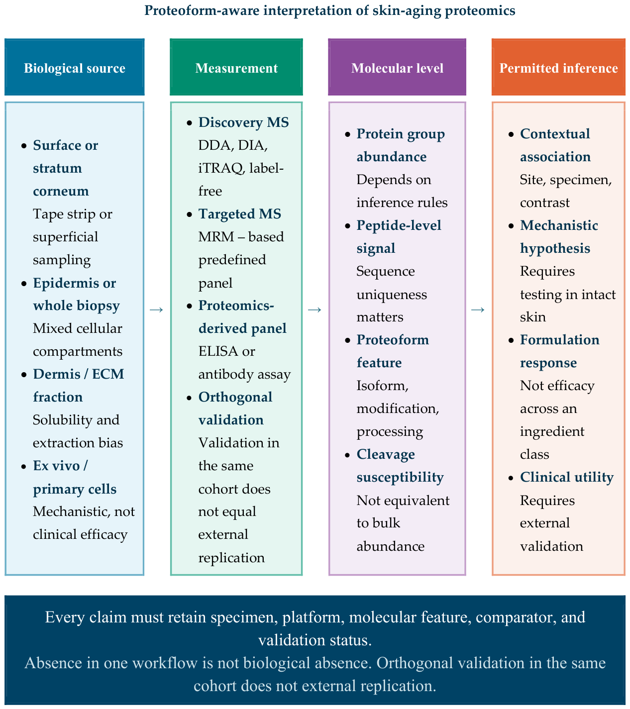
Table 3.
Descriptive summary at the domain level with selected supporting references.
| Module | Recurring signal | Supporting evidence and boundary |
|---|---|---|
| Dermal extracellular matrix and architecture | Loss, remodeling, insolubility, structural modification, or altered cleavage of collagens, proteoglycans, basement membrane, elastic fiber, and crosslinking components | Selected independent direct human and primary cell evidence [1,2,6,11,12,13,16,21,39,47]. Supports a matrix module, not a universal collagen biomarker. |
| Epidermal differentiation and barrier | Changes in keratins, desmosomal/cornification proteins, protease control, and proteins related to hydration | Selected direct human evidence [3,4,5,6,7,8,9,10,28,29,30,38], plus adjacent targeted panel evidence [20]. Site, depth, season, vehicle, and regimen remain influential. |
| Oxidative stress and proteostasis | Redox enzymes, oxidized proteins, chaperones, and metabolic stress responses vary with age and exposure | Selected independent direct human and primary cell evidence [2,5,7,14,22,23,24,28,39,45]. Report [45] reanalyzes [14] and does not add an independent unit. Strong biological coherence but strongly dependent on exposure and model. |
| Inflammaging and immune signaling | Immune, interferon, stress, and protease modules accompany older or photoexposed skin and secretion associated with senescence | Selected independent direct human and primary cell evidence [2,5,8,14,16,17,22,23,24,44]. Report [44] reanalyzes [14] and does not add an independent unit. Cytokines of low abundance are inconsistently captured. |
| Cellular senescence and cytoskeleton | Cytoskeletal, mitotic, lysosomal, and secretory programs vary with donor age or induced senescence | Selected independent direct human and primary cell evidence [8,13,14,15,16,17,18,19,21,22,32,44,45]. Reports [44,45] are linked to [14]. Mechanistic evidence exceeds direct tissue confirmation; induced senescence is not chronological aging. |
| Structural change sensitive to proteoforms | Regional peptide yield, processing, and cleavage susceptibility can change without a difference in bulk abundance | Two independent examples from direct human evidence [11,12]; intact proteoforms were not resolved. |
Convergence was stronger for biological subsystems than for individual proteins.
Table 5.
Selected direct in vivo human topical intervention arms with proteomic endpoints.
| Intervention and design | Proteomic endpoint | Main observation | Permitted inference |
|---|---|---|---|
| Quinoa bioester formulation; 61 women aged 20–80 (one participant at each year of age); paired, nonrandomized study with participant blinding and systematic balanced contralateral forearm allocation; 30 days; vehicle comparator | Untargeted LC–MS/MS of superficial samples collected by a noninvasive procedure similar to microdermabrasion; exploratory proteomic age model within the cohort; PXD062216 | Remodeling of barrier, antioxidant, and protease pathways. Score lower than vehicle in participants ≥50 (p < 0.01) but not <50 (p = 0.14); no direct interaction test; hydration difference not significant (p = 0.17) [28] | Molecular response specific to the formulation. The model was trained on pretreatment samples from the same participants and was not an externally validated clock; no claim of rejuvenation or efficacy across the ingredient class |
| Retinol 0.3% cream; 15 of 30 women aged 50–65; every other day in month 1 then daily to month 12; contralateral control product | Stratum corneum iTRAQ proteomics | Early remodeling that continued over time preceded or accompanied later morphological change [29] | Supports this concentration, vehicle, and regimen only |
| All-trans-retinoic acid 0.025% cream; 15 participants from the same trial of 30 participants; same regimen; contralateral control; separate active arm | Stratum corneum iTRAQ proteomics | Coordinated longitudinal remodeling of functions involved in biosynthesis and aging [29] | Molecular response shown; medical, regulatory, and tolerability context remains distinct |
| 20% glycerol moisturizer; 31 women aged 55–75; randomized design, with double blinding and treatment split across the face; twice daily; proteomics at day 56; vehicle comparator | Targeted MRM panel of 54 stratum corneum proteins | KLK5 and LGALS7 had significantly higher abundance in univariate analysis; KLK7 and PARK7 clustered with the active treatment profile in exploratory multivariate analysis. Both formulas improved wrinkle scores, with no incremental benefit in wrinkle score for active treatment over vehicle; other instrumental differences were observed [30] | Molecular and instrumental effects specific to the formulation; no demonstrated causal mediation and no additional efficacy for wrinkle score over vehicle |
| Lotion plus fine fiber film versus lotion alone; comparison within participants at the lower leg; 8 healthy women aged 38–54 selected for dryness score ≥2.5; daily for 14 days | Stratum corneum samples collected by tape strips; DDA nanoLC–Orbitrap proteomics; 172 proteins identified and 163 analyzed after exclusion of 9 proteins with large baseline differences between sides | Greater improvement in dryness scores and conductance with the combined regimen; the abundance of suprabasin and retroviral-like aspartic protease 1 increased, whereas annexin A2 abundance decreased relative to lotion alone [38] | Molecular and instrumental response over the short term, specific to the formulation and regimen; small selected subgroup, baseline filtering, no external cohort, and company employment limits generalizability |
The findings support molecular responses specific to the tested formulation, not general clinical efficacy.
Table 6.
Evidence ladder developed in this study and informed by proteomics for interpretive use.
| Tier | Evidence | What it can support | What it cannot support |
|---|---|---|---|
| 1 | In vivo human intervention specific to a formulation with comparator and skin proteomic endpoint | A molecular response specific to the formulation; stronger when the molecular endpoint and analysis are prespecified and linked prospectively to validated clinical outcomes | Universal ingredient efficacy, causal clinical mediation or individualized product matching without replication |
| 2 | Human ex vivo skin | Plausibility of pathways at tissue level and local response in intact human tissue | In vivo human efficacy, tolerability, adherence, durability or systemic context |
| 3 | Primary adult human skin cells or matrix derived from cells | Mechanistic direction, candidate pathways and hypotheses on responses to exposure | Topical delivery, benefit to whole skin or clinical recommendation at product level |
The tiers indicate translational proximity, not methodological quality or product performance.
Disclaimer/Publisher’s Note: The statements, opinions and data contained in all publications are solely those of the individual author(s) and contributor(s) and not of MDPI and/or the editor(s). MDPI and/or the editor(s) disclaim responsibility for any injury to people or property resulting from any ideas, methods, instructions or products referred to in the content. |
© 2026 by the authors. Licensee MDPI, Basel, Switzerland. This article is an open access article distributed under the terms and conditions of the Creative Commons Attribution (CC BY) license (http://creativecommons.org/licenses/by/4.0/).
Copyright: This open access article is published under a Creative Commons CC BY 4.0 license, which permit the free download, distribution, and reuse, provided that the author and preprint are cited in any reuse.