

Submitted:
24 September 2026
Posted:
28 September 2026
You are already at the latest version
Abstract
Neonatal hypoxic–ischemic encephalopathy (HIE) remains a major cause of neonatal mortality and long-term neurological disability. Although therapeutic hypothermia (TH) improves outcomes in eligible infants with moderate-to-severe HIE, its efficacy is incomplete, highlighting the need for additional neuroprotective strategies. Endogenous neuroprotec-tive compounds are particularly attractive candidates because they participate in intrinsic cellular defense mechanisms and can modulate components of the hypoxic–ischemic (HI) injury cascade. This review focuses on five endogenous molecules — carnosine, citicoline, kynurenic acid (KYNA), lactoferrin, and N-acetylaspartylglutamate (NAAG) — whose neuroprotective effects have been demonstrated in experimental models of neonatal HI. These molecules act through distinct but partly overlapping mechanisms involving mod-ulation of glutamatergic neurotransmission, antioxidant defense, mitochondrial protection, anti-inflammatory signaling, and inhibition of regulated cell death. Their complementary actions suggest that they may represent components of a broader endogenous neuropro-tective network rather than independent protective mechanisms. However, differences in blood–brain barrier accessibility, pharmacokinetics, therapeutic windows, and the pre-dominantly preclinical nature of the available evidence remain important limitations to translation. Future studies should define optimal dosing, establish safety in the developing brain, and determine whether rational combinations of these compounds, particularly as adjuncts to therapeutic hypothermia, can provide more effective protection against neonatal hypoxic–ischemic brain injury.
Keywords:
neonatal hypoxic–ischemic encephalopathy
; endogenous neuroprotection
; carnosine
; citicoline
; kynurenic acid
; lactoferrin
; NAAG
; therapeutic hypothermia
; oxidative stress
; neuroinflammation
1. Introduction
Over the past several decades, advances in biomedical science have significantly improved the prevention, diagnosis, and treatment of numerous diseases. These achievements have been driven largely by progress in genomics, the development of mRNA vaccines, artificial intelligence (AI)-assisted diagnostics, and gene-editing technologies such as CRISPR. AI-based algorithms have further enhanced clinical decision-making by improving diagnostic accuracy and enabling more personalized therapeutic strategies [1,2]. Major medical breakthroughs include effective management of HIV infection, the development of cancer immunotherapies, and the global eradication of smallpox. Despite these remarkable advances, many medical conditions still lack effective therapies capable of ensuring full recovery. One such condition is perinatal hypoxia–ischemia (HI), which can lead to hypoxic–ischemic encephalopathy (HIE), a severe form of neonatal brain injury.
Perinatal HI remains a major contributor to early neonatal mortality and long-term neurological morbidity worldwide, with an estimated incidence of approximately 2–6 cases per 1000 live births. Globally, HI is responsible for nearly 900,000 deaths each year, and approximately 25% of surviving infants develop permanent neurological impairments, including cerebral palsy, epilepsy, or cognitive deficits [3].
Perinatal HI results from an inadequate supply of oxygen and blood flow to the fetal or neonatal brain, which may arise from a range of antenatal, intrapartum, or postnatal events. The resulting reduction in oxygen and metabolic substrate delivery initiates a cascade of intracellular events that can ultimately lead to neuronal and glial injury and death. The pathological process is time-dependent and begins with primary energy failure characterized by ionic imbalance, membrane depolarization, and cellular swelling. These changes are accompanied by excessive release of excitatory neurotransmitters, leading to excitotoxicity. The initial phase develops within minutes after the hypoxic–ischemic insult and can result in rapid cell death due to severe osmotic swelling and membrane rupture (necrosis), while simultaneously triggering further damaging processes [4].
Following the primary phase, a secondary and more prolonged stage occurs, typically lasting from several hours to days. This phase is associated with secondary energy failure, excessive calcium influx due to excitotoxicity, mitochondrial dysfunction, and increased production of reactive oxygen species (ROS) and reactive nitrogen species (RNS). As a consequence, severe oxidative stress develops, leading to activation of degradative enzymes such as proteases, lipases, and endonucleases, which damage cellular membranes, cytoskeletal structures, and DNA.
Mitochondrial dysfunction and the release of cytochrome c activate caspase-dependent apoptotic pathways. In parallel, oxidative stress–induced depletion of glutathione (GSH) and reduced activity of glutathione peroxidase 4 (GPX4) promote the accumulation of iron-dependent lipid peroxides, triggering ferroptosis, another important mechanism of neuronal cell death [5,6]. Moreover, the developing inflammatory response, together with disruption of the blood–brain barrier (BBB), further exacerbates neuronal damage and contributes to progressive tissue injury [7,8].
Currently, therapeutic hypothermia (TH) is the standard neuroprotective treatment for eligible term and near-term neonates with moderate-to-severe HIE. This intervention is often complemented by supportive care and pharmacological approaches aimed at controlling seizures or targeting neuronal excitability, oxidative stress, inflammation, and apoptosis [7,9,10,11]. Many compounds investigated as potential adjunctive neuroprotectants were originally developed for other clinical indications. For example, allopurinol, a xanthine oxidase (XO) inhibitor commonly used to treat hyperuricemia and gout, reduces uric acid production and may limit free-radical formation. Another example is the antiepileptic drug topiramate, which inhibits voltage-gated sodium and calcium channels, enhances γ-aminobutyric acid (GABA)ergic signaling, and inhibits excitatory glutamatergic transmission, thereby reducing neuronal excitability [12].
Additional therapeutic strategies include NMDA receptor antagonists such as magnesium sulfate and memantine. Memantine is widely used in the treatment of moderate to severe Alzheimer’s disease (AD), while magnesium sulfate has diverse clinical applications. Other compounds investigated for their neuroprotective potential include edaravone, an antioxidant drug used in the treatment of amyotrophic lateral sclerosis (ALS) and acute ischemic stroke, as well as phenobarbital, which remains one of the first-line medications for controlling seizures in neonates with HIE undergoing therapeutic hypothermia (for review, see [9,10,11,12,13,14]).
In recent years, increasing attention has been directed toward nutraceuticals—bioactive compounds naturally present in foods or used as dietary supplements—that possess antioxidant, anti-inflammatory, and anti-apoptotic properties [15]. Many of these compounds originate from plants and have been derived from traditional Chinese medicine (for review, see [16]). Among them, polyphenols have been extensively investigated because of their well-documented antioxidant and neuroprotective properties. Numerous polyphenols have been tested in experimental models of HI, with resveratrol, a compound present in grapes, peanuts, and pomegranates, demonstrating particularly promising neuroprotective effects [17].
Other plant-derived compounds, including alkaloids (e.g., capsaicin), diterpenes (e.g., tanshinones), flavonoids, coumarin derivatives, and caffeine, have also been investigated as potential therapeutic agents for HIE. These compounds have been shown to attenuate oxidative stress and reduce neuronal apoptosis in experimental models [18].
Vitamins represent another group of nutraceuticals under investigation. Vitamins D, C, and E, as well as B-complex vitamins such as thiamine and folic acid, have been studied for their potential neuroprotective effects in neonatal HIE. These compounds may exert antioxidant, anti-inflammatory, and anti-ferroptotic actions, thereby counteracting oxidative damage, reducing neuronal loss, and improving neurological outcomes. In many experimental studies, they have been evaluated as adjunctive therapies to therapeutic hypothermia [5,19,20,21,22,23,24,25]. Preclinical evidence suggests that these vitamins may play a particularly important role in preventing secondary brain injury following the initial hypoxic–ischemic insult.
Another promising group of neuroprotective agents includes endogenous molecules involved in biochemical pathways associated with HI-induced neurodegeneration. Some of these compounds, such as erythropoietin and melatonin, have been extensively studied and their neuroprotective properties described in numerous experimental and clinical reports. However, clinical trials evaluating these agents—either alone or in combination with therapeutic hypothermia—have produced inconsistent or controversial results, and further clinical investigation has been recommended in most studies (for review, see [26,27,28,29,30,31,32,33]).
Despite substantial progress, there remains a need to identify endogenous neuroprotective mechanisms that could be therapeutically enhanced to complement current treatment strategies for neonatal HIE.
In this review, we focus on a group of relatively underexplored endogenous compounds involved in biochemical pathways associated with intrinsic neuroprotection against hypoxia–ischemia-induced neurodegeneration. While considerable attention has been given to well-known neuroprotective molecules such as erythropoietin and melatonin, other endogenous metabolites with potential neuroprotective properties remain less extensively discussed. The compounds included in this review were selected based not only on their established endogenous neuroprotective properties but also on the availability of experimental evidence demonstrating their effects in models of neonatal hypoxia–ischemia. We provide a comprehensive overview of these selected molecules and their mechanisms of action, with particular emphasis on their ability to modulate excitotoxicity, oxidative stress, mitochondrial dysfunction, neuroinflammation, and regulated forms of cell death. By integrating the available experimental evidence, this review aims to assess their potential as adjunctive therapeutic strategies for neonatal HIE (Figure 1).
2. Selected Endogenous Neuroprotective Compounds
The endogenous compounds discussed in this review were selected based on evidence of neuroprotective activity in experimental models of neonatal hypoxia–ischemia and their ability to modulate major mechanisms contributing to HI-induced brain injury. The five molecules described—carnosine, citicoline, kynurenic acid (KYNA), lactoferrin, and N-acetylaspartylglutamate (NAAG)—differ considerably in their physiological functions and molecular targets. Nevertheless, their neuroprotective actions converge on several interconnected components of the HI injury cascade, including excitotoxicity, oxidative stress, mitochondrial dysfunction, neuroinflammation, and regulated cell death. The following sections summarize their physiological roles, mechanisms of neuroprotection, experimental evidence in neonatal HI, and potential therapeutic relevance.
2.1. Carnosine
Carnosine (β-alanyl-L-histidine) is an endogenous dipeptide composed of β-alanine and L-histidine. It has been known for more than a century and was the first peptide isolated from muscle tissue [34]. Together with other carnosine-related dipeptides, such as anserine (β-alanyl-N3-methyl-L-histidine) and homocarnosine (g-aminobutyryl-L-histidine), carnosine is widely distributed in cardiac and skeletal muscles, where its concentration may reach up to 20 mM, and is also present in relatively high concentrations in the olfactory epithelium, ranging from 0.7 to 2.0 mM, with about 10-fold lower amounts in other parts of the brain, mainly glial cells [35,36,37,38,39]. Homocarnosine is present in the brain at much higher concentrations than carnosine, reaching up to 1 mM [40].
The non-proteinogenic b-amino acid β-alanine (3-aminopropanoic acid), a constituent of carnosine, is synthesized in the liver primarily through thymine degradation, whereas L-histidine, a proteinogenic essential amino acid, cannot be synthesized de novo in humans and must therefore be obtained through diet [41,42]. Although a portion of circulating carnosine can cross the blood–brain barrier, is rapidly hydrolyzed by serum carnosinase (CN1), which limits its bioavailability after systemic administration [43]. Significant local synthesis occurs within glial cells, particularly oligodendrocytes, through the action of the ATP-dependent enzyme carnosine synthase 1 (CARNS1), which catalyzes the formation of carnosine from β-alanine and L-histidine. This synthesis is especially prominent in the olfactory bulbs and cerebral cortex [44,45].
Neurons are unable to synthesize carnosine and instead take up the dipeptide released from oligodendrocytes. Its release is mediated by glutamate receptors and depends on elevated intracellular Ca²⁺ concentrations, suggesting a Ca²⁺-dependent vesicular release mechanism characteristic for glial exocytosis [46,47]. Neurons and astrocytes subsequently take up carnosine from the extracellular space via the energy-dependent transporter PEPT2, a saturable high-affinity transporter with broad specificity for dipeptides [41,48].
Carnosine exhibits numerous biological activities that have been observed not only in tissues where it is highly concentrated, such as muscle and brain, but also in other organs (for review, see [49]). In the central nervous system, carnosine has been shown to act as a neuromodulator involved in neuron-glia communication and may regulate the effects of copper and zinc on amino acid receptors and synaptic transmission [46,50,51,52,53]. In addition, carnosine reduces glutamate levels through upregulation of glutamate transporter 1 (GLT-1), thereby preventing excitotoxicity [54].
Carnosine also possesses anti-inflammatory properties. These effects are associated with reduced astrocyte activation, decreased release of interferon-γ (IFN-γ) [55], and downregulated expression of pro-inflammatory cytokines, including interleukin (IL)-6, IL-1β, and tumor necrosis factor-α (TNF-α). This dipeptide can simultaneously upregulate anti-inflammatory mediators such as IL-4, IL-10, and transforming growth factor-β1 (TGF-β1), which exert neuroprotective effects in experimental models of neurodegenerative diseases [56,57,58].
One of the key neuroprotective properties of carnosine is its ability to counteract oxidative stress. The imidazole ring of histidine efficiently scavenges reactive oxygen and nitrogen species, thereby reducing intracellular levels of superoxide and hydroxyl radicals and nitric oxide, and also scavenges reactive carbonyl species generated during lipid peroxidation, ultimately preventing cellular damage [59,60,61,62]. Moreover, both in vitro and in vivo studies have demonstrated that, under conditions of oxidative stress, carnosine can normalize the activity of superoxide dismutase (SOD) and GPx, restore GSH levels, reduce malondialdehyde concentrations, and prevent SOD fragmentation [63,64]. Carnosine has also been reported to protect against DNA damage and chromosomal aberrations [65,66].
Carnosine and its derivatives also act as physiological pH buffers. The dipeptide contains three ionizable groups and naturally occurs as a zwitterion, with a pKa of approximately 7 [67,68]. This buffering capacity may be particularly relevant during HI, when increased lactate production contributes to tissue acidosis and disruption of brain pH homeostasis.
Another important protective property of carnosine is its ability to counteract the formation of advanced glycation end-products (AGEs) and advanced lipoxidation end-products (ALEs). Carnosine inhibits AGE formation by preventing reactions between reactive carbonyl compounds and proteins and may also reverse preformed glycated products through transglycation [69,70,71,72,73,74,75,76,77,78,79]. In addition, it scavenges reactive α,β-unsaturated aldehydes, including 4-hydroxynonenal (HNE) and acrolein, thereby limiting ALE formation [37,80,81,82,83,84,85,86,87,88]. Carnosine may further protect cellular proteins by preventing their aggregation and promoting the degradation of damaged proteins [89,90,91,92,93,94] (for review, see [95,96]). These properties may be particularly relevant to neuroprotection, as the accumulation of AGEs, ALEs, and protein aggregates contributes to cellular dysfunction associated with aging and neurodegenerative disorders, including AD.
The beneficial effects of carnosine supplementation have been extensively investigated in experimental models of neurodegenerative disorders, including AD, Parkinson’s disease (PD), and cerebral ischemia (for review, see [97,98]). However, relatively few studies have examined the neuroprotective effects of carnosine in perinatal HI-induced brain injury.
In an experimental HI model performed in 7-day-old rats, intraperitoneal administration of carnosine (250 mg/kg) 30 minutes before HI induction exerted significant neuroprotective effects [99]. Both morphological brain injury and biochemical markers of apoptosis and oxidative stress were significantly reduced. Carnosine downregulated the mRNA expression of apoptosis-inducing factor (AIF) and caspase-3, and this was accompanied by increased SOD activity and decreased malondialdehyde levels [99].
Similar protective effects were observed when carnosine was administered at the same dose immediately after HI and again at 24 and 48 hours following the insult [100]. Furthermore, both pre- and post-treatment with carnosine improved spatial learning and memory in HI rats, functions that are typically impaired following HI.
Byun et al. (2021) investigated the combined effects of carnosine and therapeutic hypothermia in an experimental model of HI-induced brain injury [101]. Although, in this experiment, treatment with either carnosine or hypothermia alone did not produce significant neuroprotective effects, their combined administration markedly reduced brain injury. This was evidenced by decreased infarct and atrophy volumes, reduced apoptosis and gliosis, inhibition of lipid peroxidation, and decreased activity of matrix metalloproteinases. Since matrix metalloproteinases contribute to extracellular matrix degradation, BBB disruption, edema formation, and neuronal death following HI, their inhibition may represent an important mechanism underlying the protective effects of combined therapy.
2.2. Citicoline (CDP-Choline)
Citicoline is the International Nonproprietary Name (INN) for cytidine-5′-diphosphocholine (CDP-choline), originally identified by Eugene Kennedy in 1956 [102]. CDP-choline is a naturally occurring endogenous nucleotide derivative comprising cytidine and choline moieties, which serves as an intermediate in the so-called Kennedy pathway, the major biosynthetic pathway for phosphatidylcholine, the principal and essential phospholipid component of cellular membranes [103,104]. Choline itself serves as a substrate in the synthesis of acetylcholine, one of the main neurotransmitters [105,106].
Dietary choline is obtained from foods such as eggs, meat, and legumes, whereas cytidine and uridine are derived primarily from RNA in the diet. When citicoline is ingested as a dietary supplement, it is hydrolyzed in the intestine into choline and cytidine (in rats) or uridine (in humans). These metabolites enter the bloodstream and subsequently cross the BBB separately (Figure 2). The transport of choline across the BBB is mediated primarily by the major facilitator superfamily transporters FLVCR1 and FLVCR2 [107], whereas high-affinity uptake into cholinergic neurons is mainly mediated by the Na+-dependent hemicholinium-3-sensitive choline transporter 1 (CHT1) [108]; glial choline uptake involves other transporters, including choline transporter-like protein 1 (CTL1) [109]. The transport of circulating cytidine and uridine across the BBB, followed by their uptake from extracellular fluid into brain cells, is mediated by members of the equilibrative and concentrative nucleoside transporter families [110]. The transfer of these compounds into the brain extracellular space and subsequently into neurons and glia is essential for CDP-choline resynthesis via CTP-phosphocholine cytidylyltransferase (CCT) [111].
Citicoline has a long history of clinical use and investigation as a potential neuroprotective agent. Its generally favorable safety profile, supported by both in vitro and in vivo studies, makes it an attractive therapeutic candidate [112,113,114]. Its neuroprotective effects involve a broad range of cellular processes implicated in neurodegeneration.
Anti-inflammatory effects have been demonstrated in various disease models, including neurodegenerative disorders such as PD, AD, and stroke. Citicoline administration reduced levels of TNF-α, IL-1β, iNOS, NF-κB, JAK2, and STAT3 [115,116]. Citicoline also decreases phospholipase A2 activity, thereby reducing expression of cyclooxygenase-2 (COX-2), an enzyme involved in inflammatory signaling [117]. Furthermore, citicoline has been shown to regulate the expression and activity of MMP-9 and to increase expression of MMP-2 and TGF-β1 in animal models of myopia and static injury [118,119,120]. However, evidence confirming neuroprotective effects mediated specifically through COX-2 or matrix metalloproteinase pathways in the brain remains limited. Strong anti-apoptotic and antioxidant properties of citicoline have been demonstrated in numerous in vitro and in vivo studies. In in vitro models of excitotoxicity , citicoline reduced apoptotic damage in neuronal cell cultures and decreased levels of active caspase-3 and the apoptosis-associated 89-kDa cleaved PARP fragment [112,121,122].
In human astrocytes exposed to oxidative stress, citicoline reduced ROS and nitric oxide (NO) production. The maintenance of basal cytochrome c levels, a key regulator of energy metabolism and apoptosis, suggests that citicoline preserves mitochondrial integrity. Acting as an effective antioxidant, citicoline also decreased p53 levels and increased expression of sirtuin 1 (SIRT1), a critical regulator of antioxidant enzyme expression [123]. In a lipopolysaccharide (LPS)-induced model of acute kidney injury, administration of CDP-choline or choline reduced elevated ROS levels. Moreover, LPS-induced increases in the expression of biomarkers of cellular damage, including KIM-1, TNF-α, NOX4, p22phox, and NF-κB, were significantly attenuated [124].
Experimental studies investigating the neuroprotective effects of citicoline in animal models further support its antioxidant properties observed in vitro. In a mouse model of hepatic encephalopathy, citicoline administration significantly alleviated oxidative stress in brain tissue by reducing lipid peroxidation and ROS formation and restoring total antioxidant capacity. Citicoline supplementation also improved locomotor activity [125]. The antioxidant and mitochondria-protective effects of citicoline were also examined in a rat model of head irradiation injury. Citicoline administration following irradiation reduced oxidative stress, enhanced the activity of mitochondrial complexes I and II, increased ATP production, and decreased caspase-3 levels compared to irradiated controls [126].
Studies in animal models of adult brain ischemia demonstrated that administration of citicoline shortly before or after ischemia increased GSH levels and glutathione reductase activity, while preventing mitochondrial damage and cytochrome c release, thereby confirming its anti-apoptotic and antioxidant properties [127,128].
An important clinical analysis was conducted in peripheral blood samples from ischemic stroke patients receiving high dose citicoline supplementation. The study demonstrated that citicoline significantly reduced levels of lipid peroxidation markers, including arachidonic acid and total 8-iso-prostaglandin F2α (8-iso-PGF2α). Upregulation of SIRT1 was also observed [129].
The demonstrated neuroprotective, anti-apoptotic, and antioxidant properties of citicoline have prompted investigation into its use for the treatment of neonatal hypoxia–ischemia. Although several publications report beneficial effects of citicoline, the available evidence is currently derived more from randomized clinical trials than from experimental animal studies. Fiedorowicz and colleagues (2008) demonstrated that intraperitoneal administration of citicoline within 5 minutes after hypoxia–ischemia in 7-day-old rats, followed by daily injections for 7 days (100 or 300 mg/kg), markedly and dose-dependently reduced brain damage. This effect was associated with reduced activation of pro-apoptotic caspase-3 and altered expression of heat shock protein Hsp70 [130]. Similarly, neonatal rabbits subjected to hypoxic–ischemic brain injury and treated with citicoline exhibited significantly reduced brain edema and tissue damage compared with untreated controls. Reduced neuronal degeneration was also observed [131].
Randomized clinical trials have reported a significant reduction in post-discharge seizures in citicoline-treated neonates compared with controls. In addition, seizure duration was shorter, and neurodevelopmental outcomes at 9 and 12 months showed significant improvement in the citicoline-treated group [132]. Comparable findings were reported in a small pilot study by Khushdil et al. (2021) [133].
Interesting but difficult-to-interpret findings were reported by Liang et al. (2019), who compared the neuroprotective effects of monosialogangliosides and citicoline in newborns with HIE [134]. Following treatment, reductions in apoptosis and oxidative stress markers in serum and neurological dysfunction indices were observed. However, because the citicoline-treated group served as the control group, conclusions regarding the specific neuroprotective effects of citicoline remain speculative [134]. Although citicoline has been studied for a relatively long time as a potential therapeutic agent for stroke and HIE, some clinical studies have reported beneficial effects [132,133], whereas larger clinical trials and meta-analyses have not demonstrated a clear overall benefit [105,135,136,137]. These discrepancies underscore the need for further, more detailed investigation taking into account heterogeneous stroke populations, variable timing and dosing, modest effect sizes, and the confounding effects of thrombolysis or thrombectomy.
2.3. Kynurenic Acid
Kynurenic acid (KYNA) was first identified in dog urine by Justus von Liebig in 1853 [138]. Fifty years later, Ellinger recognized it as one of the first metabolites of tryptophan, again using dog urine as the source [139]. Tryptophan is an essential amino acid which cannot be synthesized by the human body and must therefore be obtained from dietary sources such as meats, eggs, and dairy products.
After crossing the BBB via the L-type amino acid transporter 1 (LAT1), tryptophan serves as a precursor for several neuroactive compounds, including serotonin and melatonin. The main catabolism route of tryptophan proceeds through the kynurenine pathway (KP) [140]. Although the final product of this pathway is nicotinamide adenine dinucleotide (NAD+), several neuroactive intermediates are generated, including the neurotoxic metabolites 3-hydroxykynurenine (3-HK) and quinolinic acid (QUIN). In a competing branch of the kynurenine pathway, kynurenine aminotransferases (KATs) convert L-kynurenine into KYNA, a metabolite generally regarded as neuroprotective, counteracting the detrimental effects of QUIN [141,142].
Because KYNA poorly crosses the BBB, brain KYNA is derived primarily from local synthesis using circulating precursors, namely tryptophan and L-kynurenine, that readily enter the brain [140] (Figure 2). Among the KAT isoforms, KAT II appears to play the predominant role in the human brain. It is mainly localized in astrocytes, where newly synthesized KYNA is rapidly released into the extracellular space, enabling its neuromodulatory and neuroprotective actions [143,144]. Nevertheless, KAT expression has also been demonstrated in neurons and microglia, depending on the brain region and enzyme isoform involved [145].
KYNA acts on both intracellular and extracellular targets. In the extracellular space, it modulates several membrane receptors, most notably ionotropic glutamate receptors and the G protein-coupled receptor 35 (GPR35). It was originally identified as an inhibitor of NMDA-sensitive glutamate receptors, acting primarily as a competitive antagonist at the strychnine-insensitive glycine-B co-agonist site of the GluN1 subunit [146,147]. KYNA has also been suggested to interact with the glutamate recognition site on the GluN2 subunit, although this remains less well established. More recent studies indicate that KYNA exerts concentration-dependent effects: concentrations slightly above physiological levels may facilitate neurotransmission, whereas inhibitory actions become apparent at higher, micromolar concentrations [148]. Similarly, the effects of KYNA on AMPA receptors are biphasic. Lower concentrations, ranging from nanomolar to micromolar levels, facilitate AMPA receptor responses, whereas high, millimolar concentrations competitively inhibit receptors through interactions with the glutamate-binding domain [149]. KYNA also interacts directly with kainate receptors, although its potency at these receptors is comparatively weaker [150].
The ability of KYNA to modulate ionotropic glutamate receptors contributes substantially to its neuroprotective profile. By attenuating glutamate receptor-mediated excitotoxicity, particularly through inhibition of NMDA receptors, KYNA may interrupt pathological cascades and limit neuronal injury [151].
Importantly, KYNA itself has direct antioxidant properties, acting as an electron donor and efficiently scavenging free radicals such as hydroxyl radicals (HO▪), superoxide anion (O2▪-) and peroxynitrite (ONOO-) produced in ischemic conditions [152,153]. These actions were confirmed in in vitro experiments, where KYNA reduced ROS production and lipid peroxidation induced by prooxidants in the rat brain homogenates. Notably, these antioxidant properties appear to be independent of KYNA-mediated modulation of NMDA and cholinergic receptors. The metal-chelating properties of KYNA were also confirmed [153]. Thus, KYNA may protect neural tissue through complementary mechanisms, combining attenuation of glutamate-mediated excitotoxicity with direct scavenging of free radicals and modulation of oxidative stress pathways.
As mentioned above, KYNA can act as an endogenous agonist of GPR35, an emerging central nervous system target expressed in neurons and glial cells [154]. Activation of GPR35 has been implicated in the regulation of neuronal excitability and neurotransmitter release, partly through inhibition of N-type calcium channels [155]. In addition, low concentrations of KYNA may interact with presynaptic α7 nicotinic acetylcholine receptors, which exhibit high calcium permeability, thereby reducing glutamate release and limiting excitotoxic damage [156].
Emerging evidence suggests a role for KYNA in mitochondrial protection. Through activation of GPR35 and the aryl hydrocarbon receptor (AhR), both of which have been suggested to localize to mitochondrial membranes, KYNA may protect mitochondria against hypoxic and ischemic damage [157]. Moreover, the mitochondrial localization of kynurenine aminotransferases raises the possibility that KYNA is present within mitochondria, where it could contribute to the regulation of mitochondrial redox homeostasis in neurodegenerative conditions [158].
KYNA also appears to regulate neuroinflammatory pathways through GPR35 signaling. Interaction between KYNA and GPR35 has been associated with suppression of NLRP3 inflammasome activation and reduced release of pro-inflammatory cytokines such as IL-1β [159,160]. KYNA has also been shown to increase the expression of peroxisome proliferator-activated receptor delta (PPARδ), which may contribute to attenuation of inflammatory responses in cerebral ischemia [161,162].
Glutamate-mediated excitotoxicity, oxidative stress, and neuroinflammation are key mechanisms underlying the development of numerous neurodegenerative disorders. Consequently, the balance between neurotoxic kynurenine metabolites, such as 3-HK and QUIN, and the neuroprotective metabolite KYNA profoundly influences neuronal survival and function. Dysregulation of the KP has been implicated in the pathogenesis of AD, PD, Huntington’s disease (HD), schizophrenia, and depression. Accordingly, the neuroprotective properties of KYNA have stimulated considerable interest in its therapeutic potential, either alone or in combination with other pharmacological interventions (for review, see [163,164]).
Experimental evidence from cerebral ischemia supports the neuroprotective role of KYNA. KYNA-containing astrocytes were detected exclusively in the ipsilateral cerebral cortex and/or striatum, at 2, 5, and 21 days following induction of experimental stroke. Moreover, elevated KYNA levels were predominantly observed within infarcted regions, suggesting that endogenous upregulation of this metabolite may represent an intrinsic protective response to ischemic injury [165]. The neuroprotective action of KYNA in cerebral ischemia is further supported by numerous in vitro and in vivo studies demonstrating that even moderate increases in brain KYNA levels, achieved either by systemic administration of high doses of KYNA or through manipulation of the kynurenine pathway, can attenuate ischemic brain damage [166,167,168] (for review, see [169,170]).
Although the protective effects of KYNA have been extensively characterized in experimental stroke models, considerably less attention has been devoted to neonatal HI. The first evidence of KYNA-mediated neuroprotection in neonatal HI was provided by Simon and colleagues (1986) in 7-day-old rats [171]. Intraperitoneal administration of KYNA between carotid artery occlusion and subsequent exposure to hypoxic conditions significantly reduced brain edema, an effect attributed to suppression of excitatory neurotransmission.
Subsequently, Nozaki and Beal (1992) showed that L-kynurenine, the precursor of KYNA, administered using the same protocol as in the study by Simon and colleagues (1986), resulted in a significant dose-dependent reduction in infarct rate and infarct volume in the ipsilateral hemisphere [172]. The treatment also reduced the expression of c-fos, a gene induced immediately by cerebral ischemia. Importantly, L-kynurenine administration significantly increased KYNA concentration in the cerebral cortex of 7-day-old rats, leading the authors to conclude that the observed neuroprotection was mediated by L-kynurenine conversion to KYNA.
Consistent with these findings, administration of KYNA immediately after HI reduced edema formation in the injured hemisphere [173]. More recently, Bratek-Gerej and colleagues (2021) demonstrated that intraperitoneal administration of KYNA at a high dose (300 mg/kg), given either 1 or 6 h after HI, reduced ischemic hemisphere damage and prevented neuronal loss in the hippocampus and cortex [174]. In addition to its structural neuroprotective effects, KYNA significantly attenuated HI-induced ROS generation while increasing GSH levels and enhancing the activity of antioxidant enzymes, including GPx), SOD, and catalase (CAT), compared with untreated animals [174]. These findings suggest that antioxidant mechanisms contribute substantially to KYNA-mediated protection following neonatal HI. However, the available data do not allow determination of whether attenuation of oxidative stress results primarily from receptor-mediated inhibition of excitotoxicity or from the direct free radical scavenging properties of KYNA. The relative contribution of NMDA receptor blockade, GPR35 activation, or direct antioxidant effects to KYNA-mediated neuroprotection in neonatal HI remains unresolved.
Despite increasing evidence supporting its beneficial actions, the physiological and pathological roles of KYNA in the brain remain incompletely understood. Nevertheless, the available data highlight its therapeutic potential in disorders characterized by excitotoxicity, oxidative stress, and neuroinflammation, including neonatal HI and HIE. Importantly, no disease has thus far been directly associated with overexpression of KATs, the enzymes responsible for the synthesis of KYNA [175,176], suggesting that enhancement of endogenous production of KYNA may represent a relatively safe therapeutic approach.
The clinical translation of KYNA as a neuroprotective agent is limited by its poor permeability across the BBB. However, KYNA possesses several advantages over its precursor, L-kynurenine, which is frequently used experimentally to elevate levels of KYNA in the brain. Unlike L-kynurenine, KYNA cannot be further metabolized into potentially neurotoxic compounds such as 3-HK and QUIN. Consequently, considerable efforts have been focused on developing strategies to improve KYNA delivery to the brain. Among these, intranasal administration of KYNA-loaded niosomal formulations has emerged as a particularly promising approach [177].
Despite the demonstrated beneficial effects of KYNA in cerebral ischemia, several challenges remain, mainly that KYNA poorly crosses the BBB and is rapidly cleared and metabolized, and that chronic elevation of KYNA may impair cognitive function through blockade of α7 nicotinic receptors [178,179]. Notably, BBB disruption may accompany HI and other ischemic brain injuries and may facilitate passive diffusion of circulating KYNA into the brain parenchyma [180] (Figure 2). This phenomenon could partly explain the neuroprotective effects observed following systemic administration of KYNA shortly before or after HI. Therefore, although challenges related to drug delivery remain, the multifaceted actions of KYNA—including modulation of excitotoxicity, antioxidant activity, mitochondrial protection, and regulation of neuroinflammation—support its continued evaluation as a potential therapeutic strategy for ischemic brain injury in both neonatal and adult populations.
2.4. Lactoferrin
Lactoferrin (Lf) is an approximately 80 kDa iron-binding glycoprotein discovered in 1939 in bovine milk [181] and isolated from human milk in the 1960s. Its iron-binding ability led to its classification as a member of the transferrin protein family (for review, see [182,183,184]).
Lf is abundant in mucosal secretions such as milk, saliva, and tears and is also present in the respiratory, urinary, genital, and gastrointestinal tracts as well as in blood, where it contributes to innate immune defense [184]. Lf synthesis and secretion are regulated by multiple tissue-dependent factors (for review, see [185]). In the healthy central nervous system, baseline Lf levels are generally low but increase during normal aging and under conditions of cellular stress associated with neurodegenerative disease. Lf can be synthesized by activated microglia and by leukocytes infiltrating the brain [186,187,188,189]. Circulating Lf can also enter the CNS through receptor-mediated transcytosis across brain capillary endothelial cells, involving receptors such as LRP1 [187,190] (Figure 2). Lf receptors have additionally been detected on neurons, particularly dopaminergic neurons in the substantia nigra, and on glial cells [191].
A major biological property of Lf is its ability to bind and sequester ferric iron (Fe3+), thereby contributing to iron homeostasis. Depending on iron saturation, Lf exists in iron-depleted (apo-Lf) or iron-bound (holo-Lf) forms [182]. Although iron sequestration is an important determinant of its biological activity, accumulating evidence indicates that Lf exerts pleiotropic neuroprotective effects through additional anti-inflammatory, antioxidant, mitochondrial, and cell-survival mechanisms.
Lf also exhibits well-documented anti-inflammatory effects in various organs, including the brain. It modulates inflammatory responses by interacting with key signaling pathways and immune cells and by suppressing the production of pro-inflammatory mediators. Lf has been shown to reduce TNF-α, IL-1β, and IL-6 while increasing anti-inflammatory cytokines such as IL-4 and IL-10, in part through modulation of NF-κB signaling (for review, see [192,193]).
In an in vitro study using astrocyte cultures Lf attenuated cell reactivity and the expression of glial fibrillary acidic protein (GFAP) and pro-inflammatory IL-6, while upregulating the expression of anti-inflammatory IL-10 and nuclear factor erythroid 2-related factor 2 (Nrf2), a transcription factor that protects cells from oxidative stress. Moreover, it was shown that Lf can upregulate the expression of SOX2, a key transcription factor involved in cellular reprogramming and neurogenesis [194]. Reduced levels of pro-inflammatory cytokines in the hippocampus and serum were observed after intragastric administration of Lf to aged mice [195].
Lf can also attenuate oxidative stress through multiple mechanisms. By binding free iron and maintaining it in a less reactive state, Lf may limit hydroxyl-radical generation through Fenton chemistry and reduce iron-dependent lipid peroxidation associated with ferroptosis. Lf has also been reported to preserve GPX4 expression or stability [196,197,198]. In addition, Lf can scavenge ROS, enhance endogenous antioxidant defenses involving SOD, CAT, and GPx, and support mitochondrial quality control, including mitophagy [199,200,201,202] (for review, see [203]).
Increased Lf expression was found in post-mortem brain tissue of AD and PD patients, most likely related to the synthesis of Lf by activated microglia and leukocytes infiltrating the brain. This increase was suggested as a defense mechanism of the brain in response to pathologies associated with the progression of these diseases. Indeed, a number of studies have shown that in AD and PD suppression of oxidative stress, inflammation, and apoptosis exerted by Lf contributes to neuroprotection (for review, see [204]). Moreover, it was demonstrated that Lf can attenuate the the pathogenic effects of apolipoprotein E4 (APOE4), the strongest known genetic risk factor for late-onset AD, and promote non-amyloidogenic pathway [205,206,207]; although the latter is still controversial [208]. Interestingly, Wang and colleagues showed that Lf exerted neuroprotective effects in both iron-free (apo-Lf) and iron-saturated (holo-Lf) forms [209].
These neuroprotective properties of Lf have made it a promising therapeutic molecule, and it has been tested in many experimental models of AD and PD, yielding encouraging results [204,210,211]. Strong neuroprotective effects of Lf have also been observed in experimental models of brain ischemia. In vitro and in vivo experiments demonstrated that Lf application significantly reduced the pro-inflammatory response and protected against oxidative stress and ferroptosis [198,212,213]. Moreover, Lf promoted macrophage-mediated clearance of dead cells and cellular debris, accelerating hematoma resolution after hemorrhagic stroke [214,215]. It was suggested that in ischemic brains Lf can activate the Kelch-like ECH-associated protein 1 (Keap1)/Nrf2 signaling pathway, which mediates the expression of many genes encoding cytoprotective, antioxidant, and anti-inflammatory proteins [216].
Only a few research teams have investigated Lf as a potential neuroprotective treatment in neonatal HI. Preclinical studies suggest that its anti-inflammatory and antioxidant properties may mitigate injury in the immature brain. In experimental HI models using 3- and 7-day-old rat pups, Lf was delivered through milk from dams whose diets were supplemented with Lf before and/or after parturition [217,218,219]. Significant reductions in brain damage and metabolic disturbances were observed in Lf-supplemented animals. Lf also reduced the expression of inflammatory mediators (IL-6, IL-1β, and TNF-α) and activation of pro-apoptotic caspase-3 after HI [219,220]. Even relatively short exposure to Lf-enriched milk provided measurable protection against HI injury [217,218]. The neuroprotective effect was dose-dependent, and very high levels of dietary supplementation were reported to be potentially detrimental [220]. HI-induced oxidative stress was attenuated through enhanced antioxidant defenses, including increased SOD and GPx activity and elevated GSH levels. Lf supplementation also improved mitochondrial function and increased expression of proteins involved in antioxidant and metabolic regulation, including Nrf2 and uncoupling protein 2 (UCP2), following experimental HI [217].
Combining Lf with therapeutic hypothermia has shown promising complementary neuroprotective effects in rat pups subjected to HI. Detailed analyses suggested that hypothermia primarily limited HI-induced disruption of neuron-astrocyte metabolic coupling, whereas Lf supported astrocytic function by modulating the expression of genes related to glutamatergic excitotoxicity and restoring transcripts involved in metabolic support [218].
The effect of maternal Lf supplementation was also studied in the lipopolysaccharide (LPS) model of infection-induced neuroinflammation applied to rat pups [199]. Lf supplementation reduced LPS-induced ventriculomegaly, brain tissue loss, and microstructural abnormalities, including myelination deficits. Decreased oxidative stress and reduced microglial activation were observed following Lf treatment. While Lf shows promising preclinical neuroprotection in neonatal HI, clinical translation will require careful dose optimization and long-term safety assessment, particularly given the potential for iron-related effects in the developing brain.
Taken together, these studies indicate that Lf can attenuate several processes contributing to HI-induced injury and neuroinflammation in the immature brain. In addition to its direct neuroprotective properties, Lf has been exploited as a brain-targeting ligand because of its capacity for receptor-mediated transcytosis across the BBB. Conjugation or coating of nanoparticles with Lf can therefore facilitate delivery of therapeutic cargo to the CNS [221].
2.5. NAAG
N-acetylaspartylglutamate (NAAG) is a dipeptide first identified at high concentrations in the mammalian nervous system in 1965 [222,223]. At that time peptides were not considered to be neurotransmitters; therefore, NAAG was thought to have a metabolic function, such as serving as an intermediate in the biosynthesis of the transmitter pool of glutamate. More than two decades later publications from Joseph Coyle’s laboratory established the neurotransmitter nature of NAAG [224,225].
Endogenous NAAG is synthesized in neurons from N-acetyl-aspartate (NAA) and glutamate via a peptide bond in an enzyme-mediated process catalyzed by NAAG synthetases (NAAGS) I and II [226]. NAAG is concentrated in neuronal synaptic vesicles particularly in glutamatergic neurons, although it was also found in GABAergic interneurons and in other non-glutamatergic neurons, including noradrenergic, serotonergic, and cholinergic neurons [227,228]. NAAG released from synaptic terminals to the synaptic cleft is rapidly degraded, mainly by the zinc metallopeptidase glutamate carboxypeptidase II (GCPII), which is expressed at high levels on the plasma membrane of astrocytes and at lower levels on microglia [229]. The released glutamate is taken up by glial and neuronal transporters, while NAA is taken up by glial cells (for review, see [226,230]).
Substantial data support the conclusion that NAAG is the most prevalent and widely distributed peptide neurotransmitter in the mammalian nervous system (for review, see [230,231]). It functions as a selective endogenous agonist for the type 3 metabotropic glutamate receptors (mGluR3), although some authors indicate that this effect could be attributed to substantial contamination of NAAG by glutamate [232]. Depending on the concentration, NAAG can exert mixed effects in interaction with NMDA receptors. At low concentrations, NAAG acts as a non-competitive antagonist (partially overcome by glycine) of NMDA receptors, but at high, pathologically relevant concentrations it can act as a weak agonist (for review, see [230,233]). Moreover, the response of NMDA receptors modulated by NAAG is pH- and subunit-dependent. Under ischemic conditions, when pH drops significantly, NAAG enhances synaptic GluN2A-containing NMDA receptors while suppressing extrasynaptic GluN2B-containing receptors [234].
Interestingly, the role of extrasynaptic NMDA receptor activation in the pathogenesis of neurodegenerative disorders, especially AD and HD as well as in ischemia/hypoxia has been documented [235,236,237]. Reduced NAA and NAAG levels have been consistently reported in AD and HD, particularly in the cortex, hippocampus, and striatum, reflecting neuronal dysfunction and loss. In parallel, alterations in GCPII indicate that impaired NAAG metabolism in these disorders is closely correlated with neuronal loss [238,239,240]. Although evidence in PD is less extensive, dysregulation of NAAG signaling has also been implicated in disease pathogenesis. Moreover, inhibition of NAAG peptidase was suggested as a target for experimental therapeutics of PD in the past decade [241,242].
In ischemic stroke, NAAG levels typically spike shortly after the initial insult to combat excitotoxicity, before subsequently declining during the progression of neuronal damage [243]. Therefore, the blockade of extrasynaptic NMDA receptors by NAAG may prevent neuronal death following HI and other neuropathological conditions associated with glutamate toxicity.
NAAG does not readily cross the BBB and is rapidly hydrolyzed by GCPII (Figure 2 and Figure 3). Consequently, GCPII inhibitors such as 2-PMPA and ZJ43 have been used experimentally to increase extracellular NAAG while limiting the generation of glutamate from NAAG hydrolysis. However, the hydrophilic character of many GCPII inhibitors restricts their penetration across the BBB, prompting investigation of alternative delivery strategies [226]. Invasive approaches such as intracerebral microinjection are useful mainly in preclinical research, whereas non-invasive intranasal delivery has been explored as a potential route to bypass the BBB [244,245].
BBB disruption during ischemic and hypoxic-ischemic injury may increase CNS exposure to systemically administered compounds that otherwise penetrate the intact BBB poorly (Figure 2). In a neonatal rat model of HI, intraperitoneally administered NAAG significantly reduced brain damage when given after right common carotid artery occlusion and before, or up to 60 min after, hypoxia [19]. The treatment also attenuated the HI-associated increase in cAMP, consistent with activation of Gi/o-coupled group II metabotropic glutamate receptor signaling [19].
Bratek-Gerej and colleagues (2022) showed that NAAG administered 1 or 6 h after experimental HI significantly reduced not only brain damage but also the expression of pro-apoptotic factors such as Bax and HtrA2/Omi, whereas expression of anti-apoptotic Bcl-2 was significantly increased compared to untreated HI animals [246]. Post-HI application of NAAG also reduced the activity of antioxidant enzymes, which was accompanied by a decrease in ROS levels, indicating suppression of oxidative stress development [247].
Application of NAAG 24 or 1 h before HI, to induce preconditioning, also significantly reduced brain damage, inhibited apoptosis and decreased oxidative stress [248,249]. Considering the fast metabolism of NAAG, the authors excluded the involvement of mGluR3 in the neuroprotective effect of NAAG administered 24 h before HI. The observed effect may be explained by the activation of NMDA receptors and induction of antioxidant defense system by NAAG pretreatment. The observation that memantine inhibited the NAAG effect supports this conclusion. Preconditioning 1 h before HI with NAAG results in a mixture of mGluR3 and NMDA receptor responses [248]. However, this theory requires further investigation.
Moreover, the studies concerning the potential beneficial effect of NAAG on recovery after cerebral ischemia, particularly neonatal HIE, should consider potential adverse effects of mGluR3 activation. Group II mGluR agonists (e.g., DCG-IV) inhibited LTP induction in hippocampal slices, showing that activation of these receptors can suppress synaptic plasticity, and impaired spatial learning in the Morris water maze, indicating that sustained group II mGluR activation can disrupt cognition. [250,251,252]. In addition, mGluR3 activation increases astrocytic glutamate transporter expression and activity, consequently chronic activation could lead to maladaptive changes in glutamate homeostasis [253,254]. The interplay between NAAG and glutamatergic neurotransmission, neuroplasticity, cognitive abilities, and other neural cells is very complex and requires further thorough investigation [255].
Given its dual action on mGluR3 and NMDA receptors, as well as its pH-dependent modulation of synaptic versus extrasynaptic NMDA receptor signaling, NAAG represents a uniquely positioned endogenous candidate for targeted neuroprotection in the immature brain; however, careful balancing of its receptor-mediated effects will be essential for future therapeutic development.
3. Summary
Despite extensive research on HIE, current therapeutic options for preventing or reducing neonatal brain injury remain limited. Current evidence indicates that HI-induced neurodegeneration is a complex process involving multiple interacting mechanisms. Consequently, effective neuroprotection is likely to require simultaneous modulation of several pathological pathways rather than targeting a single mechanism. Currently, TH remains the principal clinically established neuroprotective intervention for neonatal HIE. TH reduces cerebral metabolic demand and secondary energy failure, attenuates excitotoxicity and production of free radicals, and suppresses programmed cell death and inflammation, thereby limiting brain injury following birth asphyxia [256,257]. Its broad effects may explain its greater clinical efficacy compared with therapeutic approaches targeting individual pathological mechanisms.
Despite its beneficial effects, TH does not completely prevent neurological injury. It slows down destructive enzymatic cascades and energy depletion, delaying the speed of neurodegeneration rather than permanently halting or reversing the underlying disease process. In particular, the transition from hypothermia to rewarming may reactivate metabolic and oxidative processes and contribute to renewed cellular injury [258,259,260,261]. These limitations underscore the need for complementary therapeutic strategies capable of targeting additional mechanisms of HI-induced brain injury.
The endogenous compounds discussed in this review illustrate the multifaceted nature of endogenous neuroprotection (Figure 3). KYNA, NAAG, carnosine, citicoline, and lactoferrin differ in their primary biological functions, but their activities converge on several major pathological processes involved in HI-induced neurodegeneration. KYNA and NAAG both modulate glutamatergic neurotransmission through distinct receptor mechanisms and may therefore provide complementary control of excitotoxic signaling [148,226,227,262]. Carnosine contributes to glutamate homeostasis through regulation of GLT-1, whereas citicoline primarily supports membrane phospholipid metabolism and mitochondrial integrity, and lactoferrin exerts pleiotropic effects involving iron homeostasis, inflammation, and oxidative stress [183,263,264].
All five compounds can also influence neuroinflammatory processes. KYNA and citicoline have been shown to attenuate inflammatory signaling, while lactoferrin and carnosine reduce the expression of pro-inflammatory mediators; NAAG-related signaling may also modulate glial inflammatory responses [265,266,267,268]. Similarly, all five compounds influence oxidative stress and mitochondrial function either directly or indirectly. Particularly prominent antioxidant effects have been described for carnosine, lactoferrin, and KYNA, which can directly interact with reactive species or their upstream sources, whereas citicoline and NAAG act mainly through cellular signaling, membrane, and metabolic pathways [59,129,153,203,269]. Collectively, these compounds can modulate mitochondrial function, preserve cellular redox balance, and enhance endogenous antioxidant defenses [270,271].
An additional common feature of these compounds is their close relationship with astrocytes and other glial cells. Carnosine and KYNA are synthesized in glial cells, predominantly astrocytes, whereas lactoferrin may be synthesized by activated microglia and, once present in the extracellular space, can modulate astrocytic and microglial inflammatory responses [41,140,194,272]. NAAG metabolism is closely associated with astrocytic GCPII activity, while citicoline affects astrocytic membrane metabolism and phospholipid turnover [118,233]. These interactions further support the importance of neuron–glia communication in endogenous neuroprotection. This observation is particularly relevant to the immature brain, in which the coordinated function of neurons and glial cells is essential for maintaining metabolic and ionic homeostasis and for limiting the consequences of hypoxic–ischemic stress.
It is well established that, during evolution, vertebrates have developed common mechanisms of anoxia tolerance involving suppression of metabolism, regulation of glycolytic enzymes, suppression of ion channel activity, and increased expression and accumulation of antioxidants [273,274,275]. In this context, the findings discussed in the present review suggest the existence of a broader endogenous neuroprotective network, in which different molecules act at complementary points within the complex pathological cascade initiated by HI.
The five compounds discussed here represent only a small part of this potential endogenous defense system. Numerous other endogenous molecules with neuroprotective properties have been investigated in experimental models of perinatal HI, further supporting the concept that the brain possesses multiple intrinsic mechanisms capable of counteracting hypoxic–ischemic stress [15,275,276]. This endogenous defense network may be sufficient to maintain cellular homeostasis and limit excitotoxicity, oxidative stress, and inflammation under physiological conditions or during moderate HI. However, the severity and duration of neonatal HI may exceed the protective capacity of these mechanisms, ultimately resulting in irreversible neuronal and glial injury.
Therefore, strengthening endogenous neuroprotective mechanisms by increasing the availability or activity of endogenous protective compounds may represent a promising direction for the development of new therapeutic strategies for neonatal HIE. Importantly, their endogenous nature and generally favorable biological profiles make these molecules attractive candidates for further investigation, although their safety at pharmacological doses and in the neonatal population requires careful evaluation. Moreover, the concept of an endogenous neuroprotective network suggests that future studies should investigate rational combinations of more than one endogenous compound, rather than focusing exclusively on individual agents. Combining compounds with complementary mechanisms, particularly in conjunction with therapeutic hypothermia, may provide broader protection against the multiple pathological processes contributing to HI-induced brain injury.
4. Conclusions
In conclusion, the evidence reviewed here supports the concept that endogenous neuroprotective compounds may constitute interconnected components of a broader cellular defense system capable of counteracting several major mechanisms of neonatal HI-induced brain injury. KYNA, NAAG, carnosine, citicoline, and lactoferrin represent complementary examples of this system, influencing excitotoxicity, oxidative stress, mitochondrial dysfunction, neuroinflammation, and regulated cell death through partly overlapping and partly distinct mechanisms. Although the available evidence is predominantly preclinical, these findings provide a rationale for further investigation of endogenous neuroprotective compounds as potential adjunctive therapies for neonatal HIE. Future studies should define their optimal dosing, therapeutic windows, pharmacokinetic properties, and safety, and should particularly examine rational combinations of endogenous compounds with each other and with therapeutic hypothermia.
Author Contributions
Conceptualization, D.M. and E.S.; writing—original draft preparation, D.M. and E.S.; writing—review and editing, D.M. and E.S.; visualization, D.M. and E.S.; supervision, E.S. All authors have read and agreed to the published version of the manuscript.
Funding
This research received no external funding.
Data Availability Statement
No new data were created or analyzed in this study. Data sharing is not applicable to this article.
Conflicts of Interest
The authors declare no conflicts of interest.
Abbreviations
The following abbreviations are used in this manuscript:
| 3-HK | 3-Hydroxykynurenine |
| 8-iso-PGF2α | 8-iso-Prostaglandin F2α |
| AGEs | Advanced glycation end-products |
| AhR | Aryl hydrocarbon receptor |
| AIF | Apoptosis-inducing factor |
| ALEs | Advanced lipoxidation end-products |
| ALS | Amyotrophic lateral sclerosis |
| AMPA | α-Amino-3-hydroxy-5-methyl-4-isoxazolepropionic acid |
| APOE4 | Apolipoprotein E4 |
| ATP | Adenosine triphosphate |
| Bax | Bcl-2-associated X protein |
| Bcl-2 | B-cell lymphoma 2 |
| cAMP | Cyclic adenosine monophosphate |
| CARNS1 | Carnosine synthase 1 |
| CAT | Catalase |
| CCT | CTP-phosphocholine cytidylyltransferase |
| CDP-choline | Cytidine-5′-diphosphocholine |
| CHT1 | Choline transporter 1 |
| CN1 | Carnosinase 1 |
| COX-2 | Cyclooxygenase-2 |
| CRISPR | Clustered regularly interspaced short palindromic repeats |
| CTL1 | Choline transporter-like protein 1 |
| DCG-IV | (2S,2′R,3′R)-2-(2′,3′-Dicarboxycyclopropyl)glycine |
| GABA | γ-Aminobutyric acid |
| GCPII | Glutamate carboxypeptidase II |
| GFAP | Glial fibrillary acidic protein |
| GLT-1 | Glutamate transporter 1 |
| GluN1 | NMDA receptor subunit 1 |
| GluN2 | NMDA receptor subunit 2 |
| GluN2A | NMDA receptor subunit 2A |
| GluN2B | NMDA receptor subunit 2B |
| GPx | Glutathione peroxidase |
| GPX4 | Glutathione peroxidase 4 |
| GPR35 | G protein-coupled receptor 35 |
| GSH | Glutathione |
| HIV | Human immunodeficiency virus |
| HNE | 4-Hydroxynonenal |
| Hsp70 | Heat shock protein 70 |
| HtrA2/Omi | High-temperature requirement protein A2/Omi |
| IFN-γ | Interferon-γ |
| IL | Interleukin |
| iNOS | Inducible nitric oxide synthase |
| INN | International Nonproprietary Name |
| JAK2 | Janus kinase 2 |
| KAT | Kynurenine aminotransferase |
| Keap1 | Kelch-like ECH-associated protein 1 |
| KIM-1 | Kidney injury molecule 1 |
| KP | Kynurenine pathway |
| KYNA | Kynurenic acid |
| LAT1 | L-type amino acid transporter 1 |
| LPS | Lipopolysaccharide |
| LRP1 | Low-density lipoprotein receptor-related protein 1 |
| LTP | Long-term potentiation |
| mGluR3 | Metabotropic glutamate receptor 3 |
| MMP-2 | Matrix metalloproteinase 2 |
| MMP-9 | Matrix metalloproteinase 9 |
| NAD+ | Nicotinamide adenine dinucleotide |
| NAA | N-acetylaspartate |
| NAAG | N-acetylaspartylglutamate |
| NAAGS | NAAG synthetases |
| NF-κB | Nuclear factor kappa-light-chain-enhancer of activated B cells |
| NLRP3 | NOD-like receptor family pyrin domain containing 3 |
| NMDA | N-methyl-D-aspartate |
| NO | Nitric oxide |
| NOX4 | NADPH oxidase 4 |
| Nrf2 | Nuclear factor erythroid 2-related factor 2 |
| PARP | Poly(ADP-ribose) polymerase |
| PEPT2 | Peptide transporter 2 |
| PPARδ | Peroxisome proliferator-activated receptor delta |
| QUIN | Quinolinic acid |
| SIRT1 | Sirtuin 1 |
| SOD | Superoxide dismutase |
| STAT3 | Signal transducer and activator of transcription 3 |
| TGF-β1 | Transforming growth factor-β1 |
| TNF-α | Tumor necrosis factor-α |
| UCP2 | Uncoupling protein 2 |
| XO | Xanthine oxidase |
References
- The Future of Medicine. Nat. Med. 2025, 31(1), 1. [CrossRef] [PubMed]
- Thacharodi, A.; et al. Revolutionizing healthcare and medicine: The impact of modern technologies for a healthier future-A comprehensive review. Health Care Sci. 2024, 3(5), 329–349. [Google Scholar] [CrossRef] [PubMed]
- World Health Organization. Perinatal asphyxia. February 2026]; Available from: https://www.who.int/teams/maternal-newborn-child-adolescent-health-and-ageing/newborn-health/perinatal-asphyxia. February 2026.
- Salinska, E.; Danysz, W.; Lazarewicz, J.W. The role of excitotoxicity in neurodegeneration. Folia Neuropathol. 2005, 43(4), 322–39. [Google Scholar] [PubMed]
- Cai, Y.; Zhou, W. Ferroptosis in Neonatal Hypoxic-Ischemic Encephalopathy: Mechanisms and the Therapeutic Potential of Vitamin D/VDR Signaling. Cell Mol. Neurobiol. 2025, 45(1), 109. [Google Scholar] [CrossRef] [PubMed]
- Rocha-Ferreira, E.; Hristova, M. Plasticity in the Neonatal Brain following Hypoxic-Ischaemic Injury. Neural Plast. 2016, 2016, 4901014. [Google Scholar] [CrossRef] [PubMed]
- Leavy, A.; Jimenez Mateos, E.M. Perinatal Brain Injury and Inflammation: Lessons from Experimental Murine Models. Cells 2020, 9(12). [Google Scholar] [CrossRef] [PubMed]
- Mallard, C.; Tremblay, M.E. Z.S. Vexler, Microglia and Neonatal Brain Injury. Neuroscience 2019, 405, 68–76. [Google Scholar] [PubMed]
- Cannavo, L.; Perrone, S.; Gitto, E. Brain-Oriented Strategies for Neuroprotection of Asphyxiated Newborns in the First Hours of Life. Pediatr. Neurol. 2023, 143, 44–49. [Google Scholar] [CrossRef] [PubMed]
- Gao, H.; Jiang, H. Current status and controversies in the treatment of neonatal hypoxic-ischemic encephalopathy: A review. Medicine 2024, 103(31), e38993. [Google Scholar] [CrossRef] [PubMed]
- Gebala, P.; Janowska, J.; Sypecka, J. Pharmacological Therapies for Consequences of Perinatal Hypoxic-Ischemic Brain Injury: Where Are We Now? Int. J. Mol. Sci. 2025, 26(20). [Google Scholar] [CrossRef] [PubMed]
- Chen, G.; et al. Topiramate for hypoxic ischemic encephalopathy: A systematic review protocol. Medicine 2020, 99(17), e18704. [Google Scholar] [CrossRef] [PubMed]
- Frajewicki, A.; et al. Perinatal hypoxic-ischemic damage: review of the current treatment possibilities. Physiol. Res. 2020, 69 (Suppl 3), S379–S401. [Google Scholar] [CrossRef] [PubMed]
- Pedroza-Garcia, K.A.; Calderon-Vallejo, D.; Quintanar, J.L. Neonatal Hypoxic-Ischemic Encephalopathy: Perspectives of Neuroprotective and Neuroregenerative Treatments. Neuropediatrics 2022, 53(6), 402–417. [Google Scholar] [CrossRef] [PubMed]
- Reyes-Corral, M.; et al. Nutraceuticals in the Prevention of Neonatal Hypoxia-Ischemia: A Comprehensive Review of their Neuroprotective Properties, Mechanisms of Action and Future Directions. Int. J. Mol. Sci. 2021, 22(5). [Google Scholar] [CrossRef] [PubMed]
- Mohsenpour, H.; et al. A Review of Plant Extracts and Plant-Derived Natural Compounds in the Prevention/Treatment of Neonatal Hypoxic-Ischemic Brain Injury. Int. J. Mol. Sci. 2021, 22(2). [Google Scholar] [CrossRef] [PubMed]
- Roumes, H.; et al. Resveratrol and Some of Its Derivatives as Promising Prophylactic Treatments for Neonatal Hypoxia-Ischemia. Nutrients 2022, 14(18). [Google Scholar] [CrossRef] [PubMed]
- Pohl, F. P. Kong Thoo Lin, The Potential Use of Plant Natural Products and Plant Extracts with Antioxidant Properties for the Prevention/Treatment of Neurodegenerative Diseases: In Vitro, In Vivo and Clinical Trials. Molecules 2018, 23(12). [Google Scholar] [PubMed]
- Cai, Z.; Lin, S.; Rhodes, P.G. Neuroprotective effects of N-acetylaspartylglutamate in a neonatal rat model of hypoxia-ischemia. Eur. J. Pharmacol. 2002, 437(3), 139–45. [Google Scholar] [CrossRef] [PubMed]
- Kolnik, S.; Wood, T.R. Role of Vitamin E in Neonatal Neuroprotection: A Comprehensive Narrative Review. Life 2022, 12(7). [Google Scholar] [CrossRef] [PubMed]
- Li, S.; et al. Mechanisms of Vitamin C Regulating Immune and Inflammation Associated with Neonatal Hypoxic-Ischemic Encephalopathy Based on Network Pharmacology and Molecular Simulation Technology. Evid. Based Complement Altern. Med. 2022, 4904325. [Google Scholar]
- Liu, K.; et al. Neuroprotective effects of vitamin C on hypoxic-ischemic brain injury in neonatal mice. Pediatr. Res. 2025, 98(4), 1529–1538. [Google Scholar] [CrossRef] [PubMed]
- Lowe, D.W.; et al. Vitamin D improves functional outcomes in neonatal hypoxic ischemic male rats treated with N-acetylcysteine and hypothermia. Neuropharmacology 2017, 123, 186–200. [Google Scholar] [CrossRef] [PubMed]
- Sechi, G.P.; et al. Thiamine as a Possible Neuroprotective Strategy in Neonatal Hypoxic-Ischemic Encephalopathy. Antioxidants 2021, 11(1). [Google Scholar] [CrossRef] [PubMed]
- Sun, W.; et al. Combined Omega-3 Fatty Acid and Folic Acid Supplementation Reduces Neonatal Hypoxic-Ischemic Brain Injury via Anti-inflammatory and Anti-apoptotic Mechanisms. Iran. J. Pharm. Res. 2025, 24(1), e163943. [Google Scholar] [CrossRef] [PubMed]
- Chung, L.M.; Hariharan, G. EBNEO Commentary: Safety and efficacy of erythropoietin in neonates with hypoxic-ischaemic encephalopathy. Acta Paediatr. 2023, 112(4), 885–886. [Google Scholar] [CrossRef] [PubMed]
- Colella, M.; V. Biran, O. Baud, Melatonin and the newborn brain. Early Hum. Dev. 2016, 102, 1–3. [Google Scholar] [CrossRef] [PubMed]
- Juul, S.E.; et al. Safety of High Dose Erythropoietin Used with Therapeutic Hypothermia as Treatment for Newborn Hypoxic-Ischemic Encephalopathy: Secondary Analysis of the HEAL Randomized Controlled Trial. J. Pediatr. 2023, 258, 113400. [Google Scholar] [CrossRef] [PubMed]
- Lin, T.H.; et al. Effects of erythropoietin in neonates with hypoxic-ischemic encephalopathy receiving therapeutic hypothermia. J. Chin. Med. Assoc. 2023, 86(5), 515–522. [Google Scholar] [CrossRef] [PubMed]
- Pang, R.; et al. Melatonin for Neonatal Encephalopathy: From Bench to Bedside. Int. J. Mol. Sci. 2021, 22(11). [Google Scholar] [CrossRef] [PubMed]
- Pluta, R.; et al. Melatonin: A Potential Candidate for the Treatment of Experimental and Clinical Perinatal Asphyxia. Molecules 2023, 28(3). [Google Scholar] [CrossRef] [PubMed]
- Robertson, N.J.; et al. Melatonin augments hypothermic neuroprotection in a perinatal asphyxia model. Brain 2013, 136 Pt 1, 90–105. [Google Scholar] [CrossRef] [PubMed]
- Wisnowski, J.L.; et al. Brain Injury Outcomes after Adjuvant Erythropoietin Neuroprotection for Moderate or Severe Neonatal Hypoxic-Ischemic Encephalopathy: A Report from the HEAL Trial. Dev. Neurosci. 2024, 46(5), 285–296. [Google Scholar] [CrossRef] [PubMed]
- Gulewitsch, W.; Amiradžibi, S. Ueber das Carnosin, eine neue organische Base des Fleischextractes. Berichte Der Dtsch. Chem. Ges. 1900, 33(2), 1902–1903. [Google Scholar] [CrossRef]
- Abe, H. Role of histidine-related compounds as intracellular proton buffering constituents in vertebrate muscle. (0006-2979 (Print)).
- Crush, K.G. Carnosine and related substances in animal tissues. Comp. Biochem Physiol. 1970, 34(1), 3–30. [Google Scholar] [CrossRef] [PubMed]
- Hipkiss, A.R.; et al. Pluripotent protective effects of carnosine, a naturally occurring dipeptide. Ann. N Y Acad. Sci. 1998, 854, 37–53. [Google Scholar] [CrossRef] [PubMed]
- Mannion, A.F.; et al. Carnosine and anserine concentrations in the quadriceps femoris muscle of healthy humans. Eur. J. Appl. Physiol. Occup. Physiol. 1992, 64(1), 47–50. [Google Scholar] [CrossRef] [PubMed]
- Margolis, F.L. Carnosine in the primary olfactory pathway. Science 1974, 184(4139), 909–11. [Google Scholar] [CrossRef] [PubMed]
- Petroff, O.A.; et al. Vigabatrin increases human brain homocarnosine and improves seizure control. Ann. Neurol. 1998, 44(6), 948–52. [Google Scholar] [CrossRef] [PubMed]
- Bauer, K. Carnosine and homocarnosine, the forgotten, enigmatic peptides of the brain. Neurochem Res. 2005, 30(10), 1339–45. [Google Scholar] [CrossRef] [PubMed]
- Matthews, M.M.; Traut, T.W. Regulation of N-carbamoyl-beta-alanine amidohydrolase, the terminal enzyme in pyrimidine catabolism, by ligand-induced change in polymerization. J. Biol. Chem. 1987, 262(15), 7232–7. [Google Scholar] [CrossRef]
- Teufel, M.; et al. Sequence identification and characterization of human carnosinase and a closely related non-specific dipeptidase. J. Biol. Chem. 2003, 278(8), 6521–31. [Google Scholar] [CrossRef] [PubMed]
- Caruso, G.; Caraci, F.; Jolivet, R.B. Pivotal role of carnosine in the modulation of brain cells activity: Multimodal mechanism of action and therapeutic potential in neurodegenerative disorders. Prog. Neurobiol. 2019, 175, 35–53. [Google Scholar] [CrossRef] [PubMed]
- Schon, M.; et al. The Potential of Carnosine in Brain-Related Disorders: A Comprehensive Review of Current Evidence. Nutrients 2019, 11(6). [Google Scholar] [CrossRef] [PubMed]
- Bakardjiev, A. Carnosine and beta-alanine release is stimulated by glutamatergic receptors in cultured rat oligodendrocytes. Glia 1998, 24(3), 346–51. [Google Scholar] [CrossRef]
- Bakardjiev, A.; Bauer, K. Biosynthesis, release, and uptake of carnosine in primary cultures. Biochemistry (Mosc) 2000, 65(7), 779–82. [Google Scholar] [PubMed]
- Xiang, J.; et al. PEPT2-mediated transport of 5-aminolevulinic acid and carnosine in astrocytes. Brain Res. 2006, 1122(1), 18–23. [Google Scholar] [CrossRef] [PubMed]
- Boldyrev, A.A.; Aldini, G.; Derave, W. Physiology and pathophysiology of carnosine. Physiol. Rev. 2013, 93(4), 1803–45. [Google Scholar] [CrossRef] [PubMed]
- Trombley, P.Q.; Horning, M.S.; Blakemore, L.J. Carnosine modulates zinc and copper effects on amino acid receptors and synaptic transmission. Neuroreport 1998, 9(15), 3503–7. [Google Scholar] [CrossRef] [PubMed]
- Trombley, P.Q.; Horning, M.S.; Blakemore, L.J. Interactions between carnosine and zinc and copper: implications for neuromodulation and neuroprotection. Biochemistry (Mosc) 2000, 65(7), 807–16. [Google Scholar] [PubMed]
- Boldyrev, A. Carnosine as a modulator of endogenous Zn(2+) effects. Trends Pharmacol. Sci. 2001, 22(3), 112–3. [Google Scholar] [CrossRef] [PubMed]
- Torreggiani, A.; Bonora, S.; Fini, G. Raman and IR spectroscopic investigation of zinc(II)-carnosine complexes. Biopolymers 2000, 57(6), 352–64. [Google Scholar] [CrossRef]
- Ouyang, L.; et al. Carnosine decreased neuronal cell death through targeting glutamate system and astrocyte mitochondrial bioenergetics in cultured neuron/astrocyte exposed to OGD/recovery. Brain Res. Bull. 2016, 124, 76–84. [Google Scholar] [CrossRef] [PubMed]
- Ma, J.; et al. Protective effect of carnosine after chronic cerebral hypoperfusion possibly through suppressing astrocyte activation. Am. J. Transl. Res. 2015, 7(12), 2706–15. [Google Scholar] [PubMed]
- Chen, J.H.; et al. Protection of TGF-beta1 against neuroinflammation and neurodegeneration in Abeta1-42-induced Alzheimer’s disease model rats. PLoS ONE 2015, 10(2), e0116549. [Google Scholar] [CrossRef] [PubMed]
- Di Paola, R.; et al. Administration of carnosine in the treatment of acute spinal cord injury. Biochem Pharmacol. 2011, 82(10), 1478–89. [Google Scholar] [CrossRef] [PubMed]
- Wang, A.H.; et al. Protective effects of beef decoction rich in carnosine on cerebral ischemia injury by permanent middle cerebral artery occlusion in rats. Exp. Ther. Med. 2018, 15(2), 1321–1329. [Google Scholar] [CrossRef] [PubMed]
- Aldini, G.; et al. Understanding the antioxidant and carbonyl sequestering activity of carnosine: direct and indirect mechanisms. Free Radic. Res. 2021, 55(4), 321–330. [Google Scholar] [CrossRef] [PubMed]
- Bellia, F.; et al. Neuroprotective features of carnosine in oxidative driven diseases. Mol. Asp. Med. 2011, 32(4-6), 258–66. [Google Scholar] [CrossRef] [PubMed]
- Boldyrev, A.A.; et al. Carnosine as a natural antioxidant and geroprotector: from molecular mechanisms to clinical trials. Rejuvenation Res. 2010, 13(2-3), 156–8. [Google Scholar] [CrossRef] [PubMed]
- Caruso, G.; et al. Carnosine modulates nitric oxide in stimulated murine RAW 264.7 macrophages. Mol. Cell Biochem 2017, 431(1-2), 197–210. [Google Scholar] [CrossRef] [PubMed]
- Aruoma, O.I.; Laughton, M.J.; Halliwell, B. Carnosine, homocarnosine and anserine: could they act as antioxidants in vivo? Biochem J. 1989, 264(3), 863–9. [Google Scholar] [CrossRef] [PubMed]
- Choi, S.Y.; et al. Hydrogen peroxide-mediated Cu,Zn-superoxide dismutase fragmentation: protection by carnosine, homocarnosine and anserine. Biochim Biophys. Acta 1999, 1472(3), 651–7. [Google Scholar] [CrossRef] [PubMed]
- Gille, J.J.; et al. Effect of antioxidants on hyperoxia-induced chromosomal breakage in Chinese hamster ovary cells: protection by carnosine. Mutagenesis 1991, 6(4), 313–8. [Google Scholar] [CrossRef] [PubMed]
- Shao, L.; Li, Q.H.; Tan, Z. L-carnosine reduces telomere damage and shortening rate in cultured normal fibroblasts. Biochem Biophys. Res. Commun. 2004, 324(2), 931–6. [Google Scholar] [CrossRef] [PubMed]
- Torreggiani, A.; Tamba, M.; Fini, G. Binding of copper(II) to carnosine: Raman and IR spectroscopic study. Biopolymers 2000, 57(3), 149–59. [Google Scholar] [CrossRef]
- Vistoli, G.; et al. Predicting the physicochemical profile of diastereoisomeric histidine-containing dipeptides by property space analysis. Chirality 2012, 24(7), 566–76. [Google Scholar] [CrossRef] [PubMed]
- Alhamdani, M.S.; et al. Antiglycation and antioxidant effect of carnosine against glucose degradation products in peritoneal mesothelial cells. Nephron Clin. Pract. 2007, 107(1), c26–34. [Google Scholar] [CrossRef] [PubMed]
- Hipkiss, A.R.; Michaelis, J.; Syrris, P. Non-enzymatic glycosylation of the dipeptide L-carnosine, a potential anti-protein-cross-linking agent. FEBS Lett. 1995, 371(1), 81–5. [Google Scholar] [CrossRef] [PubMed]
- Hipkiss, A.R.; et al. Carnosine protects proteins against in vitro glycation and cross-linking. Biochem Soc. Trans. 1994, 22(4), 399S. [Google Scholar] [CrossRef] [PubMed]
- Peterszegi, G.; et al. Effect of advanced glycation end-products on cell proliferation and cell death. Pathol. Biol. 2006, 54(7), 396–404. [Google Scholar] [CrossRef] [PubMed]
- Pietkiewicz, J.; et al. Glycation of the muscle-specific enolase by reactive carbonyls: effect of temperature and the protection role of carnosine, pyridoxamine and phosphatidylserine. Protein J. 2011, 30(3), 149–58. [Google Scholar] [CrossRef] [PubMed]
- Rashid, I.; van Reyk, D.M.; Davies, M.J. Carnosine and its constituents inhibit glycation of low-density lipoproteins that promotes foam cell formation in vitro. FEBS Lett. 2007, 581(5), 1067–70. [Google Scholar] [CrossRef] [PubMed]
- Seidler, N.; Shokry, S.S.; Nauth, J. Properties of a glycation product derived from carnosine. J. Biochem. Mol. Biol. Biophys. 2001, 5, 153–162. [Google Scholar]
- Seidler, N.W. Carnosine prevents the glycation-induced changes in electrophoretic mobility of aspartate aminotransferase. J. Biochem Mol. Toxicol. 2000, 14(4), 215–20. [Google Scholar] [CrossRef]
- Seidler, N.W.; Yeargans, G.S.; Morgan, T.G. Carnosine disaggregates glycated alpha-crystallin: an in vitro study. Arch. Biochem Biophys. 2004, 427(1), 110–5. [Google Scholar] [CrossRef] [PubMed]
- Szwergold, B.S. Carnosine and anserine act as effective transglycating agents in decomposition of aldose-derived Schiff bases. Biochem Biophys. Res. Commun. 2005, 336(1), 36–41. [Google Scholar] [CrossRef] [PubMed]
- Yan, H.; Harding, J.J. Carnosine protects against the inactivation of esterase induced by glycation and a steroid. Biochim Biophys. Acta 2005, 1741(1-2), 120–6. [Google Scholar] [CrossRef] [PubMed]
- Aldini, G.; et al. The carbonyl scavenger carnosine ameliorates dyslipidaemia and renal function in Zucker obese rats. J. Cell Mol. Med. 2011, 15(6), 1339–54. [Google Scholar] [CrossRef] [PubMed]
- Carini, M.; et al. Acrolein-sequestering ability of endogenous dipeptides: characterization of carnosine and homocarnosine/acrolein adducts by electrospray ionization tandem mass spectrometry. J. Mass Spectrom. 2003, 38(9), 996–1006. [Google Scholar] [CrossRef] [PubMed]
- Cheng, J.; et al. The cytotoxic mechanism of malondialdehyde and protective effect of carnosine via protein cross-linking/mitochondrial dysfunction/reactive oxygen species/MAPK pathway in neurons. Eur. J. Pharmacol. 2011, 650(1), 184–94. [Google Scholar] [CrossRef] [PubMed]
- Hipkiss, A.R.; Chana, H. Carnosine protects proteins against methylglyoxal-mediated modifications. Biochem Biophys. Res. Commun. 1998, 248(1), 28–32. [Google Scholar] [CrossRef] [PubMed]
- Hipkiss, A.R.; et al. Protective effects of carnosine against malondialdehyde-induced toxicity towards cultured rat brain endothelial cells. Neurosci. Lett. 1997, 238(3), 135–8. [Google Scholar] [CrossRef] [PubMed]
- Hipkiss, A.R.; et al. Protective effects of carnosine against protein modification mediated by malondialdehyde and hypochlorite. Biochim Biophys. Acta 1998, 1380(1), 46–54. [Google Scholar] [CrossRef] [PubMed]
- Orioli, M.; et al. HNE Michael adducts to histidine and histidine-containing peptides as biomarkers of lipid-derived carbonyl stress in urines: LC-MS/MS profiling in Zucker obese rats. Anal. Chem. 2007, 79(23), 9174–84. [Google Scholar] [CrossRef] [PubMed]
- Pfister, F.; et al. Oral carnosine supplementation prevents vascular damage in experimental diabetic retinopathy. Cell Physiol. Biochem 2011, 28(1), 125–36. [Google Scholar] [CrossRef] [PubMed]
- Zhou, S.; Decker, E.A. Ability of carnosine and other skeletal muscle components to quench unsaturated aldehydic lipid oxidation products. J. Agric. Food Chem. 1999, 47(1), 51–5. [Google Scholar] [CrossRef] [PubMed]
- Attanasio, F.; et al. Carnosine inhibits Abeta(42) aggregation by perturbing the H-bond network in and around the central hydrophobic cluster. Chembiochem 2013, 14(5), 583–92. [Google Scholar] [CrossRef] [PubMed]
- Bharadwaj, L.A.; et al. l-carnosine and verapamil inhibit hypoxia-induced expression of hypoxia inducible factor (HIF-1 alpha) in H9c2 cardiomyoblasts. Pharmacol. Res. 2002, 45(3), 175–81. [Google Scholar] [CrossRef] [PubMed]
- Bonner, A.B.; et al. Lysosomal and nonlysosomal protease activities of the brain in response to ethanol feeding. Alcohol 1995, 12(6), 505–9. [Google Scholar] [CrossRef] [PubMed]
- Brownson, C. A.R. Hipkiss, Carnosine reacts with a glycated protein. Free Radic. Biol. Med. 2000, 28(10), 1564–70. [Google Scholar] [PubMed]
- Hipkiss, A.R.; Brownson, C.; Carrier, M.J. Carnosine, the anti-ageing, anti-oxidant dipeptide, may react with protein carbonyl groups. Mech. Ageing Dev. 2001, 122(13), 1431–45. [Google Scholar] [CrossRef] [PubMed]
- Johnson, H.A.; Devaney, K.O. Thermal instability in the endoplasmic reticulum of the rat hepatocyte. Virchows Arch. B Cell Pathol. Incl. Mol. Pathol. 1982, 40(3), 357–64. [Google Scholar] [CrossRef] [PubMed]
- Hipkiss, A.R.; Brownson, C. Carnosine reacts with protein carbonyl groups: another possible role for the anti-ageing peptide? Biogerontology 2000, 1(3), 217–23. [Google Scholar] [CrossRef] [PubMed]
- Hipkiss, A.R.; et al. Reaction of carnosine with aged proteins: another protective process? Ann. N Y Acad. Sci. 2002, 959, 285–94. [Google Scholar] [PubMed]
- Barbati, S.A.; et al. Preclinical evidence and therapeutic perspectives on carnosine for the treatment of neurodegenerative disorders. AIMS Neurosci. 2025, 12(4), 444–513. [Google Scholar] [CrossRef] [PubMed]
- Caruso, G.; et al. The therapeutic potential of carnosine: Focus on cellular and molecular mechanisms. Curr. Res. Pharmacol. Drug Discov. 2023, 4, 100153. [Google Scholar] [CrossRef] [PubMed]
- Zhang, X.; et al. Carnosine pretreatment protects against hypoxia-ischemia brain damage in the neonatal rat model. Eur. J. Pharmacol. 2011, 667(1-3), 202–7. [Google Scholar] [CrossRef] [PubMed]
- Zhang, H.; et al. Treatment with carnosine reduces hypoxia-ischemia brain damage in a neonatal rat model. Eur. J. Pharmacol. 2014, 727, 174–80. [Google Scholar] [CrossRef] [PubMed]
- Byun, J.C.; Lee, S.R.; Kim, C.S. Effects of carnosine and hypothermia combination therapy on hypoxic-ischemic brain injury in neonatal rats. Clin. Exp. Pediatr. 2021, 64(8), 422–428. [Google Scholar] [CrossRef] [PubMed]
- Kennedy, E.P.; Weiss, S.B. The function of cytidine coenzymes in the biosynthesis of phospholipides. J. Biol. Chem. 1956, 222(1), 193–214. [Google Scholar] [CrossRef]
- van der Veen, J.N.; et al. The critical role of phosphatidylcholine and phosphatidylethanolamine metabolism in health and disease. Biochim Biophys. Acta Biomembr. 2017, 1859 9 Pt B, 1558–1572. [Google Scholar] [CrossRef] [PubMed]
- Weiss, G.B. Metabolism and actions of CDP-choline as an endogenous compound and administered exogenously as citicoline. Life Sci. 1995, 56(9), 637–60. [Google Scholar] [CrossRef] [PubMed]
- Adibhatla, R.M.; Hatcher, J.F.; Dempsey, R.J. Citicoline: neuroprotective mechanisms in cerebral ischemia. J. Neurochem 2002, 80(1), 12–23. [Google Scholar] [CrossRef] [PubMed]
- Nachmansohn, D.; Machado, A.L. The Formation of Acetylcholine. A New Enzyme: “Choline Acetylase”. J. Neurophysiol. 1943, 6(5), 397–403. [Google Scholar] [CrossRef]
- Cater, R.J.; et al. Structural and molecular basis of choline uptake into the brain by FLVCR2. Nature 2024, 629(8012), 704–709. [Google Scholar] [CrossRef] [PubMed]
- Payette, D.J.; Xie, J.; Guo, Q. Reduction in CHT1-mediated choline uptake in primary neurons from presenilin-1 M146V mutant knock-in mice. Brain Res. 2007, 1135(1), 12–21. [Google Scholar] [CrossRef] [PubMed]
- Machova, E.; et al. Detection of choline transporter-like 1 protein CTL1 in neuroblastoma x glioma cells and in the CNS, and its role in choline uptake. J. Neurochem 2009, 110(4), 1297–309. [Google Scholar] [CrossRef] [PubMed]
- Cansev, M. Uridine and cytidine in the brain: their transport and utilization. Brain Res. Rev. 2006, 52(2), 389–97. [Google Scholar] [CrossRef] [PubMed]
- Fagone, P. S. Jackowski, Phosphatidylcholine and the CDP-choline cycle. Biochim Biophys. Acta 2013, 1831(3), 523–32. [Google Scholar] [PubMed]
- Matteucci, A.; et al. Neuroprotective effects of citicoline in in vitro models of retinal neurodegeneration. Int. J. Mol. Sci. 2014, 15(4), 6286–97. [Google Scholar] [CrossRef] [PubMed]
- Schauss, A.G.; et al. Single- and repeated-dose oral toxicity studies of citicoline free-base (choline cytidine 5’-pyrophosphate) in Sprague-Dawley rats. Int. J. Toxicol. 2009, 28(6), 479–87. [Google Scholar] [CrossRef] [PubMed]
- Synoradzki, K.; Swiatkiewicz, M.; Grieb, P. Cytidine does not affect acute toxicity of intravenously administered choline. Folia Neuropathol. 2024, 62(2), 120–126. [Google Scholar] [CrossRef] [PubMed]
- Salah, R.; et al. Ameliorative effect of citicoline on cyclophosphamide-induced lung injury. Open Vet. J. 2025, 15(5), 2218–2229. [Google Scholar] [CrossRef] [PubMed]
- Abdel-Aziz, N.; Saif-Elnasr, M. Citicoline modulates inflammatory signaling pathways in the spleen of rats exposed to gamma-radiation. Immunopharmacol. Immunotoxicol. 2024, 46(4), 564–571. [Google Scholar] [CrossRef] [PubMed]
- Voutsadakis, I.A. Pathogenesis of colorectal carcinoma and therapeutic implications: the roles of the ubiquitin-proteasome system and Cox-2. J. Cell Mol. Med. 2007, 11(2), 252–85. [Google Scholar] [CrossRef] [PubMed]
- Cavalu, S.; et al. Unveiling citicoline’s mechanisms and clinical relevance in the treatment of neuroinflammatory disorders. FASEB J. 2024, 38(17), e70030. [Google Scholar] [CrossRef] [PubMed]
- Gundogdu, E.B.; et al. CDP-choline modulates matrix metalloproteinases in rat sciatic injury. J. Surg. Res. 2016, 200(2), 655–63. [Google Scholar] [CrossRef] [PubMed]
- Wahyuningsih, E.; et al. The Effect of Citicoline on the Expression of Matrix Metalloproteinase-2 (MMP-2), Transforming Growth Factor-beta1 (TGF-beta1), and Ki-67, and on the Thickness of Scleral Tissue of Rat Myopia Model. Biomedicines 2022, 10(10). [Google Scholar] [CrossRef] [PubMed]
- Barrachina, M.; et al. Citicoline increases glutathione redox ratio and reduces caspase-3 activation and cell death in staurosporine-treated SH-SY5Y human neuroblastoma cells. Brain Res. 2002, 957(1), 84–90. [Google Scholar] [CrossRef] [PubMed]
- Matyja, E.; et al. CDP-choline protects motor neurons against apoptotic changes in a model of chronic glutamate excitotoxicity in vitro. Folia Neuropathol. 2008, 46(2), 139–48. [Google Scholar] [PubMed]
- Mule, S.; et al. The Combined Effect of Green Tea, Saffron, Resveratrol, and Citicoline against Neurodegeneration Induced by Oxidative Stress in an In Vitro Model of Cognitive Decline. Oxid. Med. Cell Longev. 2024, 2024, 7465045. [Google Scholar] [CrossRef] [PubMed]
- Baris, E.; et al. Choline and citicoline ameliorate oxidative stress in acute kidney injury in rats. Bratisl. Lek. Listy 2023, 124(1), 47–52. [Google Scholar] [CrossRef] [PubMed]
- Farshad, O.; et al. The Potential Neuroprotective Role of Citicoline in Hepatic Encephalopathy. J. Exp. Pharmacol. 2020, 12, 517–527. [Google Scholar] [CrossRef] [PubMed]
- Abdel-Aziz, N.; Algeda, F.R.; Shedid, S.M. The impact of citicoline on brain mitochondrial dysfunction induced in rats after head irradiation. Sci. Rep. 2025, 15(1), 25658. [Google Scholar] [CrossRef] [PubMed]
- Adibhatla, R.M.; Hatcher, J.F.; Dempsey, R.J. Effects of citicoline on phospholipid and glutathione levels in transient cerebral ischemia. Stroke 2001, 32(10), 2376–81. [Google Scholar] [CrossRef] [PubMed]
- Ghosh, S.; et al. Mannosylated liposomal cytidine 5’ diphosphocholine prevent age related global moderate cerebral ischemia reperfusion induced mitochondrial cytochrome c release in aged rat brain. Neuroscience 2010, 171(4), 1287–99. [Google Scholar] [CrossRef] [PubMed]
- Sokrateva, T.; et al. Effects of Citicoline-Based Supplementation on Lipid Peroxidation Markers and Sirtuin-1 Expression in Ischemic Stroke. Curr. Issues Mol. Biol. 2026, 48(3). [Google Scholar] [CrossRef] [PubMed]
- Fiedorowicz, M.; et al. CDP-choline (citicoline) attenuates brain damage in a rat model of birth asphyxia. Acta Neurobiol. Exp. (Wars) 2008, 68(3), 389–97. [Google Scholar] [CrossRef] [PubMed]
- Ebrahimi, S.; Esfahani, S. Ashkani; Ebrahimi, A. Comparison of the Effects of Citicoline and Piracetam on Hypoxic-ischemic Brain Damage in Neonatal Rabbits. Iran. J. Child Neurol. 2022, 16(1), 77–84. [Google Scholar] [CrossRef] [PubMed]
- Salamah, A.; et al. Citicoline in hypoxic ischemic encephalopathy in neonates: a randomized controlled trial. Ital. J. Pediatr. 2023, 49(1), 55. [Google Scholar] [CrossRef] [PubMed]
- Khushdil, A.; Ahmed, Z.; Ehsan, A. Role of Citicoline in Treatment of Moderate to Severe Birth Asphyxia: A Pilot Project. J. Coll. Physicians Surg. Pak. 2021, 31(12), 1511–1512. [Google Scholar] [CrossRef] [PubMed]
- Liang, S.P.; et al. Comparative Effects of Monosialoganglioside versus Citicoline on Apoptotic Factor, Neurological Function and Oxidative Stress in Newborns with Hypoxic-Ischemic Encephalopathy. J. Coll. Physicians Surg. Pak. 2019, 29(4), 324–327. [Google Scholar] [CrossRef] [PubMed]
- Davalos, A.; et al. Citicoline in the treatment of acute ischaemic stroke: an international, randomised, multicentre, placebo-controlled study (ICTUS trial). Lancet 2012, 380(9839), 349–57. [Google Scholar] [CrossRef] [PubMed]
- Saver, J.L. Citicoline: update on a promising and widely available agent for neuroprotection and neurorepair. Rev. Neurol. Dis. 2008, 5(4), 167–77. [Google Scholar] [PubMed]
- Shi, P.Y.; et al. Early application of citicoline in the treatment of acute stroke: A meta-analysis of randomized controlled trials. J. Huazhong Univ. Sci. Technol. Med. Sci. 2016, 36(2), 270–277. [Google Scholar] [CrossRef] [PubMed]
- Liebig, J. Ueber Kynurensäure. Justus Liebigs Ann. Der Chem. 1853, 86(1), 125–126. [Google Scholar] [CrossRef]
- Ellinger, A. Die Entsteh. Der Kynurensäure 1905, 43(3-4), 325–337.
- Rossi, F.; et al. The Synthesis of Kynurenic Acid in Mammals: An Updated Kynurenine Aminotransferase Structural KATalogue. Front Mol. Biosci. 2019, 6, 7. [Google Scholar] [CrossRef] [PubMed]
- Foster, A.C.; et al. Kynurenic acid blocks neurotoxicity and seizures induced in rats by the related brain metabolite quinolinic acid. Neurosci. Lett. 1984, 48(3), 273–8. [Google Scholar] [CrossRef] [PubMed]
- Nemeth, H.; Toldi, J.; Vecsei, L. Role of kynurenines in the central and peripheral nervous systems. Curr. Neurovasc Res. 2005, 2(3), 249–60. [Google Scholar] [CrossRef] [PubMed]
- Guidetti, P.; et al. Astrocytic localization of kynurenine aminotransferase II in the rat brain visualized by immunocytochemistry. Glia 2007, 55(1), 78–92. [Google Scholar] [CrossRef] [PubMed]
- Han, Q.; et al. Structure, expression, and function of kynurenine aminotransferases in human and rodent brains. Cell Mol. Life Sci. 2010, 67(3), 353–68. [Google Scholar] [CrossRef] [PubMed]
- Balog, E.; et al. Species-specific neuronal localization of kynurenine aminotransferase-2 in the mouse cerebellum. Neurochem Int. 2021, 142, 104920. [Google Scholar] [CrossRef] [PubMed]
- Birch, P.J.; Grossman, C.J.; Hayes, A.G. Kynurenic acid antagonises responses to NMDA via an action at the strychnine-insensitive glycine receptor. Eur. J. Pharmacol. 1988, 154(1), 85–7. [Google Scholar] [CrossRef] [PubMed]
- Kessler, M.; et al. A glycine site associated with N-methyl-D-aspartic acid receptors: characterization and identification of a new class of antagonists. J. Neurochem 1989, 52(4), 1319–28. [Google Scholar] [CrossRef] [PubMed]
- Rozsa, E.; et al. The Janus-face kynurenic acid. J. Neural Transm. 2008, 115(8), 1087–91. [Google Scholar] [CrossRef] [PubMed]
- Prescott, C.; et al. Kynurenic acid has a dual action on AMPA receptor responses. Neurosci. Lett. 2006, 402(1-2), 108–12. [Google Scholar] [CrossRef] [PubMed]
- Bertolino, M.; Vicini, S.; Costa, E. Kynurenic acid inhibits the activation of kainic and N-methyl-D-aspartic acid-sensitive ionotropic receptors by a different mechanism. Neuropharmacology 1989, 28(5), 453–7. [Google Scholar] [CrossRef] [PubMed]
- Zhang, K.; et al. NMDA receptors in neurodegenerative diseases: mechanisms and emerging therapeutic strategies. Front Aging Neurosci. 2025, 17, 1604378. [Google Scholar] [CrossRef] [PubMed]
- Genestet, C.; et al. Scavenging of reactive oxygen species by tryptophan metabolites helps Pseudomonas aeruginosa escape neutrophil killing. Free Radic. Biol. Med. 2014, 73, 400–10. [Google Scholar] [CrossRef] [PubMed]
- Lugo-Huitron, R.; et al. On the antioxidant properties of kynurenic acid: free radical scavenging activity and inhibition of oxidative stress. Neurotoxicol Teratol. 2011, 33(5), 538–47. [Google Scholar] [CrossRef] [PubMed]
- Wang, J.; et al. Kynurenic acid as a ligand for orphan G protein-coupled receptor GPR35. J. Biol. Chem. 2006, 281(31), 22021–22028. [Google Scholar] [CrossRef] [PubMed]
- Guo, J.; et al. Inhibition of N-type calcium channels by activation of GPR35, an orphan receptor, heterologously expressed in rat sympathetic neurons. J. Pharmacol. Exp. Ther. 2008, 324(1), 342–51. [Google Scholar] [CrossRef] [PubMed]
- Carpenedo, R.; et al. Presynaptic kynurenate-sensitive receptors inhibit glutamate release. Eur. J. Neurosci. 2001, 13(11), 2141–7. [Google Scholar] [CrossRef] [PubMed]
- Juhasz, L.; et al. Receptor-Mitochondria Crosstalk in the Kynurenine Metabolic Pathway: Integrating Metabolomics and Clinical Mass Spectrometry. Antioxidants 2026, 15(2). [Google Scholar] [CrossRef] [PubMed]
- Sas, K.; et al. Mitochondria, metabolic disturbances, oxidative stress and the kynurenine system, with focus on neurodegenerative disorders. J. Neurol. Sci. 2007, 257(1-2), 221–39. [Google Scholar] [CrossRef] [PubMed]
- Sun, T.; et al. Kynurenic acid ameliorates NLRP3 inflammasome activation by blocking calcium mobilization via GPR35. Front Immunol. 2022, 13, 1019365. [Google Scholar] [CrossRef] [PubMed]
- Zheng, X.; et al. Kynurenic acid/GPR35 axis restricts NLRP3 inflammasome activation and exacerbates colitis in mice with social stress. Brain Behav. Immun. 2019, 79, 244–255. [Google Scholar] [CrossRef] [PubMed]
- Chao, X.; et al. Activation of peroxisome proliferator-activated receptor beta/delta attenuates acute ischemic stroke on middle cerebral ischemia occlusion in rats. J. Stroke Cerebrovasc. Dis. 2014, 23(6), 1396–402. [Google Scholar] [CrossRef] [PubMed]
- Lee, T.; et al. Kynurenic acid attenuates pro-inflammatory reactions in lipopolysaccharide-stimulated endothelial cells through the PPARdelta/HO-1-dependent pathway. Mol. Cell Endocrinol. 2019, 495, 110510. [Google Scholar] [CrossRef] [PubMed]
- Almulla, A.F.; et al. The Tryptophan Catabolite or Kynurenine Pathway in Alzheimer’s Disease: A Systematic Review and Meta-Analysis. J. Alzheimers Dis. 2022, 88(4), 1325–1339. [Google Scholar] [CrossRef] [PubMed]
- Ostapiuk, A.; Urbanska, E.M. Kynurenic acid in neurodegenerative disorders-unique neuroprotection or double-edged sword? CNS Neurosci. Ther. 2022, 28(1), 19–35. [Google Scholar] [CrossRef] [PubMed]
- Mangas, A.; et al. Overexpression of kynurenic acid in stroke: An endogenous neuroprotector? Ann. Anat. 2017, 211, 33–38. [Google Scholar] [CrossRef] [PubMed]
- Gill, R.; Woodruff, G.N. The neuroprotective actions of kynurenic acid and MK-801 in gerbils are synergistic and not related to hypothermia. Eur. J. Pharmacol. 1990, 176(2), 143–9. [Google Scholar] [CrossRef] [PubMed]
- Moroni, F.; et al. Studies on the neuroprotective action of kynurenine mono-oxygenase inhibitors in post-ischemic brain damage. Adv. Exp. Med. Biol. 2003, 527, 127–36. [Google Scholar] [CrossRef] [PubMed]
- Salvati, P.; et al. Brain concentrations of kynurenic acid after a systemic neuroprotective dose in the gerbil model of global ischemia. Prog. Neuropsychopharmacol. Biol. Psychiatry 1999, 23(4), 741–52. [Google Scholar] [CrossRef] [PubMed]
- Cuartero, M.I.; et al. The Kynurenine Pathway in the Acute and Chronic Phases of Cerebral Ischemia. Curr. Pharm. Des. 2016, 22(8), 1060–73. [Google Scholar] [CrossRef] [PubMed]
- Yao, C.; et al. Tryptophan metabolism and ischemic stroke: An intricate balance. Neural Regen. Res. 2026, 21(2), 466–477. [Google Scholar] [CrossRef] [PubMed]
- Simon, R.P.; et al. Inhibition of excitatory neurotransmission with kynurenate reduces brain edema in neonatal anoxia. Neurosci. Lett. 1986, 71(3), 361–4. [Google Scholar] [CrossRef] [PubMed]
- Nozaki, K.; Beal, M.F. Neuroprotective effects of L-kynurenine on hypoxia-ischemia and NMDA lesions in neonatal rats. J. Cereb. Blood Flow Metab. 1992, 12(3), 400–7. [Google Scholar] [CrossRef] [PubMed]
- Andine, P.; et al. The excitatory amino acid antagonist kynurenic acid administered after hypoxic-ischemia in neonatal rats offers neuroprotection. Neurosci. Lett. 1988, 90(1-2), 208–12. [Google Scholar] [CrossRef] [PubMed]
- Bratek-Gerej, E.; et al. The Mechanism of the Neuroprotective Effect of Kynurenic Acid in the Experimental Model of Neonatal Hypoxia-Ischemia: The Link to Oxidative Stress. Antioxidants 2021, 10(11). [Google Scholar] [CrossRef] [PubMed]
- Alves, L.F.; Moore, J.B.; Kell, D.B. The Biology and Biochemistry of Kynurenic Acid, a Potential Nutraceutical with Multiple Biological Effects. Int. J. Mol. Sci. 2024, 25(16). [Google Scholar] [CrossRef] [PubMed]
- Boros, F.A.; Bohar, Z.; Vecsei, L. Genetic alterations affecting the genes encoding the enzymes of the kynurenine pathway and their association with human diseases. Mutat. Res. Rev. Mutat. Res. 2018, 776, 32–45. [Google Scholar] [CrossRef] [PubMed]
- Chettupalli, A.K.; et al. Design, optimization of Niosomal gel capability for solubility and bioavailability enhancement of Kynurenic acid after intranasal administration: In Vitro and In Vivo Evaluation. BMC Biotechnol. 2025, 25(1), 134. [Google Scholar] [CrossRef] [PubMed]
- Fukui, S.; et al. Blood-brain barrier transport of kynurenines: implications for brain synthesis and metabolism. J. Neurochem 1991, 56(6), 2007–17. [Google Scholar] [CrossRef] [PubMed]
- Hilmas, C.; et al. The brain metabolite kynurenic acid inhibits alpha7 nicotinic receptor activity and increases non-alpha7 nicotinic receptor expression: physiopathological implications. J. Neurosci. 2001, 21(19), 7463–73. [Google Scholar] [CrossRef] [PubMed]
- Olah, G.; et al. Unexpected effects of peripherally administered kynurenic acid on cortical spreading depression and related blood-brain barrier permeability. Drug Des. Devel Ther. 2013, 7, 981–7. [Google Scholar] [CrossRef] [PubMed]
- Sørensen, M.; Sørensen, S.P.L. The proteins in whey. C R Trav. Lab Carlsberg 1939, 23, 55–99. [Google Scholar] [CrossRef]
- Gonzalez-Chavez, S.A.; Arevalo-Gallegos, S.; Rascon-Cruz, Q. Lactoferrin: structure, function and applications. Int. J. Antimicrob. Agents 2009, 33(4), 301 e1–8. [Google Scholar] [CrossRef] [PubMed]
- Kowalczyk, P.; et al. The Lactoferrin Phenomenon-A Miracle Molecule. Molecules 2022, 27(9). [Google Scholar] [CrossRef] [PubMed]
- Rizzi, M.; et al. Lactoferrin, a Natural Protein with Multiple Functions in Health and Disease. Nutrients 2025, 17(21). [Google Scholar] [CrossRef] [PubMed]
- Mayeur, S.; et al. Lactoferrin, a Pleiotropic Protein in Health and Disease. Antioxid. Redox Signal 2016, 24(14), 813–36. [Google Scholar] [CrossRef] [PubMed]
- An, L.; et al. Expression and localization of lactotransferrin messenger RNA in the cortex of Alzheimer’s disease. Neurosci. Lett. 2009, 452(3), 277–80. [Google Scholar] [CrossRef] [PubMed]
- Fillebeen, C.; et al. Receptor-mediated transcytosis of lactoferrin through the blood-brain barrier. J. Biol. Chem. 1999, 274(11), 7011–7. [Google Scholar] [CrossRef] [PubMed]
- Fillebeen, C.; et al. Lactoferrin is synthesized by activated microglia in the human substantia nigra and its synthesis by the human microglial CHME cell line is upregulated by tumor necrosis factor alpha or 1-methyl-4-phenylpyridinium treatment. Brain Res. Mol. Brain Res. 2001, 96(1-2), 103–13. [Google Scholar] [CrossRef] [PubMed]
- Siebert, P.D.; Huang, B.C. Identification of an alternative form of human lactoferrin mRNA that is expressed differentially in normal tissues and tumor-derived cell lines. Proc. Natl. Acad. Sci. U S A 1997, 94(6), 2198–203. [Google Scholar] [CrossRef] [PubMed]
- Naidu, S.A.G.; et al. Lactoferrin for Mental Health: Neuro-Redox Regulation and Neuroprotective Effects across the Blood-Brain Barrier with Special Reference to Neuro-COVID-19. J. Diet. Suppl. 2023, 20(2), 218–253. [Google Scholar] [CrossRef] [PubMed]
- Faucheux, B.A.; et al. Expression of lactoferrin receptors is increased in the mesencephalon of patients with Parkinson disease. Proc. Natl. Acad. Sci. U S A 1995, 92(21), 9603–7. [Google Scholar] [CrossRef] [PubMed]
- Actor, J.K.; Hwang, S.A.; Kruzel, M.L. Lactoferrin as a natural immune modulator. Curr. Pharm. Des. 2009, 15(17), 1956–73. [Google Scholar] [CrossRef] [PubMed]
- Kim, J.W.; et al. The Multifaceted Functions of Lactoferrin in Antimicrobial Defense and Inflammation. Biomolecules 2025, 15(8). [Google Scholar] [CrossRef] [PubMed]
- Ruggiero, M.; et al. Lactoferrin Attenuates Pro-Inflammatory Response and Promotes the Conversion into Neuronal Lineages in the Astrocytes. Int. J. Mol. Sci. 2025, 26(1). [Google Scholar] [CrossRef] [PubMed]
- Zheng, J.; et al. Lactoferrin improves cognitive function and attenuates brain senescence in aged mice. J. Funct. Foods 2020, 65, 103736. [Google Scholar] [CrossRef]
- Fan, Y.G.; et al. Astrocyte-derived lactoferrin inhibits neuronal ferroptosis by reducing iron content and GPX4 degradation in APP/PS1 transgenic mice. Pharmacol. Res. 2024, 209, 107404. [Google Scholar] [CrossRef] [PubMed]
- Wang, Y.; et al. The Down-Regulation of Lactoferrin Expression Induced by Cigarette Smoke can Induce Ferroptosis in COPD. Lung 2025, 203(1), 96. [Google Scholar] [CrossRef] [PubMed]
- Xiao, Z.; et al. Reduction of lactoferrin aggravates neuronal ferroptosis after intracerebral hemorrhagic stroke in hyperglycemic mice. Redox Biol. 2022, 50, 102256. [Google Scholar] [CrossRef] [PubMed]
- Ginet, V.; et al. Lactoferrin during lactation reduces lipopolysaccharide-induced brain injury. Biofactors 2016, 42(3), 323–36. [Google Scholar] [CrossRef] [PubMed]
- Kruzel, M.L.; et al. Lactoferrin decreases LPS-induced mitochondrial dysfunction in cultured cells and in animal endotoxemia model. Innate Immun. 2010, 16(2), 67–79. [Google Scholar] [CrossRef] [PubMed]
- Ogasawara, Y.; et al. Lactoferrin directly scavenges hydroxyl radicals and undergoes oxidative self-degradation: a possible role in protection against oxidative DNA damage. Int. J. Mol. Sci. 2014, 15(1), 1003–13. [Google Scholar] [CrossRef] [PubMed]
- Pandit, S.; et al. Lactoferrin as a therapeutic modulator of oxidative stress in inflammatory conditions. Biochem Biophys. Res. Commun. 2025, 783, 152562. [Google Scholar] [CrossRef] [PubMed]
- Rascon-Cruz, Q.; et al. Antioxidant Potential of Lactoferrin and Its Protective Effect on Health: An Overview. Int. J. Mol. Sci. 2024, 26(1). [Google Scholar] [CrossRef] [PubMed]
- Yong, S.J.; et al. Neuroprotective Effects of Lactoferrin in Alzheimer’s and Parkinson’s Diseases: A Narrative Review. ACS Chem. Neurosci. 2023. [Google Scholar] [CrossRef] [PubMed]
- Abdelhamid, M.; et al. Dietary Lactoferrin Supplementation Prevents Memory Impairment and Reduces Amyloid-beta Generation in J20 Mice. J. Alzheimers Dis. 2020, 74(1), 245–259. [Google Scholar] [CrossRef] [PubMed]
- Ito, S.; et al. Cerebral clearance of human amyloid-beta peptide (1-40) across the blood-brain barrier is reduced by self-aggregation and formation of low-density lipoprotein receptor-related protein-1 ligand complexes. J. Neurochem 2007, 103(6), 2482–90. [Google Scholar] [CrossRef] [PubMed]
- Nathan, B.P.; et al. Apolipoprotein E4 inhibits, and apolipoprotein E3 promotes neurite outgrowth in cultured adult mouse cortical neurons through the low-density lipoprotein receptor-related protein. Brain Res. 2002, 928(1-2), 96–105. [Google Scholar] [CrossRef] [PubMed]
- Tsatsanis, A.; et al. The acute phase protein lactoferrin is a key feature of Alzheimer’s disease and predictor of Abeta burden through induction of APP amyloidogenic processing. Mol. Psychiatry 2021, 26(10), 5516–5531. [Google Scholar] [CrossRef] [PubMed]
- Wang, J.; et al. The protective effect of lactoferrin on ventral mesencephalon neurons against MPP + is not connected with its iron binding ability. Sci. Rep. 2015, 5, 10729. [Google Scholar] [CrossRef] [PubMed]
- Barros, C.A.; Vieira, T. Lactoferrin as a Candidate Multifunctional Therapeutic in Synucleinopathies. Brain Sci. 2025, 15(4). [Google Scholar] [CrossRef] [PubMed]
- Eker, F.; et al. Lactoferrin: neuroprotection against Parkinson’s disease and secondary molecule for potential treatment. Front Aging Neurosci. 2023, 15, 1204149. [Google Scholar] [CrossRef] [PubMed]
- Li, H.Y.; et al. Investigation and comparison of the protective activities of three functional proteins-lactoferrin, alpha-lactalbumin, and beta-lactoglobulin-in cerebral ischemia reperfusion injury. J. Dairy Sci. 2020, 103(6), 4895–4906. [Google Scholar] [CrossRef] [PubMed]
- Yang, H.G.; et al. Modulation activity of heat-treated and untreated lactoferrin on the TLR-4 pathway in anoxia cell model and cerebral ischemia reperfusion mouse model. J. Dairy Sci. 2020, 103(2), 1151–1163. [Google Scholar] [CrossRef] [PubMed]
- Zhao, X.; Kruzel, M.; Aronowski, J. Lactoferrin and hematoma detoxification after intracerebral hemorrhage. Biochem Cell Biol. 2021, 99(1), 97–101. [Google Scholar] [CrossRef] [PubMed]
- Zhao, X.; et al. Optimized lactoferrin as a highly promising treatment for intracerebral hemorrhage: Pre-clinical experience. J. Cereb. Blood Flow Metab. 2021, 41(1), 53–66. [Google Scholar] [CrossRef] [PubMed]
- Zakharova, E.T.; et al. Erythropoietin and Nrf2: key factors in the neuroprotection provided by apo-lactoferrin. Biometals 2018, 31(3), 425–443. [Google Scholar] [CrossRef] [PubMed]
- Carvalho, A.V.S.; et al. Maternal lactoferrin supplementation prevents mitochondrial and redox homeostasis dysfunction, and improves antioxidant defenses through Nrf2 and UCP2 signaling after neonatal hypoxia-ischemia. Free Radic. Biol. Med. 2025. 231, 68–79. [Google Scholar] [CrossRef] [PubMed]
- Sanches, E.; et al. Early Neuroprotective Effects of Bovine Lactoferrin Associated with Hypothermia after Neonatal Brain Hypoxia-Ischemia in Rats. Int. J. Mol. Sci. 2023, 24(21). [Google Scholar] [CrossRef] [PubMed]
- van de Looij, Y.; et al. Lactoferrin during lactation protects the immature hypoxic-ischemic rat brain. Ann. Clin. Transl. Neurol. 2014, 1(12), 955–67. [Google Scholar] [CrossRef] [PubMed]
- Sanches, E.; et al. Dose-Dependent Neuroprotective Effects of Bovine Lactoferrin Following Neonatal Hypoxia-Ischemia in the Immature Rat Brain. Nutrients 2021, 13(11). [Google Scholar] [CrossRef] [PubMed]
- Agwa, M.M.; Sabra, S. Lactoferrin coated or conjugated nanomaterials as an active targeting approach in nanomedicine. Int. J. Biol. Macromol. 2021, 167, 1527–1543. [Google Scholar] [CrossRef] [PubMed]
- Curatolo, A.; et al. Distribution of N-Acetyl-Aspartic and N-Acetyl-Aspartyl-Glutamic Acids in Nervous Tissue. J. Neurochem 1965, 12, 339–42. [Google Scholar] [CrossRef] [PubMed]
- Miyamoto, E.; Kakimoto, Y.; Sano, I. Identification of N-acetyl-alpha-aspartylglutamic acid in the bovine brain. J. Neurochem 1966, 13(10), 999–1003. [Google Scholar] [CrossRef] [PubMed]
- Blakely, R.D.; Coyle, J.T. The neurobiology of N-acetylaspartylglutamate. Int. Rev. Neurobiol. 1988, 30, 39–100. [Google Scholar] [PubMed]
- Ffrench-Mullen, J.M.; et al. N-Acetylaspartylglutamate: possible role as the neurotransmitter of the lateral olfactory tract. Proc. Natl. Acad. Sci. U S A 1985, 82(11), 3897–900. [Google Scholar] [CrossRef] [PubMed]
- Morland, C.; Nordengen, K. N-Acetyl-Aspartyl-Glutamate in Brain Health and Disease. Int. J. Mol. Sci. 2022, 23(3). [Google Scholar] [CrossRef] [PubMed]
- Benarroch, E.E. N-acetylaspartate and N-acetylaspartylglutamate: neurobiology and clinical significance. Neurology 2008, 70(16), 1353–7. [Google Scholar] [PubMed]
- Pacheco Otalora, L.F.; Moffett, J.R.; Garrido-Sanabria, E.R. Selective vulnerability of hippocampal NAAGergic neurons in experimental temporal lobe epilepsy. Brain Res. 2007, 1144, 219–30. [Google Scholar] [CrossRef] [PubMed]
- Sacha, P.; et al. Expression of glutamate carboxypeptidase II in human brain. Neuroscience 2007, 144(4), 1361–72. [Google Scholar] [CrossRef] [PubMed]
- Khacho, P. B. Wang, and R. Bergeron, The Good and Bad Sides of NAAG. Adv. Pharmacol. 2016, 76, 311–49. [Google Scholar] [PubMed]
- Neale, J.H.; Bzdega, T.; Wroblewska, B. N-Acetylaspartylglutamate: the most abundant peptide neurotransmitter in the mammalian central nervous system. J. Neurochem 2000, 75(2), 443–52. [Google Scholar] [PubMed]
- Chopra, M.; et al. The neuroactive peptide N-acetylaspartylglutamate is not an agonist at the metabotropic glutamate receptor subtype 3 of metabotropic glutamate receptor. J. Pharmacol. Exp. Ther. 2009, 330(1), 212–9. [Google Scholar] [CrossRef] [PubMed]
- Neale, J.H.; Yamamoto, T. N-acetylaspartylglutamate (NAAG) and glutamate carboxypeptidase II: An abundant peptide neurotransmitter-enzyme system with multiple clinical applications. Prog. Neurobiol. 2020, 184, 101722. [Google Scholar] [CrossRef] [PubMed]
- Khacho, P.; et al. Differential effects of N-acetyl-aspartyl-glutamate on synaptic and extrasynaptic NMDA receptors are subunit- and pH-dependent in the CA1 region of the mouse hippocampus. Neurobiol. Dis. 2015, 82, 580–592. [Google Scholar] [CrossRef] [PubMed]
- Hardingham, G.E.; Fukunaga, Y.; Bading, H. Extrasynaptic NMDARs oppose synaptic NMDARs by triggering CREB shut-off and cell death pathways. Nat. Neurosci. 2002, 5(5), 405–14. [Google Scholar] [CrossRef] [PubMed]
- Liu, Y.; et al. NMDA receptor subunits have differential roles in mediating excitotoxic neuronal death both in vitro and in vivo. J. Neurosci. 2007, 27(11), 2846–57. [Google Scholar] [CrossRef] [PubMed]
- Yu, S.P.; et al. Extrasynaptic NMDA receptors in acute and chronic excitotoxicity: implications for preventive treatments of ischemic stroke and late-onset Alzheimer’s disease. Mol. Neurodegener. 2023, 18(1), 43. [Google Scholar] [CrossRef] [PubMed]
- Jaarsma, D.; Veenma-van der Duin, L.; Korf, J. N-acetylaspartate and N-acetylaspartylglutamate levels in Alzheimer’s disease post-mortem brain tissue. J. Neurol. Sci. 1994, 127(2), 230–3. [Google Scholar] [CrossRef] [PubMed]
- Passani, L.A.; et al. N-acetylaspartylglutamate, N-acetylaspartate, and N-acetylated alpha-linked acidic dipeptidase in human brain and their alterations in Huntington and Alzheimer’s diseases. Mol. Chem. Neuropathol. 1997, 31(2), 97–118. [Google Scholar] [PubMed]
- Passani, L.A.; Vonsattel, J.P.; Coyle, J.T. Distribution of N-acetylaspartylglutamate immunoreactivity in human brain and its alteration in neurodegenerative disease. Brain Res. 1997, 772(1-2), 9–22. [Google Scholar] [CrossRef] [PubMed]
- Litim, N.; Morissette, M.; Di Paolo, T. Metabotropic glutamate receptors as therapeutic targets in Parkinson’s disease: An update from the last 5 years of research. Neuropharmacology 2017, 115, 166–179. [Google Scholar] [CrossRef] [PubMed]
- Mann, G.; et al. Therapeutic Indications of NAAG Peptidase Inhibitors for Parkinson’s Disease. Arch. Neurol. Neurosci. 2021, 10(3), 2021. [Google Scholar] [CrossRef]
- Li, K.; et al. The regulatory role of NAAG-mGluR3 signaling on cortical synaptic plasticity after hypoxic ischemia. Cell Commun. Signal 2022, 20(1), 55. [Google Scholar] [CrossRef] [PubMed]
- Xi, Z.X.; et al. N-acetylaspartylglutamate (NAAG) inhibits intravenous cocaine self-administration and cocaine-enhanced brain-stimulation reward in rats. Neuropharmacology 2010, 58(1), 304–13. [Google Scholar] [CrossRef] [PubMed]
- Zhu, H.; et al. N-acetylaspartylglutamate Inhibits Heroin Self-Administration and Heroin-Seeking Behaviors Induced by Cue or Priming in Rats. Neurosci. Bull. 2017, 33(4), 396–404. [Google Scholar] [CrossRef] [PubMed]
- Bratek-Gerej, E.; Ziembowicz, A.; Salinska, E. Group II Metabotropic Glutamate Receptors Reduce Apoptosis and Regulate BDNF and GDNF Levels in Hypoxic-Ischemic Injury in Neonatal Rats. Int. J. Mol. Sci. 2022, 23(13). [Google Scholar] [CrossRef] [PubMed]
- Bratek, E.; et al. The activation of group II metabotropic glutamate receptors protects neonatal rat brains from oxidative stress injury after hypoxia-ischemia. PLoS ONE 2018, 13(7), e0200933. [Google Scholar] [CrossRef] [PubMed]
- Bratek, E.; Ziembowicz, A.; Salinska, E. N-Acetylaspartylglutamate (NAAG) Pretreatment Reduces Hypoxic-Ischemic Brain Damage and Oxidative Stress in Neonatal Rats. Antioxidants 2020, 9(9). [Google Scholar] [CrossRef] [PubMed]
- Bratek-Gerej, E.; et al. Pretreatment with mGluR2 or mGluR3 Agonists Reduces Apoptosis Induced by Hypoxia-Ischemia in Neonatal Rat Brains. Oxid. Med. Cell Longev. 2021, 2021, 8848015. [Google Scholar] [CrossRef] [PubMed]
- Holscher, C.; Anwyl, R.; Rowan, M.J. Activation of group-II metabotropic glutamate receptors blocks induction of long-term potentiation and depotentiation in area CA1 of the rat in vivo. Eur. J. Pharmacol. 1997, 322(2-3), 155–63. [Google Scholar] [CrossRef] [PubMed]
- Higgins, G.A.; et al. Pharmacological manipulation of mGlu2 receptors influences cognitive performance in the rodent. Neuropharmacology 2004, 46(7), 907–17. [Google Scholar] [CrossRef] [PubMed]
- Lin, H.C.; et al. Activation of group II metabotropic glutamate receptors induces long-term depression of synaptic transmission in the rat amygdala. J. Neurosci. 2000, 20(24), 9017–24. [Google Scholar] [CrossRef] [PubMed]
- Aronica, E.; et al. Expression and functional role of mGluR3 and mGluR5 in human astrocytes and glioma cells: opposite regulation of glutamate transporter proteins. Eur. J. Neurosci. 2003, 17(10), 2106–18. [Google Scholar] [CrossRef] [PubMed]
- Cuellar-Santoyo, A.O.; et al. Revealing the contribution of astrocytes to glutamatergic neuronal transmission. Front Cell Neurosci. 2022, 16, 1037641. [Google Scholar] [CrossRef] [PubMed]
- Rosenberg, N.; Gerber, U.; Ster, J. Activation of Group II Metabotropic Glutamate Receptors Promotes LTP Induction at Schaffer Collateral-CA1 Pyramidal Cell Synapses by Priming NMDA Receptors. J. Neurosci. 2016, 36(45), 11521–11531. [Google Scholar] [CrossRef] [PubMed]
- Schmitt, K.R.; Tong, G.; Berger, F. Mechanisms of hypothermia-induced cell protection in the brain. Mol. Cell Pediatr. 2014, 1(1), 7. [Google Scholar] [CrossRef] [PubMed]
- Wisnowski, J.L.; et al. The effects of therapeutic hypothermia on cerebral metabolism in neonates with hypoxic-ischemic encephalopathy: An in vivo 1H-MR spectroscopy study. J. Cereb. Blood Flow Metab. 2016, 36(6), 1075–86. [Google Scholar] [CrossRef] [PubMed]
- Bhalala, U.S.; Appachi, E.; Mumtaz, M.A. Neurologic Injury Associated with Rewarming from Hypothermia: Is Mild Hypothermia on Bypass Better than Deep Hypothermic Circulatory Arrest? Front Pediatr. 2016, 4, 104. [Google Scholar] [CrossRef] [PubMed]
- Chowdhury, R.A.; et al. Electroencephalography and optical neuromonitoring predict short-term outcomes in neonates undergoing therapeutic hypothermia for hypoxic-ischemic encephalopathy. Sci. Rep. 2025, 15(1), 19879. [Google Scholar] [CrossRef] [PubMed]
- Hou, Y.; et al. Hypothermia-rewarming: A Double-edged sword? Med. Hypotheses 2019, 133, 109387. [Google Scholar] [CrossRef] [PubMed]
- Wang, B.; et al. Rewarming from therapeutic hypothermia induces cortical neuron apoptosis in a swine model of neonatal hypoxic-ischemic encephalopathy. J. Cereb. Blood Flow Metab. 2015, 35(5), 781–93. [Google Scholar] [CrossRef] [PubMed]
- Banerjee, J.; Alkondon, M.; Albuquerque, E.X. Kynurenic acid inhibits glutamatergic transmission to CA1 pyramidal neurons via alpha7 nAChR-dependent and -independent mechanisms. Biochem Pharmacol. 2012, 84(8), 1078–87. [Google Scholar] [CrossRef] [PubMed]
- Grieb, P. Neuroprotective properties of citicoline: facts, doubts and unresolved issues. CNS Drugs 2014, 28(3), 185–93. [Google Scholar] [CrossRef] [PubMed]
- Shen, Y.; et al. Carnosine protects against permanent cerebral ischemia in histidine decarboxylase knockout mice by reducing glutamate excitotoxicity. Free Radic. Biol. Med. 2010, 48(5), 727–35. [Google Scholar] [CrossRef] [PubMed]
- Arteaga Cabeza, O.; et al. Neuroprotective effects of a dendrimer-based glutamate carboxypeptidase inhibitor on superoxide dismutase transgenic mice after neonatal hypoxic-ischemic brain injury. Neurobiol. Dis. 2021, 148, 105201. [Google Scholar] [CrossRef] [PubMed]
- Baris, E.; et al. Effects of kynurenic acid and choline on lipopolysaccharide-induced cyclooxygenase pathway. 2023, 48(3), 311–318. [Google Scholar] [CrossRef]
- Li, Y.Q.; Guo, C. A Review on Lactoferrin and Central Nervous System Diseases. Cells 2021, 10(7). [Google Scholar] [CrossRef] [PubMed]
- Zhao, Q.; Liu, Z. Effect of oral ligustrazine phosphate with cerebroside carnosine on neurological function and serum inflammatory factors among patients with ischemic cerebrovascular disease: a quasi-experimental study. Eur. J. Med. Res. 2025, 30(1), 158. [Google Scholar] [CrossRef] [PubMed]
- Reddy, V.P.; et al. Carnosine: a versatile antioxidant and antiglycating agent. Sci. Aging Knowl. Environ. 2005, 2005(18), pe12. [Google Scholar] [CrossRef] [PubMed]
- Baek, S.H.; et al. Modulation of mitochondrial function and autophagy mediates carnosine neuroprotection against ischemic brain damage. Stroke 2014, 45(8), 2438–2443. [Google Scholar] [CrossRef] [PubMed]
- Hernandez-Esquivel, L.; et al. Citicoline (CDP-choline) protects myocardium from ischemia/reperfusion injury via inhibiting mitochondrial permeability transition. Life Sci. 2014, 96(1-2), 53–8. [Google Scholar] [CrossRef] [PubMed]
- Bai, C.Y.; et al. Lactoferrin alleviates LPS-induced neuroinflammation and depressive-like behavior in mice by regulating microglial M1/M2 polarization. Int. Immunopharmacol. 2026, 186, 117140. [Google Scholar] [CrossRef] [PubMed]
- Bickler, P.E.; Donohoe, P.H. Adaptive responses of vertebrate neurons to hypoxia. J. Exp. Biol. 2002, 205 Pt 23, 3579–86. [Google Scholar] [CrossRef] [PubMed]
- Bickler, P.E.; Donohoe, P.H.; Buck, L.T. Molecular adaptations for survival during anoxia: lessons from lower vertebrates. Neuroscientist 2002, 8(3), 234–42. [Google Scholar] [CrossRef] [PubMed]
- Dirnagl, U.; Simon, R.P.; Hallenbeck, J.M. Ischemic tolerance and endogenous neuroprotection. Trends Neurosci. 2003, 26(5), 248–54. [Google Scholar] [CrossRef] [PubMed]
- Hassell, K.J.; et al. New horizons for newborn brain protection: enhancing endogenous neuroprotection. Arch. Dis. Child Fetal Neonatal Ed. 2015, 100(6), F541–52. [Google Scholar] [CrossRef] [PubMed]
Figure 1.
Major pathological mechanisms underlying neonatal hypoxic–ischemic (HI) brain injury and the sites of action of selected endogenous neuroprotective compounds. Neonatal HI initiates an overlapping cascade involving excitotoxicity, oxidative stress and mitochondrial dysfunction, neuroinflammation, and neuronal death through apoptosis, necrosis, and ferroptosis. The five endogenous compounds discussed in this review—kynurenic acid (KYNA), N-acetylaspartylglutamate (NAAG), carnosine, citicoline, and lactoferrin—act at multiple points within this cascade. Thick arrows indicate predominant sites of action, whereas thin arrows indicate additional actions. The timeline illustrates the approximate progression from the primary through secondary to tertiary phases of HI injury; these phases are not strictly separated, and their underlying mechanisms overlap. The complementary actions of these compounds support the concept of a potential endogenous neuroprotective network and their further investigation as adjunctive therapeutic strategies.
Figure 1.
Major pathological mechanisms underlying neonatal hypoxic–ischemic (HI) brain injury and the sites of action of selected endogenous neuroprotective compounds. Neonatal HI initiates an overlapping cascade involving excitotoxicity, oxidative stress and mitochondrial dysfunction, neuroinflammation, and neuronal death through apoptosis, necrosis, and ferroptosis. The five endogenous compounds discussed in this review—kynurenic acid (KYNA), N-acetylaspartylglutamate (NAAG), carnosine, citicoline, and lactoferrin—act at multiple points within this cascade. Thick arrows indicate predominant sites of action, whereas thin arrows indicate additional actions. The timeline illustrates the approximate progression from the primary through secondary to tertiary phases of HI injury; these phases are not strictly separated, and their underlying mechanisms overlap. The complementary actions of these compounds support the concept of a potential endogenous neuroprotective network and their further investigation as adjunctive therapeutic strategies.
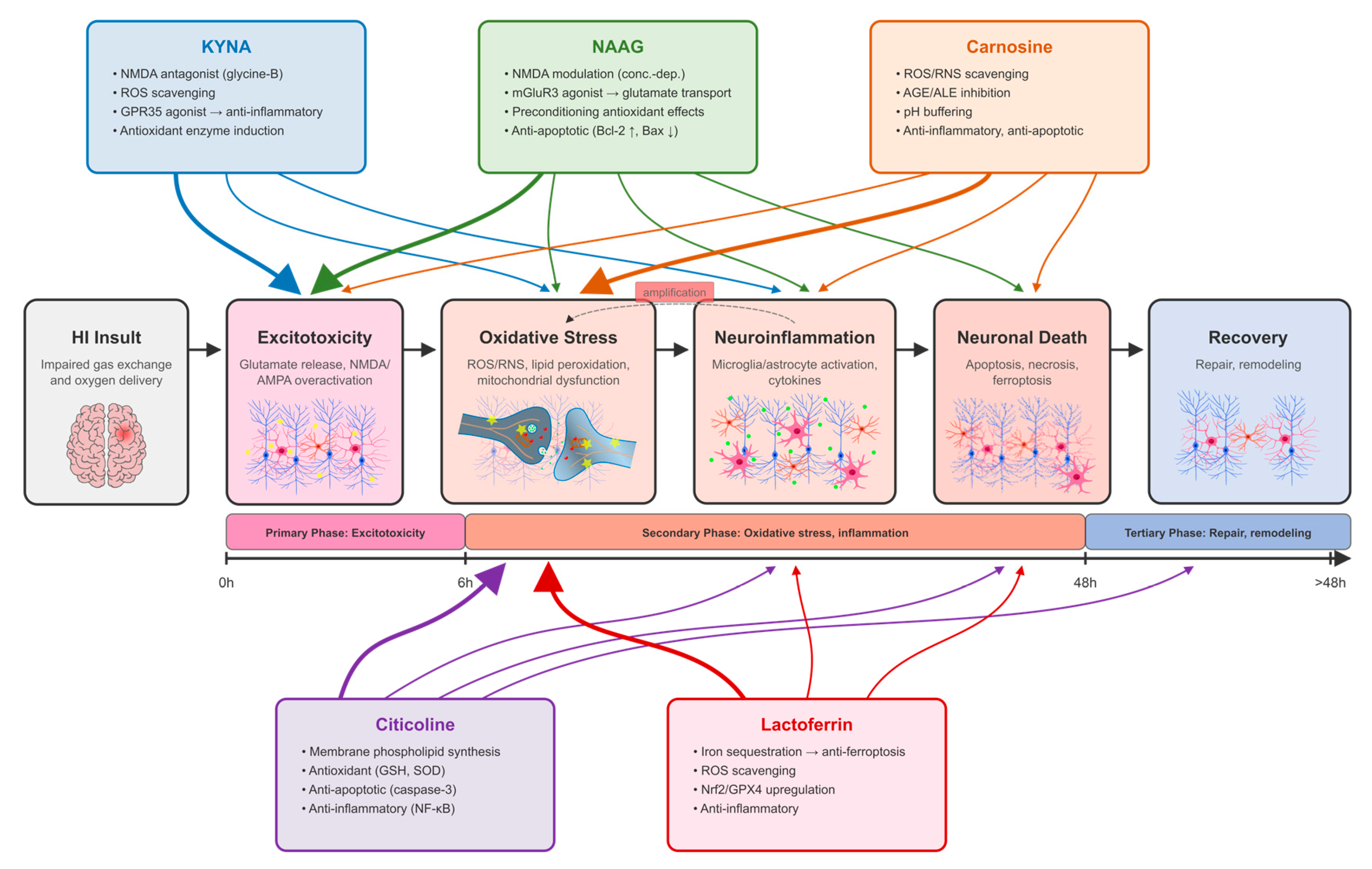
Figure 2.
Blood–brain barrier accessibility of selected endogenous neuroprotective compounds under physiological conditions and after hypoxic–ischemic injury. Under physiological conditions, BBB penetration differs considerably among the five compounds: KYNA and NAAG exhibit limited brain penetration, carnosine may cross the BBB to a limited extent, citicoline-derived metabolites/precursors can enter the brain, and lactoferrin undergoes receptor-mediated transcytosis. Following hypoxic–ischemic injury, disruption of endothelial integrity and tight junctions increases BBB permeability and may enhance brain accessibility of these compounds. The extent of this increase is likely to depend on compound-specific properties and the severity and temporal evolution of BBB disruption.
Figure 2.
Blood–brain barrier accessibility of selected endogenous neuroprotective compounds under physiological conditions and after hypoxic–ischemic injury. Under physiological conditions, BBB penetration differs considerably among the five compounds: KYNA and NAAG exhibit limited brain penetration, carnosine may cross the BBB to a limited extent, citicoline-derived metabolites/precursors can enter the brain, and lactoferrin undergoes receptor-mediated transcytosis. Following hypoxic–ischemic injury, disruption of endothelial integrity and tight junctions increases BBB permeability and may enhance brain accessibility of these compounds. The extent of this increase is likely to depend on compound-specific properties and the severity and temporal evolution of BBB disruption.
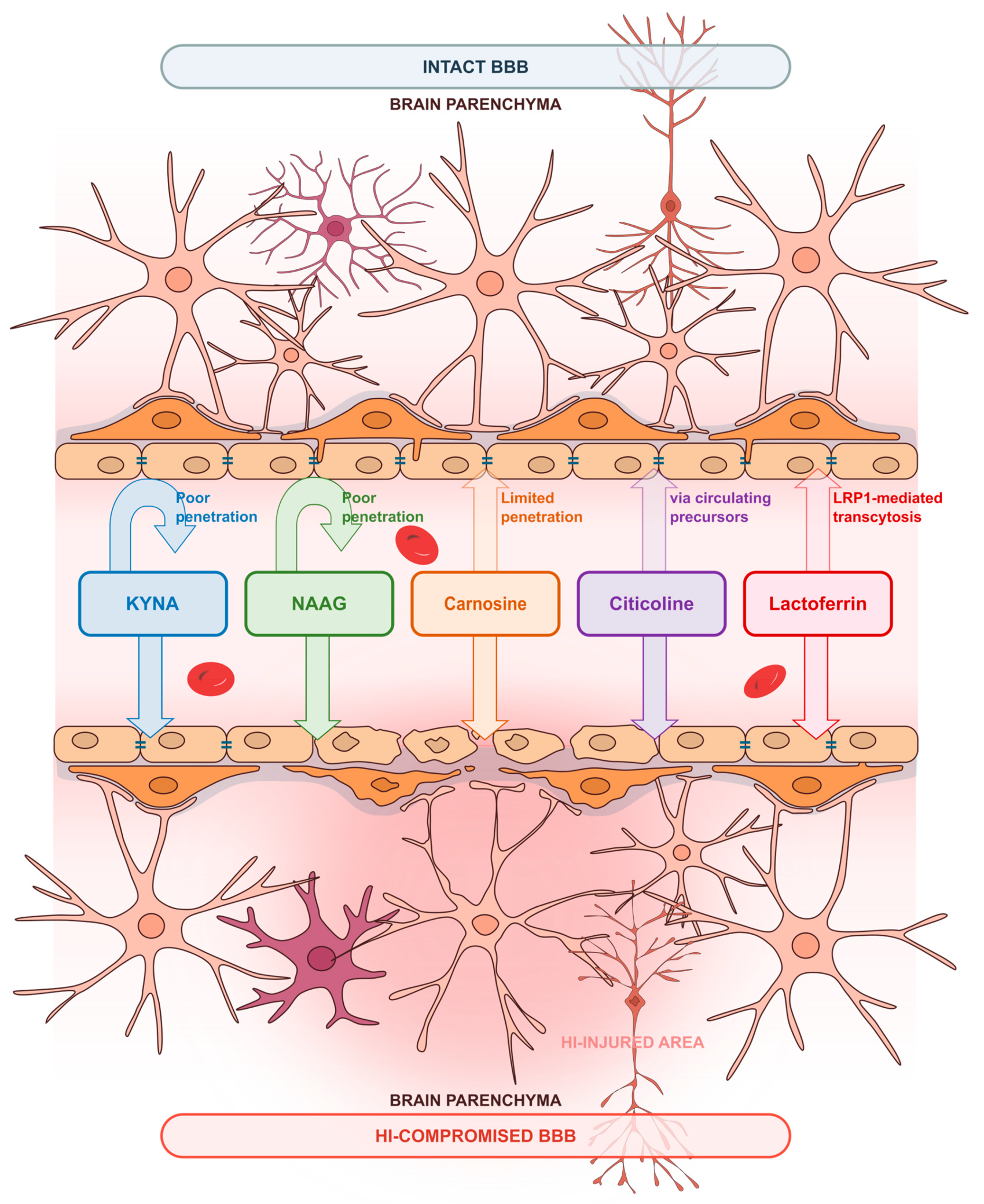
Figure 3.
Molecular targets and pathways associated with the actions of selected endogenous neuroprotective compounds in neonatal hypoxic–ischemic brain injury. KYNA, NAAG, carnosine, citicoline, and lactoferrin influence distinct but partly overlapping molecular targets and signaling pathways. KYNA and NAAG converge particularly on glutamatergic signaling through different mechanisms, whereas the other compounds influence transport systems and intracellular pathways involved in antioxidant defense, membrane homeostasis, and iron metabolism. Thick solid lines indicate established or predominant interactions, thin solid lines indicate modulatory interactions, and dashed lines indicate indirect or regulatory effects. The diversity and complementarity of these mechanisms provide a rationale for investigating combined neuroprotective strategies in neonatal HIE.
Figure 3.
Molecular targets and pathways associated with the actions of selected endogenous neuroprotective compounds in neonatal hypoxic–ischemic brain injury. KYNA, NAAG, carnosine, citicoline, and lactoferrin influence distinct but partly overlapping molecular targets and signaling pathways. KYNA and NAAG converge particularly on glutamatergic signaling through different mechanisms, whereas the other compounds influence transport systems and intracellular pathways involved in antioxidant defense, membrane homeostasis, and iron metabolism. Thick solid lines indicate established or predominant interactions, thin solid lines indicate modulatory interactions, and dashed lines indicate indirect or regulatory effects. The diversity and complementarity of these mechanisms provide a rationale for investigating combined neuroprotective strategies in neonatal HIE.
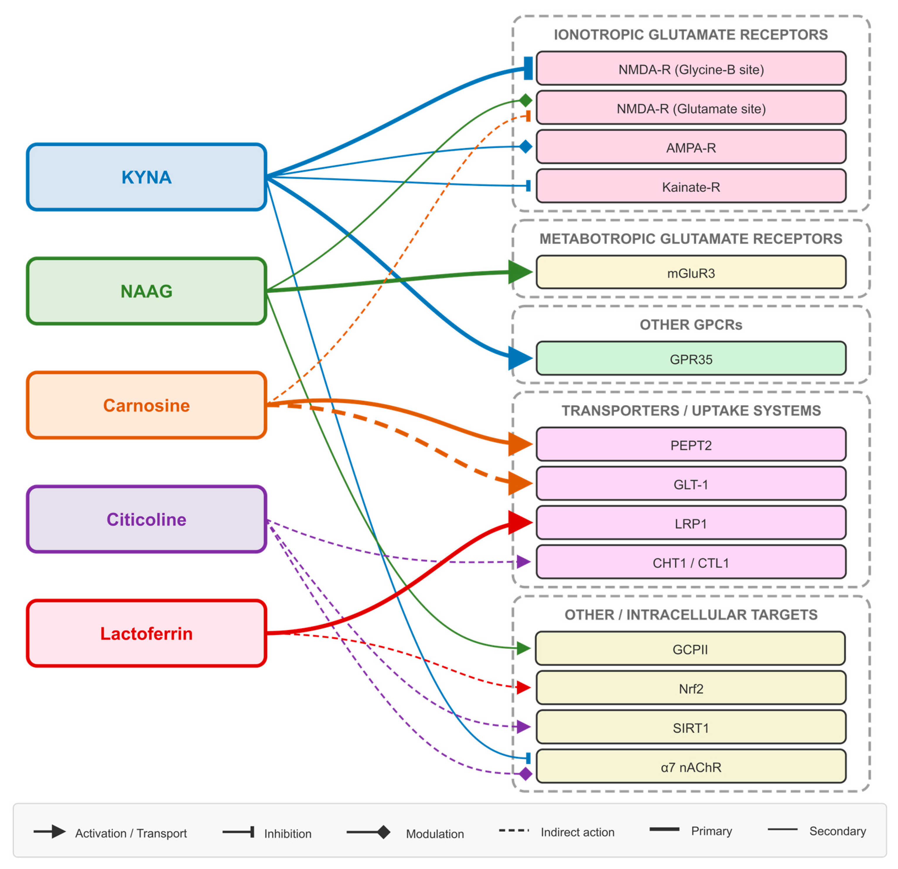
Disclaimer/Publisher’s Note: The statements, opinions and data contained in all publications are solely those of the individual author(s) and contributor(s) and not of MDPI and/or the editor(s). MDPI and/or the editor(s) disclaim responsibility for any injury to people or property resulting from any ideas, methods, instructions or products referred to in the content. |
© 2026 by the authors. Licensee MDPI, Basel, Switzerland. This article is an open access article distributed under the terms and conditions of the Creative Commons Attribution (CC BY) license (http://creativecommons.org/licenses/by/4.0/).
Copyright: This open access article is published under a Creative Commons CC BY 4.0 license, which permit the free download, distribution, and reuse, provided that the author and preprint are cited in any reuse.