

Submitted:
24 September 2026
Posted:
25 September 2026
You are already at the latest version
Abstract
Bovine mastitis remains one of the most economically significant and biologically complex diseases in dairy production, with major implications for animal welfare, antimicrobial use, milk quality and food safety. Conventional diagnostic approaches, including somatic cell count and microbiological culture, are widely used but present limitations in sensitivity, specificity and timeliness, particularly for subclinical infections. In this context, acute phase proteins (APPs) have emerged as promising host-response biomarkers that provide a direct and biologically relevant measure of mammary inflammation. This review critically evaluates the role of key APPs, along with emerging markers such as cathelicidins, lactoferrin and lipopolysaccharide-binding proteins, in identifying and monitoring bovine mastitis. Evidence indicates that APPs, particularly when measured in milk, offer superior sensitivity for early and subclinical disease compared to somatic cell count and can provide additional insights into disease severity, pathogen class, and local immune responses. Importantly, the review extends beyond diagnostics to examine the underexplored relationship between mastitis-associated inflammation, antimicrobial pharmacokinetics and the risk of drug residues in milk. Inflammatory disruption of the blood–milk barrier, altered transporter activity and changes in milk composition can significantly influence drug distribution and persistence, potentially leading to regulatory exceedances. Future perspectives highlight the integration of APPs into multi-marker diagnostic platforms, biosensor technologies, precision dairy systems and “One Health” frameworks.
Keywords:
bovine mastitis
; acute phase proteins
; haptoglobin
; serum amyloid A
; milk biomarkers
; antimicrobial residues
; pharmacokinetics
; One Health
1. Introduction
Bovine mastitis represents the most prevalent and economically consequential disease in dairy cattle worldwide. Annual losses attributable to mastitis reach billions of dollars, primarily due to decreased milk yield and quality, increased culling rates, veterinary expenditures and ongoing management costs [1,2]. Beyond its economic impact, mastitis constitutes a significant animal welfare issue, resulting in pain and morbidity for affected cows, especially in severe cases [3]. Furthermore, mastitis is the principal cause of antibiotic use in dairy herds, raising critical public health and food safety concerns in the context of global efforts to combat antimicrobial resistance [4].
The principal diagnostic approaches for bovine mastitis include somatic cell count (SCC) analysis and microbiological culture. SCC functions as an indirect marker of udder inflammation and is routinely employed in milk quality monitoring and economic evaluations. However, SCC values are influenced by physiological and environmental variables such as lactation stage, age, stress and diurnal fluctuations, thereby limiting their reliability for early or subclinical mastitis detection [5,6]. Microbiological culture remains the definitive method for identifying causative pathogens and assessing antimicrobial susceptibility, yet it is hindered by prolonged turnaround times, substantial labor requirements and inconsistent detection of pathogens with low or intermittent shedding [7]. Measurement of electrical conductivity in milk provides a rapid, automated assessment but primarily reflects epithelial integrity rather than infection or immune activation, thus restricting its diagnostic value [8]. Collectively, these limitations underscore the necessity for advanced diagnostic tools capable of providing earlier, more accurate and clinically meaningful information regarding udder health.
Interest in host-response biomarkers as supplementary or alternative approaches to traditional mastitis diagnostics is increasing. Acute phase proteins (APPs) are particularly promising in this context. APPs are plasma proteins whose concentrations change markedly in response to infection, inflammation, trauma or stress and are a central component of the innate immune system’s acute-phase response [9,10]. In cattle, the principal positive APPs are haptoglobin (Hp), serum amyloid A (SAA) and its mammary-associated isoform (MAA), and, to a lesser extent, C-reactive protein (CRP) [11]. These proteins participate in pathogen opsonisation, immune response modulation and tissue repair. Elevated levels of these markers in serum and milk are closely linked to the presence and severity of mammary gland inflammation [12].
Acute phase proteins in mastitis possess diagnostic value that extends beyond their function as general indicators of inflammation. Unlike SCC, which reflects leukocyte infiltration or electrical conductivity, which signals changes in ion flux, APPs are direct molecular products of the host inflammatory response. Measurement of these proteins in milk facilitates earlier detection of subclinical mastitis, more accurate assessment of disease severity and enhanced monitoring of therapeutic outcomes [13,14]. Moreover, the local synthesis of MAA by mammary epithelial cells imparts high specificity for intramammary disease, potentially increasing diagnostic accuracy relative to systemic biomarkers [15]. The use of biomarkers also intersects with the dynamics of udder inflammation, antimicrobial intervention and the risk of veterinary drug residues in milk. Mastitis remains the primary justification for antimicrobial administration in lactating cows and the occurrence of residues above regulatory thresholds has significant consequences for consumer safety and the integrity of dairy markets [16,17]. Inflammatory processes inherent to mastitis can substantially alter the pharmacokinetics of therapeutic agents, potentially prolonging residue persistence and increasing the risk of regulatory violations [18,19]. This intersection of disease pathophysiology, pharmacological intervention and food safety is frequently underappreciated, yet it is fundamental to protecting public health and maintaining the sustainability of the dairy industry.
This review provides a critical analysis of the literature concerning the application of biosensors for the identification and severity assessment of bovine mastitis. APPs are examined within the framework of mastitis pathogenesis and the acute phase response, with their diagnostic performance systematically compared to established methodologies. The review further addresses recent methodological advancements and challenges in APPs measurement, as well as the broader implications of mastitis-associated inflammation for drug pharmacokinetics and residue risk, including the potential for APPS concentrations to predict regulatory noncompliance. Prospective research directions include integration with precision dairy technologies, omics-based biomarker discovery and the adoption of “One Health” strategies for food safety. The overarching objective is to advance understanding of how host-response biomarkers can enhance mastitis management, promote judicious antimicrobial use and ensure the production of safe, high-quality dairy products.
2. Pathophysiology of Mastitis and the Acute Phase Response
A comprehensive understanding of the physiological mechanisms underpinning bovine mastitis and its inflammatory sequelae is fundamental to evaluating the diagnostic relevance of APPs. This section examines the etiological agents implicated in intramammary infections, delineates the cascade of immune responses following infection and discusses the interplay between local and systemic APPs in orchestrating defence against mammary inflammation.
2.1. Aetiology of Bovine Mastitis
Bovine mastitis is a multifactorial disorder primarily caused by a diverse array of microorganisms, with bacteria entering the mammary gland through the teat canal. These pathogens are generally categorised based on their origin and transmission characteristics: (i) contagious and (ii) environmental pathogens [20,21].
Contagious pathogens persist in the infected mammary gland, serving as the primary reservoir, and are mainly transmitted during milking through contaminated equipment, handlers or sanitising agents. The chief contagious agents include Staphylococcus aureus, Streptococcus agalactiae, Mycoplasma bovis and Corynebacterium bovis [22,23]. Staphylococcus aureus is particularly problematic because it can survive intracellularly within mammary cells, form biofilms and secrete virulence factors, all of which contribute to chronic, treatment-resistant infections [24,25]. This pathogen's capacity to evade immune responses and establish long-term infection is a major factor in the persistent global impact of mastitis.
Environmental pathogens are widespread in the dairy environment, inhabiting bedding, soil, manure and water sources. Infections generally develop between milkings when teats are exposed to contaminated conditions. Major environmental pathogens include coliform bacteria (notably Escherichia coli, Klebsiella spp., Enterobacter spp.), environmental streptococci (such as Streptococcus uberis and Streptococcus dysgalactiae), enterococci and coagulase-negative staphylococci (CNS) [26,27]. Coliform mastitis, especially that due to Escherichia coli, is characterised by rapid onset and severe clinical signs, with potential progression to systemic disease such as endotoxemia or septic shock. Lipopolysaccharide (LPS) in Gram-negative bacteria strongly triggers the innate immune response, fueling the inflammatory cascade underlying the acute presentation of coliform mastitis [28].
Recent research indicates that the spectrum of pathogens responsible for bovine mastitis is dynamic and subject to temporal changes. Variations in pathogen prevalence are shaped by factors such as management strategies, antimicrobial usage trends, and seasonal influences [29]. This evolving epidemiology underscores the need for continuous surveillance and the development of diagnostic tools that can reliably detect inflammatory processes regardless of the causative agent.
Coagulase-negative staphylococci, once considered minor contributors, have recently emerged as the most commonly detected organisms in bovine milk samples across various geographic areas [30]. Species like Staphylococcus chromogenes affect udder health in diverse ways. Some investigations highlight a limited inflammatory profile compared to major pathogens, whereas others note persistent subclinical infections with moderate SCC elevations [31].
When pathogens enter the mammary gland, the host initiates an inflammatory response through coordinated mechanisms to eradicate invaders and restore tissue balance. After colonisation of the teat canal and subsequent migration into the gland cistern, bacteria face the first defensive line, comprised of the keratin plug, antimicrobial substances such as lactoferrin and lysozyme and resident immune cells in the mammary tissue [32]. If these barriers are overcome, bacteria proliferate, leading to recognition of pathogen-associated molecular patterns (PAMPs) by pattern recognition receptors (PRRs) on mammary epithelial cells, macrophages, and dendritic cells. Toll-like receptors (TLRs), notably TLR2 (which detects lipoteichoic acid and peptidoglycan from Gram-positive bacteria) and TLR4 (recognising LPS from Gram-negative bacteria), are central to the downstream signalling that results in the synthesis of pro-inflammatory mediators [33].
The mammary gland's immune response to bacterial invasion involves a precise interplay between innate and adaptive components, with the innate arm predominating early on. Central to this defence is the rapid synthesis and secretion of pro-inflammatory cytokines, chemokines and other mediators that recruit and activate various immune cell populations.
Cytokine signalling constitutes the principal communication framework of the inflammatory response. Upon PRRs recognition of bacterial elements, mammary epithelial cells and tissue macrophages trigger intracellular pathways, including nuclear factor-kappa B (NF-κB) and mitogen-activated protein kinase (MAPK) cascades, that culminate in increased production of pro-inflammatory cytokines [34]. Key mediators in the initial mastitis response are interleukin-1β (IL-1β), interleukin-6 (IL-6), interleukin-8 (CXCL8) and tumour necrosis factor-alpha (TNF-α).
Recent advances in proteomics and cytokine profiling have provided greater understanding of how various pathogens elicit immune responses. For example, Winther et al. [35] observed that quarters infected with S. aureus and Streptococcus spp. exhibited substantially elevated SCC and inflammatory profiles compared to those infected with minor pathogens like S. chromogenes, where lower levels of IFN-γ, IL-10 and TNF-α were reported. Knowing these pathogen-specific immune patterns is critical for biomarker selection and interpretation, as the magnitude and nature of inflammatory mediators may differ considerably depending on the causative agent.
The JAK-STAT signalling pathway is a key regulator of the immune response in the mammary gland. It mediates how cytokines affect target cells and influences the balance between pro-inflammatory and regulatory responses [36]. Disruption in these pathways can lead to excessive tissue damage or insufficient pathogen clearance, both of which can negatively impact mastitis outcomes.
Neutrophils (NEUTs) infiltration is a key feature of the early immune response to infection in the mammary gland and the main factor driving increased somatic cell counts in milk during mastitis. In response to chemotactic signals, especially IL-8 and complement component C5a, circulating NEUT stick to vessel walls, move into mammary tissue and eventually enter the alveolar lumen. Within hours of the infection, NEUT can make up more than 90% of the somatic cell population in milk, unlike healthy milk, which primarily contains macrophages and lymphocytes [37].
Differential somatic cell count (DSCC), which quantifies the relative proportions of NEUTs, lymphocytes and macrophages in milk, is gaining prominence in mastitis diagnostics [38,39]. Studies show that DSCC profiles differ between healthy quarters and those affected by subclinical or clinical mastitis, suggesting that analysing milk cellular composition provides more nuanced information about inflammatory status than total cell counts alone.
Upon entering the mammary gland, NEUTs perform multiple antimicrobial roles, including phagocytosis of bacteria, secretion of antimicrobial peptides and generation of reactive oxygen species. Neutrophil extracellular traps (NETs), composed of DNA, histones and antimicrobial proteins. can ensnare and neutralise extracellular pathogens [40,41]. Notably, certain pathogens, particularly S. aureus, possess mechanisms to evade or degrade NETs, thereby sustaining their persistence within mammary tissue [42,43].
Although the neutrophil response is vital for bacterial clearance, excessive or prolonged activity can result in tissue injury. This damage includes destruction of mammary epithelial cells, basement membrane degradation, and fibrosis, all of which may compromise milk production even after the infection resolves [44]. Balancing protective and detrimental neutrophil functions is crucial to mastitis outcomes, underscoring the need for biomarkers that distinguish effective immune responses from pathological inflammation.
The systemic inflammatory response in mastitis, particularly in severe cases, extends beyond the mammary gland and affects the whole body through cytokines and other inflammatory mediators. In coliform mastitis, bacteria can enter the bloodstream and cause endotoxemia, producing symptoms such as fever, rapid heart rate, rumen stasis, reduced milk production in unaffected quarters, and, in some cases, cardiovascular collapse and death. This systemic response is also linked to changes in circulating white blood cell populations. Initially, neutropenia occurs as white blood cells move to the site of infection. This is followed by neutrophilia as the bone marrow releases more cells. These changes provide context for the acute phase response and the associated changes in liver protein production that are useful for diagnostics.
The acute phase response (APR) is a broad body reaction to injury, infection or inflammation. It includes various physical and metabolic changes designed to reestablish balance and fight off threats [45]. In dairy cattle, the APR is triggered by factors including bacterial and viral infections, injury, surgery, calving-related stress and metabolic conditions, with mastitis a particularly strong and well-known trigger.
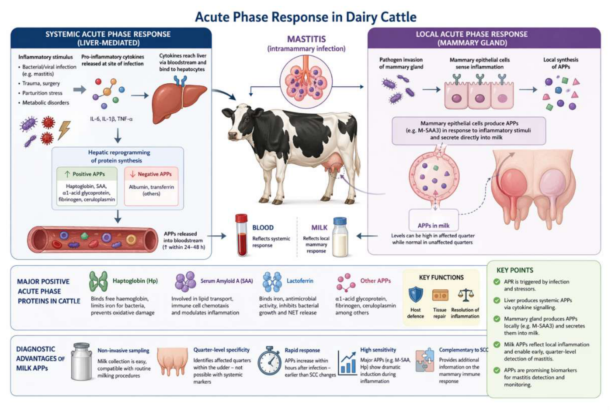
The liver plays a central role in the systemic APR through protein production. Pro-inflammatory cytokines, especially IL-6, IL-1β and TNF-α, are released from the infection site and travel through the bloodstream to the liver, where they bind to receptors on liver cells. This signalling changes liver protein production, increasing positive APPs while decreasing negative APPs such as albumin and transferrin. The response's speed and extent depend on the nature and severity of the inflammatory trigger. Levels of major APPs like Hp and SAA can rise dramatically within 24 to 48 hours after the onset of an infection [46].
In cattle, the main positive APPs include Hp, SAA, α₁-acid glycoprotein, fibrinogen and ceruloplasmin. Hp and SAA are often the most useful for diagnostics because they are low in healthy animals and increase significantly during inflammation [47]. A recent systematic review found M-SAA to be the most studied milk APP biomarker (48.5% of studies). It was followed by Hp (27.3%), cathelicidin (24.2%) and lactoferrin (21.2%), illustrating ongoing interest in these proteins for mastitis diagnostics [48].
These proteins have diverse biological functions. Hp binds free haemoglobin released during tissue damage and hemolysis. This prevents oxidative injury and sequesters iron from bacteria. SAA is involved in lipid transport, immune cell movement, and inflammation modulation. Lactoferrin is an iron-binding glycoprotein with direct antimicrobial properties. It appears at higher levels in mastitic milk and helps inhibit bacterial growth and excessive NEUTs activity, including blocking NETs release [49,50]. Together, these functions support host defence, tissue repair and inflammation resolution.
Local production in mammary tissue is a distinctive feature of the APR in response to intramammary infection. This is particularly important for mastitis diagnostics. While conventional views of the APR focus on liver production, it is now clear that tissues outside the liver, such as the mammary gland, can produce specific APPs at the site of inflammation. Mammary-associated serum amyloid A3 (M-SAA3, also known as MAA) is produced by mammary epithelial cells in response to inflammatory signals and is secreted directly into milk [51]. This local production explains why protein levels in milk frequently exceed those expected from simple movement from plasma and why milk protein levels can be high in the infected quarter while remaining normal in unaffected quarters.
Proteomic studies have confirmed and expanded on these conclusions. Satheesan et al. (2024) used comparative proteomics of milk somatic cells to identify key proteins involved in mammary immune responses during mastitis, including both APPs and other inflammatory substances. Likewise, weighted gene co-expression network analysis of the host proteome has identified protein groups strongly linked to SCC, providing a wider view of the mammary inflammatory response [52]. These studies show that at least 67 proteins are differentially expressed based on the pathogen found in milk, with 19 associated with immune functions such as TLR2 and lactoferrin, highlighting the complexity of the local inflammatory response.
Haptoglobin can also be found in milk during mastitis. Its presence indicates both local production in the mammary gland and movement from plasma due to inflammation-related increases in blood-milk barrier permeability. The shares of local versus systemic sources depend on the pathogen, disease severity, and infection stage. Thomas et al. (2015) set baseline values for Hp, M-SAA3, and CRP in composite milk from cows on a commercial dairy farm. They found notable correlations between composite SCC and Hp and identified parity as a potential confounding factor when using Hp for mastitis diagnosis.
The local APR in the mammary gland offers several possible diagnostic benefits. Measuring APPs in milk provides a non-invasive method that corresponds to regular milking practices. The quarter-level specificity of milk protein concentrations helps identify affected quarters in an udder, a finding not available from blood markers [53]. Additionally, APPs are produced locally and rapidly after infection, with increases that can be seen within hours of experimental challenge, possibly allowing earlier detection than markers that depend solely on changes in somatic cell count. These features support the rationale for studying APPs as markers for mastitis detection and monitoring, as discussed in the following sections.
Figure 1.
Pathophysiology of mastitis and acute phase response in dairy cattle.
3. Acute Phase Proteins in Bovine Mastitis
Acute phase proteins represent an essential part of the bovine innate immune response and are now recognised as among the most sensitive indicators of intramammary inflammation. Recent advances in proteomics, high-resolution mass spectrometry, transcriptomics and targeted immunoassays have substantially strengthened our understanding of APP activity in mastitis, including their tissue-specific expression, pathogen-dependent responses and diagnostic performance in milk. The classical APP, Hp, SAA and its M-SAA3 (or MAA) remain the primary focus of research; however, emerging evidence has identified additional proteins, such as cathelicidins, lactoferrin, and lipopolysaccharide-binding protein (LBP), as promising complementary biomarkers that indicate the broader immunological field of the infected udder.
Haptoglobin is a well-characterised major APP whose concentration increases substantially during mastitis. Hp binds free haemoglobin released from damaged tissues, thereby preventing oxidative injury and reducing iron availability to bacteria. Numerous studies have confirmed that Hp is highly responsive to both clinical and subclinical mastitis. Notably, Hp concentrations in milk provide greater specificity for detecting quarter-level inflammation than serum levels, since they directly reflect local mammary changes [54]. Proteomic analyses have also shown that in E. coli-induced mastitis, Hp is consistently among the most upregulated proteins, reflecting significant epithelial disruption and increased blood-milk barrier permeability [55]. Hp concentrations correlate closely with SCC, DSCC and pathogen load. Incorporating Hp into multi-marker diagnostic algorithms has been shown to boost early mastitis detection relative to SCC alone. These data position Hp as a strong and biologically reliable biomarker in contemporary mastitis diagnostics.
Serum amyloid A is considered the most sensitive bovine APP and plays a major role in directing leukocyte recruitment, inflammatory signalling and lipid transport during infection. High-throughput proteomic studies have shown that SAA rises rapidly and markedly in mastitic milk, often preceding detectable increases in SCC, making it especially well-suited for early mastitis detection [56]. Importantly, SAA expression varies by pathogen, with concentrations highest in coliform infections, particularly E. coli, and more moderate in mastitis caused by S. aureus or CNS [57,58]. These differences likely reflect variations in pathogen virulence, host immune activation, and epithelial damage, indicating the biological relevance of SAA as a functional marker of host–pathogen interaction. Because of its rapid kinetics and high sensitivity, SAA is increasingly incorporated into novel biosensing platforms for on-farm mastitis detection [59].
A notable advancement in APP research is the identification of M-SAA3 (or MAA) as a locally synthesised isoform specific to mammary tissue. Unlike systemic SAA, which is primarily produced in the liver, mammary epithelial cells generate M-SAA3 in response to infection, conferring exceptional quarter-level specificity for detecting inflammation [60]. Recent proteomic investigations have consistently identified M-SAA3 as one of the most strongly upregulated proteins in mastitic milk, regardless of breed or production context [61]. Its concentrations correlate closely with SCC, DSCC, and the extent of epithelial disruption, and, unlike systemic APP, M-SAA3 is largely unaffected by extramammary inflammatory processes [62]. These characteristics make M-SAA3 a highly promising biomarker for early, precise, quarter-level mastitis detection.
C-reactive protein, historically classified as a moderate APP in cattle, has obtained renewed attention with the advent of improved assays. Both milk and serum CRP concentrations increase during mastitis, with a tendency for greater elevation in Gram-negative compared to Gram-positive infections [63]. Although CRP is less responsive than SAA or Hp, including it in multimodal biomarker panels has been shown to improve classification of disease severity and help differentiate acute from resolving infections [63]. Given its moderate but consistent behaviour, CRP is best utilised as a supplementary marker rather than a primary diagnostic tool.
Fibrinogen, a central protein of the coagulation cascade, shows considerable elevation in systemic inflammatory states. Although its detection in milk is limited, serum fibrinogen is still a valuable indicator in mastitis cases that progress to systemic inflammatory response syndrome. Increased serum fibrinogen is associated with more severe clinical outcomes, prolonged recovery, and an elevated risk of culling, particularly in coliform mastitis [64]. As such, fibrinogen is more useful for assessing systemic severity than for routine milk-based screening.
Recent advances in proteomics and immunology have highlighted emerging APP-related biomarkers with diagnostic potential. Cathelicidins, particularly bovine cathelicidin-1, are antimicrobial peptides rapidly released by neutrophils during mastitis. Their concentrations increase sharply in milk during infection and strongly correlate with SCC, bacterial load and neutrophil activation [65,66]. Cathelicidins exhibit outstanding ability to distinguish healthy from subclinically infected quarters, especially during early inflammation when SCC remains low. Lactoferrin, an iron-binding glycoprotein with potent antimicrobial and immunomodulatory properties, also increases markedly in mastitic milk. Beyond its diagnostic value, lactoferrin helps regulate neutrophil activity and prevents excessive tissue damage [67,68]. Elevated lactoferrin levels may also indicate the severity of epithelial disruption in mastitis.
Furthermore, LBP has emerged as a highly sensitive marker of Gram-negative mastitis. LBP concentrations rise rapidly in both serum and milk following endotoxin exposure, correlating with systemic severity and inflammatory cytokine release [69]. Accordingly, LBP is valuable for differentiating pathogen classes and predicting coliform mastitis severity.
Collectively, the expanding body of research since 2018 supports a multidimensional understanding of APP behaviour in mastitis. Hp and SAA remain the most reliable and biologically precise indicators of intramammary inflammation. Complementary biomarkers, including cathelicidin-1, lactoferrin, CRP and LBP, contribute additional information regarding pathogen type, inflammatory intensity, and host tissue response. As the field progresses toward multi-marker, sensor-integrated, and data-driven diagnostic systems, APPs are poised to play a central role in future mastitis detection frameworks.

Table 1.
Major and Emerging Acute Phase Proteins in Bovine Mastitis, Their Biological Roles, Diagnostic Value and Key Recent References (2018–2025).
Table 1.
Major and Emerging Acute Phase Proteins in Bovine Mastitis, Their Biological Roles, Diagnostic Value and Key Recent References (2018–2025).
| Biomarker | Biological/Immunological Role | Diagnostic Relevance in Mastitis | Recent Supporting References |
| Haptoglobin (Hp) | Binds free Hb; limits oxidative stress; restricts iron availability to pathogens | Highly sensitive in milk; correlates with SCC, DSCC, pathogen load; strong quarter-level specificity | [27] |
| Serum Amyloid A (SAA) | Early mediator of inflammation; chemotaxis; lipid transport modulation | Most sensitive systemic/milk APP; rises rapidly (<24 h); pathogen-dependent response | [29] |
| Mammary-Associated SAA3 (M-SAA3/MAA) | Locally synthesized in mammary epithelium; secreted into milk | Strongest quarter-level specificity; minimally affected by systemic inflammation; excellent early marker | [63] |
| C-Reactive Protein (CRP) | Modulates complement; general inflammatory marker | Moderate APP; improved utility in multi-marker panels; increases more in Gram-negative mastitis | [35] |
| Fibrinogen | Coagulation factor; tissue repair; systemic inflammation | Indicates systemic severity (coliform mastitis); weak for milk diagnostics | [14] |
| Cathelicidin-1 | Neutrophil-derived antimicrobial peptide; contributes to early immune defence | Strong early mastitis indicator; differentiates healthy vs. subclinical quarters; high correlation with SCC | [28] |
| Lactoferrin | Iron-binding antimicrobial glycoprotein; suppresses NET release; modulates inflammation | Elevated in subclinical and chronic mastitis; useful adjunct for early detection and epithelial damage | [23] |
| Lipopolysaccharide-binding Protein (LBP) | Recognises and transports LPS; amplifies TLR4 signalling | Highly sensitive marker for Gram-negative mastitis; correlates with severity and cytokine release | [20] |
| Complement Proteins (e.g., C3, C5a) | Promote opsonisation and neutrophil recruitment | Differentially expressed by pathogen type; emerging proteomics-based markers | [35,63] |
| α1-Acid Glycoprotein (AAG) | Immunomodulation; binds drugs and pathogens | Moderate systemic APP; increases during chronic mastitis or systemic inflammation | [27] |
| Cathepsins / Proteolytic Enzymes | Released from neutrophils; contribute to tissue remodelling | Elevated in high-SCC and chronic mastitis; reflect epithelial damage | [63] |
4. Acute Phase Proteins as Biomarkers for Mastitis Identification
The use of APPs as biomarkers for bovine mastitis has increased because they reflect inflammatory processes in milk [70]. The most extensively studied APPs in mastitis are Hp, SAA and MAA [71]. Recent studies indicate that Hp and SAA are the primary APPs linked to both clinical and subclinical mastitis. However, their absolute concentrations vary with the causative pathogen, disease severity, lactation stage and analytical platform used [72]. Although they have potential, multiple challenges exist in using APPs as diagnostic biomarkers. Variability among analytical platforms complicates standardisation and comparison of results. The specificity of acute-phase proteins for mastitis is limited, as their concentrations may also increase in response to other inflammatory conditions [73]. In addition, the cost and accessibility of reliable assays for routine herd screening may limit their practical use at the farm level [74].
4.1. Comparison with Systemic Biomarkers
Somatic cell count remains a commonly used method for routine mastitis monitoring in dairy herds. SCC increases with inflammation, but lactation stage, stress, and environmental factors also affect levels [75]. While SCC is valuable for herd management, it lacks specificity for early detection, especially in subclinical mastitis cases [76]. In comparison, APPs such as Hp and SAA generally show greater sensitivity for detecting early or subclinical mastitis than SCC [77]. Although SCC has moderate specificity for mastitis identification, APPs may be less specific because their concentrations can increase in other inflammatory conditions unrelated to mastitis. The diagnostic performance of individual biomarkers varies, and combining SCC with APP measurements may improve both sensitivity and specificity for early mastitis detection [78].
Electrical conductivity is another method used to monitor mastitis conditions. Although electrical conductivity is rapid and easily automated, its diagnostic value is limited because the ionic shifts it measures primarily reflect mammary epithelial damage rather than direct markers of the host or pathogen. Consequently, unlike the biomarkers discussed above, electrical conductivity is not recommended for detecting subclinical mastitis [79]. The reference method for identifying the causative pathogen is still microbiological culture testing. It is time-consuming, requires trained personnel, and can miss intermittently shed or low-abundance pathogens. It is considered less suitable for rapid screening but remains an excellent confirmatory technique.
Compared with systemic methods, APPs provide distinct diagnostic advantages. Hp and SAA concentrations in both serum and milk increase significantly during subclinical and clinical mastitis. Notably, milk-based measurements are more effective than serum-based measurements for detecting subclinical mastitis [80].
4.2. Acute Phase Proteins in Milk and in Serum
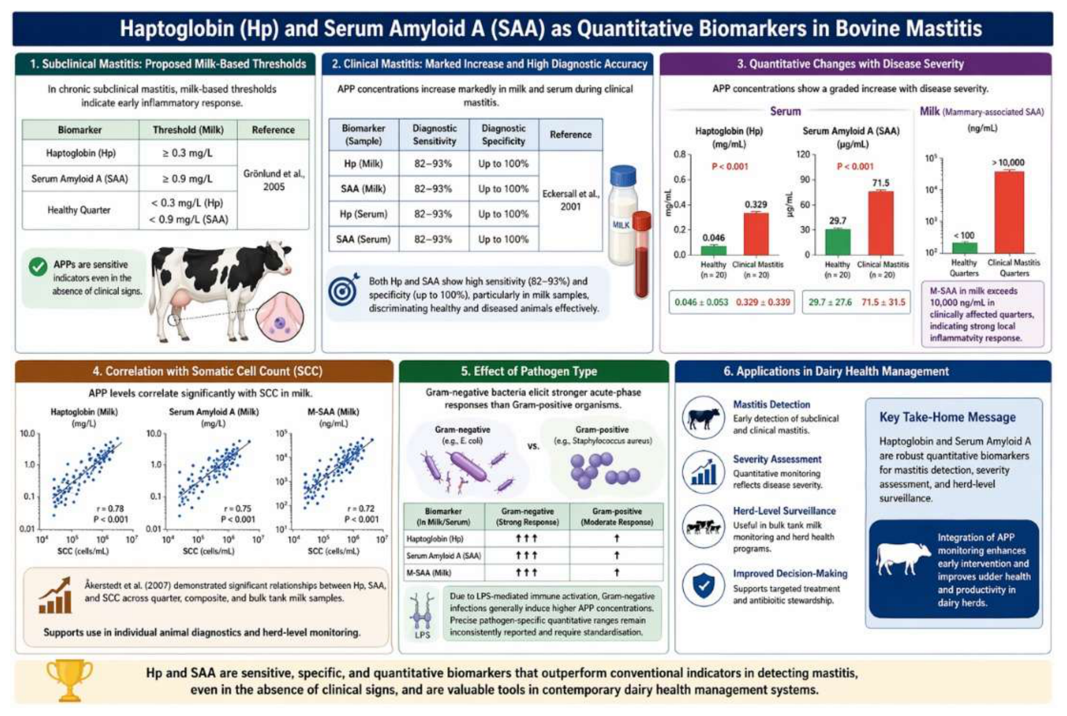
Acute phase proteins, particularly Hp and SAA, have emerged as sensitive and early indicators of inflammatory responses in bovine mastitis. In chronic subclinical mastitis, proposed milk-based thresholds are Hp ≥ 0.3 mg/L and SAA ≥ 0.9 mg/L, with healthy udder quarters typically exhibiting concentrations below these levels [81]. These findings show the utility of APPs as diagnostic markers even in the absence of overt clinical signs. In clinical mastitis, APP concentrations increase markedly in both milk and serum. Eckersall et al. (2001) showed that both Hp and SAA are significantly elevated in cows with clinical mastitis, with diagnostic sensitivities ranging from 82–93% and specificities up to 100%, particularly in milk samples. These results illustrate the strong discriminatory power of APPs between healthy and diseased animals.
Quantitative studies further demonstrate a graded response of APPs with increasing disease severity. Kováč et al. (2011) reported that serum Hp concentrations increased from 0.046 ± 0.053 mg/mL in clinically healthy cows to 0.329 ± 0.339 mg/mL in cows with clinical mastitis. Similarly, serum SAA increased from 29.7 ± 27.6 μg/mL to 71.5 ± 31.5 μg/mL in the same groups. M-SAA in milk exhibited a pronounced increase, exceeding 10,000 ng/mL in clinically affected quarters, which indicates a strong local inflammatory response. In addition to clinical classification, APP levels correlate closely with SCC, a conventional marker of udder inflammation. Åkerstedt et al. (2007) showed significant relationships between Hp, SAA, and SCC across quarter, composite, and bulk tank milk samples, supporting their use in both individual animal diagnostics and herd-level monitoring. Although APP concentrations vary by causative pathogen, the literature consistently indicates that Gram-negative bacteria elicit stronger acute-phase responses than Gram-positive organisms, likely due to lipopolysaccharide-mediated immune activation. However, precise pathogen-specific quantitative ranges remain inconsistently reported and require further standardisation. These findings show Hp and SAA as robust quantitative biomarkers for mastitis detection, severity assessment, and herd-level surveillance in contemporary dairy health management systems.
Figure 2.
Major acute phase proteins and their diagnostic roles.
4.3. Analytical Methods
Enzyme-linked Enzyme-linked immunosorbent assay (ELISA) remains the most widely used method for quantifying acute-phase proteins because of its high analytical sensitivity and specificity. However, its laboratory-based nature limits its suitability for rapid, on-farm decision-making [82,83]. Biosensors represent a promising translational approach; one study reported an electrochemical Hp biosensor for bovine milk with a working range of 0.001 to 0.32 μg/mL and a limit of detection of 0.031 μg/mL [84]. Nevertheless, validation across diverse farms, pathogens, and routine field conditions remains limited. Additionally, proteomics has proven valuable for biomarker discovery, revealing that Hp, MAA, CRP, lactoferrin, α-lactalbumin and cathelicidin are all significantly elevated in mastitic milk compared to healthy controls. This evidence suggests that APPs may be most effective as part of a broader inflammatory signature rather than as single analytes [85]. Fluorescence spectroscopy is a rapid and potentially suitable method for dairy monitoring, but it is more effective as a fingerprinting tool than as a direct APP assay. While rapid milk characterisation is useful, specificity for mastitis biomarker quantification remains lower than that of targeted immunoassays or biosensors [86].
4.4. Critical Analysis
Current literature supports the use of APPs, particularly milk Hp and MAA, as effective adjunct biomarkers for mastitis identification. Their main advantage over SCC and EC is their direct biological relevance to inflammation, while their main advantage over culture is rapidity. However, APPs do not replace culture when pathogen identification or antimicrobial decision-making is necessary. The principal unresolved issue is standardisation: although convincing evidence supports diagnostic value, consensus is lacking on universal cut-offs, preferred assay platforms, and optimal integration of APPs with SCC, culture or automated sensor data in routine herd management. Recent reviews increasingly advocate for multi-marker, platform-integrated diagnostics rather than single-test replacements.
Table 2.
Diagnostic Performance of Biomarkers in Bovine Mastitis.
| Biomarker | Sample Type | Sensitivity | Specificity | Detection Stage | Key Advantages | Key Limitations |
| Somatic Cell Count (SCC) | Milk | Moderate | Moderate | Late–subclinical to clinical | Widely available; low cost; herd-level monitoring | Affected by non-infectious factors (lactation, stress); low early sensitivity |
| Differential SCC (DSCC) | Milk | Moderate–High | Moderate | Subclinical | Provides immune cell profile; improved discrimination vs SCC | Limited standardisation; requires advanced analysis |
| Microbiological Culture | Milk | High (pathogen detection) | High | Clinical | Gold standard for pathogen identification and antimicrobial testing | Time-consuming; false negatives in intermittent shedding |
| Electrical Conductivity (EC) | Milk | Low–Moderate | Low | Late-stage | Rapid; automated; on-farm use | Reflects tissue damage, not inflammation; poor specificity |
| Haptoglobin (Hp) | Milk > Serum | High | High | Early–subclinical | Strong correlation with SCC and inflammation; quarter-level specificity | Elevated in other inflammatory conditions; assay variability |
| Serum Amyloid A (SAA) | Milk & Serum | Very High | Moderate–High | Very early | Most sensitive APP; rapid response (<24 h); detects early inflammation | Pathogen-dependent variability; limited specificity alone |
| Mammary SAA3 (M-SAA3 / MAA) | Milk | Very High | High | Very early | Mammary-specific; excellent quarter-level detection; minimal systemic interference | Limited commercial assay availability; standardisation needed |
| C-Reactive Protein (CRP) | Milk & Serum | Moderate | Moderate | Clinical | Useful in multi-marker panels; reflects severity | Lower sensitivity compared to Hp/SAA |
| Fibrinogen | Serum | Moderate | Moderate | Severe/clinical | Indicator of systemic inflammation and severity | Limited utility in milk; not suitable for early detection |
| Cathelicidin-1 | Milk | High | High | Early | Strong early marker; reflects neutrophil activation | Emerging biomarker; limited routine availability |
| Lactoferrin | Milk | Moderate–High | Moderate | Subclinical–chronic | Reflects epithelial damage; antimicrobial function | Elevated in physiological states (e.g., late lactation) |
| LBP (LPS-binding protein) | Milk & Serum | High (Gram-negative) | High | Acute | Strong indicator of Gram-negative mastitis; severity marker | Pathogen-specific; limited broad diagnostic use |
5. Mastitis Treatment, Antimicrobial Use and Implications for Milk Residues
Mastitis remains the leading disease affecting dairy cattle. It is a major contributor to antimicrobial use in food-producing animals. This highlights a crucial intersection between animal health and food safety [87,88]. Therapeutic decisions during lactation are inherently complex. These decisions require consideration of pathogen-specific responses, disease severity, and anticipated treatment effectiveness [89]. Antimicrobial treatments are more effective against streptococcal infections.
In contrast, infections caused by S. aureus have low bacteriological cure rates. This is because they can persist intracellularly and form biofilms [90,91]. Mild Gram-negative mastitis cases, especially those caused by E. coli, often resolve on their own. This diminishes the effectiveness of routine antimicrobial treatments [92]. As a result, antimicrobial use in mastitis has increasingly shifted toward selective, evidence-based strategies. These strategies incorporate bacteriological diagnosis and herd-level data. The goal is to minimise unnecessary exposure while ensuring positive clinical outcomes [93].
Intramammary administration remains the primary therapeutic route for lactating cows, underscoring the need for precise drug delivery to achieve optimal outcomes. Pharmacokinetically, the udder functions as a distinct compartment, with drug disposition influenced by systemic pharmacokinetics, milk secretion dynamics and epithelial barrier integrity [94]. β-lactam antibiotics, particularly penicillins and cephalosporins, are most commonly used in mastitis therapy due to their efficacy against Gram-positive pathogens and favourable safety profiles. Studies indicate that intramammary administration of cephapirin (275 mg per quarter, two doses at 12-hour intervals) maintains milk concentrations above the MIC90 for approximately 35 hours in infected quarters, demonstrating prolonged exposure under clinical conditions [95]. The pharmacokinetics of pirlimycin administered intramammarily are closely linked to milk production, with clearance rates strongly correlated with milk yield (r2=0.939), illustrating how physiological variables affect drug elimination (Whittem et al., 1999). Systemic therapy also contributes to antimicrobial residues in milk; for instance, ceftiofur administered intramuscularly at 1 mg/kg body weight is associated with a zero-hour milk withdrawal time under approved conditions, although residue detection in bulk milk remains a relevant analytical and operational concern [96].
Regulatory thresholds, known as maximum residue limits (MRLs), set the acceptable levels of antimicrobial residues in milk for human consumption. Withdrawal periods are established based on residue depletion kinetics to ensure compliance with these limits. The behaviour of antimicrobial residues is highly compound-specific and is influenced by both pharmacokinetic and bodily factors. For example, pharmacokinetic modelling of pirlimycin after intramammary infusion indicates that milk discard times of approximately 36 hours are required to achieve residue levels below regulatory thresholds [97,98]. In contrast, extended intramammary administration of cephapirin has been shown to produce variable elimination kinetics depending on infection status and treatment duration, indicating that pathological conditions may alter residue persistence [99]. Similarly, studies on cloxacillin show that residue depletion depends strongly on the interval between treatment and calving, with shorter dry periods associated with a higher risk of detectable residues at the onset of lactation [100]. These findings stress that withdrawal periods are not fixed pharmacological constants but are derived from population-based residue studies that integrate variability in physiology, disease status, and management practices.
Mechanistically, antimicrobials in milk are governed by passive diffusion, physicochemical properties, and mammary gland physiology [101]. Drug transfer into milk depends on molecular characteristics and the mammary gland environment. Additionally, management factors such as milking frequency can substantially affect drug depletion kinetics [102]. For example, cephapirin pharmacokinetics vary with milking frequency and dosing interval, indicating that milk removal dynamics, rather than systemic clearance alone, affect elimination [103]. Drug distribution within milk is also heterogeneous; differences in antimicrobial concentrations between milk fractions have been observed, reflecting interactions with milk components such as fat and casein [104]. Disease status further alters pharmacokinetic behaviour, as evidenced by distinct cephapirin concentration profiles in infected compared to healthy quarters [105]. Prolonged antimicrobial persistence has been reported, with ceftiofur detectable in milk for up to 108 hours after intramammary administration, particularly in cows with lower milk production [106].
From a food chemistry standpoint, the milk matrix strongly influences antimicrobial behaviour. Interactions between antimicrobials and milk proteins, particularly caseins and whey proteins, affect the proportion of free drug, changing both antimicrobial efficacy and residue detectability. Additionally, interactions with milk minerals, such as calcium, can modify the bioavailability of specific antimicrobial classes, complicating the relationship between the administered dose and the effective concentration in milk. These matrix effects matter for interpreting residue data, as analytical measurements typically reflect total concentrations rather than the pharmacologically active fraction, which may lead to over- or underestimation of biological activity depending on binding dynamics [107].
In this context, APPs such as Hp and SAA give valuable insights into the inflammatory status of the mammary gland during mastitis. These proteins are markedly elevated in both serum and milk during infection, indicating activation of the innate immune response and local inflammation. M-SAA is synthesised locally in mammary epithelial cells and secreted into milk, supporting its role as a sensitive biomarker of intramammary inflammation [108]. However, the biological mechanisms regulating APPs expression are independent of antimicrobial pharmacokinetics and residue depletion. Therefore, while APPs are useful for disease monitoring and inflammatory assessment, they do not indicate compliance with residue withdrawal requirements. Interpret APP data alongside pharmacokinetic knowledge and regulatory residue limits, underscoring the need for a multidisciplinary approach that connects veterinary therapeutics, food safety, and public health.
6. Mastitis and Milk Residues: The Hidden Connection
Mastitis, a common inflammatory disorder of the mammary gland in dairy animals, considerably changes the pharmacokinetics of veterinary drugs and, as a result, affects milk residue concentration. While withdrawal periods and MRLs are typically established under normal physiological conditions, disease states such as mastitis introduce substantial variability that current regulatory systems do not uniformly address.
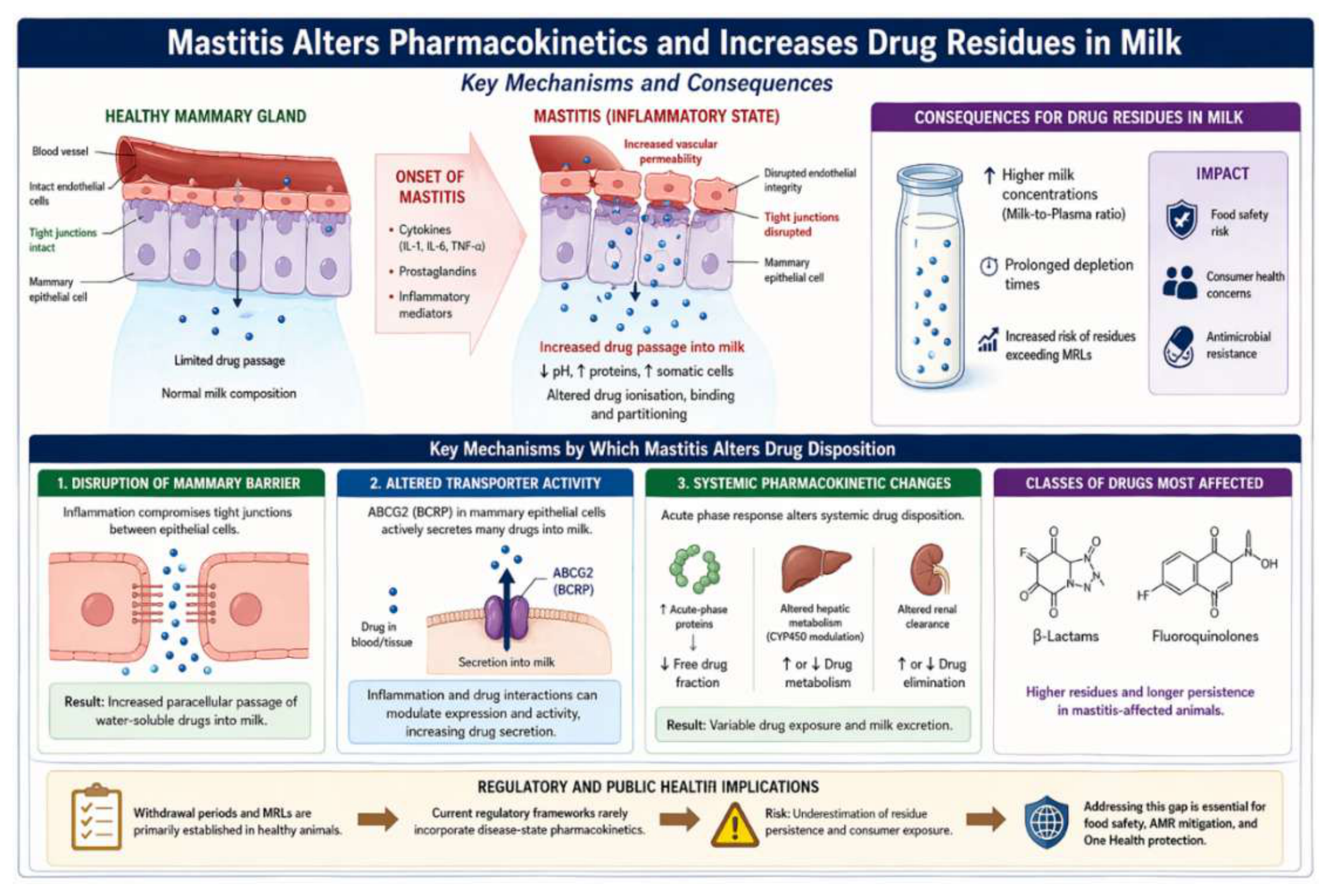
A principal feature of mastitis is increased vascular permeability within the mammary gland. Inflammatory mediators, such as cytokines and prostaglandins, compromise endothelial integrity, thereby facilitating the transfer of drugs from the systemic circulation into mammary tissue and milk. This mechanism is associated with elevated milk-to-plasma ratios for several antimicrobial agents, particularly during mastitis, and further alters drug distribution due to changes in tissue structure, local blood flow, and milk composition [109]. Inflammation also lowers milk pH and increases protein and somatic cell concentrations, factors that influence drug ionisation, protein binding, and partitioning. These physicochemical changes can improve drug retention within the mammary gland and prolong their persistence in milk.
Consequently, animals with mastitis commonly exhibit increased drug residues in milk, with both higher concentrations and prolonged depletion times reported for several classes of antimicrobials, includingβ-lactams and fluoroquinolones [110]. This situation raises significant food safety concerns, as withdrawal periods established in healthy animals may underestimate residue persistence under inflammatory conditions. First, inflammation compromises the mammary gland barrier, particularly the tight junctions that typically restrict drug passage. In mastitis, the disintegration of these junctions permits water-soluble drugs to leak into milk.
Second, alterations in transporter activity play a critical function in mediating drug secretion into milk. The ATP-binding cassette transporter G2 (ABCG2/BCRP), which is highly expressed in mammary epithelial cells, actively facilitates the secretion of numerous xenobiotics into milk [111]. Experimental studies show that ABCG2 substantially contributes to elevated milk concentrations of drugs such as fluoroquinolones [112]. Inflammation and pharmacological interactions can modulate transporter expression and activity, additionally influencing drug secretion. Third, inflammation-induced pharmacokinetic changes introduce systemic effects that modify drug disposition. The acute-phase response can alter plasma protein binding, hepatic metabolism, and renal clearance [113]. Changes in acute-phase protein levels may alter the free fraction of drugs in circulation, while cytokine-mediated modulation of cytochrome P450 enzymes can modify drug metabolism [114]. Collectively, these factors bring about variability in drug exposure and milk excretion.
To summarise, mastitis is a multifactorial determinant of changes in milk drug residues, involving structural disruption of the mammary barrier, transporter-mediated secretion, and systemic pharmacokinetic changes. However, current regulatory approaches to residue monitoring and withdrawal period determination rarely incorporate disease-state pharmacokinetics [115]. This omission may underestimate residual risks for consumers and reduce the effectiveness of policies intended to ensure food safety and mitigate antimicrobial resistance. Filling this gap is essential within a “One Health” framework, particularly for risk assessment and regulatory decision-making.
Figure 3.
Proposed link between mastitis inflammation and milk residues risk.
7. Future Perspectives
Future advancements in mastitis diagnostics, monitoring and food safety management will increasingly depend on integrative, technology-driven strategies that surpass the limitations of individual biomarkers or conventional herd-level indicators. APPs such as Hp and SAA, along with M-SAA, have already shown considerable promise as early and biologically relevant indicators of udder inflammation. However, further research and technological innovation are needed to translate these biomarkers into practical, farm-ready diagnostic systems. Recent progress in omics, biosensing, data analytics and “One Health” frameworks offers promising opportunities to improve mastitis control and milk quality at both the animal and population levels.
A key area for future development is omics-based biomarker discovery and refinement, particularly through proteomics, transcriptomics, lipidomics and integrated host–pathogen multi-omics approaches. Recent proteomic studies have identified numerous inflammation-associated proteins that vary according to pathogen type, disease severity and mammary immune response profile [116,117]. These findings suggest that APPs should be considered within a broader inflammatory network rather than as isolated markers. High-resolution proteomics is expected to reveal additional local acute-phase proteins, post-translational modifications and novel peptides derived from epithelial cells or NEUTs that may surpass current indicators for early or pathogen-specific mastitis detection. Integration of multi-omics data will also deepen mechanistic understanding of the mammary innate immune response and support the development of targeted interventions and precision therapies.
Another significant area of advancement is the development of biosensor-based and lab-on-chip diagnostic technologies for real-time APP detection. Rapid electrochemical and optical sensors for Hp, SAA and other immune markers are emerging as promising tools for on-farm mastitis screening, with recent prototypes demonstrating analytical sensitivity in the low microgram-per-litre range [118]. Integrating these systems into milking robots, in-line sampling devices, or handheld readers could shift mastitis diagnostics from reactive testing to proactive, continuous monitoring. Nevertheless, further validation in field conditions remains a key research priority before widespread implementation is feasible.
Integrating APPs biomarkers into precision dairy farming (PDF) and artificial intelligence (AI)-based decision-support systems represents another promising frontier. Automated milking systems currently generate extensive data streams on SCC, electrical conductivity, milk yield and behavioural patterns. Adding APP measurements to these datasets could enable machine-learning models to predict mastitis onset more accurately, classify disease severity and distinguish transient inflammation from true infection. Initial models combining immune biomarkers with behavioural and production data have shown encouraging results, reducing unnecessary treatments and improving intervention timing [119]. As these models advance, APPs dynamics may inform predictive algorithms to support quarter-specific treatment decisions, forecast recurrence risk and optimise selective dry-cow therapy.
Interest in applying APPs within “One Health” and food safety frameworks is increasing, particularly for their potential to indicate milk residue risk. APPs reflect both mammary barrier permeability and the severity of udder inflammation, two key determinants of antimicrobial pharmacokinetics in milk. Future research should explore whether elevated APPs concentrations could serve as practical predictors of extended drug depletion times or increased residue risk under diseased conditions. Integrating APPs measurements into withdrawal-time decision tools may enable a more adaptive, inflammation-informed approach to residue management, complementing pharmacokinetic modelling and regulatory data. Such frameworks would enhance antibiotic stewardship while reducing the risk of violative residues entering the food chain [120,121].
Another promising direction involves using APPs in selective antimicrobial therapy and personalised treatment strategies. As dairy production increasingly adopts protocols to reduce antimicrobial use, biomarkers that distinguish mild from severe infections or self-limiting from progressive cases, could transform treatment practices. SAA, M-SAA3 and cathelicidin-1 show promise for informing treatment necessity and predicting outcomes. Future clinical trials should assess whether specific APPs thresholds can safely guide treatment decisions or predict the likelihood of bacteriological cure across different pathogen groups.
Finally, the field would benefit from efforts to standardise APPs assays, establish consensus diagnostic thresholds and harmonise analytical platforms across laboratories and commercial systems. Despite extensive research, variability in assay methodologies and reference ranges remains a significant barrier to clinical adoption. Collaborative international initiatives-similar to those established for SCC, milk components and antimicrobial susceptibility testing-could accelerate the integration of APP-based diagnostics into routine dairy herd management.
In summary, the future of mastitis diagnostics lies not in a single biomarker, but in integrated, data-rich systems that combine APPs with omics-derived molecular signatures, advanced biosensing technologies and predictive analytics. As research continues to bridge immunology, engineering and dairy management, APPs are poised to become central components of next-generation mastitis control strategies, with far-reaching implications for udder health, milk quality, antimicrobial stewardship and public health.
8. Conclusions
Acute phase proteins are robust and diagnostically valuable biomarkers for bovine mastitis, offering advantages over traditional indicators such as somatic cell count and electrical conductivity. Haptoglobin, serum amyloid A and mammary-associated SAA3 demonstrate high sensitivity, strong correlation with inflammation, and early detection capabilities, particularly in subclinical disease. Evidence supports a shift from single-parameter diagnostics to integrated, multi-marker approaches combining APPs with conventional and emerging technologies. This integration improves diagnostic accuracy, allows better disease stratification, and supports more precise therapy. However, lack of standardized thresholds, variability among analytical platforms, and limited field validation remain barriers to implementation.
Beyond diagnostics, mastitis-induced inflammation significantly alters antimicrobial pharmacokinetics and raises the risk of milk residues. Inflammation-driven changes in vascular permeability, milk composition, and transporter activity can prolong drug persistence, challenging current withdrawal period guidelines. While APPs do not measure residues directly, they provide important information on inflammatory status and may inform future residue risk prediction. Integrating APP-based diagnostics with antimicrobial stewardship and food safety monitoring offers an opportunity to improve animal and public health. Future research should prioritize standardizing assays, advancing multi-omics biomarker discovery, developing biosensors, and incorporating APPs into precision dairy and predictive models.
Author Contributions
Conceptualization, K.V.A.; search of literature, K.V.A., I.K. and M.V.; writing—original draft preparation, K.V.A.; writing—specific passage, M.V. and I.K.; writing—review and editing, K.V.A. and M.V.; supervision, M.V. All authors have read and agreed to the published version of the manuscript.
Funding
This research received no external funding.
Institutional Review Board Statement
Not applicable.
Informed Consent Statement
Not applicable.
Data Availability Statement
No new data were created or analyzed in this study. Data sharing is not applicable to this article.
Acknowledgments
During the preparation of this manuscript/study, the author(s) used ChatGPT (OpenAI, GPT-4o, May 2026 version) for the purposes of proofreading, syntax improvement and figure creation. The authors have reviewed and edited the output and take full responsibility for the content of this publication.
Conflicts of Interest
The authors declare no conflicts of interest.
References
- Ceciliani, F.; Ceron, J.J.; Eckersall, P.D.; Sauerwein, H. Acute phase proteins in ruminants. J. Proteom. 2012, 75, 4207–4231. [Google Scholar] [CrossRef] [PubMed]
- Eckersall, P.D.; Bell, R. Acute phase proteins: Biomarkers of infection and inflammation in veterinary medicine. Vet. J. 2010, 185, 23–27. [Google Scholar] [CrossRef] [PubMed]
- Petersen, H.H.; Nielsen, J.P.; Heegaard, P.M.H. Application of acute phase protein measurements in veterinary clinical chemistry. Vet. Res. 2004, 35, 163–187. [Google Scholar] [CrossRef] [PubMed]
- Murata, H.; Shimada, N.; Yoshioka, M. Current research on acute phase proteins in veterinary diagnosis: An overview. Vet. J. 2004, 168, 28–40. [Google Scholar] [CrossRef] [PubMed]
- Jacobsen, S.; Niewold, T.A.; Kornalijnslijper, E.; Toussaint, M.J.M.; Gruys, E. Kinetics of local and systemic isoforms of serum amyloid A in bovine mastitic milk. Vet. Immunol. Immunopathol. 2005, 104, 21–31. [Google Scholar] [CrossRef]
- Pyörälä, S.; Hovinen, M.; Simojoki, H.; Fitzpatrick, J.; Eckersall, P.D.; Orro, T. Acute phase proteins in milk in naturally acquired bovine mastitis caused by different pathogens. Vet. Rec. 2011, 168, 535. [Google Scholar] [CrossRef] [PubMed]
- Safi, S.; Khoshvaghti, A.; Jafarzadeh, S.R.; Bolourchi, M.; Nowrouzian, I. Acute phase proteins in the diagnosis of bovine subclinical mastitis. Vet. Clin. Pathol. 2009, 38, 471–476. [Google Scholar] [CrossRef] [PubMed]
- Åkerstedt, M.; Persson Waller, K.; Sternesjö, Å. Haptoglobin and serum amyloid A in relation to the somatic cell count in quarter, cow composite and bulk tank milk samples. J. Dairy Res. 2007, 74, 198–203. [Google Scholar] [CrossRef] [PubMed]
- Grönlund, U.; Sandgren, C.H.; Waller, K.P. Haptoglobin and serum amyloid A in milk from dairy cows with chronic sub-clinical mastitis. Vet. Res. 2005, 36, 191–198. [Google Scholar] [CrossRef] [PubMed]
- Eckersall, P.D.; Young, F.J.; Nolan, A.M.; Knight, C.H.; McComb, C.; Waterston, M.M.; Hogarth, C.J.; Scott, E.M.; Fitzpatrick, J.L. Acute phase proteins in serum and milk from dairy cows with clinical mastitis. Vet. Rec. 2001, 148, 35–41. [Google Scholar] [CrossRef] [PubMed]
- Kováč, G.; Tóthová, C.; Nagy, O.; Seidel, H. Milk amyloid A and selected serum proteins in cows suffering from mastitis. Acta Vet. Brno 2011, 80, 3–9. [Google Scholar] [CrossRef]
- Molenaar, A.J.; Harris, D.P.; Rajan, G.H.; Pearson, M.L.; Callaghan, M.R.; Sommer, L.; Farr, V.C.; Oden, K.E.; Miles, M.C.; Petrova, R.S.; et al. The acute-phase protein serum amyloid A3 is expressed in the bovine mammary gland and plays a role in host defence. Biomarkers 2009, 14, 26–37. [Google Scholar] [CrossRef] [PubMed]
- Thomas, F.C.; Waterston, M.; Hastie, P.; Parkin, T.; Haining, H.; Eckersall, P.D. The major acute phase proteins of bovine milk in a commercial dairy herd. BMC Vet. Res. 2015, 11, 207. [Google Scholar] [CrossRef] [PubMed]
- Dalanezi, F.M.; Schmidt, E.M.S.; Joaquim, S.F.; Guimarães, F.F.; Guerra, S.T.; Lopes, B.C.; Cerri, R.L.A.; Chadwick, C.; Langoni, H. Concentrations of acute-phase proteins in milk from cows with clinical mastitis caused by different pathogens. Pathogens 2020, 9, 706. [Google Scholar] [CrossRef] [PubMed]
- Rainard, P.; Riollet, C. Innate immunity of the bovine mammary gland. Vet. Res. 2006, 37, 369–400. [Google Scholar] [CrossRef] [PubMed]
- Sordillo, L.M.; Streicher, K.L. Mammary gland immunity and mastitis susceptibility. J. Mammary Gland Biol. Neoplasia 2002, 7, 135–146. [Google Scholar] [CrossRef] [PubMed]
- Sordillo, L.M. Mammary gland immunobiology and resistance to mastitis. Vet. Clin. N. Am. Food Anim. Pract. 2018, 34, 507–523. [Google Scholar] [CrossRef] [PubMed]
- Wellnitz, O.; Bruckmaier, R.M. The innate immune response of the bovine mammary gland to bacterial infection. Vet. J. 2012, 192, 148–152. [Google Scholar] [CrossRef] [PubMed]
- Petzl, W.; Zerbe, H.; Günther, J.; Yang, W.; Seyfert, H.M.; Nürnberg, G.; Schuberth, H.J. Escherichia coli, but not Staphylococcus aureus, triggers an early increased expression of factors contributing to the innate immune defense in the udder of the cow. Vet. Res. 2008, 39, 18. [Google Scholar] [CrossRef] [PubMed]
- Akhtar, M.; Guo, S.; Guo, Y.; Zahoor, A.; Shaukat, A.; Chen, Y.; Umar, T.; Deng, P.G.; Guo, M. Upregulated-gene expression of pro-inflammatory cytokines via TLRs following NF-κB and MAPKs in bovine mastitis. Acta Trop. 2020, 207, 105458. [Google Scholar] [CrossRef] [PubMed]
- Khan, M.Z.; Khan, A.; Xiao, J.; Ma, Y.; Ma, J.; Gao, J.; Cao, Z. Role of the JAK-STAT pathway in bovine mastitis and milk production. Animals 2020, 10, 2107. [Google Scholar] [CrossRef] [PubMed]
- Hoppenbrouwers, T.; Sultan, A.R.; Abraham, T.E.; Lemmens-den Toom, N.A.; Hansenová Maňásková, S.; van Cappellen, W.A.; Houtsmuller, A.B.; van Wamel, W.J.B.; de Maat, M.P.M.; van Neck, J.W. Staphylococcal protein A is a key factor in neutrophil extracellular traps formation. Front. Immunol. 2018, 9, 165. [Google Scholar] [CrossRef] [PubMed]
- Okubo, K.; Kamiya, M.; Urano, Y.; Nishi, H.; Herter, J.M.; Mayadas, T.; Hirohama, D.; Suzuki, K.; Kawakami, H.; Tanaka, M.; et al. Lactoferrin suppresses neutrophil extracellular traps release in inflammation. EBioMedicine 2016, 10, 204–215. [Google Scholar] [CrossRef] [PubMed]
- Pisanu, S.; Cubeddu, T.; Pagnozzi, D.; Rocca, S.; Cacciotto, C.; Alberti, A.; Marogna, G.; Uzzau, S.; Addis, M.F. Neutrophil extracellular traps in sheep mastitis. Vet. Res. 2015, 46, 59. [Google Scholar] [CrossRef] [PubMed]
- Speziale, P.; Pietrocola, G. Staphylococcus aureus induces neutrophil extracellular traps and neutralizes their bactericidal potential. Comput. Struct. Biotechnol. J. 2021, 19, 3451–3457. [Google Scholar] [CrossRef] [PubMed]
- Caldeira, J.L.A.; Costa, D.G.; Polveiro, R.C.; Gomes do Rêgo, M.E.; Barbosa, W.F.; de Oliveira, L.L.; Moreira, M.A.S. Goat mastitis and the formation of neutrophil extracellular traps (NETs). Vet. Immunol. Immunopathol. 2024, 274, 110793. [Google Scholar] [CrossRef] [PubMed]
- Giagu, A.; Penati, M.; Traini, S.; Dore, S.; Addis, M.F. Milk proteins as mastitis markers in dairy ruminants—A systematic review. Vet. Res. Commun. 2022, 46, 329–351. [Google Scholar] [CrossRef] [PubMed]
- Niero, G.; Thomas, S.A.; Mouratidou, K.; Visentin, G.; De Marchi, M.; Penasa, M.; Cassandro, M. Lactoferrin concentration in bovine milk: Validation of radial immunodiffusion technique, sources of variation, and association to udder health status. Ital. J. Anim. Sci. 2023, 22, 230–238. [Google Scholar] [CrossRef]
- Huang, C.H.; Kayano, M.; Kusaba, N. Pathogen- and severity-dependent immune responses in bovine mastitis: Highlight the dynamics of differential somatic cell count. J. Vet. Med. Sci. 2024, 86, 7–17. [Google Scholar] [CrossRef]
- Li, M.; Li, Z.; Deng, M.; Liu, D.; Sun, B.; Liu, J.; Guo, J.; Guo, Y. Overview of bovine mastitis: Application of metabolomics in screening its predictive and diagnostic biomarkers. Animals 2024, 14, 2264. [Google Scholar] [CrossRef] [PubMed]
- Dall’Olio, E.; Andrani, M.; Baratta, M.; De Rensis, F.; Saleri, R. Functional role and diagnostic potential of biomarkers in the early detection of mastitis in dairy cows. Animals 2026, 16, 159. [Google Scholar] [CrossRef] [PubMed]
- Schukken, Y.H.; Wilson, D.J.; Welcome, F.; Garrison-Tikofsky, L.; Gonzalez, R.N. Monitoring udder health and milk quality using somatic cell counts. Vet. Res. 2003, 34, 579–596. [Google Scholar] [CrossRef] [PubMed]
- Sharma, N.; Singh, N.K.; Bhadwal, M.S. Relationship of somatic cell count and mastitis: An overview. Asian-Australas. J. Anim. Sci. 2011, 24, 429–438. [Google Scholar] [CrossRef]
- Norberg, E.; Hogeveen, H.; Korsgaard, I.R.; Friggens, N.C.; Sloth, K.H.M.N.; Løvendahl, P. Electrical conductivity of milk: Ability to predict mastitis status. J. Dairy Sci. 2004, 87, 1099–1107. [Google Scholar] [CrossRef] [PubMed]
- Winther, A.R.; da Silva Duarte, V.; Porcellato, D. Metataxonomic analysis and host proteome response in dairy cows with high and low somatic cell count: A quarter-level investigation. Vet. Res. 2023, 54, 32. [Google Scholar] [CrossRef]
- Damm, M.; Holm, C.; Blaabjerg, M.; Bro, M.N.; Schwarz, D. Differential somatic cell count—A novel method for routine mastitis screening in the frame of Dairy Herd Improvement testing programs. J. Dairy Sci. 2017, 100, 4926–4940. [Google Scholar] [CrossRef] [PubMed]
- Schwarz, D.; Diesterbeck, U.S.; König, S.; Brügemann, K.; Schlez, K.; Zschöck, M.; Wolter, W.; Czerny, C.P. Flow cytometric differential cell counts in milk for the evaluation of inflammatory reactions in clinically healthy and subclinically infected bovine mammary glands. J. Dairy Sci. 2011, 94, 5033–5044. [Google Scholar] [CrossRef] [PubMed]
- Kirkeby, C.; Toft, N.; Schwarz, D.; Farre, M.; Nielsen, S.S.; Zervens, L.; Hechinger, S.; Halasa, T. Differential somatic cell count as an additional indicator for intramammary infections in dairy cows. J. Dairy Sci. 2020, 103, 1759–1775. [Google Scholar] [CrossRef] [PubMed]
- Alhussien, M.N.; Dang, A.K. Milk somatic cells, factors influencing their release, future prospects, and practical utility in dairy animals: An overview. Vet. World 2018, 11, 562–577. [Google Scholar] [CrossRef] [PubMed]
- Alhussien, M.N.; Kaur, M.; Manjari, P.; Kimothi, S.P.; Mohanty, A.K.; Dang, A.K. A comparative study on changes in total and differential milk leukocyte counts during mastitis. Front. Vet. Sci. 2021, 8, 670811. [Google Scholar] [CrossRef] [PubMed]
- Viguier, C.; Arora, S.; Gilmartin, N.; Welbeck, K.; O’Kennedy, R. Mastitis detection: Current trends and future perspectives. Trends Biotechnol. 2009, 27, 486–493. [Google Scholar] [CrossRef] [PubMed]
- Nielen, M.; Schukken, Y.H.; Brand, A.; Haring, S.; Ferwerda-van Zonneveld, R.T. Comparison of analysis techniques for on-line detection of clinical mastitis. J. Dairy Sci. 1995, 78, 1050–1061. [Google Scholar] [CrossRef] [PubMed]
- Kamphuis, C.; Sherlock, R.; Jago, J.; Mein, G.; Hogeveen, H. Automatic detection of clinical mastitis is improved by in-line monitoring of somatic cell count. J. Dairy Sci. 2008, 91, 4560–4570. [Google Scholar] [CrossRef] [PubMed]
- Ruegg, P.L. A 100-year review: Mastitis detection, management, and prevention. J. Dairy Sci. 2017, 100, 10381–10397. [Google Scholar] [CrossRef] [PubMed]
- Bradley, A.J. Bovine mastitis: An evolving disease. Vet. J. 2002, 164, 116–128. [Google Scholar] [CrossRef] [PubMed]
- Barkema, H.W.; Schukken, Y.H.; Zadoks, R.N. Invited review: The role of cow, pathogen, and treatment regimen in the therapeutic success of bovine Staphylococcus aureus mastitis. J. Dairy Sci. 2006, 89, 1877–1895. [Google Scholar] [CrossRef] [PubMed]
- Zadoks, R.N.; Fitzpatrick, J.L. Changing trends in mastitis. Ir. Vet. J. 2009, 62, S59–S70. [Google Scholar] [CrossRef] [PubMed]
- Cobirka, M.; Tancin, V.; Slama, P. Epidemiology and classification of mastitis. Animals 2020, 10, 2212. [Google Scholar] [CrossRef] [PubMed]
- Cheng, W.N.; Han, S.G. Bovine mastitis: Risk factors, therapeutic strategies, and alternative treatments—A review. Asian-Australas. J. Anim. Sci. 2020, 33, 1699–1713. [Google Scholar] [CrossRef] [PubMed]
- Halasa, T.; Huijps, K.; Østerås, O.; Hogeveen, H. Economic effects of bovine mastitis and mastitis management: A review. Vet. Q. 2007, 29, 18–31. [Google Scholar] [CrossRef] [PubMed]
- Hogeveen, H.; Huijps, K.; Lam, T.J.G.M. Economic aspects of mastitis: New developments. N. Z. Vet. J. 2011, 59, 16–23. [Google Scholar] [CrossRef] [PubMed]
- Olde Riekerink, R.G.M.; Barkema, H.W.; Kelton, D.F.; Scholl, D.T. Incidence rate of clinical mastitis on Canadian dairy farms. J. Dairy Sci. 2008, 91, 1366–1377. [Google Scholar] [CrossRef] [PubMed]
- Levison, L.J.; Miller-Cushon, E.K.; Tucker, A.L.; Bergeron, R.; Leslie, K.E.; Barkema, H.W.; DeVries, T.J. Incidence rate of pathogen-specific clinical mastitis on conventional and organic Canadian dairy farms. J. Dairy Sci. 2016, 99, 1341–1350. [Google Scholar] [CrossRef] [PubMed]
- Keefe, G. Update on control of Staphylococcus aureus and Streptococcus agalactiae for management of mastitis. Vet. Clin. N. Am. Food Anim. Pract. 2012, 28, 203–216. [Google Scholar] [CrossRef] [PubMed]
- Rainard, P.; Foucras, G.; Fitzgerald, J.R.; Watts, J.L.; Koop, G.; Middleton, J.R. Knowledge gaps and research priorities in Staphylococcus aureus mastitis control. Transbound. Emerg. Dis. 2018, 65, 149–165. [Google Scholar] [CrossRef] [PubMed]
- Kerro Dego, O.; Vidlund, J. Staphylococcal mastitis in dairy cows. Front. Vet. Sci. 2024, 11, 1356259. [Google Scholar] [CrossRef] [PubMed]
- Smistad, M.; Bakka, H.C.; Sølverød, L.; Jørgensen, H.J.; Wolff, C. Prevalence of udder pathogens in milk samples from Norwegian dairy cows recorded in a national database in 2019. Acta Vet. Scand. 2023, 65, 19. [Google Scholar] [CrossRef]
- Zigo, F.; Farkašová, Z.; Výrostková, J.; Regecová, I.; Ondrašovičová, S.; Vargová, M.; Sasáková, N.; Pecka-Kielb, E.; Bursová, Š.; Kiss, D.S. Dairy cows’ udder pathogens and occurrence of virulence factors in staphylococci. Animals 2022, 12, 470. [Google Scholar] [CrossRef] [PubMed]
- Valckenier, D.; Piepers, S.; De Visscher, A.; De Vliegher, S. The effect of intramammary infection in early lactation with non-aureus staphylococci in general and Staphylococcus chromogenes specifically on quarter milk somatic cell count and quarter milk yield. J. Dairy Sci. 2020, 103, 768–782. [Google Scholar] [CrossRef] [PubMed]
- Taponen, S.; Myllys, V.; Pyörälä, S. Somatic cell count in bovine quarter milk samples culture positive for various Staphylococcus species. Acta Vet. Scand. 2022, 64, 32. [Google Scholar] [CrossRef] [PubMed]
- Medrano-Galarza, C.; Gibbons, J.; Wagner, S.; de Passillé, A.M.; Rushen, J. Behavioral changes in dairy cows with mastitis. J. Dairy Sci. 2012, 95, 6994–7002. [Google Scholar] [CrossRef] [PubMed]
- Duarte, V.S.; Franklin, F.V.; Krysmann, A.; Porcellato, D. Longitudinal study of the udder microbiome using genome-centric metagenomics uncovers pathogen-driven adaptation and succession. npj Biofilms Microbiomes 2025, 11, 227. [Google Scholar] [CrossRef] [PubMed]
- Satheesan, L.; Kittur, P.M.; Alhussien, M.N.; Karanwal, S.; Madhusoodan, A.P.; Alex, R.; Kamboj, A.; Dang, A.K. Comparative profiling of milk somatic cells proteomes revealed key players in mammary immune mechanisms during mastitis in tropical Sahiwal (Bos indicus) cows. Proteom. Clin. Appl. 2024, 18, e202400054. [Google Scholar] [CrossRef] [PubMed]
- Fonseca, M.; Kurban, D.; Roy, J.P.; Santschi, D.E.; Molgat, E.; Dufour, S. Usefulness of differential somatic cell count for udder health monitoring: Association of differential somatic cell count and somatic cell score with quarter-level milk yield and milk components. J. Dairy Sci. 2025, 108, 3900–3916. [Google Scholar] [CrossRef] [PubMed]
- Ramuada, M.; Tyasi, T.L.; Gumede, L.; Chitura, T. A practical guide to diagnosing bovine mastitis: A review. Front. Anim. Sci. 2024, 5, 1504873. [Google Scholar] [CrossRef]
- Keane, O.M.; Budd, K.E.; Flynn, J.; McCoy, F. Increased detection of mastitis pathogens by real-time PCR compared with bacterial culture. Vet. Rec. 2013, 173, 268. [Google Scholar] [CrossRef] [PubMed]
- Syring, C.; Boss, R.; Reist, M.; Bodmer, M.; Hummerjohann, J.; Gehrig, P.; Graber, H.U. Bovine mastitis: The diagnostic properties of a PCR-based assay to monitor Staphylococcus aureus genotype B in bulk tank milk. J. Dairy Sci. 2012, 95, 3674–3682. [Google Scholar] [CrossRef] [PubMed]
- Kajdanek, A.; Kizerwetter-Świda, M.; Rzewuska, M.; Binek, M. A rapid and inexpensive PCR test for mastitis diagnosis and identification of major mastitis pathogens. Pathogens 2024, 13, 423. [Google Scholar] [CrossRef] [PubMed]
- Martins, S.A.; Martins, V.C.; Cardoso, F.A.; Germano, J.; Rodrigues, M.; Duarte, C.; Bexiga, R.; Cardoso, S.; Freitas, P.P. Biosensors for on-farm diagnosis of mastitis. Front. Bioeng. Biotechnol. 2019, 7, 186. [Google Scholar] [CrossRef] [PubMed]
- Pol, M.; Ruegg, P.L. Treatment practices and quantification of antimicrobial drug usage in conventional and organic dairy farms in Wisconsin. J. Dairy Sci. 2007, 90, 249–261. [Google Scholar] [CrossRef] [PubMed]
- Saini, V.; McClure, J.T.; Léger, D.; Dufour, S.; Sheldon, A.G.; Scholl, D.T.; Barkema, H.W. Antimicrobial use on Canadian dairy farms. J. Dairy Sci. 2012, 95, 1209–1221. [Google Scholar] [CrossRef] [PubMed]
- Tang, K.L.; Caffrey, N.P.; Nóbrega, D.B.; Cork, S.C.; Ronksley, P.E.; Barkema, H.W.; Polachek, A.J.; Ganshorn, H.; Sharma, N.; Kellner, J.D.; Ghali, W.A. Restricting the use of antibiotics in food-producing animals and its associations with antibiotic resistance in food-producing animals and human beings: A systematic review and meta-analysis. Lancet Planet. Health 2017, 1, e316–e327. [Google Scholar] [CrossRef] [PubMed]
- Oliver, S.P.; Murinda, S.E. Antimicrobial resistance of mastitis pathogens. Vet. Clin. N. Am. Food Anim. Pract. 2012, 28, 165–185. [Google Scholar] [CrossRef] [PubMed]
- Krömker, V.; Leimbach, S. Mastitis treatment—Reduction in antibiotic usage in dairy cows. Reprod. Domest. Anim. 2017, 52, 21–29. [Google Scholar] [CrossRef] [PubMed]
- Tomanić, D.; Samardžija, M.; Kovačević, Z. Alternatives to antimicrobial treatment in bovine mastitis therapy: A review. Antibiotics 2023, 12, 683. [Google Scholar] [CrossRef] [PubMed]
- Ruegg, P.L. Making antibiotic treatment decisions for clinical mastitis. Vet. Clin. N. Am. Food Anim. Pract. 2018, 34, 413–425. [Google Scholar] [CrossRef] [PubMed]
- de Jong, E.; McCubbin, K.D.; Speksnijder, D.C.; Dufour, S.; Middleton, J.R.; Ruegg, P.L.; Lam, T.J.G.M.; Kelton, D.F.; McDougall, S.; Godden, S.M.; et al. Invited review: Selective treatment of clinical mastitis in dairy cattle. J. Dairy Sci. 2023, 106, 3761–3778. [Google Scholar] [CrossRef] [PubMed]
- Erskine, R.J.; Wagner, S.; DeGraves, F.J. Mastitis therapy and pharmacology. Vet. Clin. N. Am. Food Anim. Pract. 2003, 19, 109–138. [Google Scholar] [CrossRef] [PubMed]
- Roberson, J.R. Treatment of clinical mastitis. Vet. Clin. N. Am. Food Anim. Pract. 2012, 28, 271–288. [Google Scholar] [CrossRef] [PubMed]
- Pyörälä, S. Treatment of mastitis during lactation. Ir. Vet. J. 2009, 62, S40–S44. [Google Scholar] [CrossRef] [PubMed]
- Imperiale, F.; Lanusse, C. Pharmacokinetic-pharmacodynamic considerations for bovine mastitis treatment. In Mastitis in Dairy Cattle, Sheep and Goats; CABI: Wallingford, UK, 2021; pp. 181–210. [Google Scholar]
- Imperiale, F.; Lanusse, C. The pattern of blood–milk exchange for antiparasitic drugs in dairy ruminants. Animals 2021, 11, 2758. [Google Scholar] [CrossRef] [PubMed]
- Mahnke, H.; Ballent, M.; Baumann, S.; Imperiale, F.; von Bergen, M.; Lanusse, C.; Lifschitz, A.L.; Honscha, W.; Halwachs, S. The ABCG2 efflux transporter in the mammary gland mediates veterinary drug secretion across the blood-milk barrier into milk of dairy cows. Drug Metab. Dispos. 2016, 44, 700–708. [Google Scholar] [CrossRef] [PubMed]
- Kissell, L.W.; Leavens, T.L.; Baynes, R.E.; Riviere, J.E.; Smith, G.W. Comparison of pharmacokinetics and milk elimination of flunixin in healthy cows and cows with mastitis. J. Am. Vet. Med. Assoc. 2015, 246, 118–125. [Google Scholar] [CrossRef] [PubMed]
- Monshouwer, M.; Witkamp, R.F. Cytochromes and cytokines: Changes in drug disposition in animals during an acute phase response. Vet. Q. 2000, 22, 17–20. [Google Scholar] [CrossRef] [PubMed]
- Gorden, P.J.; van der List, M.; Lehman, F.D.; Lantz, R.K.; Constable, P.D. Elimination kinetics of cephapirin sodium in milk after an eight-day extended therapy program of daily intramammary infusion in healthy lactating Holstein-Friesian cows. J. Dairy Sci. 2013, 96, 4455–4464. [Google Scholar] [CrossRef] [PubMed]
- Stockler, R.M.; Morin, D.E.; Lantz, R.K.; Constable, P.D. Effect of milking frequency and dosing interval on the pharmacokinetics of cephapirin after intramammary infusion in lactating dairy cows. J. Dairy Sci. 2009, 92, 4262–4275. [Google Scholar] [CrossRef] [PubMed]
- Lindquist, D.A.; Baynes, R.E.; Smith, G.W. Short communication: Pharmacokinetics of intramammary hetacillin in dairy cattle milked three times per day. J. Dairy Sci. 2015, 98, 1856–1861. [Google Scholar] [CrossRef] [PubMed]
- Han, R.; Li, S.; Wang, J.; Yu, Z.; Wang, J.; Zheng, N. Elimination kinetics of ceftiofur hydrochloride in milk after an eight-day extended intramammary administration in healthy and infected cows. PLoS ONE 2017, 12, e0187261. [Google Scholar] [CrossRef] [PubMed]
- Woodward, A.P.; Whittem, T. Physiologically based modelling of the pharmacokinetics of three beta-lactam antibiotics after intra-mammary administration in dairy cows. J. Vet. Pharmacol. Ther. 2019, 42, 693–706. [Google Scholar] [CrossRef] [PubMed]
- Fischer-Tenhagen, C.; Bohm, D.; Finnah, A.; Arlt, S.; Schlesinger, S.; Borchardt, S.; Sutter, F.; Tippenhauer, C.M.; Heuwieser, W.; Venjakob, P.L. Residue concentrations of cloxacillin in milk after intramammary dry cow treatment considering dry period length. Animals 2023, 13, 2558. [Google Scholar] [CrossRef] [PubMed]
- Durel, L.; Gallina, G.; Pellet, T. Assessment of ceftiofur residues in cow milk using commercial screening test kits. Vet. Rec. Open 2019, 6, e000329. [Google Scholar] [CrossRef] [PubMed]
- Costa, L.V.; Gebara, C.; Zacaroni, O.D.; Freitas, N.E.; Silva, A.N.; Prado, C.S.; Nunes, I.A.; Cavicchioli, V.Q.; Duarte, F.O.; Minafra, C. Antibiotic residues in raw cow’s milk: A systematic review of the last decade. Foods 2024, 13, 3758. [Google Scholar] [CrossRef] [PubMed]
- Vercelli, C.; Amadori, M.; Gambino, G.; Re, G. A review on the most frequently used methods to detect antibiotic residues in bovine raw milk. Int. Dairy J. 2023, 144, 105695. [Google Scholar] [CrossRef]
- Mitchell, J.M.; Griffiths, M.W.; McEwen, S.A.; McNab, W.B.; Yee, A.J. Antimicrobial drug residues in milk and meat: Causes, concerns, prevalence, regulations, tests, and test performance. J. Food Prot. 1998, 61, 742–756. [Google Scholar] [CrossRef] [PubMed]
- Gehring, R.; Baynes, R.E.; Riviere, J.E. Application of risk assessment and management principles to the extralabel use of drugs in food-producing animals. J. Vet. Pharmacol. Ther. 2006, 29, 5–14. [Google Scholar] [CrossRef] [PubMed]
- Riviere, J.E.; Toutain, P.L. Challenges to implementing new paradigms for food animal pharmacology and therapeutics. J. Vet. Pharmacol. Ther. 2012, 35, 1–7. [Google Scholar] [CrossRef] [PubMed]
- Toutain, P.L.; Bousquet-Mélou, A. Plasma clearance. J. Vet. Pharmacol. Ther. 2004, 27, 415–425. [Google Scholar] [CrossRef] [PubMed]
- Sachi, S.; Ferdous, J.; Sikder, M.H.; Hussani, S.M.A.K. Antibiotic residues in milk: Past, present, and future. J. Adv. Vet. Anim. Res. 2019, 6, 315–332. [Google Scholar] [CrossRef] [PubMed]
- Landers, T.F.; Cohen, B.; Wittum, T.E.; Larson, E.L. A review of antibiotic use in food animals: Perspective, policy, and potential. Public Health Rep. 2012, 127, 4–22. [Google Scholar] [CrossRef] [PubMed]
- Stanek, P.; Żółkiewski, P.; Januś, E. A review on mastitis in dairy cows research: Current status and future perspectives. Agriculture 2024, 14, 1292. [Google Scholar] [CrossRef]
- Tommasoni, C.; Fiore, E.; Lisuzzo, A.; Gianesella, M. Mastitis in dairy cattle: On-farm diagnostics and future perspectives. Animals 2023, 13, 2538. [Google Scholar] [CrossRef] [PubMed]
- Du, C.; Zhao, X.; Zhang, S.; Chu, C.; Zhang, X.; Teng, Z. Milk metabolite profiling of dairy cows as influenced by mastitis. Front. Vet. Sci. 2024, 11, 1475397. [Google Scholar] [CrossRef] [PubMed]
- Pan, L.; Chen, X.; Han, D.; Li, N.; Chen, D.; Wang, J.; Chen, J.; Huo, X. Machine learning-based clinical mastitis detection in dairy cows using milk electrical conductivity and somatic cell count. Front. Vet. Sci. 2025, 12, 1671186. [Google Scholar] [CrossRef] [PubMed]
- Li, Y.; Xie, X.; Yu, Y.; Hua, S.; Zhang, Z.; Zhao, Z.; Gao, H.; Zhang, C.; Huang, M. Cross-matrix multi-omics profiling identifies host–microbe interactions and diagnostic signatures in bovine subclinical mastitis. Front. Microbiol. 2025, 16, 1613949. [Google Scholar] [CrossRef] [PubMed]
- Zhu, C.; Zhao, Y.; Yang, F.; Zhang, Q.; Zhao, X.; Yang, Z.; Dao, X.; Laghi, L. Microbiome and metabolome analyses of milk and feces from dairy cows with healthy, subclinical, and clinical mastitis. Front. Microbiol. 2024, 15, 1374911. [Google Scholar] [CrossRef] [PubMed]
- Tong, J.; Zhang, H.; Zhang, Y.H.; Xiong, B.; Jiang, L. Microbiome and metabolome analyses of milk from dairy cows with subclinical Streptococcus agalactiae mastitis—Potential biomarkers. Front. Microbiol. 2019, 10, 2547. [Google Scholar] [CrossRef] [PubMed]
- Xi, X.; Kwok, L.Y.; Wang, Y.; Ma, C.; Mi, Z.; Zhang, H. Ultra-performance liquid chromatography–quadrupole-time-of-flight mass spectrometry MSE-based untargeted milk metabolomics in dairy cows with subclinical or clinical mastitis. J. Dairy Sci. 2017, 100, 4884–4896. [Google Scholar] [CrossRef] [PubMed]
- Zandkarimi, F.; Vanegas, J.; Fern, X.Z.; Maier, C.S.; Bobe, G. Metabotypes with elevated protein and lipid catabolism and inflammation precede clinical mastitis in prepartal transition dairy cows. J. Dairy Sci. 2018, 101, 5531–5548. [Google Scholar] [CrossRef] [PubMed]
- Zhang, G.; Tobolski, D.; Zwierzchowski, G.; Mandal, R.; Wishart, D.S.; Ametaj, B.N. Identification of serum-predictive biomarkers for subclinical mastitis in dairy cows and new insights into the pathobiology of the disease. J. Agric. Food Chem. 2022, 70, 1724–1746. [Google Scholar] [CrossRef] [PubMed]
- Zwierzchowski, G.; Zhang, G.; Mandal, R.; Wishart, D.S.; Ametaj, B.N. Mass-spec-based urinary metabotyping around parturition identifies screening biomarkers for subclinical mastitis in dairy cows. Res. Vet. Sci. 2020, 129, 39–52. [Google Scholar] [CrossRef] [PubMed]
- Wang, Y.; Nan, X.; Zhao, Y.; Wang, H.; Wang, M.; Jiang, L.; et al. Coupling 16S rDNA and untargeted mass spectrometry for milk microbial composition and metabolites from dairy cows with clinical and subclinical mastitis. J. Agric. Food Chem. 2020, 68, 8737–8746. [Google Scholar] [CrossRef] [PubMed]
- Wang, Y.; Nan, X.; Zhao, Y.; Jiang, L.; Wang, M.; Wang, H.; et al. Rumen microbiome structure and metabolites activity in dairy cows with clinical and subclinical mastitis. J. Anim. Sci. Biotechnol. 2021, 12, 36. [Google Scholar] [CrossRef] [PubMed]
- Wang, Y.; Nan, X.; Zhao, Y.; Jiang, L.; Wang, H.; Zhang, F.; et al. Dietary supplementation of inulin ameliorates subclinical mastitis via regulation of rumen microbial community and metabolites in dairy cows. Microbiol. Spectr. 2021, 9, e00105-21. [Google Scholar] [CrossRef] [PubMed]
- Wang, Y.; Nan, X.; Zhao, Y.; Jiang, L.; Wang, H.; Zhang, F.; et al. Consumption of supplementary inulin modulates milk microbiota and metabolites in dairy cows with subclinical mastitis. Appl. Environ. Microbiol. 2022, 88, e02059-21. [Google Scholar] [CrossRef] [PubMed]
- Zhu, C.; Zhang, Q.; Zhao, X.; Yang, Z.; Yang, F.; Yang, Y.; et al. Metabolomic analysis of multiple biological specimens by 1H-NMR spectroscopy from dairy cows with clinical mastitis. Animals 2023, 13, 741. [Google Scholar] [CrossRef] [PubMed]
- Wall, S.K.; Wellnitz, O.; Bruckmaier, R.M.; Schwarz, D. Differential somatic cell count in milk before, during, and after lipopolysaccharide- and lipoteichoic-acid-induced mastitis in dairy cows. J. Dairy Sci. 2018, 101, 5362–5373. [Google Scholar] [CrossRef] [PubMed]
- Duse, A.; Persson Waller, K.; Pedersen, K. Microbial aetiology, antibiotic susceptibility and pathogen-specific risk factors for udder pathogens from clinical mastitis in dairy cows. Animals 2021, 11, 2113. [Google Scholar] [CrossRef] [PubMed]
- Ganda, E.K.; Bisinotto, R.S.; Decter, D.H.; Bicalho, R.C. Evaluation of an on-farm culture system (Accumast) for fast identification of milk pathogens associated with clinical mastitis in dairy cows. PLoS ONE 2016, 11, e0155314. [Google Scholar] [CrossRef] [PubMed]
- Pegolo, S.; Giannuzzi, D.; Bisutti, V.; Tessari, R.; Gelain, M.; Gallo, L.; et al. Associations between differential somatic cell count and milk yield, quality, and technological characteristics in Holstein cows. J. Dairy Sci. 2021, 104, 4822–4836. [Google Scholar] [CrossRef] [PubMed]
- Rinaldi, M.; Li, R.W.; Capuco, A.V. Inflammation and arachidonic acid signaling in dairy cow mastitis. Animals 2021, 11, 2956. [Google Scholar] [CrossRef] [PubMed]
Disclaimer/Publisher’s Note: The statements, opinions and data contained in all publications are solely those of the individual author(s) and contributor(s) and not of MDPI and/or the editor(s). MDPI and/or the editor(s) disclaim responsibility for any injury to people or property resulting from any ideas, methods, instructions or products referred to in the content. |
© 2026 by the authors. Licensee MDPI, Basel, Switzerland. This article is an open access article distributed under the terms and conditions of the Creative Commons Attribution (CC BY) license (http://creativecommons.org/licenses/by/4.0/).
Copyright: This open access article is published under a Creative Commons CC BY 4.0 license, which permit the free download, distribution, and reuse, provided that the author and preprint are cited in any reuse.