

Submitted:
24 September 2026
Posted:
25 September 2026
You are already at the latest version
Abstract
Rosai-Dorfman-Destombes disease (RDD) is a rare histiocytic disorder also known as sinus histiocytosis with massive lymphadenopathy. While, for many years, RDD was considered a polyclonal, reactive, and non-neoplastic disease, the more recent discovery of activating alterations in MAPK-pathway genes such as KRAS, NRAS, MAP2K1 and ARAF supports its current classification as a histiocytic neoplasm. In its most common classical form, RDD presents with massive, painless cervical lymphadenopathy and systemic symptoms, including fever, night sweats, fatigue, and weight loss. Extranodal involvement can also include various organs, most commonly the skin, nasal cavity, oral cavity, tonsil, salivary glands and orbit. The condition is diagnosed by histopathological examination, which reveals large pale histiocytes, often showing emperipolesis, within a lymphoplasmacytic background. The histiocytes are positive for S100 and OCT2, variably positive for CD68 and CD163, and negative for CD1a and langerin (CD207). The clinical course is variable, with spontaneous remissions or progression that may require treatment. Other patients require individualized treatment, including surgery, corticosteroids, chemotherapy, immunomodulatory therapy, targeted therapies, or radiotherapy.
Keywords:
cervical lymphadenopathy
; emperipolesis
; immunohistochemistry
; Rosai–Dorfman disease
; sinus histiocytosis with massive lymphadenopathy
1. Introduction
Rosai–Dorfman–Destombes (RDD) disease, also known as sinus histiocytosis with massive lymphadenopathy (SHML), is an idiopathic histiocytic proliferative disorder based in the lymph nodes [1,2]. In most patients, it manifests as a benign lymphoproliferative disorder of unknown etiology. The disease can be sporadic or familial, and is considered a distinct entity from cutaneous RDD, which is strictly limited to the skin [3,4]. While the condition was first described by Destombes in 1965 [5], it was later reclassified by Rosai and Dorfman as a distinct entity characterized by sinus histiocytosis and massive lymphadenopathy [6].
In the revised 2016 classification of histiocytoses, classic RDD is classified as part of the R-group of non-Langerhans cell histiocytosis, while cutaneous RDD is included in the C-group. In the former, RDD can be further divided into five subgroups: classical (nodal), familial, extranodal, neoplasia-associated, and immune disease-associated RDD [1,7]. This classification underscores the clinical heterogeneity of the condition, and highlights its hallmark characteristics, viz. histiocytes that exhibit emperipolesis and are positive for S-100, CD68, and CD163, without CD1a or CD207 expression. This classification encompasses the classical, sporadic and extranodal forms of RDD, and includes variants associated with immune-mediated conditions, such as systemic lupus erythematosus, idiopathic juvenile arthritis, autoimmune hemolytic anemia, HIV and neoplasia. In contrast, familial RDD includes H syndrome (Faisalabad syndrome) and autoimmune lymphoproliferative syndrome (ALPS)-related RDD with FAS deficiency. H syndrome is caused by mutations in the SLC29A3 gene and is inherited in an autosomal manner. ALPS-related RDD is caused by germline mutations in the TNFRSF6 gene [8].
Rosai-Dorfman-Destombes disease is now included in the 5th Edition of the World Health Organization (WHO) Classification of Myeloid and Histiocytic/Dendritic Neoplasms [9]. Its proliferating histiocytes, known as Rosai-Dorfman cells, are polyclonal, which suggests their reactive character [3]. The disovery of recurrent and somatic mutations in the MAPK signaling pathway in some patients led to a reclassification of RDD from an inflammatory disorder to a neoplastic process. Indeed, RDD has been found to follow a variable clinical course, with spontaneous remissions or progression that may require treatment.
2. Epidemiology
The prevalence of RDD is very rare, occurring with a frequency of 1 in 200,000, with about 100 new cases reported annually in the USA [10]. Only 1000 cases were reported worldwide as of 2020 [11]. The disease can appear at any age but is most common in children and adolescents, with a male-to-female ratio of 2:1. The mean age at presentation is 20.6 years, and 80% of cases have been diagnosed in patients 20 years old and younger, however, patients 70 years of age and older have also been observed [12]. The mean age of patients with cutaneous RDD is 45 years [10]. While RDD is equally common in African and White populations, it has been observed less frequently in Asian populations [13]. In contrast, cutaneous RDD is more common in White and Asian populations.
3. Etiology and Pathogenesis
It has been postulated that RDD originates from a monocyte-derived cell that develops along the macrophage differentiation pathway. The cells are large, with abundant eosinophilic cytoplasm and round-to-oval, cytologically bland nuclei. The vast majority of RDD cases are considered benign reactive proliferations, and molecular evidence of clonality has been documented in a subset. While the precise etiology of RDD remains unclear [3], infections and immunodeficiency are believed to play a part. Fro exaple, cases have been associated with human pathogens such as Brucella and Klebsiella spp., or viruses such as Epstein–Barr virus (EBV), human herpesvirus (HHV)-6, Varicella zoster virus, Cytomegalovirus (CMV), human immunodeficiency virus (HIV), and Parvovirus B19; however, no definite etiological link with RDD has been confirmed [14,15,16]. Moreover, familial cases have also been reported [17].
For many years, RDD was considered a polyclonal, reactive, and non-neoplastic disease. Molecular studies in a subset of patients have identified somatic mutations activating the mitogen-activated protein kinase (MAPK) signaling pathway. These include mutations in KRAS (KRAS proto-oncogene, GTPase), NRAS (NRAS proto-oncogene, GTPase), MAP2K1 (mitogen-activated protein kinase kinase 1), and ARAF (A-Raf proto-oncogene, serine/threonine kinase); in some patients, they have also been associated with alterations in PIK3CA (phosphatidylinositol-4,5-bisphosphate 3-kinase catalytic subunit alpha), which functions in the phosphoinositide 3-kinase–protein kinase B signaling pathway instead of the MAPK pathway [1,18,19].
In one study, one-third of RDD patients were found to dmeonstrate KRAS or MAP2K1 mutations, suggesting that a subset of cases may be clonal [20]; KRAS or MAP2K1 mutations have been found to be more common in younger patients with head and neck lymph node involvement and multifocal disease [19]. In addition, the MAPK pathway is involved in cell proliferation, differentiation and apoptosis [20]. These observations indicate that a molecularly-defined subset of RDD. BRAF, ARAF, CSF1R (Colony Stimulating Factor 1 Receptor) demonstrates clonality, and NRAS mutations have also been described in nodal and extranodal (but not cutaneous) cases [2,20,21]. Abnormal activation of the MAPK pathway can trigger the production of mammalian target of rapamycin (mTOR) and induce TNF-α production [22]. Ome reports suggest that RDD may be associated with neoplastic diseases, including Hodgkin lymphoma (HL), non-Hodgkin lymphoma (NHL), acute leukemia and sarcoma [1,23,24].
The most common lymphoma types associated with RDD include follicular lymphoma (FL) and nodular lymphocyte-predominant Hodgkin lymphoma (NLPHL) [3,23,24]. Reports also suggest a relationhip between RDD and IgG4-related diseases, with IgG4-related disease noted in up to 30% of RDD patients, and some patients presenting increased numbers of IgG4-related plasma cells in the affected organs [25,26,27,28]. Despite this, the relationship between IgG4-related disease and RDD remains unclear [3,25]. In addition, approximately 10% of cases, RDD coexists with immunological disorders, such as idiopathic juvenile arthritis, lupus or autoimmune hemolytic anemia [29]. In familial RDD, SLC29A3 (Solute Carrier Family 29 Member 3) mutations may play a role [30]. However, it should be noted that increased IgG4-positive plasma-cell counts or an elevated IgG4/IgG ratio alone do not establish IgG4-related disease in an RDD lesion.
4. The Diagnosis of RDD
A diagnosis of RDD requires an approach that integrates compatible histopathology with the clinical and radiologic setting, and that excludes dominant malignant or infectious mimics (Table 1).
4.1. Clinical Characteristics
While the clinical presentation of RDD is variable [1,2,3], it can be classified into two general clinical forms: a typical systemic form and a cutaneous form [2,31]. In around 80 % of cases, the most common symptom is massive painless cervical lymphadenopathy [2]. The classic nodal phenotype of RDD presenting as persistent cervical lymphadenopathy is illustrated in a recent Polish pediatric report of a two-year-old child [32]. However, head and neck lymph nodes, or any nodal region, may also involve other tissues and organs, including the skin, central nervous system (CNS) and gastrointestinal tract.
The most common classical type of RDD is characterized by painless cervical lymphadenopathy and systemic lymph node involvement (Figure 1 and Figure 2) [1,33,34]. However, RDD can present as either a single mass or as multiple incidences at various anatomical sites. Axillary, inguinal, and mediastinal nodes are also affected, though less frequently than cervical nodes. In one study, around 40% of patients reported extranodal involvement in a single organ system [1]. The condition is often accompanied by systemic symptoms such as fever, night sweats, fatigue and weight loss, and by laboratory abnormalities including leukocytosis, polyclonal hyperglobulinemia, an elevated erythrocyte sedimentation rate, and immunological dysfunction. Other clinical symptoms at diagnosis include neutrophilia, anemia, hypergammaglobulinemia, and an elevated erythrocyte sedimentation rate (ESR) [35,36]. Anemia is observed in 40%-50% of patients [1]; while it is usually related to chronic inflammation, other potential causes include bone marrow infiltration and autoimmune hemolysis. In 40% of patients with RDD, extranodal involvement is observed and is commonly associated with a poor prognosis [34].
4.2. Histopathological Evaluation
RDD is diagnosed by correlating tissue morphology and immunophenotype with the clinical and imaging findings (Figure 1) [2,6,37]. In lymph nodes, the sinuses are widened and filled by characteristic large histiocytes. They have round to oval vesicular nuclei, visible nucleoli, and abundant pale cytoplasm with poorly demarcated borders. Significant nuclear atypia, atypical mitotic activity, and multinucleated forms are unusual. The residual and intersinusoidal tissue commonly contains abundant polytypic plasma cells, most conspicuous in medullary cords. Neutrophils may occur, at times forming small abscesses, whereas eosinophils are usually sparse. Variable capsular and intranodal fibrosis may extend into adjacent soft tissue. In extensively involved nodes, the original architecture can be largely lost.
A typical, although variable, finding is emperipolesis: intact inflammatory cells are present within the cytoplasm of the lesional histiocytes. Lymphocytes predominate, but plasma cells, neutrophils, and erythrocytes can also be seen. This differs from phagocytosis because the internalized cells remain viable. Its presence is diagnostically helpful, but it is neither required nor unique to RDD. In particular, it may be focal or difficult to identify in extranodal lesions.
The morphology of extranodal RDD is less uniform. Pale collections of histiocytes are interspersed with plasma cell-rich and lymphocyte-rich areas, producing a light and dark appearance at low power. As true sinuses are absent, the resemblance to nodal RDD is only partial. Fibrosis, foamy histiocytes, and scant emperipolesis may dominate the biopsy and obscure the diagnosis.
The lesional histiocytes are S100-positive and express OCT2 or Cyclin D1; S100 staining may make emperipolesis easier to recognize (Figure 1 E-F). CD68 and CD163 are variably expressed. CD4, CD11c, and CD14 can also be expressed. Of note, CD1a, langerin (CD207) and ALK are absent [1,23].
According to the WHO 5th Edition Classification, the diagnosis rests on the combination of typical pale histiocytes, a plasma cell-rich inflammatory background, and S100 expression; emperipolesis is frequent but not obligatory. IgG and IgG4 stains may be useful when plasma cells are numerous. However, increased IgG4-positive plasma cells or a high IgG4/IgG ratio alone is insufficient for a diagnosis of IgG4-related disease [2,25,29,38]. RDD may occur together with lymphoma or Langerhans cell histiocytosis, and a coexisting lesion should be actively excluded.
4.3. Imaging
The initial indication of RDD is often obtained radiologically, by detecting nonspecific lymphadenopathy or identifying extranodal disease; moreover, imaging can be used to identify biopsy targets, inform prognosis, and assess treatment response [39,40]. While ultrasonography and CT are recommended as initial imaging modalities, PET-CT plays an important role in managing patients with RDD and is increasingly used to detect subclinical involvement. The technique can allow rapid initial disease characterization by revealing other potential sites of involvement and should be considered as routine in patients with suspected systemic histiocytosis. Furthermore, PET-CT has been found to provide additional diagnostic information in 30 percent of cases where CT or MRI has proven inconclusive [39]. In the scanes, RDD lesions appear hypermetabolic and are FDG-avid. The technique plays a pivotal role in characterizing disease at diagnosis and in evaluating treatment response [39,41].
4.4. Differential Diagnosis
Rosai-Dorfman-Destombes disease should be differentiated from other histiocytic disorders with similar clinical presentations, such as Langerhans Cell Histiocytosis (LCH) and Erdheim-Chester Disease (ECD) [42]. The differential diagnosis should also include infectious lymphadenopathies and atypical chronic infections (mycobacterial or fungal), certain indolent low-grade lymphomas, immunoglobulin G4 (IgG4)-related interstitial lung disease, as well as hemophagocytic syndrome, and malignant histiocytosis/histiocytic sarcoma [43]. Unlike RDD, LCH is defined by lesional cells expressing CD1a and langerin (CD207), usually with S100 positivity; emperipolesis is not characteristic [23,44]. In addition, LCH may be unifocal or multisystemic and commonly involves bone, skin, the pituitary, or lung; isolated pulmonary LCH in adults is strongly associated with smoking. Mixed histiocytosis involving Langerhans cells and Rosai-Dorfman disease was recently reported [11].
Erdheim-Chester disease is a non-LCH histiocytosis characterized by MAPK pathway hyperactivation and inflammatory symptoms [45]. Common manifestations include xanthelasma, perirenal infiltration, and sclerotic lesions in the femora and tibiae, as well as histiocyte infiltration of various organs that causes organ damage [42]. Approximately one-half of cases present BRAF V600E, and most of the remainder indicate another MAPK–ERK pathway alteration when sensitive testing is used. While ECD belongs to the L-Group of histiocytoses [23], cases with features of both ECD and RDD have also been described as a new entity [46].
5. Extranodal Localization of RDD
Over 40% of patients with RDD exhibit extranodal involvement which may involve multiple sites. The most frequent extranodal sites are skin and soft tissue (10-16%), nasal cavity and paranasal sinuses (16%), eye, orbit, and ocular and orbital tissue (11%), bone (5-10%), salivary gland (7%), central nervous system (7%), oral cavity (4%), kidney and genitourinary tract (3%), respiratory tract (3%), intrathoracic localization (2%), liver (1%), tonsil (1%), breast (0.1 to 0.2%), heart (0.1 to 0.2%), pancreas and gastrointestinal involvement (<1% ) (Figure 2) [1,34,47]. Another study found extranodal involvement in at least one site in 43% of RDD patients: these sites included the skin (20.8%), the oral cavity (27.3%), the nasal cavity/paranasal sinuses (22.9%), the lower respiratory tract (22.3%), the eyelid/orbit (22.3%), and the larynx in 16.7% [2,34,47]. Extranodal manifestations are more common in adults than in children.
5.1. Cutaneous RDD
Skin changes occur in about 10%–20% of patients with RDD. In contrast, only 3% of RDD cases demonstrate primary cutaneous RDD (CRDD); such cases lack nodal involvement and are limited to the skin [7,48,49]. CRDD is classified as a distinct entity within the ‘C group’ of histiocytoses, with different epidemiology and clinical features [23]. CRDD usually presents as asymptomatic single or multiple yellow-red to brown or purple papules, nodules, and/or plaques (Figure 3). Lesions most often arise on the face, cheeks, periorbital area, trunk, or extremities [48,50,51]. When RDD is limited to the skin, the long-term prognosis is favorable, with spontaneous regression in most patients. CRDD typically has an indolent course and may improve without treatment. In such cases, it is recommended to use conservative management with observation only, unless the lesions become disfiguring or symptomatic. Other treatment options are available, such as surgery, radiotherapy, cryotherapy, retinoids, dapsone, thalidomide, oral or intralesional corticosteroids, and low-dose methotrexate, but the strategy is not standardized [52,53]. Localized CRDD lesions can be observed or treated with local therapies, including topical or intralesional steroids, or surgery can be applied if necessary [7]. Systemic therapies are recommended for patients with extensive symptoms, multisite disease, or refractory localized disease; in such cases, corticosteroids, methotrexate, thalidomide, isotretinoin, dapsone, or cytotoxic agents are indicated [51,54].
5.2. RDD in Central Nervous System
Central nervous system (CNS) involvement is observed in less than 5% of cases [55,56]. While CNS RDD represents a particularly challenging form of RDD, both diagnostically and therapeutically, 65.5% of cases of RDD with CNS involvement are isolated, i.e. without lymphadenopathy or other extranodal sites involved [56,57]. Symptoms of CNS-localized RDD are related to the location and size of the lesion and include headache, seizures, focal neurological deficits, or cognitive impairment. Most frequently, changes present as a dural-based mass, often showing homogeneous enhancement and the dural tail sign on MRI [58,59]. This feature should be differentiated from meningioma, sarcoidosis, or infectious granulomas.
Spinal involvement is very rare and has been observed either as an isolated infiltration or in association with disseminated disease. This form of RDD is estimated to occur in only 0.6%-1% of all RDD cases and in 20-25% of CNS RDD patients [60,61]. In one report of 60 patients with spinal RDD, an epidural mass was reported in 28 (45.9%) cases, an intradural extramedullary mass in 18 (29.5%) patients, and an intramedullary mass in four (6.6%) patients [61]. One patient had both epidural and intradural extramedullary masses (1.6%), and another had both epidural and intramedullary masses. In a recent analysis of 67 studies reporting 85 patients, thoracic (61.2%) and cervical (43.5%) lesions were most common [62]. Extradural infiltration was observed in 40%, intradural-extramedullary infiltration in 24.7%, and intramedullary involvement in 31.8%. Patients with spinal localization of RDD often present with neurological symptoms involving any region of the neuraxis and typically present with symptoms related to spinal cord or nerve root compression, including back pain, radiculopathy, paraparesis or paraplegia, and tendon hyperreflexia of the lower limbs.
On CT and MRI, RDD lesions resemble meningiomas and show a characteristic dural tail sign [63]. The treatment of choice for CNS RDD is controversial and depends on extent and clinical presentation. Options include surgical resection, corticosteroids, immunomodulatory agents, chemotherapy, radiotherapy, cryosurgery, and radiosurgery. In localized disease, surgical resection and/or radiotherapy are most commonly used. However, the recurrence rate of CNS RDD after surgery is 15% [57,64]. If resection is incomplete or relapse occurs, additional treatment with corticosteroids, chemotherapy, radiotherapy, or cryosurgery should be used. Systemic therapy is also indicated in patients with multifocal, progressive, or unresectable disease [65,66]. While cases with isolated CNS RDD have a similar prognosis to those with intracranial lesions or with isolated spinal RDD [61], extramedullary spinal disease is associated with better postoperative neurological recovery and a better prognosis than intramedullary involvement [62].
5.3. Oral and Nasal Cavity
The most common extranodal sites of RDD are located around the head and neck, including the nasal cavity, oral cavity, tonsil, salivary glands, orbit, and parotid. Typical initial lymph node involvement has also been reported [67,68]. In one study, the oral cavity was involved in 27.3% of extranodal locations, the nasal cavity/paranasal sinuses in 22.9%, the eyelid/orbit in 22.3%, and the larynx in 16.7% [34]. Another report found the most common presenting symptoms to be a neck mass, nasal obstruction, and epistaxis [69]. Less commonly, loss of vision, proptosis, eye tearing, hoarseness, and hyposmia were also reported.
As RDD may originate from both the nasal cavity and the paranasal sinuses, one of the first symptoms may be sinonasal changes [70,71]. Indeed, sinonasal involvement has been noted in 11% of cases and can be difficult to diagnose because it can resemble other diseases in this location [72]. Xu et al. report a higher frequency, with nasal presentations of Rosai–Dorfman disease observed in up to 54.2% of all confirmed cases [70]. While full-body FDG-PET-CT is recommended as part of the baseline evaluation of RDD, MRI of the head may be advantageous for evaluating the sinuses and nasal skull base [73]. Ultrasonography should be used as the initial imaging modality for children up to 14 years presenting with a neck mass [74].
The most commonly-used treatment (92%) for nasal RDD is endoscopic surgery [70]. If complete resection is not feasible, laser excision is a useful therapeutic option for nasal lesions, while corticosteroids and endoscopic surgery are useful in recurrent or compressive cases. Adjuvant chemotherapy or radiotherapy may help control the disease [75]. The optimal surgical approach remains undefined.
5.4. Ophthalmologic Manifestations
Ophthalmic manifestations of RDD occur in less than 10% of cases [34]. Ophthalmic RDD symptoms involve the eye and periocular region, most commonly as an orbital mass, epibulbar mass, scleritis, corneal lesion, uveitis, choroidal mass, serous retinal detachment, and lacrimal duct obstruction [76,77,78]. Foucar et al. analyzed 36 cases with periocular involvement, including 26 in the orbit, five in the eyelid, four in the uvea, and conjunctival involvement in one case [34]. The clinical behavior of ocular RDD is chronic and unpredictable. The most common involvement includes painless orbital soft-tissue masses or rubbery, yellowish-white conjunctival/epibulbar nodules. Patients may experience proptosis, eyelid swelling, diplopia, and sometimes uveitis or optic nerve compression [76,78]. Approximately 50% of patients with ophthalmologic manifestations require treatment, most commonly surgical excision as first-line therapy. Radiotherapy, corticosteroids and chemotherapy are used in recurrent and disseminated disease [78].
5.5. Intrathoracic Localization
Intrathoracic lymphadenopathy is the most common intrathoracic localization of RDD, reported in up to 66% of patients in some series [79]. Other symptoms, such as pulmonary nodules, pleural effusion, cystic changes and interstitial lung disease, are reported in less than 3% of patients [34,80]. Diagnosis is based on imaging findings and histologic evaluation. 18FDG PET-CT may show pathologic uptake in involved organs and help assess disease extent.
Cardiac involvement is a rare manifestation of RDD, occurring in 0.1% to 0.2% of patients [80,81,82,83]. In reported cases, RDD involvement was located in the atria (more commonly the right atrium), interatrial septum, left ventricle, endocardium, myocardium, and pericardium [82,84]. The right atrium is the most commonly-affected site, followed by the left atrium, interatrial septum and left ventricle [84]. Cardiac symptoms present as chest pain, palpitations, arrhythmias, dyspnea, chest discomfort, or even severe cardiac obstruction and cardiac shock. In patients with pericardial involvement, pericardial effusion was observed [84]. Echocardiography can show intracardiac masses. However, cardiac involvement of RDD is based on histopathologic features of the excised tissue and should be differentiated from other cardiac tumors, including lymphoma [85].
5.6. Bone
Bone involvement in RDD is observed in approximately 5 to 10% of cases [86], and primary RDD of the bone without lymphadenopathy or other locations in approximately 2 to 8% [87]. Bone RDD presents as solitary or multifocal lytic lesions [86,87,88]. In approximately 75% of patients with bone lesions, extrabony manifestations were reported, mainly in soft tissues, lymph nodes, sinuses and the orbital cavity [89]. RDD is most commonly diagnosed in long bones, especially the metaphyseal heads of the femur and humerus, as well as in the pelvis, vertebrae, ribs and skull [88]. Clinical symptoms may include pain or swelling. On radiographs and CT, skeletal lesions usually appear as lytic and intramedullary, sometimes with surrounding sclerosis. Cortical destruction, periosteal reaction, and sclerosis have also been reported [90]. Primary bone RDD has been diagnosed in the axial and appendicular skeleton, including the skull, clavicle, femur, tibia, sacrum and small bones of the hands and feet [91]. The most common treatments in patients with bone RDD are surgical interventions and steroids [88,91], while less common treatments include other immunosuppressive drugs and chemotherapy. Radiotherapy in the 10-30 Gy range can induce complete resolution of bone symptoms in some patients; however, the optimal dose has not yet been established.
5.7. RDD of the Breast
Breast involvement in RDD is very uncommon. It occurs mostly in women, but a few cases have been noted in men [92]. If present, RDD in the breast manifests as a solid infiltrative mass on radiologic examination. Findings are often non-specific, resembling either tumorous or non-tumorous diseases, and are usually indistinguishable from breast cancer. An accurate diagnosis requires pathological analysis [93]. For isolated breast RDD lesions, complete surgical excision is the treatment of choice, with low recurrence rates [92]; however, in some cases with disseminated or treatment-resistant disease, systemic therapy is needed.
5.8. Other Clinical Presentations of RDD
Several other extranodal presentations of RDD have been reported, albeit as single cases. Gastrointestinal manifestations have been noted in fewer than 1% of RDD cases, with most being located beyond the pylorus, involving the ileocecal area, appendix, and distal colon [94,95]. Individual cases have been reported of pancreatic and hepatic infiltration [96], and bone marrow and spleen infiltration [97], as well as cases involving the thyroid gland, parotid gland and salivary gland [98,99,100].
6. Treatment
Largely due to the rarity of the disease, its variable clinical course, and the fact that spontaneous clinical remission occurs in 20-50% of patients, the treatment of RDD remains individualized. For asymptomatic patients or those with limited nodal involvement, observation may be appropriate [65,101], as well in patients with uncomplicated lymphadenopathy. While treatment is indicated in symptomatic patients and in those with critical organ dysfunction, no established standard management exists for symptomatic patients who require systemic therapy. In such cases, therapeutic options include surgical resection, corticosteroids, chemotherapy, immunomodulatory drugs, targeted therapies, and radiotherapy [1,47]. Localized symptomatic disease is generally treated with surgery or radiotherapy. Systemic therapy is indicated for multifocal, relapsed, or organ-threatening disease, such as central nervous system involvement or upper airway obstruction [1]. Asymptomatic cases can be observed and monitored only. Patients with symptomatic, multifocal, irresectable nodal or extranodal disease should be treated with systemic therapy.
However, realtively litte information exists regarding commonly-accepted regimens and their sequences. As such, treatment and maintenance duration vary widely, and depend on disease dissemination, organ involvement, response to therapy and relapse risk. Table 2 summarizes prospective trials and ongoing interventional studies, as well as informative retrospective treatment cohorts.
6.1. Surgery
Surgical resection can be curative, and is suitabl for isolated, extranodal, and accessible lesions, particularly in cutaneous or solitary intracranial disease [1,65,101]. It is the most effective therapy for solid CRDD, with cure rates of around 80% [1,102]. Excision is also useful for diagnosis because it provides tissue for histopathological examination.
6.2. Radiotherapy
While the effectiveness of radiatiotherapy is usually limited, it is primarily recommended for palliative care in patients with isolated, refractory disease [33]and in patients with cosmetically-sensitive or difficult-to-resect areas [103]. It is also recommended for palliative care in patients with multifocal, symptomatic disease [104]. However, data on radiation therapy doses, fractionation, technique and effectiveness in RDD are limited and no standard doses for radiotherapy in RDD have been established. Even so, doses ranging from 30 to 50 Gyhave been used in clinical practice [99,105].
6.3. Corticosteroids
Corticosteroids are commonly used as first-line treatment, but their efficacy varies. In some cases, treatment transiently decreases lymph node size and reduces fever within five days to six months [71]. Optimal steroid doses and treatment duration are unknown. Most commonly, the starting dose of oral prednisolone is 1 mg/kg of body weight [106]. Treatment should be first continued until an optimal response is achieved; following this, the dose can be reduced to 0.5 mg/kg/day in the first month, then to 0.3 mg/kg/day, and maintained for three months, and then gradually reduced to 0.1 mg/kg/day [1]. Dexamethasone can also be used in some cases, with 8 to 20 mg daily doses. Steroids are used until the best response, followed by a slow taper. The adverse effects of steroids should be carefully monitored [106,107]: in many cases, symptoms rebound after treatment, especially in patients requiring prolonged therapy or experiencing relapse, and other treatments are needed. Furthermore, steroids are often poorly tolerated and have limited activity in more advanced disease [107]. Corticosteroids should be used until the best response is achieved, followed by a slow taper. Again, it is essential to monitor patients treated with steroids for efficacy and potential side effects, as relapses of RDD lesions have often been reported shortly after treatment is discontinued [104]. In steroid-dependent and relapsing patients, prolonged low-dose immunosuppressive therapy with methotrexate, 6-mercaptopurine, azathioprine, or vinblastine should be considered to maintain remission and prevent disease reactivation [1].
6.4. Chemotherapy
Chemotherapy is recommended in selected, more advanced cases with severe symptoms and vital organ or system involvement. In such cases, the most commonly-used agents include methotrexate, vinblastine, 6-mercaptopurine (MP) and cladribine (CDA). Evidence from prospective trials and informative retrospective treatment cohorts is summarized in Table 2; outcomes not reported for an RDD subgroup should not be generalized to RDD. Prolonged low-dose immunomodulatory therapy, particularly with inter alia methotrexate, 6-mercaptopurine, azathioprine or vinblastine, has commonly been used to maintain remission and prevent relapse in steroid-dependent, relapsing or chronic disease. Methotrexate alone or in combination with other agents has been reported to achieve complete to partial responses in many cases of systemic or CRDD [108,109,110]. In patients with cutaneous RDD, oral methotrexate is given at a dose of 15- 20 mg once weekly. In some patients who obtained a CR, methotrexate was reduced to 5 mg weekly as maintenance for several months [109,110]. Methotrexate can be combined with MP, vinblastine or vinorelbine [111,112,113].
Purine analogs (mercaptopurine, azathioprine, cladribine, and clofarabine) and pyrimidine analogs (cytarabine) have also been used in RDD, mainly in relapsed patients [114,115]. Riller et al. retrospectively evaluated cladribine in 21 consecutive adults with Erdheim–Chester disease, RDD, or unclassified non-Langerhans cell histiocytosis. The respective overall clinical and radiological response rates were 62% and 43% among the whole group and 70% and 30% among the patients with RDD. Relapse occurred in four of nine radiological responders after a median of 18 months. Although all evaluable patients developed lymphopenia, clinical infections occurred in only two of nineteen, suggesting clinically-relevant activity with manageable toxicity [116].
Degar et al. conducted a phase II study of clofarabine in 25 patients, including four with non-Langerhans cell histiocytosis: two with RDD and two with malignant histiocytosis [117]. Clofarabine showed partial responses in three of foru patients, however disease-specific conclusions should be drawn cautiously due to the small size of the RDD subgroup. Of the two RDD patients, one stopped after two cycles due to illness and died from infection; the other completed six cycles and recurred after a year. Also, 80% of patients exhibited grade ≥3 toxicity, mainly hematological, indicating that the regimen has potential activity but requires close toxicity monitoring. Mercaptopurine (MP) is usually started at a daily dosage of 2.5 mg/kg [111] and is commonly used with methotrexate or methotrexate and vinblastine [111,113]. Another purine analog, azathioprine, is also effective in selected patients with RDD [114].
Cladribine has been used for many years to treat lymphoid and myeloid malignancies. It is also one of the most effective drugs for histiocytoses, including LCH, ECD, and RDD [118,119,120]. A retrospective analysis of five patients with RDD at MD Anderson Cancer Center found an 80% response rate and a median progression-free survival (PFS) of 29 months [120]. Other reports have demonstrated rapid response and durable complete remission [121,122,123]. Most commonly, cladribine is administered at 5 mg/m2 in a continuous infusion on days 1-5 every 28 days for three to six courses, although another option is 0.12 mg/kg in a two-hour infusion for five days every four weeks [122]. Patients with a clinical response to cladribine demonstrated normalization of interleukin-6 (IL-6) levels if pretreatment levels were elevated [124]. These reports support the use of cladribine in patients with high-risk, extranodal multifocal disease and organ damage. However, several side effects can complicate treatment, including severe, prolonged bone marrow aplasia [122]. In a separate Mayo Clinic cohort, four of six cladribine-treated patients responded (67%), with no relapse among responders at a median follow-up of 16 months [33].
Cytarabine and clofarabine were also active in RDD, particularly in patients with CNS involvement, as these drugs penetrate the blood–brain barrier and achieve cytotoxic concentrations in cerebrospinal fluid. In one report, cytarabine was used in two patients with CNS RDD at 1.0-1.5 g IV q12h d1-2, with dexamethasone 20 mg IV qd d1-4 for six cycles [125]. At the end of treatment, patients became asymptomatic, and repeat MRI found the brain lesions to have decreased in size. In another study, clofarabine was given at 25 mg/m2/day for 5 days every 28 days for 4-6 cycles to three patients with RDD, with good efficacy and acceptable tolerability [126]. In the retrospective clofarabine series, all three RDD patients responded and two achieved CR; a later phase II study reported non-LCH-stratum outcomes but did not provide RDD-specific efficacy.
6.5. Immunomodulatory and Immunosuppressive Agents
The immunomodulatory drugs thalidomide and lenalidomide are commonly used to treat multiple myeloma. Both agents also have therapeutic activity in some patients with RDD, and exert anti-inflammatory effects by inhibiting TNF-α and IL-6 and regulating T-helper cells. Thalidomide and lenalidomide have also been used in several cases of RDD with high response rates [1]. Among seven patients treated with thalidomide by Chen et al., five showed clinical improvement, and two showed minimal response [127]. Shahidi-Dadras et al. report an atypical case with massive cutaneous manifestation of RDD that responded well to thalidomide [128]. The starting dose of thalidomide was 100 mg/day, but was reduced to 100 mg every other day and then to 50 mg every other day due to adverse events. Response was observed during the 14 months of follow-up. The National Comprehensive Cancer Network (NCCN) recommends thalidomide for RDD patients with skin involvement (NCCN Guidelines, Version 3, 2026. Histiocytic Neoplasms).
Lenalidomide is less toxic than thalidomide and has demonstrated high activity in patients with multifocal recurrent RDD. The drug is active as a single agent [129]. However, it is more commonly used in combination with dexamethasone [130,131,132]. Chang et al. report the results of lenalidomide and dexamethasone treatment in a group of 23 patients with RDD, 13 with newly-diagnosed disease and 10 with recurrent disease [131]. Lenalidomide was administered at 25 mg/day on days 1–21, and dexamethasone 40 mg on days 1, 8, 15, and 22, in 28-day cycles for 12 cycles. The ORR was 87% (20 of 23 patients), including CR in 7 (30%). Lenalidomide and dexamethasone are well tolerated in most patients, and adverse events and toxicities are consistent with the known safety profile of these agents [132]. Median PFS and OS were not reached; the reported 2-year PFS and OS values were 69% and 100%, respectively [132].
Sirolimus is a mammalian target of rapamycin (mTOR) inhibitor that inhibits the normal development of histiocytes. It has been reported to be active in treating RDD in single cases [133,134]. In one study, sirolimus was used to treat systemic RDD complicated by multiple autoimmune manifestations refractory to several chemotherapy regimens, resulting in a CR that lasted for 23 months [134]. Elsewhere, two cases of RDD involving lymph nodes were treated with sirolimus, with good efficacy [133]. Finally, Tirado-Sánchez reported a patient with disseminated, cutaneous RDD who had previously been treated with various immunosuppressive agents, including systemic steroids, chemotherapy (cyclophosphamide, doxorubicin, vincristine, and prednisone) and radiotherapy [135]. The patient responded well to sirolimus and methylprednisolone, with a reduced incidence of new lesions and decreased size of preexisting lesions. Sirolimus is recommended for patients with ALPS and/or PIK3CA mutations [133]. While a few case reports describe the use of alpha-interferon [136,137] its role in treating RDD remains unclear.
6.6. Monoclonal Antibodies
The CD20 monoclonal antibody (Mab) rituximab was effective in some patients with autoimmune-related RDD and IgG4 disease [138,139,140]. Its mechanism of action is unclear, but is believed to be related to antibody-mediated pathogenesis. Rituximab can be used for RDD with IgG4-related RDD, RDD with nodal involvement, and coexisting cytopenias (NCCN Guidelines, Version 3, 2026. Histiocytic Neoplasms).
Interleukin-6 is a proinflammatory cytokine that stimulates the production of acute-phase reactant proteins and neutrophils and supports B-cell development. Overexpression of IL-6 has been reported in several studies of RDD patients and may play a role in disease progression [1]. The anti-IL-6 mAbs siltuximab and tocilizumab have been used in a few patients with RDD [141,142]. Siltuximab is an anti-IL-6 chimeric monoclonal antibody that reduces inflammation and acute-phase reactants and has antiangiogenic effects [141]. Siltuximab tretament achieved a CR in one heavily-pretreated patient with refractory disseminated RDD [141]. In another study, tocilizumab was found to achieve significant and durable clinical improvement in one patient with sporadic RDD and elevated IL-6 [142]. Tocilizumab is a direct IL-6 receptor inhibitor, generally well tolerated, with toxicities including upper respiratory infections, elevated transaminases, and neutropenia.
6.7. Targeted Drugs
In RDD patients with a mitogen-activated protein kinase mutation, targeted therapy with MEK pathway inhibitors, such as cobimetinib or trametinib, can be highly effective [43,143,144]. Abeykoon et al. analyzed 16 patients with RDD treated with cobimetinib at 20 to 60 mg/day for 21 days in a 28-day cycle [145]. The ORR was 63% (10 patients), including CR in five patients and partial response (PR) in another five. Somatic alterations in the KRAS or MEK genes were detected in eight (50%) patients. Eight patients with KRAS or MEK alterations had significantly higher ORR (88% vs 38%; P = 0.03) and a higher CR rate (71% vs 0%; P = 0.002). PFS was also longer in patients with KRAS or MEK mutations (one year: 100% vs 29%). Other reports have also indicated good responses to cobimetinib in RDD, particularly in patients with an activating KRAS mutation [146]. Finally, Moyon et al. treated two patients with cobimetinib for severe lung disease, with excellent pulmonary responses, confirmed by PET scan [144].
The US Food and Drug Administration approved cobimetinib in 2022 for treatment in histiocytic neoplasms. In NCCN recommendations, cobimetinib is the preferred drug for RDD with MAP kinase pathway mutation. (NCCN Guidelines, Version 3, 2026. Histiocytic Neoplasms). It should be noted, however, that the prospective cobimetinib basket trial (NCT02649972) did not report RDD-specific final efficacy; the RDD-specific 63% ORR cited above is from the separate retrospective cohort.
Another highly selective, potent MEK inhibitor is trametinib. In one study, the drug was used to treat three patients with RDD and five with ECD/RDD [147]. Trametinib was started at 1 mg orally daily and was mostly reduced to 0.5 mg daily. Two patients with ECD/RDD achieved CR, another patient PR, and another SD. Only one patient with RDD was evaluable and achieved SD. Recently, another selective MEK1/2 inhibitor, luvometinib, was used in two patients with RDD, and one responded [148].
Another potential therapeutic option involves the use of the oral JAK1/3 inhibitor tofacitinib. The JAK-STAT signaling pathway (Janus kinase-signal transducer and activator of transcription) plays an important role in various immune processes, including the synthesis of inflammatory mediators IL-6 and IFN-γ [149]. Tofacitinib was recently found to be effective in a patient with RDD [150]. Zhang et al. administered tofacitinib to patients with anaplastic lymphoma kinase (ALK)-positive histiocytosis, a unique subtype of clonal histiocytic neoplasm driven by ALK amplification and translocation [151]. The ALK gene is located on chromosome 2p23 and encodes the ALK receptor tyrosine kinase (CD246) [21].
In a few cases, ALK-positive histiocytosis has been successfully treated with crizotinib [152]. Crizotinib (Xalkori) is approved by the FDA for the treatment of ALK- or ROS1-positive lung cancer and ALK-positive relapsed or refractory systemic anaplastic large cell lymphoma. The drug is usually administered at a dose of 250 mg PO twice daily. The NCCN recommends crizotinib for patients with ALK fusion (NCCN Guidelines, Version 3, 2026. Histiocytic Neoplasms). Administering 5 mg twice daily in cases of multiple relapsed CRDD resulted in a significant reduction of skin changes [150]. ALK-positive histiocytosis is a unique subtype of clonal histiocytic neoplasm driven by ALK amplification and translocation, and is distinct from RDD subtypes [150], as such crizotinib should be discussed only in the context of a confirmed ALK fusion.
Pexidartinib (Turalio) is active in patients with CSF1R mutations, including those with a CSF1R in-frame deletion (p.S560_P566del) [21,153]. Few cases of histiocytosis and CSF1R mutation have been reported so far [143,153]. Any consideration of pexidartinib in RDD should be restricted to a confirmed CSF1R alteration and described as mutation-directed extrapolation rather than established RDD efficacy (NCCN Guidelines, Version 3, 2026. Histiocytic Neoplasms).
Larotrectinib (Vitrakvi) is an oral targeted drug for patients with a tropomyosin-positive neurotrophic tyrosine kinase receptor 1 (NTRK1) gene fusion [154]. The NCCN recommends larotrectinib for patients with an NTRK1 fusion. (NCCN Guidelines, Version 3, 2026. Histiocytic Neoplasms).
Entrectinib was designed to be active against NTRK fusion-positive tumors and to cross the blood–brain barrier. It is also a potent inhibitor of TRKA, TRKB, TRKC, ROS1, and ALK-positive cancers [155,156]. Entrectinib is active in patients with advanced or metastatic NTRK fusion-positive solid tumors [156]. For RDD, entrectinib should only be considered when a validated NTRK1/2/3 or ROS1 fusion provides a molecular rationale [155,156]. The NCCN recommends entrectinib for patients with a confirmed NTRK gene fusion (NCCN Guidelines, Version 3, 2026. Histiocytic Neoplasms).
Repotrectinib (Augtyro) inhibits TRK fusion proteins and resistant mutations. Repotrectinib was evaluated in a phase 1/2 trial of patients with a diverse range of NTRK+ solid tumors [157]. The drug showed clinical activity regardless of tumor type, NTRK gene, intracranial disease status, prior treatment or NTRK resistance mutation status. Based on the registrational phase 1/2 TRIDENT-1 study (NCT03093116) [158], the FDA granted accelerated approval of repotrectinib for patients with locally-advanced or metastatic solid tumors harboring an NTRK gene fusion. The NCCN recommends repotrectinib for RDD patients with an NTRK1 fusion (NCCN Guidelines, Version 3, 2026. Histiocytic Neoplasms).
Selpercatinib (Retevmo, Eli Lilly) is a highly-selective, potent RET inhibitor capable of penetrating the central nervous system. It has been approved by the FDA for RET fusion–positive, locally advanced or metastatic solid tumors. Rearrangements in RET (fusion gene NCOA4-RET) have been identified in a few patients with histiocytic neoplasms, including RDD [21,54]. The NCCN recommends selpercatinib for patients with RDD who have a RET fusion (NCCN Guidelines, Version 3, 2026. Histiocytic Neoplasms).
7. Prognosis
In most cases, the outcomes of Rosai-Dorfman disease are favorable; indeed, about half of all patients, particularly those with nodal or cutaneous involvement, experience spontaneous remission. However, the disease has an unpredictable course, and treatment is indicated for symptomatic patients, or those with vital organ or system involvement [1,2]. Many patients can also experience spontaneous exacerbation or remission. In some studies, 33% show residual asymptomatic lymphadenopathy, and 17% exhibit persistent disease [159].
Furthermore, central nervous system involvement and residual metabolic activity are sometimes observed after initial therapy, which is associated with an increased risk of early progression [65]. More worryingly, residual symptoms can persist for five to ten years after diagnosis, and the disease is fatal in up to 10% of patients, primarily due to direct disease, infections and amyloidosis [34]. Approximately 10% of patients have poor outcomes due to organ dysfunction, disease-related complications, infections, or amyloidosis [34,91], with unfavorable or fatal outcomes being reported mainly in patients with multiorgan involvement.
8. Conclusion
Classic RDD is a rare histiocytosis most commonly presenting as bilateral cervical lymphadenopathy, with the cutaneous form considered a separate entity. While the treatment of asymptomatic RDD is usually based on observation and monitoring, various systemic and local treatment options exist for symptomatic disease. However, despite this broad theraputic armory, the optimal treatment for RDD remains uncertain, and further studies are needed to standardize management of this rare disease and to identify the cell of origin of neoplastic RDD.
Author Contributions
Tadeusz Robak, Marcin Braun, Bartosz Puła, and Ewa Robak examined the available material, wrote the review, reviewed and revised the manuscript, and provided their approval of the final version of the manuscript. All authors agree to be accountable for all aspects of the work.
Funding
This study was not supported by any sponsor or funder.
Acknowledgments
We thank Edward Lowczowski from the Medical University of Lodz for editorial assistance.
Conflicts of Interest
The authors declare no conflicts of interest.
References
- Abla, O.; Jacobsen, E.; Picarsic, J.; Krenova, Z.; Jaffe, R.; Emile, J.F.; et al. Consensus recommendations for the diagnosis and clinical management of Rosai-Dorfman-Destombes disease. Blood 2018, 131(26), 2877–90. [Google Scholar] [CrossRef] [PubMed]
- Bruce-Brand, C.; Schneider, J.W.; Schubert, P. Rosai-Dorfman disease: an overview. J. Clin. Pathol. 2020, 73(11), 697–705. [Google Scholar] [CrossRef] [PubMed]
- Elbaz Younes, I.; Sokol, L.; Zhang, L. Rosai-Dorfman Disease between Proliferation and Neoplasia. Cancers 2022, 14(21), 5271. [Google Scholar] [CrossRef] [PubMed]
- Parrent, T.; Clark, T.; Hall, D. Cutaneous Rosai-Dorfman disease. Cutis 2012, 90(5), 237–8. [Google Scholar] [PubMed]
- Destombes, P. Adenitis with lipid excess, in children or young adults, seen in the Antilles and in Mali. (4 cases). Bull. Soc. Pathol. Exot. Fil. 1965, 58(6), 1169–75. [Google Scholar]
- Rosai, J.; Dorfman, R.F. Sinus histiocytosis with massive lymphadenopathy. A newly recognized benign clinicopathological entity. Arch. Pathol. 1969, 87(1), 63–70. [Google Scholar] [PubMed]
- Mohaghegh, F.; Saber, M.; Rajabi, P.; Sohrabi, H. Cutaneous Rosai-Dorfman Disease: A Report of 2 Cases and a Review of Recent Literature [2018–2023]. Case Rep. Dermatol. 2025, 17(1), 204–23. [Google Scholar] [CrossRef] [PubMed]
- Molho-Pessach, V.; Ramot, Y.; Camille, F.; Doviner, V.; Babay, S.; Luis, S.J.; et al. H syndrome: the first 79 patients. J. Am. Acad. Dermatol. 2014, 70(1), 80–8. [Google Scholar] [CrossRef] [PubMed]
- Khoury, J.D.; Solary, E.; Abla, O.; Akkari, Y.; Alaggio, R.; Apperley, J.F.; et al. The 5th edition of the World Health Organization Classification of Haematolymphoid Tumours: Myeloid and Histiocytic/Dendritic Neoplasms. Leukemia 2022, 36(7), 1703–19. [Google Scholar] [CrossRef] [PubMed]
- Werneck Rodrigues, D.O.; Wolp Diniz, R.; Dentz, L.C.; Costa, M.A.; Lopes, R.H.; Suassuna, L.F.; et al. Case Study: Rosai-Dorfman Disease and Its Multifaceted Aspects. J. Blood Med. 2024, 15, 123–8. [Google Scholar] [CrossRef] [PubMed]
- DeLong, A.; Othman, D. Mixed histiocytosis of Langerhans cell with Rosai-Dorfman disease. JAAD Case Rep. 2025, 59, 141–3. [Google Scholar] [CrossRef] [PubMed]
- Frater, J.L.; Maddox, J.S.; Obadiah, J.M.; Hurley, M.Y. Cutaneous Rosai-Dorfman disease: comprehensive review of cases reported in the medical literature since 1990 and presentation of an illustrative case. J. Cutan. Med. Surg. 2006, 10(6), 281–90. [Google Scholar] [CrossRef] [PubMed]
- Gaitonde, S. Multifocal, extranodal sinus histiocytosis with massive lymphadenopathy: an overview. Arch. Pathol. Lab. Med. 2007, 131(7), 1117–21. [Google Scholar] [CrossRef] [PubMed]
- Levine, P.H.; Jahan, N.; Murari, P.; Manak, M.; Jaffe, E.S. Detection of human herpesvirus 6 in tissues involved by sinus histiocytosis with massive lymphadenopathy (Rosai-Dorfman disease). J. Infect. Dis. 1992, 166(2), 291–5. [Google Scholar] [CrossRef] [PubMed]
- Mehraein, Y.; Wagner, M.; Remberger, K.; Füzesi, L.; Middel, P.; Kaptur, S.; et al. Parvovirus B19 detected in Rosai-Dorfman disease in nodal and extranodal manifestations. J. Clin. Pathol. 2006, 59(12), 1320–6. [Google Scholar] [CrossRef] [PubMed]
- Elbuluk, N.; Egbers, R.; Taube, J.M.; Wang, T.S. Cutaneous Rosai-Dorfman Disease in a Patient with Human Immunodeficiency Virus. Dermatol. Online J. 2016, 22(1), 13030/qt2162h3fj. [Google Scholar] [CrossRef]
- Magableh, H.M.; Jaber, H.D.; Magableh, A.M.; Alrabiah, M.A.; Dahhan, A.F.; Azzam, A.Z.; et al. Rosai-Dorfman Disease: Case Series and Literature Review. Cureus 2023, 15(2), e35193. [Google Scholar] [CrossRef] [PubMed]
- Shanmugam, V.; Margolskee, E.; Kluk, M.; Giorgadze, T.; Orazi, A. Rosai-Dorfman Disease Harboring an Activating KRAS K117N Missense Mutation. Head Neck Pathol. 2016, 10(3), 394–9. [Google Scholar] [CrossRef] [PubMed]
- Garces, S.; Medeiros, L.J.; Patel, K.P.; Li, S.; Pina-Oviedo, S.; Li, J.; et al. Mutually exclusive recurrent KRAS and MAP2K1 mutations in Rosai-Dorfman disease. Mod. Pathol. 2017, 30(10), 1367–77. [Google Scholar] [CrossRef] [PubMed]
- Cai, Y.; Shi, Z.; Bai, Y. Review of Rosai-Dorfman Disease: New Insights into the Pathogenesis of This Rare Disorder. Acta Haematol. 2017, 138(1), 14–23. [Google Scholar] [CrossRef] [PubMed]
- Durham, B.H.; Lopez Rodrigo, E.; Picarsic, J.; Abramson, D.; Rotemberg, V.; De Munck, S.; et al. Activating mutations in CSF1R and additional receptor tyrosine kinases in histiocytic neoplasms. Nat. Med. 2019, 25(12), 1839–42. [Google Scholar] [CrossRef] [PubMed]
- Carracedo, A.; Ma, L.; Teruya-Feldstein, J.; Rojo, F.; Salmena, L.; Alimonti, A.; et al. Inhibition of mTORC1 leads to MAPK pathway activation through a PI3K-dependent feedback loop in human cancer. J. Clin. Invest. 2008, 118(9), 3065–74. [Google Scholar] [CrossRef] [PubMed]
- Emile, J.F.; Abla, O.; Fraitag, S.; Horne, A.; Haroche, J.; Donadieu, J.; et al. Revised classification of histiocytoses and neoplasms of the macrophage-dendritic cell lineages. Blood 2016, 127(22), 2672–81. [Google Scholar] [CrossRef] [PubMed]
- González Turienzo, E.; Domínguez Celis, F.; Martínez Ruiz de Apodaca, P.; Pons Rocher, F. Coexistence of Rosai-Dorfman disease and Hodgkin's lymphoma in a patient with cervical lymphadenopathy. BMJ Case Rep. 2023, 16(9), e254152. [Google Scholar] [CrossRef] [PubMed]
- Czerwiński, J.; Pysiewicz, M.; Sołek, J.M.; Opinc, A.; Robak, T.; Makowska, J.; et al. A rare case of Rosai-Dorfman disease mimicking a malignant lymphoproliferative process and IgG4-related disease. Contemp. Oncol. (Pozn) 2025, 29(2), 206–9. [Google Scholar] [CrossRef] [PubMed]
- Menon, M.P.; Evbuomwan, M.O.; Rosai, J.; Jaffe, E.S.; Pittaluga, S. A subset of Rosai-Dorfman disease cases show increased IgG4-positive plasma cells: another red herring or a true association with IgG4-related disease? Histopathology 2014, 64(3), 455–9. [Google Scholar] [CrossRef] [PubMed]
- Liu, L.; Perry, A.M.; Cao, W.; Smith, L.M.; Hsi, E.D.; Liu, X.; et al. Relationship between Rosai-Dorfman disease and IgG4-related disease: study of 32 cases. Amer J. Clin. Pathol. 2013, 140(3), 395–402. [Google Scholar]
- Zhang, X.; Hyjek, E.; Vardiman, J. A subset of Rosai-Dorfman disease exhibits features of IgG4-related disease. Amer J. Clin. Pathol. 2013, 139(5), 622–32. [Google Scholar] [CrossRef] [PubMed]
- Ragotte, R.J.; Dhanrajani, A.; Pleydell-Pearce, J.; Del Bel, K.L.; Tarailo-Graovac, M.; van Karnebeek, C.; et al. The importance of considering monogenic causes of autoimmunity: A somatic mutation in KRAS causing pediatric Rosai-Dorfman syndrome and systemic lupus erythematosus. Clin. Immunol. 2017, 175, 143–6. [Google Scholar] [CrossRef] [PubMed]
- Morgan, N.V.; Morris, M.R.; Cangul, H.; Gleeson, D.; Straatman-Iwanowska, A.; Davies, N.; et al. Mutations in SLC29A3, encoding an equilibrative nucleoside transporter ENT3, cause a familial histiocytosis syndrome (Faisalabad histiocytosis) and familial Rosai-Dorfman disease. PLoS Genet. 2010, 6(2), e1000833. [Google Scholar] [CrossRef] [PubMed]
- Ouerradi, N.; N'Joumi, C.; Ghannam, A.; Elouali, A.; Babakhouya, A.; Rkain, M. Rosaï-Dorfman-Destombes Disease: A Clinical Case Report. Cureus 2025, 17(1), e76934. [Google Scholar] [CrossRef] [PubMed]
- Grochowska, O.; Jaremek, K.; Majk, A.; Gałązka, P.; Lewandowska, M.; Styczyński, J.; et al. Nodal form of Rosai-Dorfman-Destombes disease. Acta Haematol. Pol. 2023, 54(5), 338–40. [Google Scholar] [CrossRef]
- Goyal, G.; Ravindran, A.; Young, J.R.; Shah, M.V.; Bennani, N.N.; Patnaik, M.M.; et al. Clinicopathological features, treatment approaches, and outcomes in Rosai-Dorfman disease. Haematologica 2020, 105(2), 348–57. [Google Scholar] [CrossRef] [PubMed]
- Foucar, E.; Rosai, J.; Dorfman, R. Sinus histiocytosis with massive lymphadenopathy (Rosai-Dorfman disease): review of the entity. Semin Diagn. Pathol. 1990, 7(1), 19–73. [Google Scholar] [CrossRef] [PubMed]
- Hussain, A.; Tandon, A.; Prayaga, A.K.; Paul, T.R.; Narendra, A.M. Cytomorphology and Histology Correlation of Rosai-Dorfman Disease: A 15-Year Study from a Tertiary Referral Centre in South India. Acta Cytol. 2017, 61(1), 55–61. [Google Scholar] [CrossRef] [PubMed]
- Tummidi, S.; Singh, H.K.; Reddy, P.A.; Sindhura, M.; Kosaraju, N.; Shankaralingappa, A.; et al. ROSE in Rosai-Dorfman-Destombes (RDD) disease: a cytological diagnosis. Eur. J. Med. Res. 2021, 26(1), 34. [Google Scholar] [CrossRef] [PubMed]
- Pradhananga, R.B.; Dangol, K.; Shrestha, A.; Baskota, D.K. Sinus histiocytosis with massive lymphadenopathy (rosai-dorfman disease): a case report and literature review. Int. Arch. Otorhinolaryngol. 2014, 18(4), 406–8. [Google Scholar] [CrossRef] [PubMed]
- Zghal, M.; Makni, S.; Saguem, I.; Karra, A.; Graja, S.; Gouiaa, N.; et al. Primary cutaneous Rosai-Dorfman-Destombes disease with features mimicking IgG4-related disease: A challenging case report and literature review. Australas. J. Dermatol. 2022, 63(3), 372–5. [Google Scholar] [CrossRef] [PubMed]
- Mahajan, S.; Nakajima, R.; Yabe, M.; Dogan, A.; Ulaner, G.A.; Yahalom, J.; et al. Rosai-Dorfman Disease-Utility of 18F-FDG PET/CT for Initial Evaluation and Follow-up. Clin. Nucl. Med. 2020, 45(6), e260–e6. [Google Scholar] [CrossRef] [PubMed]
- Shah, A.S.; Shaikh, M.J.S.; Aswani, N.; Gomes Marcelino, L.; Driver, Z.V.; Nasirul Islam, G.M.; et al. Rosai-Dorfman Disease: Imaging and Updates. Radiographics 2026, 46(7), e250179. [Google Scholar] [CrossRef] [PubMed]
- Pudis, M.; Rocamora-Blanch, G.; Solanich, X.; Narváez, J.A.; Cortés-Romera, M. Diagnostic impact of 18F-FDG PET/MRI over PET/CT in histiocytoses: a prospective comparative study. Eur. J. Nucl. Med. Mol. Imaging 2026. [Google Scholar] [CrossRef] [PubMed]
- Papo, M.; Cohen-Aubart, F.; Trefond, L.; Bauvois, A.; Amoura, Z.; Emile, J.F.; et al. Systemic Histiocytosis (Langerhans Cell Histiocytosis, Erdheim-Chester Disease, Destombes-Rosai-Dorfman Disease): from Oncogenic Mutations to Inflammatory Disorders. Curr. Oncol. Rep. 2019, 21(7), 62. [Google Scholar] [CrossRef] [PubMed]
- Diamond, E.L.; Dagna, L.; Hyman, D.M.; Cavalli, G.; Janku, F.; Estrada-Veras, J.; et al. Consensus guidelines for the diagnosis and clinical management of Erdheim-Chester disease. Blood 2014, 124(4), 483–92. [Google Scholar] [CrossRef] [PubMed]
- Rastogi, A.; Jaisingh, K.; Rajurkar, K.; Saran, R.K.; Singh, M.; Baindur, S.; et al. Coexistent Rosai Dorfman disease and Langerhans cell histiocytosis in an Orbital mass: A Case Report. Eur. J. Ophthalmol. 2023, 33(3), Np60–np5. [Google Scholar] [CrossRef] [PubMed]
- Papo, M.; Emile, J.F.; Maciel, T.T.; Bay, P.; Baber, A.; Hermine, O.; et al. Erdheim-Chester Disease: a Concise Review. Curr. Rheumatol. Rep. 2019, 21(12), 66. [Google Scholar] [CrossRef] [PubMed]
- Razanamahery, J.; Diamond, E.L.; Cohen-Aubart, F.; Plate, K.H.; Lourida, G.; Charlotte, F.; et al. Erdheim-Chester disease with concomitant Rosai-Dorfman like lesions: a distinct entity mainly driven by MAP2K1. Haematologica 2020, 105(1), e5–e8. [Google Scholar] [CrossRef] [PubMed]
- Deen, I.U.; Chittal, A.; Badro, N.; Jones, R.; Haas, C. Extranodal Rosai-Dorfman Disease- a Review of Diagnostic Testing and Management. J. Community Hosp. Intern Med. Perspect. 2022, 12(2), 18–22. [Google Scholar] [CrossRef] [PubMed]
- Brenn, T.; Calonje, E.; Granter, S.R.; Leonard, N.; Grayson, W.; Fletcher, C.D.; et al. Cutaneous rosai-dorfman disease is a distinct clinical entity. Am. J. Dermatopathol. 2002, 24(5), 385–91. [Google Scholar] [CrossRef] [PubMed]
- Gaul, M.; Chang, T. Cutaneous Rosai-Dorfman disease. Cutis 2019, 103(3), 171–3. [Google Scholar] [PubMed]
- Kontrimienė, A.; Šerpytienė, E.; Macejevska, M.; Lauraitis, J.; Grigaitienė, J. Cutaneous Rosai-Dorfman disease: a case report of disease limited only to skin. Dermatol. Rep. 2026, 18(1). [Google Scholar]
- Fay, C.J.; Moslehi, D.; Iriarte, C.; Dang, T.M.; Virgen, C.A.; Russell-Goldman, E.; et al. Treatment-refractory cutaneous Rosai-Dorfman disease responsive to oral methotrexate and topical trametinib. JAAD Case Rep. 2023, 39, 74–7. [Google Scholar] [CrossRef] [PubMed]
- Tjiu, J.W.; Hsiao, C.H.; Tsai, T.F. Cutaneous Rosai-Dorfman disease: remission with thalidomide treatment. Br. J. Dermatol. 2003, 148(5), 1060–1. [Google Scholar] [CrossRef] [PubMed]
- Oka, M.; Kamo, T.; Goto, N.; Nagano, T.; Hirayama, Y.; Nibu, K.; et al. Successful treatment of Rosai-Dorfman disease with low-dose oral corticosteroid. J. Dermatol. 2009, 36(4), 237–40. [Google Scholar] [CrossRef] [PubMed]
- Konstantinou, M.P.; Tournier, E. Cutaneous Rosai Dorfman disease harboring RET and MAP2K1 mutations, successfully treated with methotrexate. Int. J. Dermatol. 2022, 61(11), 1409–11. [Google Scholar] [CrossRef] [PubMed]
- Zhang, X.; Yin, W.; Guo, Y.; He, Y.; Jiang, Z.; Li, Y.; et al. Rosai-Dorfman disease of the central nervous system: A clinical, radiological, and prognostic study of 12 cases. Front. Oncol. 2022, 12, 1013419. [Google Scholar] [CrossRef] [PubMed]
- Sandoval-Sus, J.D.; Sandoval-Leon, A.C.; Chapman, J.R.; Velazquez-Vega, J.; Borja, M.J.; Rosenberg, S.; et al. Rosai-Dorfman disease of the central nervous system: report of 6 cases and review of the literature. Medicine 2014, 93(3), 165–75. [Google Scholar] [PubMed]
- Andriko, J.A.; Morrison, A.; Colegial, C.H.; Davis, B.J.; Jones, R.V. Rosai-Dorfman disease isolated to the central nervous system: a report of 11 cases. Mod. Pathol. 2001, 14(3), 172–8. [Google Scholar] [CrossRef] [PubMed]
- Sundaram, C. Rosai-Dorfman disease and neurological manifestations. Neurol. India 2011, 59(3), 329–30. [Google Scholar] [CrossRef] [PubMed]
- Wu, M.; Anderson, A.E.; Kahn, L.B. A report of intracranial Rosai-Dorfman disease with literature review. Ann. Diagn. Pathol. 2001, 5(2), 96–102. [Google Scholar] [CrossRef] [PubMed]
- Hu, P.P.; Wei, F.; Liu, X.G.; Liu, Z.J. Diagnosis and treatment of Rosai-Dorfman disease of the spine: a systematic literature review. Syst. Rev. 2021, 10(1), 31. [Google Scholar] [CrossRef] [PubMed]
- Xu, H.; Zhang, F.; Lu, F.; Jiang, J. Spinal Rosai-Dorfman disease: case report and literature review. Eur. Spine J. 2017, 26 (Suppl 1), 117–27. [Google Scholar] [CrossRef] [PubMed]
- Azmi, A.; Perez-Chadid, D.A.; Wijaya, J.H.; Avila-Madrigal, J.P.; Gorthy, A.; Lakshman Akkineni, S.S.; et al. Spinal Rosai-Dorfman disease: systematic review and meta-analysis of clinical presentation, diagnosis, and treatment. J. Clin. Neurosci. 2026, 144, 111787. [Google Scholar] [CrossRef] [PubMed]
- Wu, L.; Xu, Y. Rosai-Dorfman disease: a rare lesion with dura tail sign mimicking spinal meningioma. Spine J. 2014, 14(12), 3058–9. [Google Scholar] [CrossRef] [PubMed]
- Rivera, D.; Pérez-Castillo, M.; Fernández, B.; Stoeter, P. Long-term follow-up in two cases of intracranial Rosai-Dorfman Disease complicated by incomplete resection and recurrence. Surg. Neurol. Int. 2014, 5, 30. [Google Scholar] [CrossRef] [PubMed]
- El Amani, M.; Haidouri, S.; Youssef, M.; Benlachgar, N.; Tazi Mezalek, Z. Rosai-Dorfman-Destombes Disease With CNS Involvement: From Treatment Response to Maintenance Strategy and Insights Into Systemic Inflammatory Dysregulation. Cureus 2026, 18(5), e109133. [Google Scholar] [CrossRef] [PubMed]
- McPherson, C.M.; Brown, J.; Kim, A.W.; DeMonte, F. Regression of intracranial rosai-dorfman disease following corticosteroid therapy. Case report. J. Neurosurg. 2006, 104(5), 840–4. [Google Scholar] [CrossRef] [PubMed]
- Silver, A.L.; Farkash, E.A.; Pitman, M.B.; Rocco, J.W. Rosai-Dorfman disease presenting in the oropharynx. Head Neck 2011, 33(11), 1660–3. [Google Scholar] [CrossRef] [PubMed]
- Oui, T.J.; Zahedi, F.D.; Husain, S.; Wan Hamizan, A.K. Rare Extranodal Manifestation of Rosai-Dorfman Disease Presenting as Nasal Obstruction and its Management. BMJ Case Rep. 2023, 16(7). [Google Scholar] [CrossRef] [PubMed]
- Azari-Yaam, A.; Abdolsalehi, M.R.; Vasei, M.; Safavi, M.; Mehdizadeh, M. Rosai-Dorfman Disease: A Rare Clinicopathological Presentation and Review of the Literature. Head Neck Pathol. 2021, 15(1), 352–60. [Google Scholar] [CrossRef] [PubMed]
- Xu, H.; Zhang, H.; Li, W.; Zhang, C.; Wang, H.; Wang, D. Nasal Presentations of Rosai-Dorfman Disease: Clinical Manifestation and Treatment Outcomes. Ear Nose Throat J. 2025, 104(11), Np770–NP8. [Google Scholar] [CrossRef] [PubMed]
- Ofori, K.A.; Rimmer, R.A. Sinonasal presentations of Rosai-Dorfman disease: a scoping review. J. Laryngol. Otol. 2025, 139(10), 1–6. [Google Scholar] [CrossRef] [PubMed]
- Dhakal, S.; Katwal, S.; Ghimire, A.; Bhusal, A.; Yogi, T.N. Uncommon presentation of Rosai-Dorfman disease: Nasal and nasopharyngeal involvement: A case report and discussion. Radiol. Case Rep. 2024, 19(3), 956–60. [Google Scholar] [CrossRef] [PubMed]
- Goyal, G.; Young, J.R.; Koster, M.J.; Tobin, W.O.; Vassallo, R.; Ryu, J.H.; et al. The Mayo Clinic Histiocytosis Working Group Consensus Statement for the Diagnosis and Evaluation of Adult Patients With Histiocytic Neoplasms: Erdheim-Chester Disease, Langerhans Cell Histiocytosis, and Rosai-Dorfman Disease. Mayo Clin. Proc. 2019, 94(10), 2054–71. [Google Scholar] [CrossRef] [PubMed]
- Gaddey, H.L.; Riegel, A.M. Unexplained Lymphadenopathy: Evaluation and Differential Diagnosis. Am. Fam. Physician 2016, 94(11), 896–903. [Google Scholar] [PubMed]
- Burgos-Sosa, E.; Mendoza, J.J.J.; Chavez-Macias, L.; Ichazo-Castellanos, J.P.; Rodriguez, M.A.G.; Garcia-Guzman, B.; et al. Rosai-Dorfman sphenoorbital histiocytosis with intraparenchymal invasion: Do we have to consider this skull base pathology as a malignant disease? Surg. Neurol. Int. 2024, 15, 337. [Google Scholar] [CrossRef] [PubMed]
- Li, H.Y.; Cui, H.G.; Zheng, X.Y.; Ren, G.P.; Gu, Y.S. Orbital Rosai-Dorfman Disease in a fifty-eight years old woman. Pak. J. Med. Sci. 2013, 29(4), 1065–7. [Google Scholar] [PubMed]
- Li, J.; Yang, R.Z.; Liu, R.; Wang, N.; Xu, L.Y.; Guo, Q.H.; et al. Extranodal Rosai-Dorfman disease with ocular involvement in 5 cases with a literature review. Int. J. Ophthalmol. 2025, 18(12), 2345–53. [Google Scholar] [CrossRef] [PubMed]
- Masoomian, B.; Lally, S.E.; Shields, J.A.; Shields, C.L. Ophthalmic Manifestations of Rosai-Dorfman Disease in Five Patients. J. Curr. Ophthalmol. 2020, 32(3), 238–43. [Google Scholar] [CrossRef] [PubMed]
- Cartin-Ceba, R.; Golbin, J.M.; Yi, E.S.; Prakash, U.B.; Vassallo, R. Intrathoracic manifestations of Rosai-Dorfman disease. Respir. Med. 2010, 104(9), 1344–9. [Google Scholar] [CrossRef] [PubMed]
- Ahuja, J.; Kanne, J.P.; Meyer, C.A.; Pipavath, S.N.; Schmidt, R.A.; Swanson, J.O.; et al. Histiocytic disorders of the chest: imaging findings. Radiographics 2015, 35(2), 357–70. [Google Scholar] [CrossRef] [PubMed]
- O'Gallagher, K.; Dancy, L.; Sinha, A.; Sado, D. Rosai-Dorfman disease and the heart. Intractable Rare Dis. Res. 2016, 5(1), 1–5. [Google Scholar] [CrossRef] [PubMed]
- Bi, Y.; Huo, Z.; Meng, Y.; Wu, H.; Yan, J.; Zhou, Y.; et al. Extranodal Rosai-Dorfman disease involving the right atrium in a 60-year-old male. Diagn. Pathol. 2014, 9, 115. [Google Scholar] [CrossRef] [PubMed]
- Marinkovic, A.; Leung, E.; Pryma, C.; Chen, L.Y.C.; Tsang, P. Isolated Cardiac Involvement in Rosai-Dorfman-Destombes Disease Causing Obstructive Shock. JACC Case Rep. 2025, 30(29), 105150. [Google Scholar] [CrossRef] [PubMed]
- Heidarian, A.; Anwar, A.; Haseeb, M.A.; Gupta, R. Extranodal Rosai-Dorfman disease arising in the heart: clinical course and review of literature. Cardiovasc Pathol. 2017, 31, 1–4. [Google Scholar] [CrossRef] [PubMed]
- Robak, T.; Kasprzak, J.D.; Jesionek-Kupnicka, D.; Chudobiński, C.; Robak, P. Cardiac Involvement in Chronic Lymphocytic Leukemia/Small Lymphocytic Lymphoma. J. Clin. Med. 2022, 11(23), 6983. [Google Scholar] [CrossRef] [PubMed]
- Duijsens, H.M.; Vanhoenacker, F.M.; ter Braak, B.P.; Hogendoorn, P.C.; Kroon, H.M. Primary intraosseous manifestation of Rosai-Dorfman disease: 2 cases and review of literature. JBR-BTR 2014, 97(2), 84–9. [Google Scholar] [CrossRef] [PubMed]
- Sun, J.J.; Emert, B.; Dry, S.; Mofakham, F.A. Isolated osseous Rosai-Dorfman disease: a case report and review of literature. Skelet. Radiol. 2025, 54(12), 2833–9. [Google Scholar] [CrossRef] [PubMed]
- Demicco, E.G.; Rosenberg, A.E.; Björnsson, J.; Rybak, L.D.; Unni, K.K.; Nielsen, G.P. Primary Rosai-Dorfman disease of bone: a clinicopathologic study of 15 cases. Amer J. Surg. Pathol. 2010, 34(9), 1324–33. [Google Scholar]
- Mosheimer, B.A.; Oppl, B.; Zandieh, S.; Fillitz, M.; Keil, F.; Klaushofer, K.; et al. Bone Involvement in Rosai-Dorfman Disease (RDD): a Case Report and Systematic Literature Review. Curr. Rheumatol. Rep. 2017, 19(5), 29. [Google Scholar] [CrossRef] [PubMed]
- George, J.; Stacy, G.; Peabody, T.; Montag, A. Rosai-Dorfman disease manifesting as a solitary lesion of the radius in a 41-year-old woman. Skelet. Radiol. 2003, 32(4), 236–9. [Google Scholar] [CrossRef] [PubMed]
- Pulsoni, A.; Anghel, G.; Falcucci, P.; Matera, R.; Pescarmona, E.; Ribersani, M.; et al. Treatment of sinus histiocytosis with massive lymphadenopathy (Rosai-Dorfman disease): report of a case and literature review. Amer J. Hematol. 2002, 69(1), 67–71. [Google Scholar] [CrossRef] [PubMed]
- Lin, Y.; Wang, Y.W.; Li, L.X.; Qiao, Z.Q.; Chen, X.; Chen, Y.D.; et al. A case report of breast Rosai-Dorfman disease and a literature review. Front. Oncol. 2025, 15, 1474931. [Google Scholar] [CrossRef] [PubMed]
- Suthar, P.P.; Sivakumar, A.; Scaria, G.; Singh, J.S. Extranodal Rosai-Dorfman Disease of Breast Mimicker of Breast Malignancy. World J. Nucl. Med. 2024, 23(2), 119–22. [Google Scholar] [CrossRef] [PubMed]
- McIntire, P.J.; Kilic, A.I.; Chen, H.H.; Atieh, M.; Wojcik, E.M.; Pambuccian, S.E. Fine-needle aspiration specimens of 3 cases of intra-abdominal Rosai-Dorfman disease with comprehensive review of the literature. J. Am. Soc. Cytopathol. 2019, 8(4), 190–205. [Google Scholar] [CrossRef] [PubMed]
- Zhao, M.; Li, C.; Zheng, J.; Yu, J.; Sha, H.; Yan, M.; Jin, J.; Sun, K.; Wang, Z. Extranodal Rosai-Dorfman disease involving appendix and mesenteric nodes with a protracted course: report of a rare case lacking relationship to IgG4-related disease and review of the literature. Int. J. Clin. Exp. Pathol. 2013, 6(11), 2569–77. [Google Scholar] [PubMed]
- Shaikh, F.; Awan, O.; Mohiuddin, S.; Farooqui, S.; Khan, S.A.; McCartney, W. 18F-FDG PET/CT Imaging of Extranodal Rosai-Dorfman Disease with Hepatopancreatic Involvement - A Pictorial and Literature Review. Cureus 2015, 7(12), e392. [Google Scholar] [CrossRef] [PubMed]
- Susai Manickam, D.J.; Pandurangan, V.; Arthur, M.; Srinivasan, D. Rosai Dorfman Disease: A Myriad of Presentations-Multisystem Involvement Including Bone Marrow. Eur. J. Case Rep. Intern Med. 2025, 12(12), 005930. [Google Scholar] [CrossRef] [PubMed]
- Vujhini, S.K.; Kolte, S.S.; Satarkar, R.N.; Srikanth, S. Fine needle aspiration diagnosis of Rosai-Dorfman Disease involving thyroid. J. Cytol. 2012, 29(1), 83–5. [Google Scholar] [CrossRef] [PubMed]
- Juskevicius, R.; Finley, J.L. Rosai-Dorfman disease of the parotid gland: cytologic and histopathologic findings with immunohistochemical correlation. Arch. Pathol. Lab Med. 2001, 125(10), 1348–50. [Google Scholar] [CrossRef]
- Panikar, N.; Agarwal, S. Salivary gland manifestations of sinus histiocytosis with massive lymphadenopathy: fine-needle aspiration cytology findings. A case report. Diagn. Cytopathol. 2005, 33(3), 187–90. [Google Scholar] [CrossRef] [PubMed]
- Shrirao, N.; Sethi, A.; Mukherjee, B. Management Strategies in Rosai-Dorfman Disease: To Do or Not To Do. J. Pediatr. Hematol. Oncol. 2016, 38(7), e248–50. [Google Scholar] [CrossRef] [PubMed]
- Al-Khateeb, T.H. Cutaneous Rosai-Dorfman Disease of the Face: A Comprehensive Literature Review and Case Report. J. Oral Maxillofac. Surg. 2016, 74(3), 528–40. [Google Scholar] [CrossRef] [PubMed]
- Lopez Navarro, O.G.; Cervera-Ceballos, E.E.; Poitevin-Chacón, A.; Perez-Lopez, H.; Flores-Vazquez, F.; Urbalejo-Ceniceros, V.I.; et al. Radiation Therapy for Primary Cutaneous Rosai-Dorfman Disease of the Face: A Case Report Resistant to Multiple Lines of Treatment and a Literature Review. Cureus 2025, 17(7), e88864. [Google Scholar] [CrossRef] [PubMed]
- Sarafoleanu, C.C.; Badea, F.C.; Georgescu, A.M.; Musat, G.C.; Andrei, A.; Tanase, I. Efficacy of Radiotherapy in Patients with Relapsing Primary Rosai-Dorfman Disease of the Nasal Cavity. Medicina 2025, 61(4), 585. [Google Scholar] [CrossRef] [PubMed]
- Toguri, D.; Louie, A.V.; Rizkalla, K.; Franklin, J.; Rodrigues, G.; Venkatesan, V. Radiotherapy for steroid-resistant laryngeal Rosai-Dorfman disease. Curr. Oncol. 2011, 18(3), e158–62. [Google Scholar] [CrossRef] [PubMed]
- Karakuş, V.; Dere, Y.M.; Soysal, D.E. Long-Lasting Follow-Up with Low-Dose Steroid in an 18-Year-Old Male with Rosai-Dorfman Disease. Case Rep. Med. 2020, 2020, 5727569. [Google Scholar] [CrossRef] [PubMed]
- Lee, C.; Choi, N.; Lee, Y.; Park, J.H.; Son, Y.I. A Case of Nasal Cavity and Laryngeal Involvement of Rosai-Dorfman Disease. Ear Nose Throat J. 2024, 103(7), 409–12. [Google Scholar] [CrossRef] [PubMed]
- Nasseri, E.; Belisle, A.; Funaro, D. Rosai-Dorfman disease treated with methotrexate and low-dose prednisone: case report and review of the literature. J. Cutan. Med. Surg. 2012, 16(4), 281–5. [Google Scholar] [CrossRef] [PubMed]
- Sun, N.Z.; Galvin, J.; Cooper, K.D. Cutaneous Rosai-Dorfman disease successfully treated with low-dose methotrexate. JAMA Dermatol. 2014, 150(7), 787–8. [Google Scholar] [CrossRef] [PubMed]
- Nadal, M.; Kervarrec, T.; Machet, M.C.; Petrella, T.; Machet, L. Cutaneous Rosai-Dorfman Disease Located on the Breast: Rapid Effectiveness of Methotrexate After Failure of Topical Corticosteroids, Acitretin and Thalidomide. Acta Derm. Venereol. 2015, 95(6), 758–9. [Google Scholar] [CrossRef] [PubMed]
- Horneff, G.; Jürgens, H.; Hort, W.; Karitzky, D.; Göbel, U. Sinus histiocytosis with massive lymphadenopathy (Rosai-Dorfman disease): response to methotrexate and mercaptopurine. Med. Pediatr. Oncol. 1996, 27(3), 187–92. [Google Scholar] [CrossRef]
- Inoue, S.; Onwuzurike, N. Venorelbine and methotrexate for the treatment of Rosai-Dorfman disease. Pediatr. Blood Cancer 2005, 45(1), 84–5. [Google Scholar] [CrossRef] [PubMed]
- Jabali, Y.; Smrcka, V.; Pradna, J. Rosai-Dorfman disease: successful long-term results by combination chemotherapy with prednisone, 6-mercaptopurine, methotrexate, and vinblastine: a case report. Int. J. Surg. Pathol. 2005, 13(3), 285–9. [Google Scholar] [CrossRef] [PubMed]
- Le Guenno, G.; Galicier, L.; Uro-Coste, E.; Petitcolin, V.; Rieu, V.; Ruivard, M. Successful treatment with azathioprine of relapsing Rosai-Dorfman disease of the central nervous system. J. Neurosurg. 2012, 117(3), 486–9. [Google Scholar] [CrossRef] [PubMed]
- Arnao, V.; Riolo, M.; Savettieri, G.; Aridon, P. Mercaptopurine Treatment in an Adult Man with Orbital and Intracranial Rosai-Dorfman Disease. Case Rep. Neurol. Med. 2016, 2016, 1030478. [Google Scholar] [CrossRef] [PubMed]
- Riller, Q.; Ben Salem, T.; Emile, J.F.; Charlotte, F.; Idbaih, A.; Faucher, B.; et al. Efficacy and Tolerance of Cladribine for Non-Langerhans Cell Histiocytosis. Eur. J. Hematol. 2026, 1–12. [Google Scholar] [CrossRef] [PubMed]
- Degar, B.A.; Abla, O.; Campbell, P.K.; Friedmann, A.M.; Henry, M.M.; Hermiston, M.L.; et al. Clofarabine for relapsed/refractory LCH and non-LC histiocytosis. Blood Adv. 2026, 10(14), 5087–96. [Google Scholar] [CrossRef] [PubMed]
- Benattia, A.; Lorillon, G.; de Margerie-Mellon, C.; Vercellino, L.; Bonniaud, P.; Marquette, C.H.; et al. Efficacy and safety of cladribine in adult pulmonary Langerhans cell histiocytosis: a phase II study. Eur. Respir. J. 2026, 67(5), 2501464. [Google Scholar] [CrossRef] [PubMed]
- Perić, P.; Antić, B.; Knezević-Usaj, S.; Radić-Tasić, O.; Radovinović-Tasić, S.; Vasić-Vilić, J.; et al. Successful treatment with cladribine of Erdheim-Chester disease with orbital and central nervous system involvement developing after treatment of Langerhans cell histiocytosis. Vojnosanit. Pregl. 2016, 73(1), 83–7. [Google Scholar] [CrossRef] [PubMed]
- Sathyanarayanan, V.; Issa, A.; Pinto, R.; Fayad, L.E.; Loghavi, S.; Hagemeister, F.; et al. Rosai-Dorfman Disease: The MD Anderson Cancer Center Experience. Clin. Lymphoma Myeloma Leuk. 2019, 19(11), 709–14. [Google Scholar] [CrossRef] [PubMed]
- Leonhardt, L.P.; Yamagata, H.; Harcha, J.; Calvo, A. Achievement of Clinical Remission in Life-Threatening Rosai-Dorfman Disease With Cladribine. Cureus 2024, 16(11), e74576. [Google Scholar] [CrossRef] [PubMed]
- Robak, T.; Braun, M.; Guminska, A.; Iskierka-Jażdżewska, E.; Robak, P. Successful treatment with cladribine in a patient with Rosai-Dorfman disease complicated by severe, prolonged marrow aplasia. Leuk. Lymphoma 2024, 65(8), 1175–80. [Google Scholar] [CrossRef] [PubMed]
- lt, Min Hyung C; sup; gt; lt; sup; et al. A Case of Recurrent Rosai-Dorfman Disease Successfully Treated with 2-Chlorodeoxyadenosine (Cladribine). Clin. Pediatr. Hematol. Oncol. 2019, 26(2), 99–104. [Google Scholar] [CrossRef]
- Aouba, A.; Terrier, B.; Vasiliu, V.; Candon, S.; Brousse, N.; Varet, B.; et al. Dramatic clinical efficacy of cladribine in Rosai-Dorfman disease and evolution of the cytokine profile: towards a new therapeutic approach. Haematologica 2006, 91((12) Suppl, Ecr52. [Google Scholar] [PubMed]
- Wang, W.; Sun, J.; Zhang, W.; Zhou, D. Successful treatment of intracranial Rosai-Dorfman disease with cytarabine and dexamethasone: case report and review of literature. Ann. Hematol. 2020, 99(5), 1157–9. [Google Scholar] [CrossRef] [PubMed]
- Simko, S.J.; Tran, H.D.; Jones, J.; Bilgi, M.; Beaupin, L.K.; Coulter, D.; et al. Clofarabine salvage therapy in refractory multifocal histiocytic disorders, including Langerhans cell histiocytosis, juvenile xanthogranuloma and Rosai-Dorfman disease. Pediatr. Blood Cancer 2014, 61(3), 479–87. [Google Scholar] [CrossRef] [PubMed]
- Chen, E.; Pavlidakey, P.; Sami, N. Rosai-Dorfman disease successfully treated with thalidomide. JAAD Case Rep. 2016, 2(5), 369–72. [Google Scholar] [CrossRef] [PubMed]
- Shahidi-Dadras, M.; Hamedani, B.; Niknezhad, N.; Ghilizadeh, N. Rosai-Dorfman disease successfully treated with thalidomide: A case report. Dermatol. Ther. 2019, 32(5), e13005. [Google Scholar] [CrossRef] [PubMed]
- Rubinstein, M.; Assal, A.; Scherba, M.; Elman, J.; White, R.; Verma, A.; et al. Lenalidomide in the treatment of Rosai Dorfman disease--a first in use report. Amer J. Hematol. 2016, 91(2), E1. [Google Scholar] [CrossRef] [PubMed]
- Liu, T.; Cao, X. Successful treatment of multisystemic Rosai-Dorfman disease with lenalidomide and dexamethasone: a case report. Ann. Hematol. 2022, 101(6), 1351–3. [Google Scholar] [CrossRef] [PubMed]
- Chang, L.; Lang, M.; Liu, T.; Lin, H.; Liu, Z.Z.; Cai, H.; et al. Lenalidomide and dexamethasone for Rosai-Dorfman disease: a single arm, single center, prospective phase 2 study. EClinicalMedicine 2024, 73, 102685. [Google Scholar] [CrossRef] [PubMed]
- Leung, E.; Fragola, N.R.; Sharp, E.W.; Harrison, R.A.; Peterson, E.; Varghese, J.; et al. Lenalidomide-Dexamethasone as an Effective and Well-Tolerated Treatment Option for Refractory Rosai-Dorfman-Destombes Disease: A Clinical and Pharmacophenomic Study. Amer J. Hematol. 2026, 101(7), 1478–83. [Google Scholar] [CrossRef] [PubMed]
- Golwala, Z.M.; Taur, P.; Pandrowala, A.; Chandak, S.; Desai, M. Sirolimus-A targeted therapy for Rosai-Dorfman disease. Pediatr. Blood Cancer 2019, 66(12), e27994. [Google Scholar] [CrossRef] [PubMed]
- Cooper, S.L.; Arceci, R.J.; Gamper, C.J.; Teachey, D.T.; Schafer, E.S. Successful Treatment of Recurrent Autoimmune Cytopenias in the Context of Sinus Histiocytosis With Massive Lymphadenopathy Using Sirolimus. Pediatr. Blood Cancer 2016, 63(2), 358–60. [Google Scholar] [CrossRef] [PubMed]
- Tirado-Sánchez, A. Recalcitrant primary cutaneous Rosai-Dorfman disease. Efficacy of sirolimus and intralesional methylprednisolone. Skin. Health Dis. 2023, 3(5), e273. [Google Scholar] [CrossRef] [PubMed]
- Kim, L.; Bavinger, J.C.; Shantha, J.G.; Costarides, A.; Grossniklaus, H.E.; Yeh, S. Long-term Management of Panuveitis and Choroidal Mass Associated with Rosai Dorfman Disease with Pegylated Interferon. Ocul. Immunol. Inflamm. 2022, 30(5), 1078–82. [Google Scholar] [CrossRef] [PubMed]
- Löhr, H.F.; Gödderz, W.; Wölfe, T.; Heike, M.; Knuth, A.; Meyer zum Büschenfelde, K.H.; et al. Long-term survival in a patient with Rosai-Dorfman disease treated with interferon-alpha. Eur. J. Cancer 1995, 31a [13–14], 2427–8. [Google Scholar] [CrossRef] [PubMed]
- Pagel, J.M.; Lionberger, J.; Gopal, A.K.; Sabath, D.E.; Loeb, K. Therapeutic use of Rituximab for sinus histiocytosis with massive lymphadenopathy (Rosai-Dorfman disease). Amer J. Hematol. 2007, 82(12), 1121–2. [Google Scholar] [CrossRef] [PubMed]
- Alqanatish, J.T.; Houghton, K.; Bond, M.; Senger, C.; Tucker, L.B. Rituximab treatment in a child with rosai-dorfman disease and systemic lupus erythematosus. J. Rheumatol. 2010, 37(8), 1783–4. [Google Scholar] [CrossRef] [PubMed]
- Al-Kutubi, H. Rituximab in the treatment of Rosai-Dorfman syndrome with IgG4 disease. J. Amer Acad. Dermatol. 2019, 81(4), AB269. [Google Scholar] [CrossRef]
- Lee, H.; King, G.; Garg, K.; Pan, Z.; Tobin, J.; Robinson, W.A. Successful treatment of disseminated Rosai-Dorfman disease with siltuximab. Haematologica 2018, 103(7), e325–e8. [Google Scholar] [CrossRef] [PubMed]
- Gonzales, M.R.; White, J.C.; Mangum, D.S.; Powell, J.L. Sporadic extranodal Rosai-Dorfman-Destombes disease treated with tocilizumab. Pediatr. Blood Cancer 2022, 69(12), e29754. [Google Scholar] [CrossRef] [PubMed]
- Abeykoon, J.P.; Lasho, T.L.; Dasari, S.; Rech, K.L.; Ranatunga, W.K.; Manske, M.K.; et al. Sustained, complete response to pexidartinib in a patient with CSF1R-mutated Erdheim-Chester disease. Amer J. Hematol. 2022, 97(3), 293–302. [Google Scholar] [CrossRef] [PubMed]
- Moyon, Q.; Boussouar, S.; Maksud, P.; Emile, J.F.; Charlotte, F.; Aladjidi, N.; et al. Lung Involvement in Destombes-Rosai-Dorfman Disease: Clinical and Radiological Features and Response to the MEK Inhibitor Cobimetinib. Chest 2020, 157(2), 323–33. [Google Scholar] [PubMed]
- Abeykoon, J.P.; Rech, K.L.; Young, J.R.; Ravindran, A.; Ruan, G.J.; Dasari, S.; et al. Outcomes After Treatment With Cobimetinib in Patients With Rosai-Dorfman Disease Based on KRAS and MEK Alteration Status. JAMA Oncol. 2022, 8(12), 1816–20. [Google Scholar] [CrossRef] [PubMed]
- Jacobsen, E.; Shanmugam, V.; Jagannathan, J. Rosai-Dorfman Disease with Activating KRAS Mutation - Response to Cobimetinib. N Engl. J. Med. 2017, 377(24), 2398–9. [Google Scholar] [CrossRef] [PubMed]
- Aaroe, A.; Kurzrock, R.; Goyal, G.; Goodman, A.M.; Patel, H.; Ruan, G.; et al. Successful treatment of non-Langerhans cell histiocytosis with the MEK inhibitor trametinib: a multicenter analysis. Blood Adv. 2023, 7(15), 3984–92. [Google Scholar] [CrossRef] [PubMed]
- Cao, X.X.; Zhu, Q.; Cai, Z.; Ma, J.; Zhou, H.; Chang, L.; et al. Luvometinib in patients with Langerhans cell histiocytosis, Erdheim-Chester disease, and other histiocytic neoplasms: a single-arm, multicentre, phase 2 study. EClinicalMedicine 2025, 88, 103486. [Google Scholar] [CrossRef] [PubMed]
- Fortelny, N.; Farlik, M.; Fife, V.; Gorki, A.D.; Lassnig, C.; Maurer, B.; et al. JAK-STAT signaling maintains homeostasis in T cells and macrophages. Nat. Immunol. 2024, 25(5), 847–59. [Google Scholar] [CrossRef] [PubMed]
- Zhang, Y.; Zheng, P.; Hu, B.; Chen, L. Tofacitinib combined with fire needle therapy for refractory cutaneous Rosai-Dorfman disease: a first case report and literature review. Front Med. 2026, 13, 1771685. [Google Scholar] [CrossRef] [PubMed]
- Liu, W.; Liu, H.J.; Wang, W.Y.; Tang, Y.; Zhao, S.; Zhang, W.Y.; et al. Multisystem ALK-positive histiocytosis: a multi-case study and literature review. Orphanet J. Rare Dis. 2023, 18(1), 53. [Google Scholar] [CrossRef] [PubMed]
- Zhou, Y.; Hurtado-Castillo, M.; Pandey, O. Case report: ALK-positive histiocytosis presented as bilateral synchronous breast masses with long-term remission on crizotinib. Front Med. 2023, 10, 1288849. [Google Scholar] [CrossRef] [PubMed]
- Smith, K.E.R.; Acosta-Medina, A.A.; Dasari, S.; Ranatunga, W.; Rech, K.L.; Ravindran, A.; et al. Personalized Medicine in Histiocytic Disorders: Novel Targets in Patients Without MAPK Alterations. JCO Precis Oncol. 2024, 8, e2400471. [Google Scholar] [CrossRef] [PubMed]
- Drilon, A.; Laetsch, T.W.; Kummar, S.; DuBois, S.G.; Lassen, U.N.; Demetri, G.D.; et al. Efficacy of Larotrectinib in TRK Fusion-Positive Cancers in Adults and Children. N Engl. J. Med. 2018, 378(8), 731–9. [Google Scholar] [CrossRef] [PubMed]
- Bedics, G.; Csóka, M.; Reiniger, L.; Varga, E.; Liptai, Z.; Papp, G.; et al. Novel actionable ROS1::GIT2 fusion in non-Langerhans cell histiocytosis with central nervous system involvement. Acta Neuropathol. 2023, 145(1), 153–6. [Google Scholar] [CrossRef] [PubMed]
- Taylor, J.; Pavlick, D.; Yoshimi, A.; Marcelus, C.; Chung, S.S.; Hechtman, J.F.; et al. Oncogenic TRK fusions are amenable to inhibition in hematologic malignancies. J. Clin. Invest. 2018, 128(9), 3819–25. [Google Scholar] [CrossRef] [PubMed]
- Besse, B.; Lin, J.J.; Bazhenova, L.; Goto, K.; de Langen, A.J.; Kim, D.W.; et al. Repotrectinib in NTRK fusion-positive advanced solid tumors: a phase 1/2 trial. Nat. Med. 2026, 32(2), 682–9. [Google Scholar] [CrossRef] [PubMed]
- Drilon, A.; Cho, B.C.; Camidge, D.R.; Nagasaka, M.; Besse, B.; Solomon, B.; et al. Characterization and clinical management of adverse events following treatment with repotrectinib: a TRIDENT-1 analysis. Oncologist 2026, 31(6). [Google Scholar] [CrossRef] [PubMed]
- Shiran, M.S.; Tan, G.C.; Kenali, M.S.; Sabariah, A.R.; Pathmanathan, R. Multifocal nodal and extranodal Rosai-Dorfman disease initially diagnosed as histiocytic lymphoma. Malays. J. Pathol. 2008, 30(1), 63–5. [Google Scholar] [PubMed]
- Goyal, G.; Tazi, A.; Go, R.S.; Rech, K.L.; Picarsic, J.L.; Vassallo, R.; et al. International expert consensus recommendations for the diagnosis and treatment of Langerhans cell histiocytosis in adults. Blood 2022, 139(17), 2601–21. [Google Scholar] [CrossRef] [PubMed]
- Fajgenbaum, D.C.; Uldrick, T.S.; Bagg, A.; Frank, D.; Wu, D.; Srkalovic, G.; et al. International, evidence-based consensus diagnostic criteria for HHV-8-negative/idiopathic multicentric Castleman disease. Blood 2017, 129(12), 1646–57. [Google Scholar] [CrossRef] [PubMed]
- van Rhee, F.; Oksenhendler, E.; Srkalovic, G.; Voorhees, P.; Lim, M.; Dispenzieri, A.; et al. International evidence-based consensus diagnostic and treatment guidelines for unicentric Castleman disease. Blood Adv. 2020, 4(23), 6039–50. [Google Scholar] [CrossRef] [PubMed]
- Deshpande, V.; Zen, Y.; Chan, J.K.; Yi, E.E.; Sato, Y.; Yoshino, T.; et al. Consensus statement on the pathology of IgG4-related disease. Mod. Pathol. 2012, 25(9), 1181–92. [Google Scholar] [CrossRef] [PubMed]
- La Rosée, P.; Horne, A.; Hines, M.; von Bahr Greenwood, T.; Machowicz, R.; Berliner, N.; et al. Recommendations for the management of hemophagocytic lymphohistiocytosis in adults. Blood 2019, 133(23), 2465–77. [Google Scholar] [CrossRef] [PubMed]
- Shinohara, Y.; Nakayama, S.; Aoki, M.; Nishio, J. Histiocytic Sarcoma: A Review and Update. Int. J. Mol. Sci. 2025, 26(17). [Google Scholar] [CrossRef] [PubMed]
- Diamond, E.L.; Durham, B.H.; Ulaner, G.A.; Drill, E.; Buthorn, J.; Ki, M.; et al. Efficacy of MEK inhibition in patients with histiocytic neoplasms. Nature 2019, 567(7749), 521–4. [Google Scholar] [CrossRef] [PubMed]
Figure 1.
Classic nodal Rosai–Dorfman disease. Painless bilateral cervical lymphadenopathy in an otherwise asymptomatic patient (A,B). Lymph node with marked sinus expansion by pale histiocytes in a lymphoplasmacytic background (C, H&E). Large histiocytes show emperipolesis (D, H&E). Strong S100 expression in the histiocytes, highlighting emperipolesis (E,F). The patient was treated with corticosteroids.
Figure 1.
Classic nodal Rosai–Dorfman disease. Painless bilateral cervical lymphadenopathy in an otherwise asymptomatic patient (A,B). Lymph node with marked sinus expansion by pale histiocytes in a lymphoplasmacytic background (C, H&E). Large histiocytes show emperipolesis (D, H&E). Strong S100 expression in the histiocytes, highlighting emperipolesis (E,F). The patient was treated with corticosteroids.
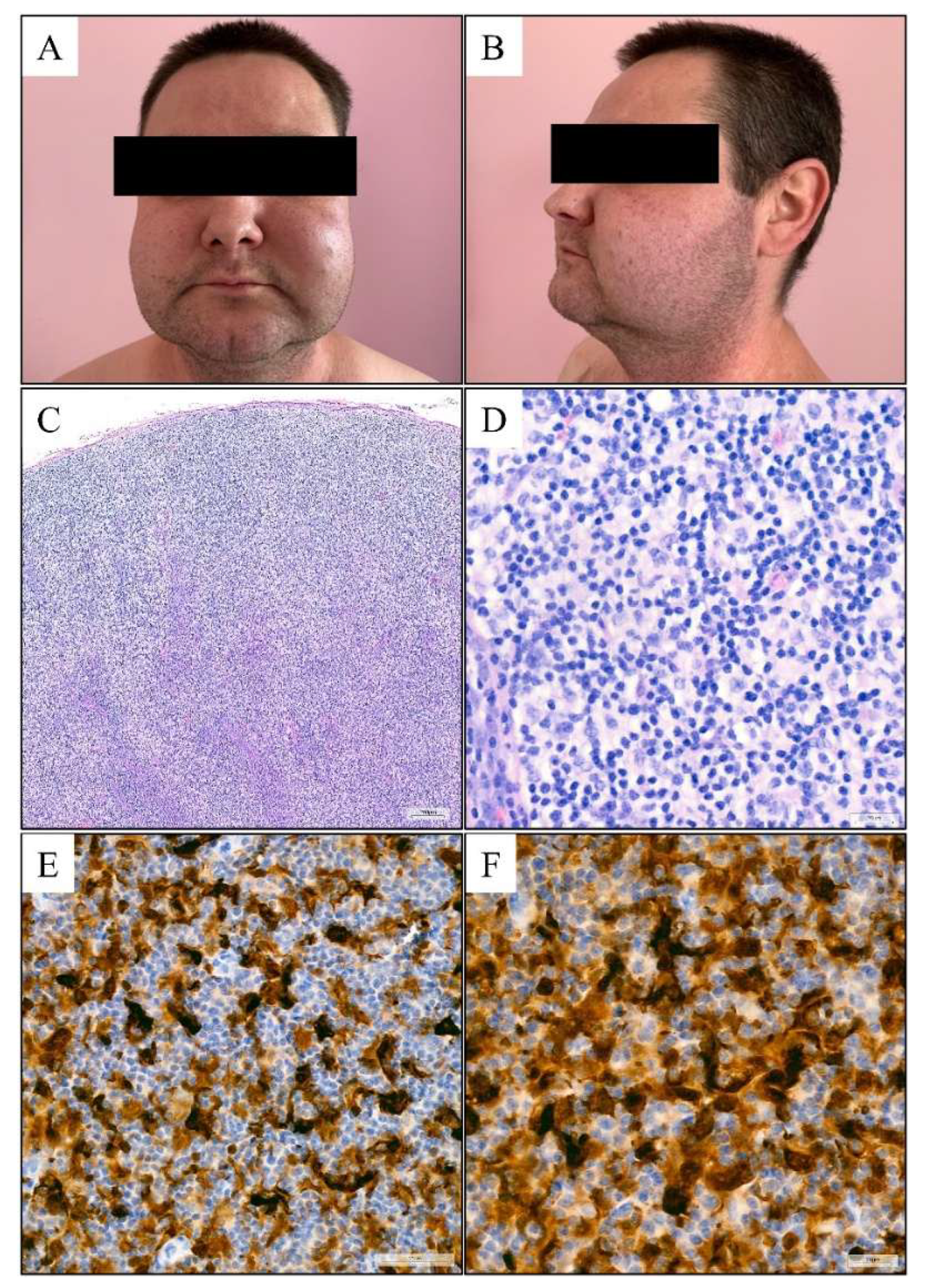
Figure 2.
Reported clinical manifestations of Rosai-Dorfman-Destombes disease. Ranges reflect heterogeneous retrospective cohorts and differing denominators; sites may coexist, and values are not additive.
Figure 2.
Reported clinical manifestations of Rosai-Dorfman-Destombes disease. Ranges reflect heterogeneous retrospective cohorts and differing denominators; sites may coexist, and values are not additive.
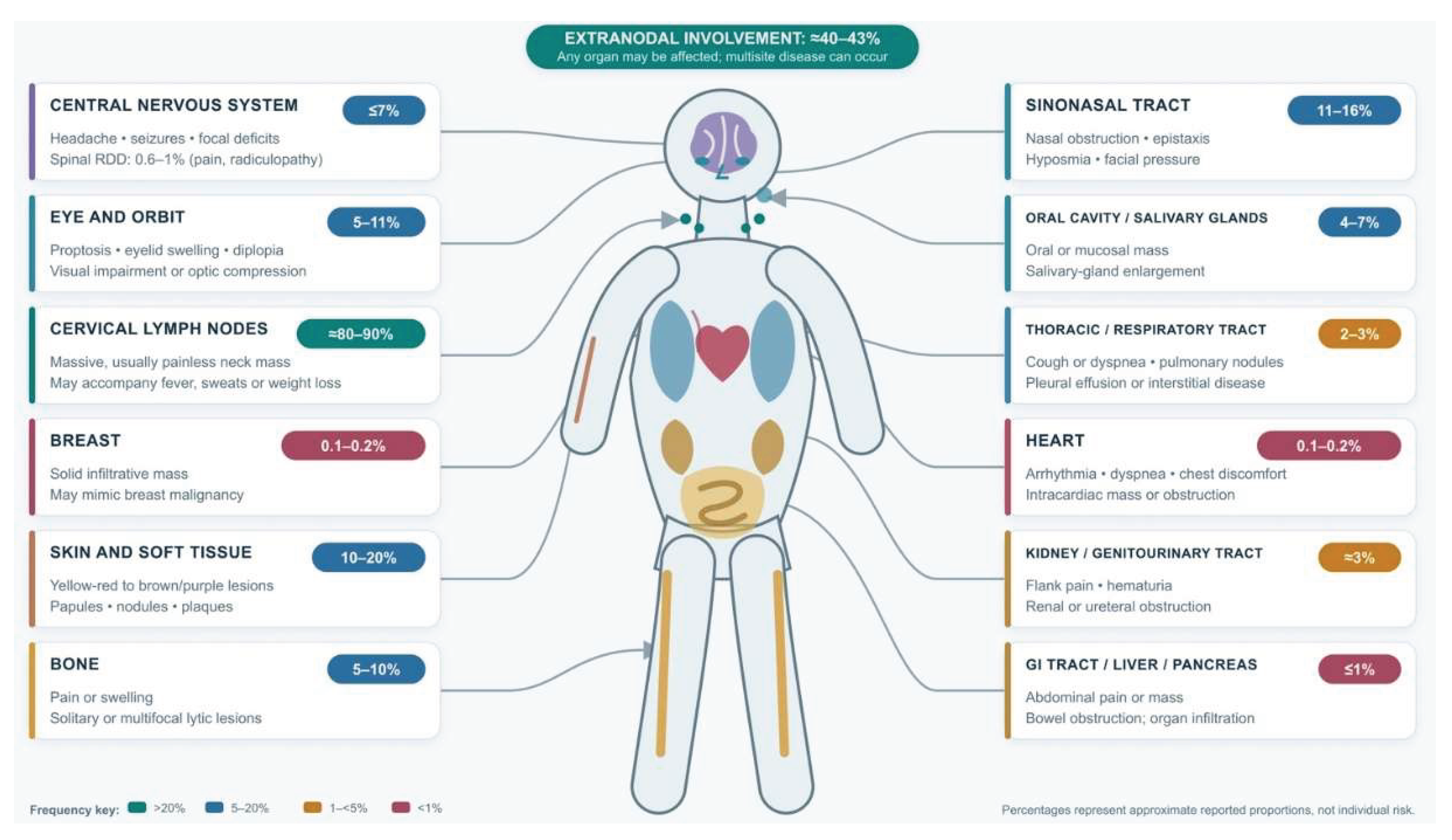
Figure 3.
Cutaneous changes in the course of RDD presenting as single or multiple yellow-red to brown or purple papules, nodules, and/or plaques (A) with some nodules undergo breakdown (B).
Figure 3.
Cutaneous changes in the course of RDD presenting as single or multiple yellow-red to brown or purple papules, nodules, and/or plaques (A) with some nodules undergo breakdown (B).
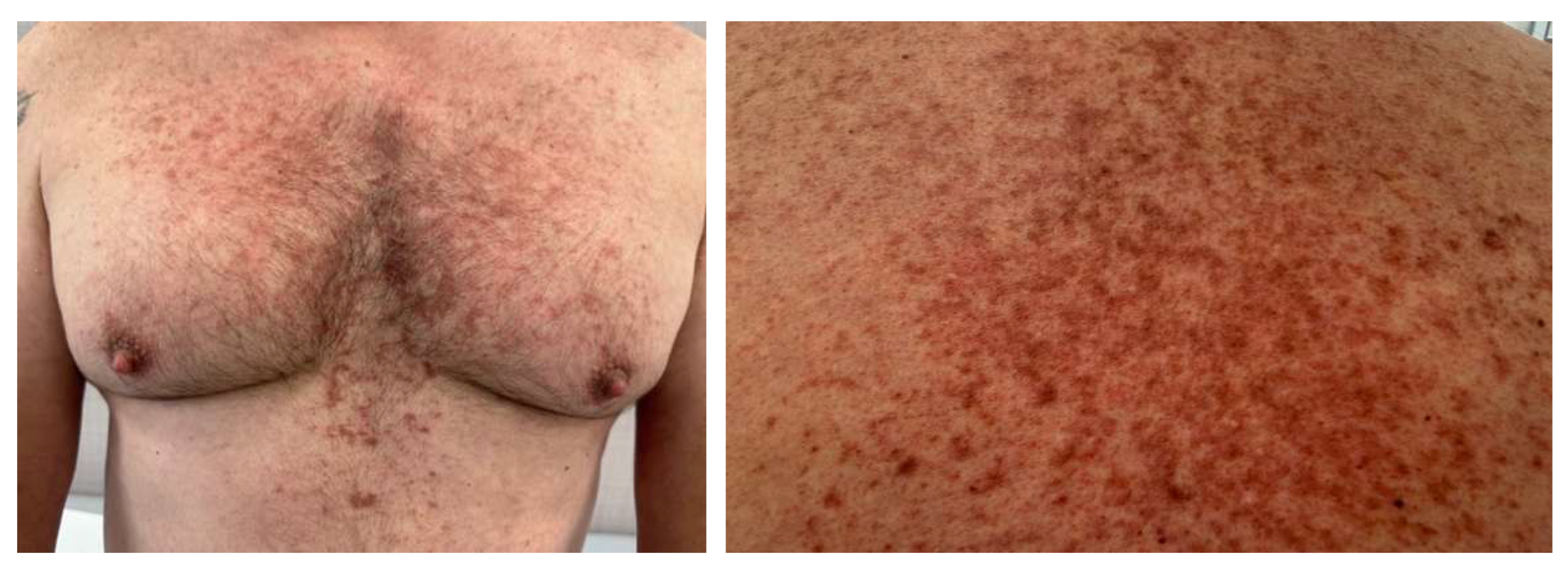

Table 1.
Diagnostic and clinical features of Rosai–Dorfman–Destombes disease and major differential diagnoses. Mixed histiocytoses and disease overlap can occur. Castleman disease and IgG4-related disease are not histiocytic neoplasms but are included because they are important clinicopathologic mimics. Molecular findings may guide therapy but do not replace morphologic and clinicoradiologic correlation.
Table 1.
Diagnostic and clinical features of Rosai–Dorfman–Destombes disease and major differential diagnoses. Mixed histiocytoses and disease overlap can occur. Castleman disease and IgG4-related disease are not histiocytic neoplasms but are included because they are important clinicopathologic mimics. Molecular findings may guide therapy but do not replace morphologic and clinicoradiologic correlation.
| Entity | Diagnostic framework and clinical pattern | Histopathology | Immunophenotype and molecular findings | Laboratory and imaging pattern | Key discriminator from RDD |
|
Rosai–Dorfman–Destombes disease [1,2] Non-Langerhans histiocytic neoplasm |
• Required synthesis: compatible lesional histology plus the correct clinical/radiologic setting and exclusion of a dominant malignant or infectious process; histology alone may represent a minor reactive component. • Classic nodal phenotype: massive, painless bilateral cervical lymphadenopathy; fever, night sweats and weight loss may occur. • Extranodal disease: ~43%; skin/subcutis, upper airway/sinonasal tract, orbit, bone and dura/CNS are characteristic sites. Multisystem disease occurs in a minority. |
• Nodal disease: expanded sinuses containing large histiocytes with abundant pale or ‘watery-clear’ cytoplasm, round/oval hypochromatic nuclei and prominent nucleoli; plasma-cell-rich background. • Emperipolesis of intact lymphocytes, plasma cells and/or neutrophils is strongly supportive but may be focal, especially extranodally, and is neither obligatory nor entirely specific. • Extranodal lesions are often fibrotic, with sparse lesional histiocytes and less conspicuous emperipolesis. |
• Typical IHC: S100+, CD68+, CD163 variable/usually+, fascin+; OCT2 and cyclin D1 often positive; CD1a− and langerin/CD207−. • Molecular: activating MAPK-pathway alterations in a subset, including KRAS, NRAS, MAP2K1 and ARAF; a driver alteration supports clonality/therapy selection but is not required for diagnosis. |
• No specific serum biomarker. Possible neutrophilic leukocytosis, anemia, elevated ESR/CRP and polyclonal hypergammaglobulinemia; abnormalities are variable and reflect disease burden/inflammation. • FDG-PET/CT is useful for staging and response assessment; lesions are often FDG-avid but the pattern is not specific. |
• Best-recognized constellation: large pale S100+/CD68+ histiocytes with emperipolesis and absence of CD1a/CD207, integrated with RDD-compatible organ distribution. • Emperipolesis preserves the internalized cell; it must not be confused with destructive phagocytosis/hemophagocytosis. |
|
Erdheim–Chester disease [43] L-group histiocytic neoplasm |
• Usually adults aged 40–70 years; bone pain, diabetes insipidus, orbital/CNS, cardiovascular, pulmonary and retroperitoneal manifestations. • Diagnosis requires characteristic tissue findings in the appropriate radiologic/clinical context; biopsy is recommended even when imaging is highly suggestive. |
• Bland foamy/lipid-loaded histiocytes in a xanthogranulomatous infiltrate with variable fibrosis; Touton giant cells are common but not required. • Emperipolesis is not a dominant feature. |
• CD68+, CD163+, factor XIIIa+; CD1a− and CD207−; S100 usually negative or only weak/focal. • BRAF V600E in roughly one-half of cases; most cases harbour another MAPK–ERK pathway alteration when sensitive testing is used. |
• Inflammatory markers may be elevated; organ-specific renal, cardiac and endocrine abnormalities are common. • Near-symmetric distal femoral/proximal and distal tibial osteosclerosis or radionuclide uptake; perinephric ‘hairy kidney’ and periaortic ‘coated aorta’ are highly characteristic. |
• Long-bone osteosclerosis plus perinephric/periaortic infiltration strongly favours ECD. • The usual S100-negative/focal phenotype, factor XIIIa positivity and xanthogranulomatous fibrosis contrast with RDD. |
|
Langerhans cell histiocytosis [160] L-group dendritic-cell neoplasm |
• Unifocal or multifocal bone lesions, skin disease, pituitary involvement/diabetes insipidus, multisystem disease; isolated pulmonary LCH is strongly smoking-associated in adults. • Definitive diagnosis generally requires lesional biopsy with the characteristic pattern and CD1a/CD207-positive cells; a typical pulmonary clinicoradiologic presentation is a limited exception. |
• Intermediate-sized Langerhans cells with reniform nuclei, longitudinal nuclear grooves and eosinophilic cytoplasm in an eosinophil-rich mixed inflammatory background. • Birbeck granules may be seen ultrastructurally but are not needed when langerin is demonstrated. |
• CD1a+, langerin/CD207+, S100+ and usually CD68 with a Golgi-dot pattern; CD163, CD14 and OCT2 generally negative. • BRAF V600E, MAP2K1 and other MAPK–ERK alterations are frequent. |
• No single diagnostic serum marker; cytopenias, liver dysfunction or inflammatory abnormalities depend on organ involvement. • Lytic bone lesions; pulmonary nodules and cysts with upper/mid-lung predominance; pituitary stalk thickening may occur. |
• Diffuse CD1a and CD207 expression is decisive and excludes conventional RDD. • Nuclear grooves and an eosinophil-rich infiltrate support LCH; emperipolesis is not characteristic. |
|
Castleman disease [161,162] Lymphoproliferative disorder; UCD, iMCD or HHV-8–associated MCD |
• UCD: one nodal station, often asymptomatic or causing local compression. MCD: multicentric adenopathy with systemic inflammation, organomegaly and fluid accumulation. • iMCD requires both major criteria (compatible excisional-node histology and nodes ≥1 cm in ≥2 stations), ≥2 minor criteria including ≥1 laboratory criterion, and exclusion of mimics. |
• Regressed/atretic germinal centres, expanded ‘onion-skin’ mantle zones, prominent follicular dendritic-cell networks and penetrating hyalinized ‘lollipop’ vessels; interfollicular hypervascularity. • Plasmacytic-spectrum disease shows hyperplastic germinal centres and sheet-like polytypic plasmacytosis. |
• No lesional S100+/CD68+ histiocytic population defining the process. Plasma cells are usually polytypic in iMCD. • HHV-8 LANA-1 positivity establishes HHV-8–associated MCD in the correct setting; HHV-8 must be excluded for iMCD. |
• MCD: elevated CRP/ESR, anemia, hypoalbuminemia, renal dysfunction/proteinuria, thrombocytosis or thrombocytopenia and polyclonal hypergammaglobulinemia; an IL-6-driven inflammatory syndrome is typical. • TAFRO phenotype: thrombocytopenia, anasarca, fever/inflammation, reticulin myelofibrosis/renal dysfunction and organomegaly. |
• Follicular/vascular architectural abnormalities—not sinus expansion by S100+ histiocytes—define Castleman disease. • Emperipolesis is not a defining feature. Castleman-like changes require exclusion of infection, autoimmune disease, lymphoma and IgG4-RD. |
|
IgG4-related disease [163] Immune-mediated fibroinflammatory disease |
• Tumefactive, often multiorgan disease affecting pancreas/biliary tree, salivary and lacrimal glands, orbit, kidney, lung, aorta and retroperitoneum. • Diagnosis integrates typical organ involvement, morphology and IgG4 immunostaining; classification criteria are not identical to clinical diagnostic criteria. |
• Dense lymphoplasmacytic infiltrate, storiform fibrosis and obliterative phlebitis are the core pattern; tissue eosinophilia may occur. • Lymph nodes may lack storiform fibrosis and obliterative phlebitis, making nodal disease less specific. |
• Increased IgG4+ plasma cells and an IgG4/IgG ratio usually >40% support the diagnosis, but absolute cut-offs are organ- and specimen-specific and are insufficient alone. • No defining S100+/OCT2+/cyclin D1+ histiocytic proliferation. |
• Serum IgG4 may be elevated but can be normal and lacks specificity. Polyclonal hypergammaglobulinemia, eosinophilia, elevated IgE and hypocomplementemia (especially renal disease) may occur. • Imaging reflects organ-specific swelling, masses or fibroinflammatory encasement. |
• Storiform fibrosis/obliterative phlebitis plus a typical organ pattern favours IgG4-RD. • RDD may contain abundant IgG4+ plasma cells and can exceed a 40% IgG4/IgG ratio; this finding alone must not be used to reclassify RDD as IgG4-RD. |
|
Hemophagocytic lymphohistiocytosis [164] Severe hyperinflammatory syndrome |
• Acute/subacute fever, splenomegaly, hepatitis, cytopenias, coagulopathy and multiorgan dysfunction; often triggered by infection, malignancy or autoimmune/autoinflammatory disease in adults. • HLH-2004: molecular diagnosis or ≥5 of 8 criteria; in adults, apply with clinical judgement because the criteria were developed in children. |
• Activated macrophages with hemophagocytosis may be present in marrow, spleen, liver or nodes, but hemophagocytosis is neither required nor specific and may be absent early. • No characteristic RDD-like sinus histiocytosis or lesional emperipolesis phenotype. |
• No disease-defining IHC profile. Genetic and functional testing (including cytotoxicity/NK-cell studies) supports primary/familial forms. • Macrophage and T-cell activation, rather than a discrete S100+ histiocytic tumour population, drives the syndrome. |
• HLH-2004 variables: cytopenias in ≥2 lineages, triglycerides ≥3.0 mmol/L and/or fibrinogen ≤1.5 g/L, ferritin ≥500 µg/L, sCD25 ≥2400 U/mL and low/absent NK activity, plus fever and splenomegaly. • Adult ferritin is often far above the formal threshold but is not specific by itself. |
• Rapid systemic hyperinflammation, cytopenias, hypofibrinogenemia/hypertriglyceridemia and very high ferritin favour HLH. • Hemophagocytosis entails destructive ingestion; RDD emperipolesis contains viable intact cells. |
|
Histiocytic sarcoma [165] Aggressive histiocyte/macrophage neoplasm |
• Aggressive nodal or extranodal destructive mass, often in gastrointestinal tract, skin/soft tissue or spleen; localized or disseminated disease with constitutional symptoms. • Diagnosis requires malignant histiocytic morphology, confirmation of macrophage lineage and broad exclusion of lymphoma, melanoma, carcinoma, myeloid and dendritic-cell neoplasms. |
• Sheets of large pleomorphic cells with irregular vesicular nuclei, prominent nucleoli, abundant eosinophilic cytoplasm, marked atypia and atypical mitoses; necrosis and hemophagocytosis may occur. • Destructive growth and high-grade cytology are central. |
• CD163+, CD68+, lysozyme+ and PU.1+; S100 variable, often focal/weak; CD1a and CD207 usually negative. High proliferative fraction is common. • MAPK-pathway and CDKN2A alterations may occur but are not disease-specific. |
• Possible cytopenias, elevated LDH and inflammatory markers, depending on tumour burden and marrow/organ involvement. • Imaging shows destructive mass lesions rather than the stereotyped RDD distribution. |
• Marked pleomorphism, atypical mitoses, necrosis, destructive invasion and high proliferation favour histiocytic sarcoma. • RDD histiocytes are cytologically bland; emperipolesis of intact cells is supportive of RDD but not a hallmark of sarcoma. |
Abbreviations. CNS, central nervous system; CRP, C-reactive protein; ECD, Erdheim–Chester disease; ESR, erythrocyte sedimentation rate; FDG-PET/CT, 18F-fluorodeoxyglucose positron-emission tomography/computed tomography; HHV-8, human herpesvirus 8; HLH, hemophagocytic lymphohistiocytosis; iMCD, idiopathic multicentric Castleman disease; LCH, Langerhans cell histiocytosis; MCD, multicentric Castleman disease; RDD, Rosai–Dorfman–Destombes disease; TAFRO, thrombocytopenia, anasarca, fever, reticulin myelofibrosis/renal dysfunction and organomegaly; UCD, unicentric Castleman disease.
Table 2.
Therapeutic clinical trials and informative published treatment cohorts in Rosai–Dorfman–Destombes disease.
Table 2.
Therapeutic clinical trials and informative published treatment cohorts in Rosai–Dorfman–Destombes disease.
| Study / author | NCT or registry | Phase | Investigational treatment | No. of cases | ORR | CR | Median PFS | Median OS |
| A. Prospective trials with published or posted efficacy results | ||||||||
| Lenalidomide and dexamethasone in RDD — Chang et al., 2024 [131] | NCT04924647 | II | Lenalidomide + dexamethasone | 23 RDD | 87% (20/23) | 30% (7/23) | Not reached; 2-y PFS 69% | Not reached; 2-y OS 100% |
| Single-agent cobimetinib in histiocytic disorders — Diamond et al., 2019; final registry results [166] | NCT02649972 | II | Cobimetinib | 35 enrolled; 29 evaluable. Initial report: 18 total, 2 RDD | 74.3% ITT (26/35); 89.7% evaluable (26/29) | 60.0% ITT (21/35); 72.4% evaluable (21/29) | Not reached in initial report; 1-y PFS 94%. Final registry: NR | NR; OS was not a reported final-registry efficacy endpoint |
| Luvometinib in histiocytic neoplasms — Cao et al., 2025 [148] | No NCT; CTR20221069 / ChiCTR2300067955 | II | Luvometinib | 30 enrolled; 29 efficacy-evaluable; RDD n=2 | 82.8% overall; RDD 50% (1/2) | 48.3% overall; RDD-specific NR | Not reached overall; 12-mo PFS 74.4% | Not reached overall; 12-mo OS 100% |
| Clofarabine for recurrent/refractory LCH and related disorders — Degar et al., 2026 [117] | NCT02425904 | II | Clofarabine | 25 total; non-LCH n=5, 4 evaluable; RDD eligible, enrolled RDD count NR | Non-LCH stratum 75% (3/4); not RDD-specific | 0% in non-LCH stratum (all 3 responses were PR) | Median NR; 1-y PFS 50% in non-LCH stratum | Median NR; 1-y OS 75% in non-LCH stratum |
| B. Ongoing or results-not-posted registered interventional studies | ||||||||
|
Multicentre RD-regimen study — Cao, study chair Recruiting |
NCT07187167 | II | Lenalidomide + dexamethasone; lenalidomide maintenance | 40 planned RDD | Not available | Not available | Not available | Not available |
|
NACHO-COBI — Allen, study chair Recruiting histiocytosis |
NCT04079179 | II | Cobimetinib | 90 planned; RDD eligible | Not available | Not available | Not available | Not available |
|
Mirdametinib in histiocytic disorders — Kumar/Bartlett, PIs [168] Recruiting |
NCT06153173 | II | Mirdametinib | 40 planned; RDD eligible | Not available | Not available | Not available | Not available |
|
Central China RDD Registry — Du, study chair Interventional phase IV; status unknown |
NCT05284942 | IV | Mycophenolate mofetil + oral prednisone | 20 planned; RDD/LCH | Not available | Not available | Not available | Not available |
|
Q702 in haematologic malignancies — Abeykoon, PI Recruiting basket |
NCT06712810 | I | Q702 (Axl/Mer/CSF1R inhibitor) | 46 planned; recurrent/refractory RDD eligible | Not available | Not available | Not available | Not available |
| C. Informative published retrospective cohorts (not prospective RDD trials) | ||||||||
| Cobimetinib in RDD by KRAS/MEK status — Abeykoon et al., 2022 [145] | None | Retrospective | Cobimetinib | 16 RDD | 63% (10/16) | 31% (5/16) | NR; 1-y PFS 100% with KRAS/MEK alteration vs 29% without | NR |
| Trametinib multicentre analysis — Aaroe et al., 2023 [147] | None; observational data included some patients on NCT02478931 | Retrospective | Trametinib (occasionally with another agent) | 26 total; RDD n=3, ECD/RDD n=5; 17 evaluable overall | 71% overall (12/17); RDD-specific summary NR | 12% overall (2/17); both CRs in ECD/RDD overlap | Not reached; 1-y PFS 94.1% overall | Not reached; 3-y OS 90.1% overall |
| Mayo Clinic RDD cohort — Goyal et al., 2020 [33] | None | Retrospective | Cladribine subgroup | 64 RDD total; cladribine n=6 | 67% for cladribine | NR | NR; no relapse among responders at median 16-mo follow-up | NR; no RDD-attributed deaths among 49 with complete follow-up |
| Clofarabine salvage series — Simko et al., 2014 [126] | None | Retrospective | Clofarabine | 18 total; RDD n=3 | RDD 100% (3/3) | RDD 67% (2/3) | RDD median NR; whole-cohort 1-y PFS 62% | RDD median NR; whole-cohort 1-y OS 94% |
| Cladribine in non-Langerhans histiocytosis — Riller et al., 2026 [116] | None | Retrospective | Cladribine | 21 total; RDD subgroup size not stated in indexed abstract | RDD: 70% clinical; 30% radiologic | NR | NR; among all radiologic responders, median time to relapse 18 mo | NR |
Notes: ORR is CR/CMR plus PR/PMR, according to the study-specific criteria. NR means not reported; ‘not reached’ is used only when explicitly stated. Landmark PFS/OS estimates are shown because they must not be converted to medians. NCT02649972 final registry results provide overall basket counts but no histology-stratified final response. NCT02425904 reports a non-LCH stratum in which RDD was eligible, but not RDD-specific outcomes. For NCT04924647, the publication enrolled 23 patients, whereas the older registry record lists an estimated sample of 16. Abbreviations: CMR, complete metabolic response; CR, complete response; CSF1R, colony-stimulating factor 1 receptor; ECD, Erdheim–Chester disease; ITT, intention-to-treat; LCH, Langerhans cell histiocytosis; mo, months; ORR, overall response rate; OS, overall survival; PFS, progression-free survival; PI, principal investigator; PMR, partial metabolic response; PR, partial response; RD, lenalidomide–dexamethasone; RDD, Rosai–Dorfman–Destombes disease; y, years.
Disclaimer/Publisher’s Note: The statements, opinions and data contained in all publications are solely those of the individual author(s) and contributor(s) and not of MDPI and/or the editor(s). MDPI and/or the editor(s) disclaim responsibility for any injury to people or property resulting from any ideas, methods, instructions or products referred to in the content. |
© 2026 by the authors. Licensee MDPI, Basel, Switzerland. This article is an open access article distributed under the terms and conditions of the Creative Commons Attribution (CC BY) license (http://creativecommons.org/licenses/by/4.0/).
Copyright: This open access article is published under a Creative Commons CC BY 4.0 license, which permit the free download, distribution, and reuse, provided that the author and preprint are cited in any reuse.