

Submitted:
23 September 2026
Posted:
24 September 2026
You are already at the latest version
Abstract
Meissner corpuscles are specialized cutaneous mechanosensory end organs primarily located in the dermal papillae of human glabrous skin and specialized mucocutaneous territories. Traditionally regarded as rapidly adapting tactile receptors composed of a sensory axon surrounded by passive lamellar cells, current evidence supports a more complex organization in which the axon, terminal glial cells (TGCs), extracellular matrix, and endoneurial capsule form an integrated mechanosensory unit. This review provides an updated synthesis of the development, anatomical distribution, structural organization, molecular phenotype, mechanotransduction, functional neurobiology, aging, and pathological alterations of human Meissner corpuscles. Particular attention is given to the molecular identity of TGCs and to their emerging role as active components of corpuscular function. PIEZO2 remains the best-established mediator of primary mechanotransduction in the sensory axon, whereas human TGCs express several mechanosensing-associated proteins and experimental evidence suggests a potential glial contribution to corpuscular function. Human studies also demonstrate substantial regional and age-related variability in corpuscular density and phenotype. Importantly, Meissner corpuscles undergo quantitative, structural, immunophenotypic, and molecular alterations in a broad spectrum of peripheral and central neurological disorders. Recent evidence of disease-associated molecular pathology, including intracorpuscular phosphorylated TDP-43 in amyotrophic lateral sclerosis, further expands their potential relevance as accessible peripheral indicators of neurological disease. Finally, emerging three-dimensional imaging, tissue-clearing, spatial molecular profiling, functional imaging, and quantitative digital approaches provide new opportunities for investigating human corpuscular organization. Collectively, current evidence supports viewing the Meissner corpuscle as a dynamic neuro-glial mechanosensory organ whose biological and clinical significance extends beyond its classical role in discriminative touch.
Keywords:
Meissner corpuscle
; terminal glial cells
; mechanotransduction
; PIEZO2
; mechanosensation
; tactile receptors
; glabrous skin
; sensory neuropathies
; neurodegenerative diseases
; touch
1. Introduction
Touch is fundamental to object exploration, skilled manipulation, postural control, and social interaction. In humans, its discriminative component depends on specialized low-threshold mechanoreceptive systems that transform skin deformation into precisely patterned neural activity [1,2,3]. Among cutaneous sensory end organs, Meissner corpuscles occupy a distinctive position because of their preferential localization in regions of high tactile acuity and their association with rapidly adapting type 1 low-threshold mechanoreceptive afferents [4,5]. Their study therefore lies at the intersection of human anatomy, peripheral sensory neurobiology, and the mechanobiology of touch.
Meissner corpuscles are small, encapsulated sensory organs located primarily within the dermal papillae of glabrous skin. They are especially abundant in the digital pads and palmar skin but also occur in plantar and selected mucocutaneous territories [6,7,8,9]. Myelinated Aβ afferents supplying these organs lose their myelin sheath before entering the corpuscle and form complex terminal profiles among stacks of lamellar cells enclosed by a delicate connective-tissue capsule [10,11,12]. Functionally, this arrangement is associated with sensitivity to dynamic skin deformation, motion across the skin, and low-frequency flutter, properties that contribute to grip control, texture exploration, and fine tactile discrimination [1,2,4].
For much of the twentieth century, the Meissner corpuscle was portrayed as a relatively simple encapsulated receptor in which the axon constituted the active sensory element and the non-neuronal cells provided structural support. This interpretation no longer accommodates the available anatomical and molecular evidence. Ultrastructural, immunohistochemical, and three-dimensional studies have disclosed a highly ordered organ comprising distinct but interacting axonal, terminal glial cells, extracellular-matrix, and capsular compartments [10,11,12,13]. The spatial organization and molecular specialization of these compartments indicate that mechanical signals reach the sensory axon within a highly organized corpuscular microenvironment rather than an isolated axonal ending.
This conceptual revision has been reinforced by advances in molecular mechanotransduction. The identification of PIEZO2 as a principal mechanically activated ion channel in low-threshold touch afferents provided a molecular link between tissue deformation and neuronal excitation [5,14,15]. PIEZO2 immunoreactivity has subsequently been demonstrated in human cutaneous sensory formations, including Meissner corpuscles, although its precise compartmental distribution and functional interactions with other mechanosensitive molecules remain incompletely resolved [16,17,18]. At the same time, the lamellar cells (widely regarded as specialized terminal Schwann-related glia) have emerged as biologically active elements with potential structural, trophic, and modulatory functions [12,19].
The biological relevance of Meissner corpuscles extends beyond normal tactile physiology. Their density, dimensions, morphology, and molecular phenotype vary across anatomical territories and throughout the human lifespan [7,8,9,20]. Corpuscular loss and structural disorganization accompany aging and have also been described in diabetic and other peripheral neuropathies, following nerve injury, and during reinnervation [6,20,21]. Because these sensory structures are superficially located and can be evaluated in skin-biopsy material, they have attracted interest as structural correlates of sensory dysfunction and as potential indicators of peripheral nerve integrity [22,23]. Nevertheless, the extent to which corpuscular alterations are disease-specific, precede functional impairment, or can serve as clinically useful biomarkers remains uncertain.
Despite more than a century and a half of investigation, knowledge of human Meissner corpuscles remains fragmented. Most contemporary syntheses address them only as one component of the broader cutaneous mechanoreceptor system, whereas the last dedicated review from our group was published in 2012 [6]. Since then, substantial advances have been made in development, molecular phenotyping, mechanosensitive ion-channel biology, terminal glial-cell identity, age-related variation, digital distribution, three-dimensional imaging, and the analysis of corpuscles in disease [11,12,13,18,20,24,25]. An updated, human-focused synthesis is therefore warranted.
Such a synthesis also requires critical attention to the evidential basis of current concepts. The accessibility of animal models has generated important mechanistic insights, but species differences in skin organization, corpuscular distribution, cellular composition, and sensory coding limit their direct extrapolation to humans [13,26,27]. This limitation is illustrated by the dual TrkB-positive and Ret-positive afferent organization reported in murine and avian Meissner-like corpuscles, whereas TrkB and RET colocalize within a single axon in human digital corpuscles [28]. Conversely, studies of human material often provide exceptional anatomical resolution but are constrained by sample availability, variable tissue preservation, heterogeneous anatomical sites, and predominantly descriptive or cross-sectional designs. Integrating these complementary bodies of evidence without conflating them is essential for defining what is established, what remains inferential, and which questions require direct investigation in human tissue.
In this review, we critically examine the human Meissner corpuscle from its classical morphological description to its current interpretation as a molecularly specialized neuroglial mechanosensory organ. We integrate evidence on historical development, anatomical distribution, ontogeny and maturation, age-related change, ultrastructure, cellular and extracellular-matrix composition, mechanotransduction, pathology, and emerging investigative technologies. Particular emphasis is placed on the coordinated contributions of the sensory axon, terminal glial cells, capsule, and extracellular matrix, and on the distinction between direct human observations and model-derived mechanisms. By integrating these domains, we aim to provide an anatomically grounded framework for understanding normal tactile function, sensory decline, and the biological and clinical relevance of human Meissner corpuscles.
2. Historical Perspective
2.1. The Original Description of Meissner Corpuscles
The tactile corpuscles that now bear Meissner’s name were first described by Rudolf Wagner and Georg Meissner in 1852 in the dermal papillae of human glabrous skin, particularly in the hand, and were soon examined in greater detail by Meissner in his monograph on the anatomy and physiology of the skin [29,30]. Their characteristic localization in regions of high tactile acuity led from the outset to the interpretation that they represented specialized peripheral organs of touch.
2.2. Classical Histology and Early Functional Interpretations
Classical histology established the basic organization of the corpuscle as a superficial papillary structure supplied by a myelinated sensory fibre that loses its myelin before forming intracorpuscular terminals among lamellar elements. Detailed light-microscopic studies also demonstrated the complexity of its innervation and the presence and organization of a capsule, providing the anatomical basis for its classification as a rapidly adapting tactile end organ [6,31,32].
2.3. The Ultrastructural Era
A major advance in Meissner-corpuscle research occurred with the introduction of transmission electron microscopy. Ultrastructural studies revealed axonal profiles interdigitating with flattened lamellar cells, specialized axon–glial interfaces and an organized extracellular compartment, establishing that the non-neuronal component is an integral part of corpuscular architecture rather than a simple supporting framework [10,33].
2.4. Immunohistochemistry and the Molecular Revolution
From the late twentieth century onward, immunohistochemistry allowed the neural, glial and connective-tissue compartments to be defined molecularly. Neuronal markers delineated the sensory axon, whereas S100 proteins and vimentin characterized the TGCs; subsequent studies identified a much broader repertoire of cytoskeletal, calcium-binding, trophic and mechanosensing-associated proteins [10,25,34]. Extracellular-matrix and endoneurial markers further demonstrated a specialized corpuscular microenvironment and an endoneurial-type capsule [11].
2.5. From Tactile Receptor to Neuro-Glial Mechanosensory Unit
The identification of mechanosensing-associated molecules, especially PIEZO2, together with the increasingly complex molecular phenotype recognized in TGCs, changed the classical interpretation of the Meissner corpuscle [12,16,35]. These findings support a more integrated view of its neuronal and non-neuronal components. Direct functional evidence for active glial mechanosensation, however, derives principally from experimental models and should not be extrapolated uncritically to humans.
3. Anatomical Distribution of Human Meissner Corpuscles
3.1. General Distribution in Human Glabrous Skin
Human Meissner corpuscles are preferentially located in dermal papillae of glabrous or specialized mucocutaneous skin. Their distribution is markedly heterogeneous: they are particularly abundant in the hand, whereas substantially lower numbers occur in several other anatomical territories traditionally considered tactilely sensitive [6,7].
3.2. Fingertips and Palmar Skin
The fingertips constitute the principal human territory for Meissner corpuscles. Density is highest distally and is greater in fingertip skin than in more proximal digital regions or the palm; substantial regional and interindividual variation is also present [8,36]. Recent mapping of 17 regions of the human hand by our group further supports a strongly non-uniform distribution, with anatomical location and age emerging as the major determinants of corpuscular density [19].
3.3. Plantar Skin
Meissner corpuscles are also present in plantar glabrous skin, including the toes, but their abundance and structural characteristics differ from those of the fingers. Direct comparison of digital skin from the hand and foot confirms that anatomical site must be taken into account when corpuscular density is used for quantitative or clinical studies [9].
3.4. Facial Glabrous Regions
Meissner corpuscles have been reported in the lips and other specialized mucocutaneous territories, although they are much less numerous than in the fingertips and their contribution to regional tactile acuity should not be inferred solely from receptor density [7].
3.5. External Genitalia
Meissner corpuscles or closely related corpuscular morphotypes occur in human external genital skin, but their abundance varies considerably with the precise anatomical site. In the male prepuce, Meissner corpuscles are a prominent sensory corpuscular population and express molecular features associated with mechanosensation, including PIEZO2 [37,38,39].
3.6. Regional and Interindividual Variability
Regional variability is a defining feature of human Meissner-corpuscle distribution. Differences occur not only between body territories but also within the same hand and between individuals; age is an especially important source of variation, whereas sex and laterality appear to have substantially smaller effects in current human datasets [8,19].
3.7. Functional Significance of Distribution Patterns
The density of Meissner corpuscles in distal glabrous skin is consistent with their role in dynamic touch, low-frequency vibration and the sensory control of object manipulation. Nevertheless, receptor density is only one component of tactile performance, which also depends on innervation, receptive-field organization, skin biomechanics and central processing [1,6].
4. Development, Maturation and Ageing
4.1. Chronology of Human Meissner-Corpuscle Development
Human Meissner corpuscles develop relatively late compared with several other cutaneous sensory end organs, and their maturation extends from fetal life into the postnatal period. Systematic immunohistochemical analysis of developing human digital skin demonstrates that histogenesis begins during mid-gestation. Sensory axons reach the dermal papillae at approximately 20–24 weeks of estimated gestational age (WEGA), before recognizable TGCs are present [24].
By approximately 36–40 WEGA, rudimentary Meissner corpuscles become recognizable. Rounded S100-positive Schwann-related cells become associated with the axonal profiles and subsequently acquire vimentin immunoreactivity and a progressively flattened morphology. Maturation continues after birth, and an adult-like morphology and immunohistochemical organization are reached at approximately 8 months, although maturation is not completely synchronous [24,40].
4.2. Molecular Maturation of Corpuscular Components
The structural maturation of human Meissner corpuscles is accompanied by a dynamic molecular program. NCAM is detectable in early axonal profiles, becomes weak or undetectable during late gestation and the early postnatal period, and reappears during the first months after birth, becoming prominent at the axon–TGC interface [24].
Maturation of the extracellular and capsular compartments follows a prolonged temporal sequence. Type IV collagen, as a major component of the basal-lamina specialization of the extracellular matrix, becomes detectable during late gestation and progressively acquires its mature distribution after birth. CD34-positive cells associated with the endoneurial capsule appear postnatally [24].
4.3. Acquisition of Mechanoreceptive Competence
Morphological maturation and acquisition of molecular competence for mechanotransduction are temporally dissociated. PIEZO2 becomes detectable in the axonal component of developing human Meissner corpuscles at approximately 36 WEGA and persists thereafter [17]. Its presence before completion of corpuscular maturation indicates prenatal establishment of molecular mechanotransductive competence.
This should not be interpreted as direct evidence of fully mature fetal receptor physiology. PIEZO2 loss-of-function in humans profoundly impairs discriminative touch and proprioception [15], but direct electrophysiological recordings from developing human Meissner corpuscles are lacking.
4.4. Molecular Control of Meissner-Corpuscle Formation
Experimental studies identify neurotrophin signaling as a major determinant of Meissner-corpuscle development. Meissner corpuscles fail to develop normally in mice lacking functional TrkB signaling, and BDNF deficiency profoundly disrupts development whereas NT-4 deficiency does not reproduce the same phenotype [41,42]. TrkB expression in human TGCs provides direct human anatomical evidence for this signaling system, although its developmental function in human TGCs remains unproven [43].
Intrinsic transcriptional programs also contribute. Shox2 is required for specification of subsets of low-threshold mechanoreceptive neurons [44]. In c-Maf-deficient mice, Meissner corpuscles are markedly reduced and remaining corpuscles display rudimentary or abnormal morphology [45]. Ret signaling has a distinct role: Meissner corpuscles can remain present in Ret-deficient mice but are frequently underdeveloped or disorganized [46].
Target-derived signals from glabrous skin also instruct end organ morphogenesis. BMP5 and BMP7 are enriched in developing glabrous skin, and disruption of type I BMP-receptor signaling in sensory neurons results in abnormal Meissner-corpuscle morphology in mice [47]. An equivalent BMP-dependent mechanism has not been demonstrated directly in humans.
4.5. Activity-Dependent Maturation
Mechanically evoked neuronal activity contributes to developmental refinement of cutaneous mechanosensory end organs. In experimental models, loss of Piezo2 does not prevent initial Meissner-corpuscle formation but produces enlarged, structurally disorganized end organs with abnormal sensory innervation [48]. Human PIEZO2 expression precedes completion of structural maturation [17], but activity-dependent maturation has not been demonstrated directly in humans.
4.6. Integrated Developmental Model
Current evidence supports a sequential but overlapping model in which intrinsic neuronal specification, neurotrophin signaling, target-derived signals, glial differentiation and mechanically evoked activity contribute at different stages to the assembly of the mature sensory end organ. Human evidence defines a complementary chronology: axonal arrival during mid-gestation, formation of rudimentary corpuscles during late gestation, subsequent TGC differentiation, prenatal PIEZO2 expression and continued postnatal maturation of axon–TGC, extracellular-matrix and capsular compartments [17,24].
4.7. Dependence of Meissner-Corpuscle Integrity on Sensory Innervation
The structural and molecular integrity of adult Meissner corpuscles is critically dependent on the preservation of sensory innervation. Human studies demonstrate that alterations affecting the supplying sensory axon are accompanied by abnormalities of corpuscular immunohistochemical phenotype, morphology, size and density [49,50,51]. Anatomical persistence of a corpuscle should therefore not be interpreted as evidence of preserved molecular integrity or normal sensory function.
Experimental studies in rats provide mechanistic evidence concerning regenerative capacity. Recovery after denervation depends strongly on developmental stage [52,53]. The broader experimental literature indicates that surviving terminal Schwann cells, capsular structures and basal-lamina pathways can provide a substrate for reinnervation [54]. Direct human evidence demonstrates reoccupation and remodelling of residual corpuscular structures by regenerating sensory axons after peripheral nerve injury [55].
4.8. Ageing Changes
Ageing is one of the major biological determinants of human Meissner-corpuscle organization. Human morphometric and immunohistochemical studies demonstrate progressive remodelling of multiple components rather than simple numerical loss [11,20].
Quantitative changes are among the most consistently documented features. Ageing is associated with a progressive reduction in corpuscular density and changes in size, shape and position [20]. In-vivo reflectance confocal microscopy confirms an inverse relationship between age and density [23], while systematic mapping of the human hand identifies age and anatomical location as major determinants [19].
Age-related remodelling also affects the neural and TGC compartments and molecular profiles associated with mechanosensation and neurotrophic signaling [20]. SOX10 persists in TGCs throughout life, indicating retention of Schwann-lineage identity despite selective molecular remodelling [56]. The endoneurial capsule also changes, with reduced and discontinuous CD34 immunoreactivity in older individuals [11].
Age-related corpuscular changes occur in parallel with deterioration of tactile performance, although the contribution of corpuscular aging cannot be isolated from concurrent changes in skin biomechanics, peripheral nerves and central somatosensory processing [19,23,57]. Consequently, quantitative studies require anatomically standardized and age-adjusted sampling.
5. Structural Organization of Human Meissner Corpuscles
5.1. General Architecture
Human Meissner corpuscles are small, encapsulated sensory end organs located predominantly within the dermal papillae of glabrous skin, immediately beneath the epidermis. In conventional two-dimensional sections, mature corpuscles typically appear elongated, ovoid or ellipsoidal, with their major axis oriented approximately perpendicular to the skin surface (Figure 1). Their dimensions and apparent morphology vary according to anatomical site, age, individual differences and plane of section [8,58,59].
5.2. Shape and Three-Dimensional Organization
The classical morphology of Meissner corpuscles has been derived predominantly from two-dimensional histological and ultrastructural sections. Multiple intracorpuscular axonal profiles may represent branches or different segments of the same parent afferent and therefore cannot, by themselves, establish multiple innervation [33,58,59].
Recent volumetric approaches have substantially refined this view. In mice, two molecularly distinct Aβ-LTMR populations associated with TrkB and Ret can innervate Meissner corpuscles [26]. High-resolution FIB-SEM subsequently demonstrated both doubly and singly innervated mouse Meissner corpuscles and complex relationships between intracorpuscular axons and lamellar cells [13].
Three-dimensional FIB-SEM analysis of the avian Meissner-like (Grandry) corpuscle similarly revealed afferent endings interdigitated with stacked lamellar cells, with individual corpuscles receiving either one or two afferents [27].
These experimental observations establish that dual afferent innervation is a genuine biological feature in at least some vertebrate species. In human digital Meissner corpuscles, however, the great majority are supplied by a single parent axon despite multiple intracorpuscular neural profiles, and TrkB and RET can coexist within the same afferent [28].
5.3. The Axonal Compartment
Human Meissner corpuscles are supplied by myelinated Aβ sensory afferents that lose their myelin sheath before or upon entering the corpuscle and subsequently form complex intracorpuscular terminal profiles among TGCs [33,58,59]. Multiple profiles should not automatically be interpreted as multiple innervation; recent analysis indicates that the great majority of human digital Meissner corpuscles are supplied by a single parent axon [28].
This organization contrasts with experimental models in which singly and doubly innervated corpuscles are directly demonstrated [13,26,27].
After losing its myelin sheath, the parent afferent follows a complex and highly convoluted intracorpuscular course. True terminal branching may also occur, but conventional two-dimensional sections do not always allow branching to be distinguished unequivocally from repeated profiles of a tortuous axonal trajectory. A complete high-resolution reconstruction of this trajectory in the human Meissner corpuscle remains lacking.
5.4. The Terminal Glial Cells (TGCs)
TGCs constitute the principal non-neuronal cellular component of the Meissner corpuscle. In mature human corpuscles, they are flattened cells arranged in close spatial relationship with intracorpuscular axonal profiles, producing the characteristic lamellar organization observed by light and electron microscopy [33,58,59].
Developmental and immunohistochemical evidence supports a Schwann-cell lineage for TGCs. During human corpuscular development, S100-positive Schwann-related cells become associated with the sensory axon and progressively acquire the flattened morphology characteristic of mature TGCs [24].
5.5. Axon-TGC Relationships
A defining structural feature of the human Meissner corpuscle is the intimate spatial relationship between intracorpuscular axonal profiles and TGCs. Classical electron microscopy demonstrated extensive areas of close apposition between the axolemma and TGC membranes [33]. From human ultrastructural evidence alone, these contacts should be interpreted primarily as anatomical specializations.
Recent three-dimensional studies in experimental mechanosensory corpuscles provide a mechanistic framework. Mouse Meissner corpuscles show organized axonal protrusions and contacts with adjacent non-neuronal cells [13], while avian Grandry corpuscles provide direct electrophysiological evidence that lamellar cells are mechanically responsive and can influence afferent activity [27]. Equivalent functional specializations have not been demonstrated directly in human TGCs.
Taken together, the close axon–TGC relationships observed in human Meissner corpuscles and mechanistic evidence from experimental corpuscular models support the possibility that corpuscular architecture contributes to transmission and preprocessing of mechanical forces before or during activation of the sensory afferent; this biomechanical role remains unproven directly in humans.
5.6. The Endoneurial Capsule
The human Meissner corpuscle is surrounded by a thin endoneurial capsule that separates the corpuscular core from the surrounding connective tissue of the dermal papilla [11,58,59]. This capsule is structurally distinct from the TGC compartment and contains flattened CD34-positive cells [11]. The age-related reduction and discontinuity of the CD34 phenotype indicate remodelling of the capsular compartment during the human lifespan [20].
5.7. Vascular Relationships
The vascular relationships of human Meissner corpuscles remain poorly understood. Their location within dermal papillae places them in close anatomical proximity to the superficial dermal microvasculature, but vessels specifically associated with the corpuscle have not been systematically investigated.
In acute complex regional pain syndrome, both Meissner-corpuscle density and CD31-positive papillary microvessels are reduced compared with healthy controls [60]. These findings demonstrate coexistence of sensory-corpuscular and microvascular abnormalities within the same cutaneous territory but do not establish a direct causal relationship or a specialized neurovascular unit.
5.8. Structural Plasticity
Meissner-corpuscle architecture remains plastic after maturation. Human observations following peripheral nerve injury demonstrate that corpuscular morphology and molecular organization can be profoundly altered when sensory innervation is disrupted, while residual corpuscular structures may persist and subsequently undergo remodelling during reinnervation [49,50,51,55]. Structural recovery does not necessarily imply complete restoration of the original molecular phenotype or sensory function.
6. Molecular Identity of Human Meissner Corpuscles (Table 1)
6.1. General Molecular Organization
The application of immunohistochemical and molecular approaches has profoundly changed the characterization of human Meissner corpuscles. A sensory end organ originally defined primarily by morphology, location and innervation is now recognized as a molecularly heterogeneous structure in which the sensory axon, TGCs, extracellular matrix and endoneurial capsule display distinct but spatially integrated molecular profiles [10,25,34].
Interpretation of these molecular profiles requires caution. Immunohistochemical detection of a protein establishes its presence and cellular or subcellular localization, but does not by itself demonstrate functional activity within the corpuscle. Throughout this section, proteins demonstrated directly in human Meissner corpuscles are distinguished from functional roles established experimentally in other systems.

Table 1.
Molecular Signature of Human Meissner Corpuscles.
| Axons | Terminal glial cells (TGCs) | References |
|---|---|---|
| Cytoskeletal proteins | Cytoskeletal proteins | |
| NFP + | — | [34] |
| — | Vimentin + | [63] |
| — | Nestin + (~7.6%; 5.3–11.6%) | [64] |
| — | GFAP − | [63] |
| Calcium-binding proteins | Calcium-binding proteins | |
| — | S100/S100β + | [63,67] |
| Calbindin-D28k + | Calbindin-D28k + | [34,67] |
| Calretinin + | Calretinin + | [34] |
| Parvalbumin + | Parvalbumin + | [34,67] |
| Neurocalcin + | — | [68] |
| Cytoplasmic proteins | Cytoplasmic proteins | |
| PGP9.5 + | — | [34] |
| NSE + | — | [34] |
| Synaptophysin + | — | [17] |
| Bcl-2 + | Bcl-2 − | [61] |
| Growth factors and related receptors | Growth factors and related receptors | |
| p75NTR + | p75NTR + | [51,65] |
| TrkA + | TrkA + | [51,105] |
| TrkB − | TrkB + | [43] |
| EGFR + | EGFR + | [105] |
| Extracellular matrix-related proteins* | Extracellular matrix-related proteins* | |
| — | Laminin + | [71] |
| — | Collagen IV + | [71] |
| — | Heparan-sulfate proteoglycans + | [11] |
| — | Chondroitin sulfate − | [20] |
| Mechanosensing proteins | Mechanosensing proteins | |
| ASIC1 − | ASIC1 − | [35] |
| ASIC2 + | ASIC2 + (4.1–100%, site-dependent) | [35] |
| ASIC3 − | ASIC3 − | [35] |
| β-ENaC + | β-ENaC − | [35] |
| γ-ENaC + | γ-ENaC − | [35] |
| PIEZO1 + (~56%) | − in digital MCs; +28.6% in one labium minus specimen | [18,35] |
| PIEZO2 + (~85%) | − in digital MCs; +44.2% in one labium minus specimen | [16,18,35] |
| TRPA1 + | + fingers 18.2%; toes 13.3% | [35] |
| TRPC1 + | TRPC1 − | [35] |
| TRPC6 + | TRPC6 − | [35] |
| TRPV4 + | + 5.7–14.9%, site-dependent; − lips/nipple | [35] |
| TMEM150C/Tentonin-3 + | weak + (<2%) | [62] |
| Other proteins | Other proteins | |
| — | SOX10 + (nuclear) | [56] |
| — | WT1 + (cytoplasmic)† | [69] |
| — | AQP4 +† | [39] |
| MBP − | MBP − | [106] |
6.2. Molecular Identity of the Sensory Axon
The intracorpuscular axon of human Meissner corpuscles can be identified immunohistochemically by a broad repertoire of neuronal proteins. Classical pan-neuronal and axonal markers, including PGP9.5, neuron-specific enolase (NSE) and neurofilament proteins (NFP), delineate the complex neural profiles coursing through the corpuscular core [10,25,34].
Synaptophysin is selectively expressed in the axonal component of human cutaneous sensory end organs, including Meissner corpuscles [17]. Its presence should not be interpreted as evidence of conventional synaptic transmission within the corpuscle.
The intracorpuscular axon also expresses Bcl-2, whereas TGCs are Bcl-2-negative [61]. Its functional significance in the mechanosensory axon remains unknown.
In human digital Meissner corpuscles, PIEZO1 and PIEZO2 are localized to the sensory axon and are not detected in TGCs [18]. Exceptionally, TGC-associated PIEZO1 and PIEZO2 immunoreactivity was observed in Meissner corpuscles from a single labium minus specimen, a finding requiring confirmation in larger series [35]. Tentonin-3/TMEM150C is also predominantly axonal, with only occasional weak TGC labelling [62].
6.3. Molecular Identity of the TGCs
TGCs display a complex molecular phenotype consistent with their peripheral glial lineage. In human Meissner corpuscles, they express proteins associated with Schwann-cell differentiation and phenotype, including S100 proteins, vimentin and SOX10, together with a broader repertoire of cytoskeletal, calcium-binding, neurotrophic and growth-associated proteins [10,12,25,34,56]. None of these proteins individually constitutes a specific marker of TGCs; identification depends on molecular phenotype, characteristic morphology and location.
A distinctive feature of the cytoskeletal phenotype of human TGCs is expression of vimentin in the absence of GFAP. TGCs display S100 and strong cytoplasmic vimentin immunoreactivity, whereas GFAP is absent [10,25,34,63].
The cytoskeletal heterogeneity of human TGCs is further illustrated by nestin. In adult human Meissner corpuscles, nestin is expressed by a small subpopulation of TGCs that coexpress S100 and vimentin [64]. These cells were proposed to represent an incompletely differentiated Schwann-related population retaining progenitor-like properties; their regenerative potential remains to be established.
Neurotrophin receptors constitute an important component of the TGC molecular phenotype. Human studies demonstrate p75NTR and TrkA-related immunoreactivity and show that their expression changes after disruption of normal innervation [51,65,66]. TrkB is expressed by human TGCs [43]. EGFR immunoreactivity is present in both TGCs and the intracorpuscular axon [66].
Calcium-binding proteins constitute another conspicuous component of the molecular phenotype of human Meissner corpuscles. S100β, calbindin-D28k, calretinin and parvalbumin have been demonstrated in human Meissner corpuscles [34,67]. Neurocalcin is localized to the axonal component [68].
Human TGCs also express mechanosensitivity-associated ion-channel proteins. PIEZO1 and PIEZO2 are not characteristic components of digital TGCs, whereas ASIC2, TRPV4 and TRPA1 show TGC immunoreactivity in subsets of corpuscles and territories [35]. PIEZO1/2 are treated as mechanotransducers; ASIC2, TRPV4 and TRPA1 as mechanosensitive or mechanosensing-associated proteins. Immunoreactivity does not establish functional channel activity.
6.4. Molecular Identity of the Endoneurial Capsule
The endoneurial capsule of human Meissner corpuscles is characterized by CD34-positive cells, molecularly distinct from the vimentin-positive TGCs [11]. CD34 immunoreactivity decreases and becomes discontinuous with aging, whereas other specific molecular markers of the human capsular compartment remain poorly characterized [11].
6.5. Extracellular Matrix
The extracellular matrix (ECM) constitutes an integral component of the human Meissner corpuscle and is highly organized in relation to its cellular elements. Within this extracellular compartment, the basal lamina represents a specialized form of ECM associated predominantly with the TGCs. Laminin and type IV collagen are major components of this specialized extracellular matrix [71].
The molecular composition of the corpuscular ECM extends beyond laminin and type IV collagen. Heparan-sulfate proteoglycans are present in human Meissner corpuscles and show a distribution closely associated with type IV collagen, supporting their localization within the basal-lamina specialization of the ECM [72]. In contrast, chondroitin sulfate is not detected in human Meissner corpuscles [73].
The highly ordered distribution of the ECM places it at key cellular interfaces and may contribute to corpuscular architecture and mechanical coupling. The specific contribution of individual ECM molecules to mechanotransduction in human Meissner corpuscles has not been demonstrated.
7. Mechanotransduction and Neuro-Glial Communication in Human Meissner Corpuscles
7.1. From an Axon-Centred to a Neuro–Glial Model of Mechanotransduction
Mechanotransduction is the process by which mechanical deformation of the skin is converted into electrical activity in sensory afferents. In Meissner corpuscles, this process has traditionally been attributed almost exclusively to the intracorpuscular sensory axon. The discovery of mechanically activated ion channels, particularly PIEZO2, provided a molecular basis for this classical neuronal model and established the sensory afferent as a principal site of mechanical signal transduction [5,14,15].
However, the structural complexity of the Meissner corpuscle and accumulating evidence concerning TGCs indicate that mechanosensation may involve the integrated behaviour of several corpuscular compartments. The sensory axon remains the best-established site of primary mechanotransduction, but TGCs, extracellular matrix and the specialized axon–glial interfaces may influence how mechanical forces are transmitted, detected and modulated within the receptor.
7.2. PIEZO2 and Neuronal Mechanotransduction
PIEZO2 is the best-established mechanically activated ion channel involved in low-threshold touch. Genetic studies in mice demonstrate that Piezo2 is a major transducer of mechanical forces in somatosensory neurons, whereas humans carrying loss-of-function PIEZO2 variants show profound impairment of discriminative touch and proprioception [14,15]. Direct human anatomical evidence is consistent with this functional role. PIEZO2 immunoreactivity has been demonstrated in the axonal component of human digital Meissner corpuscles [16,18]. During development, PIEZO2 becomes detectable in the axons of human Meissner corpuscles by approximately 36 weeks of estimated gestational age, indicating acquisition of molecular competence for mechanotransduction before completion of adult corpuscular maturation [17].
Subsequent immunohistochemical studies confirmed PIEZO2 expression in adult human digital Meissner corpuscles and also identified PIEZO1 immunoreactivity within these sensory structures [18]. These observations demonstrate the presence of PIEZO proteins in human Meissner corpuscles but, as with all immunohistochemical evidence, protein localization alone cannot establish channel activity or define its precise contribution to mechanical coding.
High-resolution three-dimensional studies in experimental animals have provided important mechanistic information. Handler et al. [13] demonstrated that Piezo2 is concentrated in the sensory axon membrane, including numerous axonal protrusions that establish adherens-junction-like contacts with surrounding non-neuronal cells. This organization supports a model in which deformation of the corpuscle mechanically loads axonal protrusions and gates nearby Piezo2 channels. Although derived from mouse end organs, this model provides an important structural framework for interpreting human corpuscular mechanotransduction.
7.3. Terminal Glial Cells as Potential Mechanosensory Elements
Human evidence is presently molecular rather than functional. The demonstration of mechanosensitivity-associated proteins in TGCs of Meissner corpuscles, particularly ASIC2 and TRPV4 and, in selected specimens, other candidate channels, provides biological plausibility for an active sensory role [35]. This interpretation is reinforced by their broader molecular phenotype and by their close structural relationship with both axonal terminals and the extracellular matrix.
Direct functional evidence comes primarily from experimental models. Ojeda-Alonso et al. [74] identified molecularly distinct Schwann-cell populations associated with mouse Meissner corpuscles and showed that manipulation of these cells modifies rapidly adapting mechanoreceptor function. Optogenetic inhibition of Sox10-positive Schwann cells increased perceptual thresholds for low-intensity vibrotactile stimuli, demonstrating that non-neuronal corpuscular cells are functionally required for normal touch sensitivity.
These experimental findings provide strong support for a neuro–glial model of touch, but they cannot yet be assumed to apply identically to the human Meissner corpuscle. At present, no direct electrophysiological demonstration exists showing mechanically evoked currents in isolated human Meissner-corpuscle TGCs. Consequently, the most rigorous interpretation is that human TGCs possess a molecular phenotype compatible with mechanosensory participation, while their precise functional contribution remains to be established.
7.4. Axon-Glial Communication and Mechanical Coupling
Whatever the independent mechanosensitivity of TGCs, the close anatomical association between the axonal and glial compartments is likely to influence corpuscular function. Ultrastructural observations demonstrate extensive axon–glial interfaces, and modern three-dimensional reconstructions reveal specialized contacts capable of mechanically coupling neuronal terminals to surrounding cells [10,13].
The axonal protrusions described in experimental mechanosensory end organs are particularly relevant. These structures are tethered to adjacent non-neuronal cells by adherens junctions and may transmit tensile forces directly to regions of axonal membrane containing Piezo2 [13]. In this model, non-neuronal cells need not themselves generate the primary receptor potential to participate critically in mechanotransduction: they may form part of the mechanical apparatus that controls deformation of the sensory axon.
7.5. An Integrated Model of Human MeissnercCorpuscle Mechanotransduction
Current evidence supports a model in which the sensory axon, particularly axonal PIEZO2, represents the best-established site of primary mechanotransduction in human Meissner corpuscles [16,18]. Experimental studies indicate that non-neuronal corpuscular cells and specialized axon–glial interactions may contribute to modulation and mechanical coupling [13,74]. Whether comparable mechanisms operate in human TGCs remains unknown. Thus, the concept of the human Meissner corpuscle as an integrated neuro–glial mechanosensory organ should presently be regarded as a working model rather than a fully demonstrated mechanism.
8. Functional Neurobiology of Human Meissner Corpuscles
8.1. Rapidly Adapting Low-Threshold Mechanoreceptors
Meissner corpuscles are innervated by rapidly adapting type I (RAI) Aβ low-threshold mechanoreceptive afferents, characterized by small receptive fields and phasic responses to skin deformation. Their discharge occurs predominantly during changes in stimulus amplitude, particularly at stimulus onset and release, whereas sustained indentation evokes little or no maintained activity. This response profile makes RAI afferents particularly sensitive to temporal changes in skin deformation and provides the physiological basis for the contribution of Meissner corpuscles to dynamic touch [75,76].
8.2. Dynamic Touch, Motion and Texture
RAI afferents are particularly sensitive to dynamic skin deformation generated by moving contact, changes in friction and relative motion between the skin and an object. During active exploration, these time-varying mechanical events produce spatial and temporal patterns of afferent activity that contribute to the detection of motion and surface properties. Meissner-associated afferents therefore participate in texture perception, particularly when tactile information is generated by movement across a surface, acting in concert with other cutaneous mechanoreceptor populations [75,76].
8.3. Frequency Tuning and Vibration
RAI afferents associated with Meissner corpuscles are particularly sensitive to low-frequency vibration and flutter, with maximal sensitivity typically occurring around 30–50 Hz. Their responses decline at higher frequencies, where Pacinian-associated afferents become progressively more sensitive. This frequency tuning allows Meissner afferents to encode rapidly changing skin deformation generated during intermittent contact, object motion and manipulation [76].
8.4. Grip Control and Object Manipulation
RAI afferents play a major role in the sensory control of precision grip. They respond vigorously to localized and partial slips arising at the skin–object interface, providing rapid feedback about changes in friction and object stability. These signals contribute to the automatic adjustment of grip force to load requirements, allowing corrective responses before overt object slippage occurs. This feedback is particularly important during manipulation of objects whose weight, surface properties or frictional conditions change unpredictably [2,77,78].
8.5. Sensorimotor Integration
Tactile signals conveyed by RAI afferents are continuously integrated with motor commands during object manipulation. Their rapid responses to changes at the skin–object interface provide feedback that allows ongoing motor commands to be adjusted as contact conditions evolve. This sensory information contributes to predictive and corrective control of hand actions, linking peripheral tactile events with sensorimotor processes involved in dexterous manipulation [2]. Tactile information conveyed by RAI afferents is ultimately integrated with signals from other mechanoreceptor classes within central somatosensory networks, contributing to the perception and sensorimotor control of dynamic touch [2,76].
9. Meissner Corpuscles in Neurological Disease: Cutaneous Assessment and Biomarker Potential (Table 2)
9.1. Rationale
Peripheral nerve fibres and Meissner corpuscles share major neural and Schwann-cell-related molecular components, allowing skin biopsy to provide information on structural and molecular changes affecting the peripheral sensory nervous system. Importantly, skin biopsy is considerably less invasive than conventional peripheral nerve biopsy and permits direct examination of sensory axons together with their specialized terminal end organs. Meissner corpuscles are particularly suitable for this approach because they are superficially located, structurally discrete and accessible in human glabrous skin. Disease-related changes may involve corpuscular density, architecture, axonal content, TGC molecular phenotype or disease-associated molecular signals [6,79].
Assessment can be performed by skin biopsy with immunohistochemistry or immunofluorescence and confocal microscopy, and at selected sites by in vivo reflectance confocal microscopy. Interpretation requires strict control of anatomical site and age because normal corpuscular density is region-dependent and declines with ageing [22,23].
Table 2.
Human disorders with documented involvement of Meissner corpuscles.
| Disorder | Main finding in Meissner corpuscles | Reference(s) |
|---|---|---|
| Diabetic neuropathy | Reduced corpuscular density; severe depletion in painful neuropathy, with loss of axonal and TGC markers and reduced PIEZO2, ASIC2 and TRPV4 immunoreactivity | [37,80,81] |
| Charcot–Marie–Tooth disease | Reduced Meissner-corpuscle density, with age-related decline documented in CMT1A | [83,107] |
| HIV-associated neuropathy | Reduced Meissner-corpuscle density, also detectable in some HIV-positive subjects without clinically evident distal sensory polyneuropathy | [84] |
| Nerve entrapment, transection and regeneration | Altered corpuscular morphology and immunophenotype after denervation, with persistence and subsequent axonal reoccupation/remodeling during regeneration | [49,50,51,55] |
| Systemic sclerosis | Reduced Meissner-corpuscle density associated with cutaneous sensory denervation | [85,86] |
| SCN9A-related congenital insensitivity to pain | Absence of Meissner corpuscles | [87] |
| Ross syndrome | Reduced Meissner-corpuscle density and structural abnormalities | [96] |
| POEMS syndrome | Marked loss of Meissner corpuscles | [97] |
| Spinobulbar muscular atrophy (Kennedy disease) | Loss of Meissner corpuscles | [95] |
| Friedreich ataxia | Markedly reduced Meissner-corpuscle density | [94] |
| Parkinson disease | Reduced Meissner-corpuscle density | [88,90,91] |
| Amyotrophic lateral sclerosis | Reduced Meissner-corpuscle density and intracorpuscular pTDP-43 accumulation | [91,92,93] |
| Spinal cord injury | Altered morphology and immunophenotype of Meissner corpuscles below the lesion | [49,51] |
| Local skin disorders | Local skin disorders | Local skin disorders |
| Pachyonychia congenita | Reduced Meissner-corpuscle density in affected plantar skin | [99] |
| Nevus sebaceus | Hyperplastic Meissner-like corpuscles with structural abnormalities | [100] |
9.2. Peripheral Nervous System and Systemic Disorders
9.2.1. Diabetic Neuropathy
Diabetic neuropathy provides one of the strongest human examples of Meissner-corpuscle pathology. Human studies demonstrate reduced corpuscular density, with severe depletion in painful neuropathy, together with loss of axonal and TGC markers and reduced immunoreactivity for PIEZO2, ASIC2 and TRPV4 [21,80,81].
9.2.2. Charcot–Marie–Tooth Disease
9.2.3. HIV-Associated Neuropathy
In HIV infection, in vivo reflectance confocal microscopy demonstrated reduced Meissner-corpuscle density in distal sensory polyneuropathy and in some HIV-positive subjects without clinically evident distal polyneuropathy [84].
9.2.4. Nerve Entrapment, Transection and Regeneration
Human peripheral nerve injury demonstrates that persistence of the Meissner-corpuscle structure does not necessarily imply preservation of its normal innervation or molecular phenotype. Nerve entrapment and transection may produce altered TGC and axonal immunoreactivity, whereas residual corpuscular structures can subsequently be reoccupied and remodelled by regenerating sensory axons [49,50,51,55].
9.2.5. Other Peripheral and Systemic Disorders
Meissner-corpuscle abnormalities have also been documented in several peripheral and systemic conditions. Reduced corpuscular density has been reported in systemic sclerosis, whereas complete absence of Meissner corpuscles was observed in a patient with SCN9A-related congenital insensitivity to pain [85,86,87]. In acute complex regional pain syndrome, bilateral reduction of Meissner-corpuscle density, including complete absence in some patients, has been reported despite unilateral clinical disease [60].
9.3. Central Nervous System and Neurodegenerative Disorders
9.3.1. Parkinson Disease and Synucleinopathies
9.3.2. Amyotrophic Lateral Sclerosis
Amyotrophic lateral sclerosis (ALS) provides particularly compelling evidence of Meissner-corpuscle involvement in neurodegenerative disease. Human skin-biopsy studies demonstrated reduced corpuscular density, with progressive loss across clinical stages [91,92]. More recently, phosphorylated TDP-43 (pTDP-43) has been demonstrated and quantified within Meissner corpuscles, with pTDP-43 signal and the pTDP-43/PGP ratio distinguishing ALS from healthy controls and non-ALS neuropathy/neuronopathy groups [93]. These findings extend Meissner-corpuscle pathology in ALS from quantitative structural alterations to disease-associated intracorpuscular molecular changes.
9.3.3. Friedreich Ataxia
Friedreich ataxia is associated with a marked reduction in Meissner-corpuscle density. In vivo reflectance confocal microscopy demonstrated reduced corpuscular density at several glabrous-skin sites, with values related to disease severity and duration [94]. Whether these changes provide a useful longitudinal measure remains to be established.
9.3.4. Spinal Cord Injury
In human spinal cord injury, Meissner corpuscles below the lesion may retain a largely preserved immunohistochemical phenotype, although S100 immunoreactivity in TGCs can be irregular or absent. These changes are substantially less severe than those observed after peripheral nerve transection [49,50], emphasizing the importance of peripheral axonal continuity for maintenance of the corpuscular phenotype.
9.3.5. Other Neurological Disorders
Meissner-corpuscle involvement has also been documented in several less frequently studied neurological disorders. Loss or reduced density of Meissner corpuscles has been reported in spinobulbar muscular atrophy (Kennedy disease), Ross syndrome and POEMS syndrome [95,96,97]. The available evidence is more limited than for the major disorders discussed above.
9.3.6. Dementia and Neuropsychiatric Disorders
An early quantitative study reported alterations of Meissner corpuscles in patients with presenile dementia [98], but this observation has not been substantiated by a substantial body of subsequent Meissner-specific research. Evidence for direct structural or molecular involvement of Meissner corpuscles in dementia and neuropsychiatric disorders therefore remains very limited.
9.4. Local Skin Disorders
Local skin pathology may also be associated with structural abnormalities of Meissner corpuscles. In pachyonychia congenita, affected plantar skin shows a reduced density of Meissner corpuscles compared with unaffected skin and anatomically matched controls [99]. Hyperplastic Meissner-like corpuscles have also been demonstrated in nevus sebaceus of the labium minus, displaying increased size and abnormal structural organization [100]. These observations indicate that corpuscular remodelling may occur locally within diseased skin, although the significance of local Meissner-corpuscle changes in skin disease remains poorly understood.
9.5. Conceptual Synthesis: From Receptor Counts to Molecular Biomarkers
Human disease studies reveal four distinct levels of Meissner-corpuscle pathology: quantitative changes in corpuscular density; structural abnormalities of corpuscular organization; alterations of the axonal or TGC immunophenotype; and disease-associated molecular pathology, exemplified by intracorpuscular pTDP-43 in ALS. These levels should be distinguished, since persistence of a morphologically recognizable corpuscle does not necessarily imply preservation of its innervation or molecular phenotype.
Whether these measures can serve as clinically useful biomarkers will require standardized anatomical sampling, age-adjusted reference values and prospective validation in longitudinal studies.
10. Emerging Technologies and Future Directions
10.1. Multiplex and Spatial Protein Imaging
Multiplex immunofluorescence and spatial proteomic imaging can substantially increase the number of molecular markers analysed simultaneously while preserving tissue architecture. In human skin, imaging mass cytometry enables highly multiplexed protein mapping with retention of spatial cellular relationships, whereas multiplex fluorescence combined with high-resolution confocal microscopy permits detailed localization of multiple markers in human neural tissue [101,102]. Applied to Meissner corpuscles, these approaches could refine simultaneous characterization of axonal, TGC and extracellular-matrix molecular profiles while preserving their spatial organization.
10.2. Three-Dimensional Imaging and Tissue Clearing
Three-dimensional imaging and tissue-clearing approaches allow cutaneous structures to be examined without the spatial fragmentation inherent in conventional histological sectioning. iDISCO-based clearing has been successfully applied to intact human skin, and Skin-iDISCO+ has been optimized for immunolabelling and three-dimensional analysis of human skin using confocal and light-sheet microscopy [103]. Although iDISCO-based approaches have not yet been reported specifically for human Meissner corpuscles, their demonstrated feasibility in intact human skin provides a strong methodological basis for volumetric analysis of corpuscular architecture, innervation and spatial distribution.
10.3. Spatial and Single-Cell Molecular Profiling
Spatial transcriptomics and single-cell RNA sequencing are now providing detailed molecular maps of human skin at cellular resolution. Recent spatial analysis of adult human skin across multiple anatomical sites has identified Schwann cells within their native tissue context [104]. These approaches have not yet been specifically applied to TGCs of human Meissner corpuscles, but provide a methodological framework for defining their molecular identity and determining whether distinct TGC profiles occur across anatomical sites, aging or disease.
10.4. Functional Imaging and Mechanobiology
Functional imaging combined with controlled mechanical stimulation could provide direct information on cellular responses within mechanosensory corpuscles. Experimental studies support the feasibility of this approach, but comparable functional analysis of intact human Meissner corpuscles remains lacking [27,74].
10.5. Quantitative Digital Image Analysis
Digital image analysis permits reproducible quantification of human Meissner-corpuscle density in histological and in vivo imaging datasets, with high inter- and intra-observer reliability reported for reflectance confocal microscopy. Semi-automated image-analysis workflows have also been applied to human skin biopsies [8,60]. Fully automated identification and segmentation of human Meissner corpuscles has not yet been validated and will require anatomically supervised training and validation datasets.
10.6. Priorities for Future Research
Future research on human Meissner corpuscles should prioritize three unresolved questions: the three-dimensional organization of the axon–TGC complex; the molecular and functional identity of TGCs; and the evaluation of corpuscular structural and molecular alterations as candidate indicators of neurological disease. Progress will require anatomically standardized human material and integration of structural, molecular and functional approaches.
11. Conclusions
Human Meissner corpuscles are specialized cutaneous mechanosensory end organs whose organization extends beyond the classical concept of a sensory axon surrounded by passive lamellar cells. Current evidence defines an integrated structure comprising the sensory axon, TGCs, a specialized extracellular matrix and an endoneurial capsule, each displaying distinct structural and molecular properties.
The sensory axon remains the best-established site of primary mechanotransduction, particularly through PIEZO2, whereas molecular evidence in humans and functional evidence from experimental models suggest a potential contribution of TGCs to corpuscular function. Human studies further demonstrate substantial regional and age-related variability and show that Meissner corpuscles undergo quantitative, structural, immunophenotypic and, in some diseases, disease-associated molecular alterations. These observations extend their relevance beyond tactile reception to the investigation of sensory-system pathology.
Future studies integrating high-resolution three-dimensional imaging, spatial molecular profiling and functional approaches in anatomically standardized human material should clarify axon–TGC interactions, define TGC molecular heterogeneity and establish the clinical significance of Meissner-corpuscle alterations in neurological Supplementary.
Author Contributions
E.A. and P.C. conducted the quantitative analysis; Y.G-M. and J.A.V. designed the study, analyzed the data, and wrote the manuscript. All authors have read and agreed to the published version of the manuscript.
Funding
No fundings.
Data Availability Statement
Statement: The data that support the findings of this study are available from the corresponding author upon reasonable request.
Conflicts of Interest
The authors declare no conflict of interest.
References
- Johnson, K.O.; Yoshioka, T.; Vega-Bermudez, F. Tactile functions of mechanoreceptive afferents innervating the hand. J. Clin. Neurophysiol. 2000, 17(6), 539–558. [Google Scholar] [CrossRef] [PubMed]
- Johansson, R.S.; Flanagan, J.R. Coding and use of tactile signals from the fingertips in object manipulation tasks. Nat. Rev. Neurosci. 2009, 10(5), 345–359. [Google Scholar] [CrossRef] [PubMed]
- Abraira, V.E.; Ginty, D.D. The sensory neurons of touch. Neuron 2013, 79(4), 618–639. [Google Scholar] [CrossRef]
- Zimmerman, A.; Bai, L.; Ginty, D.D. The gentle touch receptors of mammalian skin. Science 2014, 346(6212), 950–4. [Google Scholar] [CrossRef] [PubMed]
- Handler, A.; Ginty, D.D. The mechanosensory neurons of touch and their mechanisms of activation. Nat. Rev. Neurosci. 2021, 22(9), 521–537. [Google Scholar] [CrossRef] [PubMed]
- Vega, J.A.; López-Muñiz, A.; Calavia, M.G.; García-Suárez, O.; Cobo, J.; Otero, J.; Arias-Carrión, O.; Pérez-Piñera, P.; Menéndez-González, M. Clinical implication of Meissner’s corpuscles. CNS Neurol. Disorders–Drug Targets 2012, 11(7), 856–868. [Google Scholar] [CrossRef] [PubMed]
- Kim, S.H.; Lee, Y.H. Re-evaluation of the distribution of Meissner’s corpuscles in human skin. Anat. Cell Biol. 2020, 53(3), 325–329. [Google Scholar] [CrossRef] [PubMed]
- Ciano, J.; Beatty, B.L. Regional variation in the density of Meissner’s corpuscles in human fingers. Ann. Anat. 2022, 243, 151946. [Google Scholar] [CrossRef] [PubMed]
- Cuendias, P.; del Río, R.; García-Suárez, O.; Cobo, R.; Aragona, M.; Feito, J.; Martín-Biedma, B.; Vega, J.A.; García-Mesa, Y. Differences between finger and toe Meissner corpuscles: searching for the optimal place to analyze Meissner corpuscles in cutaneous biopsy. Transl. Res. Anat. 2023, 31, 100234. [Google Scholar] [CrossRef]
- Vega, J.A.; García-Suárez, O.; Montaño, J.A.; Pardo, B.; Cobo, J.M. The Meissner and Pacinian sensory corpuscles revisited: new data from the last decade. Microsc. Res. Tech. 2009, 72(4), 299–309. [Google Scholar] [CrossRef] [PubMed]
- García-Piqueras, J.; Cobo, R.; Cárcaba, L.; García-Mesa, Y.; Feito, J.; Cobo, J.; García-Suárez, O.; Vega, J.A. The capsule of human Meissner corpuscles: immunohistochemical evidence. J. Anat. 2020, 236(5), 854–861. [Google Scholar] [CrossRef] [PubMed]
- Suazo, I.; Vega, J.A.; García-Mesa, Y.; García-Piqueras, J.; García-Suárez, O.; Cobo, T. The lamellar cells of vertebrate Meissner and Pacinian corpuscles: development, characterization, and functions. Front. Neurosci. 2022, 16, 790130. [Google Scholar] [CrossRef] [PubMed]
- Handler, A.; Zhang, Q.; Pang, S.; Nguyen, T.M.; Iskols, M.; Nolan-Tamariz, M.; Cattel, S.; Plumb, R.; Sanchez, B.; Ashjian, K.; Shotland, A.; Brown, B.; Kabeer, M.; Turecek, J.; DeLisle, M.M.; Rankin, G.; Xiang, W.; Pavarino, E.C.; Africawala, N.; Santiago, C.; Lee, W.C.A.; Xu, C.S.; Ginty, D.D. Three-dimensional reconstructions of mechanosensory end organs suggest a unifying mechanism underlying dynamic, light touch. Neuron 2023, 111(20), 3211–3229.e9. [Google Scholar] [CrossRef] [PubMed]
- Ranade, S.S.; Woo, S.H.; Dubin, A.E.; Moshourab, R.A.; Wetzel, C.; Petrus, M.; Mathur, J.; Bégay, V.; Coste, B.; Mainquist, J.; Wilson, A.J.; Francisco, A.G.; Reddy, K.; Qiu, Z.; Wood, J.N.; Lewin, G.R.; Patapoutian, A. Piezo2 is the major transducer of mechanical forces for touch sensation in mice. Nature 2014, 516(7529), 121–125. [Google Scholar] [CrossRef] [PubMed]
- Chesler, A.T.; Szczot, M.; Bharucha-Goebel, D.; Čeko, M.; Donkervoort, S.; Laubacher, C.; Hayes, L.H.; Alter, K.; Zampieri, C.; Stanley, C.; Innes, A.M.; Mah, J.K.; Grosmann, C.M.; Bradley, N.; Nguyen, D.; Foley, A.R.; Le Pichon, C.E.; Bönnemann, C.G. The role of PIEZO2 in human mechanosensation. N. Engl. J. Med. 2016, 375(14), 1355–1364. [Google Scholar] [CrossRef] [PubMed]
- García-Mesa, Y.; García-Piqueras, J.; García, B.; Feito, J.; Cabo, R.; Cobo, J.; Vega, J.A.; García-Suárez, O. Merkel cells and Meissner’s corpuscles in human digital skin display Piezo2 immunoreactivity. J. Anat. 2017, 231(6), 978–989. [Google Scholar] [CrossRef] [PubMed]
- García-Mesa, Y.; Feito, J.; Cuendias, P.; García-Piqueras, J.; Germanà, A.; García-Suárez, O.; Martín-Biedma, B.; Vega, J.A. The acquisition of mechanoreceptive competence by human digital Merkel cells and sensory corpuscles during development: an immunohistochemical study of PIEZO2. Ann. Anat. 2022, 243, 151953. [Google Scholar] [CrossRef] [PubMed]
- García-Mesa, Y.; Cuendias, P.; Alonso-Guervós, M.; García-Piqueras, J.; Martín-Biedma, B.; Cobo, T.; García-Suárez, O.; Vega, J.A. Immunohistochemical detection of PIEZO1 and PIEZO2 in human digital Meissner corpuscles. Ann. Anat. 2024, 252, 152200. [Google Scholar] [CrossRef] [PubMed]
- Amigo, I.; García-Mesa, Y.; Cuendias, P.; Feito, J.; García-Suárez, O.; Suazo, I.; Fernández, Z.; Vega, J.A. Mapping the distribution of Meissner corpuscles throughout the human hand: sex- and age-dependent variations. Manuscript accepted for publication in The Anatomical Record. 2026. Submission ID: 114fdd11-276a-4d70-82fe-bd531fd66615. Submission ID: 114fdd11-276a-4d70-82fe-bd531fd66615.
- García-Piqueras, J.; García-Mesa, Y.; Cárcaba, L.; Feito, J.; Torres-Parejo, I.; Martín-Biedma, B.; Cobo, J.; García-Suárez, O.; Vega, J.A. Ageing of the somatosensory system at the periphery: age-related changes in cutaneous mechanoreceptors. J. Anat. 2019, 234(6), 839–852. [Google Scholar] [CrossRef] [PubMed]
- García-Mesa, Y.; Feito, J.; González-Gay, M.; Martínez, I.; García-Piqueras, J.; Martín-Cruces, J.; Viña, E.; Cobo, T.; García-Suárez, O. Involvement of cutaneous sensory corpuscles in non-painful and painful diabetic neuropathy. J. Clin. Med. 2021, 10(19), 4609. [Google Scholar] [CrossRef] [PubMed]
- Herrmann, D.N.; Boger, J.N.; Jansen, C.; Alessi-Fox, C. In vivo confocal microscopy of Meissner corpuscles as a measure of sensory neuropathy. Neurology 2007, 69(23), 2121–2127. [Google Scholar] [CrossRef] [PubMed]
- Creigh, P.D.; Du, K.; Wood, E.P.; Mountain, J.; Sowden, J.; Charles, J.; Behrens-Spraggins, S.; Herrmann, D.N. In vivo reflectance microscopy of Meissner corpuscles and bedside measures of large fiber sensory function: A normative data cohort. Neurology 2022, 98(7), e750–e758. [Google Scholar] [CrossRef] [PubMed]
- Feito, J.; García-Suárez, O.; García-Piqueras, J.; García-Mesa, Y.; Pérez-Sánchez, A.; Suazo, I.; Cabo, R.; Suárez-Quintanilla, J.; Cobo, J.; Vega, J.A. The development of human digital Meissner’s and Pacinian corpuscles. Ann. Anat. 2018, 219, 8–24. [Google Scholar] [CrossRef] [PubMed]
- Cobo, R.; García-Piqueras, J.; Cobo, J.; Vega, J.A. The Human Cutaneous Sensory Corpuscles: An Update. J. Clin. Med. 2021, 10(2), 227. [Google Scholar] [CrossRef] [PubMed]
- Neubarth, N.L.; Emanuel, A.J.; Liu, Y.; Springel, M.W.; Handler, A.; Zhang, Q.; Lehnert, B.P.; Guo, C.; Orefice, L.L.; Abdelaziz, A.; DeLisle, M.M.; Iskols, M.; Rhyins, J.; Kim, S.J.; Qiu, Z.; Segal, J.; Ma, S.; Harvey, C.D.; Ginty, D.D. Meissner corpuscles and their spatially intermingled afferents underlie gentle touch perception. Science 2020, 368(6497), eabb2751. [Google Scholar] [CrossRef] [PubMed]
- Nikolaev, Y.A.; Ziolkowski, L.H.; Pang, S.; Li, W.P.; Feketa, V.V.; Xu, C.S.; Gracheva, E.O.; Bagriantsev, S.N. 3D architecture and a bicellular mechanism of touch detection in mechanosensory corpuscle. Sci. Adv. 2023, 9(37), eadi4147. [Google Scholar] [CrossRef] [PubMed]
- Cuendias, P.; García-Mesa, Y.; Suazo, I.; Alonso-Guervós, M.; García-Piqueras, J.; Abreu-Velez, A.M.; García-Suárez, O.; Feito, J.; Vega, J.A. Human digital Meissner’s corpuscles are not doubly innervated. Manuscript submitted to The Anatomical Record. 2026. Submission ID: 2f96142d-d5b4-4244-9367-2f226f56c412. Submission ID: 2f96142d-d5b4-4244-9367-2f226f56c412.
- Wagner, R.; Meissner, G. Ueber das Vorhandensein bisher unbekannter eigenthümlicher Tastkörperchen (Corpuscula tactus) in den Gefühlswärzchen der menschlichen Haut und über die Endausbreitung sensitiver Nerven. Göttingische Gelehrte Anzeigen 1852, 2, 17–31. [Google Scholar]
- Meissner, G. Beiträge zur Anatomie und Physiologie der Haut; Voss: Leipzig, 1853. [Google Scholar]
- Cauna, N. Nerve supply and nerve endings in Meissner’s corpuscles. Am. J. Anat. 1956, 99(2), 315–350. [Google Scholar] [CrossRef] [PubMed]
- Cauna, N. Structure and origin of the capsule of Meissner’s corpuscle. Anat. Rec. 1956, 124(1), 77–93. [Google Scholar] [CrossRef] [PubMed]
- Cauna, N.; Ross, L.L. The fine structure of Meissner’s touch corpuscles of human fingers. J. Biophys. Biochem. Cytol. 1960, 8(2), 467–482. [Google Scholar] [CrossRef] [PubMed]
- Vega, J.A.; Haro, J.J.; Del Valle, M.E. Immunohistochemistry of human cutaneous Meissner and Pacinian corpuscles. Microsc. Res. Tech. 1996, 34(4), 351–361. [Google Scholar] [CrossRef]
- Amigo, I.; García-Mesa, Y.; Cuendias, P.; Feito, J.; García-Suárez, O.; Abreu-Velez, A.M.; Suazo, I.; Vega, J.A. Exploratory immunohistochemical mapping of mechanosensitivity-associated ion channel proteins in terminal glial cells of human Meissner and Pacinian corpuscles. Int. J. Mol. Sci. 2026, 27(14), 6525. [Google Scholar] [CrossRef] [PubMed]
- Kelly, E.J.; Terenghi, G.; Hazari, A.; Wiberg, M. Nerve fibre and sensory end organ density in the epidermis and papillary dermis of the human hand. Br. J. Plast. Surg. 2005, 58(6), 774–779. [Google Scholar] [CrossRef] [PubMed]
- García-Mesa, Y.; García-Piqueras, J.; Cobo, R.; Martín-Cruces, J.; Suazo, I.; García-Suárez, O.; Feito, J.; Vega, J.A. Sensory innervation of the human male prepuce: Meissner’s corpuscles predominate. J. Anat. 2021, 239(4), 892–902. [Google Scholar] [CrossRef] [PubMed]
- Vega, J.A.; Aiello, V.; Martín-Cruces, J.; Suazo, I.; Jones, R.; Musa, W.; Szebeni-Varga, B.; García-Suárez, O.; García-Mesa, Y. Sexual mechanosensitivity: age-related changes in the innervation of the human prepuce. J. Clin. Med. 2025, 14(13), 4730. [Google Scholar] [CrossRef] [PubMed]
- Cepeda-Emiliani, A.; Otero-Alén, M.; Suárez-Quintanilla, J.; Gándara-Cortés, M.; García-Caballero, T.; Gallego, R.; García-Caballero, L. The sensory penis: a comprehensive immunohistological and ontogenetic exploration of human penile innervation. Andrology 2026, 14(3), 661–701. [Google Scholar] [CrossRef] [PubMed]
- Vega, J.A.; García-Mesa, Y.; Cuendias, P.; Martín-Cruces, J.; Cobo, R.; García-Piqueras, J.; Suazo, I.; García-Suárez, O. Development of vertebrate cutaneous end-organ complexes. Curr. Top. Dev. Biol. 2025. [Google Scholar] [CrossRef] [PubMed]
- González-Martínez, T.; Germanà, G.P.; Monjil, D.F.; Silos-Santiago, I.; de Carlos, F.; Germanà, G.; Cobo, J.; Vega, J.A. Absence of Meissner corpuscles in the digital pads of mice lacking functional TrkB. Brain Res. 2004, 1002, 120–128. [Google Scholar] [CrossRef] [PubMed]
- González-Martínez, T.; Fariñas, I.; Del Valle, M.E.; Feito, J.; Germanà, G.; Cobo, J.; Vega, J.A. BDNF, but not NT-4, is necessary for normal development of Meissner corpuscles. Neurosci. Lett. 2005, 377, 12–15. [Google Scholar] [CrossRef] [PubMed]
- Calavia, M.G.; Feito, J.; López-Iglesias, L.; de Carlos, F.; García-Suárez, O.; Pérez-Piñera, P.; Cobo, J.M.; Vega, J.A. The lamellar cells in human Meissner corpuscles express TrkB. Neurosci. Lett. 2010, 468, 106–109. [Google Scholar] [CrossRef] [PubMed]
- Abdo, H.; Li, L.; Lallemend, F.; Bachy, I.; Xu, X.J.; Rice, F.L.; Ernfors, P. Dependence on the transcription factor Shox2 for specification of sensory neurons conveying discriminative touch. Eur. J. Neurosci. 2011, 34(10), 1529–1541. [Google Scholar] [CrossRef] [PubMed]
- Wende, H.; Lechner, S.G.; Cheret, C.; Bourane, S.; Kolanczyk, M.E.; Pattyn, A.; Reuter, K.; Munier, F.L.; Carroll, P.; Lewin, G.R.; Birchmeier, C. The transcription factor c-Maf controls touch receptor development and function. Science 2012, 335(6074), 1373–1376. [Google Scholar] [CrossRef] [PubMed]
- Luo, W.; Enomoto, H.; Rice, F.L.; Milbrandt, J.; Ginty, D.D. Molecular identification of rapidly adapting mechanoreceptors and their developmental dependence on Ret signaling. Neuron 2009, 64(6), 841–856. [Google Scholar] [CrossRef] [PubMed]
- Koutsioumpa, C.; Santiago, C.; Jacobs, K.; Lehnert, B.P.; Ginty, D.D. Skin type-dependent development of murine mechanosensory neurons. Dev. Cell 2023, 58(20), 2032–2047.e6. [Google Scholar] [CrossRef] [PubMed]
- Santiago, C.; Sharma, N.; Africawala, N.; Siegrist, J.; Handler, A.; Tasnim, A.; et al. Activity-dependent development of the body’s touch receptors. Neuron 2025, 113, 1758–1773.e9. [Google Scholar] [CrossRef] [PubMed]
- Márquez, J.; Pérez-Pérez, M.; Naves, F.J.; Vega, J.A. Effect of spinal cord and peripheral nerve injury on human cutaneous sensory corpuscles: an immunohistochemical study. J. Peripher. Nerv. Syst. 1997, 2, 49–59. [Google Scholar] [PubMed]
- Albuerne, M.; López, S.; Naves, F.J.; Martínez-Almagro, A.; Represa, J.; Vega, J.A. S100α and S100β proteins in human cutaneous sensory corpuscles: effects of nerve and spinal cord injury. Anat. Rec. 1998, 251, 351–359. [Google Scholar] [CrossRef]
- López, S.M.; Pérez-Pérez, M.; Márquez, J.M.; Naves, F.J.; Represa, J.; Vega, J.A. p75 and TrkA neurotrophin receptors in human skin after spinal cord and peripheral nerve injury, with special reference to sensory corpuscles. Anat. Rec. 1998, 251, 371–383. [Google Scholar] [CrossRef]
- Zelená, J.; Jirmanová, I.; Nitatori, T.; Ide, C. Effacement and regeneration of tactile lamellar corpuscles of rat after postnatal nerve crush. Neuroscience 1990, 39(2), 513–522. [Google Scholar] [CrossRef] [PubMed]
- Jirmanová, I.; Dubový, P.; Zelená, J. Regeneration of tactile lamellar corpuscles of the rat after postnatal freeze injury. Anat. Embryol. 1997, 195(4), 363–374. [Google Scholar] [CrossRef] [PubMed]
- Dubový, P.; Aldskogius, H. Degeneration and regeneration of cutaneous sensory nerve formations. Microsc. Res. Tech. 1996, 34(4), 362–375. [Google Scholar] [CrossRef]
- Provitera, V.; Piscosquito, G.; Manganelli, F.; Mozzillo, S.; Caporaso, G.; Stancanelli, A.; Borreca, I.; Di Caprio, G.; Santoro, L.; Nolano, M. A model to study myelinated fiber degeneration and regeneration in human skin. Ann. Neurol. 2020, 87(3), 456–465. [Google Scholar] [CrossRef] [PubMed]
- Feito, J.; García-Mesa, Y.; Cuendias, P.; Calvo-Enrique, L.; Malmierca-González, S.I.; Salazar-Blazquez, R.; Suazo, I.; García-Suárez, O.; Vega, J.A. Persistent SOX10 expression in terminal glial cells of human cutaneous sensory corpuscles throughout life. Ann. Anat. 2026, 268, 153354. [Google Scholar] [CrossRef] [PubMed]
- Infante, V.H.P.; Fehlberg, M.; Saikumar, S.; Drewing, K.; Meinke, M.C.; Bennewitz, R. The role of skin hydration, skin deformability, and age in tactile friction and perception of materials. Sci. Rep. 2025, 15, 9935. [Google Scholar] [CrossRef] [PubMed]
- Malinovský, L.; Páč, L. Morphology of sensory corpuscles in mammals. Acta Fac. Medicae Univ. Brun. 1982, 79, 7–219. [Google Scholar]
- Zelená, J. Nerves and Mechanoreceptors: The Role of Innervation in the Development and Maintenance of Mammalian Mechanoreceptors; Chapman & Hall: London, 1994. [Google Scholar]
- Mehling, K.; Becker, J.; Chen, J.; Scriba, S.; Kindl, G.; Jakubietz, R.; Sommer, C.; Hartmannsberger, B.; Rittner, H.L. Bilateral deficiency of Meissner corpuscles and papillary microvessels in patients with acute complex regional pain syndrome. Pain 2024, 165, 1613–1624. [Google Scholar] [CrossRef] [PubMed]
- González-Martínez, T.; Monjil, D.F.; Aguado-Barrios, A.; Cobo, J.; Germanà, G.; Vega, J.A. Bcl-2 immunoreactivity in human cutaneous Meissner and Pacinian corpuscles. Neurosci. Lett. 2006, 394(1), 13–16. [Google Scholar] [CrossRef] [PubMed]
- Suazo, I.; García-Mesa, Y.; Martín-Cruces, J.; Cuendias, P.; Cobo, T.; García-Suárez, O.; Vega, J.A. Immunohistochemical detection of Tentonin-3/TMEM150C in human dorsal root ganglion, cutaneous end-organ complexes, and muscle spindles. Brain Sci. 2025, 15(4), 337. [Google Scholar] [CrossRef] [PubMed]
- Haro, J.J.; Vega, J.A.; del Valle, M.E.; Calzada, B.; Zaccheo, D.; Malinovský, L. Immunohistochemical study of sensory nerve formations in human glabrous skin. Eur. J. Morphol. 1991, 29(4), 271–284. [Google Scholar] [PubMed]
- Calavia, M.G.; Viña, E.; Menéndez-González, M.; López-Muñiz, A.; Alonso-Guervós, M.; Cobo, J.; Otero, J.; Arias-Carrión, O.; Vega, J.A.; García-Suárez, O. Evidence of nestin-positive cells in the human cutaneus Meissner and Pacinian corpuscles. CNS Neurol. Disorders–Drug Targets 2012, 11(7), 869–877. [Google Scholar] [CrossRef] [PubMed]
- Vega, J.A.; del Valle, M.E.; Haro, J.J.; Calzada, B.; Suárez-Garnacho, S.; Malinovský, L. Nerve growth factor receptor immunoreactivity in Meissner and Pacinian corpuscles of the human digital skin. Anat. Rec. 1993, 236, 730–736. [Google Scholar] [CrossRef] [PubMed]
- Vega, J.A.; Vázquez, E.; Naves, F.J.; Calzada, B.; del Valle, M.E.; Represa, J.J. Expression of epidermal growth factor receptor (EGFr) immunoreactivity in human cutaneous nerves and sensory corpuscles. Anat. Rec. 1994, 240(1), 125–130. [Google Scholar] [CrossRef] [PubMed]
- del Valle, M.E.; Vázquez, E.; Represa, J.; Malinovsky, L.; Vega, J.A. Immunohistochemical localization of calcium-binding proteins in the human cutaneous sensory corpuscles. Neurosci. Lett. 1994, 168(1-2), 247–250. [Google Scholar] [CrossRef] [PubMed]
- Galeano, R.M.; Germanà, A.; Vázquez, M.T.; Hidaka, H.; Germanà, G.; Vega, J.A. Immunohistochemical localization of neurocalcin in human sensory neurons and mechanoreceptors. Neurosci. Lett. 2000, 279(2), 89–92. [Google Scholar] [CrossRef] [PubMed]
- Cepeda-Emiliani, A.; Otero-Alén, M.; García-Caballero, T.; Gallego, R.; García-Caballero, L. The human mechanosensory corpuscles: a new Schwann cell localization of the Wilms’ tumor protein WT1. J. Histochem. Cytochem. 2025, 73(5-6), 197–221. [Google Scholar] [CrossRef] [PubMed]
- Cepeda-Emiliani, A.; García-Caballero, T.; Otero-Alén, M.; Suárez-Quintanilla, J.; Gándara-Cortés, M.; Gallego, R.; García-Caballero, L. Aquaporin-4 localization in terminal glial cells of human mechanosensory corpuscles: an immunohistochemical study of penile tissues. J. Histochem. Cytochem. 2026. [Google Scholar] [CrossRef] [PubMed]
- Vega, J.A.; Esteban, I.; Naves, F.J.; del Valle, M.E.; Malinovsky, L. Immunohistochemical localization of laminin and type IV collagen in human cutaneous sensory nerve formations. Anat. Embryol. 1995, 191(1), 33–39. [Google Scholar] [CrossRef] [PubMed]
- García-Piqueras, J.; García-Suárez, O.; García-Mesa, Y.; García-Fernández, B.; Quirós, L.M.; Cobo, R.; Martín-Biedma, B.; Feito, J.; Vega, J.A. Heparan sulfate in human cutaneous Meissner’s and Pacinian corpuscles. Anat. Rec. 2020, 303(8), 2262–2273. [Google Scholar] [CrossRef] [PubMed]
- García-Piqueras, J.; Cárcaba, L.; García-Mesa, Y.; Feito, J.; García, B.; Viña, E.; Suárez-Quintanilla, J.; Cobo, J.; Vega, J.A.; García-Suárez, O. Chondroitin sulfate in human cutaneous Meissner and Pacinian sensory corpuscles. Anat. Rec. 2019, 302(2), 325–331. [Google Scholar] [CrossRef] [PubMed]
- Ojeda-Alonso, J.; Calvo-Enrique, L.; Paricio-Montesinos, R.; Kumar, R.; Zhang, M.D.; Poulet, J.F.A.; Ernfors, P.; Lewin, G.R. Sensory Schwann cells set perceptual thresholds for touch and selectively regulate mechanical nociception. Nat. Commun. 2024, 15, 898. [Google Scholar] [CrossRef] [PubMed]
- Johansson, R.S.; Vallbo, A.B. Tactile sensibility in the human hand: relative and absolute densities of four types of mechanoreceptive units in glabrous skin. J. Physiol. 1979, 286, 283–300. [Google Scholar] [CrossRef] [PubMed]
- Johnson, K.O. The roles and functions of cutaneous mechanoreceptors. Curr. Opin. Neurobiol. 2001, 11(4), 455–461. [Google Scholar] [CrossRef] [PubMed]
- Johansson, R.S.; Westling, G. Roles of glabrous skin receptors and sensorimotor memory in automatic control of precision grip when lifting rougher or more slippery objects. Exp. Brain Res. 1984, 56(3), 550–564. [Google Scholar] [CrossRef] [PubMed]
- Johansson, R.S.; Westling, G. Signals in tactile afferents from the fingers eliciting adaptive motor responses during precision grip. Exp. Brain Res. 1987, 66(1), 141–154. [Google Scholar] [CrossRef] [PubMed]
- García-Suárez, O.; García-Mesa, Y.; García-Piqueras, J.; Salvo, G.; Cobo, J.L.; Alba, E.; Cobo, R.; Feito, J.; Vega, J.A. The cutaneous biopsy for the diagnosis of peripheral neuropathies: Meissner’s corpuscles and Merkel’s cells. In Demystifying Polyneuropathy - Recent Advances and New Directions; IntechOpen, 2018. [Google Scholar] [CrossRef]
- Peltier, A.C.; Myers, M.I.; Artibee, K.J.; Hamilton, A.D.; Yan, Q.; Guo, J.; et al. Evaluation of dermal myelinated nerve fibers in diabetes mellitus. J. Peripher. Nerv. Syst. 2013, 18, 162–167. [Google Scholar] [CrossRef] [PubMed]
- Creigh, P.D.; McDermott, M.P.; Sowden, J.E.; Ferguson, M.; Herrmann, D.N. In-vivo reflectance confocal microscopy of Meissner’s corpuscles in diabetic distal symmetric polyneuropathy. J. Neurol. Sci. 2017, 378, 213–219. [Google Scholar] [CrossRef] [PubMed] [PubMed Central]
- Almodovar, J.L.; Ferguson, M.; McDermott, M.P.; Lewis, R.A.; Shy, M.E.; Herrmann, D.N. In vivo confocal microscopy of Meissner corpuscles as a novel sensory measure in CMT1A. J. Peripher. Nerv. Syst. 2011, 16, 169–174. [Google Scholar] [CrossRef] [PubMed]
- Manganelli, F.; Nolano, M.; Pisciotta, C.; Provitera, V.; Fabrizi, G.M.; Cavallaro, T.; Stancanelli, A.; Caporaso, G.; Shy, M.E.; Santoro, L. Charcot-Marie-Tooth disease: new insights from skin biopsy. Neurology 2015, 85(14), 1202–1208. [Google Scholar] [CrossRef] [PubMed]
- Almodovar, J.L.; Schifitto, G.; McDermott, M.P.; Ferguson, M.; Herrmann, D.N. HIV neuropathy: an in vivo confocal microscopic study. J. NeuroVirology 2012, 18, 503–510. [Google Scholar] [CrossRef] [PubMed]
- Provitera, V.; Nolano, M.; Pappone, N.; di Girolamo, C.; Stancanelli, A.; Lullo, F.; Crisci, C.; Santoro, L. Distal degeneration of sensory and autonomic cutaneous nerve fibres in systemic sclerosis. Ann. Rheum. Dis. 2005, 64, 1524–1526. [Google Scholar] [CrossRef] [PubMed]
- Provitera, V.; Nolano, M.; Pappone, N. Axonal degeneration in systemic sclerosis can be reverted by factors improving tissue oxygenation. Rheumatology 2007, 46, 1739–1741. [Google Scholar] [CrossRef] [PubMed]
- Marchi, M.; Provitera, V.; Nolano, M.; Romano, M.; Maccora, S.; D’Amato, I.; Salvi, E.; Gerrits, M.; Santoro, L.; Lauria, G. A novel SCN9A splicing mutation in a compound heterozygous girl with congenital insensitivity to pain, hyposmia and hypogeusia. J. Peripher. Nerv. Syst. 2018, 23(3), 202–206. [Google Scholar] [CrossRef] [PubMed]
- Nolano, M.; Provitera, V.; Estraneo, A.; Selim, M.M.; Caporaso, G.; Stancanelli, A.; et al. Sensory deficit in Parkinson’s disease: evidence of a cutaneous denervation. Brain 2008, 131, 1903–1911. [Google Scholar] [CrossRef] [PubMed]
- Nolano, M.; Provitera, V.; Manganelli, F.; Iodice, R.; Stancanelli, A.; Caporaso, G.; et al. Loss of cutaneous large and small fibers in naïve and L-dopa-treated Parkinson’s disease patients. Neurology 2017, 89, 776–784. [Google Scholar] [PubMed]
- Nolano, M.; Caporaso, G.; Manganelli, F.; Stancanelli, A.; Borreca, I.; Mozzillo, S.; Tozza, S.; Dubbioso, R.; Iodice, R.; Vitale, F.; Koay, S.; Vichayanrat, E.; Valerio da Silva, F.; Santoro, L.; Iodice, V.; Provitera, V. Phosphorylated α-synuclein deposits in cutaneous nerves of early parkinsonism. J. Park. Dis. 2022, 12(8), 2453–2468. [Google Scholar] [CrossRef]
- Nolano, M.; Provitera, V.; Manganelli, F.; Iodice, R.; Caporaso, G.; Stancanelli, A.; Marinou, K.; Lanzillo, B.; Santoro, L.; Mora, G. Non-motor involvement in amyotrophic lateral sclerosis: new insight from nerve and vessel analysis in skin biopsy. Neuropathol. Appl. Neurobiol. 2017, 43(2), 119–132. [Google Scholar] [CrossRef] [PubMed]
- Nolano, M.; Provitera, V.; Caporaso, G.; Fasolino, I.; Borreca, I.; Stancanelli, A.; Iuzzolino, V.V.; Senerchia, G.; Vitale, F.; Tozza, S.; Ruggiero, L.; Iodice, R.; Ferrari, S.; Santoro, L.; Manganelli, F.; Dubbioso, R. Skin innervation across amyotrophic lateral sclerosis clinical stages: new prognostic biomarkers. Brain 2024, 147(5), 1740–1750. [Google Scholar] [CrossRef] [PubMed]
- Nolano, M.; Provitera, V.; Caporaso, G.; Areniello, A.R.; Borreca, I.; Stancanelli, A.; Senerchia, G.; Iuzzolino, V.V.; Fasolino, I.; Vitale, F.; Ciccarelli, G.; Dell’Aversana, D.; Masciarelli, F.; Tozza, S.; Iodice, R.; Santoro, L.; Manganelli, F.; Dubbioso, R. Phosphorylated TDP-43 pathology in skin and muscle tissue of patients with amyotrophic lateral sclerosis. Neurology 2026, 106(10), e214940. [Google Scholar] [CrossRef] [PubMed]
- Creigh, P.D.; Mountain, J.; Sowden, J.E.; Eichinger, K.; Ravina, B.; Larkindale, J.; Herrmann, D.N. Measuring peripheral nerve involvement in Friedreich’s ataxia. Ann. Clin. Transl. Neurol. 2019, 6(9), 1718–1727. [Google Scholar] [CrossRef] [PubMed]
- Manganelli, F.; Iodice, V.; Provitera, V.; Pisciotta, C.; Nolano, M.; Perretti, A.; Santoro, L. Small-fiber involvement in spinobulbar muscular atrophy (Kennedy’s disease). Muscle Nerve 2007, 36, 816–820. [Google Scholar] [CrossRef] [PubMed]
- Nolano, M.; Provitera, V.; Perretti, A.; Stancanelli, A.; Saltalamacchia, A.M.; Donadio, V.; Manganelli, F.; Lanzillo, B.; Santoro, L. Ross syndrome: a rare or a misknown disorder of thermoregulation? A skin innervation study on 12 subjects. Brain 2006, 129, 2119–2131. [Google Scholar] [CrossRef] [PubMed]
- Santoro, L.; Manganelli, F.; Bruno, R.; Nolano, M.; Provitera, V.; Barbieri, F. Sural nerve and epidermal vascular abnormalities in a case of POEMS syndrome. Eur. J. Neurol. 2006, 13, 99–102. [Google Scholar] [CrossRef] [PubMed]
- Hunter, R.; Ridley, A.; Malleson, A. Meissner corpuscles in skin biopsies of patients with presenile dementia: a quantitative study. Br. J. Psychiatry 1969, 115, 347–349. [Google Scholar] [CrossRef] [PubMed]
- Pan, B.; Byrnes, K.; Schwartz, M.; Hansen, C.D.; Campbell, C.M.; Krupiczojc, M.; et al. Peripheral neuropathic changes in pachyonychia congenita. Pain 2016, 157, 2843–2853. [Google Scholar] [CrossRef] [PubMed]
- Feito, J.; Cebrián-Muiños, C.; Alonso-Morrondo, E.J.; García-Mesa, Y.; García-Piqueras, J.; Cobo, R.; García-Suárez, O.; Vega, J.A. Hyperplastic sensory corpuscles in nevus sebaceus of labia minora pudendi. A case report. J. Cutan. Pathol. 2018, 45(10), 777–781. [Google Scholar] [CrossRef] [PubMed]
- Veenstra, J.; Dimitrion, P.; Yao, Y.; Zhou, L.; Ozog, D.M.; Mi, Q.S. Research Techniques Made Simple: Use of Imaging Mass Cytometry for Dermatological Research and Clinical Applications. J. Investig. Dermatol. 2021, 141(4), 705–712.e1. [Google Scholar] [CrossRef] [PubMed]
- Rosen, G.A.; Kirsch, D.; Nicks, R.; et al. SHARD: an improved method for staining and visualizing multiplex immunofluorescence in optically cleared postmortem human brain tissue. Front. Neurosci. 2024, 18, 1474617. [Google Scholar] [CrossRef] [PubMed]
- Pechtimaldjian, L.; Cario, M.; Teillon, J.; Marais, S.; Dupichaud, S.; Rezvani, H.R.; Moisan, F. Skin-iDISCO+: An optimized tissue-clearing and labeling protocol for morphometric analysis of human cutaneous vasculature. STAR Protoc. 2025, 6(1), 103524. [Google Scholar] [CrossRef] [PubMed]
- Restrepo, P.; Wilder, A.; Houser, A.; Sandhu, H.S.; Ramirez, A.; Hren, M.G.; Gill, R.; Kazmi, A.; Chen, L.; Nigro, A.; Imanishi, I.; Demircioglu, D.; Hasson, D.; Soto, A.; McQuillan, S.; Gonzalez-Kozlova, E.; Brody, R.; Ungar, B.; Kasper, M.; Lu, C.P.; Torina, P.; Lewin, J.M.; Gnjatic, S.; Ma, S.; Ji, A.L. Single-cell spatial transcriptomic analysis of human skin anatomy. Nat. Genet. 2026, 58(4), 903–915. [Google Scholar] [CrossRef] [PubMed]
- Vega, J.A.; Vázquez, E.; Naves, F.J.; del Valle, M.E.; Calzada, B.; Represa, J.J. Immunohistochemical localization of the high-affinity NGF receptor (gp140-TrkA) in the adult human dorsal root and sympathetic ganglia and in the nerves and sensory corpuscles supplying digital skin. Anat. Rec. 1994, 240(4), 579–588. [Google Scholar] [CrossRef] [PubMed]
- García-Suárez, O.; Montaño, J.A.; Esteban, I.; González-Martínez, T.; Alvarez-Abad, C.; López-Arranz, E.; Cobo, J.; Vega, J.A. Myelin basic protein-positive nerve fibres in human Meissner corpuscles. J. Anat. 2009, 214(6), 888–893. [Google Scholar] [CrossRef] [PubMed]
- Hartmannsberger, B.; Doppler, K.; Stauber, J.; Schlotter-Weigel, B.; Young, P.; Sereda, M.W.; Sommer, C. Intraepidermal nerve fibre density as biomarker in Charcot–Marie–Tooth disease type 1A. Brain Commun. 2020, 2(1), fcaa012. [Google Scholar] [CrossRef] [PubMed]
Figure 1.
Structural and immunohistochemical organization of human Meissner corpuscles. The schematic illustrates the main corpuscular components: sensory axon, terminal glial cells (TGCs), cellular capsule, Schwann cells and endoneurium. Light microscopy shows NFP immunoreactivity in the axonal component and S100P and vimentin immunoreactivity in TGCs. Double immunofluorescence for NFP/S100P demonstrates the close relationship between the axon (red) and TGCs (green), whereas CD34/S100P immunofluorescence identifies the cellular capsule (CD34, red) surrounding the TGC compartment (S100P, green).
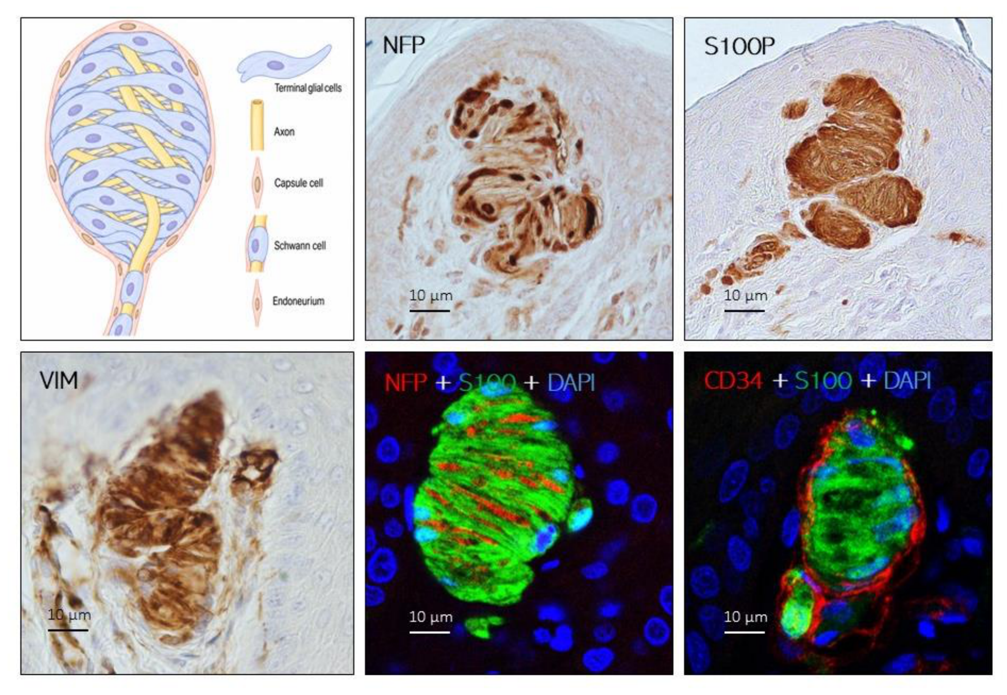
Disclaimer/Publisher’s Note: The statements, opinions and data contained in all publications are solely those of the individual author(s) and contributor(s) and not of MDPI and/or the editor(s). MDPI and/or the editor(s) disclaim responsibility for any injury to people or property resulting from any ideas, methods, instructions or products referred to in the content. |
© 2026 by the authors. Licensee MDPI, Basel, Switzerland. This article is an open access article distributed under the terms and conditions of the Creative Commons Attribution (CC BY) license (http://creativecommons.org/licenses/by/4.0/).
Copyright: This open access article is published under a Creative Commons CC BY 4.0 license, which permit the free download, distribution, and reuse, provided that the author and preprint are cited in any reuse.